nih public access j. claire godbersen clin cancer res...
TRANSCRIPT

The Nedd8-activating enzyme inhibitor MLN4924 thwartsmicroenvironment-driven NFκB activation and inducesapoptosis in chronic lymphocytic leukemia B-cells
J. Claire Godbersen1, Leigh Ann Humphries1, Olga V. Danilova2, Peter E. Kebbekus1,Jennifer R. Brown3, Alan Eastman4, and Alexey V. Danilov1
1Medicine, Dartmouth-Hitchcock Medical Center, Lebanon, NH2Pathology, Dartmouth-Hitchcock Medical Center, Lebanon, NH3Medical Oncology, Dana-Farber Cancer Institute, Boston, MA4Department of Pharmacology and Toxicology, Geisel School of Medicine at Dartmouth, Hanover,NH
AbstractBackground—Stromal-mediated signaling enhances NFκB pathway activity in chroniclymphocytic leukemia B-cells (CLL), leading to cell survival and chemoresistance. Ubiquitinationof IκBα may partially account for constitutive activation of NFκB. MLN4924 is an investigationalagent that inhibits the Nedd8-activating enzyme, thereby neutralizing Cullin-RING ubiquitinligases and preventing degradation of their substrates.
Experimental Design—We conducted a pre-clinical assessment of MLN4924 in CLL. PrimaryCLL cells were co-cultured in vitro with CD40L-expressing stroma to mimic the pro-survivalconditions present in lymphoid tissue. The effect of MLN4924 on CLL cell apoptosis, NFκBpathway activity, Bcl-2 family members and cell cycle was assessed by flow cytometry, westernblotting, PCR and immunocytochemistry.
Results—CD40L-expressing stroma protected CLL cells from spontaneous apoptosis andinduced resistance to multiple drugs, accompanied by NFκB activation and Bim repression.Treatment with MLN4924 induced CLL cell apoptosis and circumvented stroma-mediatedresistance. This was accompanied by accumulation of phospho-IκBα, decreased nucleartranslocation of p65 and p52 leading to inhibition of both canonical and non-canonical NFκBpathways, and reduced transcription of their target genes, notably chemokines. MLN4924promoted induction of Bim and Noxa in the CLL cells leading to rebalancing of Bcl-2 familymembers towards the pro-apoptotic BH3-only proteins. siRNA-mediated knockdown of Bim orNoxa decreased sensitivity to MLN4924. MLN4924 enhanced the antitumor activity of theinhibitors of BCR-associated kinases.
Conclusions—MLN4924 disrupts NFκB activation and induces Bim expression in CLL cellsthereby preventing stroma-mediated resistance. Our data provide rationale for further evaluationof MLN4924 in CLL.
Corresponding author: Alexey V. Danilov, Norris Cotton Cancer Center, 1 Medical Center Drive, Lebanon, NH, USA, 03766; tel.603-650-9474, [email protected].
Author contributionsAVD is the principal investigator and takes primary responsibility for the paper. AVD and JCG designed research. JCG, AVD, LAH,OVD, PEK performed experiments. AVD and JCG analyzed data. AE contributed vital reagents. JCG, AE, AVD wrote the paper.
Conflict of Interest: the authors have nothing to disclose.
NIH Public AccessAuthor ManuscriptClin Cancer Res. Author manuscript; available in PMC 2015 March 15.
Published in final edited form as:Clin Cancer Res. 2014 March 15; 20(6): 1576–1589. doi:10.1158/1078-0432.CCR-13-0987.
NIH
-PA Author Manuscript
NIH
-PA Author Manuscript
NIH
-PA Author Manuscript

Keywordschronic lymphocytic leukemia; apoptosis; MLN4924; NF-κB; Bim
IntroductionInefficient B-cell apoptosis is considered the dominant defect in CLL (1, 2). Still, up to 2%of the circulating CLL B-cell pool is renewed daily (3). Gene expression profiling identifiedlymph nodes as the site of cell activation and proliferation with upregulation of NFκB andB-cell receptor (BCR) signaling (4). Constitutive and BCR-dependent activation of NFκB isan important feature in CLL B-cells and predicts poor disease outcome (5-7). Targetingtyrosine kinases within the BCR-signaling cascade has proven a promising therapeuticstrategy with novel inhibitors of Bruton tyrosine kinase (ibrutinib) and phosphoinositide 3-kinase delta (PI-3Kδ) [idelalisib, formerly CAL-101] currently in clinical trials (8, 9).However BCR-independent activation of NFκB may lead to tumor resistance. NFκB activitymay be driven via microenvironmental stimuli, examples of which include macrophage-mediated B cell–activating factor of the TNF family/a proliferation-inducing ligand (BAFF/APRIL), T-cell-mediated-CD40L signaling, and Toll-like receptor signaling (7, 10, 11).Thus, neutralization of NFκB is a promising strategy in CLL, as it has the potential to targetthe proliferative pool of CLL cells and could hypothetically lead to sensitization to bothconventional chemotherapy and BCR-targeting agents. The NFκB pathway has beensuccessfully targeted in CLL in vitro (12). However, lack of clinical advances with thoseagents necessitates development of novel approaches.
MLN4924 is an investigational small molecule inhibitor of NEDD8-activating enzyme(NAE) which has shown promising pre-clinical activity in hematologic malignancies,including acute myeloid leukemia and lymphoma (13, 14). NAE is necessary for activationof Cullin-RING ubiquitin ligases. In vitro MLN4294 leads to accumulation of Cullin-RINGE3 ligase (CRL) substrates, including IκBα, Nrf-2, p27 and Cdt1 (13, 15). Disruptedubiquitination of IκB in the presence of MLN4924 results in inactivation of the NFκBcanonical pathway in several tumor types (14, 16). Since the NFκB pathway ispredominantly active in the lymphatic tissue, we proposed that its pharmacologicalinhibition will target CLL cells within their supportive microenvironment. Our pre-clinicalwork demonstrates for the first time that MLN4924 shows promising ex vivo activity againstprimary neoplastic B cells derived from patients with CLL. MLN4924 abrogates NFκBpathway activation in CLL cells co-cultured with CD40L-expressing stroma. This results inenhanced expression of the pro-apoptotic BH3-only proteins Bim and Noxa and circumventsstroma-mediated resistance. Furthermore, cooperation between MLN4924 and the BCR-targeting agent CAL-101 warrants exploration of its clinical activity in CLL.
MethodsPatient samples, CLL and stromal cell co-cultures
Following Institutional Review Board approval and provision of written informed consent,peripheral blood was obtained from 42 patients with B-CLL at Dartmouth-HitchcockMedical Center. The median time from diagnosis to study entry was 4 years; 37 patients(88%) were untreated. Blood was also obtained from 7 healthy volunteers. Standard Ficoll-Hypaque (Amersham, Piscataway, NJ) techniques were used to isolate peripheral bloodmononuclear cells (PBMCs). Such CLL samples had more than 90% CD5+/CD19+ cells asdetermined by flow cytometry. CLL cells were cultured in RPMI 1640 supplemented with15% fetal bovine serum, 100 U/mL penicillin, and 100 μg/mL streptomycin, 2 mM L-glutamine, 25 mM HEPES, 100 μM minimum essential medium non-essential amino acids
Godbersen et al. Page 2
Clin Cancer Res. Author manuscript; available in PMC 2015 March 15.
NIH
-PA Author Manuscript
NIH
-PA Author Manuscript
NIH
-PA Author Manuscript

and 1 mM sodium pyruvate (Lonza, Walkersville, MD). 10 CLL samples with 17p deletionwere obtained from the CLL Center at Dana-Farber Cancer Institute. All experiments wereperformed with freshly isolated cells except the viability assays involving the latter, whichwere performed with viably frozen cells.
Mouse fibroblast cell line (L cells) engineered to express CD40L (L4.5) was given to us byDr. Sonia Neron (Quebec, Canada) (17). Parental L cells were obtained from AmericanType Culture Collection (Manassas, VA). All were maintained in RPMI 1640 medium with10% FBS and penicillin-streptomycin. CLL cells were cultured under standardized conditionon stroma as previously described (18). Briefly, stromal cells were seeded to achieve80-100% confluence on the following day when CLL cells were plated at a 50:1 ratio andincubated at 37°C in 5% CO2 in presence or absence of 10 ng/ml IL4 (Cell Signaling,Danvers, MA). For comparison, cells were cultured in suspension (off stroma) at the samedensity. Cultures were then treated with drugs for the indicated time periods. At harvest,CLL cells were gently washed off the stromal layer. When harvested for protein and mRNAanalysis, CLL cells were transferred to a new plate and incubated for an additional 60minutes for stroma re-attachment to minimize contamination of CLL cells.
For BCR stimulation, CLL cells were seeded at a density of 5×10⁵ per well in 24-well platespre-coated with 10 μg/well of rabbit anti-human IgM antibody (Jackson ImmunoresearchLaboratories, Baltimore, MD).
Cell viability testing and drugsCLL cell apoptosis was measured in duplicate as previously described using the ApoScreenAnnexin V apoptosis Kit (19). Briefly, cells were resuspended in 150 μl of Annexin Vbinding buffer containing 1 μl of Annexin V-PE, 1 μl of 7-AAD and 1 μl of CD19- or CD3-FITC mAbs (Southern Biotech, Birmingham, AL) followed by flow cytometry on aFACSCalibur (Becton Dickinson, Palo Alto, CA). MLN4924 was provided by MillenniumPharmaceuticals, Inc. (Cambridge, MA). CAL-101, ibrutinib and bortezomib were obtainedfrom Selleck Chemicals (Houston, TX); BMS-345541, fludarabine, chlorambucil,bendamustine and U0126 - from Sigma Aldrich (St. Louis, MO). Survival of the murinestromal cells was analyzed in a caspase-3 activity assay (Cell Signaling).
ImmunoblottingCells were lysed in RIPA buffer (20 mM Tris, 150 mM NaCl, 1% NP-40, 1 mM NaF, 1 mMSodium phosphate, 1 mM NaVO3, 1 mM EDTA, 1 mM EGTA, supplemented with proteaseinhibitor cocktail (Roche, Indianapolis, IN) and 1 mM PMSF). Proteins were analyzed byimmunoblotting as previously described (19). The following antibodies were used: Bcl-2,Bcl-xL, Bim, phospho-Bim, phospho-IκBα, cleaved PARP, Mcl-1, p65/RelA, p52/p100,XIAP, FOXO3A (Cell Signaling), Bcl2-A1 (Abcam, Cambridge, MA), NEDD8 (Epitomics,Burlingame, CA), p27 (Santa Cruz, Biotechnology, Santa Cruz, CA), Noxa (Imgenex, SanDiego, CA), β-actin (Sigma Aldrich), horseradish peroxidase-conjugated anti-mouse andanti-rabbit antibodies (BioRad).
Reverse transcription polymerase chain reaction (RT-PCR)CLL cells were negatively selected using B-cell Isolation Kit (Miltenyi Biotec#130-093-660, Auburn, CA). This method achieved >99% B-cell purity as determined byflow cytometry. Total RNA was isolated using RNeasy Mini Kit (Qiagen, Valencia, CA).cDNA was synthesized from 500 ng RNA using the iScript cDNA Synthesis Kit (BioRad,Hercules, CA). Quantitative real time PCR (qRT-PCR) was performed in a C1000 ThermalCycler (BioRad) using Universal PCR Master Mix according to the manufacturer'sinstructions (Applied Biosystems, Foster City, CA), with template cDNA and gene specific
Godbersen et al. Page 3
Clin Cancer Res. Author manuscript; available in PMC 2015 March 15.
NIH
-PA Author Manuscript
NIH
-PA Author Manuscript
NIH
-PA Author Manuscript
probes. The following probes were used: BIM: Hs00708019_s1; NOXA: Hs00560402_m1,PUMA: Hs00248075_m1; CXCR4: Hs00607978_s1; CXCR5: Hs00540548_s1; BCL2A1:Hs00187845_m1; BCL-xL: Hs00236329_m1; CFLAR: Hs00153439_m1; CCND1:Hs00765553_m1 STAT5A: Hs00234181_m1. Amplification of the sequence of interest wascompared to a reference probe (18S RNA, #4308329; all from Life Technologies, Carlsbad,CA).
All samples were analyzed in duplicate. We used the comparative Ct method for relativequantitation (2−ΔΔCt, where ΔΔCt=ΔCtP-ΔCtK; P=Probe and K=reference sample).
Immunocytochemistry3×105 cells were adhered onto polylysine D-coated coverslips (Sigma Aldrich) during a 45-minute incubation at 37°C, fixed in 10% formalin (Fisher Scientific, Pittsburgh, PA) andpermebealized in 1% triton-X 100 in PBS. Coverslips were blocked for 30 minutes in 5%bovine serum albumin (Sigma-Aldrich) in PBS with 0.1% Tween-20, probed with p65/RelA, p52/p100 (Cell Signaling) or cleaved PARP (Thermo Scientific, Lafayette, CO)antibodies and then with Alexa Fluor 594 goat anti-mouse antibodies (Life Technologies).Coverslips were mounted with anti-fading ProLong Gold Solution (Life Technologies) with4′,6-diamidino-2-phenylindole (for nuclear counterstaining). Fluorescent images werecaptured with an F-view II monochrome camera (Olympus, U-CMAD3) mounted on anOlympus BX51 microscope.
NFκB activity assayCells were treated as described above. Nuclear fractions were isolated and subsequentlyanalyzed for NFκB activity as per manufacturer's instructions using the NFκB p50/p65Transcription Factor Assay Kit (Cat# ab133128; Abcam).
siRNA-mediated gene silencingElectroporation of siRNA into CLL cells was performed using Amaxa Human B-cellNucleofection Kit (Amaxa, Cologne, Germany). 1×10⁷ PBMCs were mixed with 100 μL ofAmaxa B-cell nucleofector solution, and 2 μg of siRNA was nucleofected using programX-05. Transfection efficiency, assessed by transfection with 2 μg pMaxGFP plasmid, was30-60% with cell viability of 50-80% at 24 hours. siRNA oligos were synthesized byDharmacon (Lafayette, CO). Sense strand against human Bim1:GACCGAGAAGGUAGACAAUUU, Bim2: CUACCUCCCUACAGACAGAUU, Noxa8:GUAAUUAUUGACACAUUUCUU, Noxa9: AGUCGAGUGUGCUACUCAATT, Puma1:GCCUGUAAGAUACUGUAUAUU.
IGHV Mutation statusFollowing RNA and cDNA isolation (as above), IGHV mutations were analyzed using theIGH Somatic Hypermutation Assay v2.0 (Invivoscribe, San Diego, CA). Briefly, PCR wasperformed using the supplied master mixes and Amplitaq Gold DNA polymerase (AppliedBiosystems) on a Veriti Thermal Cycler Model #9902 (Applied Biosystems) to amplify theIGH sequence fragment between the leader (VHL) and joining (J) regions as per themanufacturer's instructions. To confirm amplification of a single clonal product in theexpected size range, an aliquot of each sample was run on a 1.5% agarose gel and separatedby gel electrophoresis. Samples were then submitted for DNA sequencing using the suppliedsequencing primers. The NCBI IgBLAST tool was used to determine the % divergence ofeach clonal sequence. Samples which showed less than 2% divergence from germlinesequence were deemed to have unmutated IGHV.
Godbersen et al. Page 4
Clin Cancer Res. Author manuscript; available in PMC 2015 March 15.
NIH
-PA Author Manuscript
NIH
-PA Author Manuscript
NIH
-PA Author Manuscript

Microarray AnalysisThe RNA gene expression microarray experiments were carried out by the DartmouthGenomics & Microarray Laboratory (Lebanon, NH, USA). Beadarrays with probes for allknown human genes (Illumina, San Diego, CA) were used for RNA profiling. Reversetranscription using an oligo(dT) primer bearing a T7 promoter and the high yieldArrayScriptTM reverse transcriptase were used to make cDNA. The cDNA was madedouble-stranded and purified to use as a template for in vitro transcription with T7 RNAPolymerase and the included biotin-NTP mix. The labeled cRNA was purified and 1.5 µgused for hybridization to the beadarrays for 16 hours at 55°C. Following hybridization, thebeadarrays were washed and stained with streptavidin-Cy3 (GE Healthcare, Piscataway,NJ). Fluorescent images were obtained with an Illumina 500GX scanner and processed withthe BeadScan software (Illumina). Full results are available at http://www.ncbi.nlm.nih.gov/geo/query/acc.cgi?acc=GSE44864.
Statistical analysisStatistical analysis was performed with Student t test (paired or unpaired) in GraphPadPrism software (LaJolla, CA). p<0.05 was considered to be statistically significant.Microarray data were analyzed for functional significance using Pathway Studio software(Ariadne Genomics/Elsevier, Rockville, MD). Fisher exact test was used to identifyontology groups and pathways statistically enriched in the gene set. Data is presented asmean±SE.
ResultsMLN4924 reduces neddylation of cullins and promotes apoptosis in CLL
We first investigated whether NAE inhibition induced apoptosis in CLL. CLL cellsincubated with 1 μM MLN4924 for 24 hours demonstrated enhanced apoptosis compared tovehicle control (12.8±1.7% Annexin V+ cells; Fig. 1A). While CLL cells exhibited variablesensitivity to MLN4924, we did not find distinct responses depending on either BCR heavy-chain immunoglobulin gene (IGHV) mutational status (Fig. 1B), ZAP-70 or CD38expression (data not shown) or cytogenetic abnormalities, including 17p deletion (Fig. 1C).
MLN4924 selectively inhibits modification of cullin proteins by NEDD8 thus preventingCRL functional activity and ultimately leading to accumulation of their substrates (15). Inthe presence of MLN4924, CLL cells demonstrated a dose-dependent decrease inneddylation of cullins as early as 2 hours (Fig. 1D). This effect was more pronounced atlater time points when it was also seen at lower drug concentrations. We investigated theeffect of MLN4924 on several CRL substrates and observed a concomitant accumulation ofphospho-IκBα. CLL cell apoptosis, both spontaneous and drug-induced, was detected at 10hours and prominent at 24 hours, but was more pronounced in the presence of MLN4924, asevidenced by PARP cleavage. The cell cycle inhibitor p27Kip1 is another CRL substrate andis highly expressed in circulating CLL cells (20). Interestingly, MLN4924 had no effect onp27Kip1 protein levels, pointing to a relatively low turnover of this cell-cycle regulator inresting peripheral blood CLL cells (Fig.1D).
NAE inhibition-mediated abrogation of NFκB reverses the stroma-mediated protection inCLL
Given the importance of the microenvironment in sustaining CLL cell survival andproliferation in general and the NFκB pathway activity in particular, we further studied theeffects of MLN4924 in a stromal co-culture system. Stroma cells present in the lymph nodeand bone marrow establish direct cell-cell contact with CLL cells engaging multiple pro-
Godbersen et al. Page 5
Clin Cancer Res. Author manuscript; available in PMC 2015 March 15.
NIH
-PA Author Manuscript
NIH
-PA Author Manuscript
NIH
-PA Author Manuscript

survival pathways (18), among which the tumor necrosis factor receptor family, includingCD40, plays a prominent role. CD40L+ stroma partially rescued CLL cells fromspontaneous apoptosis (Fig. 2A, white bars). We and others have previously shown thatCD40L induces drug resistance in CLL (21-23). We confirmed those findings bydemonstrating that the CD40L-expressing (but not parental) stroma induced CLL cellresistance to common chemotherapy agents and CAL-101 (Fig. 2B). In agreement withearlier work (24) the CD40L-expressing stroma activated both canonical and non-canonicalNFκB pathways in CLL cells after 16-24 hours of co-culture. Activation of the non-canonical NFκB pathway was readily demonstrated by the emergence of p52, a cleavedproduct of p100 (Fig. 2C). Canonical NFκB pathway activation was confirmed bydemonstrating nuclear translocation of p65/RelA, as discussed below. NFκB activation wasaccompanied by induction of anti-apoptotic proteins Mcl-1 and Bcl-xL on the CD40L-expressing stroma while Bcl-2 levels remained constant (Fig. 2C).
Unexpectedly, we found that CD40L-expressing stroma did not elicit resistance toMLN4924 (Fig. 2A, gray and black bars). Interestingly, 1 μM MLN4924 induced moreapoptosis on stroma (55.1±4.2% CLL cells) than off stroma (42.4±2.9%; p=0.015). Thus,CD40L+ (but not parental) L cells also appeared to sensitize CLL cells towards aneddylation inhibitor (Fig. 2A). Similarly, apoptosis occurred irrespective of commongenetic features albeit CLL samples carrying deletion 17p were less sensitive to MLN4924compared to non-17p CLL (Supplementary Fig. 1A-B; p<0.05). The size of a 17p− clonewithin each sample did not correlate with apoptotic response, indicating its susceptibility tothe drug (Supplementary Fig. 1C). Neither protection from spontaneous apoptosis norsensitivity of CLL cells to MLN4924 was altered by addition of IL4 (Fig. 2A). Importantly,MLN4924 did not lead to caspase activation and did not impair CD40L expression in thestroma (Supplementary Fig. 2). Finally, normal B cells and T cells exhibited decreasedsensitivity to MLN4924 compared to CLL cells (Fig. 2D).
Treatment with MLN4924 in the CD40L+ co-culture system resulted in dose-dependentreversal of p100 cleavage and accumulation of phosphorylated IκBα in CLL cells (Fig. 3A).CLL cells demonstrated strong predominantly nuclear staining for both p65/RelA and p52upon co-culture with CD40L-expressing stroma, indicating that the NFκB pathway wasactive (Fig. 3B). However, treatment with MLN4924 resulted in a shift towards cytoplasmicstaining for p65 and p100/p52, thus indicating abrogation of both canonical and non-canonical NFκB pathways. Meanwhile, incubation of CLL cells with the parental controlstroma did not result in nuclear translocation of either p65 or p52 (Fig. 3B). Addition of IL4led to a mild increase in p100 processing compared with CD40L alone, yet this was reversedby MLN4924 (Fig. 3C). We have noted no effect on IκBα under those conditions (Fig. 3C).Similarly, upon treatment with MLN4924, the p65/RelA subunit shifted into the cytoplasmin CLL cells exposed to IL4 (Supplementary Fig. 3). We then sought to confirm that CLLcells undergoing apoptosis in response to MLN4924 had lost NFκB activity. Indeed, cellswhich demonstrated cleaved PARP expression did not show nuclear expression of p65(Supplementary Fig. 3).
We next employed gene expression profiling to determine which pathways were deregulatedby NAE inhibition in CLL and whether the transcriptional targets of NFκB were affected.Of the genes incorporated in the probe set, 7254 were expressed in CLL. Using a cutoff of atleast 1.5-fold change we identified 977 genes whose expression was significantly affectedby MLN4924 (p<0.01). A set of 559 down regulated genes was analyzed for functionalsignificance. We determined that receptor signaling and expression target pathwaysinvolving NFκB were most significantly associated with the downregulated genes(p<0.0001). Of the >400 known NFκB transcriptional targets (http://www.bu.edu/nf-kb/gene-resources/target-genes/) 181 genes were expressed in CLL (Fig. 3D and
Godbersen et al. Page 6
Clin Cancer Res. Author manuscript; available in PMC 2015 March 15.
NIH
-PA Author Manuscript
NIH
-PA Author Manuscript
NIH
-PA Author Manuscript

Supplementary Table) and 79 showed a 50% change in expression (p<0.01). We notedreduced transcription of several groups of NFκB target genes, including anti-apoptotic Bcl-2family members, genes involved in cell cycle progression, and chemokines (p<0.01).Interestingly, we found a significant downregulation of a number of important cytokineligands and receptors expressed by the CLL cells which are downstream targets of NFκB,including CCL5, CCL22, CXCR7, CXCR5 and CD40 (Supplementary Table). Chemokinereceptors CXCR4 and CXCR5 are vital for CLL cell homing (25). We observeddownregulation of the NFκB transcriptional target CXCR5 (26), but not CXCR4, by RT-PCR (Supplementary Fig. 4A). Intriguingly, we detected a 7.7-fold reduction in miR-155, anoncogenic micro-RNA which is predominantly expressed by proliferative CLL cells and isregulated by NFκB (27, 28). Finally, we confirmed that MLN4924 abrogated NFκB activityusing an assay which measures p65 and p50 binding to the DNA sequence containing theNFκB response element. In this assay, NFκB activity was reduced in the presence ofMLN4924 in a dose-dependent manner (Supplementary Fig. 4B), consistent with previouslyappreciated effect on IκBα (Fig. 3B). We then analyzed several NFκB transcriptional targetsto confirm that the inhibitory effect of MLN4924 was dose-dependent (Supplementary Fig.4C).
Thus, CLL cells exhibited NFκB activation in stromal co-cultures which was blocked byMLN4924, leading to enhanced apoptosis.
NAE inhibition rebalances Bcl-2 family members towards the pro-apoptotic BH3-onlyproteins in CLL
While circulating CLL lymphocytes express almost exclusively Bcl-2, cells in the stromalniche have been shown to express other pro-survival Bcl-2 family members. A balancebetween them and their relative ratio to the pro-apoptotic multi-BH proteins Bax and Bakdetermines cell fate (29). BH3-only proteins Puma, Noxa, Bim and others also interact withthe anti-apoptotic Bcl-2 family members. Recent efforts have seen emergence of Bcl-2inhibitors and BH3-mimetics (e.g., ABT-263) which have shown promise in treatment ofCLL (29). We investigated whether changes in transcription of the anti-apoptotic proteinsBcl-2 and Bcl-xL as identified by gene expression profiling resulted in a significantreduction in the corresponding protein levels to explain enhanced apoptosis in response toMLN4924 on CD40L-expressing stroma.
As described above, CD40L-expressing stroma induced Bcl-xL and Mcl-1 in CLL (Fig. 2Cand 4A). Additionally, we observed a significant repression of Bim mRNA, but not Noxa orPuma mRNA, in cells cultured on the CD40L+ stroma vs. control stroma (Fig. 4B).Furthermore, CD40L+ but not parental L cells resulted in repression of Bim protein in 4/6tested CLL samples (Fig. 4C). Consistent with previous reports that unmanipulated L cellsdo not induce either Mcl-1 or Bcl-xL (30), we observed no change in their expression inCLL cells co-cultured on the parental stroma (Fig. 2C and 4C).
Contrary to the microarray findings, Bcl-2 protein expression remained unchanged upontreatment with MLN4924. Meanwhile, of 10 CLL samples tested, modest downregulation ofBcl-xL was detected in 6 samples upon treatment with MLN4924 for 48 hours on CD40L+
stroma, while expression of Mcl-1, XIAP and Bcl2A1 were stable (Fig. 4A). We thenstudied the effect of MLN4924 on the pro-apoptotic BH3-only proteins. Gene expressionprofiling experiments revealed a 2-fold upregulation of Bim transcript by MLN4924(Supplementary Table). We confirmed upregulation of Bim mRNA and protein levels (Fig.4A and 4B). Bim protein induction was evident by 8 hours of exposure to 1 μM MLN4924.Importantly, Bim induction also occurred in the CLL samples which showed no Bimrepression by stroma and no drug-mediated change in anti-apoptotic proteins. BMS-345541,an IκB kinase inhibitor, also led to induction of Bim in CLL cells, confirming the
Godbersen et al. Page 7
Clin Cancer Res. Author manuscript; available in PMC 2015 March 15.
NIH
-PA Author Manuscript
NIH
-PA Author Manuscript
NIH
-PA Author Manuscript

importance of NFκB in Bim regulation (Supplementary Fig. 5). NFκB inhibition waspreviously shown to upregulate other BH3-only proteins, namely Noxa, in CLL (31). Wefound that Noxa mRNA and protein as well as Puma mRNA were also induced byMLN4924 in CD40L-expressing stroma co-cultures (Fig. 4B and 4D).
We further sought to confirm the role of BH3-only proteins in MLN4924-mediatedapoptosis. siRNA-mediated knockdown yielded consistent reduction in baseline Bim andblunted its upregulation upon treatment with MLN4924 (Fig. 5A). Bim short isoform (S),the most cytotoxic, was no longer detected. This was accompanied by a reduction inapoptosis, despite reduced Bcl-xL expression. These findings were confirmed in anexpanded cohort of CLL samples (Fig. 5B). In agreement with earlier data on theinvolvement of NFκB in regulation of Noxa in CLL, apoptosis was decreased in Noxa-suppressed CLL cells, while manipulation of Puma had no effect (Fig. 5A andSupplementary Fig. 6). Combined knockdown of Bim and Noxa further suppressed CLL cellapoptosis (Fig. 5C).
Bortezomib was previously shown to induce Noxa in CLL cells (32). Expectedly, treatmentof CLL cells with bortezomib resulted in sensitization to MLN4924 (Fig. 5D). Noxainduction was enhanced when MLN4924 was combined with bortezomib compared witheither drug alone (Fig. 5D). Meanwhile, CD40L-mediated downregulation of Bim isdependent on activation of extracellular signal-regulated kinase (ERK) (33). While bothMLN4924 and U0126 (a mitogen-activated protein kinase inhibitor) induced Bim in CLLcells, the increase in toxicity of the combination was not as pronounced, albeit statisticallysignificant (p<0.01, paired t-test), possibly due to a mild cooperative effect on Bimexpression. Thus we found that Bim is frequently downregulated in a CD40L-expressingmicroenvironment. Meanwhile, NAE inhibition-mediated inactivation of NFκB results inrebalancing of the Bcl-2 family members towards the pro-apoptotic BH3-only proteins Bimand Noxa in CLL cells, which are required for apoptosis induction in this setting.
MLN4924 sensitizes CLL cells to the BCR-targeting agentsSince NFκB activation is one of the dominant pathways ensuring CLL cell survival inresponse to BCR signaling, we hypothesized that MLN4924 may enhance the pro-apoptoticeffects of the novel BCR kinase inhibitors (9). We first determined whether MLN4924 hadan impact on BCR signaling-mediated survival of the CLL cells. As expected, IgMstimulation resulted in CLL cell rescue from spontaneous apoptosis in a subset of patientsamples (34), and this was reversed by MLN4924, once again emphasizing the importanceof NFκB activation in BCR-mediated survival (Fig. 6A). We then explored the combinedeffect of MLN4924 and BCR-targeting agents CAL-101 and ibrutinib (8, 9). While CLLcells were resistant to both CAL-101 and ibrutinib in the CD40L-expressing system (but noton parental L cells - not shown), co-incubation with either agent and 0.25 μM MLN4924 ledto an increase in cell death compared to MLN4924 alone (Fig. 6B).
DiscussionGene expression profiling of peripheral blood CLL cells had initially characterized them asquiescent lymphocytes related to memory cells (2). Subsequently, it was established that, inaddition to cell accumulation, proliferation of the neoplastic B-cells provides a significantcontribution to the malignant clone (3). Lymph nodes and bone marrow were identified asthe preferential sites of activation and proliferation of the CLL cells (4, 5). BCR genesignature along with the NFκB, NFAT and other proliferation/cell cycle functional gene setsare overrepresented in the lymph node, while bone marrow-resident CLL cells showdecreased apoptotic priming (4, 22). Signals transduced through the BCR, CD40 and Toll-like receptors converge on NFκB, leading to high NFκB activity in the CLL cells resident in
Godbersen et al. Page 8
Clin Cancer Res. Author manuscript; available in PMC 2015 March 15.
NIH
-PA Author Manuscript
NIH
-PA Author Manuscript
NIH
-PA Author Manuscript
the microenvironment. Hewamana et al. demonstrated that DNA binding of the Rel Asubunit is enhanced in CLL compared with normal B-cells, providing evidence forconstitutive NFκB activation in CLL (5).
NFκB activation occurs through the canonical and non-canonical pathways (35). In thecanonical pathway, the IκB kinase complex phoshorylates IκBα, triggering itsubiquitination and leading to nuclear translocation of the NFκB dimers, predominantly p50/Rel A and p50/c-Rel. Non-canonical activation is based on the proteosome-assistedprocessing of the precursor protein p100 with nuclear translocation of p52/Rel B (35, 36).Inappropriate degradation of IκBα is among the potential reasons for constitutive activationof NFκB in cancer (37). Thus, blocking proteosomal degradation of IκBα is a promisingtherapeutic approach. MLN4924 is a recently discovered inhibitor of NAE. MLN4924 bindsNAE at its active site forming a covalent MLN4924-NEDD8 adduct thus preventing themodification of cullin proteins by Nedd8 and leading to accumulation of CRL substrates(15). In this work we demonstrate that MLN4924 efficiently reverses cullin neddylation inCLL cells in vitro, a process accompanied by the accumulation of phospho-IκBα andsubsequent apoptosis. Accumulation of IκBα in CLL occurred rapidly and at concentrationsof MLN4924 sufficient to induce apoptosis (14).
To study the impact of MLN4924 on the microenvironment-mediated NFκB activation, weco-cultured CLL cells with CD40L-expressing stroma. Such a strategy has been shown toactivate NFκB and counter the spontaneous apoptosis of the CLL cells in vitro (24, 38).Importantly, CLL cells exposed to CD40L-expressing stroma have decreased “priming” toundergo apoptosis and acquire resistance to both conventional and novel therapeutic agentssuch as ABT-737 and CAL-101 (21-23, 25). In agreement with earlier reports wedemonstrated activation of both canonical and non-canonical NFκB pathways in CLL cellsco-cultured with the CD40L-expressing stroma (but not with parental stroma), thus creatinga partial recapitulation of the lymph node microenvironment (24). Importantly, we foundthat CD40L signaling may lead to repression of Bim.
While CLL cells co-cultured with CD40L-expressing stroma were resistant to multiplechemotherapy agents, the protective effects were abrogated by MLN4924. We furtherobserved that NAE inhibition prevented nuclear translocation of the NFκB pathwayeffectors p65/Rel A and p52 in CLL cells. Attenuated ubiquitination of phospho-IκBαresulted in an increased retention of p65 in the cytoplasm thus leading to inactivation of thecanonical pathway. Meanwhile, a non-canonical pathway effector p52 is generated when anE3 ligase induces processing of the NFκB2 precursor protein p100 (36). Since p100 proteinexpression was not affected by MLN4924 in CLL, while p52 was reduced, it is likely thatinactivation of the non-canonical pathway occurred due to E3 ligase inhibition andsubsequent reduction in proteosomal processing of p100. It has been previously reported thatboth canonical and non-canonical NFκB pathways are activated in CLL lymph nodes,pointing to the biological relevance of our findings (4, 24). Interestingly, MLN4924 alsoinduced apoptosis of CLL cells cultured off stroma. Using electrophoretic mobility shiftassays, it was shown that peripheral blood CLL cells also demonstrate increased NFκBacitvity compared to normal B cells. It is possible that inhibition of this “baseline” NFκBactivity accounts for modest apoptosis in the presence of MLN4924 in this setting (39).
Abrogation of NFκB activity in CLL cells by MLN4924 resulted in decreased transcriptionof its nuclear targets in a dose-dependent manner. Among those, we found a significantreduction in transcription of genes involved in cell cycle and CLL cell-derived chemokinesmediating microenvironment dependence (CXCR5, CCL17, CCL22, etc), some of which areinduced via CD40 (40, 41). Furthermore, recent work demonstrates that NFκB upregulatesthe expression of cytokines and adhesion molecules crucial for CLL survival in the stroma
Godbersen et al. Page 9
Clin Cancer Res. Author manuscript; available in PMC 2015 March 15.
NIH
-PA Author Manuscript
NIH
-PA Author Manuscript
NIH
-PA Author Manuscript
itself (42). Hence MLN4924 may disrupt the chemokine network and cell-cell interactions inthe protective microenvironment leading to reduced tissue homing in CLL. In addition,MLN4924 may shift the balance of CD40 signaling towards pro-apoptotic events (43).
While NAE inhibition had minimal effect on expression of the anti-apoptotic Bcl-2 familymembers, it led to induction of Bim and Noxa. Bim plays an important role in apoptosisregulation in CLL (44). Bim is phosphorylated and ubiquitinated in response to BCRsignaling leading to CLL cell survival and disease progression (44). Bim is capable ofbinding to all Bcl-2 proteins with high affinity, activating the pro-death Bax and/or Bak(45). We found that MLN4924 induced all Bim splice variants (BimEL/L/S) in CLL but theexact mechanism remains unclear. MLN4924 did not induce Forkhead box 3A transcriptionfactor, a regulator of Bim (45) (Supplementary Fig. 7). Earlier reports suggest that NFκBneutralizes Bim via a c-Rel-dependent mechanism in B-cells (46). Additionally, transgenicexpression of p52 led to repression of Bim and defective apoptosis of mouse lymphocytes(47). Furthermore, degradation of at least one Bim isoform, BimEL is proposed to occur withthe involvement of CRL (48). We found that Bim transcription is upregulated by MLN4924in CLL cells, implicating de novo synthesis in its induction. While we did not detect Bimphosphorylation in CLL (Supplementary Fig. 7), we did not fully exclude a possibility ofattenuated degradation of Bim. . Meanwhile, Noxa has been designated as a putative CRLtarget and therefore NAE inhibition may abrogate its degradation in CLL (49). Diminisheddegradation of Bim and Noxa may be contributing to the pro-apoptotic effect of MLN4924in CLL cells cultured off stroma.
Since NFκB is among the terminal effectors of BCR signaling, our findings that MLN4924abolished the protective effect of BCR stimulation suggest that it may overcome theenhanced responsiveness to BCR signaling and the unfavorable prognosis rendered byunmutated IGHV in CLL (50). In CD40L+ stromal co-cultures, CLL cells were rescued fromthe pro-apoptotic effects of the PI3-Kδ inhibitor CAL-101 and Bruton tyrosine kinaseibrutinib, an effect reversed by MLN4924. Hence, microenvironment-mediated NFκBactivation may enhance CLL cell survival independent of BCR signaling and induceresistance to BCR-targeting agents. Remarkable efficacy of MLN4924 in this settingsuggests addiction to the NFκB pathway and justifies further investigation of those drugcombinations in the clinic.
In summary, we demonstrate that MLN4924 effectively inhibits cullin neddylation in CLLcells. In a model which mimics the lymph node microenvironment, this leads to inactivationof the NFκB pathway, re-expression of Bim and Noxa and prevents stroma-mediated drugresistance. MLN4924 shows cooperation with the BCR-targeting agents . Our datacombined with the new knowledge regarding the indispensability of stromal NFκB to CLLcell survival (42) justify further studies of the NAE inhibitor MLN4924 in CLL.
Supplementary MaterialRefer to Web version on PubMed Central for supplementary material.
AcknowledgmentsWe would like to thank Dr Allison Berger, Ph.D. (Millennium Pharmaceuticals, Inc.) for the helpful discussionsand Dr Mark Israel, M.D. for use of the microscope.
Financial support: Support to AVD provided by a National Cancer Institute new faculty award(3P30CA023108-31S4) to the Norris Cotton Cancer Center. AE and AVD are supported by a translational researchaward from the Leukemia & Lymphoma Society. JRB is supported by the Leukemia Lymphoma Society and theAmerican Cancer Society and is a Scholar in Clinical Research of the Leukemia and Lymphoma Society.
Godbersen et al. Page 10
Clin Cancer Res. Author manuscript; available in PMC 2015 March 15.
NIH
-PA Author Manuscript
NIH
-PA Author Manuscript
NIH
-PA Author Manuscript

References1. Danilov AV, Danilova OV, Klein AK, Huber BT. Molecular pathogenesis of chronic lymphocytic
leukemia. Curr Mol Med. 2006; 6:665–75. [PubMed: 17022736]
2. Rosenwald A, Alizadeh AA, Widhopf G, Simon R, Davis RE, Yu X, et al. Relation of geneexpression phenotype to immunoglobulin mutation genotype in B cell chronic lymphocyticleukemia. The Journal of experimental medicine. 2001; 194:1639–47. [PubMed: 11733578]
3. Messmer BT, Messmer D, Allen SL, Kolitz JE, Kudalkar P, Cesar D, et al. In vivo measurementsdocument the dynamic cellular kinetics of chronic lymphocytic leukemia B cells. J Clin Invest.2005; 115:755–64. [PubMed: 15711642]
4. Herishanu Y, Perez-Galan P, Liu D, Biancotto A, Pittaluga S, Vire B, et al. The lymph nodemicroenvironment promotes B-cell receptor signaling, NF-kappaB activation, and tumorproliferation in chronic lymphocytic leukemia. Blood. 2011; 117:563–74. [PubMed: 20940416]
5. Hewamana S, Lin TT, Rowntree C, Karunanithi K, Pratt G, Hills R, et al. Rel a is an independentbiomarker of clinical outcome in chronic lymphocytic leukemia. Journal of clinical oncology :official journal of the American Society of Clinical Oncology. 2009; 27:763–9. [PubMed:19124804]
6. Liu Z, Hazan-Halevy I, Harris DM, Li P, Ferrajoli A, Faderl S, et al. STAT-3 activates NF-kappaBin chronic lymphocytic leukemia cells. Mol Cancer Res. 2011; 9:507–15. [PubMed: 21364020]
7. Nishio M, Endo T, Tsukada N, Ohata J, Kitada S, Reed JC, et al. Nurselike cells express BAFF andAPRIL, which can promote survival of chronic lymphocytic leukemia cells via a paracrine pathwaydistinct from that of SDF-1alpha. Blood. 2005; 106:1012–20. [PubMed: 15860672]
8. Woyach JA, Johnson AJ, Byrd JC. The B-cell receptor signaling pathway as a therapeutic target inCLL. Blood. 2012; 120:1175–84. [PubMed: 22715122]
9. Stevenson FK, Krysov S, Davies AJ, Steele AJ, Packham G. B-cell receptor signaling in chroniclymphocytic leukemia. Blood. 2011; 118:4313–20. [PubMed: 21816833]
10. Furman RR, Asgary Z, Mascarenhas JO, Liou HC, Schattner EJ. Modulation of NF-kappa Bactivity and apoptosis in chronic lymphocytic leukemia B cells. J Immunol. 2000; 164:2200–6.[PubMed: 10657675]
11. Arvaniti E, Ntoufa S, Papakonstantinou N, Touloumenidou T, Laoutaris N, Anagnostopoulos A, etal. Toll-like receptor signaling pathway in chronic lymphocytic leukemia: distinct gene expressionprofiles of potential pathogenic significance in specific subsets of patients. Haematologica. 2011;96:1644–52. [PubMed: 21750087]
12. Pickering BM, de Mel S, Lee M, Howell M, Habens F, Dallman CL, et al. Pharmacologicalinhibitors of NF-kappaB accelerate apoptosis in chronic lymphocytic leukaemia cells. Oncogene.2007; 26:1166–77. [PubMed: 16924235]
13. Swords RT, Kelly KR, Smith PG, Garnsey JJ, Mahalingam D, Medina E, et al. Inhibition ofNEDD8-activating enzyme: a novel approach for the treatment of acute myeloid leukemia. Blood.2010; 115:3796–800. [PubMed: 20203261]
14. Milhollen MA, Traore T, Adams-Duffy J, Thomas MP, Berger AJ, Dang L, et al. MLN4924, aNEDD8-activating enzyme inhibitor, is active in diffuse large B-cell lymphoma models: rationalefor treatment of NF-{kappa}B-dependent lymphoma. Blood. 2010; 116:1515–23. [PubMed:20525923]
15. Soucy TA, Smith PG, Rolfe M. Targeting NEDD8-activated cullin-RING ligases for the treatmentof cancer. Clin Cancer Res. 2009; 15:3912–6. [PubMed: 19509147]
16. Wei D, Li H, Yu J, Sebolt JT, Zhao L, Lawrence TS, et al. Radiosensitization of human pancreaticcancer cells by MLN4924, an investigational NEDD8-activating enzyme inhibitor. Cancerresearch. 2012; 72:282–93. [PubMed: 22072567]
17. Neron S, Suck G, Ma XZ, Sakac D, Roy A, Katsman Y, et al. B cell proliferation following CD40stimulation results in the expression and activation of Src protein tyrosine kinase. Int Immunol.2006; 18:375–87. [PubMed: 16415104]
18. Kurtova AV, Balakrishnan K, Chen R, Ding W, Schnabl S, Quiroga MP, et al. Diverse marrowstromal cells protect CLL cells from spontaneous and drug-induced apoptosis: development of a
Godbersen et al. Page 11
Clin Cancer Res. Author manuscript; available in PMC 2015 March 15.
NIH
-PA Author Manuscript
NIH
-PA Author Manuscript
NIH
-PA Author Manuscript

reliable and reproducible system to assess stromal cell adhesion-mediated drug resistance. Blood.2009; 114:4441–50. [PubMed: 19762485]
19. Humphries LA, Godbersen JC, Danilova OV, Kaur P, Christensen BC, Danilov AV. Pro-apoptoticTP53 homolog TAp63 is repressed via epigenetic silencing and B-cell receptor signalling inchronic lymphocytic leukaemia. British journal of haematology. 2013; 163:590–602. [PubMed:24117128]
20. Frenquelli M, Muzio M, Scielzo C, Fazi C, Scarfo L, Rossi C, et al. MicroRNA and proliferationcontrol in chronic lymphocytic leukemia: functional relationship between miR-221/222 cluster andp27. Blood. 2010; 115:3949–59. [PubMed: 20203269]
21. Soderquist R, Bates DJ, Danilov AV, Eastman A. Gossypol overcomes stroma-mediated resistanceto the BCL2 inhibitor ABT-737 in chronic lymphocytic leukemia cells ex vivo. Leukemia. 2013;27:2262–4. [PubMed: 23640104]
22. Davids MS, Deng J, Wiestner A, Lannutti BJ, Wang L, Wu CJ, et al. Decreased mitochondrialapoptotic priming underlies stroma-mediated treatment resistance in chronic lymphocyticleukemia. Blood. 2012; 120:3501–9. [PubMed: 22955911]
23. Vogler M, Butterworth M, Majid A, Walewska RJ, Sun XM, Dyer MJ, et al. Concurrent up-regulation of BCL-XL and BCL2A1 induces approximately 1000-fold resistance to ABT-737 inchronic lymphocytic leukemia. Blood. 2009; 113:4403–13. [PubMed: 19008458]
24. Tromp JM, Tonino SH, Elias JA, Jaspers A, Luijks DM, Kater AP, et al. Dichotomy in NF-kappaBsignaling and chemoresistance in immunoglobulin variable heavy-chain-mutated versus unmutatedCLL cells upon CD40/TLR9 triggering. Oncogene. 2010; 29:5071–82. [PubMed: 20581863]
25. Burger JA, Ghia P, Rosenwald A, Caligaris-Cappio F. The microenvironment in mature B-cellmalignancies: a target for new treatment strategies. Blood. 2009; 114:3367–75. [PubMed:19636060]
26. Liu R, Zhao X, Gurney TA, Landau NR. Functional analysis of the proximal CCR5 promoter.AIDS Res Hum Retroviruses. 1998; 14:1509–19. [PubMed: 9840284]
27. Wang M, Tan LP, Dijkstra MK, van Lom K, Robertus JL, Harms G, et al. miRNA analysis in B-cell chronic lymphocytic leukaemia: proliferation centres characterized by low miR-150 and highBIC/miR-155 expression. J Pathol. 2008; 215:13–20. [PubMed: 18348159]
28. Tili E, Michaille JJ, Cimino A, Costinean S, Dumitru CD, Adair B, et al. Modulation of miR-155and miR-125b levels following lipopolysaccharide/TNF-alpha stimulation and their possible rolesin regulating the response to endotoxin shock. J Immunol. 2007; 179:5082–9. [PubMed:17911593]
29. Billard C. Design of novel BH3 mimetics for the treatment of chronic lymphocytic leukemia.Leukemia. 2012; 26:2032–8. [PubMed: 22453662]
30. Cosimo E, McCaig AM, Carter-Brzezinski LJ, Wheadon H, Leach MT, Le Ster K, et al. Inhibitionof NF-kappaB-mediated signaling by the CDK inhibitor CR8 overcomes pro-survival stimuli toinduce apoptosis in chronic lymphocytic leukemia cells. Clin Cancer Res. 2013
31. Tromp JM, Geest CR, Breij EC, Elias JA, van Laar J, Luijks DM, et al. Tipping the Noxa/Mcl-1balance overcomes ABT-737 resistance in chronic lymphocytic leukemia. Clin Cancer Res. 2012;18:487–98. [PubMed: 22128299]
32. Smit LA, Hallaert DY, Spijker R, de Goeij B, Jaspers A, Kater AP, et al. Differential Noxa/Mcl-1balance in peripheral versus lymph node chronic lymphocytic leukemia cells correlates withsurvival capacity. Blood. 2007; 109:1660–8. [PubMed: 17038534]
33. Hallaert DY, Jaspers A, van Noesel CJ, van Oers MH, Kater AP, Eldering E. c-Abl kinaseinhibitors overcome CD40-mediated drug resistance in CLL: implications for therapeutic targetingof chemoresistant niches. Blood. 2008; 112:5141–9. [PubMed: 18796631]
34. Deglesne PA, Chevallier N, Letestu R, Baran-Marszak F, Beitar T, Salanoubat C, et al. Survivalresponse to B-cell receptor ligation is restricted to progressive chronic lymphocytic leukemia cellsirrespective of Zap70 expression. Cancer research. 2006; 66:7158–66. [PubMed: 16849562]
35. Perkins ND. Integrating cell-signalling pathways with NF-kappaB and IKK function. Nat Rev MolCell Biol. 2007; 8:49–62. [PubMed: 17183360]
36. Sun SC. The noncanonical NF-kappaB pathway. Immunol Rev. 2012; 246:125–40. [PubMed:22435551]
Godbersen et al. Page 12
Clin Cancer Res. Author manuscript; available in PMC 2015 March 15.
NIH
-PA Author Manuscript
NIH
-PA Author Manuscript
NIH
-PA Author Manuscript
37. Luo JL, Kamata H, Karin M. IKK/NF-kappaB signaling: balancing life and death--a new approachto cancer therapy. J Clin Invest. 2005; 115:2625–32. [PubMed: 16200195]
38. Cuni S, Perez-Aciego P, Perez-Chacon G, Vargas JA, Sanchez A, Martin-Saavedra FM, et al. Asustained activation of PI3K/NF-kappaB pathway is critical for the survival of chroniclymphocytic leukemia B cells. Leukemia. 2004; 18:1391–400. [PubMed: 15175625]
39. Hewamana S, Alghazal S, Lin TT, Clement M, Jenkins C, Guzman ML, et al. The NF-kappaBsubunit Rel A is associated with in vitro survival and clinical disease progression in chroniclymphocytic leukemia and represents a promising therapeutic target. Blood. 2008; 111:4681–9.[PubMed: 18227347]
40. Davids MS, Burger JA. Cell Trafficking in Chronic Lymphocytic Leukemia. Open J Hematol.2012:3. [PubMed: 22844583]
41. Scielzo C, Apollonio B, Scarfo L, Janus A, Muzio M, Ten Hacken E, et al. The functional in vitroresponse to CD40 ligation reflects a different clinical outcome in patients with chroniclymphocytic leukemia. Leukemia. 2011; 25:1760–7. [PubMed: 21709686]
42. Lutzny G, Kocher T, Schmidt-Supprian M, Rudelius M, Klein-Hitpass L, Finch AJ, et al. ProteinKinase C-beta-Dependent Activation of NF-kappaB in Stromal Cells Is Indispensable for theSurvival of Chronic Lymphocytic Leukemia B Cells In Vivo. Cancer Cell. 2013; 23:77–92.[PubMed: 23328482]
43. Messmer D, Kipps TJ. CD154 gene therapy for human B-cell malignancies. Ann N Y Acad Sci.2005; 1062:51–60. [PubMed: 16461788]
44. Paterson A, Mockridge CI, Adams JE, Krysov S, Potter KN, Duncombe AS, et al. Mechanisms andclinical significance of BIM phosphorylation in chronic lymphocytic leukemia. Blood. 2012;119:1726–36. [PubMed: 22160382]
45. Gillings AS, Balmanno K, Wiggins CM, Johnson M, Cook SJ. Apoptosis and autophagy: BIM as amediator of tumour cell death in response to oncogene-targeted therapeutics. FEBS J. 2009;276:6050–62. [PubMed: 19788418]
46. Banerjee A, Grumont R, Gugasyan R, White C, Strasser A, Gerondakis S. NF-kappaB1 and c-Relcooperate to promote the survival of TLR4-activated B cells by neutralizing Bim via distinctmechanisms. Blood. 2008; 112:5063–73. [PubMed: 18805964]
47. Wang Z, Zhang B, Yang L, Ding J, Ding HF. Constitutive production of NF-kappaB2 p52 is nottumorigenic but predisposes mice to inflammatory autoimmune disease by repressing Bimexpression. J Biol Chem. 2008; 283:10698–706. [PubMed: 18281283]
48. Dehan E, Bassermann F, Guardavaccaro D, Vasiliver-Shamis G, Cohen M, Lowes KN, et al.betaTrCP- and Rsk1/2-mediated degradation of BimEL inhibits apoptosis. Mol Cell. 2009;33:109–16. [PubMed: 19150432]
49. Jia L, Yang J, Hao X, Zheng M, He H, Xiong X, et al. Validation of SAG/RBX2/ROC2 E3ubiquitin ligase as an anticancer and radiosensitizing target. Clin Cancer Res. 2010; 16:814–24.[PubMed: 20103673]
50. Lanham S, Hamblin T, Oscier D, Ibbotson R, Stevenson F, Packham G. Differential signaling viasurface IgM is associated with VH gene mutational status and CD38 expression in chroniclymphocytic leukemia. Blood. 2003; 101:1087–93. [PubMed: 12393552]
Godbersen et al. Page 13
Clin Cancer Res. Author manuscript; available in PMC 2015 March 15.
NIH
-PA Author Manuscript
NIH
-PA Author Manuscript
NIH
-PA Author Manuscript
Translational Relevance
Chronic lymphocytic leukemia (CLL) is the most common type of leukemia in theWestern Hemisphere. Successful use of purine analogue-containing chemo-immunotherapy regimens has extended survival of younger patients with CLL. However,eventual progression to fludarabine-resistant disease, lack of low-risk curative strategiesand limited therapeutic options in the elderly warrant exploration of novel treatmentstrategies. The tumor microenvironment in the lymphatic tissues provides pro-survivaland pro-proliferative stimuli and harbors chemoresistant CLL clones. We demonstratethat MLN4924, an investigational inhibitor of Nedd8-activating enzyme exhibitscytotoxicity towards CLL cells incubated in the microenvironment-mimicking conditionswhere other drugs are ineffective. MLN4924 disrupts key pathways implicated inprogressive disease - NFκB signaling and the anti-apoptotic Bcl-2 family members. Ourresults justify the development of MLN4924 in CLL.
Godbersen et al. Page 14
Clin Cancer Res. Author manuscript; available in PMC 2015 March 15.
NIH
-PA Author Manuscript
NIH
-PA Author Manuscript
NIH
-PA Author Manuscript
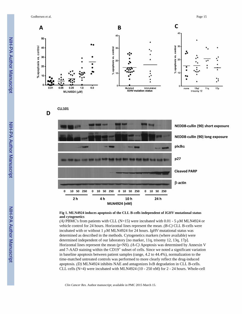
Fig 1. MLN4924 induces apoptosis of the CLL B-cells independent of IGHV mutational statusand cytogenetics(A) PBMC's from patients with CLL (N=15) were incubated with 0.01 - 5 μM MLN4924 orvehicle control for 24 hours. Horizontal lines represent the mean. (B-C) CLL B-cells wereincubated with or without 1 μM MLN4924 for 24 hours. IgHV mutational status wasdetermined as described in the methods. Cytogenetics markers (where available) weredetermined independent of our laboratory [no marker, 11q, trisomy 12, 13q, 17p].Horizontal lines represent the mean (p=NS). (A-C) Apoptosis was determined by Annexin Vand 7-AAD staining within the CD19+ subset of cells. Since we noted a significant variationin baseline apoptosis between patient samples (range, 4.2 to 44.4%), normalization to thetime-matched untreated controls was performed to more clearly reflect the drug-inducedapoptosis. (D) MLN4924 inhibits NAE and antagonizes IκB degradation in CLL B-cells.CLL cells (N=4) were incubated with MLN4924 (10 - 250 nM) for 2 - 24 hours. Whole-cell
Godbersen et al. Page 15
Clin Cancer Res. Author manuscript; available in PMC 2015 March 15.
NIH
-PA Author Manuscript
NIH
-PA Author Manuscript
NIH
-PA Author Manuscript

protein lysates were subjected to immunoblotting. A representative image from 1 of 4experiments is shown.
Godbersen et al. Page 16
Clin Cancer Res. Author manuscript; available in PMC 2015 March 15.
NIH
-PA Author Manuscript
NIH
-PA Author Manuscript
NIH
-PA Author Manuscript

Fig. 2. MLN4924 abrogates the pro-survival effects of CD40L-expressing stroma in CLL cells(A) CLL cells (n=20) were cultured on CD40L-expressing or parental L cells for 24 hours,followed by incubation with 0.25 or 1 μM MLN4924 or vehicle control for 48 hours. As areference, cells were treated off stroma. Cells were also cultured in the presence of 10 ng/mlIL4 (N=8). Apoptosis within CD19+ subset of cells was determined by Annexin V and 7-AAD staining. Data are the mean ± SE. *, p<0.01 compared to untreated control; **, p<0.05compared to off stroma. (B) CLL cells (N=6) were co-cultured with CD40L-expressing orparental L cells for 24 hours, followed by incubation with the indicated drugs or withvehicle control for 48 hours. As a reference, cells were treated off stroma. Apoptosis withinthe CD19+ subset of cells was determined by Annexin V and 7-AAD staining and
Godbersen et al. Page 17
Clin Cancer Res. Author manuscript; available in PMC 2015 March 15.
NIH
-PA Author Manuscript
NIH
-PA Author Manuscript
NIH
-PA Author Manuscript

normalized to the untreated controls. Data are the mean ± SE. * - p<0.05 compared to offstroma or parental stroma. (C) CLL cells were co-cultured with CD40L-expressing (leftpanel) or parental stroma (right panel except for lane 5) for 2 - 24 hours. Whole-cell proteinlysates were subjected to immunoblotting. Results from 1 of 3 experiments are shown. (D)Peripheral blood mononuclear cells from patients with CLL or healthy volunteers werecultured on CD40L-expressing stroma for 24 hours, followed by incubation with 1 μMMLN4924 or vehicle control for 48 hours. Apoptosis within CD19+ and CD3+ subset ofcells was determined by Annexin V and 7-AAD staining and normalized to the time-matched untreated controls (p<0.0001).
Godbersen et al. Page 18
Clin Cancer Res. Author manuscript; available in PMC 2015 March 15.
NIH
-PA Author Manuscript
NIH
-PA Author Manuscript
NIH
-PA Author Manuscript

Fig. 3. MLN4924 reverses NFΔB activation in CLL cells(A) CLL cells were co-cultured with CD40L-expressing stroma for 24 hours andsubsequently incubated with the indicated doses of MLN4924 for 8 - 48 hours. Whole-celllysates were subjected to immunoblotting. Results from 1 of 4 experiments are shown. (B)CLL cells (N=8) were cultured on CD40L-expressing or parental stroma for 24 hours,followed by incubation with 1 μM MLN4924 for additional 24 hours and immunostainedwith p65 or p52/p100 antibodies. Nuclei were counterstained with 4,6-diamino-2-phenylindole. A representative case of CLL cells cultured on CD40L-expressing stroma inpresence or absence of MLN4924 is shown. 100 cells per sample were scored for expression
Godbersen et al. Page 19
Clin Cancer Res. Author manuscript; available in PMC 2015 March 15.
NIH
-PA Author Manuscript
NIH
-PA Author Manuscript
NIH
-PA Author Manuscript

of p65 or p52 in the nuclear compartment. A comparison of nuclear expression of p65 andp52 in CLL cells co-cultured with either parental or CD40+ L cells, the latter with or without1 μM MLN4924, is shown in the lower panels. Data are the mean ± SE. (C) CLL cells wereco-cultured with CD40L-expressing stroma for 24 hours in the presence or absence of 10 ng/ml IL4. Thereafter, cell were incubated with 1 μM MLN4924 for 24 hours. Whole-celllysates were subjected to immunoblotting. Results from 1 of 3 experiments are shown. (D)Gene expression profiling reveals a decrease in NFκB-driven gene signature pattern upontreatment with MLN4924. CLL cells were co-cultured with CD40L-expressing stroma for18 hours and treated or not with 1 μM MLN4924 for additional 24 hours. RNA was isolatedfrom the purified CLL B-cells and microarray analysis was performed as described in themethods. The heat map (left panel) represents a change in expression of the 181 putativeNFκB target genes. Blue represents gene downregulation and yellow represents geneupregulation across the individual CLL samples (N=11).
Godbersen et al. Page 20
Clin Cancer Res. Author manuscript; available in PMC 2015 March 15.
NIH
-PA Author Manuscript
NIH
-PA Author Manuscript
NIH
-PA Author Manuscript
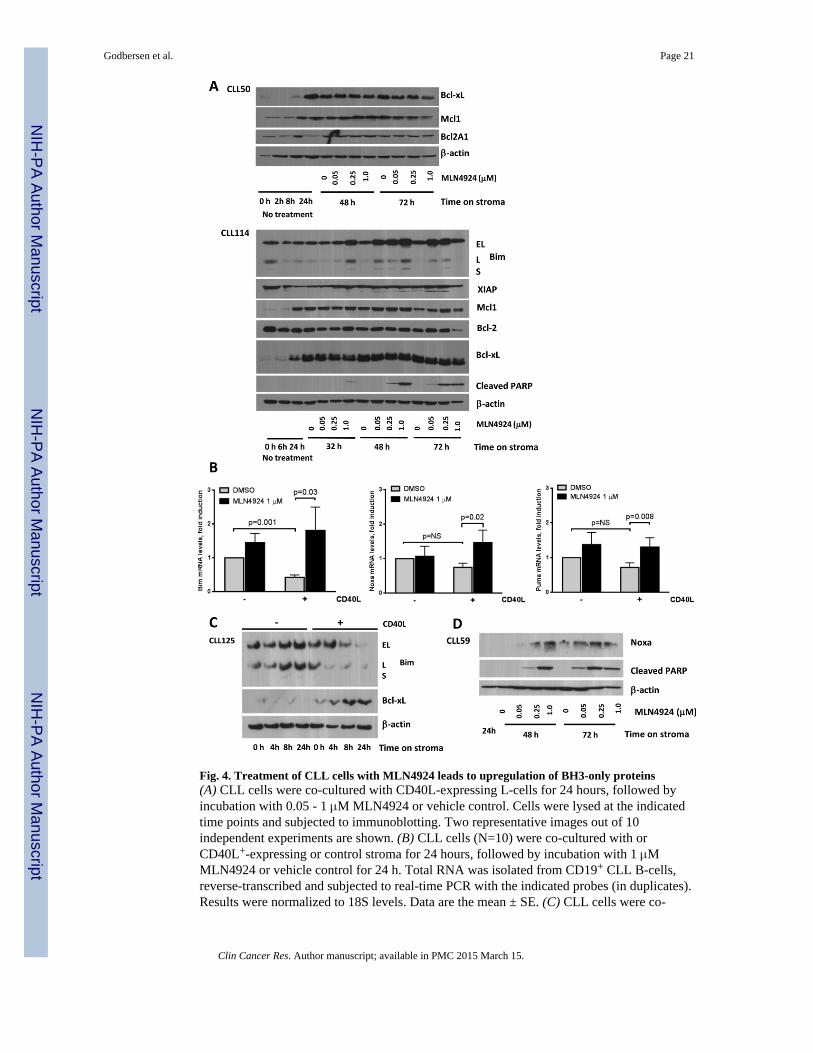
Fig. 4. Treatment of CLL cells with MLN4924 leads to upregulation of BH3-only proteins(A) CLL cells were co-cultured with CD40L-expressing L-cells for 24 hours, followed byincubation with 0.05 - 1 μM MLN4924 or vehicle control. Cells were lysed at the indicatedtime points and subjected to immunoblotting. Two representative images out of 10independent experiments are shown. (B) CLL cells (N=10) were co-cultured with orCD40L+-expressing or control stroma for 24 hours, followed by incubation with 1 μMMLN4924 or vehicle control for 24 h. Total RNA was isolated from CD19+ CLL B-cells,reverse-transcribed and subjected to real-time PCR with the indicated probes (in duplicates).Results were normalized to 18S levels. Data are the mean ± SE. (C) CLL cells were co-
Godbersen et al. Page 21
Clin Cancer Res. Author manuscript; available in PMC 2015 March 15.
NIH
-PA Author Manuscript
NIH
-PA Author Manuscript
NIH
-PA Author Manuscript

cultured with CD40L-expressing or parental L cells for 4 - 24 hours. Whole-cell proteinlysates were subjected to immunoblotting. Results from 1 of 6 experiments are shown. (D)CLL cells were co-cultured with CD40L-expressing stroma for 24 hours, followed byincubation with MLN4924 or vehicle control. Cell were lysed at the indicated time pointsand subjected to immunoblotting. A representative image out of 5 independent experimentsis shown.
Godbersen et al. Page 22
Clin Cancer Res. Author manuscript; available in PMC 2015 March 15.
NIH
-PA Author Manuscript
NIH
-PA Author Manuscript
NIH
-PA Author Manuscript

Fig. 5. BH3-only proteins Bim and Noxa are required for apoptosis induction by MLN4924(A) CLL cells were transfected with two individual siRNA's against Bim or control siRNAusing Amaxa program X-05. Immediately after nucleofection, cells were cultured onCD40L-expressing stroma for 24 hours, followed by incubation with 0.5 μM MLN4924 orvehicle control for 24 h. Whole-cell lysates were subjected to immunoblotting. Arepresentative blot of three independent experiments is shown. (B-C) CLL cells weretransfected with siRNA against Bim (N=7), Noxa (N=7), Puma (N=4), both Bim and Noxa(panel C, N=5) or control siRNA using Amaxa program X-05. Immediately afternucleofection, cells were co-cultured with the CD40L-expressing stroma for 24 hours,followed by incubation with 0.25 or 1 μM MLN4924 or vehicle control for 24 h. Apoptosis
Godbersen et al. Page 23
Clin Cancer Res. Author manuscript; available in PMC 2015 March 15.
NIH
-PA Author Manuscript
NIH
-PA Author Manuscript
NIH
-PA Author Manuscript
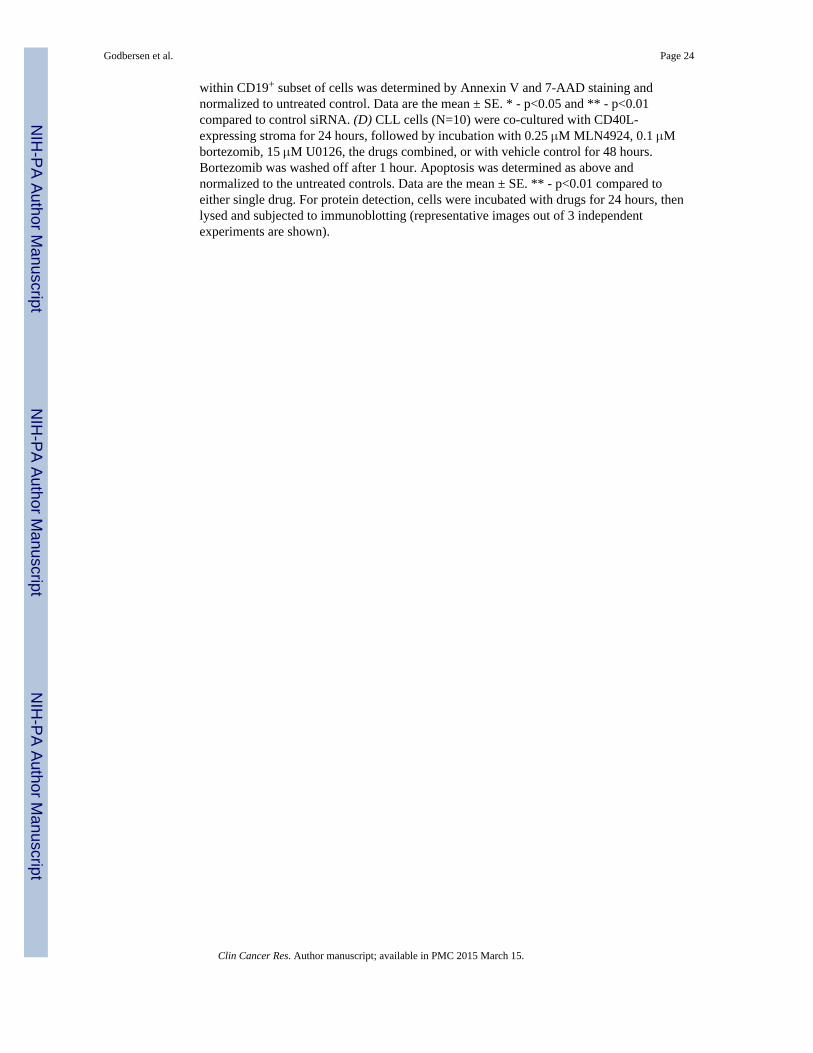
within CD19+ subset of cells was determined by Annexin V and 7-AAD staining andnormalized to untreated control. Data are the mean ± SE. * - p<0.05 and ** - p<0.01compared to control siRNA. (D) CLL cells (N=10) were co-cultured with CD40L-expressing stroma for 24 hours, followed by incubation with 0.25 μM MLN4924, 0.1 μMbortezomib, 15 μM U0126, the drugs combined, or with vehicle control for 48 hours.Bortezomib was washed off after 1 hour. Apoptosis was determined as above andnormalized to the untreated controls. Data are the mean ± SE. ** - p<0.01 compared toeither single drug. For protein detection, cells were incubated with drugs for 24 hours, thenlysed and subjected to immunoblotting (representative images out of 3 independentexperiments are shown).
Godbersen et al. Page 24
Clin Cancer Res. Author manuscript; available in PMC 2015 March 15.
NIH
-PA Author Manuscript
NIH
-PA Author Manuscript
NIH
-PA Author Manuscript

Fig. 6. MLN4924 enhances the activity of BCR signaling inhibitor CAL-101 and alkylatingagents in CLL(A) CLL cells (N=12) were stimulated with immobilized anti-IgM (10 μg/mL) for 1 hour ornot and cultured in the presence of 0.25 or 1 μM MLN4924 or vehicle control for 24 hours(in duplicates). (B) CLL cells (N=8) were co-cultured with CD40L-expressing stroma for 24hours, followed by incubation with 0.25 μM MLN4924, 1 μM CAL-101, 1 μM ibrutinib, thedrugs combined, or with vehicle control for 48 hours.
Godbersen et al. Page 25
Clin Cancer Res. Author manuscript; available in PMC 2015 March 15.
NIH
-PA Author Manuscript
NIH
-PA Author Manuscript
NIH
-PA Author Manuscript