nih public access processing in the hippocampus of the...
TRANSCRIPT

Maternal Immune Activation Alters Nonspatial InformationProcessing in the Hippocampus of the Adult Offspring
Hiroshi T. Ito1, Stephen E. P. Smith1,2, Elaine Hsiao1, and Paul H. Patterson1,*1Division of Biology, California Institute of Technology, Pasadena, CA 91125
AbstractThe observation that maternal infection increases the risk for schizophrenia in the offspring suggeststhat the maternal immune system plays a key role in the etiology of schizophrenia. In a mouse model,maternal immune activation (MIA) by injection of poly(I:C) yields adult offspring that displayabnormalities in a variety of behaviors relevant to schizophrenia. As abnormalities in thehippocampus are a consistent observation in schizophrenia patients, we examined synaptic propertiesin hippocampal slices prepared from the offspring of poly(I:C)- and saline-treated mothers.Compared to controls, CA1 pyramidal neurons from adult offspring of MIA mothers display reducedfrequency and increased amplitude of miniature excitatory postsynaptic currents. In addition, thespecific component of the temporoammonic pathway that mediates object-related informationdisplays increased sensitivity to dopamine. To assess hippocampal network function in vivo, we usedexpression of the immediate early gene, c-Fos, as a surrogate measure of neuronal activity. Comparedto controls, the offspring of poly(I:C)-treated mothers display a distinct c-Fos expression pattern inarea CA1 following novel object, but not novel location, exposure. Thus, the offspring of MIAmothers may have an abnormality in modality-specific information processing. Indeed, the MIAoffspring display enhanced discrimination in a novel object recognition, but not in an object location,task. Thus, analysis of object and spatial information processing at both synaptic and behaviorallevels reveals a largely selective abnormality in object information processing in this mouse model.Our results suggest that altered processing of object-related information may be part of thepathogenesis of schizophrenia-like cognitive behaviors.
Keywordsdopamine; hippocampus; schizophrenia; poly(I:C); temporoammonic pathway
INTRODUCTIONSchizophrenia is a major psychiatric disorder with a strong genetic contribution (Burmeisteret al., 2003; Bertolino and Blasi, 2009). Nonetheless, epidemiologic evidence indicates thatgenetic factors alone cannot explain the pathogenesis. For example, the concordance for
© 2010 Elsevier Inc. All rights reserved.* Correspondence should be addressed to : Division of Biology, 216-76, Caltech, Pasadena, CA 91125. [email protected]. T) 626-395-6826F) 626-395-5805 .2Current address: Departments of Neurology and Pathology, Harvard Medical School and Beth Israel Deaconess Medical Center, Boston,MAPublisher's Disclaimer: This is a PDF file of an unedited manuscript that has been accepted for publication. As a service to our customerswe are providing this early version of the manuscript. The manuscript will undergo copyediting, typesetting, and review of the resultingproof before it is published in its final citable form. Please note that during the production process errors may be discovered which couldaffect the content, and all legal disclaimers that apply to the journal pertain.
NIH Public AccessAuthor ManuscriptBrain Behav Immun. Author manuscript; available in PMC 2011 August 1.
Published in final edited form as:Brain Behav Immun. 2010 August ; 24(6): 930–941. doi:10.1016/j.bbi.2010.03.004.
NIH
-PA Author Manuscript
NIH
-PA Author Manuscript
NIH
-PA Author Manuscript
schizophrenia in monozygotic twins is approximately 48% (Gottesman, 1991). Furthermore,while the concordance between monozygotic twins who share a placenta is 60%, theconcordance rate between such twins who do not share a placenta is only 10% (Davis et al.,1995; Patterson, 2007). These studies suggest the importance of the fetal environment.Supporting this idea, Mednick et al. (1988) reported that fetuses gestating during a viralepidemic are at elevated risk for developing schizophrenia. Subsequent prospective studieshave shown that maternal infections of various types increase the risk for schizophrenia in theoffspring 3–7 fold (reviewed by Patterson, 2009; Brown and Derkits, 2010).
Based on this evidence, several animal models of MIA have been established (reviewed byMeyer et al., 2009; Patterson, 2009). Among these, administration of the synthetic dsRNA,poly(I:C) can effectively induce MIA, resulting in offspring that display a variety of behaviorsand neuropathologies that are consistent with those seen in schizophrenia patients (Meyer andFeldon, 2009; Patterson, 2009). Thus, this animal model is useful for investigating thepathophysiology of schizophrenia.
Clinical studies reveal an important role for dopamine (DA)-mediated signaling in thepathophysiology of schizophrenia. For example, drugs that increase DA release induce severalaspects of schizophrenic psychosis in normal adults, and exacerbate psychotic symptoms inpatients with schizophrenia (Angrist and Vankammen, 1984; Lieberman et al., 1987).Moreover, all drugs currently in widespread use for the treatment of schizophrenia block DAD2 receptors (Creese et al., 1976). Other studies suggest that there is a deficit in DA D1receptor-mediated transmission in prefrontal areas of schizophrenic patients (Davis et al,1991; Toda and Abi-Dargham, 2007). Indeed, imaging studies of patients reveal an increasedD2 receptor density in the striatum (Weinberger and Laruelle, 2001) and a decreased D1receptor density in the prefrontal cortex (Okubo et al., 1997).
Deficits in other cortical regions may also play a key role in the pathophysiology ofschizophrenia. Among them, hippocampal abnormalities are commonly found (Heckers andKonradi, 2002). Lipska et al. (1993) suggested that the important variable is the developmentalperiod during which the hippocampal damage takes place, because lesion of the adult rathippocampus fails to produce schizophrenia-like behaviors, while hippocampal disruption inneonatal stages causes these behavioral alterations to emerge in adulthood. Most importantlyin the present context are recent studies showing a reciprocal functional interaction betweenthe DA system and the hippocampus (Lisman and Grace, 2005), and DA D1- and D2-likereceptors are highly expressed in the hippocampus (Wamsley et al. 1989; Meador-Woodruffet al., 1994; Khan et al., 1997). Together, these studies indicate that hippocampal dysfunctionparticipates in the pathogenesis of schizophrenia.
Considering these findings, we used the poly(I:C) MIA mouse model to investigate thepathogenesis of schizophrenia-like behaviors. We focus on the hippocampal network and theinfluence of DA, conducting experiments at the synaptic as well as the behavioral level. Thedata suggest a link between synaptic dysfunction, DA and altered behavior.
MATERIALS AND METHODSAnimals
Pregnant C57BL/6J mice were injected either i.v. with 5 mg/kg or i.p. with 20 mg/kg poly(I:C)potassium salt freshly dissolved in 0.9% sterile saline on E12.5. Both doses and routes ofadministration have been used to induce MIA and behaviors relevant to schizophrenia in adultoffspring (Meyer et al., 2006; Smith et al., 2007). Control females were injected with the samevolume of saline. The offspring were undisturbed until weaning on P21. Offspring werebehaviorally tested from 6 to 11 weeks for pre-pulse inhibition (PPI), latent inhibition and open
Ito et al. Page 2
Brain Behav Immun. Author manuscript; available in PMC 2011 August 1.
NIH
-PA Author Manuscript
NIH
-PA Author Manuscript
NIH
-PA Author Manuscript

field exploration, to confirm behavioral deficits observed in previous studies (Shi et al., 2003;Smith et al., 2007; data not shown), and for Morris water maze, object location and novel objectrecognition. Five litters each from saline- and poly(I:C)-treated mothers were generated (3 byi.p. injection and 2 by i.v. injection), and four litters each were used for analysis in the presentstudy. One pair of litters generated by i.p injection was not included because control and MIAoffspring did not show significant differences in PPI and latent inhibition. The other excludedpair was used for other experiments. The litters generated by i.p. injection (24 offspring pairsin total) were used for the analysis in Figs. 1A, 1B, 2D, 2E and for all of the behavioral analysisin Fig. 5. The rest of experiments, including those in supplemental figures, were conductedusing the litters generated by i.v. injection (28 offspring pairs in total). No striking differencesin PPI, latent inhibition or open field behavior was observed between the offspring of i.p. andi.v. injected mothers. To avoid influences of prior behavioral testing, animals used forelectrophysiology, immunohistochemisty and c-Fos expression analysis were sacrificed atleast 3 days after the last behavioral test.
Hippocampal slice preparationFor each experiment, hippocampal slices were made from paired adult offspring (7–15 weekold, a pair of the same age and sex for each set of experiments were randomly selected fromeach litter without regard to the results of behavioral tests) from saline- and poly(I:C)-treatedmothers. In brief, a vibrating microtome (Leica VT1000S) was used to cut transversehippocampal sections from the intermediate part of a dorsoventral axis of the hippocampus(400 μm thickness) in ice-cold, oxygenated artificial cerebrospinal fluid (ACSF) containing(in mM) 119 NaCl, 2.5 KCl, 1.3 MgSO4, 2.5 CaCl, 1.0 NaH2PO4, 26.2 NaHCO3, 11.0 glucose.Slices were recovered at room temperature for at least 2 hours in an interface chamber, andthen transferred to a submerged recording chamber perfused with ACSF. Concentric bipolartungsten electrodes (FHC) and stimulus isolators (Axon Instruments) were used for thestimulation.
ElectrophysiologyExtracellular field potential recordings were made with 1–3 MΩ resistance microelectrodesfilled with 3 M NaCl using a bridge amplifier (Axoclamp 2B, Molecular Devices). Therecordings were made at 25°C. Whole-cell voltage-clamp recordings from CA1 pyramidalneurons were obtained without visualization with an Axopatch 200B (Molecular Devices). Theinternal solution of whole-cell patch pipettes contained (in mM) 115 cesium gluconate, 20cesium chloride, 10 sodium phosphocreatine, 10 HEPES, 0.2 EGTA, 2 MgATP, 0.3 NaGTP(pH 7.3). The membrane capacitance was cancelled and series resistance was compensated(60–70%) for paired-pulse facilitation experiments, but uncompensated for miniaturerecordings. Recordings were discarded when the series resistance was over 20 MΩ or eitherseries or membrane resistance changed more than 20% (30% for mIPSC recordings) duringdata acquisition. The amplitude of test pulses was 20–40 A for recordings from SC-CA1synapses and 50–150 A for recordings from TA-CA1 synapses, and the duration of pulse was100 μs. The test pulses were applied once every 30 s. Dopamine was obtained from Sigma. Allother drugs were obtained from Tocris. For mEPSC recordings, whole-cell patch clamprecordings were obtained from CA1 pyramidal neurons in extracellular solution containingTTX (1 μM) and bicuculline (10 μM) at 25 °C. For mIPSC recordings, the internal solution ofpatch pipettes was a cesium chloride-based solution (in mM): 115 cesium chloride, 20 KCl,10 sodium phosphocreatine, 10 HEPES, 0.2 EGTA, 2 MgATP, 0.3 NaGTP (pH 7.3). Therecordings were made at 28 °C, using TTX (1 μM), NBQX (20 μM) and APV (50 μM) to blockexcitatory synaptic transmission, and the extracellular potassium concentration was increasedfrom 2.5 mM to 5 mM to enhance the frequency of miniature synaptic events. Membranevoltage was clamped at −70 mV. For the analysis of paired-pulse facilitation, whole-cell patchclamp recordings were obtained from CA1 pyramidal neurons. The membrane potential was
Ito et al. Page 3
Brain Behav Immun. Author manuscript; available in PMC 2011 August 1.
NIH
-PA Author Manuscript
NIH
-PA Author Manuscript
NIH
-PA Author Manuscript

clamped at −60 mV, and recordings were made at 25 °C under bicuculline (10 μM) andCGP55845 (1 μM) to block inhibitory synaptic transmission. The interstimulus interval was50 msec. The LTP induction protocol was a single train of 100 pulse stimuli at 100 Hz. Allstimulus pulses were of the same length and amplitude as test pulses. The baseline fEPSP slopeprior to LTP induction was adjusted within the range of 0.2–0.3 mV/ms.
Behavioral manipulations for c-Fos expression analysisAll the behavioral manipulations were carried out at night (12–4 am) to maximize activeexploration of the environment. The objects used for novel object exposure were two smallchildren’s toys, made of either plastic or wood. The new cage for novel place exposure was inthe same color and dimensions as the original cage, but had new woodchip bedding of adifferent scent and texture than the prior bedding, and the new cage did not have a food boxon the ceiling. Such sensory cue changes in the environmental context, without geometricchanges in the cage configuration, are sufficient for remapping of hippocampal place cellactivities (Anderson and Jeffery, 2003), implying that animals should recognize the new cageas a different context. More radical changes in the new cage configuration were not made inan effort not to introduce fear or anxiety in the mice.
Behavioral testsWater maze task—Water maze testing followed previously published procedures(Zuckerman and Weiner 2005). Briefly, mice were introduced to the maze (water made opaquewith non-toxic latex paint) in a random spot and allowed 60 s to find a platform submerged 1cm under the water. The walls of the room contained visual cues (four 0.5 m diameter piecesof different color paper cut into different shapes). Mice were given four trials per session, twosessions per day separated by approx 4 hours. In between trials mice were allowed to recoverfor approx 5 min in a cage lined with paper towels and warmed with an electric heating pad.If the mice found the platform within 60 s, the time to find the platform was recorded and themouse was removed after 10 s on the platform; if it did not find the platform it was placed onthe platform for 10 s and the time was recoded as 60 s. On the afternoon of the 4th day, theplatform was removed and swim pattern was recorded for 60 s (probe trial). On the dayfollowing the probe trial, the platform was placed in the opposite quadrant of the pool, andthree more sessions were conducted with the platform in the novel location. Finally, the waterlevel was dropped and four visible platform trials were conducted.
Novel object recognition and object location recognition tasks—Mice were firstallowed to explore the environment (a 50 cm-square white plastic box) for 10 min. They werethen removed for 5 minutes, during which time two different objects were placed in oppositecorners of the box, and the mice were allowed to explore the objects for 5 min. The mice wereagain removed from the box for five minutes, and for the novel object recognition test, one ofthe objects was replaced with a novel object (the target object), and the mice were allowed toexplore for another 5 min. On the following day, the object location test was administered. Theprocedure was identical to the previous one except that new objects were used, and rather thanreplacing the target object during the second phase of the test, it was moved 90 degrees to adifferent corner of the box (see fig. 7D). To eliminate experimental bias due to innate preferencefor an object, the five objects used (a plastic pickle, a “mini-koosh” ball, a plastic top, a smallmetal knife, and a small candle) were pseudo-randomized such that different mice saw differentcombinations of objects at each stage of the test; however no innate preferences for an objectwere observed.
Ito et al. Page 4
Brain Behav Immun. Author manuscript; available in PMC 2011 August 1.
NIH
-PA Author Manuscript
NIH
-PA Author Manuscript
NIH
-PA Author Manuscript
Data analysisElectrophysiology—Data were collected using a custom-written program (LabView dataacquisition system; National Instruments) for extracellular recordings, or DigiData 1200 andpClamp 9 (Molecular Devices) for whole-cell recordings. After recordings were stabilized,current traces were acquired for 3 min. The mEPSCs were detected using a template-matchingalgorithm of Clampfit 9 (Molecular Devices). All numerical values listed represent mean ±s.e.m. Student’s t-test was performed to analyze the signficance of the data.
Immunohistochemistry—Slices (400 μm thickness) were prepared from randomlyselected pairs of animals using the same procedures as for electrophysiology recordings. Aftercutting, slices were quickly fixed in 4% paraformaldehyde in phosphate-buffered saline (PBS)for at least 2 days. Thin (50 μm) sections were cut with a vibrating microtome (Leica VT1000S).
The sections were incubated overnight with either of 1:250 concentration of anti-c-Fos (sc-52)(Santa Cruz), 1:1000 of anti-synaptophysin (Millipore), 1:1000 of anti-synapsin I (Millipore),1:1000 of anti-Bassoon (Stressgen), 1:1000 of NeuN (Millipore), 1:1000 of GluR1 (Millipore),or 1:1000 of anti-MAP2 (Sigma) antibodies. The incubation was carried out at roomtemperature in Tris-buffered saline containing 0.2% Triton X-100, BSA 2%, NGS 4%,followed by 4 hrs of secondary-antibody incubation with 1:1000 of Alexa 488-conjugated anti-rabbit and 1:1000 of Alexa 543-conjugated anti-mouse antibodies (Invitrogen).
For the analysis of immunohistochemistry experiments, images were obtained with Zeiss LSM510 laser scanning confocal microscopes using a Plan-Neofluor 10×/0.3 air objective. Alexa488 and 546 were visualized by excitation with the 488 line of an argon ion laser and the 543nm line of a HeNe laser, respectively. The optical section was 20 μm and fluorescent signalswere acquired throughout the section thickness. Each 50 μm section was obtained from adifferent 400 μm slice and two sections were analyzed from each animal. Slices were obtainedfrom the same septo-temporal position in all experiments. To count the number of c-Fospositive neurons, fluorescent signals of less than the mean + 2 SD were excluded. Thenautomated particle analysis was carried out using ImageJ (NIH) based on the following criteria:the particle size was larger than 39 μm2 and the circularity was larger than 0.5. Statisticaldifferences between animals groups were assessed by ANOVA.
Behavioral analysisWater maze task: All trials were recorded by a camera suspended over the pool, and data wasanalyzed with Ethovision (Noldus). Swim speed, mean distance swam and performance in thevisible platform task was not significantly different between groups (data not shown).
Novel object recognition and object location tasks: The trials were recorded by an overheadcamera, and object investigation was measured as the number of times a mouse brought itsnose within 2 cm of the object, by an observer blind to the identification of each mouse. Datawas expressed as percent nose pokes to the target object (nose-pokes to target / [nose-pokes totarget + nose-pokes to non-target object] × 100), where the target object is represented by thenovel object during the novel object recognition task and by the re-located object during theobject location task.
RESULTSCA1 pyramidal neurons in the offspring of poly(I:C)-treated mothers display a reducedfrequency and increased amplitude of mEPSCs
A number of studies indicate that the brains of schizophrenia patients exhibit a reduction inthe volume of the hippocampus (Nelson et al., 1998; Heckers and Konradi, 2002). Although
Ito et al. Page 5
Brain Behav Immun. Author manuscript; available in PMC 2011 August 1.
NIH
-PA Author Manuscript
NIH
-PA Author Manuscript
NIH
-PA Author Manuscript

most studies on schizophrenia patients report no significant change in neuronal density in thehippocampus (Dwork, 1997; Harrison, 1999), many post-mortem studies have reported anabnormal expression of synaptic proteins, including synaptophysin (Eastwood and Harrison,1995; Davidsson et al., 1999), synapsin I (Browning et al., 1993), SNAP-25 (Young et al.,1998) and spinophilin (Law et al., 2004).
Therefore, we asked whether the MIA offspring display abnormalities in synaptic number orefficacy in CA1 pyramidal neurons. We observe an increased amplitude, but decreasedfrequency in miniature excitatory postsynaptic currents (mEPSCs) (amplitude: saline 8.5 ± 0.3pA, poly(I:C) 9.9 ± 0.5 pA; frequency: saline 0.95 ± 0.15 Hz, poly(I:C) 0.60 ± 0.05 Hz) (Fig.1A). There is no significant difference in the kinetics of mEPSC waveforms (Fig. 1B) ormembrane properties (membrane capacitance: saline 220 ± 36 pF, poly(I:C) 210 ± 33 pF,membrane resistance: saline 194 ± 62 MΩ, poly(I:C) 149 ± 55 MΩ, in mean ± SD), suggestingthat the observed differences in mEPSC amplitude and frequency are primarily due to alteredsynaptic properties. The decrease in mEPSC frequency suggests either presynaptic dysfunctionor a reduction in excitatory synapse number per neuron. To assess presynaptic function, weanalyzed paired-pulse facilitation but do not find a significant difference between the groups(saline: 2.1 ± 0.2, poly(I:C): 2.0 ± 0.2; ratio of 2nd to 1st EPSC amplitude) (Fig. 1C), suggestingthat presynaptic function in the experimental group is normal and that the reduced mEPSCfrequency is likely due to a reduction in excitatory synapse number. The increase in mEPSCamplitude in the offspring of poly(I:C)-treated mothers may be a compensatory response forthe reduction of mEPSC frequency (Turrigiano et al., 1998;Sutton et al., 2006). We also testedsynaptic plasticity at Schaffer-collateral-CA1 synapses. When LTP was induced by a singletrain of 100 stimuli at 100 Hz, the magnitude of LTP in slices prepared from the offspring ofpoly(I:C)-treated mothers is similar to controls (saline: 1.45 ± 0.05, poly(I:C): 1.36 ± 0.07;mean fEPSP slope at 55 – 60 min after LTP induction relative to the baseline) (Fig. 1D). Takentogether, these results indicate that the adult offspring of poly(I:C)-treated mothers display areduced number of normally functioning excitatory synapses on CA1 pyramidal neurons.
We next performed immunohistochemistry of the synaptic proteins synaptophysin and GluR1in area CA1. Dendritic distribution of these proteins is similar between control and MIAoffspring. In contrast, we found that a cytoskeletal protein, MAP2, shows a distinct distributionalong the dendritic axis of CA1 pyramidal neurons in MIA offspring (Supplemental Fig. 1),which is consistent with a study on schizophrenia patients showing an elevated expression ofMAP2 in the hippocampus (Cotter et al., 2000).
To examine inhibitory synaptic transmission, we recorded mIPSCs from CA1 pyramidalneurons. We do not find a significant difference in either amplitude or frequency of mIPSCsbetween groups (amplitude: saline 13.9 ± 1.1 pA, poly(I:C) 13.6 ± 1.0 pA, frequency: saline4.9 ± 0.3 Hz, poly(I:C) 4.3 ± 0.5 Hz) (Fig. 1E), suggesting that the function of inhibitorysynapses is normal in area CA1.
Temporoammonic-CA1 synapses in MIA offspring display increased sensitivity to dopamineMany lines of evidence indicate that DA signaling plays an important role in hippocampalfunction, as well as in the pathophysiology of schizophrenia. For example, DA neurons in thesubstantia nigra compacta and ventral tegmental area innervate the hippocampus (Swanson,1987; Gasbarri et al., 1997) and release DA when animals are exposed to novel environments(Ihalainen et al., 1999). This neurotransmitter influences hippocampal synaptic plasticity(Huang and Kandel, 1995; Otmakhova and Lisman, 1996; Li et al., 2003) and frequency-dependent synaptic transmission (Ito and Schuman, 2007). Furthermore, disruption of DA-mediated signaling impairs hippocampal-dependent learning (Gasbarri et al. 1996; El-Ghundiet al., 1999; Rossato et al., 2009). Therefore, we investigated DA-mediated synaptic controlin hippocampal area CA1. As previous studies showed a selective influence of DA on the
Ito et al. Page 6
Brain Behav Immun. Author manuscript; available in PMC 2011 August 1.
NIH
-PA Author Manuscript
NIH
-PA Author Manuscript
NIH
-PA Author Manuscript

temporoammonic (TA)-CA1 synapses compared to Schaffer-collateral-CA1 synapses(Otmakhova and Lisman, 1999; Ito and Schuman, 2007; Supplemental Fig. 2), we examinedDA modulation of TA-CA1 synapses. The TA pathway includes axonal populations from boththe medial (MEC) and lateral (LEC) entorhinal cortexes (Steward, 1976; Witter and Amaral,2004). These projections are topographically organized along the transverse axis of area CA1,such that the projections from MEC make synapses at proximal CA1 (close to CA3), whilethose from the LEC project to distal CA1 (close to subiculum) (Fig. 2A; Steward, 1976; Witterand Amaral, 2004). Previous anatomical studies in the mouse brain showed a topographicprojection of the TA pathway similar to that observed in the rat brain (van Groen et al.,2003). Our immunohistochemical analysis of the presynaptic proteins synapsin I and bassoonin the stratum lacunosum-moleculare indeed indicates expression differences between distaland proximal regions of area CA1 (Supplemental Fig. 3). Thus, the presynaptic properties ofthe proximal and distal TA-CA1 synapses in the topographic projection of entorhinal-corticalfibers may be distinct. This organization of presynaptic proteins is observed in both MIA andcontrol offspring (Supplemental Fig. 3).
We recorded fEPSPs simultaneously from proximal and distal TA-CA1 synapses. Theapplication of DA (20 μM; Otmakhova and Lisman 1999) to slices prepared from control miceinduces a significantly larger depression at proximal TA-CA1 synapses compared to distal TA-CA1 synapses (proximal TA: 57.6 ± 2.2%; distal TA: 65.3 ± 2.3%, relative to baseline; Fig.2B and 4C). Furthermore, we found that another neuromodulator, norepinephrine (NE), alsodifferentially controls TA synapses made by MEC and LEC inputs (Supplemental Fig. 4).These data indicate that the neuromodulators, DA and NE, can differentially control MEC andLEC inputs to area CA1 of the mouse hippocampus.
In slices prepared from MIA offspring, DA induces a depression comparable to that observedin control slices at proximal TA-CA1 synapses (proximal TA: 55.4 ± 3.1%, relative to baseline;Fig. 2C). At distal TA-CA1 synapses, however, the slices prepared from MIA offspring showa significantly larger depression compared to controls (distal TA: 56.3 ± 1.4%, relative tobaseline; Fig. 2C). To further examine DA sensitivity at distal TA-CA1 synapses, theneurotransmitter was applied sequentially from low to high concentration to acute slices (Fig.2D) and DA-mediated depression quantified. We find that, compared to controls, the amountof depression is significantly larger at each DA concentration in the slices prepared from theMIA group (Fig. 2E). These results indicate that the adult offspring of poly(I:C)-treatedmothers display an enhanced sensitivity to DA selectively at LEC inputs.
The offspring of poly (I:C)-treated mothers display a distinct c-Fos expression pattern inhippocampal area CA1 following novel object exposure
While the major afferent inputs to the hippocampus are provided by the entorhinal cortex(Cajal, 1911), recent studies demonstrated that two subdivisions of the entorhinal cortex, MECand LEC, provide distinct information modalities to the hippocampus. Spatial information iscarried by axons from the MEC, whereas nonspatial, or object information is carried by axonsfrom the LEC (Haagreaves et al., 2005; Knierim et al., 2006; Manns and Eichenbaum, 2006).Because MIA offspring display higher sensitivity to DA in the LEC projection to area CA1,these animals may exhibit abnormal object information processing.
One of the major features shared by hippocampal and DA-releasing neurons in vivo is themodulation of neuronal activity by stimulus novelty (Knight, 1996; Schultz, 1998; Horvitz,2000; Rutishauser et al., 2006). Therefore, we examined how hippocampal neurons areactivated in vivo during novel object exposure using immunostaining for an immediate-earlygene product, c-Fos (Morgan and Curran, 1991). Immediate early gene expression in restinganimals is very low (e.g. Supplemental Fig. 5C), but rapidly increases following patternedneuronal activity that induces synaptic plasticity (Cole et al., 1989), suggesting that c-Fos
Ito et al. Page 7
Brain Behav Immun. Author manuscript; available in PMC 2011 August 1.
NIH
-PA Author Manuscript
NIH
-PA Author Manuscript
NIH
-PA Author Manuscript

expression can be used as a surrogate marker for synaptic modification (Guzowski et al.,2005). Following accomodation to the home cage for several days, control and experimentalmice were exposed to novel objects in the home cage (Fig. 3A). After 2 hrs of exposure, animalswere sacrificed and immunohistochemistry performed. Control mice show differential c-Fosexpression between proximal and distal CA1 pyramidal neurons (Figs. 3B, C and SupplementalFig. 5A). In contrast, MIA offspring do not show clear differential c-Fos activation betweenproximal and distal CA1 pyramidal neurons (Figs. 3B, C and Supplemental Fig. 5A). Theseresults suggest that MIA offspring display abnormal object information processing in thehippocampus.
We also examined c-Fos expression after animals were exposed to a novel cage environment.Following accommodation to the home cage for several days, animals were placed in a newcage, which lacked a food box and contained new bedding with a different texture and scentthan the prior bedding. After 2 hrs of such novel location exposure, animals were sacrificedand immunohistochemistry performed (Fig. 4A). In contrast to the results after novel objectexposure (Fig. 3), we observe a similar c-Fos expression pattern in the transverse-axis of areaCA1 between the offspring of saline- and poly(I:C)-treated mothers (Figs. 4B, C andSupplemental Fig. 5B). Thus, MIA offspring appear to have a selective abnormality in object,but not spatial, information processing. This could be due to hyper-DA sensitivity in LECinputs at TA-CA1 synapses because our previous studies indicate that neuromodulators playa key role in novel object-driven differential c-Fos expression between proximal and distalCA1 (Ito and Schuman, submitted).
The offspring of poly(I:C)-treated mothers display behavioral inflexibility and abnormal novelobject recognition
Our slice recording and c-Fos expression analyses indicate that MIA offspring have a selectiveabnormality in nonspatial information processing in the hippocampus. To examine if theseanimals display a corresponding behavioral abnormality, we tested the performance ofhippocampus-dependent behavior using the Morris water maze task (Morris, 1984).
We do not find a significant difference in the learning of the initial platform location betweenexperimental and control groups (Fig. 5A, B), suggesting that the MIA offspring have normalability to acquire spatial navigation memory. After animals learned the initial platform location,we moved the platform to a different location. Two-way ANOVA with session number andprenatal treatment as variables reveals a significant effect of prenatal treatment (T1,222=5.693,p < 0.05), as well as a significant effect of session number (T3,222=5.875, p < 0.01) with nointeraction between the two variables, indicating that while both groups improve theirperformance over time, the MIA offspring display a significantly slower learning of the newplatform location (Fig. 5C). Thus, although the MIA offspring have normal ability to learn aspatial context per se, they have difficulty in adapting to a change introduced in a previously-learned context.
To further test this idea, we examined how these animals perform in novel object recognitionand object location tasks. We placed two different objects in a test cage and allowed animalsbecome familiar with them. One object was subsequently moved to a new location (objectlocation test) (Fig. 5D). Both groups of animals display a higher preference for the movedobject, and we do not find a significant difference in the magnitude of preference betweengroups (Fig. 5E). In another set of experiments, after familiarization with the two objects, anovel object replaced one of the familiar ones at the same location (novel object recognitiontest) (Fig. 5D). Although both groups of animals display preference for the new object, theMIA offspring display a significantly stronger preference than control animals (Fig. 5F). Thus,the MIA offspring display abnormally high sensitivity to a novel object, but not to a novel
Ito et al. Page 8
Brain Behav Immun. Author manuscript; available in PMC 2011 August 1.
NIH
-PA Author Manuscript
NIH
-PA Author Manuscript
NIH
-PA Author Manuscript
location, which may be due to altered processing of nonspatial information in the hippocampusof these animals. This is consistent with the slice recording and c-Fos findings.
DISCUSSIONReduced excitatory input on CA1 pyramidal neurons
Our electrophysiological studies demonstrate that the offspring of poly(I:C)-treated mothersdisplay a decreased number, but enhanced efficacy, of excitatory synapses on CA1 pyramidalneurons. Synaptic abnormalities have also been observed in area CA3 in schizophrenia patients,including abnormal mRNA expression of presynaptic proteins (Harrison and Eastwood,2001).
We do not observe a significant abnormality in the mIPSC amplitude or frequency in theoffspring of poly(I:C)-treated animals. Although several studies have reported a decreasednumber of GABAergic neurons in the hippocampus, most of these differences were observedin area CA2/3, not in area CA1 (Benes et al., 1996; Benes et al., 1997; Heckers and Konradi,2002). Thus, GABAergic input to area CA1 pyramidal neurons appears normal in both ourmodel and in patients.
Increased dopamine sensitivity in the temporoammonic pathwayCompared to the Schaffer-collateral pathway in area CA1, the TA pathway has been a relativelyunexplored circuit in the hippocampus. Many recent findings highlight its unique role inhippocampal function, however (Brun et al., 2002; Remondes and Schuman, 2004; Nakashibaet al., 2008). Some interesting features of this pathway include its topographic projectionpattern and its sensitivity to neuromodulators.
A topographic projection in which LEC- or MEC-derived axons terminate in different regionsallows the EC to send nonspatial and spatial information to distinct neuronal populations inarea CA1 (Steward, 1976; Witter and Amaral, 2004). The efferents from area CA1 are alsotopographically organized such that neurons in proximal CA1 send projections back to theMEC, while neurons in distal CA1 project back to the LEC (Tamamaki and Nojyo, 1995).Thus, two independent circuit loops for nonspatial and spatial information exist between theEC and CA1. This architecture, based on the TA pathway, may allow the hippocampus toindependently process nonspatial and spatial information, providing a unique role for the TApathway in the hippocampus.
Sensitivity to neuromodulators is another feature of the TA pathway. In recordings from controlmouse hippocampal slices, DA induces a larger depression at proximal compared to distal TA-CA1 synapses. However, hippocampal slices prepared from offspring of poly(I:C)-treatedmothers exhibit a significantly larger DA-induced depression, selectively at distal TA-CA1synapses compared to controls, which may abolish the differential control of LEC and MECinputs by DA. Our present observations, based on both electrophysiology and c-Fos expression,suggest that the offspring of poly(I:C)-treated mothers have altered DA-mediated control ofthe TA pathway and may have an abnormality in object information processing. This alteredDA signaling may be due to the abnormal development of the DA system recently observedin MIA offspring (Vuillermot, 2010), although the hippocampus and EC in this animal modelhave not yet been examined for expression of DA receptor or DA transporter expression withregard to the TA pathway. It is worth noting that the antipsychotic drug, clozapine, effectivelyblocks DA-induced depression at TA-CA1 synapses (Otmakhova and Lisman, 1999),indicating that the TA pathway may be a locus for clozapine action in schizophrenia.
The proper integration between Schaffer-collateral- and TA-CA1 synapses is critical forcontrolling spike initiation and synaptic plasticity in CA1 pyramidal neurons (Dvorak-Carbone
Ito et al. Page 9
Brain Behav Immun. Author manuscript; available in PMC 2011 August 1.
NIH
-PA Author Manuscript
NIH
-PA Author Manuscript
NIH
-PA Author Manuscript

and Schuman, 1999; Remondes and Schuman, 2002; Jarsky et al., 2005; Ang et al., 2005;Dudman et al., 2007). Thus, abnormal DA-mediated control of TA-CA1 synapses, togetherwith reduced number of excitatory inputs on CA1 pyramidal neurons, will likely lead to alteredsynaptic plasticity or information integration in CA1 pyramidal neurons in the MIA offspring.
Perseveration behavior and hypersensitivity to a novel objectOur behavioral analyses indicate that the offspring of poly(I:C)-treated mothers showbehavioral inflexibility in the Morris water maze task and increased sensitivity in the novelobject recognition task. The normal acquisition of an initial platform location in the water mazetask indicates that MIA offspring have normal spatial navigation, such as in self-localization,route learning and motor function for swimming (Redish and Touretzky, 1998). A selectivedeficit in learning a moved platform location suggests that these animals have difficulty inadapting to a modification introduced in previously acquired information, which maycorrespond to perseveration behavior. Such behavior is defined as contextually inappropriateand unintentional repetition, and is often observed in schizophrenia patients (Crider, 1997).These patients have difficulty in switching behavioral-strategy, or reversal learning, and tendto repeat the same response or strategy (Fey, 1951; Nolan, 1974; Floresco et al., 2008).Interestingly, a recent human study indicates that maternal infection is correlated in theoffspring with impaired performance in the Wisconsin card sorting test, which provides a goodmeasure of perseveration behavior (Brown et al., 2009). That is, the subset of schizophreniasubjects born to infected mothers display a more severe behavioral deficit in this test thanschizophrenia subjects born to non-infected mothers. In experimental animals, the offspringof poly(I:C)-treated mothers display deficits in reversal learning in a left-right discriminationtask (Meyer et al., 2006). Perseveration behavior is also observed in hippocampus-lesionedanimals in the Morris water maze task (Whishaw and Tomie, 1997). Both hippocampus-lesioned animals (Kim and Frank, 2009) and rodents injected with a DA receptor agonist(Boulougouris et al., 2009) exhibit impaired reversal learning. Thus, abnormalities in thehippocampus and the DA system may play a key role in perseveration behavior.
In light of perseveration behavior in the water maze, the increased sensitivity to a novel objectthat we observe in the MIA offspring may be due to an enhanced expectation for the originalobject, which will increase novelty for a switched object. Importantly, the abnormal preferencein the MIA offspring was observed selectively in the novel object recognition (nonspatialinformation), but not in the object location (spatial information), task. These altered behaviorsin the MIA offspring may be due to abnormal processing of nonspatial information in thehippocampus, as observed in our slice physiology and c-Fos expression analyses. Contrary toour results, Ozawa et al. (2006) and Ibi et al. (2009) reported a deficit in a novel objectrecognition test. However, they used a more severe regimen of poly(I:C) treatment for 5-6consecutive days at a different time during development, and they employed a longer timeaway from the object, making the test more of a memory test than a novelty preference test.Moreover, Golan et al. (2005) obtained a result similar to ours in a novel object recognitiontest using maternal injection of lipopolysaccharide to mimic maternal bacterial infection. Itshould also be noted that, in the water maze test, Zuckerman and Weiner (2005) reported somesimilarities and some differences in results compared to ours. However, they used rats andadministered poly(I:C) at a much later time in gestation.
Integration of information processed in parallelA major feature of brain function is parallel information processing. For example, visualinformation is processed in two distinct information streams: a ventral stream that subservesobject recognition, or “what” perception, and a dorsal stream that primarily subserves spatialinformation, or “where” perception (Ungerleider and Haxby, 1994). The distributedinformation, processed in different brain areas, must be integrated for coherent perception.
Ito et al. Page 10
Brain Behav Immun. Author manuscript; available in PMC 2011 August 1.
NIH
-PA Author Manuscript
NIH
-PA Author Manuscript
NIH
-PA Author Manuscript

Recent imaging and physiology studies report abnormal visual object recognition inschizophrenia patients (Doniger et al., 2002; Wynn et al., 2008). Wynn et al. (2008) measuredactivity in early retinotopically organized areas (V1–V4), motion-sensitive areas (human areaMT) and object-recognition areas (lateral occipital complex), and found that schizophreniapatients display more widely-distributed activation in areas involved in object-recognition thancontrols. Thus, the abnormal distribution of activity in object-selective cortex in schizophreniapatients may indicate a problem in the integration of spatial and nonspatial information.
In the hippocampus, the integration of spatial and nonspatial information is critical forconstructing a neural representation of environmental context. As such, the hippocampus playsan essential role in contextual memory formation. Interestingly, several studies indicate thatschizophrenia patients have a severe problem in contextual memory formation (Boyer et al.,2007; Rizzo et al., 1996; Danion et al., 1999), although other types of memory, which do notrequire contextual information, are relatively intact. Our findings raise the question of whetherthis memory deficit could be due to abnormal DA-mediated control of the TA pathway in thehippocampus. Interestingly, a recent study using model simulation predicts a dominance ofobject over spatial information processing in the medial temporal lobe of schizophrenia patients(Talamini and Meeter, 2009). Thus, the altered hippocampal information processing weobserve in MIA offspring may underlie some schizophrenia-like behaviors of these animals,such as in prepulse inhibition, latent inhibition and perseverative behavior. Furtherinvestigation based on this perspective may shed new light on the pathophysiology ofschizophrenia.
Supplementary MaterialRefer to Web version on PubMed Central for supplementary material.
AcknowledgmentsWe thank Prof. Erin M. Schuman (Caltech/Howard Hughes Medical Institute) and the HHMI for support in theelectrophysiology and c-Fos experiments. The other experiments were supported by grants to P.H.P. from the NationalInstitute of Mental Health and the McKnight Foundation. H.T.I. was supported by the Nakajima Foundation. S.E.P.S.was supported by the Autism Speaks Foundation. E.H. was supported by an NIH training grant.
REFERENCESAnderson MI, Jeffery KJ. Heterogeneous modulation of place cell firing by changes in context. J Neurosci
2003;23:8827–8835. [PubMed: 14523083]Ang CW, Carlson GC, Coulter DA. Hippocampal CA1 circuitry dynamically gates direct cortical inputs
preferentially at theta frequencies. J Neurosci 2005;25:9567–9580. [PubMed: 16237162]Angrist B, Vankammen DP. CNS stimulants as tools in the study of schizophrenia. Trends Neurosci
1984;7:388–390.Benes FM, Khan Y, Vincent SL, Wickramasinghe R. Differences in the subregional and cellular
distribution of GABAA receptor binding in the hippocampal formation of schizophrenic brain.Synapse 1996;22:338–349. [PubMed: 8867028]
Benes FM, Wickramasinghe R, Vincent SL, Khan Y, Todtenkopf M. Uncoupling of GABA(A) andbenzodiazepine receptor binding activity in the hippocampal formation of schizophrenic brain. BrainRes 1997;755:121–129. [PubMed: 9163547]
Bertolino A, Blasi G. The genetics of schizophrenia. Neurosci. 2009 in press.Boulougouris V, Castane A, Robbins TW. Dopamine D2/D3 receptor agonist quinpirole impairs spatial
reversal learning in rats: investigation of D3 receptor involvement in persistent behavior.Psychopharmacol (Berl.) 2009;202:611–620.
Ito et al. Page 11
Brain Behav Immun. Author manuscript; available in PMC 2011 August 1.
NIH
-PA Author Manuscript
NIH
-PA Author Manuscript
NIH
-PA Author Manuscript

Boyer P, Phillips JL, Rousseau FL, Ilivitsky S. Hippocampal abnormalities and memory deficits: newevidence of a strong pathophysiological link in schizophrenia. Brain Res Rev 2007;54:92–112.[PubMed: 17306884]
Brown AS, Vinogradov S, Kremen WS, Poole JH, Deicken RF, Penner JD, McKeague IW, KochetkovaA, Kern D, Schaefer CA. Prenatal exposure to maternal infection and executive dysfunction in adultschizophrenia. Am J Psychiat 2009;166:683–690. [PubMed: 19369317]
Brown AS, Derkits EJ. Prenatal infection and schizophrenia: A review of epidemiologic and translationalstudies. Amer J Psychiat. 2010 in press.
Browning MD, Dudek EM, Rapier JL, Leonard S, Freedman R. Significant reductions in synapsin butnot synaptophysin specific activity in the brains of some schizophrenics. Biol Psychiat 1993;34:529–535. [PubMed: 8274580]
Brun VH, Otnass MK, Molden S, Steffenach HA, Witter MP, Moser MB, Moser EI. Place cells and placerecognition maintained by direct entorhinal-hippocampal circuitry. Science 2002;296:2243–2246.[PubMed: 12077421]
Burmeister M, McInnis MG, Zollner S. Psychiatric genetics: progress amid controversy. Nat Rev Genet2008;9:527–540. [PubMed: 18560438]
Cajal, SRY. Histologie du Systeme nerveux de l’hommes et des vertebres. Maloine; Paris: 1911.Cole AJ, Saffen DW, Baraban JM, Worley PF. Rapid increase of an immediate early gene messenger
RNA in hippocampal neurons by synaptic NMDA receptor activation. Nature 1989;340:474–476.[PubMed: 2547165]
Cotter D, Wilson S, Roberts E, Kerwin R, Everall IP. Increased dendritic MAP2 expression in thehippocampus in schizophrenia. Schizophr Res 2000;41:313–323. [PubMed: 10708340]
Creese I, Burt DR, Snyder SH. Dopamine receptor binding predicts clinical and pharmacologicalpotencies of antischizophrenic drugs. Science 1976;192:481–483. [PubMed: 3854]
Crider A. Perseveration in schizophrenia. Schizophr Bull 1997;23:63–74. [PubMed: 9050113]Danion JM, Rizzo L, Bruant A. Functional mechanisms underlying impaired recognition memory and
conscious awareness in patients with schizophrenia. Arch Gen Psychiat 1999;56:639–644. [PubMed:10401510]
Davidsson P, Gottfries J, Bogdanovic N, Ekman R, Karlsson I, Gottfries CG, Blennow K. The synaptic-vesicle-specific proteins rab3a and synaptophysin are reduced in thalamus and related cortical brainregions in schizophrenic brains. Schizophr Res 1999;40:23–29. [PubMed: 10541003]
Davis KL, Kahn RS, Ko G, Davidson M. Dopamine in schizophrenia: a review and reconceptualization.Am J Psychiat 1991;148:1474–1486. [PubMed: 1681750]
Davis JO, Phelps JA, Bracha HS. Prenatal development of monozygotic twins and concordance forschizophrenia. Schizophr Bull 1995;21:357–366. [PubMed: 7481567]
Doniger GM, Foxe JJ, Murray MM, Higgins BA, Javitt DC. Impaired visual object recognition and dorsal/ventral stream interaction in schizophrenia. Arch Gen Psychiat 2002;59:1011–1020. [PubMed:12418934]
Dudman JT, Tsay D, Siegelbaum SA. A role for synaptic inputs at distal dendrites: instructive signalsfor hippocampal long-term plasticity. Neuron 2007;56:866–879. [PubMed: 18054862]
Dvorak-Carbone H, Schuman EM. Patterned activity in stratum lacunosum moleculare inhibits CA1pyramidal neuron firing. J Neurophysiol 1999;82:3213–3222. [PubMed: 10601455]
Dwork AJ. Postmortem studies of the hippocampal formation in schizophrenia. Schizophr Bull1997;23:385–402. [PubMed: 9327505]
Eastwood SL, Harrison PJ. Decreased synaptophysin in the medial temporal lobe in schizophreniademonstrated using immunoautoradiography. Neurosci 1995;69:339–343.
El-Ghundi M, Fletcher PJ, Drago J, Sibley DR, O’Dowd BF, George SR. Spatial learning deficit indopamine D(1) receptor knockout mice. Eur J Pharmacol 1999;383:95–106. [PubMed: 10585522]
Fey ET. The performance of young schizophrenics and young normals on the Wisconsin Card SortingTest. J Consult Psychol 1951;15:311–319. [PubMed: 14861329]
Floresco SB, Zhang Y, Enomoto T. Neural circuits subserving behavioral flexibility and their relevanceto schizophrenia. Behav Brain Res. 2008
Ito et al. Page 12
Brain Behav Immun. Author manuscript; available in PMC 2011 August 1.
NIH
-PA Author Manuscript
NIH
-PA Author Manuscript
NIH
-PA Author Manuscript
Gasbarri A, Sulli A, Innocenzi R, Pacitti C, Brioni JD. Spatial memory impairment induced by lesion ofthe mesohippocampal dopaminergic system in the rat. Neuroscience 1996;74:1037–1044. [PubMed:8895872]
Gasbarri A, Sulli A, Packard MG. The dopaminergic mesencephalic projections to the hippocampalformation in the rat. Prog Neuropsychopharmacol Biol Psychiatry 1997;21:1–22. [PubMed:9075256]
Golan HM, Lev V, Hallak M, Sorokin Y, Huleihel M. Specific neurodevelopmental damage in miceoffspring following maternal inflammation during pregnancy. Neuropharmacology 2005;48:903–917. [PubMed: 15829260]
Gottesman, II. Schizophrenia Genesis: The Origins of Madness. W.H. Freeman & Company; New York,NY: 1991.
Guzowski JF, Timlin JA, Roysam B, McNaughton BL, Worley PF, Barnes CA. Mapping behaviorallyrelevant neural circuits with immediate-early gene expression. Curr Opin Neurobiol 2005;15:599–606. [PubMed: 16150584]
Hargreaves EL, Rao G, Lee I, Knierim JJ. Major dissociation between medial and lateral entorhinal inputto dorsal hippocampus. Science 2005;308:1792–1794. [PubMed: 15961670]
Harrison PJ. The neuropathology of schizophrenia. A critical review of the data and their interpretation.Brain 1999;122(Pt 4):593–624. [PubMed: 10219775]
Harrison PJ, Eastwood SL. Neuropathological studies of synaptic connectivity in the hippocampalformation in schizophrenia. Hippocampus 2001;11:508–519. [PubMed: 11732704]
Heckers S, Konradi C. Hippocampal neurons in schizophrenia. J Neural Transm 2002;109:891–905.[PubMed: 12111476]
Horvitz JC. Mesolimbocortical and nigrostriatal dopamine responses to salient non-reward events.Neuroscience 2000;96:651–656. [PubMed: 10727783]
Huang YY, Kandel ER. D1/D5 receptor agonists induce a protein synthesis-dependent late potentiationin the CA1 region of the hippocampus. Proc Natl Acad Sci U S A 1995;92:2446–2450. [PubMed:7708662]
Ibi D, Nagai T, Kitahara Y, Mizoguchi H, Koike H, Shiraki A, Takuma K, Kamei H, Noda Y, Nitta A,Nabeshima T, Yoneda Y, Yamada K. Neonatal polyI:C treatment in mice results in schizophrenia-like behavioral and neurochemical abnormalities in adulthood. Neurosci Res 2009;64:297–305.[PubMed: 19447299]
Ihalainen JA, Riekkinen P Jr. Feenstra MG. Comparison of dopamine and noradrenaline release in mouseprefrontal cortex, striatum and hippocampus using microdialysis. Neurosci Lett 1999;277:71–74.[PubMed: 10624812]
Ito HT, Schuman EM. Frequency-dependent gating of synaptic transmission and plasticity by dopamine.Front Neural Circuits 2007;1:1. [PubMed: 18946543]
Jarsky T, Roxin A, Kath WL, Spruston N. Conditional dendritic spike propagation following distalsynaptic activation of hippocampal CA1 pyramidal neurons. Nat Neurosci 2005;8:1667–1676.[PubMed: 16299501]
Khan ZU, Gutierrez A, Martin R, Penafiel A, Rivera A, De La Calle A. Differential regional and cellulardistribution of dopamine D2-like receptors: an immunocytochemical study of subtype-specificantibodies in rat and human brain. J Comp Neurol 1998;402:353–371. [PubMed: 9853904]
Kim SM, Frank LM. Hippocampal lesions impair rapid learning of a continuous spatial alternation task.PLoS One 2009;4:e5494. [PubMed: 19424438]
Knierim JJ, Lee I, Hargreaves EL. Hippocampal place cells: parallel input streams, subregionalprocessing, and implications for episodic memory. Hippocampus 2006;16:755–764. [PubMed:16883558]
Knight R. Contribution of human hippocampal region to novelty detection. Nature 1996;383:256–259.[PubMed: 8805701]
Law AJ, Weickert CS, Hyde TM, Kleinman JE, Harrison PJ. Reduced spinophilin but not microtubule-associated protein 2 expression in the hippocampal formation in schizophrenia and mood disorders:molecular evidence for a pathology of dendritic spines. Am J Psychiat 2004;161:1848–1855.[PubMed: 15465982]
Ito et al. Page 13
Brain Behav Immun. Author manuscript; available in PMC 2011 August 1.
NIH
-PA Author Manuscript
NIH
-PA Author Manuscript
NIH
-PA Author Manuscript

Li S, Cullen WK, Anwyl R, Rowan MJ. Dopamine-dependent facilitation of LTP induction inhippocampal CA1 by exposure to spatial novelty. Nat Neurosci 2003;6:526–531. [PubMed:12704392]
Li XG, Somogyi P, Ylinen A, Buzsaki G. The hippocampal CA3 network: an in vivo intracellular labelingstudy. J Comp Neurol 1994;339:181–208. [PubMed: 8300905]
Lieberman JA, Kane JM, Alvir J. Provocative tests with psychostimulant drugs in schizophrenia.Psychopharmacol (Berl.) 1987;91:415–433.
Lipska BK, Jaskiw GE, Weinberger DR. Postpubertal emergence of hyperresponsiveness to stress andto amphetamine after neonatal excitotoxic hippocampal damage: a potential animal model ofschizophrenia. Neuropsychopharmacol 1993;9:67–75.
Lisman JE, Grace AA. The hippocampal-VTA loop: controlling the entry of information into long-termmemory. Neuron 2005;46:703–713. [PubMed: 15924857]
Manns JR, Eichenbaum H. Evolution of declarative memory. Hippocampus 2006;16:795–808. [PubMed:16881079]
Meador-Woodruff JH, Grandy DK, Van Tol HH, Damask SP, Little KY, Civelli O, Watson SJ Jr.Dopamine receptor gene expression in the human medial temporal lobe. Neuropsychopharmacology1994;10:239–248. [PubMed: 7945734]
Mednick SA, Machon RA, Huttunen MO, Bonett D. Adult schizophrenia following prenatal exposure toan influenza epidemic. Arch Gen Psychiat 1988;45:189–192. [PubMed: 3337616]
Megias M, Emri Z, Freund TF, Gulyas AI. Total number and distribution of inhibitory and excitatorysynapses on hippocampal CA1 pyramidal cells. Neurosci 2001;102:527–540.
Meyer U, Nyffeler M, Engler A, Urwyler A, Schedlowski M, Knuesel I, Yee BK, Feldon J. The time ofprenatal immune challenge determines the specificity of inflammation-mediated brain and behavioralpathology. J Neurosci 2006;26:4752–4762. [PubMed: 16672647]
Meyer U, Feldon J. Prenatal exposure to infection: a primary mechanism for abnormal dopaminergicdevelopment in schizophrenia. Psychopharmacol (Berl). 2009 in press.
Meyer U, Feldon J, Fatemi SH. In vivo rodent models for the experimental investigation of prenatalimmune activation effects in neurodevelopmental brain disorders. Neurosci Behav Rev. 2009 inpress.
Morgan JI, Curran T. Stimulus-transcription coupling in the nervous system: involvement of the inducibleproto-oncogenes fos and jun. Annu Rev Neurosci 1991;14:421–451. [PubMed: 1903243]
Morris R. Developments of a water-maze procedure for studying spatial learning in the rat. J NeurosciMeth 1984;11:47–60.
Nakashiba T, Young JZ, McHugh TJ, Buhl DL, Tonegawa S. Transgenic inhibition of synaptictransmission reveals role of CA3 output in hippocampal learning. Science 2008;319:1260–1264.[PubMed: 18218862]
Nelson MD, Saykin AJ, Flashman LA, Riordan HJ. Hippocampal volume reduction in schizophrenia asassessed by magnetic resonance imaging: a meta-analytic study. Arch Gen Psychiat 1998;55:433–440. [PubMed: 9596046]
Nolan JD. A within-subjects analysis of discrimination shift behavior in schizophrenics. J AbnormPsychol 1974;83:497–511. [PubMed: 4455713]
Okubo Y, Suhara T, Suzuki K, Kobayashi K, Inoue O, Terasaki O, Someya Y, Sassa T, Sudo Y,Matsushima E, Iyo M, Tateno Y, Toru M. Decreased prefrontal dopamine D1 receptors inschizophrenia revealed by PET. Nature 1997;385:634–636. [PubMed: 9024661]
Otmakhova NA, Lisman JE. D1/D5 dopamine receptor activation increases the magnitude of early long-term potentiation at CA1 hippocampal synapses. J Neurosci 1996;16:7478–7486. [PubMed:8922403]
Otmakhova NA, Lisman JE. Dopamine selectively inhibits the direct cortical pathway to the CA1hippocampal region. J Neurosci 1999;19:1437–1445. [PubMed: 9952420]
Ozawa K, Hashimoto K, Kishimoto T, Shimizu E, Ishikura H, Iyo M. Immune activation duringpregnancy in mice leads to dopaminergic hyperfunction and cognitive impairment in the offspring:a neurodevelopmental animal model of schizophrenia. Biol Psychiat 2006;59:546–54. [PubMed:16256957]
Patterson PH. Maternal effects on schizophrenia risk. Science 2007;318:576–577. [PubMed: 17962542]
Ito et al. Page 14
Brain Behav Immun. Author manuscript; available in PMC 2011 August 1.
NIH
-PA Author Manuscript
NIH
-PA Author Manuscript
NIH
-PA Author Manuscript

Patterson PH. Immune involvement in schizophrenia and autism: Etiology, pathology and animal models.Behav Brain Res 2009;204:313–324. [PubMed: 19136031]
Redish AD, Touretzky DS. The role of the hippocampus in solving the Morris water maze. Neural Comput1998;10:73–111. [PubMed: 9501505]
Remondes M, Schuman EM. Direct cortical input modulates plasticity and spiking in CA1 pyramidalneurons. Nature 2002;416:736–740. [PubMed: 11961555]
Remondes M, Schuman EM. Role for a cortical input to hippocampal area CA1 in the consolidation ofa long-term memory. Nature 2004;431:699–703. [PubMed: 15470431]
Rizzo L, Danion JM, Van Der Linden M, Grangé D, Rohmer JG. Impairment of memory for spatialcontext in schizophrenia. Neuropsychol 1996;10:376–384.
Rossato JI, Bevilaqua LR, Izquierdo I, Medina JH, Cammarota M. Dopamine controls persistence oflong-term memory storage. Science 2009;325:1017–1020. [PubMed: 19696353]
Rutishauser U, Mamelak AN, Schuman EM. Single-trial learning of novel stimuli by individual neuronsof the human hippocampus-amygdala complex. Neuron 2006;49:805–813. [PubMed: 16543129]
Schultz W. Predictive reward signal of dopamine neurons. J Neurophysiol 1998;80:1–27. [PubMed:9658025]
Sorra KE, Harris KM. Occurrence and three-dimensional structure of multiple synapses betweenindividual radiatum axons and their target pyramidal cells in hippocampal area CA1. J Neurosci1993;13:3736–3748. [PubMed: 8366344]
Steward O. Topographic organization of the projections from the entorhinal area to the hippocampalformation of the rat. J Comp Neurol 1976;167:285–314. [PubMed: 1270625]
Sutton MA, Ito HT, Cressy P, Kempf C, Woo JC, Schuman EM. Miniature neurotransmission stabilizessynaptic function via tonic suppression of local dendritic protein synthesis. Cell 2006;125:785–799.[PubMed: 16713568]
Swanson, L. Handbook of Chemical neuroanatomy. Elsevier; Amsterdam: 1987. The limbic region I.The septohippocampal system; p. 125-227.
Talamini LM, Meeter M. Dominance of objects over context in a mediotemporal lobe model ofschizophrenia. PLoS One 2009;4:e6505. [PubMed: 19652706]
Tamamaki N, Nojyo Y. Preservation of topography in the connections between the subiculum, field CA1,and the entorhinal cortex in rats. J Comp Neurol 1995;353:379–390. [PubMed: 7538515]
Toda M, Abi-Dargham A. Dopamine hypothesis of schizophrenia: making sense of it all. Curr PsychiatRep 2007;9:329–336.
Turrigiano GG, Leslie KR, Desai NS, Rutherford LC, Nelson SB. Activity-dependent scaling of quantalamplitude in neocortical neurons. Nature 1998;391:892–896. [PubMed: 9495341]
Ungerleider LG, Haxby JV. ‘What’ and ‘where’ in the human brain. Curr Opin Neurobiol 1994;4:157–165. [PubMed: 8038571]
van Groen T, Miettinen P, Kadish I. The entorhinal cortex of the mouse: organization of the projectionto the hippocampal formation. Hippocampus 2003;13:133–149. [PubMed: 12625464]
Vuillermot S, Weber L, Feldon J, Meyer U. A longitudinal examination of the neurodevelopmental impactof prenatal immune activation in mice reveals primary defects in dopaminergic development relevantto schizophrenia. J Neurosci 2010;30:1270–1287. [PubMed: 20107055]
Wamsley JK, Gehlert DR, Filloux FM, Dawson TM. Comparison of the distribution of D-1 and D-2dopamine receptors in the rat brain. J Chem Neuroanat 1989;2:119–137. [PubMed: 2528968]
Weinberger, D.; Laruelle, M. Neuropsychopharmacology. Lippincott Williams & Wilkins; Philadelphia:2001. Neurochemical and neuropharmacological imaging in schizophrenia.
Whishaw IQ, Tomie JA. Perseveration on place reversals in spatial swimming pool tasks: further evidencefor place learning in hippocampal rats. Hippocampus 1997;7:361–370. [PubMed: 9287076]
Witter, MP.; Amaral, DG. Hippocampal Formation. In: Paxinos, G., editor. The Rat Nervous System.Elsevier academic press; Amsterdam: 2004.
Wynn JK, Green MF, Engel S, Korb A, Lee J, Glahn D, Nuechterlein KH, Cohen MS. Increased extentof object-selective cortex in schizophrenia. Psychiat Res 2008;164:97–105.
Young CE, Arima K, Xie J, Hu L, Beach TG, Falkai P, Honer WG. SNAP-25 deficit and hippocampalconnectivity in schizophrenia. Cereb Cortex 1998;8:261–268. [PubMed: 9617921]
Ito et al. Page 15
Brain Behav Immun. Author manuscript; available in PMC 2011 August 1.
NIH
-PA Author Manuscript
NIH
-PA Author Manuscript
NIH
-PA Author Manuscript

Zuckerman L, Weiner I. Maternal immune activation leads to behavioral and pharmacological changesin the adult offspring. J Psychiatr Res 2005;39:311–323. [PubMed: 15725430]
Ito et al. Page 16
Brain Behav Immun. Author manuscript; available in PMC 2011 August 1.
NIH
-PA Author Manuscript
NIH
-PA Author Manuscript
NIH
-PA Author Manuscript
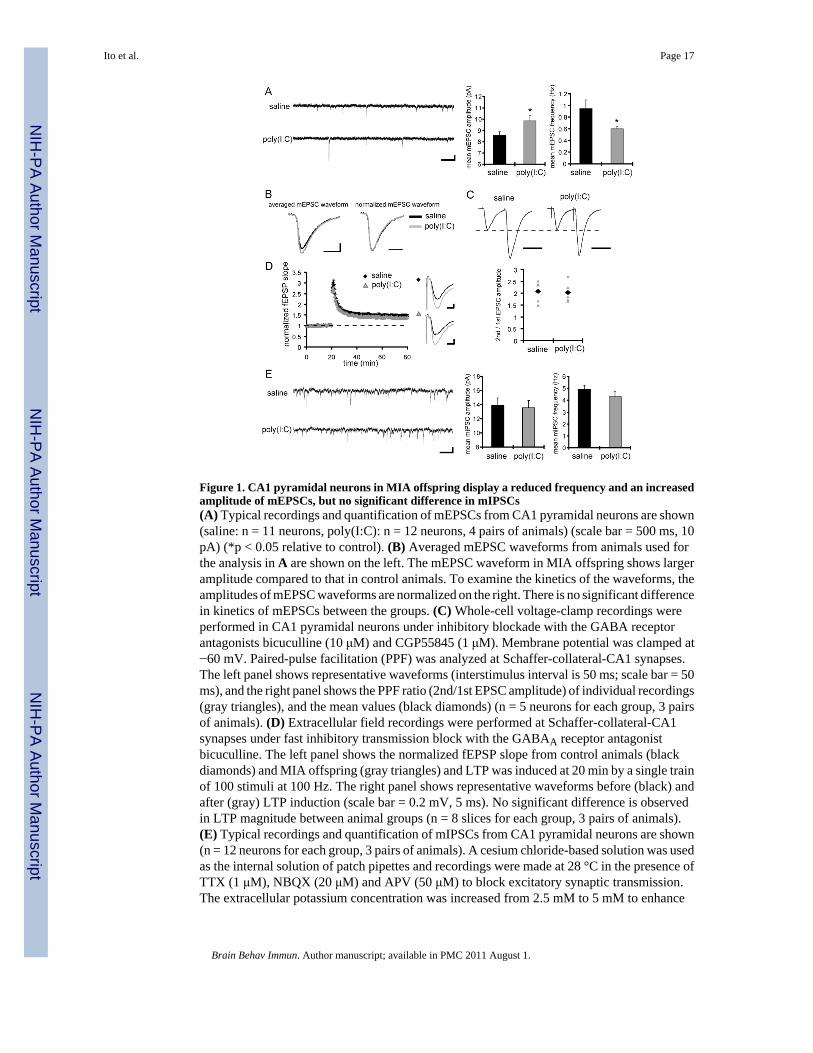
Figure 1. CA1 pyramidal neurons in MIA offspring display a reduced frequency and an increasedamplitude of mEPSCs, but no significant difference in mIPSCs(A) Typical recordings and quantification of mEPSCs from CA1 pyramidal neurons are shown(saline: n = 11 neurons, poly(I:C): n = 12 neurons, 4 pairs of animals) (scale bar = 500 ms, 10pA) (*p < 0.05 relative to control). (B) Averaged mEPSC waveforms from animals used forthe analysis in A are shown on the left. The mEPSC waveform in MIA offspring shows largeramplitude compared to that in control animals. To examine the kinetics of the waveforms, theamplitudes of mEPSC waveforms are normalized on the right. There is no significant differencein kinetics of mEPSCs between the groups. (C) Whole-cell voltage-clamp recordings wereperformed in CA1 pyramidal neurons under inhibitory blockade with the GABA receptorantagonists bicuculline (10 μM) and CGP55845 (1 μM). Membrane potential was clamped at−60 mV. Paired-pulse facilitation (PPF) was analyzed at Schaffer-collateral-CA1 synapses.The left panel shows representative waveforms (interstimulus interval is 50 ms; scale bar = 50ms), and the right panel shows the PPF ratio (2nd/1st EPSC amplitude) of individual recordings(gray triangles), and the mean values (black diamonds) (n = 5 neurons for each group, 3 pairsof animals). (D) Extracellular field recordings were performed at Schaffer-collateral-CA1synapses under fast inhibitory transmission block with the GABAA receptor antagonistbicuculline. The left panel shows the normalized fEPSP slope from control animals (blackdiamonds) and MIA offspring (gray triangles) and LTP was induced at 20 min by a single trainof 100 stimuli at 100 Hz. The right panel shows representative waveforms before (black) andafter (gray) LTP induction (scale bar = 0.2 mV, 5 ms). No significant difference is observedin LTP magnitude between animal groups (n = 8 slices for each group, 3 pairs of animals).(E) Typical recordings and quantification of mIPSCs from CA1 pyramidal neurons are shown(n = 12 neurons for each group, 3 pairs of animals). A cesium chloride-based solution was usedas the internal solution of patch pipettes and recordings were made at 28 °C in the presence ofTTX (1 μM), NBQX (20 μM) and APV (50 μM) to block excitatory synaptic transmission.The extracellular potassium concentration was increased from 2.5 mM to 5 mM to enhance
Ito et al. Page 17
Brain Behav Immun. Author manuscript; available in PMC 2011 August 1.
NIH
-PA Author Manuscript
NIH
-PA Author Manuscript
NIH
-PA Author Manuscript

the frequency of miniature synaptic events. No significant difference is observed in mIPSCamplitude or frequency between the groups.
Ito et al. Page 18
Brain Behav Immun. Author manuscript; available in PMC 2011 August 1.
NIH
-PA Author Manuscript
NIH
-PA Author Manuscript
NIH
-PA Author Manuscript

Figure 2. The offspring of poly(I:C)-treated mothers display increased DA-induced depression atdistal temporoammonic-CA1 synapses(A) A schematic diagram is shown depicting the two distinct axonal projections in the TApathway from MEC and LEC. (B) Extracellular field recordings are obtained simultaneouslyfrom proximal and distal TA-CA1 synapses. For each recording from proximal or distal TA-CA1 synapses, the left panels show the normalized fEPSP slopes from control (black diamonds)and MIA offspring (gray triangles), and the right panels show representative traces of fEPSPwaveforms before (black) and after (gray) DA application (scale bar = 0.05 mV, 5 ms). Thetest pulse was applied every 30 sec and DA (20 μM) was bath applied during the time indicatedby the thick line (*p < 0.05 relative to control) (saline: n = 7 slices, poly(I:C): n = 6 slices, 4pairs of animals). (C) In hippocampal slices prepared from control animals, DA induces asignificantly larger depression at proximal compared to distal TA-CA1 synapses. While theslices prepared from MIA offspring show a normal DA-induced depression at proximalsynapses, they exhibit a significantly larger depression at distal TA-CA1 synapses, comparedto controls (*p < 0.05). (D) Hippocampal slices prepared from MIA offspring show increasedDA-induced depression at distal TA-CA1 synapses. The DA concentration was increased every10 min, from 1 to 20 μM (*p < 0.05 relative to control). (E) Analysis of the data in D is shown.The mean fEPSP slopes at 5–10 min after the application of each designated concentration ofDA were analyzed. Slices prepared from MIA offspring show a significantly larger depressionat each DA concentration examined (n = 12 slices for each group, 8 pairs of animals) (*p <0.05 relative to control).
Ito et al. Page 19
Brain Behav Immun. Author manuscript; available in PMC 2011 August 1.
NIH
-PA Author Manuscript
NIH
-PA Author Manuscript
NIH
-PA Author Manuscript

Figure 3. The offspring of poly(I:C)-treated mothers display abnormal c-Fos expression in areaCA1 pyramidal neurons following novel object exposure(A) A schematic diagram of the behavioral procedure is shown. After mice were accommodatedto their new home cages for a few days, two novel objects were placed in the cage. After a 2hr exposure to allow c-Fos gene activation to be expressed as protein, the hippocampus wasprocessed for immunohistochemistry. (B) Examples of c-Fos expression in the pyramidal layerof area CA1 are shown. The pyramidal layer was divided into equal proximal and distal regions.The c-Fos signals that are larger than the mean + two times the standard deviation, werequantified using ImageJ (see Methods for details) (scale bar = 100 μm). (C) The number of c-Fos signals was quantified in proximal and distal regions of the CA1 pyramidal layer. A two-way ANOVA was performed with 2 variables: animal group (saline vs. poly(I:C)) and CA1subregion (distal vs. proximal), and revealed a significant interaction (*p < 0.05) (n = 6 pairsof animals, 2 sections analyzed and averaged from each).
Ito et al. Page 20
Brain Behav Immun. Author manuscript; available in PMC 2011 August 1.
NIH
-PA Author Manuscript
NIH
-PA Author Manuscript
NIH
-PA Author Manuscript

Figure 4. The offspring of poly(I:C)-treated mothers display comparable c-Fos expression in areaCA1 pyramidal neurons following novel place exposure(A) A schematic diagram of the behavioral procedure is shown. After mice were accommodatedto the new home cage for a few days, they were moved to a novel cage. After a 2 hr exposure,immunohistochemistry on the hippocampus was carried out. (B) Examples of c-Fos expressionin the pyramidal layer of area CA1 are shown. The pyramidal layer was divided into equalproximal and distal regions. The c-Fos signals that are larger than the mean + two times thestandard deviation, were quantified using ImageJ (see Methods for details) (scale bar = 100μm). (C) The number of c-Fos signals was quantified in proximal and distal regions of CA1pyramidal layer (n = 5 pairs of animals, 2 sections analyzed from each).
Ito et al. Page 21
Brain Behav Immun. Author manuscript; available in PMC 2011 August 1.
NIH
-PA Author Manuscript
NIH
-PA Author Manuscript
NIH
-PA Author Manuscript
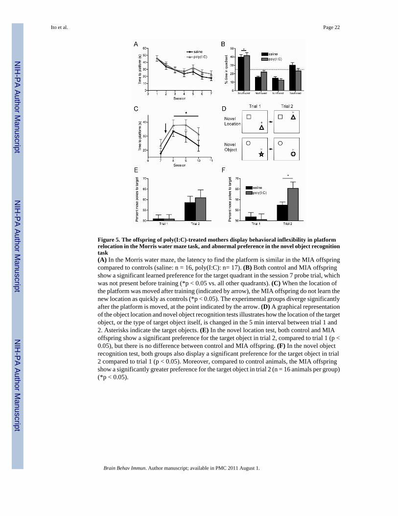
Figure 5. The offspring of poly(I:C)-treated mothers display behavioral inflexibility in platformrelocation in the Morris water maze task, and abnormal preference in the novel object recognitiontask(A) In the Morris water maze, the latency to find the platform is similar in the MIA offspringcompared to controls (saline: n = 16, poly(I:C): n= 17). (B) Both control and MIA offspringshow a significant learned preference for the target quadrant in the session 7 probe trial, whichwas not present before training (*p < 0.05 vs. all other quadrants). (C) When the location ofthe platform was moved after training (indicated by arrow), the MIA offspring do not learn thenew location as quickly as controls (*p < 0.05). The experimental groups diverge significantlyafter the platform is moved, at the point indicated by the arrow. (D) A graphical representationof the object location and novel object recognition tests illustrates how the location of the targetobject, or the type of target object itself, is changed in the 5 min interval between trial 1 and2. Asterisks indicate the target objects. (E) In the novel location test, both control and MIAoffspring show a significant preference for the target object in trial 2, compared to trial 1 (p <0.05), but there is no difference between control and MIA offspring. (F) In the novel objectrecognition test, both groups also display a significant preference for the target object in trial2 compared to trial 1 (p < 0.05). Moreover, compared to control animals, the MIA offspringshow a significantly greater preference for the target object in trial 2 (n = 16 animals per group)(*p < 0.05).
Ito et al. Page 22
Brain Behav Immun. Author manuscript; available in PMC 2011 August 1.
NIH
-PA Author Manuscript
NIH
-PA Author Manuscript
NIH
-PA Author Manuscript