nitroglycerin safety in inferior st elevation …
TRANSCRIPT

Northern Michigan University Northern Michigan University
NMU Commons NMU Commons
DNP Scholarly Projects Student Works
5-2021
NITROGLYCERIN SAFETY IN INFERIOR ST ELEVATION NITROGLYCERIN SAFETY IN INFERIOR ST ELEVATION
MYOCARDIAL INFARCTION (STEMI) PATIENTS: A MYOCARDIAL INFARCTION (STEMI) PATIENTS: A
RETROSPECTIVE REVIEW RETROSPECTIVE REVIEW
Olivia Montgomery [email protected]
Follow this and additional works at: https://commons.nmu.edu/dnp
Part of the Nursing Commons
Recommended Citation Recommended Citation Montgomery, Olivia, "NITROGLYCERIN SAFETY IN INFERIOR ST ELEVATION MYOCARDIAL INFARCTION (STEMI) PATIENTS: A RETROSPECTIVE REVIEW" (2021). DNP Scholarly Projects. 19. https://commons.nmu.edu/dnp/19
This Scholarly Project is brought to you for free and open access by the Student Works at NMU Commons. It has been accepted for inclusion in DNP Scholarly Projects by an authorized administrator of NMU Commons. For more information, please contact [email protected],[email protected].

NITROGLYCERIN SAFETY IN INFERIOR ST ELEVATION MYOCARDIAL
INFARCTION (STEMI) PATIENTS: A RETROSPECTIVE REVIEW
By
Olivia Montgomery
DNP PROJECT
Submitted to
Northern Michigan University
In partial fulfillment of the requirements
For the degree of
DOCTOR OF NURSING PRACTICE
School of Nursing
March 29, 2021

SIGNATURE APPROVAL FORM
NITROGLYCERIN SAFETY IN INFERIOR ST ELEVATION MYOCARDIAL
INFARCTION (STEMI) PATIENTS: A RETROSPECTIVE REVIEW
This DNP Project by Olivia Montgomery is recommended for approval by the student’s
Faculty Chair, Committee, and Department Head in the School of Nursing
Kristi Robinia PhD, RN_______________________________ 05/11/2021
Committee Chair: Date
Katie Menard PhD, RN _______ 05/11/2021
First Reader: Date
Terry Durley DNP, MPA, CRNA, FNP-C_________________ 05/11/2021
Second Reader: Date
Kristi Robinia PhD, RN_______________________________ 05/11/2021
Department Head: Date

i
ABSTRACT
NITROGLYCERIN SAFETY IN INFERIOR ST ELEVATION MYOCARDIAL
INFARCTION (STEMI) PATIENTS: A RETROSPECTIVE REVIEW
By
Olivia Montgomery
Nitroglycerin (NTG) is medication used to reduce chest pain (Boden et al., 2015)
and is the suggested analgesic for angina associated with ST elevation myocardial
infarction (STEMI) (de Alencar Neto, 2018). Due to the potential for right ventricular
(RV) infarct and hemodynamic collapse in inferior STEMI patients (Nagam, Vinson, &
Levis, 2017), the American Heart Association (AHA) recommends avoidance of NTG in
patients with suspected RV infarct (Antman et al., 2004). The purpose of this DNP
project was to explore the safety of NTG use in the treatment of patients diagnosed with
STEMI by evaluating the effects of NTG on hemodynamic measures and angina. Data
were collected via a retrospective chart review at a rural Midwestern hospital and
analyzed via Fisher’s Exact and multiple linear regression analyses. There were no
significant differences between STEMI groups for occurrence of hypotension (p=0.521),
bradycardia (p=0.064), medical need for hemodynamic support (p=0.530), or cardiac
arrhythmia (p=0.465). For this sample of patients, the results show no difference in
adverse occurrences between patients who received NTG with a diagnoses of inferior
STEMI versus patients who received NTG with a diagnoses of non-inferior STEMI. A
recommendation is that patients with identified inferior STEMI receive a right-sided
ECG to evaluate for RV infarct (Bischof et al., 2018), in order to guide individualized

ii
patient care including the use of NTG, which was shown to significantly reduce chest
pain in this sample of patients (p<0.001).

iii
Copyright by
OLIVIA MONTGOMERY
May 11, 2021

iv
DEDICATION
This DNP project is dedicated to my husband, Adam, and to my family and friends for
their continued support throughout my education and career.
v
ACKNOWLEDGEMENTS
The author would like to thank Dr. Kristi Robinia, the DNP project chair, for her support,
guidance, and advice; Joe Ackerman, the research sponsor, for his direction and
willingness to assist; and the Montgomery & Johnson families, for their extensive support
throughout the entire process of conducting this project. Words cannot describe the level
of gratitude for these individuals, as their contributions facilitated the successful
completion of this project.

vi
TABLE OF CONTENTS
List of Tables………………………………………………………………vi
Chapter One………………………………………………………………... 1
Chapter Two…………………………………………………………….......8
Chapter Three…………………………………………………………….... 25
Chapter Four……………………………………………………………...... 32
Discussion………………………………………………………………….. 41
Conclusion…………………………………………………………………. 58
References………………………………………………………………….. 61
Appendix A: Johns Hopkins Approval for Model Use…….……....……….72
Appendix B: Institutional Review Board Approval…….……....…………. 74
Appendix C: Chart Review Methodology…………………………………. 76
Appendix D: Software Citations…………………………………………… 78

vii
LIST OF TABLES
Table 1: Initial Route and Dosage of Nitroglycerin………………………………….33
Table 2: Health History Diagnoses of 2019 STEMI Patients…………………...........34
Table 3: STEMI Location and Culprit Vessels ……………………………………....35
Table 4: Medications Administered to 2019 STEMI Patients………………………..36
Table 5: Effects Estimates of Variables on Systolic Blood Pressure……………........38
Table 6: Effects Estimates of Variables on Heart Rate….…………………………....40
Table 7: Effects Estimates of Variables on Chest Pain Rating……………………….41

1
Chapter One
Introduction
Nitroglycerin (NTG) is the most commonly prescribed short-acting nitrate to
reduce the intensity of chest pain in those experiencing angina (Boden, Padala, Cabral,
Buschmann, & Sidhu, 2015). NTG is a front-line medication used to relieve chest pain in
individuals experiencing acute coronary syndrome (ACS) (Boden et al., 2015) and is also
the most frequently delivered treatment to individuals having a myocardial infarction
(MI) (Ferreira & Mochly-Rosen, 2012). Although there is no study identifying reduction
of mortality, NTG remains a suggested analgesic medication for angina associated with
ST elevation myocardial infarction (STEMI) as well as for the treatment of hypertension
(de Alencar Neto, 2018).
ACS represents the signs and symptoms a person experiences relating to
myocardial ischemia or infarction, with or without electrocardiogram (ECG) ST segment
changes (Camm & Camm, 2016) and includes unstable angina, non-ST elevation
myocardial infarction (NSTEMI) and STEMI. The most damaging and life-threating
diagnosis associated with ACS is STEMI. The diagnosis of STEMI occurs when there is
complete occlusion of a coronary artery causing cardiac tissue death, which is the cause
of angina, and is identified by ST segment elevation on an ECG (Camm & Camm, 2016).
The purpose of this DNP project was to explore the safety of NTG use in the treatment of
patients diagnosed with STEMI by evaluating the effects of NTG on hemodynamic
measures and angina.

2
Significance and Background
The American Heart Association (AHA) continues to designate cardiovascular
disease as the leading cause of mortality for both men and women in the United States
(Benjamin et al., 2018). Between the years 2011 and 2014, there were 8.7 million
individuals diagnosed with angina, and in 2014 alone, there were 957,000 individuals
diagnosed with MIs (Benjamin et al., 2018). The total number of individuals with angina
and MIs suggests a significant number of individuals who may have received NTG or
been provided with a NTG prescription. The rapid onset of NTG effects results in venous
and arterial dilation, ultimately increasing blood flow to the heart and reducing
myocardial stress, which are desired during a STEMI (Ferreira & Mochly-Rosen, 2012).
Currently, the AHA advises extreme caution with the use of NTG, or against the
use of NTG entirely, with inferior STEMIs with potential right ventricular (RV)
infarction and failure (Antman et al., 2004, O’Connor et al., 2010). RV infarction most
commonly occurs with the occlusion of the marginal branch of the right coronary artery
(RCA), resulting in dyskinetic RV wall motion, decreased compliance of the ventricle,
and subsequent reduced stroke volume (Namana et al., 2018). Clinical indications that are
more specific to RV infarction and pending RV failure include elevated jugular
distention, clear lung sounds with auscultation, hypotension, pulsus parodoxus, and a
tricuspid murmur (Namana et al., 2018).
The reasoning behind avoiding NTG use in patients with potential RV
involvement is that the medication could potentiate hypotension and cardiac arrhythmias
(Antman et al., 2004, O’Connor et al., 2010). An inferior STEMI, which is identified on a
standard 12-lead ECG as ST segment elevation in leads II, III and aVF (Taglieri et al.,

3
2014), often indicates occlusion of the RCA (Namana et al., 2018). Because occlusion of
this coronary artery is also is the most common to cause RV infarct, concern for RV
involvement may influence clinical interventions.
Inferior wall MIs represent 40% to 55% of all MIs (Jaton, 2017; Warner &
Tivakaran, 2020), while the occurrence of right ventricle (RV) infarct has been reported
to occur in approximately 20% to 50% of inferior STEMIs (Bischof, Worrall, & Smith,
2018; Nagam, Vinson, & Levis, 2017). A standard 12-lead ECG provides limited data on
RV infarct (Nagam et al., 2017), and therefore, it is recommended that a right-sided ECG
be obtained when there is inferior STEMI identification on a standard 12-lead ECG
(Antman et al., 2004).
Nagam et al. (2017) discuss how a conventional 12-lead ECG largely shows
involvement of the left ventricle, while minimally showing RV wall involvement. A
right-sided ECG analyzes leads V1R-V6R, allowing for improved evaluation of the RV
(Somers, Brady, Bateman, Mattu, & Perron, 2003). With the right-sided ECG, precordial
leads mirror the locations of the standard 12-lead but on the right side of the patient’s
chest (Somers et al., 2003). Because lead V4R has high sensitivity and specificity for
identifying RV involvement, when obtained via a right-sided ECG, this lead may provide
pertinent information on the status of the RV and help guide treatment of patients
diagnosed with inferior STEMI (Nagam et al., 2017; Somers et al., 2003).
NTG has systemic venous dilating properties, which reduces cardiac preload
(Ferreira & Mochly-Rosen, 2012) and may contribute to hypotension and decreased
cardiac output, especially in those with RV infarct (Antman et al., 2004). The effects of
NTG generally last less than 30 minutes, with a standard sublingual dose of 0.4 mg

4
resulting in temporary coronary artery dilation. Higher doses of intravenous infusions
between 160 and 600 micrograms per minute result in sustained systemic arterial and
venous dilation (Jaton, 2017). The effects of NTG are transient and the benefits of NTG
for angina reduction in patients with inferior STEMIs requires evaluation on a patient-
specific basis.
The use of opioid medications, such as morphine (de Alencar Neto, 2018), as an
analgesic for angina should be avoided unless angina is unresponsive to NTG (Frampton,
Devries, Welch, & Gersh, 2020). Evaluation of morphine, which was traditionally used to
reduce angina during ACS and STEMIs, has been shown to reduce the therapeutic effects
of commonly used medications, such as ticagrelor, prasugrel, and clopidogrel, all of
which are used for platelet inhibition before and after percutaneous coronary intervention
(PCI) (Frampton et al., 2020; Ibanez et al., 2018). Although morphine is still listed as a
potential analgesic for angina resulting from STEMI (Antman et al., 2008), it should be
noted that newer research implies the risks of reducing therapeutic effects of antiplatelet
medications outweigh the benefits of morphine and potentially other opioids for analgesia
(Duarte et al., 2019; Frampton et al., 2020; Koh, Fernando, Peter, & Stub, 2019).
Treating angina associated with STEMI becomes challenging when risks could impact
patient morbidity and mortality. An individualized approach to patient care requires
ongoing analysis of the safety and efficacy of medications currently used in the treatment
of STEMI.
Purpose of DNP Project
The purpose of this DNP project was to explore the safety of NTG use in the
treatment of patients diagnosed with STEMI by evaluating the effects of NTG on
5
hemodynamic measures and angina. Comparisons occurred between patients who
received NTG with the diagnosis of inferior STEMI to those diagnosed with non-inferior
STEMI, all of whom presented to a rural Midwestern hospital for treatment. The hospital
used in this project serves as the only PCI center in a large geographical region spanning
approximately 16,000 square miles, providing services to individuals that reside in the
town in which the hospital is located, as well as to individuals in the many surrounding
rural communities. NTG administration was tracked retrospectively via healthcare
professionals’ documentation, in order to assess the safety implications of using NTG for
angina reduction in this rural area. As the development of hemodynamic instability is a
major concern resulting from administration of NTG to patients with inferior STEMIs
due to RV infarct (Antman et al., 2004), the following research questions were explored:
1) Does the administration of NTG result in hypotension more often in inferior
STEMI patients than non-inferior STEMI patients?
2) Do patients with inferior STEMIs require additional interventions more
frequently to correct hypotension or bradycardia than non-inferior STEMI patients
following NTG administration?
3) Do inferior STEMI patients experience more bradycardia and cardiac
arrhythmias compared to non-inferior STEMI patients?
4) Does NTG significantly reduce chest pain levels reported by STEMI patients?
Methods
A retrospective chart review examined charts of previously hospitalized patients
who were diagnosed with inferior or non-inferior STEMI who presented to the rural
Midwestern hospital between January 1, 2019 and December 31, 2019. Systolic blood

6
pressure (SBP), heart rate, cardiac arrhythmias, need for hemodynamic interventions, and
patient reported chest pain were compared between patients diagnosed with inferior
STEMI to patients diagnosed with non-inferior STEMI. All patients in both groups also
had received NTG, which was identified while examining each individual’s chart.
The sample consisted of patients who had received NTG in conjunction with the
diagnosis of STEMI. The listed discharge diagnosis of STEMI, which was determined by
a cardiology physician, was how the presence of STEMI was determined in this project.
Data were grouped according to patient diagnoses of inferior versus non-inferior STEMI,
and the outcome measurements of SBP, heart rate, and occurrence of cardiac arrhythmias
were compared between the two groups. Reduction in reported angina experienced by
patients following NTG administration was also examined for both groups, as well as the
use of fluid resuscitation or vasopressors in response to hypotension or bradycardia
following NTG administration.
Theoretical Framework
The theoretical framework used for this project was the Johns Hopkins Nursing
Evidence-Based Practice (JHNEBP) model (© The Johns Hopkins Hospital/ The Johns
Hopkins University, permissions viewable in Appendix A). The JHNEBP model is an
evidence-based practice model that allows for nurses and other interdisciplinary team
members to identify and make changes to practice in order to provide evidence-based
individualized care to patients (McEwen & Wills, 2018). For the purposes of this project,
results will be reported to the hospital STEMI coordinator in order to add to discussions
on the care of patients presenting with ACS at this hospital. Based on the JHNEBP
model, the problem identified is that inferior STEMI patients may receive generalized

7
treatment based on current AHA guidelines for RV infarct (Antman et al., 2004), which
may not account for individualized potentially beneficial interventions, especially in the
absence of diagnostic measures to identify RV infarct. Specifically, this project seeks to
add to the evidence regarding the impact of NTG on the hemodynamic outcome variables
of SBP, heart rate, cardiac arrhythmias and chest pain experienced by patients diagnosed
with STEMI. Ultimately, the goal of this project is to contribute data to the conversation
of how to safely improve the quality and individualization of care to patients diagnosed
with STEMI.

8
Chapter Two
RV infarct has been reported to occur in as low as 20% of patients (Bischof et al.,
2018) and as high as 50% of patients (Nagam, Vinson, & Levis, 2017) with inferior
STEMI identification on ECG. The most common culprit lesion causing RV infarct is the
acute marginal branch of the RCA with ST elevation on ECG in lead V1, II, III, and aVF
(Namana et al., 2018). Although RV infarct may occur with other culprit lesions, such as
the left circumflex artery (Namana et al., 2018), the RCA is often suspected in
individuals with inferior STEMI identification on ECG.
As inferior STEMIs represent approximately 40% to 55% of all STEMI cases
(Jaton, 2017; Warner & Tivakaran, 2020), and with current recommendations to withhold
NTG from patients with RV infarct (Antman et al., 2004), it is pertinent to include
methods to identify RV infarct in patient care (Ibanez et al., 2018). Without proper
identification of RV infarct in inferior STEMI patients, hesitancy in the provision of NTG
may exist due to the associated occurrence of RCA and RV involvement observed in
patients with inferior STEMIs (Namana et al., 2018). NTG may provide necessary
analgesia for angina experienced by patients throughout their MI, as angina has been
identified as a common symptom experienced by both men and women during a STEMI
(Sederholm Lawesson et al., 2018). Recent literature has warned that angina treated with
morphine may result in lengthened hospital admission, increased infarct size and
subsequent higher mortality rates (McCarthy, Bhambhani, Pomerantsev, & Wasfy, 2018).
A reason cited for these adverse effects is that morphine decreases the therapeutic action
of certain antiplatelet medications (Duarte et al., 2019; Frampton et al., 2020; Koh et al.,
2019).

9
Nurses have an ethical standard that applies to providing evidence-supported
analgesia to individuals experiencing pain, in addition to providing patients with
individualized care (Stokes, 2018). With interdisciplinary involvement, healthcare
providers should evaluate various modalities for pain reduction in patients. For acute pain
in patients experiencing STEMI, reduction of angina needs to consider an individualized
approach to guide the provision or withholding of medications. Re-establishment of
blood flow to the occluded coronary artery with PCI will ultimately relive angina
experienced by patients, however, angina should be treated to reduce myocardial stress in
the interim (Ibanez et al., 2018). Because of the current recommendations for NTG use in
STEMIs (Antman et al., 2004), inferior STEMI patients are potentially blanketed into a
group that may not receive NTG with subsequent angina reduction, especially in the
absence of interventions to specifically identify RV infarct. Although caution for adverse
events and unwanted side effects needs to be considered in all patients with all
medications, it is necessary to address each patient case individually and to uphold the
ethical implications of managing pain. To provide evidence-driven and individualized
care, it is important to evaluate the safety of NTG use between inferior STEMI and non-
inferior STEMI patients, and to question whether current practices are best for patients.
Literature Review
A review of literature was conducted using Northern Michigan University’s
online Lydia Olson Library, CINAHL, PubMed, and Google Scholar. Online searches
focused on finding information relating to NTG, ECG, inferior STEMI, RV infarct, and
opioid medications.

10
The Use of NTG
In a retrospective chart review of patients with suspected STEMI in a prehospital
setting, Bosson et al. (2019) evaluated blood pressure changes, pain scores and heart rates
after the administration of NTG. The study reviewed records from July, 2015 to
December, 2016, from three PCI centers to which patients were transported in Los
Angeles County, California. A 12-lead ECG was performed on patients with suspected
cardiac-related chest pain. Suspected STEMI was determined by software analysis in
combination with paramedic interpretation, with additional assistance provided by online
medical control (Bosson et al., 2019). Bosson et al. (2019) describe the primary treatment
of persistent angina was 0.4 mg sublingual NTG, which was repeated twice if pain
persisted. Patients with initial SBP < 100 mmHg were not included in the study, as they
were ineligible for NTG or opioid medications.
Bosson et al. (2019) identified change in blood pressure as a key safety issue that
was evaluated in the study in patients that received NTG compared to those who did not.
Triage SBP < 100 mmHg, bradycardia or heart rate < 60 beats per minute, drop in
systolic blood pressure > 30 mmHg and cardiac arrest occurring out of the hospital were
secondary safety outcomes evaluated by the researchers. Pain was evaluated on a 0 to 10
scale to assess for efficacy of NTG in angina reduction, which was compared to pain
reported by individuals who did not receive NTG (Bosson et al., 2019). Subgroups,
including individuals who received PCI to the right coronary artery (RCA), were
evaluated for the same outcome criteria and compared to individuals who had PCI to
other coronary arteries.
STEMI was confirmed in 193 patients, with 155 individuals receiving NTG

11
(Bosson et al., 2019). The average chest pain score was reduced by 2.4 on a 0 to 10 point
scale in STEMI patients treated with NTG (n=145), while all individuals who did not
receive NTG had an average reduction in chest pain score of 1.4. Bosson et al. (2019)
identified that a pain reduction of 1.39 was the minimum for reduction that represented
clinical significance. When comparing chest pain reduction to the clinically significant
value of 1.39, both groups had reduced angina, but the individuals treated with NTG had
what was considered to be a significant reduction in angina (p <0.001), compared to those
who did not receive NTG (p=0.5). Bosson et al. (2019) report that of the 75 patients with
RCA occlusions, 80% received NTG. Comparable occurrence of hypotension and
bradycardia resulted in the patients receiving PCI for non-RCA occlusions RR 0.64 (95%
CI, 0.21, 1.95) versus those with RCA occlusion RR 1.30 (95% CI, 0.57, 2.94).
Bosson et al. (2019) discuss the significant reduction in pain following
administration of NTG. There also was no increased risk of hypotension or bradycardia
with pre-hospital administration of NTG at arrival to the emergency department (ED).
The researchers concluded that the protocols directing withholding NTG from patients
diagnosed with inferior STEMI may need to be revisited. Limitations of the study
included that only 193 patients of the 780 suspected actually had STEMIs, which limited
the assessment of hemodynamics in this group. Although the number of patients with
actual STEMI was significantly smaller than the suspected number, the researchers
provide information on all patients as well as subgroups, which is suggested as evidence
for the safety of NTG use in pre-hospital settings.
In a similar study, Robichaud et al. (2016) conducted a retrospective chart review
between February, 2010 and July, 2012 in Quebec, Canada, to analyze the impact of

12
NTG on blood pressure in patients experiencing inferior STEMI as compared to those
diagnosed with non-inferior STEMI. Hypotension was defined as systolic blood pressure
(SBP) < 90 mmHg and was compared between inferior and non-inferior STEMI groups.
Additionally, researchers evaluated any reductions in SBP > 30 mmHg between inferior
and non-inferior patient groups. The chart review included 805 STEMI patients, all of
whom received NTG prior to hospital arrival (Robichaud et al., 2016).
Robichaud et al. (2016) discovered that patients with inferior STEMIs did have
more frequent initial hypotension than non-inferior STEMI patients, with 9.9% of inferior
STEMI patients presenting with hypotension, compared to 4.9% of patients with non-
inferior STEMIs. Patients that presented with hypotension would not meet criteria to
receive NTG based on blood pressure regardless of infarct location, and therefore were
not included in the study (Robichaud et al., 2016). With regard to bradycardia, 1.1% of
inferior STEMI patients (n=5/474) experienced heart rates < 50 beats per minute, while
no patients with non-inferior STEMIs had heart rates documented at < 50 beats per
minute (n=0/347).
Following statistical analyses, Robichaud et al. (2016) reported that based on
computer-interpreted ECGs, there was no statistically significant difference in incidence
of hypotension between inferior STEMI patients (n= 38/466, 8.2%) and non-inferior
STEMI patients (n= 30/339, 8.9%) following NTG administration (p= 0.73). There was
also no significant difference between the two groups for decrease in SBP > 30 mmHg,
which occurred in 23.4% of inferior STEMI patients and in 23.9% of non-inferior STEMI
patients (p = 0.87). Robichaud et al. (2016) described that based on the results of their
research, ST segment elevation in lead III that measured greater than ST segment

13
elevation in lead II, which would suggest RV involvement, was not a good predictor for
patients who would be at risk for developing hypotension. However, Robichaud et al.
(2016) observed lower rates of hypotension than their expected rate of 15%, even given
their large sample size. This was identified as a limitation to the study as, Robichaud et
al. (2016) estimated a need to have a much larger sample size of approximately 38,000
individuals in order to show the same occurrence of hypotension among inferior and non-
inferior STEMI groups using a power of 80%.
Another retrospective chart review evaluating the effectiveness of sublingual
NTG in reducing chest pain and adverse events associated with NTG administration was
completed by Engleberg et al. (2000). The researchers reviewed all advanced life support
(ALS) cases in the Emergency Medical Services’ (EMS) System of Suffolk County, New
York, for which IRB approval had been granted. Patients included in this study had
received NTG from an emergency medical technician (EMT) or paramedic prior to
hospital arrival between January, 1993, and June, 1994. A single dose of 0.4 mg
sublingual NTG was provided to 1,662 patients. NTG was administered to individuals
reporting chest pain (n=901), respiratory distress likely caused from congestive heart
failure (n=251), or a combination of both (n=510). The researchers analyzed chest pain
rating reported by patients, hemodynamic measurements, including systolic and diastolic
blood pressure, as well as the occurrence of adverse events.
Of the 1,662 patients who received NTG in this study, there were a total of 779
records with complete data on chest pain ratings. Chest pain was rated by patients using a
verbal numeric scale ranging from 0 to 10, with 0 representing no pain and the most
severe pain rated at 10. Chest pain scores were reported before and after receiving NTG,

14
with a mean decrease in chest pain reported by patients after receiving NTG of 2.6 (95%
CI [2.4, 2.8]). After receiving NTG, the mean SBP in all patients decreased by 11.8
mmHg (95% CI [10.7, 13.0]), while heart rate had a mean increase by 2.7 beats per
minute (95% CI [0.6, 4.9]).
Engleberg et al. (2000) reported 12 adverse events within the study, which
included patients that experienced a drop in SBP of > 100 mmHg that responded to fluid
or elevation of the patients’ legs (n=6), hypotension following receiving NTG with a
documented SBP of < 90 mmHg (n=4), syncope (n=1) and profound bradycardia and
hypotension (n=1). Zero deaths were reported. Of these patients, individuals with a > 100
mmHg decrease in SBP had initial SBP > 210 mmHg prior to receiving NTG, and no
adverse events were associated with individuals who self-administered NTG prior to
EMS arrival. In total, the estimated adverse events for the study were reported as 0.7%
(95% CI [0.4, 1.3]).
The researchers (Engleberg et al., 2000) reported that the average reduction in
chest pain ratings was 2.6 on the 10-point scale which equated to an approximate 40%
reduction of pain, although pain was completely relieved in only 10% of patients. In
discussion of limitations, Engleberg et al. (2000), point out that final diagnoses were not
included, meaning generalizations should not be made in relation to patients experiencing
cardiac ischemia or MI, but suggest future investigation of the effects of NTG in this
patient population. This study documents the importance of NTG’s role in chest pain
reduction in patients and a low occurrence of adverse events associated with
administration of NTG, regardless of diagnosis.

15
Bosson et al. (2019), Engleberg et al. (2000), and Robichaud et al. (2016) all cite
a retrospective study conducted by Ferguson, Diver, Boldt, and Pasternak (1989) whom
identified a significant occurrence of hypotension in inferior STEMI patients with
suspected RV infarct. In discussion of their research, Ferguson et al. (1989) report
concomitant administration of calcium channel blockers in several of the patients
included in their study, noting that this may have potentiated the occurrence of observed
hypotension. A prospective study to identify patients with RV infarct via multiple
diagnostic modalities, as well as their response to NTG, was recommended by the
researchers (Ferguson et al., 1989).
Robichaud et al. (2016) discuss how the study conducted by Ferguson et al.
(1989) was a singular study supporting the recommendations that currently stand about
NTG use in inferior STEMI patients. As the occurrence of RV infarct in patients with
inferior STEMIs is considered highly variable (Ferguson et al., 1989), the risk of using
this medication in inferior STEMI patients with the potential of RV infarct was higher
than the benefits. With more current retrospective reviews that refute avoidance of NTG
in inferior STEMI patients (Bosson et al, 2019; Robichaud et al., 2016), and the apparent
time gap in related research, it is suggested that further investigation into this topic is
warranted.
The Use of ECG Findings
Another component in evaluating the safety of NTG for patients experiencing
inferior STEMI is the use of ECG to determine RV infarct. Bischof et al. (2018)
conducted a retrospective review of patients with inferior STEMIs to evaluate ECG
sensitivity and specificity for identification of RV infarct and the roles of ST segment

16
depression or elevation in standard 12-lead ECGs. Patients included in their study were
those that presented to Hennepin County Medical Center, Minnesota, who had ST
elevation > 1 mm identified in inferior leads (II, III, aVF), between January, 2002 and
March, 2008. IRB approval was granted for this project.
Manual measurement of ST segments in leads I, II, III, aVF, aVL, V1 and V2 was
completed by researchers (Bischof et al., 2018) and compared between patients who were
categorized into two groups, right ventricular myocardial infarction (RVMI) and non-
RVMI. Culprit lesions were previously identified in the cardiac catheterization
laboratory, which was the basis for categorizing patients into the respective groups.
RVMI patients had culprit lesions identified in the RCA proximal to the RV marginal
branch, which is the artery that supplies the RV myocardium. Non-RVMI patients had
occlusions in the middle or distal RCA or circumflex artery. Subsequently, ST elevation
or depression measured by researchers was compared to evaluate the sensitivity and
specificity of ST segment changes on ECG in identifying RVMI and non-RVMI infarcts.
Bischof et al. (2018) reported a sample size of 43 patients in the RVMI group and
106 patients in the non-RVMI group. In ECG analysis, there was no difference in ST
segment depression in lead I between RVMI patients (n=37, 86%) and non-RVMI
patients (n=85, 80%) (p=0.54), with sensitivity and specificity of lead I in determining
RVMI reported as 86% and 20%, respectively. A lower frequency of non-RVMI patients
had ST elevation in V1 (n=17, 16%, 95% CI [10, 24]) compared to patients with RVMI
(n=15, 35%, 95% CI [22, 50]), which was a significant finding (p=0.015). Specificity and
sensitivity for RVMI was thus calculated to be 84% (95%, CI [76, 90]) and 35%,
respectively, for ST elevation in lead V1.

17
In the absence of ST segment depression in lead V2, sensitivity for RVMI was
found to be higher (69%, 95% CI [44, 86]) when compared to the presence of ST
segment depression in lead V2 (15%, 95% CI [6, 32]), which was a significant finding
(p< 0.001). In lead V1 in RVMI patients, ST segment depression was present in V2 in
85% of patients who did not have concurrent ST segment elevation in lead V1. Bischof et
al. (2018) thus concluded that ST segment depression in lead I was not dependable in
differentiating patients with or without RV infarct. However, ST segment elevation in V1
was found to be specific for RVMI, with higher sensitivity identified in the absence of ST
segment depression in lead V2.
Bischof et al. (2018) did identify limitations within their study, including wide
confidence intervals and a small sample size of patients identified with RVMI. A
prospective study was suggested, in addition to further investigation of ECG analysis in
patients with RV dysfunction in relation to culprit lesions, which may alter sensitivity and
specificity of leads I and V1 in identification of RV dysfunction. Due to lack of
consistent sensitivity and specificity of leads in a standard 12-lead ECG, the researchers
made an important recommendation, which was to obtain a right-sided ECG, specifically
lead V4R, to accurately diagnose RV infarct (Bischof et al., 2018).
The importance of ECG analysis for patients presenting with STEMI is magnified
in the presence of inferior infarct identification, as treatments such as the administration
of NTG hinge on the potential for RV infarct. The European Society of Cardiology
(ESC), American College of Cardiology (ACC), and the AHA (Ibanez et al., 2018;
O’Gara et al., 2013) recommend obtaining right precordial views of the heart, specifically
V4R, to evaluate for ST elevation that would signify RV infarct in patients with inferior

18
STEMI identification. As completion of a right-sided ECG involves inversion of leads
and few additional supplies, it could easily be completed in a prehospital or emergency
room setting and provide identification of RV infarct using leads with the highest
sensitivity for diagnosis (O’Gara et al., 2013).
The Use of Opioid Medications
The ESC report reduction in patients’ pain as essential during STEMI, as
vasoconstriction occurs as a sympathetic nervous system response to pain, ultimately
increasing the workload of the heart (Ibanez et al., 2018). The ESC states the use of
intravenous opioids for pain should be considered but with a low level of evidence to
support opioid use. Recommendations also warn of the effects of morphine and the
reduction of therapeutic effects of ticagrelor, prasugrel, and clopidogrel antiplatelet
medications (Ibanez et al., 2018) when provided to patients concomitantly. The ESC also
recommends the administration of one of the previously mentioned antiplatelet
medications in a prehospital setting or prior to PCI due to improved patient outcomes,
with a high level of evidence to support the recommendation (Ibanez et al., 2018). With a
higher level of evidence and recommendation to provide these important antiplatelet
medications, treating pain with opioid medications, such as morphine, may be
counterintuitive due to the reduction in antiplatelet efficacy.
The AHA and ACC continue to list morphine as the analgesic of choice for pain
management in patients experiencing STEMI (O’Gara et al., 2013). This is despite the
growing research showing reduced efficacy of recommended antiplatelet medications for
patients experiencing STEMI that receive morphine and other opioid medications
(Frampton et al., 2020). In the same AHA/ACC guidelines, it is recommended to provide

19
ticagrelor, prasugrel, or clopidogrel, as soon as possible to patients prior to or at the time
of PCI. Again, this recommendation is provided with a higher level of evidence to
support the therapeutic effects of antiplatelet medications and improved patient outcomes
when compared to the use of opioid medications to treat chest pain associated with
STEMI.
Clinical trials conducted by researchers Hobl et al. (2014) and Kubica et al.
(2016) evaluated the effects of morphine on clopidogrel and ticagrelor, respectively. The
results of the study conducted by Hobl et al. (2014) showed significant reduction in the
active metabolite of clopidogrel and decreased gastric absorption of the medication in
individuals who received morphine. Similarly, Kubica et al. (2016) reported significant
reduced blood plasma concentrations and bioavailability of ticagrelor in individuals who
received morphine for pain. Both clinical trials show that morphine reduces the efficacy
of the antiplatelet medications, warranting exploration into alternative pain relief
measures.
The Platelet Aggregation with tiCagrelor Inhibition and FentanYl (PACIFY) trial
was conducted by Ibrahim et al. (2018) to evaluate effects of intravenous fentanyl on
ticagrelor. Fentanyl, which is a potent opioid medication that is frequently provided to
patients during PCI (Ibrahim et al., 2018), was evaluated due to the previously identified
adverse effects of morphine on antiplatelet medications. The results from this clinical
trial showed reduction in the efficacy of ticagrelor in individuals who received fentanyl
when compared to those who did not receive the opioid medication, indicating that
morphine is not the sole opioid with this type of interaction.

20
It is suggested that opioid medications are reserved for chest pain that is
unresponsive to NTG (Frampton et al., 2020), as commonly used opioid medications
reduce the efficacy of important antiplatelet medications that are currently recommended
to be provided as early as possible during a STEMI. Chest pain associated with STEMI is
important for providers to manage (Ibanez et al., 2018; Ibrahim et al., 2018), indicating
the need to explore medication options and their potential impact on patient outcomes.
With the knowledge that opioid medications may reduce the efficacy of antiplatelet
medications, the use of NTG remains a viable option in treating chest pain in patients
experiencing STEMI.
In summary, several studies have been conducted to evaluate the safety of NTG
use in patients with the diagnosis of STEMI, including inferior STEMI, in relation to
hemodynamic outcome variables, as well as to analyze NTG’s ability to reduce chest pain
in patients (Bosson et al., 2019; Engleberg et al., 2000; Robichaud et al., 2016). Bischof
et al. (2018) recommend other measures to identify RV infarct, due to the lack of
sensitivity and specificity in identifying RV infarct with a standard 12-lead ECG within
their study. Based on this literature review, further investigation into NTG use in STEMI
patients, and specifically inferior STEMI patients, is warranted. Furthermore, patient care
should be individualized to optimize patient outcomes, including provision of analgesic
medication for chest pain and identification of RV infarct in patients with inferior STEMI
identified on an ECG. The purpose of this DNP project was to explore the safety of NTG
use in the treatment of patients diagnosed with STEMI by evaluating the effects of NTG
on hemodynamic measures and angina. From this, this DNP project may potentially add
21
to evidence for future recommendations designed to individualize care for patients, as
well as the safe use of NTG in patients presenting with the diagnosis of STEMI.
Theoretical Framework
The theoretical framework that is the basis of this project is that patients should
receive evidence-based individualized care. Nurses and interdisciplinary team members
have the capability of identifying areas in practice that propose a certain question or
problem, and may use the Johns Hopkins Nursing Evidence-Based Practice (JHNEBP)
model as an evidenced-based practice model to make changes to practice (McEwen &
Wills, 2018; Dang & Dearholt, 2018). For the purposes of this project, providing
additional data with evidence to a hospital allows for discussions on current practice and
contributes to debate on changes if warranted.
The JHNEBP model consists of a question pertaining to practice, evidence
relating to the question, and subsequent translation to practice (McEwen & Wills, 2018).
The initial step of this theoretical model is to question certain problems or concerns that
are identified in practice (Dang & Dearholt, 2018). For this project, it is not clear if
inferior STEMI patients receive individualized care based on the AHA guidelines to
withhold NTG without additional interventions, such as the recommended right-sided
ECG.
Following the JHNEBP model, the identification of an evidence-based practice
(EBP) question for this project was strictly based on using a standard 12-lead ECG and
whether exclusive standard views of the heart are enough to provide appropriate and
patient-specific interventions to inferior STEMI patients. From this, the questions for this
project are as follows:

22
1) Does the administration of NTG result in hypotension more often in inferior
STEMI patients than non-inferior STEMI patients?
2) Do patients with inferior STEMIs require additional interventions more
frequently to correct hypotension or bradycardia than non-inferior STEMI patients
following NTG administration?
3) Do inferior STEMI patients experience more bradycardia and cardiac
arrhythmias compared to non-inferior STEMI patients?
4) Does NTG significantly reduce chest pain levels reported by STEMI patients?
As NTG is used to reduce angina, patient report of angina rating will be compared among
inferior and non-inferior STEMI groups for analysis of therapeutic reduction in angina
intensity.
The initial practice question in the JHNEBP model requires recruitment of an
interdisciplinary team. The Northern Michigan University Graduate Nursing Committee
approval for graduate research relating to this project in conjunction with the author’s
DNP Chair approval were the initial steps towards developing a team. Cardiologists,
Emergency Department physicians, Emergency Medical Service (EMS) personnel,
quality improvement and the STEMI coordinator are all key stakeholders in this project,
as STEMI patients are cared for by several different teams prior to and at the time of PCI.
The acronym Patient population, Intervention, Comparison, Outcome and Time table
(PICOT) format is used in the JHNEBP model to refine the evidence-based practice
question (McEwen & Wills, 2018). As applied to this DNP project PICOT was
formulated as:

23
▪ The Patient population consisted of patients with inferior and non-inferior
STEMIs presenting to the rural Midwestern hospital for PCI intervention.
▪ The Intervention is the administration of NTG to patients in inferior and non-
inferior STEMI groups, which was reviewed in a retrospective manner due to
the controversy and ethics of a prospective study evaluating the same topic.
▪ Comparison and Outcomes consisted of a comparison of data between the
inferior and non-inferior STEMI groups in terms of hemodynamic
measurements, additional interventions required in each group, and angina
experienced between the two groups.
▪ Time was defined as patients presenting to the rural Midwestern hospital
between January 1, 2019 and December 31, 2019, with inclusion of patients
experiencing STEMI and meeting other inclusion criteria presenting to the
facility.
The next section of the JHNEBP model relates to evidence. Based on the
JHNEBP model, the problem identified is that patients are categorized into an inferior
STEMI group and may not receive patient-specific interventions but more general
treatment based on inferior infarct identification on a standard 12-lead ECG due to the
potential of RV involvement. Medication, such as NTG, may be withheld based on
guidelines for RV infarct (Antman et al., 2004), as the occurrence of RV infarct is largely
associated with inferior STEMI. To improve quality of care and individualized care to
patients with STEMI identification, research addressing blood pressure and patient
hemodynamic stability between inferior and non-inferior STEMI groups should be
utilized (Jaton, 2017; Robichaud et al., 2016). Research utilization should help in guiding

24
the use of NTG for patients experiencing angina during a STEMI, whether an inferior or
non-inferior infarct is identified. A review of patient charts at a rural Midwestern hospital
was conducted to analyze the previously identified research questions. A review of
literature with appraisal of information relating to the problem signifies the external
search for evidence. From this, both internal and external evidence will be used to
facilitate discussions on current policy.
Translation of evidence to practice will depend on the fit and feasibility of
additional interventions at the rural Midwestern hospital. Findings of internal and
external evidence appraisal, and guidance gained from the literature on how to implement
any changes to practice will be disseminated to the hospital through the STEMI
coordinator.

25
Chapter Three
RV infarct in inferior STEMI patients has led to the recommendation by the AHA
to use extreme caution with the use of NTG or exclude NTG entirely as a medication to
treat chest pain in this patient population due to the possibility of hypotension and cardiac
arrhythmias (Antman et al., 2004). Several studies (Bosson et al., 2019; Engleberg et al.,
2000; Robichaud et al., 2016) have evaluated the safety of prehospital NTG in suspected
STEMI and actual STEMI patients, as well as the significance of NTG in chest pain
reduction. Similar to previous studies, this DNP project explored the safety of NTG by
evaluation of hemodynamic measures and angina in patients experiencing STEMIs in a
rural Midwestern setting.
Sample and Setting
Data collection for this DNP project was conducted at a rural Midwestern hospital
through a retrospective chart review. The designated hospital serves as the only PCI
center for STEMI patients in the region, providing much needed services to the
community in which it is located, as well as to numerous outlying communities. The
sample consisted of STEMI patients presenting to the designated hospital, either directly
or by transfer to the hospital from an outlying area.
This project focused on a convenience sample of patients, all of whom presented
to the designated hospital with STEMI between January 1, 2019 and December 31, 2019.
As the purpose of this DNP project was to evaluate the safety of NTG, only STEMI
patients (confirmed on 12-lead ECG by a cardiologist) who received NTG were included
in this project and subsequent data analysis. Additional inclusion criteria were the
requirements that individuals were 18 years of age or older with chest pain of suspected
cardiac origin. Chest pain of cardiac origin must not have resulted from trauma and

26
patients must have been eligible to receive NTG upon first medical contact. As alternate
diagnoses may mimic a STEMI on an ECG (Gu, Svilaas, van der Horst, & Zijlstra, 2008),
final discharge diagnosis of STEMI was used in conjunction with preliminary STEMI
identification on ECG analysis.
The universal definition of STEMI on ECG is new ST-elevation at the J-point ≥
0.1 mV in two contiguous leads, other than V2 and V3 (Thygesen et al., 2012). Inferior
STEMI is identified by ST elevation ≥ 0.1 mV in leads II, III and aVF (Robichaud et al.,
2016; Thygesen et al., 2012). Non-inferior STEMI patients are identified by having ST
elevation ≥ 0.1 to 0.2 mV in other contiguous leads (Thygesen et al., 2012). For the
purposes of this project, STEMI location was determined by a member of the
interventional cardiology group, and was noted in the patient chart along with the culprit
vessel causing the STEMI noted in the catheterization note. Four cardiologists, all having
between 10 and 30 years of experience, were active in treating STEMI patients during the
study timeframe. If patients had infarction in the inferior region as well as another region,
they were included in the inferior STEMI group.
Exclusion criteria for this study included anyone who did not receive NTG based
on initial hemodynamics, pregnant women, anyone with thoracic trauma, and those who
did not receive NTG based on allergy or recent ingestion of medications for which NTG
would be contraindicated. Due to variance in protocols among medical providers and
regional services, patients with initial SBP < 100 mmHg and heart rate < 60 beats per
minute were still included in this study if they received NTG. Prisoners and individuals
with cognitive disabilities were excluded as vulnerable populations. To reduce bias in

27
interpretation while communicating level of chest pain, those individuals who listed their
primary language as something other than English, were excluded from the study.
Necessary sample size was calculated using Creative Research Systems (2012)
sample size calculator, and was based on the population of STEMI patients who received
NTG. The necessary sample size was determined using a 95% confidence level (⍺=0.05)
and margin of error of 5%. The necessary sample size based on 76 STEMI patients who
received NTG was 64 individuals. Due to incomplete data recorded on two patients, data
were collected on 74 STEMI patients that received NTG in 2019.
IRB Approval Process
This project was presented to and approved by the university IRB and hospital
IRB in May of 2020. During data collection, patient information was de-identified,
relinquishing the necessity for consent from patients included in the study. Stored data
were stripped of any patient name, address or birth date to guard anonymity. University
IRB approval documents may be viewed in Appendix B. To preserve hospital anonymity,
IRB approval documents are not included from the facility and identifying information
found on the university IRB was removed. The university IRB acknowledges the receipt
of the hospital IRB.
Design and Procedures
The retrospective chart review and data collection occurred May through June of
2020, and included STEMI patients presenting to the rural Midwestern hospital from
January 1, 2019 through December 31, 2019. Patients were identified using the STEMI
tracking system used by the hospital and STEMI coordinator. Charts were reviewed for
final diagnosis of STEMI, ECG identification of STEMI, chest pain analysis,

28
hemodynamic trends, as well as administration of NTG. Chart review methodology is
located in Appendix C. Patients that did not meet inclusion criteria were marked as
ineligible, with no further data collection.
Measures and Instruments
Outcome measures were recorded for each patient upon initial contact with
medical personnel, and included SBP, heart rate, and chest pain intensity. For patients
that self-administered NTG, initial patient hemodynamics were used from first medical
contact, with note of self-administration doses. Repeat hemodynamic measurements
following NTG administration were recorded from the patient chart based on the
pharmacokinetic onset of NTG. For sublingual and intravenous NTG, post-NTG
hemodynamics were recorded within a three and fifteen minute period following the
administration of the medication. Both sublingual and intravenous NTG have an average
onset between one and three minutes, with average maximum effects occurring between
five and seven minutes after administration (Food and Drug Administration, 2011; U.S.
National Library of Medicine, 2020a, 2020b). Patients who received transdermal NTG
had repeat vital signs recorded between 30 and 60 minutes, due to the delayed onset of
therapeutic effects and absorption (U.S. National Library of Medicine, 2020c).
Chest pain intensity was rated by patients on a 0-10 Likert scale, with 0
representing no chest pain, and 10 representing the maximum rating for chest pain
intensity. Initial and repeat analysis of chest pain levels were recorded at the same
interval as repeat hemodynamics to allow for analysis of maximal therapeutic effects on
chest pain. Initial and repeat SBP were measured on each patient with a manual

29
sphygmomanometer or automatic external blood pressure monitor, while heart rate was
obtained from an external cardiac monitoring device or via palpation.
As in other studies that have evaluated the safety of NTG use in STEMI patients
(Bosson et al., 2019; Engleberg et al., 2000; Robichaud et al., 2016), hypotension was
defined as SBP < 90 mmHg and bradycardia as a heart rate < 60 beats per minute.
Interventions to correct hypotension, including a fluid bolus or vasoactive medication,
were recorded for each patient if they were required. Chest pain was recorded based on
the verbal-numeric scale previously mentioned.
Multiple brands of 12-lead ECG machines were used to collect preliminary ECGs
on each patient. Due to monitor variation in sensitivity and specificity in the
identification of STEMI (Garvey, Zegre-Hemsey, Gregg & Studnek, 2016) and low
positive predictive value of computer interpretation of STEMI (Wilson et al., 2013),
automated ECG interpretations were not used to determine eligibility. A discharge
diagnosis of STEMI, which was made by an interventional cardiologist, was essential for
identifying eligible individuals for this study. Patients’ medical histories were also
obtained from chart data, which allowed for evaluation of relationships between
comorbidities and impact on outcome variables.
Data Analysis
Data were categorized during extraction from the patients’ charts. The presence of
STEMI was grouped into either inferior or non-inferior categories. Descriptive statistics
(mean + standard deviation, percentage) were computed for clinical characteristics of the
study population (age, gender, and need for intervention following NTG administration).

30
Inferential statistical analyses were computed by statistician J. Rich (personal
communication, August 16, 2020) using various software programs (Appendix D).
Fisher’s Exact tests were used to analyze differences in occurrence of
hypotension, the occurrence of bradycardia, the need for a fluid bolus or vasoactive
medication, as well as the occurrence of cardiac arrhythmias or cardiac arrest, between
inferior STEMI patients compared to non-inferior STEMI patients. Multiple linear
regressions were used to analyze for a difference in the effect of NTG on SBP between
inferior and non-inferior STEMI patients as well as to compare differences in heart rate
among STEMI groups, while controlling for comorbidities and interventions.
To evaluate chest pain, a mixed effects model was used to initially evaluate for
interactions between time and STEMI location, as well as for morphine administration,
and their effects on chest pain. This allowed for evaluation of potential differences
between inferior and non-inferior STEMI groups and to determine if NTG reduced chest
pain in one group significantly more than in the other. A multivariable linear regression
was then used to evaluate for a significant reduction in chest pain levels in all STEMI
patients when comparing pre-NTG administration and post-NTG administration chest
pain ratings. This model controls for comorbidities, age and other medications that were
administered to the patients in the study, while analyzing for a significant difference in
chest pain rating following NTG administration.
For all regression models, an initial best subsets regression was completed and
included all of the following variables: age, hypertension, previous MI, gender, diabetes
mellitus, chronic obstructive pulmonary disease (COPD), cardiac arrhythmias, coronary
artery disease (CAD), STEMI location, obesity, analgesic medications, bradycardia,

31
hypotension, vasopressor or fluid bolus, smoking history, morphine, and the use of an
antihypertensive. Following the best subsets regression, Akaike information criterion
(AIC) was used as an automated model selection. This allowed for the inclusion of
variables that had a stronger association to the response variable of interest within each
separate regression. Following statistical analyses, results and discussion of the research
questions within this DNP project were compiled, which are presented in the subsequent
chapter. Additionally, recommendations for future practice and research were identified
from analyses within this project.

32
Chapter Four
The purpose of this DNP project was to explore the safety of NTG use in the
treatment of patients diagnosed with STEMI by evaluating the effects of NTG on
hemodynamic measures and angina. The intent of statistical analyses was to compare
differences in SBP, heart rate, cardiac arrhythmias, and chest pain ratings, between
inferior and non-inferior STEMI patients that presented at one rural Midwestern hospital
between January 1, 2019 and December 31, 2019. The research questions for this project
were:
1) Does the administration of NTG result in hypotension more often in inferior
STEMI patients than non-inferior STEMI patients?
2) Do patients with inferior STEMIs require additional interventions more
frequently to correct hypotension or bradycardia than non-inferior STEMI patients
following NTG administration?
3) Do inferior STEMI patients experience more bradycardia and cardiac
arrhythmias compared to non-inferior STEMI patients?
4) Does NTG significantly reduce chest pain levels reported by STEMI patients?
Similar to previous studies (Bosson et al., 2019; Engleberg et al., 2000; Robichaud et al.,
2016), this project seeks to provide further information on the use of NTG in STEMI
patients, as well as to provide recommendations for individualized patient care.
Demographics and Descriptive Statistics
Seventy-four patients who presented to the Midwestern hospital with the
diagnosis of STEMI in 2019 received NTG and were eligible for inclusion in this project.
Initial routes of administration of NTG included sublingual (n= 47), intravenous (n= 22),
33
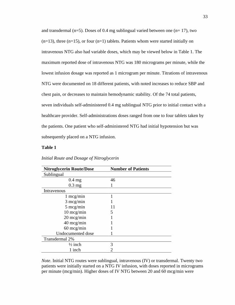
and transdermal (n=5). Doses of 0.4 mg sublingual varied between one (n= 17), two
(n=13), three (n=15), or four (n=1) tablets. Patients whom were started initially on
intravenous NTG also had variable doses, which may be viewed below in Table 1. The
maximum reported dose of intravenous NTG was 180 micrograms per minute, while the
lowest infusion dosage was reported as 1 microgram per minute. Titrations of intravenous
NTG were documented on 18 different patients, with noted increases to reduce SBP and
chest pain, or decreases to maintain hemodynamic stability. Of the 74 total patients,
seven individuals self-administered 0.4 mg sublingual NTG prior to initial contact with a
healthcare provider. Self-administrations doses ranged from one to four tablets taken by
the patients. One patient who self-administered NTG had initial hypotension but was
subsequently placed on a NTG infusion.
Table 1
Initial Route and Dosage of Nitroglycerin
Nitroglycerin Route/Dose Number of Patients
Sublingual
0.4 mg
0.3 mg
46
1
Intravenous
1 mcg/min 1
3 mcg/min 1
5 mcg/min 11
10 mcg/min 5
20 mcg/min 1
40 mcg/min 1
60 mcg/min
Undocumented dose
1
1
Transdermal 2%
½ inch 3
1 inch 2
Note. Initial NTG routes were sublingual, intravenous (IV) or transdermal. Twenty two
patients were initially started on a NTG IV infusion, with doses reported in micrograms
per minute (mcg/min). Higher doses of IV NTG between 20 and 60 mcg/min were

34
documented on patients with initial SBP > 150 mmHg. IV NTG infusions were started on
additional patients (n=23), totaling 45 documented NTG infusions. Transdermal dosage
of ½ inch equates to 7.5 mg of NTG and 1 inch equates to 15 mg, which is gradually
absorbed with therapeutic onset between 30 and 60 minutes (U.S. National Library of
Medicine, 2020c). Transdermal 2% NTG paste of ½ inch equates to approximately 5
mcg/min of IV NTG, while 1 inch equates to a range of 10 to 39 mcg/min of IV NTG
(Esposito, Dunham, Granger, Tudor, & Granger, 1998).
Male patients (n= 53) represented a larger proportion of the sample than female
patients (n=21), representing approximate male and female patient percentages of 72 and
28, respectively. The average age (± SD) of all individuals included in this project was
62.8 years (± 11.8 years). Female age was higher with an average of 63.7 (± 12.3) years,
while males had an average age of 62.5 (± 11.7) years. The most common cardiovascular
risk factors that were recorded in patients’ health histories included hypertension (n= 41),
hyperlipidemia (n=34), and any history of smoking (n=32). Additional comorbidities and
cardiovascular risk factors are provided below in Table 2.
Table 2
Health History Diagnoses of 2019 STEMI Patients
Comorbidity Occurrence
Hypertension 41
Hyperlipidemia 34
Smoking 32
Coronary Artery Disease 19
Diabetes Mellitus 17
Obesity 11
Previous MI 11
Chronic Obstructive Pulmonary Disease 6
Note. Diagnoses in patient health histories as listed within the patient chart. Patient
occurrence represents total number of patients with the specific diagnosis, with 74 total
patients. Additional diagnostic histories by number of occurrence included cerebral
vascular accident (n=3), chronic kidney disease (n=4), obstructive sleep apnea (n=3),
previous coronary artery bypass grafting (n=3), and atrial fibrillation (n=3). Although
several additional diagnoses were present in the study population, they were not included
for the purposes of this study.

35
Inferior STEMI was the diagnosis in a larger number of patients (n=44) than non-
inferior STEMI patients (n=30). Of the patients in the inferior STEMI group, the majority
of patients had the culprit vessel identified as the right coronary artery (RCA) (n=21),
while non-inferior STEMI patients had the left anterior descending (LAD) coronary
artery (n=21) identified as the most common culprit vessel. Seven patients in the inferior
STEMI group had multiple vessels occluded, while three patients in the non-inferior
STEMI group had multiple vessels occluded. Takotsubo cardiomyopathy was identified
in one patient within the non-inferior STEMI group (data located in Table 3).
Table 3
STEMI Location and Culprit Vessels
Number of Patients
Vessel Inferior Non-inferior
RCA 21 -
LAD 4 21
DIAG - 3
OM1/OM2 2 -
CIRC 7 -
PDA 1 -
Multiple 7 3
Takotsubo - 1
Unknown 2 2
Total 44 30
Note. Vessel location as identified in cardiac catheterization laboratory. Unknown is due
to no catheterization or no specification noted. Coronary arteries by name and
abbreviation: Right coronary artery (RCA). Left anterior descending (LAD). Diagonal
(DIAG). Obtuse marginal (OM1/OM2). Circumflex (CIRC). Posterior descending artery
(PDA). Takotsubo cardiomyopathy is an acute condition that often results in elevated
cardiac markers and ECG changes (Boyd & Solh, 2020) and can result from a strong
emotional or physical event that causes left ventricular dilation and acute heart failure.
With Takotsubo cardiomyopathy, ST- segment elevation can occur, with diagnosis made
following an absence of plaque rupture or obstructed coronary artery (Boyd & Solh,
2020).

36
Bradycardia was observed in approximately 31% of patients (n=23) and was the
most common cardiac aberration. Hypotension occurred in 8% of patients (n=6), while
cardiac arrhythmias occurred in 12% of patients (n=9). Cardiac arrhythmias included
ventricular fibrillation (n=3), ventricular tachycardia (n=3) and symptomatic bradycardia
(n=3). Additional medications that were provided to patients included vasopressors, fluid
boluses, antihypertensive medications, and analgesics (illustrated below in Table 4).
Table 4
Medications Administered to 2019 STEMI Patients
Medications Number of Patients
Fluid Bolus/Vasopressor
Normal Saline
Dopamine
Atropine
Norepinephrine
Epinephrine
25
1
3
1
1
Analgesics/Anxiolytics/Sedatives
Morphine 28
Fentanyl 8
Hydromorphone 1
Lorazepam 2
Diazepam 1
Midazolam 1
Diprivan 1
Antihypertensive
Metoprolol 19
Diltiazem 1
Hydralazine 1
Note. The above table includes alternate medications that patients received in addition to
NTG. The number of patients who received a medication does not account for potentially
receiving medications within the same class or a different class. Patients that received a
fluid bolus or vasopressor (n=28), analgesia/anxiolytic/sedative (n=38), or an
antihypertensive(s) (n=19) represents the total number of individual patients that received
medications within the specific grouping.

37
Inferential Statistics to Answer Research Questions
Research Question 1: Does the administration of NTG result in hypotension more
often in inferior STEMI patients than non-inferior STEMI patients?
Inferior STEMI patients had 1.429 times the risk of developing hypotension than
non-inferior STEMI patients after receiving NTG (OR 1.429, 95% CI [0.248, ∞]). The
results were not significant (p=0.521), with four inferior STEMI patients and two non-
inferior STEMI patients experiencing hypotension after receiving NTG (comparisons
illustrated in Figure 1).
Figure 1
STEMI Patient Events by Occurrence
Note. Total number of patients (n=73) compared for hypotension and bradycardia. Total
number of patients (n=74) for occurrence of administration of corrective medication, as a
fluid bolus or vasoactive medication to correct hypotension or bradycardia, and
occurrence of cardiac arrhythmias. There were no significant differences in occurrence of
hypotension (p=0.521), administration of corrective medications (p=0.530), bradycardia
(p=0.064), and cardiac arrhythmias (p=0.465).
4
17 17
6
2
11
6
3
0
2
4
6
8
10
12
14
16
18
Hypotension Corrective Med Bradycardia Cardiac Arrhytmia
Inferior Non-inferior

38
There was a significant difference in SBP noted between inferior STEMI patients
and non-inferior STEMI patients after receiving NTG (p=0.012), with inferior STEMI
patients’ mean SBP reduced by 15.23 mmHg more than non-inferior STEMI patients
after receiving NTG. After receiving NTG, inferior STEMI patients had a mean
difference in SBP of -19 mmHg (95% CI [-27.4, -10.74]), compared to non-inferior
STEMI patients, who had a mean difference in SBP of -10.9 mmHg (95% CI [-20.6, -
1.1]). Table 5 below indicates the control variables with significant impacts on mean SBP
which included antihypertensive medications (p=0.025) and diabetes mellitus (p=0.023),
with a mean SBP decrease of 15.47 mmHg and mean SBP increase of 16.03 mmHg for
each variable, respectively.
Table 5
Effects Estimates of Variables on Systolic Blood Pressure
Variable Estimate Std. Error t p
Inferior STEMI -15.23 5.90 -2.58 0.012*
Hypertension 9.98 5.82 1.72 0.091
Diabetes Mellitus 16.03 6.86 2.34 0.023*
Obesity 13.14 8.38 1.57 0.122
Morphine 6.35 5.76 1.10 0.274
Antihypertensive -15.47 6.74 -2.29 0.025*
Vasopressor/Bolus 5.34 5.91 0.90 0.370
(Intercept) -18.59 6.43 -2.89 0.005
Note. P-values are of absolute t-values for each variable. Intercept is included for a point
of reference, although no inferences will be made based on intercept data. Variables
included above were decided with AIC, an automated model selection, to allow for
inclusion of variables that had a stronger association to the response variable (SBP).
*p < 0.05

39
Research Question 2: Do patients with inferior STEMIs require additional
interventions more frequently to correct hypotension or bradycardia than non-
inferior STEMI patients following NTG administration?
When analyzing the need for a fluid bolus or corrective medication for
hypotension or bradycardia, the odds of requiring one of these interventions was 1.086
times the risk for inferior STEMI patients compared to non-inferior STEMI patients (OR
1.086, 95% CI [0.439, ∞]). A total of 17 inferior STEMI patients and 11 non-inferior
STEMI patients received a fluid bolus or corrective medication in this study, equating to
38.6% of inferior STEMI patients and 36.6% of non-inferior STEMI patients (illustrated
in to Figure 1). The results were not significant (p=0.530).
Research Question 3: Do inferior STEMI patients experience more bradycardia and
cardiac arrhythmias compared to non-inferior STEMI patients?
The occurrence of bradycardia was not significantly different between inferior
and non-inferior STEMI groups (p=0.064), with inferior STEMI patients having 2.582
times the risk of bradycardia than non-inferior STEMI patients (OR 2.582, 95% CI
[0.940, ∞]). Approximately 40% of inferior STEMI patients (n=17) had at least one
documented occurrence of bradycardia, while approximately 20% of non-inferior STEMI
patients (n=6) had at least one documented occurrence of bradycardia. There also was no
significant difference in heart rate between inferior STEMI and non-inferior STEMI
groups after NTG (p=0.239), with a lower mean difference in heart rate of 2.84 beats per
minute observed in inferior STEMI patients following NTG.
The administration of an antihypertensive and the occurrence of a cardiac
arrhythmia each significantly reduced mean heart rate (p<0.001, p=0.015), with a

40
reduction in mean heart rate of 14 beats per minute associated with antihypertensive
medications while cardiac arrhythmias were associated with a mean reduction in heart
rate of approximately nine beats per minute. Other variables evaluated with heart rate
may be viewed below in Table 6. The risk for developing a cardiac arrhythmia was 1.415
times higher for inferior STEMI patients (n=6) than non-inferior STEMI patients (n=3)
(OR 1.415, 95% CI [0.340, ∞]), however, this was not significant (p=0.465).
Table 6
Effects Estimates of Variables on Heart Rate
Variable Estimate Std. Error t p
Inferior STEMI -2.84 2.39 -1.19 0.239
Diabetes Mellitus -3.59 2.84 -1.26 0.211
Smoking -4.03 2.37 -1.70 0.094
Morphine 3.34 2.39 1.40 0.167
Antihypertensive -14.02 2.76 -5.09 <0.001**
Cardiac Arrhythmia -8.65 3.47 -2.50 0.015*
(Intercept) 6.09 2.44 2.49 0.015
Note. P-values are of absolute t-values for each variable. Intercept is included for a point
of reference, although no inferences will be made based on intercept data. Significant
effects were found for antihypertensive medications and cardiac arrhythmias.
*p < 0.05; **p < 0.001
Research Question 4: Does NTG significantly reduce chest pain levels reported by
STEMI patients?
Chest pain rating following the administration of NTG did not differ significantly
between inferior and non-inferior STEMI groups (p=0.391). When rating chest pain on
the numerical 0 to 10 Likert scale after receiving NTG, inferior STEMI patients reported
less of a mean chest pain score reduction, with mean chest pain scores 0.61 higher than
chest pain scores reported by non-inferior STEMI patients. For all STEMI patients in this

41
project, NTG administration did significantly reduce the mean chest pain score reported
by the patients by 1.87 on the 0 to 10 Likert scale (p<0.001). The administration of
morphine also was associated with significantly increased chest pain levels (p=0.008),
with a mean chest pain score increase of 1.96 (Refer to Table 7 below).
Table 7
Effects Estimates of Variables on Chest Pain Rating
Variable Estimate Std. Error t p
Post-NTG CP -1.87 0.35 -5.31 <0.001**
Inferior STEMI -0.99 0.76 -1.30 0.200
Age -0.05 0.03 -1.47 0.148
CAD 1.24 1.30 -0.96 0.342
Previous MI -1.46 1.47 -0.99 0.326
Obesity -1.90 1.17 -1.62 0.114
Morphine 1.96 0.71 2.77 0.008*
Analgesics 1.25 1.12 1.11 0.274
(Intercept) 9.32 2.11 4.42 <0.001
Note. Post-NTG CP represents chest pain (CP) ratings following the administration of
NTG, with significant reductions for all STEMI patients (p<0.001). Morphine was
associated with significantly higher CP ratings (p=0.008), while all other variables did
not have a significant effect on CP changes before and after receiving NTG. Initial
comparison to evaluate differences in CP reported by inferior and non-inferior STEMI
patients after receiving NTG was not significant (p=0.391), but due to model selection,
STEMI location was included and again not significant (p=0.200).
*p < 0.05; **p < 0.001
Discussion
The route and dosage of NTG administered to STEMI patients varied greatly
within this project. Bosson et al. (2019) and Engelberg et al. (2000) reported the use of
only 0.4 mg sublingual NTG in patients within their studies. Robichaud et al. (2016) also
reported 0.4 mg sublingual NTG, but included 0.4 mg intranasal NTG spray as a
treatment modality for patients as well. Although this project did not seek to evaluate the
variability of different routes of administration of NTG, with the account of

42
pharmacokinetics and therapeutic onset for each route. The variation was attempted to be
accounted for by documenting medication doses, times, and routes for all medications
during data extraction to allow for a more complete evaluation.
The four patients diagnosed with inferior STEMI that experienced hypotension
had variable initial NTG doses and routes. One patient developed hypotension with a
documented SBP of 89 mmHg following three doses of 0.4 mg sublingual NTG. Another
patient developed hypotension and subsequent ventricular fibrillation following two
doses of 0.4 mg sublingual NTG approximately three minutes following NTG
administration. Successful management of the patient’s condition occurred, and the
patient was then placed on five micrograms per minute of intravenous NTG. The third
patient with inferior STEMI had hypotension following a single dose of 0.4 mg
sublingual NTG, but was eventually placed on a NTG intravenous infusion at five
micrograms per minute without further incident. The fourth patient with inferior STEMI
had “hypotension” documented by an EMS transport crew for a patient who was on a
titrated dose of NTG, which was at 20 micrograms per minute at the time of
discontinuation. SBP documented prior to discontinuation was consistently > 90 mmHg,
with ability to down-titrate the NTG infusion. This patient was restarted on the NTG
infusion per the cardiologist upon arriving to the cardiac catheterization laboratory.
The two patients diagnosed with non-inferior STEMI who developed hypotension
both had received intravenous NTG. One patient had an undocumented amount of NTG,
but also received diltiazem, metoprolol, and lorazepam before becoming hypotensive.
The second patient was on five micrograms per minute of intravenous NTG when they

43
developed symptomatic bradycardia and hypotension. The patient responded to
interventions and remained on the intravenous NTG without further adverse events.
The occurrence of hypotension observed in this project following administration
of NTG does not appear to occur in a predictable fashion, with hypotension occurring
after variable doses and routes of NTG. Interestingly, three of the six patients who
developed hypotension remained on intravenous NTG, which supports continued use and
titration of NTG in an individualized manner in response to each patient. Although it is
unclear within this project, it is possible that healthcare provider preference or medical
training played a role in the variability observed between NTG dosages and routes.
However, regardless of the doses and routes of NTG that were provided to patients in this
project, there was no significant difference in occurrence of hypotension between patients
diagnosed with inferior STEMI compared to those with non-inferior STEMI.
The doses reported in previous studies ranged from a single dose (Engelberg et
al., 2000), to three doses (Bosson et al., 2019), while Robichaud et al. (2016) accounted
for unlimited doses. As in this project, Bosson et al. (2019) and Robichaud et al. (2016)
did not specifically investigate the effects of multiple doses of NTG, but rather for the
occurrence of hypotension, bradycardia, and adverse events. In this project, the observed
initiation of intravenous NTG infusions after alternative sublingual or transdermal routes,
in conjunction with intravenous dose titrations, provides information about how
management and care of patients diagnosed with STEMI varies within the patient
encounter. This observation likely relates to adapting to the patients’ needs rather than
adhering to an undeviating plan of care, ultimately supporting individualized patient care.

44
Robichaud et al. (2016) reported several of the same comorbidities and
cardiovascular risk factors that were controlled for in this project, including previously
diagnosed CAD, diabetes mellitus, hypertension, and hyperlipidemia. Demographic
variables were controlled for in statistical analyses within this project to allow for
identification of potential influences on outcome variables, although the aims of this
project were not focused on detailed demographic analyses. Demographics were included
to allow for simple comparisons between patients within this project to demographic
information of patients with STEMIs in other studies.
In the analysis of gender frequency, females represented approximately 28% of
the total patients included in this project. The approximate occurrence of female patients
with STEMIs reported by Bosson et al. (2019), Engelberg et al. (2000), and Robichaud et
al. (2016) was 39%, 48%, and 30%, respectively. O’Gara et al. (2013) reported female
patients representing 30% of all STEMI patients in their study as well. Robichaud et al.
(2016) and O’Gara et al. (2013) have the most comparable female frequencies reported
when compared to the frequency of female patients found with this project.
The average age of all patients in this project (62.8 ± 11.8 years) is similar to that
noted in a study that evaluated demographics of STEMI patients, with researchers
reporting an average age of 62 (±13.9) years for all patients (Boyer et al., 2012). The
study conducted by Bosson et al. (2019), which had similar research questions to those
analyzed in this project, reported a median age of 63 (IQR 54-75) for patients that
received NTG. With this information, it does not appear that patients in this project were
considerably different in demographics, with gender and age occurrences that are
comparable to those reported in other studies.

45
In evaluating STEMI location, inferior STEMI patients represented approximately
59% of the patients included in this study, compared to the approximate 47% of patients
in the study by Robichaud et al. (2016). A higher percentage of patients experienced an
inferior STEMI in this project than the predicted range of 40% to 50% of patients with
STEMIs (Warner & Tivakaran, 2020). The RCA was the most commonly occluded
vessel in the inferior STEMI group, which is most often suspected with inferior STEMI
identification on a standard 12-lead ECG (Namana et al., 2018). The left circumflex
artery was the second most commonly occluded coronary artery in this project for
patients with inferior STEMI, which may indicate left dominant circulation in these
patients (Namana et al., 2018). For the non-inferior STEMI group, the LAD was the most
commonly occluded vessel, which is consistent with anterior STEMI and left ventricular
involvement (Taglieri et al., 2014). The observations of occurrence in this project support
the most likely culprit vessels associated with each STEMI location (Namana et al., 2018;
Taglieri et al., 2014), but also that inferior STEMI is not always associated with RCA
occlusion.
It is important to note that the inferior STEMI patients in this project represent a
subgroup of all STEMI patients that presented to the rural Midwestern hospital in 2019. It
is not clear how this translates to the entire STEMI population that is treated at this
facility, but does suggest the prevalence of patients that receive NTG with the diagnosis
of inferior STEMI. Although RV infarct was not evaluated for specifically within this
project, there were no documented additional diagnostic interventions in patients
presenting with inferior STEMI. As RV infarct is a contraindication to administration of

46
nitrates to patients (Antman et al., 2004; Ibanez, 2018), specific identification of RV
involvement would be beneficial.
The patient with Takotsubo cardiomyopathy was included in this study due to
their clinical presentation, treatment, and diagnosis of STEMI within their medical
record. Boyd and Solh (2020) discuss how closely ACS and Takotsubo may present,
including ST segment elevation on a 12-lead ECG tracing. The potential for
hemodynamic instability in patients with Takotsubo is related to left ventricular
dysfunction and acute heart failure (Templin et al., 2015) and may be similar to that of
STEMI patients. The inclusion criteria were met for this patient, even though the patient
did not have a culprit vessel identified as causing ST-segment elevation due to lack of
thrombotic or plaque occlusion. Had the patient not had STEMI included in the discharge
diagnosis made by a cardiologist, the patient would have been excluded from this study.
Discussion of Research Question 1: Does the administration of NTG result in
hypotension more often in inferior STEMI patients than non-inferior STEMI
patients?
With regard to blood pressure, there is very weak evidence in this project to
support any significant difference in occurrence of hypotension, defined as SBP < 90
mmHg, between inferior STEMI patients and non-inferior STEMI patients. The results in
this project were similar to those reported by Robichaud et al. (2016), with no significant
difference in occurrence of hypotension between inferior and non-inferior STEMI groups
(p=0.80). Inferior STEMI patients in this project did, however, have a significantly
greater reduction in mean SBP compared to non-inferior STEMI patients following
receipt of NTG. Although the mean reduction in SBP between inferior and non-inferior

47
STEMI groups was significant (p=0.012) after receiving NTG, the lower observed SBP
in the inferior STEMI group did not equate to a significantly higher occurrence of
hypotension, as previously reported. This shows that patients with inferior STEMIs
experienced increased effects from NTG compared to those with non-inferior STEMIs
within this group of patients, without hemodynamic instability.
For all patients that received NTG in the study conducted by Bosson et al. (2019)
there was a median change in SBP of -11 mmHg (IQR -26, 0), while in the study by
Engleberg et al. (2000), the mean decrease in SBP in patients after receiving NTG was
-11.8 mmHg (95% CI [-10.7, -13]). In this project, non-inferior STEMI patients had
similar reduction in mean SBP compared to those in the previously listed studies, with
mean SBP decrease of -10.9 mmHg, while inferior STEMI patients had a mean SBP
decrease of -19 mmHg. The previously listed studies did not compare differences in SBP
between inferior and non-inferior STEMI patients, whereas this project did, showing a
significant difference in mean SBP reduction between the two groups. The difference in
mean SBP may suggest increased effects of NTG on patients with inferior STEMIs,
although Robichaud et al. (2016) found no significant difference in occurrence of drop in
SBP ≥30 mmHg following NTG between inferior and non-inferior STEMI groups
(p=0.87). The decrease in SBP ≥ 30 mmHg as investigated in previous studies (Bosson et
al. 2019; Robichaud et al. 2016) was a threshold used to evaluate the sensitivity of
patients to nitrates and how those with RV infarct would have a higher incidence in larger
SBP decreases. Neither study found significant results, indicating the need to reconsider a
blanket avoidance of nitrates in inferior STEMI patients. Although this project did not
evaluate for a specific level of decrease in SBP, the lack of significance in comparing

48
occurrence of hypotension adds to the evidence supplied by previous studies, as patients
diagnosed with inferior STEMI in this project did not experience hemodynamic
instability after receiving NTG.
Antihypertensive medications were found to be a significant factor in reducing the
mean SBP while all other variables were controlled for in this project. This suggests that
the medications were effective in decreasing blood pressure, but also that
antihypertensive medications may have contributed to the observed difference in
decreased mean SBP between each inferior and non-inferior STEMI groups. This
observation warrants further investigation, as the effects of antihypertensive medications
were not compared with statistical analyses between patients with inferior and non-
inferior STEMIs in this project. Although morphine and hemodynamic supportive
measures did not have significant effects on SBP in this project, the regression model
identified these variables as affecting the outcome variable of SBP more than other
demographic variables or comorbidities.
Although it is not clear why NTG was associated with a significantly increased
mean SBP in patients with diabetes, there are some potential explanations. Muntner,
Whelton, Woodward, and Carey (2018) note that 66.3% of patients with diabetes have
concurrent hypertension by American Diabetes Association (ADA) guidelines, while
77.1 % of patients with diabetes have hypertension by the ACC and AHA guidelines. The
high prevalence in general, regardless of which guideline is used for defining
hypertension, illustrates the close relationship between diabetes and hypertension. This
may represent an overall higher SBP in patients presenting with STEMI with concurrent
diabetes mellitus, however, this was not analyzed within this project. Additionally,

49
patients with diabetes may not respond to NTG with arterial dilation as well due to
impaired nitric oxide pathways, which can occur with damaged endothelium (Tessari et
al., 2010; Vavuranakis et al., 1999). As nitroglycerin converts to nitric oxide within the
body (Kim, Kerndt, & Schaller, 2020), reduced efficacy may result given impaired
metabolic pathways in diabetes. Although this is not a definite explanation for the
observed significant increase in this project, it may indicate a need to investigate other
factors relating to STEMI patients with diabetes mellitus and how standard medications
may impact that specific population.
Discussion of Research Question 2: Do patients with inferior STEMIs require
additional interventions more frequently to correct hypotension or bradycardia
than non-inferior STEMI patients following NTG administration?
In comparing inferior and non-inferior STEMI groups and the number of patients
requiring a medication to correct hypotension or bradycardia, there is a lack of evidence
to support any true difference in need between the groups based on the occurrences
observed in this project. Individuals who received these pharmacological interventions
may not have needed the specific intervention based on hemodynamic data reviewed
within the charts. Multiple individuals in both inferior and non-inferior STEMI groups
received fluid boluses without concomitant hypotension, with no clear basis for
administration identified during the chart review. The fluid boluses may have been
administered as a prophylactic measure or fluid challenge to the patients, but were not
documented as such. A total of 28 patients received a fluid bolus or vasoactive
medication, while only six patients experienced true hypotension and three experienced
symptomatic bradycardia. The patients with hypotension and bradycardia all responded

50
to fluid or medication administration and remained stable within the timeframe of interest
for this project. Fluid bolus and vasoactive medication were included in the multivariable
linear regression in the analysis of SBP following administration of NTG, however, these
variables were not associated with a significant increase in SBP (refer to Table 5). Six
patients experienced a ventricular arrhythmia, but none died within the timeframe of
interest in this project.
Discussion of Research Question 3: Do inferior STEMI patients experience more
bradycardia and cardiac arrhythmias compared to non-inferior STEMI patients?
Inferior STEMI patients in this project had lower mean heart rates than non-
inferior STEMI patients, which ultimately was not a significant finding. In the entire
observed population, there were 23 patients who had bradycardia, with a higher
occurrence in inferior STEMI patients. Three patients in this project had symptomatic
bradycardia, all of whom had inferior STEMIs, but all of whom also responded to
medical interventions. Many patients with bradycardia were either noted to have stable
bradycardia upon initial contact or have one documented heart rate below 60. This is
similar to the observations made in Bosson et al. (2019), with 75% of bradycardic
patients having a heart rate between 50 and 59 beats per minute, with a comparable
occurrence of bradycardia noted in patients with RCA and non-RCA occlusions.
Antihypertensive medications were observed to have a significant effect on heart
rate in this project. Of these medications, metoprolol was administered to 100% (n=19) of
patients who received an antihypertensive medication. Diltiazem (n=1) and hydralazine
(n=1) were also administered to separate patients in addition to at least one dose of
metoprolol. Metoprolol, which is a cardiac-selective medication categorized as a beta-

51
blocker, has the ability to cause a decrease in heart rate, cardiac output, and blood
pressure (Morris & Dunham, 2020). Similarly, Roolvink et al. (2016) identified a
significant heart rate reduction in STEMI patients who received metoprolol. As the
predominant antihypertensive medication provided to patients in this project, metoprolol
and its effects on outcome variables of heart rate and SBP had strong hemodynamic
influences identified in patients within this project.
When controlling for other variables, cardiac arrhythmias also affected mean
heart rate in patients, with a mean reduction of approximately eight beats per minute.
This observation points to the potential for adverse events following NTG administration,
with lower heart rates observed in patients who experienced a cardiac arrhythmia.
However, there is no evidence in this project that the occurrence of a cardiac arrhythmia
differed significantly between inferior and non-inferior patients. STEMI location in this
project was not a strong predictor of an adverse event occurring, while other variables
with strong effects on SBP and heart rate may have been more likely to contribute to the
occurrence.
Discussion of Research Question 4: Does NTG significantly reduce chest pain levels
reported by STEMI patients?
There was no significant difference in chest pain reduction between inferior and
non-inferior STEMI patients, meaning no single group experienced a greater benefit from
NTG in this project. Interestingly, morphine was associated with increased mean chest
pain, which might be attributed to higher initial and subsequent chest pain scores reported
by individuals who received this medication. This may be because morphine was truly
reserved for patients with higher pain ratings that were not responsive to NTG, as

52
recommended by Frampton et al. (2020), since the use of morphine inhibits the
therapeutic effects of commonly used antiplatelet medications.
Kendrick and Strout (2005) identified a change in pain rated on the numeric pain
scale of 1.39 ± 1.05 (95% CI, 1.27-1.51) to be significant, which is how chest pain was
evaluated in this project. As the verbal analog scale (VAS) for pain can be correlated with
the numeric pain scale (Holdgate, Asha, Craig, & Thompson, 2003), pain reduction
around 1.3 was also considered significant in an alternative study using VAS to assess
pain (Todd, Funk, Funk, & Bonacci, 1996). The mean reduction in chest pain scores in
this project was 1.9 for all STEMI patients, representing a significant reduction in pain
and demonstrating the benefit of NTG and its ability to reduce chest pain in STEMI
patients in this project. Mean chest pain reduction reported by both Bosson et al. (2019)
and Engleberg et al. (2000), was 2.6, again providing support for the role of NTG in chest
pain reduction, including chest pain experienced by STEMI patients. The results from
this project, as well as the results in previous studies, indicate that patients experiencing
STEMI benefit from the effects of NTG by significant reductions in chest pain.
Clinical Implications for Practice
This DNP project contributes to clinical practice by identifying variables that
influence hemodynamic variables as well as differences that occur in inferior and non-
inferior STEMI patients who received NTG. NTG, which was provided to a large number
of patients with the diagnosis of inferior STEMI, did not result in a significant difference
in occurrence of hypotension when compared to non-inferior STEMI patients. Although
there should not be generalizations transferred directly to practice, there are implications
for provision of care to patients on an individual basis.

53
The concern relating to NTG administration to patients with inferior STEMI
identified on a 12-lead ECG is RV infarct and the potential for hemodynamic collapse. In
this project, RV infarct was not evaluated for specifically. Even though patients with
inferior STEMI received NTG with few adverse events, it is unclear if these patients did
have RV involvement that precipitated adverse events. Further investigation in the effects
of NTG on patients with inferior STEMI and concomitant RV infarct would be
beneficial, as well as diagnostic tools to identify RV involvement.
One intervention that could assist healthcare providers in identifying RV infarct
would be to perform a right-sided ECG. Increasing views of the heart by adding
additional leads to a standard 12-lead ECG can increase sensitivity to diagnosing STEMI,
improve infarct location identification, and identify infarctions that may not be visible on
a standard 12-lead ECG (Tragardh, Claesson, Wagner, Zhou, & Pahlm, 2007; Vogiatzis
et al., 2019). Bischof et al. (2018) recommend evaluation of lead V4R to identify RV
involvement during an acute MI, because of low sensitivity and specificity of standard
12-lead ECG analysis. Similarly, Kanovsky et al. (2016) discuss the value of the right-
sided ECG in identifying RV infarct, as patients with occlusions to the RCA, circumflex
artery and LAD may have RV involvement that may not be recognized using standard
12-lead ECG analysis. No patients in this study had a right-sided ECG analysis, which
would be one recommendation for future patients presenting to the rural Midwestern
hospital. The right precordial leads would assist healthcare providers in identifying RV
involvement, which would help to guide individualized treatment.
This project identified that NTG was associated with a significant reduction in
chest pain experienced by patients with the diagnosis of STEMI. Ethically, healthcare

54
providers should be providing analgesia to patients, and doing so with evidence to
support such interventions. As newer research shows that opioids reduce the efficacy of
antiplatelet medications used in STEMI patients (Frampton et al., 2020), it is important to
use medications that provide the most overall benefit. As adverse events were not
significantly different between inferior and non-inferior STEMI patients in this project,
the safety of NTG by these standards supports the viability of this medication as a
possible analgesic choice for both inferior and non-inferior STEMI patients.
As patients with RV infarct have higher immediate mortality rates (Noguchi et al.,
2019), implementing a right-sided ECG would help to more easily identify RV infarct,
guiding treatment of the patient. Providers would have additional information from which
to base decisions, including making informed decisions on whether or not to withhold
NTG from patients. Additionally, with the identification of RV infarct, providers would
be able to anticipate the need for fluid resuscitation, vasoactive medications and potential
external pacing (Inohara, Kohsaka, Fukuda, & Menon, 2013; Noguchi et al., 2019), and
respond to the patients’ needs in the event of hemodynamic collapse.
Recommendations for Future Research
Several recommendations for future research can be made based on results in this
DNP project. The first recommendation would be for a prospective study to evaluate the
effects of NTG on hemodynamic variables and chest pain on inferior and non-inferior
STEMI patients. This project, as a retrospective review, has limitations in generalizability
to STEMI patients and cannot explain direct causality, for which a prospective study
would be necessary.

55
In a prospective study or similar future retrospective studies, more strict criteria
may be warranted when evaluating the occurrence of bradycardia, with modifications to
what identifies a patient as bradycardic. Recommendations may be to include patients
that experience symptomatic bradycardia or consistently documented bradycardia with
heart rates < 60 beats per minute. This way, a transient or asymptomatic decrease in heart
rate would not be considered as an adverse event in patients. In this project, one
occurrence of a documented heart rate below 60 beats per minute was counted toward an
occurrence of bradycardia, even though only 3 patients had hemodynamic instability and
several others had one heart rate below 60 beats per minute, providing an occurrence rate
but not necessarily a clear picture of patient condition.
As NTG doses and routes of administration varied among patients within this
project, a future prospective study or similar retrospective study would control for this
variable. Although it is possible to evaluate patient hemodynamics based on therapeutic
onset and pharmacokinetic properties of NTG, a controlled dose and route would help
remove potential limitations with the results. With the removal of variation, the effects of
NTG on hemodynamic variables may be more clearly identified in STEMI patients.
The effects of metoprolol on STEMI patients should also be evaluated, as this
medication served as a variable that had significant effects on both SBP and heart rate
within this project. Research has been conducted to evaluate potential benefits of
metoprolol prior to PCI and its impact on infarct size (García-Ruiz et al., 2016; Roolvink
et al., 2016), however, the data does not have consistent results with regard to the
medication’s ability to reduce infarct size and mortality during the acute phase in STEMI

56
patients. As 19 patients in this study received this medication, further investigation on its
effects on this patient population would be beneficial.
Another recommendation for future research would be to include the
identification of RV infarct within the study sample. This could be done in comparative
analyses between STEMI patients with and without RV infarct, but also as a simple
observation of occurrence. Because RV infarction occurs at a variable frequency within
inferior STEMI patient populations (Bischof et al., 2018; Nagam et al., 2017), further
analysis into the specific topic would be a beneficial contribution to the knowledge
regarding STEMI patients. Although this project did not seek to identify patients with RV
infarct within the sample, the results of the effects of NTG on inferior STEMI patients
contribute to the research suggesting the elimination of avoidance of NTG in individuals
with inferior STEMI identification. Additional diagnostic tools, such as the right-sided
ECG, may be utilized to identify RV involvement with the goal of removing hesitancy in
the provision of NTG to patients with inferior STEMI identified on ECG analysis.
Strengths
To mitigate potential errors that may occur with retrospective chart reviews, this
DNP project aimed to follow recommendations on how to conduct such a project (Kaji,
Schriger, & Green, 2014; Vassar & Holzmann, 2013). The sample size exceeded the
minimum number of individuals that was determined by the sample size calculator,
reducing probability of type II error. Although several patients were excluded from the
multiple variable regressions evaluating blood pressure and heart rate due to incomplete
data for these specific analyses models, there were 66 and 67 patients, which exceeded
the minimum of 64 individuals that was determined by the sample size calculator. IRB

57
approval was obtained from both the hospital and through Northern Michigan University
prior to data collection, and a data template was used to organize extracted data to allow
for organization and to maintain patient confidentiality. These measures as recommended
by Vassar and Holzmann (2013), increase the strength of studies by adhering to
systematic data extraction and upholding patient confidentiality. Additionally, exclusion
criteria for this project addressed vulnerable populations, which addresses ethical
considerations that may arise when conducting a retrospective chart review (Vassar &
Holzmann, 2013).
Limitations
One of the main limitations identified in this project is the retrospective design
and subsequent sampling technique. Because the sampling of patients was a convenience
sample, bias is not accounted for due to the non-random sampling techniques used.
Because of this, generalizations cannot be made about entire STEMI populations (Vassar
& Holzmann, 2013), as analyses describe only the observations within the specific
sample of patients within this project. Although effects estimates were provided for
variables within this project, their influence on outcome measures should be viewed as
associations and only applied to patients within this project due to lack of randomization.
Additionally, selection bias is introduced to the project with the exclusion of patients
from statistical analyses due to missing data (Kaji et al., 2014). Although the minimum
number of required individuals as determined by the sample size calculator was surpassed
with each analysis, it is important to address potential selection bias resulting from the
exclusion of patients. Other limitations in this project include conducting the project at a
single facility and including patients that presented to the facility within a one-year time-

58
frame. Including data on patients from additional locations and extending the time frame
from which patients could be included may have been beneficial in reducing bias as well
as when drawing observational inferences. Additionally, the doses and routes of NTG
administration were not controlled for within this project, and may be considered a
limitation due to the high variability among patients.
Conclusion
As NTG remains a frontline medication used in STEMI patients, both to reduce
angina and as an antihypertensive (de Alencar Neto, 2018), this DNP project sought to
contribute further to what is known about the effects and safety of NTG in STEMI
patients. The purpose of this DNP project was to explore the safety of NTG use in the
treatment of patients diagnosed with STEMI by evaluating the effects of NTG on
hemodynamic measures and angina. Outcome variables of SBP, heart rate, occurrence of
cardiac arrhythmias, and need for medical interventions to correct hemodynamic
instability thus were compared between inferior and non-inferior STEMI patients who
received NTG. As the AHA discourages the use of NTG in patients with RV infarct
(Antman et al., 2004), further investigation into the provision of NTG to patients with
inferior STEMI and RV infarct is warranted. Although identification of the occurrence of
RV infarct was not conducted within this project, diagnostic measures to improve
identification of RV infarct could provide valuable information to healthcare providers,
and subsequently used to guide individualized patient care.
In this project, there was no significant difference in occurrence of hypotension in
inferior STEMI patients and non-inferior STEMI patients who received NTG. Mean SBP
was lower in inferior STEMI patients, however, not to the extent of instability or

59
significant increased need for medical intervention. There was also no significant
difference in occurrence of cardiac arrhythmias or bradycardia between the two groups,
but inferior STEMI patients did have lower mean heart rates. Additionally, NTG
provided patients with significant reductions in chest pain in both inferior and non-
inferior STEMI groups, supporting its therapeutic use in this patient population. Overall,
these results suggest the general safety of NTG in patients eligible to receive it, even
without specific diagnostic measures to identify RV involvement.
For advanced practice nurses, the information in this project may be a valuable
component to their role in providing individualized patient care. As a member of the
interdisciplinary team, it is necessary to consider individualized treatment options for
patients. This includes medications, associated safety concerns prior to administration,
and potential adverse events that may occur in patient populations. Advanced practice
nurses may use clinical judgement and incorporate additional diagnostic tools in the
treatment of STEMI patients, including the use of right-sided ECG analysis in inferior
STEMI patients. This may help guide the provision of medications such as NTG, which
may relieve angina associated with STEMI, and ultimately improve the level of
individualized patient care.
In this project, NTG use in inferior STEMI patients did not result in significantly
more adverse events than in non-inferior STEMI patients, even without the identification
of RV infarct. This suggests reevaluating the avoidance of NTG based on concerns of
hemodynamic adverse events due to potential RV infarct. Inferior STEMI patients should
receive a right-sided ECG to detect RV infarct, with lead V4R serving as the most
sensitive marker (O’Gara et al., 2013). Additional views of the heart and higher

60
sensitivity in detecting the presence of RV infarct with a right-sided ECG can help guide
healthcare providers in providing highly-individualized care. As in similar studies
(Bosson et al., 2019; Engleberg et al., 2000; Robichaud et al., 2016), a prospective study
to evaluate the safety and effects of NTG in STEMI patients is recommended.

61
References
Antman, E. M. et al. (2004). ACC/AHA guidelines for the management of patients with
ST-elevation myocardial infarction. ACC/AHA Practice Guidelines, e82-e293.
Retrieved from https://www.ahajournals.org/doi/pdf/10.1161/circ.110.9.e82
Antman, E. M. et al. (2008). 2007 focused update to the ACC/AHA 2004 guidelines for
the management of patients with ST-elevation myocardial infarction. Journal of
the American College of Cardiology, 51(2), doi:10.1016/j.jacc.2007.10.001
Benjamin, E. J., Virani, S. S., Callaway, C. W., Chamberlain, A. M., Chang, A. R.,
Cheng, S., . . . Muntner, P. (2018). Heart disease and stroke statistics - 2018
update: A report from the American Heart Association. Circulation, 137(12), e67-
e492. doi:10.1161/CIR.0000000000000558
Bischof, J. E., Worrall, C. I., & Smith, S. W. (2018). In inferior myocardial infarction,
neither ST elevation in lead V1 nor ST depression in lead I are reliable findings
for the diagnosis of right ventricular infarction. Journal of Electrocardiology,
51(6), 977-980. doi:10.1016/j.jelectrocard.2018.08.010
Boden, W. E., Padala, S. K., Cabral, K. P., Buschmann, I. R., & Sidhu, M. S. (2015).
Role of short-acting nitroglycerin in the management of ischemic heart disease.
Drug Design, Development and Therapy, 9(1), 4793–4805.
doi:10.2147/DDDT.S79116
Bosson, N., Isakson, B., Morgan, J. A., Kaji, A. H., Uner, A., Hurley, K., … Niemann, J.
T. (2019). Safety and effectiveness of field nitroglycerin in patients with
suspected ST elevation myocardial infarction. Prehospital Emergency Care,
23(5), 603–611. doi:10.1080/10903127.2018.1558318

62
Boyd, B., & Solh, T. (2020). Takotsubo cardiomyopathy: Review of broken heart
syndrome. Journal of the American Academy of Physician Assistants, 33(3), 24-
29. doi:10.1097/01.JAA.0000654368.35241.fc
Boyer, N. M., Laskey, W. K., Cox, M., Hernandez, A. F., Peterson, E. D., Bhatt, D. L. …
Fonarow, G. C. (2012). Trends in clinical, demographic, and biochemical
characteristics of patients with acute myocardial infarction from 2003 to 2008: a
report from the American heart association get with the guidelines coronary artery
disease program. Journal of the American Heart Association, 1(4), e001206.
doi:10.1161/JAHA.112.001206
Camm, C. F., & Camm, J. A. (2016). Clinical Guide to Cardiology. Retrieved from
https://ebookcentral-proquest-com.nmu.idm.oclc.org
Creative Research Systems. (2012). Sample size calculator. Retrieved from
https://www.surveysystem.com/sscalc.htm
Dang, D., & Dearholt, S. (2018). Johns Hopkins Nursing Evidence-Based Practice Third
Edition: Model and Guidelines (3rd ed). Indianapolis, IN: Sigma Theta Tau
International. Retrieved from https://search-ebscohost-
com.nmu.idm.oclc.org/login.aspx?direct=true&db=e000xna&AN=1625431
&site=ehost-live
de Alencar Neto, J. N. (2018). Morphine, oxygen, nitrates, and mortality reducing
pharmacological treatment for acute coronary syndrome: An evidence-based
review. Cureus, 10(1), e2114. doi:10.7759/cureus.2114
Duarte, G. S., Nunes-Ferreira, A., Rodrigues, F. B., Pinto, F. J., Ferreira, J. J., Coasta, J.,
& Caldeira, D. (2019) Morphine in acute coronary syndrome: systematic review

63
and meta-analysis. BMJ Open, 9(3), e025232. doi:10.1136/bmjopen-2018-025232
Engleberg, S., Singer, A. J., Moldashel, J., Sciammarella, J., Thode, H. C., & Henry, M.
(2000). Effects of prehospital nitroglycerin on hemodynamics and chest pain
intensity. Prehospital Emergency Care, 4(4), 290–293.
doi:10.1080/10903120090940967
Esposito, G. A., Dunham, G., Granger, B. B., Tudor, G. E., & Granger, C. B. (1998).
Converting IV nitroglycerin therapy to nitroglycerin ointment therapy: A
comparison of two methods. American Journal of Critical Care, 7(2), 123-130.
doi:10.4037/ajcc1998.7.2.123
Ferguson, J. J., Diver, D. J., Boldt, M., & Pasternak, R. C. (1989). Significance of
nitroglycerin-induced hypotension with inferior wall acute myocardial infarction.
American Journal of Cardiology, 64(5), 311-314. doi: 10.1016/0002-
9149(89)90525-0
Ferreira, J. C., & Mochly-Rosen, D. (2012). Nitroglycerin use in myocardial infarction
patients: Risks and benefits. Circulation Journal, 76(1), 15-21.
doi:10.1253/circj.cj-11-1133
Food and Drug Administration. (2011). Nitrostat. Retrieved from
https://www.accessdata.fda.gov/drugsatfda_docs/label/2011/021134s005lbl.pdf
Frampton, J., Devries, J. T., Welch, T. D., & Gersh, B. J. (2020). Modern management of
ST-segment elevation myocardial infarction. Current Problems in Cardiology,
45(3), 1-80. doi:10.1016/j.cpcardiol.2018.08.005
García-Ruiz, J., Fernández-Jiménez, R., García-Alvarez, A., Pizarro, G., Galán-Arriola,
C., Fernández-Friera, L.,… Ibáñez, B. (2016). Impact of the timing of metoprolol

64
administration during STEMI on infarct size and ventricular function. Journal of
the American College of Cardiology, 67(18), 2093–2104.
doi:10.1016/j.jacc.2016.02.050
Garvey, J. L., Zegre-Hemsey, J., Gregg, R., & Studnek, J. R. (2016).
Electrocardiographic diagnosis of ST segment elevation myocardial infarction:
An evaluation of three automated interpretation algorithms. Journal of
Electrocardiology, 49(5), 728-732. doi:10.1016/j.jelectrocard.2016.04.010
Gu, Y. L., Svilaas, T., van der Horst, I. C., & Zijlstra, F. (2008). Conditions mimicking
acute ST-segment elevation myocardial infarction in patients referred for primary
percutaneous coronary intervention. Netherlands Heart Journal, 16(10), 325–331.
doi:10.1007/bf03086173
Hobl, E., Stimpfl, T., Ebner, J., Schoergenh, C.,Derhaschnig, U., Sunder-Plassmann,
R.,… Jilma, B. (2014). Morphine decreases clopidogrel concentrations and
effects: A randomized, double-blind, placebo-controlled trial. Journal of the
American College of Cardiology, 63(7), 630-635. doi:10.1016/j.jacc.2013.10.068
Holdgate, A., Asha, S., Craig, J., & Thompson, J. (2003). Comparison of a verbal
numeric rating scale with the visual analogue scale for the measurement of acute
pain. Emergency Medicine, 15(5‐6), 441–446. doi:10.1046/j.1442-
2026.2003.00499.x
Ibanez, B., James, S., Agewall, S., Antunes, M. J., Bucciarelli-Ducci, C., Bueno,
H.,…Widimsky, P. (2018). 2017 ESC guidelines for the management of acute
myocardial infarction in patients presenting with ST-segment elevation. European
Heart Journal, 39(21), 119-177. doi:10.1093/eurheartj/ehx393

65
Ibrahim, K., Shah, R., Goli, R., Kickler, T., Clarke, W., Hasan, R.,… McEvoy, J. (2018).
Fentanyl delays the platelet inhibition effects of oral ticagrelor: Full report of the
PACIFY randomized clinical trial. Thrombosis and Haemostasis, 118(8), 1409-
1418. doi:10.1055/s-0038-1666862
Inohara, T., Kohsaka, S., Fukuda, K., & Menon, V. (2013). The challenges in the
management of right ventricular infarction. European Heart Journal: Acute
Cardiovascular Care, 2(3), 226-234. doi:10.1177/2048872613490122
Jaton, E. (2017). Inferior wall acute myocardial infarction: Not as preload dependent as
once thought? Air Medical Journal, 36(1), 27-29. doi:10.1016/j.amj.2016.10.008
Kaji, A. H., Schriger, D., & Green, S. (2014). Looking through the retrospectoscope:
Reducing bias in emergency medicine chart review studies. Annals of Emergency
Medicine, 63(3), 292-298. doi:10.1016/j.annemergmed.2014.03.025
Kanovsky, J., Kala, P., Novotny, T., Benesova, K., Holicka, M., Jarkovsky, J., . . . Malik,
M. (2016). Association of the right ventricle impairment with
electrocardiographic localization and related artery in patients with ST-elevation
myocardial infarction. Journal of Electrocardiology, 49(6), 907-910.
doi:10.1016/j.jelectrocard.2016.08.001
Kendrick, D., & Strout, T. (2005). The minimum clinically significant difference in
patient-assigned numeric scores for pain. The American Journal of Emergency
Medicine, 23(7), 828-832. doi:10.1016/j.ajem.2005.07.009
Kim, K. H., Kerndt, C. C., Schaller, D. J. (2020). Nitroglycerin. In: StatPearls [Internet].
Retrieved from https://www.ncbi.nlm.nih.gov/books/NBK482382/

66
Koh, J. Q., Fernando, H., Peter, K., & Stub, D. (2019). Opioids and ST elevation
myocardial infarction: A systematic review. Heart, Lung & Circulation, 28(2),
697-706. doi:10.1016/j.hlc.2018.12.015
Kubica, J., Adamski, P., Ostrowska, M., Sikora, J., Kubica, J. M., Sroka, W. D,… &
Koziński, M. (2016). Morphine delays and attenuates ticagrelor exposure and
action in patients with myocardial infarction: The randomized, double-blind,
placebo-controlled impression trial. European Heart Journal, 37(3), 245–252.
doi:10.1093/eurheartj/ehv547
McCarthy, C. P., Bhambhani, V., Pomerantsev, E., & Wasfy, J. H. (2018). In-hospital
outcomes in invasively managed acute myocardial infarction patients who receive
morphine. Journal of Interventional Cardiology, 31(2), 150–158.
doi:10.1111/joic.12464
McEwen, M., & Wills, E. M. (2018). Evidence-based practice and nursing theory. In M.
McEwen, & E. Wills, Theoretical basis for nursing, (5th ed., pp. 253-272).
Philadelphia, PA: Wolters Kluwer.
Morris, J., & Dunham, A. (2020). Metoprolol. In: StatPearls [Internet]. Retrieved from
https://www.ncbi.nlm.nih.gov/books/NBK532923/
Muntner, P., Whelton, P. K., Woodward, M., & Carey, P. M. (2018). A comparison of
the 2017 American College of Cardiology/American Heart Association blood
pressure guideline and the 2017 American Diabetes Association diabetes and
hypertension position statement for U.S. adults with diabetes. Diabetes Care,
41(11), 2322-2329. doi:10.2337/dc18-1307

67
Nagam, M. R., Vinson, D. R., & Levis, J. T. (2017). ECG diagnosis: Right ventricular
myocardial infarction. The Permanente Journal, 21(1), 16–105.
doi:10.7812/TPP/16-105
Namana, V., Gupta, S. S., Abbasi, A. A., Raheja, H., Shani, J., & Hollander, G. (2018).
Right ventricular infarction. Cardiovascular Revascularization Medicine, 19(1),
43-50. doi:10.1016/j.carrev.2017.07.009
Noguchi, M., Sakakura, K., Akashi, N., Adachi,Y., Watanabe,Y., Taniguchi,Y., …
Fujita, H. (2019). The comparison of clinical outcomes between inferior ST-
elevation myocardial infarction with right ventricular infarction versus without
right ventricular infarction. International Heart Journal, 60(3), 560-568.
doi:10.1536/ihj.18-515
O’Connor, R. E., Brady, W., Brooks, S. C., Diercks, D., Egan, J., Ghaemmaghami, C.
…Yannopoulos, D. (2010). Part 10: Acute coronary syndromes. Circulation,
122(18), S787-S817. doi:10.1161/circulationaha.110.971028
O’Gara, P. T., Kushner, F. G., Ascheim, D. D., Casey, D. E.,, Chung, M. K., de Lemos, J.
A., … Zhao, D. X. (2013). 2013 ACCF/AHA guideline for the management of
ST-elevation myocardial infarction. Journal of the American College of
Cardiology, 61(4), e78-e140. doi:10.1016/j.jacc.2012.11.019
Robichaud, L., Ross, D., Proulx, M., Légaré, S., Vacon, C., Xue, X., & Segal, E. (2016).
Prehospital nitroglycerin safety in inferior ST elevation myocardial infarction.
Prehospital Emergency Care, 20(1), 76-81. doi:10.3109/10903127.2015.1037480
Roolvink, V., Ibáñez, B., Ottervanger, J. P., Pizarro, G., van Royen, N., Mateos, A., . . .
van’t Hof, A. W. (2016). Early intravenous beta-blockers in patients with ST-

68
segment elevation myocardial infarction before primary percutaneous coronary
intervention. Journal of the American College of Cardiology, 67(23), 2705-2715.
doi:10.1016/j.jacc.2016.03.522
Sederholm Lawesson, S., Isaksson, R., Thylén, I., Ericsson, M., Ängerud, K., & Swahn,
E. (2018). Gender differences in symptom presentation of ST-elevation
myocardial infarction – an observational multicenter survey study. International
Journal of Cardiology, 264(1), 7-11. doi:10.1016/j.ijcard.2018.03.084
Somers, M. P, Brady, W. J., Bateman, D. C., Mattu, A., & Perron, A. D. (2003).
Additional electrocardiographic leads in the ED chest pain patient: right
ventricular and posterior leads. American Journal of Emergency Medicine, 21(7),
563-573. doi:10.1016/j.ajem.2003.08.008.
Stokes, L. (2018). ANA position statement: The ethical responsibility to manage pain and
the suffering it causes. The Online Journal of Issues in Nursing, 24(1), 3.
doi:10.3912/OJIN.Vol24No01PoSCol01.
Taglieri, N., Saia, F., Alessi, L., Cinti, L., Reggiani, M. L. B., Lorenzini, M., . . . Rapezzi,
C. (2014). Diagnostic performance of standard electrocardiogram for prediction
of infarct related artery and site of coronary occlusion in unselected STEMI
patients undergoing primary percutaneous coronary intervention. European Heart
Journal: Acute Cardiovascular Care, 3(4), 326-339.
doi:10.1177/2048872614530665
Templin, C., Ghadri, J. R., Diekmann, J., Napp, L. C., Bataiosu, D. R., Jaguszewski, M.,
… Luscher, T. F. (2015). Clinical features and outcomes of Takotsubo (stress)

69
cardiomyopathy. New England Journal of Medicine, 373, 929–938.
doi:10.1056/NEJMoa1406761
Tessari, P., Cecchet, D., Cosma, A., Bettore, M., Coracina, A., Millioni, R., …Vedovato,
M. (2010). Nitric oxide synthesis is reduced in subjects with type 2 diabetes and
nephropathy. Diabetes, 59(9), 2152–2159. doi:10.2337/db09-1772
Thygesen, K., Alpert, J. S., Jaffe, A. S., Simoons, M. L., Chaitman, B. R., & White, H. D.
(2012). Third universal definition of myocardial infarction. Circulation, 126(16),
2020-2035. doi:10.1161/CIR.0b013e31826e1058
Todd, K. H., Funk, K. G., Funk, J. P. & Bonacci, R. (1996). Clinical significance of
reported changes in pain severity. Annals of Emergency Medicine, 27(4), 485-489.
doi:10.1016/S0196-0644(96)70238-X
Tragardh, E., Claesson, M., Wagner, G. S., Zhou, S., & Pahlm, O. (2007). Detection of
acute myocardial infarction using the 12-lead ECG plus inverted leads versus the
16-lead ECG (with additional posterior and right-sided chest electrodes). Clinical
Physiology and Functional Imaging, 27(6), 368-374. doi:10.1111/j.1475-
097X.2007.00761.x
U.S. National Library of Medicine. (2020a). Nitroglycerin in dextrose – nitroglycerin
injection. Retrieved from
https://dailymed.nlm.nih.gov/dailymed/drugInfo.cfm?setid=457a026c-ebce-4701-
9deb-2d7652759a99
U.S. National Library of Medicine. (2020b). Nitroglycerin tablet, orally disintegrating.
Retrieved from
70
https://dailymed.nlm.nih.gov/dailymed/drugInfo.cfm?setid=041f127e-5166-4484-
bc1a-0a373d1187ae
U.S. National Library of Medicine. (2020c). Nitro-bid nitroglycerin ointment. Retrieved
from https://dailymed.nlm.nih.gov/dailymed/drugInfo.cfm?setid=e464e9bb-48e8-
4b9f-9fff-e220cfbac0c5
Vassar, M., & Holzmann, M. (2013). The retrospective chart review: Important
methodological considerations. Journal of Educational Evaluation for Health
Professions, 10, 12. doi:10.3352/jeehp.2013.10.12
Vavuranakis, M., Stefanadis, C., Triandaphyllidi, E., Toutouzas, K., & Toutouzas, P.
(1999). Coronary artery distensibility in diabetic patients with simultaneous
measurements of luminal area and intracoronary pressure: Evidence of impaired
reactivity to nitroglycerin. Journal of the American College of Cardiology, 34(4),
1075–1081. doi:10.1016/S0735-1097(99)00331-9
Vogiatzis, I., Koulouris, E., Ioannidis, A., Sdogkos, E., Pliatsika, M., Roditis, P., &
Goumenakis, M. (2019). The importance of the 15-lead versus 12-lead ECG
recordings in the diagnosis and treatment of right ventricle and left ventricle
posterior and lateral wall acute myocardial infarctions. Acta Informatica Medica,
27(1), 35-39. doi:10.5455/aim.2019.27
Warner, M. J., & Tivakaran, V. S. (2020). Inferior myocardial infarction. In: StatPearls
[Internet]. Retrieved from https://www.ncbi.nlm.nih.gov/books/NBK470572/
Wilson, R. E., Kado, H. S., Percy, R. F., Butterfield, R., Sabato, J., Strom, J. A., & Box,
L. C. (2013). An algorithm for identification of ST-elevation myocardial infarction

71
patients by emergency medicine services. American Journal of Emergency
Medicine, 31(7), 1098-1102. doi:10.1016/j.ajem.2013.04.013

72
Appendix A
JHNEBP MODEL AND
TOOLS- PERMISSION
Thank you for your submission. We are happy to give you permission to use the JHNEBP
model and tools in adherence of our legal terms noted below:
• You may not modify the model or the tools without written approval from Johns
Hopkins.
• All reference to source forms should include “©The Johns Hopkins Hospital/The Johns
Hopkins University.”
• The tools may not be used for commercial purposes without special permission.
If interested in commercial use or discussing changes to the tool, please
email [email protected].

73
Downloads:
JHNEBP Tools-Printable Version
JHNEBP Tools-Electronic Version
Do you prefer hands-on learning?
We are offering a 5-day intensive Boot Camp where you will learn and master the entire
EBP process from beginning to end. Take advantage of our retreat-type setting to focus
on your project, collaborate with peers, and get the expertise and assistance from our
faculty. Click HERE to learn more about EBP Boot Camp. Group rates available,
email [email protected] to inquire.
Go back to the form

74
Appendix B
Graduate Studies and Research Marquette, MI 49855-5301
906-227-2300 www.nmu.edu/graduatestudies/
Memorandum
TO: Olivia Montgomery
School of Nursing
CC: Kristi Robinia
School of Nursing
FROM: Lisa Schade Eckert Dean of Graduate Education and Research
DATE: May 15, 2020
SUBJECT: IRB Reciprocal Approval NMU Approval Number: HS20-1131
“A retrospective review to evaluate and compare the use of nitroglycerin in
patients diagnosed with inferior ST elevated myocardial infarction and
patients diagnosed with non-inferior ST elevated myocardial infarction”
IRB Approval Date: 5/15/2020
Proposed Project Dates: 5/20/2020 – 6/27/2020
This IRB proposal, “A retrospective review to evaluate and compare the use of nitroglycerin in patients diagnosed with inferior ST elevated myocardial infarction and patients diagnosed with
non- inferior ST elevated myocardial infarction,” has been approved under the reciprocal
review process.
The study is approved by Upper Peninsula Health Systems Marquette “hospital” IRB.
Include the NMU proposal number (HS20-1131) and the contact information of the NMU
researcher and the NMU IRB Administrator on all research materials and on any correspondence
regarding this project at Northern Michigan University.
Any changes or revisions to your approved research plan must be approved by the
Institutional Review Board (IRB) prior to implementation.
If you do not complete your project within 12 months from the date of your approval
notification, you must submit a Project Renewal Form for Research Involving Human
Subjects. You may apply for a one-year project renewal up to four times.

75
All forms can be found at the NMU Grants and Research
website: http://www.nmu.edu/grantsandresearch/node/102

76
Appendix C
Chart Review Methodology
1. Obtain Institutional Review Board (IRB) approval
2. Construct template for data extraction
3. Obtain list of patient’s with STEMI
a. Contingent on STEMI tracking system
b. May include individuals that activated a STEMI alert – requiring further
inquiry
4. Access patient information
a. Begin with searching for discharge diagnosis of STEMI
b. Not all patients that activate a STEMI response are true STEMIs
c. If patient has discharge diagnosis of STEMI, continue with chart review
for that patient
d. If discharge diagnosis does not include STEMI, patient excluded from
project
5. Review chart for patient inclusion and exclusion criteria
a. Inclusion criteria
i. Received NTG
ii. Chest pain of suspected cardiac origin
iii. 18 years of age or older
iv. Diagnosis of STEMI (ECG/expert cardiologist diagnosis)
b. Exclusion criteria
i. Did not receive NTG
ii. Pregnant women
iii. Thoracic trauma
iv. Vulnerable populations
v. English as secondary language
6. Begin extraction of data into template
a. Patient demographics: Age, gender, past medical history relating to
cardiac risk factor (ie: Previous MI, Diabetes mellitus, smoking)
b. Identify through documentation on patient location and region of STEMI
i. Culprit vessel causing STEMI (coronary artery)
ii. Inferior/Anterior/Posterior/Lateral
iii. Obtained from cardiology documentation
c. Extract all heart rates, blood pressures, oxygen saturations, respirations
with associated time (oxygen saturation and respirations not evaluated in
this project)
d. Extract patient rated chest pain and times
e. Extract all medications with associated administration time, route, and
dose
f. Extract arrhythmias noted in chart and time of occurrence

77
7. Sort data
a. Hemodynamics prior to administration of NTG and first documented after
NTG based on medication onset and half-life to be used for ratio level
analyses on effects of NTG
b. Review all hemodynamics for each patient
i. Note if patient was hypotensive or bradycardic based on
parameters
ii. Note if patient experienced a cardiac arrhythmia or adverse event
iii. Note corrective medications, either as vasopressor or fluid bolus
and if patient was responsive medically to interventions
8. Analyze data based on specific research question

78
Appendix D
Software Citations
Name Citation
R R Core Team (2020). R: A language and environment for statistical computing.
R Foundation for Statistical Computing, Vienna, Austria. URL https://www.R-
project.org/.
cAIC4 Saefken, B., Ruegamer, D., Kneib, T., and Greven, S. (2018), Conditional Model
Selection in Mixed-Effects Models with cAIC4, ArXiv e-prints 1803.05664.
lme4 Douglas Bates, Martin Maechler, Ben Bolker, Steve Walker (2015). Fitting
Linear Mixed-Effects Models Using lme4. Journal of Statistical Software,
67(1), 1-48.
doi:10.18637/jss.v067.i01.
lmerTest Kuznetsova A, Brockhoff PB, Christensen RHB (2017). “lmerTest Package: Tests
in Linear Mixed Effects Models.” Journal of Statistical Software, 82(13), 1-26.
doi:
10.18637/jss.v082.i13 (URL: https://doi.org/10.18637/jss.v082.i13).
lmSubsets Hofmann M, Gatu C, Kontoghiorghes E, Colubi A, Zeileis A (2020). lmSubsets:
Exact Variable-Subset Selection in Linear Regression. R package version 0.5-1,
(URL: https://CRAN.R-project.org/package=lmSubsets).
psych Revelle, W. (2020) psych: Procedures for Personality and Psychological Re-
search, Northwestern University, Evanston, Illinois, USA, https://CRAN.R-
project.org/package=psych Version = 2.0.7,.
tidyverse Wickham et al., (2019). Welcome to the tidyverse. Journal of Open Source
Software,
4(43), 1686, https://doi.org/10.21105/joss.01686
xtable David B. Dahl, David Scott, Charles Roosen, Arni Magnusson and Jonathan
Swin-
ton (2019). xtable: Export Tables to LaTeX or HTML. R package version 1.8-4.
https://CRAN.R-project.org/package=xtable
yarrr Nathaniel Phillips (2017). yarrr: A Companion to the e-Book “YaRrr!: The Pirate’s
Guide to R”. R package version 0.1.5. https://CRAN.R-project.org/package=yarrr