nmr nuclear magnetic resonance by kurtis lukey and matt loschiavo
TRANSCRIPT

NMRNuclear Magnetic Resonance
By Kurtis Lukey and Matt Loschiavo

What is NMR?• Nuclear Magnetic Resonance is a
phenomenon whish occurs when the nuclei of certain atoms are immersed in a static magnetic field and exposed to a second oscillating magnetic field. Some nuclei experience this phenomenon, and others do not, dependent upon whether they possess a property called spin.

Pacific Northwest National Laboratory's high magnetic field (800 MHz, 18.8 T) NMR spectrometer being loaded with a sample.
900MHz, 21.2 T NMR Magnet at HWB-NMR, Birmingham, UK being loaded with a sample

What is Spin?• Spin is a fundamental property of nature like electrical charge
or mass. Spin comes in multiples of ½ and can be positive or negative. Protons, electrons and neutrons possess spin.
• When placed in a magnetic field of strength B, a particle with a net spin can absorb a photon, of frequency v.
• The frequency v depends on the gyromagnetic ratio, y of the particle.
v =y B

Spectroscopy
• Nuclear magnetic resonance spectroscopy is the use of the NMR phenomenon to study physical, chemical, and biological properties of matter. As a consequence, NMR spectroscopy finds applications in several areas of science. NMR spectroscopy is routinely used by chemists to study chemical structure using simple one-dimensional techniques.

• NMR spectroscopy is replacing x-ray crystallography for the determination of protein structure
• Spectroscopy is the study of the interaction of electromagnetic radiation with matter

History of NMR• Nuclear magnetic resonance was first described and measured in molecular beams
by Isidor Rabi in 1938. Eight years later, in 1946, Felix Bloch and Edward Mills Purcell refined the technique for use on liquids and solids, for which they shared the Nobel Prize in physics in 1952.
• Purcell had worked on the development and application of RADAR during World War II at Massachusetts Institute of Technology's Radiation Laboratory. His work during that project on the production and detection of radiofrequency energy, and on the absorption of such energy by matter, preceded his discovery of NMR.
• They noticed that magnetic nuclei, like 1H and 31P, could absorb RF energy when placed in a magnetic field of a strength specific to the identity of the nuclei. When this absorption occurs, the nucleus is described as being in resonance. Interestingly, for analytical scientists, different atoms within a molecule resonate at different frequencies at a given field strength. The observation of the resonance frequencies of a molecule allows a user to discover structural information about the molecule.
• The development of nuclear magnetic resonance as a technique of analytical chemistry and biochemistry parallels the development of electromagnetic technology and its introduction into civilian use.
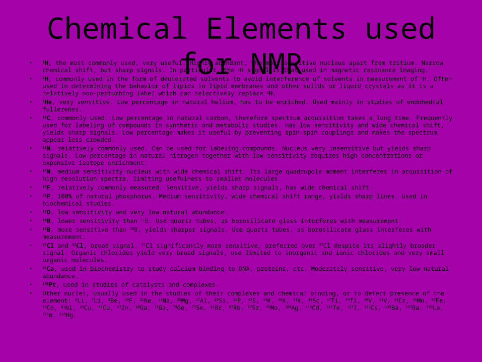
Chemical Elements used for NMR• 1H, the most commonly used, very useful. Highly abundant, the most sensitive nucleus apart from tritium. Narrow chemical shift,
but sharp signals. In particular, the 1H signal is that used in magnetic resonance imaging. • 2H, commonly used in the form of deuterated solvents to avoid interference of solvents in measurement of 1H. Often used in
determining the behavior of lipids in lipid membranes and other solids or liquid crystals as it is a relatively non-perturbing label which can selectively replace 1H.
• 3He, very sensitive. Low percentage in natural helium, has to be enriched. Used mainly in studies of endohedral fullerenes. • 13C, commonly used. Low percentage in natural carbon, therefore spectrum acquisition takes a long time. Frequently used for
labeling of compounds in synthetic and metabolic studies. Has low sensitivity and wide chemical shift, yields sharp signals. Low percentage makes it useful by preventing spin-spin couplings and makes the spectrum appear less crowded.
• 15N, relatively commonly used. Can be used for labeling compounds. Nucleus very insensitive but yields sharp signals. Low percentage in natural nitrogen together with low sensitivity requires high concentrations or expensive isotope enrichment.
• 14N, medium sensitivity nucleus with wide chemical shift. Its large quadrupole moment interferes in acquisition of high resolution spectra, limiting usefulness to smaller molecules.
• 19F, relatively commonly measured. Sensitive, yields sharp signals, has wide chemical shift. • 31P, 100% of natural phosphorus. Medium sensitivity, wide chemical shift range, yields sharp lines. Used in biochemical studies. • 17O, low sensitivity and very low natural abundance. • 10B, lower sensitivity than 11B. Use quartz tubes, as borosilicate glass interferes with measurement. • 11B, more sensitive than 10B, yields sharper signals. Use quartz tubes, as borosilicate glass interferes with measurement. • 35Cl and 37Cl, broad signal. 35Cl significantly more sensitive, preferred over 37Cl despite its slightly broader signal. Organic chlorides
yield very broad signals, use limited to inorganic and ionic chlorides and very small organic molecules. • 43Ca, used in biochemistry to study calcium binding to DNA, proteins, etc. Moderately sensitive, very low natural abundance. • 195Pt, used in studies of catalysts and complexes. • Other nuclei, usually used in the studies of their complexes and chemical binding, or to detect presence of the element: 6Li, 7Li,
9Be, 19F, 21Ne, 23Na, 25Mg, 27Al, 29Si, 31P, 33S, 39K, 40K, 41K, 45Sc, 47Ti, 49Ti, 50V, 51V, 53Cr, 55Mn, 57Fe, 59Co, 61Ni, 63Cu, 65Cu, 67Zn, 69Ga, 71Ga, 73Ge, 77Se, 81Br, 87Rb, 87Sr, 95Mo, 109Ag, 113Cd, 125Te, 127I, 133Cs, 135Ba, 137Ba, 139La, 183W, 199Hg.
Applications
• The use of nuclear magnetic resonance best known to the general public is in magnetic resonance imaging for medical diagnosis, however, it is also widely used in chemical studies. Biochemical information can also be obtained from living tissue (e.g human brain tumours) with the technique known as in vivo magnetic resonance spectroscopy.
Magnetic Resonance Imaging• Magnetic resonance imaging (MRI) is a medical imaging technique
primarily used in Radiology to visualize the structure and function of the body. It provides detailed images of the body in any plane. MR has much greater soft tissue contrast than computed tomography (CT) making it especially useful in neurological, musculoskeletal, cardiovascular, and oncological imaging. Unlike CT it uses no ionizing radiation, but uses a powerful magnetic field to align the nuclear magnetization of (usually) hydrogen atoms in water in the body. Radiofrequency fields are used to systematically alter the alignment of this magnetization, causing the hydrogen nuclei to produce a rotating magnetic field detectable by the scanner. This signal can be manipulated by additional magnetic fields to build up enough information to reconstruct an image of the body.


The End