no. val. 261, issue of 15, pp. the journal biological … · 2001-07-10 · the formation of the...
TRANSCRIPT

THE JOURNAL OF BIOLOGICAL CHEMISTRY 0 1986 by The American Society of Biological Chemists, Inc.
Val. 261, No. 8, Issue of March 15, pp. 3661-3669,1986 Printed in U.S.A.
Biosynthesis of Riboflavin ENZYMATIC FORMATION OF 6,7-DIMETHYL-8-RIBITYLLUMAZINE FROM PENTOSE PHOSPHATES*
(Received for publication, April 24, 1985)
Peter NielsenS, Gerhard Neubergerg, Isao FujiiP, David H. Bowng, Paul J. Kellers, Heinz G. Floss§, and Adelbert BacherS From the $Lehrstuhl fur Organische Chemie und Bwchemie, Technische Uniuersitat Miinchen, Lichtenbergstr. 4, 0-8046 Garching, Federal Republic of Germany and the §Department of Chemistry, The Ohio State University, Columbus, Ohio 43210
The xylene ring of riboflavin originates by dismu- tation of the precursor, 6,7-dimethyl-8-ribitylluma- zine. The formation of the latter compound requires a 4-carbon unit as the precursor of carbon atoms 6a, 6, 7, and 7a of the pyrazine ring. The formation of ribo- flavin from GTP and ribose phosphate by cell extract from Candida guilliermondii has been observed by Logvinenko et al. (Logvinenko, E. M., Shavlovsky, G. M., Zakal’sky, A. E., and Zakhodylo, I. V. (1982) Biokhimiya 47, 931-936). We have studied this en- zyme reaction in closer detail using carbohydrate phos- phates as substrates and synthetic 5-amino-6-ribity- lamino-2,4-(lH,3H)-pyrimidinedione or its 5’-phos- phate as cosubstrates. Several pentose phosphates and pentulose phosphates can serve as substrate for the formation of riboflavin with similar efficiency. The reaction requires Mg2+. Various samples of ribulose phosphate labeled with 14C or 13C have been prepared and used as enzyme substrates. Radioactivity was ef- ficiently incorporated into riboflavin from [ 1-14C]ri- bulose phosphate, [3,5-14C]ribulose phosphate, and [5- 14C]ribulose phosphate, but not from [4-14C]ribulose phosphate. Label from [ l-13C]ribose 5-phosphate was incorporated into C6 and C8a of riboflavin. [2,3,5- 13C3]Ribose 5-phosphate yielded riboflavin containing two contiguously labeled segments of three carbon at- oms, namely 5a, 9a, 9 and 8,7,7a. 5-Amino-6-[1’-14C] ribitylamino-2,4( lH,3H)-pyrimidlnedione transferred radioactivity exclusively to the ribityl side chain of riboflavin in the enzymatic reaction. It follows that the 4-carbon unit used for the biosynthesis of 6,7-di- methyl-8-ribityllumazine consists of the pentose car- bon atoms 1, 2, 3, and 5 in agreement with earlier in vivo studies.
The biosynthesis of riboflavin leads from GTP to 5-amino- 6 - ribitylamino - 2,4( 1H,3H) -pyrimidinedione 5‘ -phosphate (Compound 1, Fig. 1) (3-6) which is converted to 6,7-di-
*This work was supported by grants from the Deutsche For- schungsgemeinschaft and the Fonds der Chemischen Industrie (to A. B.), by National Institutes of Health Grant GM 32910 and a Hum- boldt Senior United States Scientist Award ( t o H. G. F.), by a North Atlantic Treaty Organization travel grant (to A. B. and P. J. K.), and by postdoctoral fellowships from the Ohio State University Graduate School (to I. F.), the National Institutes of Health (GM 10448 to D. H. B.), and the Alexander von Humboldt Stiftung (to P. J. K.). Preliminary communications on part of this work have been pub- lished (1,2). The costs of publication of this article were defrayed in part by the payment of page charges. This article must therefore be hereby marked “advertisement” in accordance with 18 U.S.C. Section 1734 solely to indicate this fact.
methyl-8-ribityllumazine (Compound 2, Fig. 1) by the addi- tion of a 4-carbon unit. Dismutation of 2 yields riboflavin and the pyrimidine 4 (for reviews see Refs. 7 and 8). The structure and origin of the 4-carbon unit required for the formation of 2 remained elusive for several decades in spite of considerable efforts. Early work by Plaut and Broberg (9, 10) showed that both terminal carbon atoms of glucose, i.e. C1 and C6, were incorporated into the xylene ring of riboflavin with similar efficiency (9, 10). Subsequent studies led to the suggestion of acetoin (11, 12), diacetyl (13), pyruvate (14), tetroses (15), pentoses (16, 17), and 5-amino-6-ribitylamino- 2,4(1H,3H)-pyrimidinedione (4) (18, 19) as the ultimate source of the 4-carbon moiety. Work by Alworth and co- workers (17) specifically suggested the incorporation of C1 of ribose into the benzenoid moieties of dimethylbenzimidazole and riboflavin.
We have previously studied the incorporation of a wide variety of 13C-labeled precurso,rs into riboflavin by the flavi- nogenic fungus Ashbya gossypii (20-25). A large number of incorporation experiments with a variety of single and mul- tiple 13C-labeled precursors has been performed and can be summarized as follows. (i) Diacetyl and other symmetrical molecules can be ruled out definitely as precursors. (ii) Carbon atoms 1*-3* are biosynthetically equivalent to Cl-C3 of the pentose pool (see Fig. 1 for explanation of the arbitrary nomenclature used). (iii) C4* of riboflavin is equivalent to C5 of the pentose pool. (iv) C4 of the pentose pool has no equivalent in the 4-carbon moiety; it is eliminated during the biosynthetic process through a rearrangement of the carbon skeleton in which C3 and C5 are reconnected intramolecu- larly. Consequently, the carbon atoms 3 and 5 of the precursor ribose are contiguous in the heterocyclic moieties of the products 2 and 3.
Several attempts have been made to study the formation of the xylene ring in uitro. The enzymatic formation of ribofla- vin from 5-amino-6-ribitylamino-2,4(1H,3H)-pyrimidine- dione (4) and acetoin or pyruvate by cell extract of Ererno- thecium ashbyii has been reported (12, 14). Plaut has sug- gested that these results may be explained by the enzymatic formation of diacetyl which would subsequently react nonen- zymatically with the pyrimidine 4 to yield the lumazine 2 (26).
More recently, Hollander et al. (19) reported on the for- mation of riboflavin from 1 or 4 by cell extracts of Escherichia coli. Surprisingly, this reaction required no second substrate. It was, therefore, proposed, in agreement with an earlier hypothesis by Bresler et al. (18), that 4 can donate its ribityl group as a 4-carbon precursor by a dismutation reaction which would be formally analogous to the formation of riboflavin from 2.
3661

3662 Enzymatic Formation of 6,7-
/ H-C-OH 2 H-C-OH
0 ;H,OH
$HZ H
H-C-OH H-$-OH
4 , R = H ~ - 6 - 0 ~ H+OH 3 H-?-OH
CHZOH
FIG. 1. Biosynthesis of riboflavin. Numbers with asterisks rep- resent an arbitrary nomenclature to illustrate the formation of the 4- carbon unit and the regiochemistry of the last reaction step (dismu- tation of 2).
Logvinenko and co-workers (27-29) have recently described the formation of 2 from GTP by cell-free extracts of the yeast Candida guilliermondii (27-29). The reaction could proceed with ribose phosphate or, less efficiently, with hexose phos- phates as cosubstrate. The question then arises whether this i n uitro reaction follows the same mechanism as the biosyn- thesis of the vitamin in uiuo.
We have used a variety of ribose phosphate and ribulose phosphate samples labeled with I4C or 13C to characterize the enzymatic reaction in closer detail. The results are in full agreement with those of the i n vivo experiments. The evidence leaves no doubt that the reaction in the cell-free system is equivalent to the true biosynthetic pathway.
EXPERIMENTAL PROCEDURES'
RESULTS
We studied the enzymatic formation of riboflavin from synthetic pyrimidine substrates by cell extracts of C. guillier- mondii. The reaction requires the addition of 4 carbon atoms. As shown in Table I, each of two pentose phosphates and two pentulose phosphates could serve as donor of the 4-carbon unit. The highest yield of riboflavin was obtained with ribose phosphate as substrate. Unphosphorylated pentoses and hex- ose phosphates could not serve as precursors.
The pyrimidine 4 and its 5"phosphate 1 could both serve as precursors for the in uitro formation of the heterocyclic moiety of the vitamin (Table 11). Yields of riboflavin were similar with both types of pyrimidine substrate.
Oxidized and reduced pyrimidine nucleotide cofactors (1 mM) did not increase the formation of riboflavin, nor did
"Experimental Procedures" are presented in miniprint at the end of this paper. Miniprint is easily read with the aid of a standard magnifying glass. Full size photocopies are available from the Journal of Biological Chemistry, 9650 Rockville Pike, Bethesda, MD 20814. Request Document No. 85M-1368, cite the authors, and include a check or money order for $4.80 per set of photocopies. Full size photocopies are also included in the microfilm edition of the Journal that is available from Waverly Press.
.Dimethyl-8-ribityllumazine TABLE I
Enzymatic formation of riboflavin by cell extract from C. guilliermondii
The reaction mixture contained 0.72 mM 1, 38 mM Tris hydro- chloride, pH 8.0, 3.8 mM MgC12, 10 mM ditbioerythritol, 0.95 mM of the respective carbohydrate and 0.6 mg of protein; total volume, 400 pl. Incubation was at 37 "C for 1 h. Riboflavin was monitored by bioassay with Lactobacillus casei. Results are corrected for blank values (approximately 1.6 pM riboflavin).
Substrate Riboflavin formed
None Arabinose 5-phosphate Fructose Fructose 6-phosphate Glucose Glucose 6-phosphate Ribose Ribose 5-phosphate Ribulose Ribulose 5-phosphate Xylulose 5-phosphate
PM
0 4.5 0.1 0.3 0.1 0.3 0 9.0 0 5.4 8.2
TABLE I1 Enzymatic formation of riboflavin by cell extract from C.
guilliermondii Reaction mixtures contained 40 mM Tris hydrochloride, pH 8.0, 4
mM MgC12, 1 mM ribose phosphate, pyrimidine substrate as indicated, and protein. Results are corrected for blank values. For other details see Table I.
Substrate Riboflavin formed
None 1 (0.63 mM) 4 (0.44 mM)
P M
0 8.1 5.3
TABLE I11 Enzymatic formation of riboflavin by cell extract from C.
guilliermondii Reaction mixtures contained 30 mM Tris hydrochloride, pH 8.2, 2
mM MgC12, 1.5 mM 1, 5 mM ribose phosphate, and protein. Data are corrected for blank values. For other details see Table I.
Addition Riboflavin formed
None NADH (1 mM) NAD (1 mM) NADPH (1 mM) NADP (1 mM) ATP (2 mM) EDTA (10 mM)
7% 100 80 37 83 34 98 4 . 0
ATP (2 mM) and thiamine pyrophosphate. The reaction was completely inhibited by 10 mM EDTA (Table 111). Cell extract dialyzed against buffer containing EDTA could be partially reactivated by the addition of an excess of Mg2+.
In order to study the fate of individual pentose carbon atoms in closer detail, we prepared enzymatically a variety of ribulose 5-phosphate samples specifically labeled with 14C. Commercial samples of radioactive glucose and glycerol served as precursors. 6-Phosphogluconate has a high retention time on the anion exchanger Dowex 1-X8 and, therefore, can be easily separated from the other intermediates occurring in the reaction sequence. We thus chose to isolate this intermediate in radiochemically pure form. It was subsequently converted to the desired product, ribulose 5-phosphate. It should be mentioned that radioactive ribulose 5-phosphate samples de- composed rapidly even at the temperature of liquid nitrogen.
Enzymatic Formation of 6,7-Dimethyl-8-ribityllumazine 3663
A loss of more than 50% usually occurred within several days. Throughout this study, the ribulose 5-phosphate samples were, therefore, prepared immediately prior to further use.
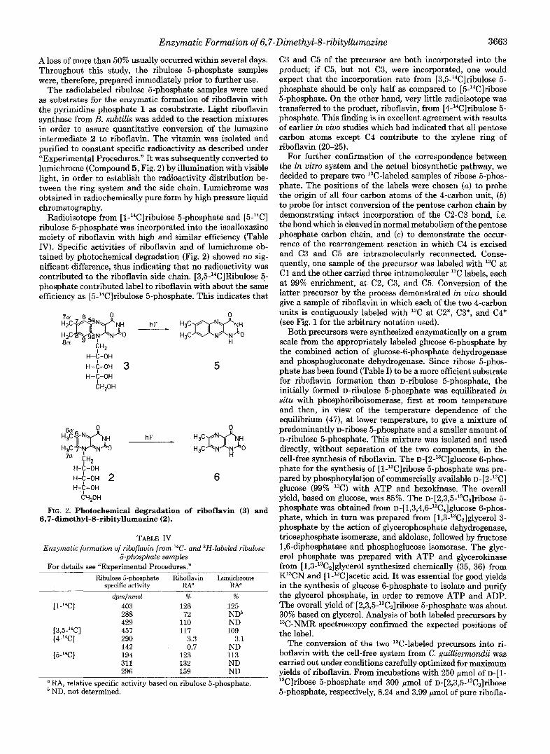
The radiolabeled ribulose 5-phosphate samples were used as substrates for the enzymatic formation of riboflavin with the pyrimidine phosphate 1 as cosubstrate. Light riboflavin synthase from B. subtilis was added to the reaction mixtures in order to assure quantitative conversion of the lumazine intermediate 2 to riboflavin. The vitamin was isolated and purified to constant specific radioactivity as described under "Experimental Procedures." It was subsequently converted to lumichrome (Compound 5, Fig. 2) by illumination with visible light, in order to establish the radioactivity distribution be- tween the ring system and the side chain. Lumichrome was obtained in radiochemically pure form by high pressure liquid chromatography.
Radioisotope from [l-14C]ribulose 5-phosphate and [5-14C] ribulose 5-phosphate was incorporated into the isoalloxazine moiety of riboflavin with high and similar efficiency (Table IV). Specific activities of riboflavin and of lumichrome ob- tained by photochemical degradation (Fig. 2) showed no sig- nificant difference, thus indicating that no radioactivity was contributed to the riboflavin side chain. [3,5-14C]Ribulose 5- phosphate contributed label to riboflavin with about the same efficiency as [5-14C]ribulose 5-phosphate. This indicates that
H-C-OH ~ - 6 - 0 ~ 3 H-C-OH
CH,OH
5
H-C-OH H-+-OH 2 H-?-OH
CH,OH
6
FIG. 2. Photochemical degradation of riboflavin (3) and 6,7-dimethyl-8-ribityllumazine (2).
TABLE IV Enzymatic formation of riboflavin from 14C- and 3H-labeled ribulose
5-phosphate samples For details see "Experimental Procedures."
Ribulose 5-phosphate Riboflavin Lumichrome snecific activitv RA" RA"
dpmlnmol ~~ ~~
% %
[ 1 -14C] 403 128 125
[3,5-'4C] 457 117 109 [4-'4C] 290 3.3 3.1
[5"4C] 194 123 113
288 72 NDb 429 110 ND
142 0.7 ND
311 132 ND 296 159 ND
a RA, relative specific activity based on ribulose 5-phosphate. ND, not determined.
C3 and C5 of the precursor are both incorporated into the product; if C5, but not C3, were incorporated, one would expect that the incorporation rate from [3,5-'4C]ribulose 5- phosphate should be only half as compared to [5-14C]ribose 5-phosphate. On the other hand, very little radioisotope was transferred to the product, riboflavin, from [4-14C]ribulose 5- phosphate. This finding is in excellent agreement with results of earlier i n vivo studies which had indicated that all pentose carbon atoms except C4 contribute to the xylene ring of riboflavin (20-25).
For further confirmation of the correspondence between the in vitro system and the actual biosynthetic pathway, we decided to prepare two 13C-labeled samples of ribose 5-phos- phate. The positions of the labels were chosen (a ) to probe the origin of all four carbon atoms of the 4-carbon unit, (b) to probe for intact conversion of the pentose carbon chain by demonstrating intact incorporation of the (2243 bond, i.e. the bond which is cleaved in normal metabolism of the pentose phosphate carbon chain, and (c ) to demonstrate the occur- rence of the rearrangement reaction in which C4 is excised and C3 and C5 are intramolecularly reconnected. Conse- quently, one sample of the precursor was labeled with 13C at C1 and the other carried three intramolecular 13C labels, each at 99% enrichment, at C2, C3, and C5. Conversion of the latter precursor by the process demonstrated in vivo should give a sample of riboflavin in which each of the two 4-carbon units is contiguously labeled with 13C at C2*, C3*, and C4* (see Fig. 1 for the arbitrary notation used).
Both precursors were synthesized enzymatically on a gram scale from the appropriately labeled glucose 6-phosphate by the combined action of glucose-6-phosphate dehydrogenase and phosphogluconate dehydrogenase. Since ribose 5-phos- phate has been found (Table I) to be a more efficient substrate for riboflavin formation than D-ribulose 5-phosphate, the initially formed D-ribulose 5-phosphate was equilibrated in situ with phosphoriboisomerase, first at room temperature and then, in view of the temperature dependence of the equilibrium (47), at lower temperature, to give a mixture of predominantly D-ribose 5-phosphate and a smaller amount of D-ribulose 5-phosphate. This mixture was isolated and used directly, without separation of the two components, in the cell-free synthesis of riboflavin. The D-[2-13C]gl~~ose 6-phos- phate for the synthesis of [1-l3C]ribose 5-phosphate was pre- pared by phosphorylation of commercially available D-[Z-~~C] glucose (99% 13C) with ATP and hexokinase. The overall yield, based on glucose, was 85%. The D-[2,3,5-13C3]ribose 5- phosphate was obtained from D-[1,3,4,6-13C4]glucose 6-phos- phate, which in turn was prepared from [1,3-13C2]glycerol 3- phosphate by the action of glycerophosphate dehydrogenase, triosephosphate isomerase, and aldolase, followed by fructose 1,6-diphosphatase and phosphoglucose isomerase. The glyc- erol phosphate was prepared with ATP and glycerokinase from [1,3-'3C2]glycerol synthesized chemically (35, 36) from K13CN and [l-13C]acetic acid. It was essential for good yields in the synthesis of glucose 6-phosphate to isolate and purify the glycerol phosphate, in order to remove ATP and ADP. The overall yield of [2,3,5-13Cz]ribose 5-phosphate was about 30% based on glycerol. Analysis of both labeled precursors by 13C-NMR spectroscopy confirmed the expected positions of the label.
The conversion of the two 13C-labeled precursors into ri- boflavin with the cell-free system from C. guilliermonclii was carried out under conditions carefully optimized for maximum yields of riboflavin. From incubations with 250 pmol of ~ - [ 1 - 13C]ribose 5-phosphate and 300 pmol of D-[2,3,5-'3C3]ribo~e 5-phosphate, respectively, 8.24 and 3.99 pmol of pure ribofla-

3664 Enzymatic Formation of 6,7"Dimethyl-8-ribityllumazine
FIG. 3. 13C NMR spectrum of ri- boflavin formed in vitro from [l- 13C]ribose 5-phosphate.
I* I 5a
160
vin were isolated. These two samples were subjected to anal- ysis by 13C NMR spectroscopy. The spectrum of riboflavin from ~-[l-l~C]ribose 5-phosphate is shown in Fig. 3. Absolute 13C enrichments were calculated using the average of the ribityl carbon signals as natural abundance standard (1.1%). The data (Scheme A) show high enrichment of the two 1* positions, i.e. carbon atoms 6 and 8a (see Fig. 2 for systematic nomenclature of carbon atoms), confirming that C1 of the pentose phosphate gives rise to C1* of the 4-carbon unit as demonstrated in vivo (23). A small amount of label seems to be present at C2*, i.e. carbon atoms 5a and 8 of riboflavin (see Fig. 1 and Scheme A for numbering). This, too, is con- sistent with observations made in the in vivo feeding experi- ments, which had shown some scrambling of label from C1 to C2 in the metabolism of ribose (23, 25).
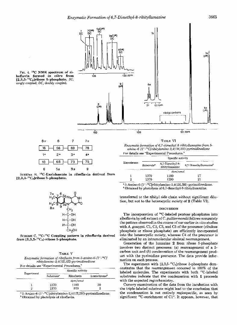
The 13C NMR spectrum of riboflavin from D-[2,3,5-l3C3] ribose 5-phosphate is shown in Fig. 4. The carbons of the two 4-carbon moieties show the expected enrichments (Scheme B). The rather complex 13C-13C coupling patterns are most easily deduced for carbons which have the fewest enriched one-bond carbon neighbors. Thus, the optimal sites for ex- amination of coupling are 8a (l*), 5a, (2*), 9a ( 3 7 , and 7a (47 (see Scheme C). The other four signals (6, 7, 8, and 9) yield entirely consistent information, but the patterns are somewhat more complicated. Carbon atoms 5a (2*) and 7a (4*) each show coupling to one other labeled carbon. Notably, 97% of the total signal due to 701 is one-bond carbon coupled. The 3* positions, i.e. carbons 7 and 9a, appear predominantly as doubly coupled species. In addition, a small amount of singly coupled species is discernible, reflecting again some metabolism of ribose 5-phosphate in the system (see below). Carbon atoms 8 and 9 show more complex patterns due to a combination of single coupling to the 3* position and statis- tical coupling to each other as a result of the combination of two highly enriched 4-carbon units. A small amount of scram- bling of label into the 1* position, i.e. C6 and C8a, is also evident, with about 48% of the 801 signal showing statistical coupling to the 56% enriched C8. This presumably reflects
160 do ppm
8 a 8 7 7a
2.5 H 0.9 H 1.7 1 1* 2 * 3* 4*
19.6 H 2.8 1.4 1.8
6 5a 9 a 9 SCHEME A. "C-Enrichments (a) in riboflavin derived from
[ l-13C]ribose 5-phosphate.
the same process which leads to scrambling from C1 to C2 of the pentose. A small amount of labeling of the 1* position from D-[2,3-13Cz]ribo~e has also been observed in the i n vivo experiments. In summary, the data confirm that the pentose carbons 2, 3, and 5 correspond to C2*, C3*, C4* of the alloxazine moiety, with loss of the pentose C4.
Studies by Hollander et al. (19) had suggested that the xylene moiety of riboflavin can be formed from the ribityl side chain of the pyrimidine 4 by enzymes from E. coli. In order to check whether this reaction occurs in C. guillier- mondii, we synthesized 5-amin0-6-[1'-'~C]ribitylamino- 2,4(1H,3H)-pyrimidinedione. Treatment of this compound with cell extract of C. guilliermondii in the presence of unla- beled ribose 5-phosphate afforded riboflavin which was pho- tochemically degraded to lumichrome (Compound 5, Fig. 2). The pyrimidine contributed its isotope to riboflavin with high efficiency. However, the lumichrome fragment was devoid of radioactivity (Table V). It follows that radioactivity from the purine precursor was exclusively incorporated into the ribityl side chain of the vitamin.
In a similar experiment, 5-amin0-6-[l'-~~C]ribitylamino- 2,4(1H,3H)-pyrimidinedione was treated with cell extract of C. guilliermondii mutant E6 using unlabeled ribose 5-phos- phate as second substrate. This mutant is devoid of riboflavin synthase. Hence, the experiment yields the riboflavin precur- sor, 6,7-dimethyl-8-ribityllumazine (2). Photochemical deg- radation yielded 6,7-dimethyllumazine (Compound 6, Fig. 2). As in the previous experiment, the radioactive label was
Enzymatic Formation of 6,7-Dimethyl-8-ribitylEumazine 3665
FIG. 4. 13C NMR spectrum of ri- boflavin formed in vitro from [2,3,5-13C3]ribose 5-phosphate. SC, singly coupled; DC, doubly coupled.
I I 135 130 PPm
7c
i . 1
ribityl carbons
7a (4*)
lh
8a 8 7 7a
I 16 H 56 H 69 H 78 I 1* 2* 3* 4*
IO H 68 H 73 H 75 ] 6 5a 9 a 9
SCHEME B. 13C-Enrichments in riboflavin derived from [2,3,5-”C3]ribose 5-phosphate.
H-$-OH H-?-OH H-?-OH
CHZOH
SCHEME C. 13C-13C Coupling pattern in riboflavin derived from [2,3,5-13C3]-ribose 5-phosphate.
TABLE V Enzymatic formation of riboflavin from 5-amim-6-[1 ‘-l4C1
ribitylamino-2,4(lH,3H)-pyrimidinedione For details see “Experimental Procedures.”
Experiment Specific activity
Substrate” Riboflavin Lumichromeb dpmlnmol
1 1370 1100 30 2 1370 970 2
a 5-Amino-6-[l’-’4C]ribitylamino-2,4(1H,3H)-pyrimidinedione. * Obtained by photolysis of riboflavin.
TABLE VI Enzymatic formation of 6,7-dimethyl-8-ribityllumazim from 5- amin0-6-[l’-‘~C]ribitylamino-2,4(1H,3H)-pyrimidinedione
For details see “Experimental Procedures.” Specific activity
Experiment 6,7-Dimethyl-8- ribitvllumazine 6,7-Dimethylluma~ine~
dpmlnmol 1 1370 1180 17 2 1370 1290 21
a 5-Amin0-6-[1‘-’~C]ribitylamino-2,4(1H,3H)-pyrimidinedione. Obtained by photolysis of 6,7-dimethyl-8-ribityllumazine.
transferred to the ribityl side chain without significant dilu- tion, but not to the heterocyclic moiety of 2 (Table VI).
DISCUSSION
The incorporation of 14C-labeled pentose phosphates into riboflavin by cell extract of C. guillierrnondii follows accurately the pattern observed in the course of our earlier in vivo studies with A. gossypii. C1, C2, C3, and C5 of the precursor (ribulose phosphate or ribose phosphate) are efficiently incorporated into the heterocyclic moiety, whereas C4 of the precursor is eliminated by an intramolecular skeletal rearrangement.
Generation of the lumazine 2 from ribose 5-phosphate involves two distinct processes: (a) rearrangement of a 5- carbon unit and ( b ) condensation of the rearrangement prod- uct with the pyrimidine precursor. The data provide infor- mation on each process.
The experiment with [2,3,5-13C3]ribose 5-phosphate dem- onstrates that the rearrangement occurred in 100% of the labeled molecules. The experiments with both 13C-labeled substrates indicate that the condensation with 1 proceeds with the expected regiochemistry.
Cursory examination of the data from the incubation with the triple-labeled substrate might lead to the conclusion that the condensation is not entirely regiospecific, as seen by significant 13C-enrichment of C1*. It appears, however, that

3666 Enzymatic Formation of 6,7-Dimethyl-8-ribityllumazine
the I* labeling is due to metabolism of the proffered pentose interchanging C-1 and C-2, perhaps via the pentose phosphate shunt. This idea is supported by the observation that the sum of the enrichments of C1* and C2* equals the enrichments of both C3* and C4* and that the amount of 1*-2* coupling is only what is expected on a statistical basis. If the C1* labeling were due to inversion of the 4-carbon unit, C1* would be coupled to C2* to the same extent as C4* to C3* (i.e. 97%), which is clearly not observed. The interchange of pentose C1 and C2 also explains the presence of singly coupled species at C3*. This interchinge also occurred approximately 20% of the time with the sample from [l-13C]ribose 5-phosphate, as well as in previous in vivo experiments.
The specific incorporation rates of carbon from pentose precursors observed in the present experiments present an unresolved problem. Since the 4-carbon unit is duplicated in the last biosynthetic step, 14C-labeled riboflavin should have a relative specific activity of 200% of that of the precursor. The observed values (about 120%) were lower than the ex- pected value. Substantial isotopic dilution has also been ob- served in the experiment with [1-l3C]ribose 5-phosphate, but little dilution occurred in the experiment with [2,3,5-13C3] ribose phosphate. The origin of the isotopic dilution is un- known. The dialyzed cell extracts used in this study contained a small amount of unlabeled riboflavin, but it appears likely that some unlabeled riboflavin was also formed de novo during the enzyme incubation from an unknown precursor present in the cell extract. We expect that this problem will be resolved by further work with purified enzymes instead of crude cell extracts.
Other authors have reported on the formation of riboflavin from 4 by partially purified enzymes from E. coli. This reac- tion proceeded in the absence of a second substrate, and it has been suggested that the ribityl side chain of 4 was the source of the carbon atoms of the xylene ring (19). In our experiments, the isotope from l'-14C-labeled 4 was not incor- porated into the heterocyclic moieties of 2 and 3, whereas it was incorporated into the side chain without significant di- lution. Although it is conceivable that different biosynthetic pathways could operate in the yeast C. guilliermondii and the bacterium E. coli, earlier in vivo experiments with Salmonella typhimurium agree with the biosynthetic pattern reported in this paper. Thus, a mutant of S. typhimurium with an absolute requirement for guanosine incorporated label from [1',2',3',4',5'-14C]guanosine into the ribityl side chain, but not into the isoalloxazine moiety of riboflavin (48). I n vivo studies with B. subtilis have indicated the same biosynthetic pattern as in A. gossypii and C. guilliermondii, i.e. formation of C1* from C1 and extrusion of C4 of the carbohydrate precursor (49). We conclude that a pentose phosphate is the actual precursor of the 4-carbon unit both in bacteria and fungi.
Since various carbohydrate phosphates can serve as sub- strates in the enzyme reaction studied, the structure of the direct riboflavin precursor remains elusive. In particular, we cannot decide whether it is a pentose phosphate or a pentulose phosphate. Apparently, various carbohydrate phosphates are rapidly interconverted by enzymes present in the crude cell extract.
We have recently obtained evidence for the formation of an aliphatic compound preliminarily designated as "X" which is produced by the cell extract of a C. guilliermondii mutant from ribose phosphate (50). Compound X can be converted to 2 in the presence of 4 by cell extract from a different C. guilliermondii mutant. The structure of compound X has not yet been determined. However, it was shown that the com-
pound is inactivated by treatment with phosphatase suggest- ing the presence of a phosphoric acid moiety. It is not known whether compound X contains 5 or 4 carbon atoms.
On the basis of the data presented here it is clear that the in vitro system reflects the biosynthetic processes observed i n vivo, as opposed to an artefactual production of riboflavin. Thus, we may confidently bring together information gained from in vivo and in vitro studies to suggest a hypothetical scheme for the biogenesis of lumazine 2 (Figs. 5 and 6). The 4-carbon unit is depicted (Fig. 5) as being generated via a radical mechanism, initiated by abstraction of a hydrogen atom from C3 of ribose 5-phosphate. This latter step has precedent, e.g. in the ribonucleotide reductase reaction (51). However, at this stage an equivalent cationic mechanism, initiated by abstraction of a hydride from C3, is equally likely. Condensation of the proposed 4-carbon precursor with 4 to give 2 (Fig. 6) follows a plausible mechanistic pathway, based on known chemistry of these compounds. The proposed mech- anism is not in conflict with any of the information currently available on riboflavin biosynthesis; however, a number of alternative mechanisms cannot be ruled out.
It should be noted that the unphosphorylated pyrimidine 4 and its 5"phosphate 1 can both serve as substrates for the
OH OH dH bH OH OH
0 4* I*
H w H 3 J HCOOH cHpQ ,?"h/.. CH3
OH 0 on o FIG. 5. Hypothetical mechanism for the formation of a 4-
carbon moiety from "ribose 5-phosphate.
0 0
H-C-OH 4 H-C-OH
CHzOH
~ - 6 - 0 ~ H-C-OH
CH,OH
H-C-OH I -
H-C-OH H-&OH 2 H-C-OH H-C-OH H-C-OH
C H P H CH,OH
H-&-OH H-?-OH H-C-OH
CHzOH
FIG. 6. Hypothetical mechanism for the formation of 6,7- dimethyl-8-ribityllumazine.

Enzymatic Formation of6,7-Dimethyl-8-ribityZlumazine 3667
formation of 2. This could imply that the respective enzyme accepts both pyrimidines. However, it is also possible that the phosphate 1 is enzymatically dephosphorylated by the cell extract prior to incorporation into 2 and riboflavin. Recent studies with a lumazine synthase/riboflavin synthase complex (i.e. “heavy riboflavin synthase”) from B. subtilis suggest that the committed precursor for the lumazine 2 is the unphos- phorylated pyrimidine 4.2
The availability of a cell-free system capable of synthesizing riboflavin from 1 and a pentose phosphate opens the way to the mechanistic investigation of the intriguing reactions in- volved in this biosynthesis and to the characterization of the intermediates and enzymes involved. The demonstration that the cell-free process shows the same features as the biosyn- thetic pathway in vivo provides assurance that information gleaned from the in vitro system does relate to the normal biosynthesis of riboflavin in the intact organism.
Acknowledgments-We thank Prof. H. Schirmer, Heidelberg, for helpful discussions. We are indebted to the Los Alamos Stable Isotope Resource, supported under National Institutes of Health Grant RR 02231, for l3C-labe1ed materials, and to the Ohio State University Chemical Instrument Center for mass spectra. We thank Degussa AG for a generous gift of reagents. The secretarial assistance of Astrid Konig and Angelika Kohnle is gratefully acknowledged.
REFERENCES
1. Nielsen, P., Neuberger, G., Floss, H. G., and Bacher, A. (1984) Biochem. Biophys. Res. Commun. 118,814-820
2. Bacher, A., Nielsen, P., Neuberger, G., and Floss, H. G. (1984) in Flavins and Flavoproteins (Bray, R. C., Engel, P. C., and Mayhew, S. G., eds) pp. 799-802, Walter de Gruyter, Berlin
3. Limens, F., Oltmanns, 0.. and Bacher, A. (1967) Z. Naturforsch. 22b, 755-758
. . . . .
4. Burrows. R. B.. and Brown. G. M. (1978) J. Bacterwl. 136.657- 667
Senyuta, E. Z. (1980) Biokhimiya 4 5 , 1284-1292
. ,
5. Logvinenko, E. M., Shavlovsky, G. M., Zakal’sky, A. E., and
6. Nielsen, P., and Bacher, A. (1981) Biochim. Biophys. Acta 6 6 2 ,
7. Plaut, G. W. E. (1971) in Comprehensive Biochemistry (Florkin, M., and Stotz, E. H., eds) Vol. 21, pp. 11-45, Elsevier Scientific Publishing Co., Amsterdam
8. Plaut, G. W. E., Smith, C. M., and Alworth, W. L. (1974) Annu. Rev. Bwchem. 43,899-922
9. Plaut, G. W. E. (1954) J. Biol. Chem. 211,111-116
312-317
10. Plaut, G. W. E., and Broberg, P. L. (1956) J. Biol. Chem. 219 ,
11. Goodwin, T. W., and Treble, D. H. (1959) Biochem. J. 70,14P-
12. Kishi, T., Asai, M., Masuda, T., and Kuwada, S. (1959) Chem.
13. Bryn, K., and Stbrmer, F. C. (1976) Biochim. Biophys. Acta 428 ,
14. Katagiri, H., Takeda, I., and Imai, K. (1959) J. Vitaminol. (Kyoto)
15. Alworth, W. L., Baker, H. N., Winkler, M. F., Keenan, A. M., Gokel, G. W., and Wood, F. L. I11 (1970) Biochem. Biophys. Res. Commun. 40,1026-1031
131-138
15P
Phurm. Bull. (Tokyo) 7,515-519
257-259
5,287-297
G. Neuberger and A. Bacher, manuscript in preparation.
16. Ali, S. N., and al-Khalidi, U. A. S. (1966) Biochem. J. 98, 182-
17. Alworth, W. L., Dove, M. F., and Baker, H. N. (1977) Biochem-
18. Bresler, S. E., Perumov, D. A., Chernik, T. P., and Glazunov, E.
19. Hollander, I. J., Braman, J. C., and Brown, G. M. (1980) Biochem.
20. Bacher, A., Le Van, Q., Biihler, M., Keller, P. J., Eimicke, V.,
21. Keller, P. J., Le Van, Q., Bacher, A., Kozlowski, J. F., and Floss,
22. Keller, P. J., Le Van, Q., Bacher, A., and Floss, H. G. (1983)
23. Bacher, A., Le Van, Q., Keller, P. J., and Floss, H. G. (1983) J.
24. Floss, H. G., Le Van, Q., Keller, P. J., and Bacher, A. (1983) J.
25. Bacher, A., Le Van, Q., Keller, P. J., and Floss, H. G. (1985) J.
26. Plaut, G. W. E. (1961) Annu. Rev. Biochem. 30 , 409-446 27. Logvinenko, E. M., Shavlovsky, G. M., Zakal’sky, A. E., and
Zakhodylo, I. V. (1982) Biokhimiya 47,931-936 28. Logvinenko, E. M., Shavlovsky, G. M., and Tsarenko, N. Y.
(1984) Biokhimiya 49,43-50 29. Logvinenko, E. M., Shavlovsky, G. M., and Tsarenko, N. Y.
(1985) Biokhimiya 5 0 , 744-748 30. Plaut, G. W. E., and Harvey, R. A. (1971) Methods Enzymol. 18 ,
31. Nielsen, P., and Bacher, A. (1983) in Chemistry and Siology of Pteridines (Blair, J. A., ed) pp. 705-709, Walter de Gruyter, Berlin
188
istry 16,526-531
A. (1976) Genetika 12,83-91
Biophys. Res. Commun. 94, 515-521
and Floss, H. G. (1982) J. Am. Chem. SOC. 104,3754-3755
H. G. (1983) J. Am. Chem. Soc. 105,2505-2507
Tetrahedron 39,3471-3481
Biol. Chem. 2 5 8 , 13431-13437
Am. Chem. SOC. 105,2493-2494
Am. Chem. SOC. 107,6380-6385
515-538
32. Bacher, A. (1984) Z. Naturforsch. 39b, 252-258 33. Cresswell, R. M., and Wood, H. C. S. (1960) J. Chem. SOC. 4768-
34. Kuhn, R., and Cook, A. H. (1937) Chem. Ber. 70,761-768 35. Ott, D. G. (1981) Synthesis with Stable Isotopes of Carbon, Nitro-
gen and Oxygen, pp. 33-35,57, Wiley-Interscience, New York 36. Murray, A., and Williams, D. L. (1958) Organic Synthesis with
Isotopes, pp. 931-932, Interscience Publishers, Inc., New York 37. Bacher, A., Baur, R., Eggers, U., Harders, H.-D., Otto, M. K.,
and Schnepple, H. (1980) J. Biol. Chem. 255,632-637 38. Klein, G., and Bacher, A. (1980) 2. Naturforsch. 35b, 482-484 39. Oltmanns, O., Bacher, A., and Lingens, F. (1968) 2. Naturforsch.
23b, 1556 40. Shavlovsky, G. M., Sibirny, A. A., Kshanovskaya, B. V., Koltun,
L. V., and Logvinenko, E. M.. (1979) Genetika 15,1561-1568 41. Longenecker, J. P., and Williams, J. F. (1980) J. Labelled Compd.
Radiopharm. 18,309-317 42. Jermyn, M. A. (1975) Anal. Biochem. 6 8 , 332-335 43. Bergmeyer, H. (1974) Methods of Enzymatic Analysis, p. 1343,
44. Rozutal, M., and Tomaszewski, L. (1974) Clin. Chim. Acta 5 0 ,
45. Chen, P. S., Toribata, T. Y., and Warner, H. (1956) Anal. Chem.
46. Bergmeyer, H. (1974) Methods of Enzymatic Analysis, p. 1238,
47. Axelrod, B. (1955) Methods Enzymol. 1,363-366 48. Mailiinder, B., and Bacher, A. (1976) J. Biol. Chem. 251, 3623-
49. Le Van, Q., Keller, P. J., Bown, D. H., Floss, H. G., and Bacher,
50. Neuberger, G., and Bacher, A. (1985) Biochem. Biophys. Res.
51. Stubbe, J. A. (1983) Mol. Cell. Biochem. 105, 7428-7435
4775
Academic Press, New York
311-317
28,1756-1758
Academic Press, New York
3628
A. (1985) J. Bacteriol. 162, 1280-1284
Commun. 127,175-181
Continued on next page.

Enzymatic Formation of 6,7-Dimethyl-8-ribityllumazine SUPPLEMENTARY MATERIAL TO Biosynthesis of Riboflavin. Enzymatic Foormatian of 6.7-Dimethyl-8-ribityllu~~ zine from Pentose Phosphates
Peter Nielsen, Gerhard Neuberger, Isao Fujii, David H. Bown, Paul J. Keller Heinr G. Floss and Adelbert BaCher
EXPERIMENTAL PROCEDURES
Chemicals - 5-Nitroso-6-ribitylamino-2.4(1H.3H)-pyrimidi"~dio"~ 1301, 5-nitro s o - 6 - r i b i t y l a m i n o - Z , 4 ( l H , 3 H ) - p y r l m i d i n e d i o n e 5"phosphate 131). 5-nitro-6 ribitylamino-2,4(1H.3H)-pyrimidinedione 5"phosphate (32), 5-nitro-6-chloro
dimethyllumazine (34) were prepared as described. [2-14ClGlucose, [6-14Clglu 2,4(1H,3Hl-pyrimidinedione 133). 6.7-dimethyl-8-ribityllumazine (30). and 6.7
Amersham/Buchler. Braunschweig. D-[2-'3C1Glucose (99 % l3C1 was obtained fro cose, [l-14Clglycerol, [2-14c]glycerol and [l-14Clribose were purchased fro
Omicron Biochemicals. Ithaca, N.Y., and 11,3-13C21glycerol (99 % 13C) wa
~ l a m o s Stable Isotope Resource, by standard literature procedures (35. 36) synthesized from [l-13Clacetic acid and K13CN, kindly provrded by the La
Ribose 5-phosphate and Plorisil were Obtained from Sigma.
~ n z me5 - Light riboflavin synthase of Bacillus subtilis was purified to a &ic activity of 18,000 " m o l m g - l h-l as described earlier (37). Triose phosphate isomerase w a s purchased from Boehringer. Other enzymes used were obtained from Sigma.
~icroarganisms - Lactobacillus & ATCC 7469 was a gift of Dr. Bernhard Maillnder, Pfizer Inc., Karlsruhe. -guillrermonaiiATCC 9058 was ob- tained from the American Type Culture Collection and Was grown aS described earlier (38). Frozen cell paste was stored at -20 OC. A mutant E6 was derived from the C.guilliermondii wild type strain after treatment with ethyl m e - thanesulfonate (39, 401. The mutant accumulated 6.7-dimethyl-8-ribityllumazine and required the addition of riboflavin I200 m g / l ) to the growth medium.
Preparation of cell extract - Frozen 5 guilliermondii cells were disrupted by passage through an X-press. The frozen plug w a s thawed and the suspension was centrifuged. The supernatant was dialyzed against 0.1 M Tris hydrochloride pH 7.5 containing 10 mM MgC12.
Enzyme Substrates - 5-Amino-6-ribitylamino-Z.4(lH,3H)-pyrimidi"~dion~ ( 4 ) was prepared by catalytic hydrogenation of an aqueous solution of 5-nitroso-6- ribitylamino-2,4(1H,3H)-pyrimidinedione over palladium on charcoal at room temperature and atmospheric pressure. The Catalyst w a s removed by filtration through a Millipore fllter. The solution was used mmediately.
pared by catalytic hydrogenation of 5-nitroso-6-ribitylamino-2,4llH,3HI-pyri- 5-Amino-6-~ibitylamino-Z,4llH,3H)-pyrimidi"=di~~~ 5"phosphate (1) was pre-
midinedione 5"phosphate or 5-nitro-6-ribitylamino-2.4(lH,3H)-pyrimidi~~dion~ 5"phosphate as described above. The solution was used immediately.
Preparation of isotopically carbohydrate phosphates Radioactive phos ho luconate from lucose - Reaction mixtures contamed 0.25 M Tris hydrochlarBdegpH 7.8, 1 K M h l 5 m M NADP+, 15 mM ATP, 5.6 " m o l I5 uci) of glucose appropriately labeled with l4C, 50 U of hexokinase, and 50 u of glucose phosphate dehydrogenase in a total volume of 400 "1. The reaction mixture was incubated at 30 OC for 30min. The mixture was dilutedto a v o l u m e of 10 ml with deionized water and placed on a c o l u m n of D O W ~ X 1x8 (formate f o r m , 200-400 mesh, 0.9 x 15 cm). The column was washed with 25 ml of deioni-
acid and 0.1 M ammonium formate. The flow rate was 0.6 ml/min. Fractions were zed water and subsequently developed with an eluent containing 0.1 M formic
collected and monitored by liquid scintillation counting of aliquots. 6-Pho5- phogluconate was eluted from 80 to 120 ml. Fractions were combined and lyophi- lized. The radiochemical yield was 60-83 %.
114ClPhosphogluconate from glycerol - Phosphogluconate w a s prepared by modi-
hydrochloride pH 7.8, 20 mM MgC12, 13 mM ATP, 27 mM NAD+, 20 mM fructose 6- fication of a published procedure 141). Reaction mixtures contained 0.1 M Tris
phoshate, 27 mM sodium pyruvate, 1.3 "mol I10 uci) of 14C-labeled glycerol, 34 U of glycerokinase, 1.6 U of glycerol 3-phosphate dehydrogenase, 350 U of triose phosphate isomerase, 1.5 u of transaldolase, and 5.5 U of lactate dehy- drogenase in a total volume of 300 ul. 14C-Labeled glycerol w a s purchased as
use. The reaction mixture was incubated at 30 OC for 2 h. an ethanol solution. The ethanol w a s removed by a Stream of nitrogen prior to
Phosphoglucose isomerase I10 ul was added and the mixture was again incubated
phosphate which had been formed in the first reaction Step. for 30 mi" at 37 Oc in order to produce glucose 6-phosphate from fructose 6-
-_
Glucose 6-phosphate was further converted to 6-phosphogluconate by the addi- tion of 6.1 mg (7 umoll of NADP+, 50 u of glucose phosphate dehydrogenase, and 250 ul of 0.25 M Tris hydrochloride pH 7.9 followed by incubation at 30 OC for
described above. The radiochemical yield was 27-46 %. 40 mi". The reaction mixture was subjected to ion exchange chromatography as
Radioactive ribulose 5-phosphate from 6-phosphogluconate - The reaction mix- ture contained 90 mM Tris hydrochloride pH 7.8, 7 mM NADP*, 0.3-2 UM 6- phosphogluconate from the previous step, and 5 u of 6-phosphogluconate dehy- drogenase in a total volume of 0.9 ml. The mixture w a s incubated at 37 OC for 90 mi". diluted to 10 ml with deionized water, and placed on a column of Dowex 1x8 (200-400 mesh, formate farm, 0.9 x 15 cm). The column w a s developed with a Solntion containing 0.1 M formic acid and 30 mM ammonium formate. The flow rate was 0.6 ml per mi". FractlOnS were collected and monitored by liquid scintillation counting of aliquots. Ribulose 5-phosphate was eluted from 35-65 ml. Fractions were pooled and lyophlllzed. The radiochemical yleld based on 6- phosphogluconate Was 20-45%.
"~ D-[l-13Clribase 5 - p h 0 ~ p h a t e / D - [ l - ~ ~ C l r i b u l o s e 5-phosphate - ATP 16.15 mmol) adjusted to 6.75 Wlth 1 N NaOH. Dlthiothreitol I 1 6 8 u m o l l , D-[2-13C191ucase and MgC12.6 H20 (6.11 mmol) were dissolved in 240 ml H20 and the pH W a s
mixture was incubated at 29.5 OC. while the pH was maintained at 6.6 by (1.0 g) and hexokinase 1602 IO), dissolved in 10 m l H20. w a s added. and the
titration with 1 N NaOH. When the pH change stopped. after about 30 min, incubation was continued far another h, the mixture w a s then cooled in ice and 50 ml of 50 % (w/vl trichloroacetic acid was added. Precipitated protein w a s
removed by centrifugation and the supernatant was adjusted to pH 7.0 with 50 % (w/v) NaOH. Barium acetate 124.4 mmol) was then added, a small amoilnt of precipitate of Ba-ADP was removed by centrifugation, and ethanol was added to the supernatant to a final concentration of 80 % (v /v ) . After standing over- night at 0 OC, the precipitated Ba Salt of glucose 6-phosphate was collected by centrifugation and washed with 80 % lV/V) ethanol. The salt was suspended in ethanol. The ethanol was evaporated and the residue was dried in a vacnum to give 4.0 9 of crude product. This material w a s dissolved in 40 m l Of H20 with the aid of some HC1. 6.66 mmol Na2S04 was added and. after neutralization with NaOH, the BaS04 was removed by centrifugation. The Supernatant was ap- plied to a column (4x20 c m ) Of Dowex 1x8 (formate form, 100-200 mesh), which was then washed with H20 and eluted with a gradient of ammonium formate buffer
taining glucose 6-phosphate, as determined by the anthrone/sulfuric acid (1 1 Of H20 Vs 1 1 of 0.4 M HCOOH/0.12 M ammonium formate). The fractions con-
reaction (42). were pooled and the product was precipitated as the barium
pure, based on enzymatic analysls with glucose 6-phosphate dehydrogenase (431. salt. The resulting barium glucose 6-phosphate (2.53 g) was essentially 100 %
Yield. 88.5 %, based on glucose.
a-Ketoglutarate 115.8 mmol), NH4Cl 120.1 mmoll, dithioerythritol (219 mmol),
pH 1.9. The pH w a s adjusted to 7.8 with 1 N NaOH and 0,401 m m o l of NADP+ and and MgC12-6 H20 12.08 mmol) were dissolved in 150 ml of 0.2 M Tris HC1 buffer,
2.53 g of barlum [2-13C1glucose 6-phosphate. dissolved in 50 ml water, were added. The reaction w a s started by the addition of 294 U of glucose 6-phos- phate dehydrogenase. 214 u of 6-phosphogluconate dehydrogenase and 209 U of glutamate dehydrogenase. After one h incubation at 27 OC, 1020 U Of phosphori- bolsomerase was added and the incubation was continued for one h at 27 OC and then over night at 4 OC. Protein was removed by trichloroacetic acid I30 ml 50 % sol. w/v) precipitation, the supernatant was neutralized with 50 % NaOH, and th; barium salts were preclpltated by addltion of 10 mmol of barium acetate
purified by chromatography on a D O W ~ X 1 column as descrlbed above for barium and ethanol to 80 8 v/v. The crude mixture of barium salts 15.19 g) was
glucose 6-phosphate. The fractions, as determined by the orcinol reaction
harium salts. The product, 2.19 g, was found by enzymatic assay 143) to 144). w e r e combined, and the sugar phosphates were again precipitated a5 the
contain 4.1 mmol of ribose 5-phosphate and 0.59 mmol of ribulose 5-phosphate, correspondrng to an overall yield of 84.8 8 based on [2-13Clglucose.
D-[2,3,5-13C31ribose 5-pho~phate/D-[2,3.5-~~C,lribulose 5- hoS hate - I1.3- T3C21Glycerol 11.44 91, ATP I23 mmol), dithiothreitol 10.6 :mol:, and MgC12
Glycerokinase ( 6 0 0 u ) was then added and the mixture was incubated at room I23 m m o l l were dissolved in 600 ml of H20 and the pH w a s adjusted to 9.6.
temperature while the pH was maintained at 9-9.5 by titration with 1 N NaOH. After COnSUmptlOn of 15.5 m l of 1 N NaOH, the pH change Stopped and the mixture w a s incubated for one more h. The pH w a s then adjusted to 1.1 and 50
centrifugation and the Supernatant loaded On a column of D O W ~ X 1x8 (4x20 cm, mmol of BaCl2 was added. The precipitated barium salt of ADP Was removed by
the first fractions eluted with ammonium formate buffer (gradient of 1 1 of formate form). a-Glycerophosphate appeared in the "on-adsorbed fraction and in
H20 vs 1 1 of 1.3 M HCOOH/ammonium formate, pH 3). Precipitation of the barium salt with acetone w a s attempted, but w a s unsuccessful. The purifLcatlon on D O W ~ X 1x8 w a s repeated and the fractions containing o-glycerophosphate, as determined by phosphate assay I451 were pooled. Barium acetate I50 mmol) was added, and ethanol w a s subsequently added to a final concentration of 8 0 %
phosphate based on enzymatic assay 146). This corresponds to an isolation (v/vl. The precipitated harium salt (6.04 gl contained 12.3 mmol of 0-glycero-
yield of 79.3 % based on the amount of glycerol reacted (15.5 mmole by titra- tion) and a total yield of at least 59.3 % based on startlng glycerol.
The [ 1,3-13C21-a-glycerophosphate was converted into glucose 6-phosphate in
Of HC1, and barium ions were removed by addition of Na2S04 and centrifugation. two batches. The barium Salt (7.25 m m o l ) was dissolved in water with the aid
The supernatant w a s added to 1 1 of 0.2 M Tris HC1 buffer, pH 7.9, containing 9.1 mmol of NAD', 73 mmol of sodium pyruvate, and 1 mmol Of dithiothreitol. a- Glycerophosphate dehydrogenase I500 U1 and L-lactate dehydrogenase I 5 0 0 0 Ul were then added, and the formation of dihydroxyacetone phosphate was followed by assay for alkali-labile phosphate. When about 40 % of the o-glycerophos- phate had been convertedto dihydroxyacetone phosphate, 15,000 U of triose- phosphate isomerase and 500 u of aldolase were added and the formation of fructose 1.6-diphosphate was followed by the anthrone/sulfuric acid reaction (42). When fructose diphosphate formation had reached its maximum, fructose-
the pH w a s adjusted to 9. Then phosphoglucose isomerase I880 U) was added and 1,6-diphosphatase (100 U ) and MgC12 (to a concentration of 1 mM) was added and
the react~on mixture was incubated over night. The pH was adjusted to 7.5. barium acetate (294 mmol) was added and yellow colored impurities were removed by treatment with activated charcoal (55 91, which w a s filtered off and washed with 0.01 M NH40H. Ethanol was added to the combined filtrate and washings to a final concentration of 80 8 v/v. The precipitated crude barium Salt was purified on a D O W ~ X 1x8 column (4x20 cm. formate form) as described above. The fractions containing the ~roduct were pooled and the barium Salt W a S again prepared. The product (835 mg) was found by enzymatic assay to contain 1.28 mmol of glucose 6-phosphate and 0.16 mmol of fructose 6-phosphate. In another batch, 5.0 mmol of a-glycerophosphate was converted to 749 mg of barium salt containing 1.09 mmol of glucose +phosphate and 0.22 mmol of fructose 6- phosphate. The combined yield from o-glycerophosphate w a s 44.7 %.
The mixture of [1,3,4,6-13C4]g1ucose 6-phosphate and [l,3.4,6-13C41fru~tose 6- phosphate was converted to a mixture of [2,3.5-l3C ribose 5-phosphate and
except that 1000 u of phosphoglucose isomerase were included in the reaction [2,3,5-13C3]~ibulo~e 5-phosphate as described for [~-'3Clglucose 6-phosphate.
mixture. From 1.75 mmol of the hexose phosphate mixture, we obtained 1.03 g of purified pentose phosphate barium salt, which according to enzymatic assay contained 2.07 m m o l of ribose 5-phosphate and 0.27 m m o l of ribulose 5-phos- phate. This corresponds to a yield of 85.1 8 based on hexose phosphate or 30.2 % overall yleld based on consumed glycerol.
The samples underwent some decomposition upon prolonged storage, as evidenced by reexaminingthe "C-NMR Spectrum of an aliquotafter 6 months at-20 OC.
Preparation of 5-nitro-6-[l'-14C]ribitylamino-2,4(1H.3H)-py~imidi"~d~o"~
lyophilized in a 1.5 ml plastic cup. Subsequent to the addition of 0.15 ml of [l-14C]Ribose oxime - ~n aqueous solution of [1-14Clribose (8mg. 50 U C ~ I w a s
a 1.3 M hydroxylamine solution in absolute methanol (30). the mixture was kept for 12 h at room temperature and Subsequently for 24 h at 4 'C. Crystals of

Enzymatic Formation of 6,7-Dimethyl-8-ribityllumazine 3669
ribose oxime were collected on a fritted glass disk by Suction and washed
was to dryness and yielded approximately 2 a9 of ribose oxime three eimes with 70 u l of ethanol yielding 2.8 mg of white solid. The filtrate
Which was separately converted to 5-nitro-6-ll'-14ClribitylaminO-2,~(lH~3H)- pyrimidinediono.
[1-14c]~ibit lamine - A suspension of 1.1 mg of PtGZ in 2 m l of acetic acid was bydrogenaied-fF20 mi". [1-14C1Ribose oxime (2.8 mgl was added and hydro- genated for 12 h at room temperature and atmospheric pressure. Platinum was removed by filtration, and the solut ion was evaporated to dryness under redu- ced pressure. The oily residue was dissolved in l o m l of water and placed OD a column of AG 50 W X 8 (H' form. 200-400 mesh, 0 . 8 X 12.5 C m l . The Column Was washed with 100 m l of deionized water. Ribitylamine w a s subsequently eluted with 50 m l of 0.2 M NX~OH. The effluent was evaporated to dryness under reduced pressure. Deionized water was added and evaporation was repeated three times.
-
n i y 6.3 mg of 5-nitro-C-chloro-2,4(lH.3~I-pyrimidinedionc in 350 ul of etha- 5-Nitro-6-[l'-14Clribitylami"o-2;4(lH,3HJ-pyrimidi~edio~e - A solution contai-
no1 was added to the ribitylamine. Phosphate buffer pH 8.0 (0.1 M, 1.5 nil1 WaS
under reduced pressure. and the residue was dissolved i n 25 ml of deionized added brd the Solution was kept at 40 OC for 24 h. The solvent was evaporated
RP18, 16 x 250 mm; eluent. 20 mM ammonium formate pH 3.8; flow rate, 6-12 water. The solution Was passed through a preparative HPLC Column ILichrosorb
ml/minJ. Fractions were collected and lyophilized. Yield, 11.0 umol. Speoific activity , 1320 dpm/nmol. Radiochemical yield based on Il-14Clribose, 13.2 %.
The second crop of [l-14C]ribose oxime obtaioed at the end of step 1
additional amount of 8 umol of 5-nitro-6-[l~-14Clribitylamino-2,411H,3Kl-pyri- was separately oarried through the subsequent steps yielding an
midinedione with a specific activity of 1370 dpm/nmol. The total radiochemical yield based an ribose was 23 %. Both batches appeared pur@ as judged by irnaly- tical HPLC and thin layer chromatography (cellulose, 1-butanol/acetic acid/wa- ter 50:15:35, v/v; Rf = 0.62). They showed the expected absorption spectrum (xmax F 227 nm, 323 nm, ~ ~ ~ ~ / c ~ ~ ~ = 1.95 at p H 1.01.
" Enzyme Studies
All experiments Were performed i n dim light to minimize photolysis of pro- ducts. The pyrimidiile substrates used are very oxygen sensitive. They were
sure to molecular oxygen. freshly prepared for each experiment, and efforts were made to minimize expo-
~- Riboflavin from 14C-labeled ribulose 5-phosphate - Assay mixtures con- tained 0.1 M Tris hydrochloride pH 7.5, lo mH dithioerythritol, 10 m~ ~gel,, 1
phate, 1 mM radiolabeled ribulose phosphate. 1000 u of iight riboflavin syn- thase from B. silbtilis, and dialyzed cell extract from & gullliermondii RTCC 9058 (3 mg of protein) in a total volume of 2 ml. The mixture was incubated
using b. casei. Riboflavin (25 nmol dissolved in 400 ul of water) was added as for 1 h at 30 OC. An aliquot (100 ul] w a s retrieved for bioassay of riboflavin
carrier to the assay mixture, and the solution was passed through a column of Florisil (0.5 X 7 em). The column was washed with 10 m l of deronized water. Fiuorescent material was eluted with 5 m l of acetone/2 M NH~OH (~:1, The eluate w a s evaporated to dryness Under reduced pressure. The residue dissolved in 0.4 m l of deianized water and freed from so,lids by centrifugation in a Beckmann Airfuge at 100,000 g for 10 m i " . The sLpernatant was passed through a reversed phase HPLC column (Nuclaosil lo cis, 0.4 x 25 cm, flow rate 2 ml/min; eluent. 20 % methanol in 0.1 M ammonium formate). m e e l u a t e w a s monitored photometrically (254 nm and 405 nm). The fraction containing flavin (retention time, 30 ni.n) w a s collected and evaporated to dryness undei reduced pressure.. The residue was dissolved in 0.3 m l of deionized water, and aliqilots were used for riboflavin bioassay and for liquid scintillation cow-
____- mM rreshly prepared 5-amino-6-ribitylamimo-2.4(1W,3H)-p~~imidi~~d~o"~ 5'-phos-
ting. The remaining material was converted to lumichrome.
Riboflavin from [1-I3CIribose 5-phos hate - The reaction mixture contained 0.1
phosphate, 0.9 mM freshly prepared 5-amino-6-ribitylamino-2,4[lX,3H)-pyrisi- M Tris XCl pH 7.5, 5 mlrl MgCIZ, 10 m z dithioerythritol, 1 m M [1-13C]ribose 5-
dinedione S'-phosphate (1). &Rd dialyzed crude cell extract f r~m c- guillier- OC for 2 h, 5.0 ml of 18.6 mM 1. 16 ml of 7.7 m M [1-13Clribose 5-phosphate, mondii (1.3 g of protein1 i n a total volume of 132 ml. After incubation at 30
and a solution of 100,000 U of light riboflavin Synthase in 6 ml of 0.1 M Tris
HC1 were edded, and the incubation at 30 OC was continued for 7.5 h. The reaction was stopped by the addition of 24 g of trichloroacetic acid. Protein was removed by centrifugation at 32,000 9 for 20 nin. The supernatant was brought to pH 7 by the addition of 30 g of sodium acetate. Water was added to a f i n a l volume of 500 ml. The Solution w a s applied to a column of rlorisil (0.8 x 11.5 om). The column w-5 Washed with 500 m l of deionized water. Pluo-
v/vl. The eluate was evaporated to dryness under reduced pressure. The residue rescent material was eluted with a mixture of acetone/2 M NH~OH (50 ml, 1:1,
was dissolved in 30 m l o f water. l'he solutlon was placed on a column of AG 50 WX8 IH' form, 1.0 x 20 om). The column was deveLoped with 0.1 M KC1. Fractions containing riboflavin were combined and concentrated to dryness under reduced pressure. Yield, 3.1 mg.
Riboflavin Exom [2,3,5-13C3]ribose 5-phosphate - The reaction mixture aontai- ned 0.1 M Pris HC1 pH 7.5, 5 mM MgClZ. 2 m M CaCl2, 10 ml< dithioerythritol, 1.1 mM [2,3,5-13C,Iribose 5-phosphatel 0.65 mM 1, and dialyzed crude cell extract from C.guill;ermtn (1.2 g of protein) in a total volume of 136 ml. After incubation at 3 0 OC for 2 h, 4.0 m l of 135 mM 1. 10 ml of 14.7 mM [2.3,5- 13C31ribose 5-ph06phater and 75,000 U of light riboflavin synthase dissolved in 13 ml of 0.1 M TriS WCl were added, and the incubation was continued for 9.5 h. RiboElavin was purified 2s described above. Yield, 1.5 mg.
Riboflavin from 5-amino-6-I.l'-14C1ribityiamino-Z,4l1H,3H)-pyrimidineaione - A solution of 5-nitro-6-[1'-*4C]ribitylamino-2.4l1H.3H~-pyrimidinadione (1.0 mg, 3.2 u m o l ) in 1. ml O f deionized water w a s hydrogenated over palladium on Char- coal. The suspension was passed through a nitrocellulose filter to remove the catalyst. The filter was Wasbed With 200 u l of Water. An aliquot of the fil- trate i0.46 m i containing 1.4 uno1 of the pyrimidinedionel was mixed with 1.14 m l of a solution containing 0.14 M Tris hydrochloride pH 7.5, 14 mM MgCl2, 14 m M dithioerythrit.ol, 1.4 mM ribose 5-phosphate, and cell extract from C. guil- liermondii ATCC 9058 (12 mg of protein). The mixture Was incubated for 2 h at 37 OC. L'rght riboflavin synthase from 0. subtilis 12200 0) wa§ added and the incubation was continued for another 1.5 h. Riboflavin was isolated and puri- fied as described above.
pyrimidinedione - Assay mixtures contained 0.53 mM 5-amino-6-Il'-1'Clribityl- 6.7-Dimethyl -8- r ib i ty l lumazina from 5-amino-6 - [1 ' -14C]~ ib i ty l a . i no -2 ,4 ( lH ,3Hl -
amino-2,4(1H.3Hl-pyrinidineiiione [freshly prepared as described above), 0.1 M Tris hydrochloride pH 7.5, l o mM MgC12, 10 mM dithioerythritol, 1 InM ribose 5- phosphate, and cell extract from 5 guilliermre mutant E6 (15 mg of pro-
mixture was passed through a column of Florisil (0.8 x 10 cm). The column was teinl in a total volume of 1.6 nl. After incubation for 2 h at 37 OC the
washed with 10 ml of deionized Water. Fluorescent material was eluted with acetone/2 M NH40H (15 m l , 1:l. "/VI. The eluate was brought t o dryness under reduced pressare. The residue was passed through a reversed phase HPLC Oolumn
formate buffer pH 3.8). The effluent was monitored photometrically (254 and (Nucleosil 10 CIS, 0.4 x 25 om; flow rate, 1 ml/min; eluent, 20 mM ammonium
405 nm). The fraction containing 6,7-dimethyl-8-ribityllu~a~i~e (retention time, 32 min) was collected and lyophilized.
"
Degradations
Fhoeocheinical degradation of riboflavin - Riboflavin was dissolved in buffer containing 0.25 M formic acid and 0.25 M ammonium formate pH 3.8. The eolutian was illuminated far 2 h by a 500 N lamp (distance, 0.1 m). The solution was passed through a HPLC column (Nucleoail 10 C18. 0.4 x 25 ~ m , flow rate, 2 m l / m i n ; eluent. 34 % methanol in 0.1 M ammonium formate pi3 6.31. Absorbance was monitozed at 254 and 365 nm. The fraction containing lumichrome ( 5 ) (re- tention time, 17 minl was collected and lyophilized.
Shotochemical degradation 6.l-dimethyl-8-ribityllu~n+ - A Solution of 6,7-dimethyl-8-ribityllumazine in 0.1 M ammonLum formate buffer pH 3.5 w a s
column (Nucleosil 10 C18, 0.4 x 25 cm; eluent, 10% methanol in 0.1 M ammonium illuminated f a 1 h as described above. The solution was passed through a HPLC
formate pH 6.3; flow rate, 1 ml/minl. Absorbance was monitored at 254 and 340 nm. The fraction Containing 6.7-dimethyllumazine (61 (retention time, 25 min) wa5 collected and Lyophilized.
NMR SpectroscOpy
'H-decoupled 13C-NMR spectra Were obtained a t 7.0 T on a Bruker WM-300 NMR spectrometar. Samples of r iboflavin were dissolved in approximately 0.b m l of D@X30-~6. The Spectral acquisition parameters were: Pulse angle - 60 O, repeti-
broadening, Chemical shifts are referenced to tetramethylsilane, tion tile = 2.0 sec, Spectral width = 15.15 KHz. 32 K data sets, 1.0 HZ line