nobel prize for mri imaging denied to raymond - fonar mri prize for mri damadian, gbk, chem...
TRANSCRIPT

Chem. Educator 2014, 19, 73–90 73
© 2014 The Chemical Educator, S1430-4171(14)12540-2, Published 03/21/2014, 10.1333/s00897142540a, 19140073.pdf
Nobel Prize for MRI Imaging Denied to Raymond V. Damadian a Decade Ago
George Kauffman*
Department of Chemistry, California State University, Fresno, Fresno, CA 93740-8034, [email protected]
Abstract: The 2003 Nobel Prize in Physiology or Medicine was awarded to chemist Paul Christian Lauterbur and physicist Sir Peter Mansfield “for their discoveries concerning magnetic resonance imaging.” Raymond Vahan Damadian, M.D. correctly claimed that he had invented MRI and that Lauterbur and Mansfield had merely refined the technology. Because Damadian was not included although the Nobel statutes permit the award to be made to three living individuals, his omission was deliberate. Possible purported reasons for his rejection have included the fact that he was a physician not an academic scientist, his intensive lobbying for the prize, his supposedly abrasive personality, and his active support of creationism, none of which constitute grounds for the denial. This article surveys previous contributions to nuclear magnetic resonance for which Nobel Prizes were awarded, explores Damadian’s personal and professional career, and concludes that Damadian’s seminal discovery preceded and was more fundamental than Lauterbur’s developments.
According to the late Ulf Lagerkvist (1926–2010), a member of the Swedish Academy of Sciences, who participated in judging nominations for the Chemistry Prize,
It is in the nature of the Nobel Prize that there will always be a number of candidates who obviously deserve to be rewarded but never get the accolade. Of course it is an impossible task that the Academy has struggled with for more than a century. Maybe one can claim that it has done a fairly good job after all. Nevertheles there have been unfortunate sins of omission and the most outrageous is that committed against Dmitri Mendeleev. It is indeed a pity that his name does not appear in the distinguished list of laureates. It would certainly have added to both the prestige of the Nobel Prize and the Royal Swedish Academy of Sciences [1].
Although the denial of the Nobel Prize in 1905 and 1906 to Mendeleev for the periodic table, the Rosetta Stone of chemistry, is probably the best known example of outrageous disgrace in the awarding of the prizes, another case of bias that occurred almost a century later— a decade ago—deserves our attention.
The Shameful Wrong That Must Be Righted
Usually, a losing candidate merely accepts the injustice. But in the case of the 2003 Nobel Prize in Physiology or Medicine of $1.3 million, awarded ten years ago, to University of Illinois chemist Paul Christian Lauterbur (1929–2007) (Figure 1) and University of Nottingham, UK physicist Sir Peter Mansfield (b. 1933) (Figure 2) “for their discoveries concerning magnetic resonance imaging,” the undoubtedly deserving candidate, Raymond Vahan Damadian, M.D. (b. 1936) (Figure 3), an American of Armenian-French descent, did not take this injustice lying down. A group called “The Friends of Raymond Damadian” protested the denial with full-page advertisements, “The Shameful Wrong That Must Be Righted” (Figure 4) in the New York Times, Washington Post, the Los Angeles Times,
* Series Editor contribution.
and Stockholm’s Dagens Nyheter. His exclusion scandalized the scientific community in general and the Armenian community in particular. He correctly claimed that he had invented MRI and that Lauterbur and Mansfield had merely refined the technology [2, 3].
Because Damadian was not included in the award even though the Nobel statutes permit the award to be made to as many as three living individuals, his omission was clearly deliberate. The possible purported reasons for his rejection have included the fact that he was a physician not an academic scientist, his intensive lobbying for the prize although other candidates have done so, his supposedly abrasive personality, and his active support of creationism. None of these constitute valid grounds for the denial.
The careful wording of the prize citation reflects that fact that the laureates did not come up with the idea of applying nuclear magnetic resonance (NMR) [4, 5] (the term was later changed to avoid the public’s fear of the word “nuclear” even though nuclear energy is not involved in the procedure) to medical imaging. Today magnetic resonance imaging (MRI) [6, 7] is universally used to image every part of the body and is particularly useful in diagnosing cancer, strokes, brain tumors, multiple sclerosis, torn ligaments, and tendonitis, to name just a few conditions. An MRI scan is the best way to see inside the human body without cutting it open.
Development of NMR and Nobel Prizes
In a classic case of unintended consequences [8], Chancellor Adolf Hitler (1889–1945) told pioneer quantum physicist and 1918 Nobel Physics laureate Max Planck (1858–1947), “If the dismissal of Jewish scientists means the annihilation of contemporary German science, then we shall do without science for a few years” [9]. By stripping Germany’universities of its Jewish professors, including some of the world’s finest minds, the arrogant Führer rendered his Heimat scientifically crippled for years as American universities, eager to beef up the theoretical part of their

74 Chem. Educator, Vol. 19, 2014 George B. Kauffman
© 2014 The Chemical Educator, S1430-4171(14)12540-2, Published 03/21/2014, 10.1333/s00897142540a, 19140073.pdf
Figure 1. Paul Christian Lauterbur (1929–2007). Courtesy, Nobel Foundation.
Figure 2. Sir Peter Mansfield (b. 1933). Courtesy, Nobel Foundation.
Figure 3. Raymond Vahan Damadian, M.D. (b. 1936), 1988.
Figure 4. “The Shameful Wrong That Must Be Righted,” New York Times, October 2003.
physics research programs, welcomed the émigrés with open arms. Hitler had unwittingly engineered not only a mass exodus of the originators of the “new” physics, contributing in the process to his own eventual demise, but he had also enriched America with scientists who would contribute enormously to its postwar leadership in nuclear physics, microwave technology, solid state physics, and—in our case at hand—nuclear magnetic resonance, for which a number of Nobel Prizes were awarded [10, 11].
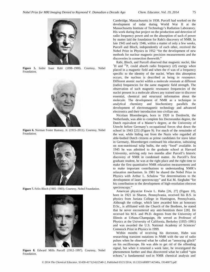
Nuclear magnetic reaonance was first described and measured in molecular beams by Isidor Isaac Rabi (1898–1988) [12] (Figure 5), who extended the Stern-Gerlach experiment [13]. Rabi was born into an Orthodox Jewish family in Rymanów, Galicia, in what was then part of the Austro-Hungarian Empire. At the age of three, he and his mother, Scheindel, joined his father, David, who had earlier emigrated to the United States. He was raised in a two-room apartment in New York City’s Lower East Side and later stated,
My mother made me a scientist without ever intending to. Every other Jewish mother in Brooklyn would ask her child after school: So? Did you learn anything today? But not my mother. “Izzy,” she would say, “did you ask a good question today?” That difference—asking good questions—made me become a scientist [14].
After studies at Cornell University and Columbia University, Rabi returned to Europe and worked with many of the leading physicists of the time. In 1929 he returned to the United States, where Columbia offered him a faculty position, making him the university’s first Jewish faculty member. His discovery of NMR [15–20] led to his receipt in 1944 of the Nobel Prize in Physics “for his resonance method for recording the magnetic properties of atomic nuclei” [21].
Norman Foster Ramsey, Jr. (1915–2011) [22] (Figure 6) was only 22 years old when he became the only graduate student to work with Rabi and his associates on the development of NMR. The next summer, while working alone on the noisy 10th-floor molecular-beam vacuum pumps of Columbia’s Pupin Hall., he became the first member of Rabi’s team and determined the magnetic moments of the proton and the deuteron and the radiofrequency spectrum of hydrogen in various magnetic fields. The six-peak spectrum that he and the rest of the Rabi team subsequently acquired became the first example of multiple-line spectroscopy with coherent electromagnetic radiation, a characteristic of radiofrequency, microwave, and laser spectroscopy. In 1949 he developed the first successful chemical shift theory, and in 1989 he received the Nobel Prize in Physics “for the invention of the separated oscillatory fields method and its use in the hydrogen maser and other atomic clocks.”
Felix Bloch (1905–1983) [23] (Figure 7) was born in Zürich, Switzerland to Jewish parents, attended the Eidgenössische Technische Hochschule (ETH) there, became Privatdozent (unsalaried lecturer) at the Universität Leipzig, was dismissed in 1933 upon Hitler’s rise to power, and emigrated to the United States, where he became Professor of Physics at Stanford University, Palo Alto, CA.
Edward Mills Purcell (1912–1997) [24] (Figure 8) was born in Taylorville, Illinois, sang in the boys choir of the local Presbyterian church, graduated as an electrical engineering major from Purdue University, studied in Europe, and received his Ph.D degree in physics from Harvard University,
Nobel Prize for MRI Imaging Denied to Raymond V. Damadian a Decade Ago Chem. Educator, Vol. 19, 2014 75
© 2014 The Chemical Educator, S1430-4171(14)12540-2, Published 03/21/2014, 10.1333/s00897142540a, 19140073.pdf
Figure 5. Isidor Isaac Rabi (1898–1988). Courtesy, Nobel Foundation.
Figure 6. Norman Foster Ramsey, Jr. (1915–2011). Courtesy, Nobel Foundation.
Figure 7. Felix Bloch (1905–1983). Courtesy, Nobel Foundation.
Figure 8. Edward Mills Purcell (1912–1997). Courtesy, Nobel Foundation.
Cambridge, Massachusetts in 1938. Purcell had worked on the development of radar during World War II at the Massachusetts Institute of Technology’s Radiation Laboratory. His work during that project on the production and detection of radio frequency power and on the absorption of such rf power by matter laid the foundation for Rabi's discovery of NMR. In late 1945 and early 1946, within a matter of only a few weeks, Purcell and Bloch, independently of each other, received the Nobel Prize in Physics in 1952 “for the development of new methods for nuclear magnetic precision measurements and the discoveries in connection therewith.”
Rabi, Bloch, and Purcell observed that magnetic nuclei, like 1H and 31P, could absorb radio frequency (rf) energy when placed in a magnetic field and when the rf was of a frequency specific to the identity of the nuclei. When this absorption occurs, the nucleus is described as being in resonance. Different atomic nuclei within a molecule resonate at different (radio) frequencies for the same magnetic field strength. The observation of such magnetic resonance frequencies of the nuclei present in a molecule allows any trained user to discover essential, chemical and structural information about the molecule. The development of NMR as a technique in analytical chemistry and biochemistry parallels the development of electromagnetic technology and advanced electronics and their introduction into civilian use.
Nicolaas Bloembergen, born in 1920 in Dordrecht, the Netherlands, was able to complete his Doctorandus degree, the Dutch equivalent of a Master’s degree, at the University of Utrecht before Germany’s occupational forces shut down the school in 1943 [25] (Figure 9). For much of the remainder of the war, while hiding out from the Nazis who regarded all able-bodied Dutch citizens as prime candidates for slave labor in Germany, Bloembergen continued his education, subsisting on non-nutritional tulip bulbs, the only “food” available. In 1945 he was admitted to the graduate school at Harvard University, arriving only two months after Purcell’s historic discovery of NMR in condensed matter. As Purcell’s first graduate student, he was at the right place and the right time to make the first quantitative NMR relaxation measurements and to make important contributions to understanding NMR’s relaxation mechanism. In 1981 he shared the Nobel Prize in Physics with Arthur L. Schalow “for determinations to the development of laser spectroscopy” and Kai M. Siegbahn “for his contribution to the development of high-resolution electron spectroscopy.”
American physicist Erwin L. Hahn [26, 27] (Figure 10), born in 1921 in Sharon, Pennsylvania, received his B.S. in physics from Juniata College in Huntington, Pennsylvania. Although the college, which later awarded him an honorary D.Sc., is affiliated with the Church of the Brethren, he stated that he never encountered any anti-Semitism there [28]. He received his M.S. and Ph.D. degrees from the University of Illinois at Urbana-Champaign, He served as Professor of Physics at the University of California, Berkeley (1955–1991) and was awarded the U.S. National Academy of Sciences’ Comstock Prize in Physics in 1999.
Within months of receiving his doctorate, Hahn was researching transient nutations in NMR with the use of radio pulses when he observed what he called an “annoying glitch” on his oscilloscope. He was able to get rid of the offending signal, but when it returned a week later, he investigated the phenomenon further and thus discovered what he called “spin echoes,” a fundamental tool in NMR chemical analysis and

76 Chem. Educator, Vol. 19, 2014 George B. Kauffman
© 2014 The Chemical Educator, S1430-4171(14)12540-2, Published 03/21/2014, 10.1333/s00897142540a, 19140073.pdf
Figure 9. Nicolaas Bloembergen (b. 1920).
Figure 10. Erwin L. Hahn (b. 1921).
Figure 11. Richard R. Ernst (b. 1933). Courtesy, Nobel Foundation.
later, a major weapon in MRI’s armamentarium of diagnostic pulse sequences.
Richard R. Ernst [29] (Figure 11) was born in 1933 in Winterthur, Switzerland, only a few miles from Felix Bloch’s birthplace. Both NMR pioneers also graduated from the Eidgenössische Technische Hochschule (ETH). Their paths crossed again in 1963 in Palo Alto, California when Ernst joined Varian Associates, a company closely linked to Stanford University for whom Bloch had once been a consultant. Because of the important contributions to Fourier transform NMR that Ernst made at Varian and for later contributions that he made to two-dimensional NMR spectroscopy, in 1991 he became the second Swiss native to receive a Nobel Prize for work in NMR—this time in
chemistry—“for his contributions to the development of the methodology of high resolution nuclear magnetic resonance (NMR) spectroscopy.”
Early Studies of MRI Imaging
In 1952 Herman Yaggi Carr (1924–2008) [30–32], a noted American physicist, pioneer of the NMR profiling of nuclear spins, and longtime professor (1952–1987) at Rutgers University, New Brunswick, New Jersey, received his B.S. (summa cum laude, 1948), M.S. (1949), and Ph.D. (1953) degrees from Harvard University, Cambridge, Massachusetts. In his doctoral dissertation of 1952, supervised by Edward M. Purcell, he used the magnetic field gradient to produce a one-dimensional profile of the nuclear spins of a sample [33–35]. As a professed pacifist, he served on the National General Board of Church and Society of the United Methodist Church where he was a representative to Russia during glasnost promoting Christianity and world peace. He was recognized and honored in 1995 with the Francis Asbury Award for fostering United Methodist Ministries in Higher Education. There was some controversy when Carr was not awarded the prize jointly with Lauterbur and Mansfield.
In 1960 24-year-old Vladislav Aleksandrovich Ivanov (then a Soviet army lieutenant, now Professor, Doctor of Chemical Sciences, and Head of the Department of Measurement Technologies and Computer Tomography at the Saint Petersburg Institute of Precision Mechanics and Optics) was serving at a rocket airfield in the town of Suchan (Maritime Territory) and was engaged in natural objects navigation based on the magnetic field of the earth. He filed four patent applications with the U.S.S.R. State Committee for Inventions and Discovery at Leningrad, all of which were rejected at the time because they were considered unfeasible. One of these, “A means of investigating the internal structures,” Application No. 659411/26, was registered on March 21, 1960, for a Magnetic Resonance Imaging device [36–39].
However, the original idea of applying NMR to medical imaging (MRI) was first proposed by Raymond V, Damadian, M.D. [40], who was granted a U.S. patent for an MRI scanner, and founded a company to manufacture such scanners.
Dr. Damadian’s Early Years
Raymond Vahan Damadian was born in the Manhattan borough of New York City on March 16, 1936, the son of Vahan and Odette Damadian (née Yazedjian) [10]. Vahan, who was a photoengraver at the New York World, a newspaper that became the World-Telegram in 1931, was born in Turkey in 1903. At the age of 12, together with his mother and sister, he escaped the 1915 Armenian genocide. After World War II the Damadians were reunited in the United States with two of Vahan’s brothers, who had emigrated earlier. In 1932 Vahan married Odette Yazedjian, the daughter of a French mother and Armenian father. The family soon moved to Elmhurst, Queens, where Raymond’s sister Claudette was born on October 9, 1937. When Raymond was two years old, the family settled in the Forest Hills section, where he ultimately was to acquire considerable skills in tennis, sing in the community’s Congregational church, play the violin at church services, and from the age of eight years commute to New York City’s Juilliard School of Music, becoming an accomplished violinist at a young age (Figures 12–16).

Nobel Prize for MRI Imaging Denied to Raymond V. Damadian a Decade Ago Chem. Educator, Vol. 19, 2014 77
© 2014 The Chemical Educator, S1430-4171(14)12540-2, Published 03/21/2014, 10.1333/s00897142540a, 19140073.pdf
Figure 12. Raymond Damadian as an Infant of 3-1/2 Weeks with His Father, Vahan Damadian.
Figure 13. Raymond Damadian as a Toddler with His Father, Vahan Damadian.
Figure 14. Vahan and Odette Damadian, Raymond Damadian’s Parents.
Figure 15. Raymond Damadian (2nd from left) with His Parents and Sister, Claudette (3rd from left), at His Graduation from Public School 101 in 1949.
Figure 16. Vahan Damadian, Raymond Damadian’s Father.
One of the powerful influences in Damadian’s life was his French maternal grandmother, Jeanne Pénot Yazedjian (Figure 17). During most of his young formative years, her loving, doting, disciplining ways were a potent combination. She would get him to sit on her lap and teach him to read, which wasn’t easy because she couldn’t speak English. When he was still a boy, he lost his grandmother to a slow death by cancer. As a medical student, he vowed to find a way to overcome this dreaded disease.
After his graduation in 1949 from Public School 101, located in the Forest Hills Gardens section of Queens, Damadian, during the early 1950s, attended the Forest Hills High School, which was becoming known for its excellent science program, outstripping even the Bronx High School of Science in the number of Westinghouse Science Talent Search Awards won by its students. The teacher who made the greatest impression on him was his mathematics instructor, Wally Manheim, who assigned him special challenging problems.
In 1951, during his junior year in high school at the age of 15, Damadian competed with about 10,000 other students for an Advanced Placement Scholarship offered by the Ford Foundation for university admission without completing high school. After a winnowing-down process, he became one of 200 students who were given the opportunity to attend, on the basis of a draft-like selection system, either Yale University, Columbia University, the University of Wisconsin, or the University of Chicago. He was drafted by the University of Wisconsin, Madison in the fall of 1952. Before accepting the Ford Foundation fellowship, he considered the odds of succeeding in a career as a concert violinist or a champion tennis professional (as a member of the West Side Tennis Club, he had been playing championship tennis at the Junior Davis Cup level).
Damadian’s presence on the Madison campus was noted in a front-page article in the school newspaper, the Daily Cardinal, titled “Percival Suckthumb Arrives,” accompanied by an illustration of the fictional Little Lord Fauntleroy dressed in black velvet and sporting a lace collar beneath his prepubescent curls. Despite the so-called “thick skin” developed during his youth, which may have appeared impervious to his often heartless peers, he was extremely sensitive to their pointed barbs. He recalled, “I sang soprano in boys choirs, played tennis, was chided for being an egghead. My preferences always seemed to leave me feeling different” [10].
Although Damadian never officially registered in a pre-medical program, by the end of his sophomore year, he knew that he wanted to become a physician. With a major in mathematics, a minor in chemistry, and science courses like biology and physics, he already possessed all the prerequisites needed for medical school. Except for summer vacations, he had been away from home for four years and had experienced living in a dormitory, a fraternity house, and finally a rooming house. He received his B.S. degree in mathematics from the University of Wisconsin, Madison in 1956. For his M.D. degree he preferred to live at home in part to save money. He applied and was accepted by the Albert Einstein College of Medicine in the Washington Heights section of New York City, the first medical school in the United States under Jewish auspices.
Damadian admits that his performance at medical school was not all that it could have been because of the massive

78 Chem. Educator, Vol. 19, 2014 George B. Kauffman
© 2014 The Chemical Educator, S1430-4171(14)12540-2, Published 03/21/2014, 10.1333/s00897142540a, 19140073.pdf
Figure 17. Jeanne Pénot Yazedjian, Raymond Damadian’s French Maternal Grandmother.
memorization of data required. It was only later, when he began ruminating in the course of his research on the material that he had once devoured with little discrimination, that he began to realize some of the benefits of some of the material that he had learned in medical school. Taking an interest in transplant immunology, he decided to focus on internal medicine:
It was the most analytical of the disciplines where chemistry came into play. It was the most fun. It was solving puzzles. The internist is the problem solver of medicine. The surgeon does his thing. The obstetrician delivers babies. But it’s the internist who tries to solve the peculiar diagnostic problems. He’s the gumshoe of medicine [10].
By the time that he graduated, Damadian decided that he would prefer to work in a research laboratory rather than the doctor’s office of a clinician. If his research was successful, he would ultimately be able to help millions instead of the thousands to which a practitioner is limited in his daily practice:
It seemed to me that given the proper diligence, a serious effort at experimental research could result in an important contribution to medicine. I didn’t know if anything would come of research I might do, but I knew that I wanted to try my hand at an effort to relieve patient suffering on a broader scale, through research, than would be possible as a practitioner [10].
Beginning in 1956 to help pay for his expenses in medical school Damadian took summer jobs as a tennis pro to the wealthy clientele at the Dune Deck Hotel in Westhampton, Long Island. Coincidentally, Charlie Brukl, a child friend, frequent tennis rival, and fellow member of the West Side Tennis Club, also was a summer teaching pro at the nearby Westhampton Bath and Tennis Club that summer and came to know Donna Terry (b. 1938), who worked at the soda fountain at Speed’s Pharmacy in Westhampton. The next summer, at Brukl’s suggestion, Damadian began dating her.
Donna attended Houghton College, a Wesleyan Methodist liberal arts institution in Houghton, New York, an hour’s drive from Buffalo, and transferred two years later to Columbia University in New York City to obtain her registered nurse (R.N.) degree. On June 4, 1960, less than one week after Damadian received his M.D. degree from the Einstein College of Medicine (May 31, 1960), Damadian married Donna at the East Quogue Methodist Church, East Quogue, Long Island, New York. She received her R.N. degree from Columbia a year later.
According to Damadian, Donna was always “a steadying influence and an unfailing supporter of extraordinary gentleness.” She endured enormous risks as he embarked on the course of building the first NMR body scanner and risked the family finances to establish the first MRI scanner company. She helped to keep him going with her prayer and encouragement, protecting both him and her household. She supplied the peace and understanding needed to fulfill the “mission” and raise their three children: Timothy (b. 1964) is an executive at FONAR Corporation, the MRI company founded by his father, having participated from his youth in FONAR’s founding and its growth and development. Jevan (b. 1966) is an electrical engineer at FONAR, active in digital design. Daughter, Keira (b. 1973), graduated from Geneva College, a Presbyterian liberal arts institution in Beaver Falls, Pennsylvania, a suburb of Pittsburgh, where she majored in Bible studies and minored in missions (Figures 18–20).
Damadian fulfilled both his internship (now referred to as first-year residency) and residency requirements at the Downstate Medical Center in Brooklyn, part of the State University of New York (SUNY) system. It has since been renamed the State of New York Health Science Center. As a medical resident in the Department of Internal Medicine he became extremely interested in the process by which the delicate balance of fluids and electrolytes is maintained by the human body. Because the kidney regulates these electrolyte balances, which is central to the maintenance of the very cellular voltages that govern life itself, Damadian narrowed his research focus and began his investigative career by studying how the kidney worked and how it was always able to excrete exactly the proper amount of electrolytes and water, at the same time retaining what it needs and excreting the rest. Although he failed to realize it at the time, the encouragement of the Department Chairman, Ludwig W. Eichna, M.D. (1908–2002), that his medical officers and faculty pursue research that probed the nature of the life process at the most “basic” level possible, put Damadian on a research track, right at the beginning of his medical research, that would eventually lead him, clue by clue, to the seminal and fundamental event in magnetic resonance scanning—his discovery that disease could be detected on the basis of NMR’s T1 and T2 relaxation times.
On completion of his residency program at Downstate, Damadian and Donna moved to St. Louis, Missouri, where for one year, Damadian worked with Neal S. Bricker, M.D., as a postdoctoral fellow in nephrolgy in the Renal Division of the Department of Internal Medicine of Washington University Medical Center. Damadian considered his time at Washington University to have sent him down a wrong but useful track for several years, useful in that it eventually caused him to question the existence of a biochemical mechanism, which for most scientists was accepted as fact.
Although there are more sodium atoms than potassium atoms in the human body, inside the cell the ratio is reversed, an anomaly explained by what was dubbed the “sodium pump.” From his research on kidney nephrons at both Downstate and Washington University, it was obvious to Damadian that this sodium pump was something that was very central to the physiology of life itself, since it was directly involved in generating the electric voltages that govern it.
At staff scientific conferences directed by Dr. Bricker, the role of the major physiological ions, sodium (Na+) and potassium (K+), in producing the electric voltages (the cellular potentials) that govern the electricity of life (e.g., the muscular

Nobel Prize for MRI Imaging Denied to Raymond V. Damadian a Decade Ago Chem. Educator, Vol. 19, 2014 79
© 2014 The Chemical Educator, S1430-4171(14)12540-2, Published 03/21/2014, 10.1333/s00897142540a, 19140073.pdf
Figure 18. Raymond Damadian and his Wife, Donna Damadian, with Their Children (left to right) Timothy, Jevan, and Keira.
Figure 19. Raymond Damadian (upper left) with His Sister, Claudette (lower right), Their Parents Vahan and Odette Damadian, and the Grandchildren, Calton Chan and Keira Damadian (held by Odette), Jevan Damadian (in front of Dr. Damadian), and Timothy (behind Claudette).
Figure 20. Raymond and Donna Damadian (center) with Donna’s Parents, Amy and Donald Terry (front); Donna’s Brother Thomas and His Wife Sherry (upper left); and Donna’s Brother, David, and His Wife, Constance (right).
contractions that enable its motion, and cerebration), was a central topic. The voltage was known to originate within each cell of the body and was known to arise from the high potassium content within the interior of the cell relative to the cell's low potassium exterior. If one inserted the values of the external and internal concentration of this electrically charged potassium ion (a 1+ charge) into the well known Nernst equation for computing battery voltages, the voltage computed was the same as the voltage measured by inserting a voltage probe directly into the cell's interior, thus establishing that the asymmetric concentrations of the charged potassium ion inside and outside the cell were the origin of the living cell's voltage. Conversely though, the exterior of the cell was dominated by another charged species, the 1+ charged sodium (Na+) ion, whose concentration values inside and outside the cell were just the reverse (high outside – low inside) of its potassium ion distribution.
These ion asymmetries across the cell's outer membrane naturally gave rise to the scientific question: Why was potassium the selected ion for the living cell's interior and not
the sodium ion? This question was key to any understanding of how the “electricity of life” is generated. The popular explanation was that the cell possessed on its external membrane a protein (the “sodium pump”) that made the ion choice and “pumped” potassium into the cell and sodium out of the cell. Damadian, intrigued by this phenomena and discovering that this “pump” had never been isolated and characterized, became focused on a desire to do so. Since Bricker’s laboratory was not organized for the protein chemistry needed to isolate the sodium pump, Damadian became a United States Public Health Postdoctoral Research Fellow at Harvard Medical School’s Biophysics Laboratory, headed by Arthur Kaskel Solomon (1912–2002), in Boston. Dr. Solomon proposed isolating a mutant of the bacterium Escherichia coli that was deficient in potassium accumulation and that a protein fractionation of both the parent and mutant bacteria should identify the protein dissimilar between the two strains which would identify the “pump” protein. Within six months he did isolate a mutant Escherichia coli deficient in potassium accumulation.
Damadian later credited Solomon with stimulating his own interest in instrument development:
Dr. Solomon taught that a lot of your ability to go forward in science is dependent to a considerable degree on the instruments you are able to make. The kind of precision that his laboratory was capable of in making instruments brought me the truth of that and when it came time for me to go ahead and get into NMR, I was not intimidated by the technology. I was eager to build instruments [10].
During his time at Harvard, Damadian audited Edward Purcell’s quantum physics course, where for the first time he was introduced by an NMR pioneer to NMR spectroscopy, a field, which, during Damadian’s undergraduate years at the University of Wisconsin, was virtually unknown. As a medical student at Albert Einstein, Damadian had been automatically deferred from the draft and during his residency at Downstate, he was deferred under a military medical deferment program, the Berry Plan, designed to supply the armed forces with the medical specialists that it needed. With the Vietnam War accelerating, he received his orders from the U.S. Air Force to commence active duty. Taking along his potassium-transport mutants, Captain Damadian was sent to the Brooks Air Force Base in San Antonio, Texas, where he served in the School of Aerospace Medicine-Physiological Chemistry Section. Fortunately, his commanding officer, Lieutenant Colonel Lou Bitter, who possessed a doctorate in physiology, permitted him to continue the work that he began at Harvard, provided that he also conduct experiments on hydrazine (N2H4), the liquid rocket propellant needed by the Air Force. After several years of work, Damadian concluded that there was no conclusive scientific evidence for a “sodium pump” and began to pursue an alternative explanation. He termed it the “ion exchanger resin theory.”
By 1967, Damadian had fulfilled his military obligation and was ready to resume his civilian medical career. From a number of offers, he decided to accept the one from Ludwig W. Eichna, M.D., Chairman of the Department of Internal Medicine at SUNY’s Downstate Medical Center in Brooklyn, under whom he had completed his residency program and whose encouragement and recommendation had led him to pursue his work at Harvard. Not long after his arrival as Assistant Professor of Medicine and Biophysics, he acquired

80 Chem. Educator, Vol. 19, 2014 George B. Kauffman
© 2014 The Chemical Educator, S1430-4171(14)12540-2, Published 03/21/2014, 10.1333/s00897142540a, 19140073.pdf
two graduate student assistants, Lawrence Minkoff and Michael Goldsmith, who chose him as their thesis professor.
For two decades chemists had used NMR spectroscopy to analyze chemical compounds, revolutionizing research in the process. However, relatively little work had been done biologicaly and nothing medically. In the late summer of 1969 Damadian collaborated with scientist-physician-mathematician Freeman Widener Cope, M.D., the father of modern supramolecular biology, of the Aerospace Medical Research Laboratory, U. S. Naval Air Development Center, Warminster, Pennsylvania, where he and Damadian were the first to detect and measure potasium in living cells by NMR [41] (Figure 21) and demonstrate, measuring the potassium NMR relaxation time, that the measured relaxation time was much shorter than the relaxation time of potassium in aqueous solution, strongly suggesting that cell potassium was complexed to “fixed charge” counter-ions and not in free solution as assumed by the sodium pump theory.
This work, performed on a pulsed NMR spectrometer at NMR Specialties, New Kensington, Pennsylvania, near Pittsburgh, led Damadian, about nine months later, to determine whether or not cancerous tissue might exhibit different water proton relaxation times than normal tissue (Figure 22). Without Cope’s influence, Damadian would never have learned how to use NMR equipment and would never have tried to measure potassium in the Dead Sea bacterium, Halobacter halobium, which contains 20 times the normal amount of intracellular potassium. Damadian tracked down a supply of these Halobacteria to enhance Cope and Damadian's experimental effort to measure a potassium NMR signal in a living organism. According to Damadian,
Had I never met Cope and been introduced to the NMR at his urging, I would never have had the NMR scanning idea. I comment on this to stress the imprint of the life of my dear friend on humanity and on science lest the enormity of his contributions pass unnoticed [42].
Evidence that Damadian began thinking early on of using NMR for non-invasive, in vivo detection of cancer in humans is provided in a letter that he sent in 1969, 11 days after their successful potassium experiment, to Dr. George Mirick (1909–1983) of the Health Research Council of the City of New York, the agency that had named Damadian a Career Scientist (Figure 23). After requesting $89,000 to purchase a pulsed NMR spectrometer for his Brooklyn laboratory to follow up this promising line of research, Damadian added the following postscript:
I am enclosing a first draft of the manuscript we are submitting to Science [43] for publication. I want to mention that our findings have powerful application in anti-cancer technology. Malignant cells have marked alterations in the physical structure of their protoplasm. To the best of my knowledge, it is generally true that all malignant cells have been marked by elevated cell potassium values and depressed Ca++ levels. I am very much interested in the potential of NMR spectroscopy for early non-destructive detection of internal malignancies [underlining included in the original letter]. To the extent that our primary research objectives will permit, I will make every effort myself and through collaborators, to establish that all tumors can be recognized by their potassium relaxation times or H2O-proton spectra and proceed with the development of instrumentation and probes that can be used to scan the body externally for early signs of malignancy. Detection of internal tumors during the earliest stages of their genesis
should bring us very close to the total eradication of this disease [10, 44].
In June 1970 Damadian persuaded NMR Specialties President Paul J. Yajko (1935–2007) to let him return to NMR Specialties to attempt to make the cancer measurements. Obsessed with the idea of using NMR to explore the human body for cancer, Damadian had collected a menagerie of rats afflicted with Walker sarcoma tumors.
Recalling the first time that he compared relaxation times of tumors to normal tissue, Damadian said,
Never having operated an NMR machine before (Freeman operated the NMR for the K39 experiment), the nest of coaxial cables, vacuum tube amplifiers and digital programmer units that constituted the electronics was very effective at intimidating beginners. After a few more days of making NMR measurements on my own in order to be confident I was measuring T1 in the different normal rat tissues reliably, I decided to attempt the cancer measurement (The date was June 18, 1970). To my mind, this was the measurement that would make or break my NMR body scanner idea. I needed that abnormal cancer signal if there was to be any hope of a human scanner that could hunt down cancer deposits in the body. I held my breath and made the first measurement. It was different—dramatically different! [10].
Using an NMR spectrometer for analyzing the amplitudes and signal decay times (relaxation times) of the nuclear resonance signals of test-tube samples, Damadian found that there was a lag in T1 and T2 relaxation times between the nuclear magnetic resonance (NMR) signals of normal and malignant tissues, allowing him to distinguish between normal and cancerous tissue in rats implanted with tumors. In 1971 he published the seminal article for NMR (Figure 24) use in human body imaging:
At present, early detection of internal neoplasms is hampered by the relatively high permeability of many tumors to x-rays. In principle, nuclear magnetic resonance (NMR) techniques combine many of the desirable features of an external probe for the detection of internal cancer. Magnetic resonance measurements cause no obvious deleterious effects on biologic tissue, the incident radiation consisting of common radio frequencies at right angles to a static magnetic field. The detector is external to the sample, and the method permits one to resolve the information emitted by the sample to atomic dimensions. Thus the spectroscopist has available for study a wide range of nuclei for evidence of deviant chemical behavior.
Spin echo nuclear magnetic resonance measurements may be used as a method for discriminating between malignant tumors and normal tissue. Measurements of spin-lattice (T1) and spin-spin (T2) magnetic relaxation times were made in six normal tissues in the rat (muscle, kidney, stomach, intestine, brain, and liver) and in two malignant solid tumors, Walker sarcoma and Novikoff hepatoma. Relaxation times for the two malignant tumors were distinctly outside the range of values for the normal tissues studied, an indication that the malignant tissues were characterized by an increase in the motional freedom of tissue water molecules. The possibility of using magnetic relaxation methods for rapid discrimination between benign and malignant surgical specimens has also been considered. Spin-lattice relaxation times for two benign fibroadenomas were distinct from those for both malignant tissues and were the same as those of muscle [45].

Nobel Prize for MRI Imaging Denied to Raymond V. Damadian a Decade Ago Chem. Educator, Vol. 19, 2014 81
© 2014 The Chemical Educator, S1430-4171(14)12540-2, Published 03/21/2014, 10.1333/s00897142540a, 19140073.pdf
Figure 21. Raymond Damadian (left) and Freeman W. Cope, M.D. (right), 1969.
Figure 22. Raymond Damadian Analyzing Tissue Samples with NMR at the Downstate Medical Center (SUNY), Brooklyn, NY, 1970.
In accordance with his original 1969 proposal and aspiration that NMR could be used to scan the human body, the fact that NMR could detect cancer cells in a test tube, reinforced the prospect that it could be used to detect cancer cells in the live human body if the NMR machine were large enough. Furthermore, if NMR worked for cancer, perhaps the same analytical power that it brought to in vivo cancer could be used for in vivo detection of a wide variety of diseases. Damadian’s 1971 Science article [45] was the first one ever to propose the ground-breaking idea that disease could be externally detected in humans using NMR and that different tissues—both diseased and normal—exhibited different NMR relaxation rates.
While waiting for the Science article to appear, Damadian forged ahead on a number of fronts, all of which were intended to turn his new-found dream into reality, even though he was well aware of the risks involved:
When the idea of the scanner and the measurements of cancerous tissue occurred to me, I thought that actually performing the experiments would be running off on a tangent. I had been proceeding nicely down a main highway of research in salt and water biophysics for almost eight years and here was I flirting with an idea that would detour directly into the unknown [46].
However, as far as he was concerned, once Damadian took that course and was committed, there was no turning back:
Once you get a strong idea, you can become its prisoner. When the idea is compelling enough, it can be difficult to evade its allure. It seems to repeatedly force itself into consciousness to remind you of your failure to act [46].
Damadian himself supplied the link required to go from the test-tube NMR of chemical solutions to the MRI of human tissues. In his 1971 Science article (Figure 24) he showed that the NMR relaxations of cancerous tissue were markedly prolonged relative to healthy tissue, and, in performing the relaxation measurements of the healthy tissues in order to provide the needed comparison with cancerous tissue, he performed the first NMR relaxation measurements of a full spectrum of healthy tissues. For the first time, a diverse sampling of healthy tissues had their NMR relaxations measured at the same time, at the same frequency, and by direct pulsed NMR measurements of the relaxation instead of by indirect and inexact estimates obtained from the line width of frequency spectra. The measurements allowed direct comparisons of a full spectrum of healthy tissues to be made for the first time.
The marked variation of the NMR relaxations among healthy tissues, as reported by Damadian in his 1971 Science article [45] (Figure 24), is the reason that MRI images reveal such exceptional anatomical detail. As the source of the extraordinary contrast range available in MRI, the diverse tissue relaxations enabled MRI to picture soft-tissue anatomy with far more detail than had ever been possible in medical imaging.
In a June 1992 article in Physics Today Felix Werner Wehrli, former MRI Imaging scientist and Manager of the NMR Applications Division at the General Electric Company, acknowledged the fundamental importance of the discovery of tissue relaxations:
It was recognized early on that in most diseased tissues, such as tumors, the relaxation times are prolonged (R. Damadian 1971, Science 171, 1151). This difference provides the basis for image contrast between normal and pathological tissues [47].
Wehrli's conclusion regarding the benefits of Damadian's discovery of the tissue relaxation differences between cancer tissue and normal tissue when employed to generate an image is illustrated in Figure 25. A T2 MRI image of an acoustic neuroma of the auditory nerve, illustrates the powers of the tissue relaxation differences of diseased tissue discovered by Damadian to generate the image contrast necessary to visualize diseased tissue on an MRI image.
Wehrli emphasized the key role that the newly uncovered tissue relaxation differences play in creating the detail of MRI images when he pointed out that the few percent differences in proton density between tissues were too small to explain “the marked contrast found within soft tissue….the clue is magnetic relaxation, a phenomenon which has no counterpart in X-ray CT” [48].
Although many individuals in the scientific and NMR community considered Damadian’s ideas far-fetched, and he had few supporters at this time, in 1971, the same year that his Science article [45] appeared, he received a grant from the National Institutes of Health (NIH) to continue his work because of Dr. Ken Olson, Director of the Cancer Diagnosis Program of the NIH, and his belief based on his examination of Damadian's data published in Science that Damadian's discovery had real potential for advancing the goals of early cancer detection. Damadian proposed to use whole body scanning by NMR for medical diagnosis in a patent application, “Apparatus and Method for Detecting Cancer in Tissue,” filed on March 17, 1972, one day after his 36th

82 Chem. Educator, Vol. 19, 2014 George B. Kauffman
© 2014 The Chemical Educator, S1430-4171(14)12540-2, Published 03/21/2014, 10.1333/s00897142540a, 19140073.pdf
Figure 23. Raymond Damadian’s Letter to George S. Mirick, M.D., September 17, 1969.
birthday; U.S. Patent No. 3789832, issued February 5, 1974 (Figure 26). This was the first patent in the field of MRI, which, as of February 21, 2013, had 4,552 patents. By February 1976 he was able to scan the interior of a live mouse using his FONAR method (Field fOcused Nuclear mAgnetic Resonance) [49] (Figure 27).
In 1977 Damadian created the first MRI scanner, christened “Indomitable” [50], now preserved in the Smithsonian Institution, Washington, D.C. (Figures 28–30). The first attempt at a whole-body human scan, based on the relaxation differences among the body’s tissues, was made with Damadian sitting in the scanner (Figure 31). A blood-pressure cuff was affixed to his right arm, an electrocardiogram (EKG)
was wired to his chest, and oxygen was kept handy. A cardiologist (standing at left in the figure) was present in case the magnetic field produced any strange cardiac effect on Damadian. However, no signal was received from the scanner. The team decided that Damadian was oversized for the cardboard vest housing the antenna and that the antennna must have been detuned. A thinner “guinea pig” was therefore needed.
Although Larry Minkoff was slender and fit the antenna vest better, he was not anxious to risk his health for an experiment that had never been done before. However, on July 2, 1977, ten days after the failed attempt, he told Damadian that he would go into the machine. That very evening, shortly before

Nobel Prize for MRI Imaging Denied to Raymond V. Damadian a Decade Ago Chem. Educator, Vol. 19, 2014 83
© 2014 The Chemical Educator, S1430-4171(14)12540-2, Published 03/21/2014, 10.1333/s00897142540a, 19140073.pdf
Figure 24. Raymond Damadian’s Seminal Article, “Tumor Detection by Nuclear Magnetic Resonance,” Science March 19, 1971, 171, 1151–1153.
Figure 25. Acoustic Neuroma of the Auditory Nerve.
midnight, he removed his shirt and put on the cardboard vest (Figure 32). As soon as the machine was turned on, there was a signal. Four hours and 45 minutes after he had been incrementally moved via the adjustable seat into different positions, the first whole-body human NMR image scan—a cross-section of Minkoff’s chest, which revealed heart, lungs, vertebræ, and musculature—was complete (Figure 33).
Minkoff had to be moved to 106 positions with 20 to 30 signals taken from each position. Each of the 106 numerical values was given a corresponding color, which, when drawn on a sheet of grid paper, indicated a rough, but otherwise accurate, representation of Minkoff’s chest. The data was later fed into a computer to produce the first image of the live human body, known as “Mink 5” (Figures 34 and 35).
An ecstatic Damadian penned his jubilant reaction in Michael Goldsmith’s notebook:
4:45 A.M. FANTASTIC SUCCESS!
First Human Image
Complete in Amazing Detail
Showing Heart
Lungs
Vertebra
Musculature (Figure 36)

84 Chem. Educator, Vol. 19, 2014 George B. Kauffman
© 2014 The Chemical Educator, S1430-4171(14)12540-2, Published 03/21/2014, 10.1333/s00897142540a, 19140073.pdf
Figure 26. “Apparatus and Method for Detecting Cancer in Tissue,” U.S. Patent No. 3789832, Feb. 5, 1974.
Figure 27. Damadian, R.; Minkoff, L.; Goldsmith, M.; Stanford, M.; Koutcher, J. Field Focusing Nuclear Magnetic Resonance (FONAR): Visualization of a Tumor in a Live Animal. Science December 24, 1976, 194, 1430-1432.
Figure 28. Dr. Damadian and “Indomitable” in the Smithsonian Institution.
Figure 29. Vahan Damadian (left) and his son, Raymond Damadian (right), share a light-hearted moment during the ceremony in which “Indomitable” was inducted into the Smithsonian Institution in Washington, DC.
Congratulatory telegrams poured in from all over the world, including one from Mansfield.
Damadian’s “amazing detail” claim seems amusing when contrasted with the images that his scanners produce today, but the first human body MRI images were far ahead of the pictures generated by the first CT (computed tomography) scanners only a few years earlier. The first CT pictures required specialists to explain to physicians and radiologists what they were viewing. They also involve the use of X-rays.
Damadian’s scanner dramatically changed the practice of medicine. “The MRI is a window into the entirety of the human body. It is just as though one were at the anatomy table” [51].
MRI scanners make use of the fact that body tissue contains lots of water (H2O), and hence protons (1H nuclei), which will be aligned in a large magnetic field. Each water molecule contains two protons. When a person is inside the scanner’s powerful magnetic field, the average magnetic moment of many protons becomes aligned with the direction of the field. A radio frequency (rf) current is briefly turned on, producing a varying electromagnetic field. When this electromagnetic field has just the right frequency, known as the resonance frequency, some of these magnetically aligned protons absorb the radiated energy of the rf beam and “spin flip” to a higher energy level. After the rf electromagnetic field is turned off, these spins return to their thermodynamic equilibrium energies, and the bulk magnetization becomes realigned with the static magnetic field. During this relaxation, the absorbed rf energy that generated the “spin flip” is emitted as a radio frequency signal (the “nuclear resonance signal”—electromagnetic radiation in the rf range) that is measured with receiver coils [52].
Information about the origin of the signal in 3-dimensional space can be obtained by applying additional magnetic fields (magnetic field gradients) during the scan. These additional magnetic field gradients across the sample can be used to dictate the frequency of the signal produced by the atoms at each spatial location in the body (spatial excitation), which enables the encoding of spatial information [53]. The 3D

Nobel Prize for MRI Imaging Denied to Raymond V. Damadian a Decade Ago Chem. Educator, Vol. 19, 2014 85
© 2014 The Chemical Educator, S1430-4171(14)12540-2, Published 03/21/2014, 10.1333/s00897142540a, 19140073.pdf
Figure 30. Raymond Damadian, Lawrence Minkoff, and Michael Goldsmith (left to right) stand next to the “Indomitable.”
Figure 31. Raymond Damadian in the “Indomitable,” late June, 1977.
Figure 32. Larry Minkoff in the “Indomitable,” July 2, 1977.
Figure 33. Data from Michael Goldsmith’s notebook where he and Damadian recorded signals received from Larry Minkoff’s chest on the night of the first whole-body MRI scan.
Figure 34. First Image of the Live Human Body, Known as “Mink 5.”
images obtained in MRI can be rotated along arbitrary orientations and manipulated by the doctor to be better able to detect tiny changes of structures within the body. These fields, generated by passing electric currents through gradient coils, make the magnetic field strength vary depending on the position within the magnet. In addition, protons in different tissues return to their equilibrium state—“relax”—at different relaxation rates, as shown by Damadian in his 1971 Science paper.
In early 1978 Damadian established the FONAR Corporation in Melville, NY, of which he is President and Chairman, to produce MRI scanners [54]. From the very beginning of his MRI research, his wife Donna’s brother, David Terry, played an active role in Damadian’s scanner project, including raising funds to help finance the building of the scanner. In the same year, together with Damadian, he produced the first scans of patients with cancer.
Later in 1978 Damadian completed his design of the first practical permanent magnet for an MRI scanner, christened “Jonah.” By 1980 his QED 80, the first-ever commercial MRI scanner, was completed. The MRI imaging industry then expanded rapidly with more than a dozen different manufacturers. In 1985 the FONAR MRI scanner at the University of California, Los Angeles Medical Center became the world’s first MRI machine to be used to perform interventional surgery. In that same year FONAR introduced the world’s first mobile MRI scanner. On October 6, 1997 the Rehnquist U.S. Supreme Court awarded Damadian $128,705,766 from the General Electric Company for infringement of his patent [55].
In 2007 FONAR’s Upright Multi-Position MRI was recognized as “The Invention of the Year” by the Intellectual Properties Owners Association Education Foundation [56]. Damadian also collaborated with Wilson Greatbatch (1919–2011) [57, 58], an early developer of the implantable pacemaker, to develop an MRI-compatible pacemaker. In 1996 Greatbach received the Lemelson-MIT Lifetime Achievement Award, which Damadian received in 2001.
Honors and Awards
In 1988 President Ronald Reagan (1911–2004) honored Damadian by awarding him the National Medal of Technology, which was shared with Paul C. Lauterbur “for their independent contributions in conceiving and developing the application of magnetic resonance technology to medical uses including whole-body scanning and diagnostic imaging” (Figures 37 and 38). In 1989 Damadian was inducted into the National Inventors Hall of Fame of the United States Patent Office, along with John Deere (1804–1886), founder of the John Deere Tractor Company and inventor of the steel plow; Irving Langmuir (1881–1957), inventor of the high-vacuum electron tube and gas-filled incandescent light; and George Westinghouse (1846–1914), founder of the Westinghouse Corporation and inventor of the air brake. In the induction ceremonies President George H. W. Bush acknowledged his efforts to strengthen the “spirit of invention” in the United States through Invent America!, a program designed to encourage invententiveness among the nation’s youth. Bush stated:

86 Chem. Educator, Vol. 19, 2014 George B. Kauffman
© 2014 The Chemical Educator, S1430-4171(14)12540-2, Published 03/21/2014, 10.1333/s00897142540a, 19140073.pdf
Figure 35. First Image of the Live Human Body, Known as “Mink 5.”
Figure 36. Damadian’s jubilant hand-written notation, “Fantastic Success,” marked the historic event of the first image of the live human body.
Figure 37. President Ronald Reagan (left) presents National Medal of Technology to Dr. Damadian (right), 1988, while Dr. William R. Graham, the President’s Science Advisor (center) looks on.
Figure 38. Dr. Damadian’s National Medal of Technology.
I have seen Dr. Damadian at work, captivating young imaginations with the fires of his own. I would not be surprised to see him joined in the Hall of Fame by some of those promising young minds. All it takes is imagination and encouragement, and he is an ideal source of both. He is living, reassuring proof that the spirit of invention continues to thrive in our great Nation [51].
As mentioned previously, Damadian’s full-body scanner (“Indomitable”) was given to the Smithsonian Institution in the 1980s. In 1999 Damadian was made an Honorary Alumnus by the Alumni Association of the Downstate Medical Center College of Medicine. In 2001 the Lemelson-MIT Prize Program bestowed its Lifetime Achievement Award on him as “the man who invented the MRI scanner” [59]. In 2003 he was honored with the Innovation Award in Bioscience from The Economist, and he was named Knights of Vartan 2003 “Man of the Year.” On March 18, 2004 the Franklin Institute of Philadelphia bestowed its Bower Award in Business Leadership on him for his development of MRI.
The Priority Issue
Even before filing his patent application Damadian announced in 1969 his intention to build a machine large enough to scan human beings, which would require a scale-up in size from the NMR spectrometers used for chemical analysis. Furthermore, unlike a test-tube-full of uniformly mixed chemicals, the spectrometer would have to incorporate a method for spatial localization. In order to be diagnostically useful it would have to be capable of identifying and separating the cancer signal from all the other NMR signals transmitted by the body.
In Spring 1971, six months before Lauterbur witnessed—on September 2, 1971—Saryan’s successful repeating of Damadian’s tissue NMR relaxation discovery and therefore six months before he entered the field, a second article appeared in the SUNY Downstate Reporter [60, 61]. “Not until [the article’s author, science writer writer Ed] Edelson, pinned me down on how the needed spatial localization was to be achieved, did I think it through and find a solution,” Damadian later told James Mattson [46].
According to Edelson,
The proposed NMR device for detecting cancer in humans would not have to be highly elaborate. It would consist of a large coil to emit radio waves and a movable magnet to create the magnetic field required. The coil would be wrapped around the patient’s chest, while the magnet passed back and forth across the body. A detector would pick up the NMR emissions for analysis [60].
Reaction to Damadian’s 1971 Science article came quickly, and research scientists from around the world attempted to repeat his experiments, prominent among whom were Donald P. Hollis, who had established a Nuclear Magnetic Resonance Laboratory at the Department of Physiological Chemistry, Johns Hopkins School of Medicine, Baltimore, Maryland and Leon A. Saryan [62], a graduate student in biochemistry there. Since a pulsed NMR spectrometer like the one that Damadian had used was unavailble at Johns Hopkins, Hollis borowed time, as Damadian had done more than a year earlier, on one of the machines at NMR Specialties in New Kensington.
In early 1971 Hollis and his collaborators confirmed “in a general way” the T1s of the liver tumors that they tested were “very different from that of normal liver” and also confirmed

Nobel Prize for MRI Imaging Denied to Raymond V. Damadian a Decade Ago Chem. Educator, Vol. 19, 2014 87
© 2014 The Chemical Educator, S1430-4171(14)12540-2, Published 03/21/2014, 10.1333/s00897142540a, 19140073.pdf
“that the water of malignant tissue is in a significantly less ordered state than the of normal tissue judging by its significantly longer T1” [63].
Paul C. Lauterbur (1929–2007), then Associate Professor of Chemistry at the State University of New York (SUNY) at Stony Brook on Long Island, had earlier resided in the Pittsburgh area, where he had carried out NMR research on silicon and carbon-13 for the Dow Corning Corporation. He had become a member of the Board of Directors for NMR Specialties, where Damadian performed his original NMR measurements of cancer tissue. In May 1971, as a member of the board, he was notified that the company had engaged in dubious business practices and was consequently in imminent danger of bankruptcy. In order to prove that it would change its ways the company needed to appoint a new interim president. Because academic duties at Stony Brook were not demanding his attention that summer, Lauterbur agreed to assume the position and began a series of long commutes between Stony Brook and Pittsburgh. If Paul Yajko instead of Lauterbur had been at NMR Specialties, the history of MRI scanning might have developed quite differently.
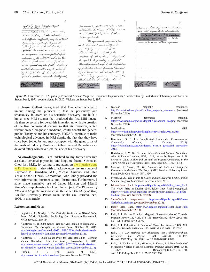
Lauterbur was aware of Damadian’s work, but he did not realize its full impact until he observed essentially the same experiment—the rats’ tumors removed and NMR measurements measured—repeated by Leon A. Saryan that summer. Noting the consistent differences in relaxation times obtained from various normal and malignant tissues, he began to wonder about ways to map these differences in the human body. Later that evening, September 2, 1971, he proposed the first step in accomplishing that objective when he thought of superimposing on the strong, uniform magnetic field passing through the patient a weaker, non-uniform magnetic field. The second step—using a series of gradients together with back projection reconstruction—came more than a year later. Lauterbur added this contribution on October 30, 1972. According to Ros Herman, “He wrote out his proposal at the beginning of a notebook, together with a reference to Damadian’s Science paper on cell differences to show its applications, dated all this, and asked a colleague to countersign it” [64, 65] (Figure 39). Thus Lauterbur had specifically acknowledged that he had been inspired by Damadian’s earlier work.
Considering the idea of body scanning, Lauterbur would later claim that he was unaware that Damadian had plans for anything except test-tube biopsies. However, in the very first paragraph of his 1971 Science article Damadian had indicated that he intended to use NMR as a scanning device: “In principle, nuclear magnetic resonance (NMR) techniques combine many of the desirable features of an external probe for the detection of internal cancer” [45].
In the 1973 Nature letter in which Lauterbur first published his idea, he proposed that “a possible application of considerable interest at this time would be to the in vivo study of malignant tumors, which have been shown to give proton nuclear magnetic resonance signals with much longer spin-lattice relaxation times than those in the corresponding normal tissues” [66]. Rather than citing the obvious, which was Damadian’s 1971 Science article [45], Lauterbur cited a follow-up study to Damadian’s by Weisman et al., which had been published in Science in 1972 [67] and which had referenced Damadian’s work. The omission justifiably irked Damadian.
Damadian’s seminal contribution was publicly acknowledged by Lauterbur in a 1986 reprint of the speech that Lauterbur gave when he accepted the Charles F. Kettering Prize from General Motors for his contributions to cancer detection by zeugmatography [68]:
The attention of the medical community was first attracted by the report of Damadian that some animal tumors have remarkably long proton NMR relaxation times. Efforts to reproduce these results and to explore their significance were soon underway in other laboratories….It was measurements that I observed Saryan carrying out in September of 1971 that caught my attention….Even normal tissues differed markedly among themselves in NMR relaxation times, and I wondered whether there might be some way to noninvasively map out such quantities within the body [69].
Undoubtedly, both Damadian and Lauterbur made major contributions to MRI imaging and scanning. Without Damadian’s relaxation discoveries that showed sharp discrimination between tissues and particularly a serious disease like cancer, there would have been no reason to entertain or even consider a method for displaying the relaxation differences so that they could be visualized as an image. Furthermore, except for the relaxation differences discovered by Damadian, there would be no reason to expect that such an image would show anything, i.e., that any tissue NMR contrast existed with which to make an image.
Science and technology are two distinctly different enterprises. Science is the branch of knowledge dedicated to compiling factual information and understanding natural phenomena. It precedes technology, and technology cannot advance without it. Without science’s new knowledge of natural phenomena, technology’s new methods for exploiting and taking advantage of nature’s secrets cannot be created. The new scientific information is necessarily the first step.
Moreover, there is no doubt that Damadian’s seminal discovery preceded Lauterbur’s developments. Professor Henry Wallman (1913–1992) was a member of the Royal Swedish Academy of Sciences for 25 years, a principal reviewer of the technical publications of candidates on behalf of the Nobel Committees in physics, chemistry, and economics, and a participant in the selection of the Nobel Prizes in these fields. After a comprehensive review of the documentary record of the origins of MRI, Wallman, himself a professor of applied electronics who invented X-ray televsision, for which he was named an honorary doctor of medicine, stated in writing letters recommending Damadian for important awards: “I am of the definite opinion that Dr. Damadian’s contribution was both prior to and more fundamental that Dr. Lauterbur’s” [the underlining is included in the original statement].
Professor Abraham Gelbart (1911–1994), Chairman of the Gelbart Institute for Advanced Mathematics at Bar-Ilan University, Ramat Gan, Israel and collaborator with Albert Einstein at the Institute for Advanced Study at Princeton, New Jersey, independently reached a similar conclusion to that of Professor Wallman’s, his friend of almost 60 years:
The key to the development of MRI was a physician-scientist [Damadian] who could bridge the disciplines of modern physics and medicine and see the connections to make it happen—a rare confluence of circumstances [10, p 676].
88 Chem. Educator, Vol. 19, 2014 George B. Kauffman
© 2014 The Chemical Educator, S1430-4171(14)12540-2, Published 03/21/2014, 10.1333/s00897142540a, 19140073.pdf
Figure 39. Lauterbur, P. C. “Spatially Resolved Nuclear Magnetic Resonance Experiments,” handwritten by Lauterbur in laboratory notebook on September 2, 1971, countersigned by G. D. Vickers on September 3, 1971.
Professor Gelbart recognized that Damadian is clearly unique among the pioneers in that he personally and tenaciously followed up his scientific discovery. He built a human-size MRI scanner that produced the first MRI image. He then personally followed this invention up with the creation of the first commercial scanner so that his invention, which revolutionized diagnostic medicine, could benefit the general public. Today he and his company, FONAR, continue to make technological advances in MRI despite the fact that they have since been joined by and must compete with the giant firms of the medical industry. Professor Gelbart viewed Damadian as a devoted father who never left the side of his discovery.
Acknowledgments. I am indebted to my former research assistant, personal physician, and longtime friend, Steven H. Chooljian, M.D., for calling to my attention the injustice done to Dr. Damadian. I also wish to acknowledge the assistance of Raymond V. Damadian, M.D., Michael Guarino, and Ellen Yeske of the FONAR Corporation, who kindly provided me with information, documents, and illustrations. Furthermore, I have made extensive use of James Mattson and Merrill Simon’s comprehensive book on the subject, The Pioneers of NMR and Magnetic Resonance in Medicine: The Story of MRI; Bar-Ilan University Press: Dean Books Co.: Jericho, NY, 1996, in this article.
References and Notes
1. Lagerkvist, U; Norrby, E. The Periodic Table and a Missed Nobel Prize; World Scientific Publishing Co.: Singapore/Hackensack, NJ/London, 2012; p 114.
2. Kauffman, G. B. 2003 Nobel Prize for MRI denied to Raymond V. Damadian. The Collegian at Fresno State, October 20, 2013. http://collegian.csufresno.edu/2013/10/20/2003-nobel-prize-for-mri-denied-to-raymond-v-damadian (accessed November 2013).
3. Kauffman, G. B. 2003 Nobel Prize for MRI Denied to Raymond Vahan Damadian. Armenian Weekly, November 7, 2013. http://www.armenianweekly.com/2013/11/07/2003-nobel-prize-for-mri-denied-to-raymond-vahan-damadian (accessed November 2013).
4. Hornak, J. P. The Basics of NMR. http://www.cis.rit.edu/htbooks/nmr (accessed November 2013).
5. Nuclear magnetic resonance. http://en.wikipedia.org/wiki/Nuclear_magnetic_resonance (accessed November 2013).
6. Magnetic resonance imaging. http://en.wikipedia.org/wiki/Magnetic_resonance_imaging (accessed November 2013).
7. MedlinePlus. MRI. http://www.nlm.nih.gov/medlineplus/ency/article/003335.htm (accessed November 2013).
8. Kauffman, G. B. It’s Complicated: Unintended Consequences. Community Alliance, 16 (October, 2013). http://fresnoalliance.com/wordpress/?p=8376 (accessed November 2013).
9. Hartshorne, E. Y. The German Universities and National Socialism; Allen & Unwin: London, 1937; p 112; quoted by Beyerchen, A. D. Scientists Under Hitler: Politics and the Physics Community in the Third Reich; Yale University Press: New Haven, CT, 1977; p 43.
10. Mattson, J.; Simon, M. The Pioneers of NMR and Magnetic Resonance in Medicine: The Story of MRI; Bar-Ilan University Press: Dean Books Co.: Jericho, NY, 1996.
11. Myers, M. A. Prize Fight: The Race and the Rivalry to be the First in Science; Palgrave Macmillan: New York, NY, 2012.
12. Isidore Isaac Rabi. http://en.wikipedia.org/wiki/Isidor_Isaac_Rabi; The Nobel Prize in Physics 1944. Isidor Isaac Rabi-Biographical. http://www.nobelprize.org/nobel_prizes/physics/laureates/1944/rabi-bio.html (accessed November 2013).
13. Stern-Gerlach experiment. http://en.wikipedia.org/wiki/Stern-Gerlach_experiment (accessed November 2013).
14. Isidor Isaac Rabi. http://en.wikiquote.org/wiki/Isidor_Isaac_Rabi (accessed November 2013).
15. Rabi, I. I. On the Principal Magnetic Susceptibilities of Crystals. Physical Review 1927, 29, 174–185. Bibcode:1927PhRv...29..174R. doi:10.1103/PhysRev.29.174.
16. Rabi, I. I. Refraction of Beams of Molecules. Nature 1929, 123, 163–164. Bibcode:1929Natur.123..163R. doi:10.1038/123163b0.
17. Rabi, I. I. Zur Methode der Ablenkung von Molekularstrahlen. Zeitschrift für Physik 1929, 54(3-4), 190–197. Bibcode:1929ZPhy...54..190R. doi:10.1007/BF01339837.
18. Rabi, I. I.; Zacharias, J. R.; Millman, S.; Kusch, P. A New Method of Measuring Nuclear Magnetic Moment. Physical Review 1938, 53(4), 318–327. Bibcode:1938PhRv...53..318R. doi:10.1103/PhysRev.53.318. PMID 9981980.

Nobel Prize for MRI Imaging Denied to Raymond V. Damadian a Decade Ago Chem. Educator, Vol. 19, 2014 89
© 2014 The Chemical Educator, S1430-4171(14)12540-2, Published 03/21/2014, 10.1333/s00897142540a, 19140073.pdf
19. Rabi, I. I.; Millman, S.; Kusch, P.; Zacharias, J. R. The Molecular Beam Resonance Method for Measuring Nuclear Magnetic Moments. The Magnetic Moments of 3Li6, 3Li7 and 9F
19. Physical Review Letters 1939, 55, 526–535. Bibcode:1939PhRv...55..526R. doi:10.1103/PhysRev.55.526. http://www.uni-saarland.de/fak7/becher/vorlesungen/WS1011/uebungen/Rabi1939.pdf (accessed November 2013).
20. Rabi, I. I.; Zacharias, J. R.; Millman, S.; Kusch, P. Milestones in Magnetic Resonance: ‘a new method of measuring nuclear magnetic moment’. 1938. J. Magnetic Resonance Imaging March-April 1992, 2(2), 131–133. www.ncbi.nlm.nih.gov/pubmed/1562763 (accessed November 2013).
21. The Nobel Prize in Physics 1944. Isidor Isaac Rabi. http://www.nobelprize.org/nobel_prizes/physics/laureates/1944 (accessed November 2013).
22. The Nobel Prize in Physics 1989. Norman F. Ramsey-Biographical. http://www.nobelprize.org/nobel_prizes/physics/laureates/1989/ramsey-bio.html (accessed November 2013). In Nobel Foundation, Nobel Lectures Including Presentation Speeches and Laureates’ Biographies, Chemistry 1989; Frängsmyr, T., Editor-in-Charge; Malmström, B. G., Editor; World Scientific Publishing Co.: Singapore; River Edge, NJ; London, 1990.
23. Felix Bloch. http://en.wikipedia.org/wiki/Felix_Bloch (accessed November 2013).
24. The Nobel Prize in Physics 1952. E. M. Purcell-Biographical. http://www.nobelprize.org/nobel_prizes/physics/laureates/1952/purcell-bio.html (accessed November 2013).
25. The Nobel Prize in Physics 1981. Nicolaas Bloembergen-Biographical. http://www.nobelprize.org/nobel_prizes/physics/laureates/1981/bloembergen-bio.html (accessed November 2013).
26. Erwin Hahn. http://en.wikipedia.org/wiki/Erwin_Hahn (accessed November 2013).
27. physics@berkeley. Faculty. http://physics.berkeley.edu/index.php?option=com_dept_management&act=people&Itemid=299&task=view&id=239 (accessed November 2013).
28. Oral History Transcript—Erwin L. Hahn. Interview by Joan Bromberg, August 21, 1986. http://www.aip.org/history/ohilist/4652.html (accessed November 2013).
29. The Nobel Prize in Chemistry 1991. Richard R. Ernst-Biographical. http://www.nobelprize.org/nobel_prizes/chemistry/laureates/1991/ernst-bio.html (accessed November 2013).
30. Herman Yaggi Carr, professor, researcher. http://www.app.com/article/B1/20080416/NEWS04/804160351 (accessed December 2013).
31. Lee, V. S. Herman Yaggi Carr, PhD (1924-2008): A tribute. Journal of Magnetic Resonance Imaging June 2009, 29(6), 1243–1247. http://onlinelibrary.wiley.com/doi/10.1002/jmri.21772/full (accessed December 2023).
32. Herman Carr. http://en.wikipedia.org/wiki/Herman_Carr (accessed December 2013).
33. Carr, Herman Yaggi (1952). Free Precession Techniques in Nuclear Magnetic Resonance (PhD thesis). Cambridge, MA: Harvard University. OCLC 76980558.
http://lms01.harvard.edu/F/A33CPQLDNRHPDJQHJK8B45AQ9UIF54PG7RDXD51YNLGPJ1P5IV-01276?func=find-acc&acc_sequence=036694378 (accessed December 2013).
34. Carr, Herman Y. (July 2004). Field Gradients in Early MRI. Physics Today (American Institute of Physics) 57 (7): 83. Bibcode:2004PhT....57g..83C. doi:10.1063/1.1784322.
35. Grant, D. M.; Harris, R. K. Encyclopedia of Nuclear Magnetic Resonance; John Wiley and Sons: Hoboken, NJ; 1996; Vol. 1, p 253.
36. MacWilliams, B. Russian claims first in magnetic imaging. Nature November 27, 2003, 426(6965), 375. Bibcode:2003Natur.426..375M. doi:10.1038/426375a. PMID 14647349.
http://www.nature.com/nature/journal/v426/n6965/full/426375a.html (accessed December 2013).
37. Bateneva,T.; Leskov, S. the Nobel Prize Should Have Gone to Lt. Ivanov. The Great Britain-Russia Society Reviews. http://www.gbrussia.org/reviews.php?id=68 (accessed December 2013).
38. Bateneva, T.; Leskov, S. Izvestia, October 31, 2003. Russian Researcher Vladislav Ivanov Could Have Received the 2003 Nobel Prize in Medicine. http://www.informnauka.ru/eng/2003/2003-10-31-03_396_e.htm (accessed December 2013).
39. Vladislav Ivanov. Privet Nobeliu ot Ivanova (in Russian). http://www.peoples.ru/science/professor/ivanov (accessed December 2013).
40. Raymond Vahan Damadian. http://en.wikipedia.org/wiki/Raymond_Vahan_Damadian (accessed December 2013).
41. Cope, F. W. Nuclear Magnetic Resonance Evidence Using D2O for Structured Water in Muscle and Brain. Biophys. J. March 1969, 9(3), 303–319. http://www.ncbi.nlm.nih.gov/pmc/articles/PMC1367570/pdf/biophysj00689-0036.pdf (accessed January 2014).
42. How the Gerson therapy heals. http://gerson-research.org/docs/HildenbrandGLG-1990-5 (accessed January 2014).
43. The letter was actually published in Nature. Cope, F.; Damadian, R. Cell Potassium by 39K Spin Echo Nuclear Magnetic Resonance. Nature 1970, 228, 76–77. http://www.nature.com/nature/journal/v228/n5266/abs/228076a0.html (accessed January 2014).
44. Damadian, R. V., letter to George S. Mirick, MD, Scientific Director, The Health Research Council of the City of New York, September 17, 1969.
45. Damadian, R. Tumor Detection by Nuclear Magnetic Resonance. Science, March 19, 1971, 171, 1151–1153. http://www.sciencemag.org/content/171/3976/1151.full.pdf (accessed January 2014). According to the Science Citation Index, this article was the most widely cited publication in the field of MRI from 1970 to 1989, the years of the maximum rate of growth of MRI installations. Because of its exceptional citation frequency, it was designated a “Citation Classic” by the publishers of the Science Citation Index in 1987.
46. Damadian, R. V., personal communication with James Mattson, June 12, 1995.
47. Wehrli, F. W. The Origins and Future of Nuclear Magnetic Resonance Imagining. Physics Today June 1992, 34–42. http://scitation.aip.org/content/aip/magazine/physicstoday/article/45/6/10.1063/1.881310 (accessed January 2014).)
48. Wehrli, F. W. The significance of contrast in NMR images. Radiology 1983, 147, 12 (back cover).
49. Damadian, R.; Minkoff, L.; Goldsmith, M.; Stanford, M.; Koutcher, J. Field Focusing Nuclear Magnetic Resonance (FONAR): Visualization of a Tumor in a Live Animal. Science December 24, 1976, 194, 1430–1432. http://www.sciencemag.org/content/194/4272/1430.full.pdf (accessed February 2014).
50. Kleinfield, S. A Machine Called Indomitable; Time Books: New York, 1985. http://www.muebooks.com/a-machine-called-indomitable-PDF-1008051 (accessed January 2014).
51. Shames, C. Interview with Dr. Raymond V. Damadian. Alumni Today, pp 14–16. http://www.downstate.edu/alumni/pdf/damadian.pdf (accessed January 2014).
52. Filler, A. G. The History, Development and Impact of Computed Imaging in Neurological Diagnosis and Neurosurgery: CT, MRI, and DTI. http://precedings.nature.com/documents/3267/version/3/files/npre20093267-3.pdf?origin=publication_detail (accessed November 2013).
53. k-space (MRI). http://en.wikipedia.org/wiki/K-space_(MRI) (accessed January 2014).

90 Chem. Educator, Vol. 19, 2014 George B. Kauffman
© 2014 The Chemical Educator, S1430-4171(14)12540-2, Published 03/21/2014, 10.1333/s00897142540a, 19140073.pdf
54. Fonar History—Maker of the Upright Multi-position MRI. http://fonar.com/history.htm (accessed January 2014).
55. Fonar v. General Electric. http://en.wikipedia.org/wiki/Fonar_v._General_Electric (acessed January 2014).
56. Intellectual Property Owners Association. http://en.wikipedia.org/wiki/Intellectual_Property_Owners_Association (accessed January 2014).
57. Wilson Greatbatch. http://en.wikipedia.org/wiki/Wilson_Greatbatch (accessed January 2014).
58. Adam, J. Innovative Lives. Making Hearts Beat. http://invention.smithsonian.org/centerpieces/ilives/lecture09.html (accessed January 2014).
59. Celebrate Downstate 2011. Raymond V. Damadian, MD. http://www.downstate.edu/celebrate/honorees/damadian.html (accessed January 2014).
60. Edelson, E. Basic research leads to radio signals from cancer signals. Downstate Reporter, Downstate Medical Center, Spring 1971, 2(2), 1, 16.
61. Dr. Raymond Damadian—Originator of the Concept of Whole-Body NMR Scanning (MRI) and Discoverer of the NMR Tissue Relaxation Differences That Made It Possible. http://www.downstate.edu/brooklynhistoryofmedicine/pdf/exhibits/damadian.pdf (accessed January 2014).
62. Leon Saryan, Ph.D. http://www.yatedo.com/p/Leon+Saryan,+Ph.D./normal/cbd1ea3625e69dc262ef3c17cf504dc6 (accessed January 2014).
63. Hollis, D. P.; Saryan, L. A.; Morris, H. P. A nuclear magnetic resonance study of water in two Morris hepatomas. Johns Hopkins Medical Journal December 1972, 131(6), 441–444. http://www.ncbi.nlm.nih.gov/pubmed/?term=Johns+Hopkins+Medical+Journal+1972%2C+131%2C+441 (accessed January 2014).
64. Herman, R. A chemical clue to disease. New Scientist March 15, 1979, 876.
65. Lauterbur, P. C. “Spatially Resolved Nuclear Magnetic Resonance Experiments,” handwritten by Lauterbur in a laboratory notebook on September 2, 1971, countersigned by G. D. Vickers on September 3, 1971.
66. Lauterbur, P. C. Image Formation by Induced Local Interactions: Examples Employing Nuclear Magnetic Resonance. Nature March 16, 1973, 242, 190–191. http://www.nature.com/nature/journal/v242/n5394/pdf/242190a0.pdf (accessed January 2014).
67. Weisman, I.; Bennett, L. H.; Maxwell, Sr., L. R.; Woods, M. W.; Burk, D. Recognition of Cancer in vivo by Nuclear Magnetic Resonance. Science December 22, 1972, 178, 1288–1290. http://www.sciencemag.org/content/178/4067/1288.full.pdf (accessed 2014).
68. Definition: ‘Zeugmatrography’. Term coined by Lauterbur in 1972 for the joining of a magnetic field and spatially defined radiofrequency field gradients to generate a two-dimensional display of proton density and relaxation times in tissues. http://www.medilexicon.com/medicaldictionary.php?t=100372 (accessed January 2014).
69. Lauterbur, P. C. Cancer Detection by Nuclear Magnetic Resonance Zeugmatographic Imaging. Accomplishments in Cancer Research, 1985 Prize Year, General Motors Cancer Research Foundation; J. B. Lippincott Co.: Philadelphia, PA, 1986; also published in Cancer May 1986, 57, 1899–1904. http://onlinelibrary.wiley.com/store/10.1002/1097-0142(19860515)57:10%3C1899::AID-CNCR2820571002%3E3.0.CO;2-4/asset/2820571002_ftp.pdf?v=1&t=hqlxfj3q&s=918b01a2814e65544b2ef6c36ed236db9f66cb7b (accessed January 2014).