non accidental trauma pediatric critical care medicine emory university children’s healthcare of...
TRANSCRIPT

NON ACCIDENTAL TRAUMA
Pediatric Critical Care MedicineEmory University
Children’s Healthcare of Atlanta
2
Introduction• >40% Of Death in children <12mos• #1 cause of death is head injury• 30% of head injury may be misdiagnosed
• 4 of 5 deaths cause by head injury can be prevented if early diagnosis during prior medical evaluation

3
Epidemiology• Most often < 1 yr of age• Battering is the most common mechanism of
injury in children 3-5 mos• Incidence of inflicted TBI is similar in US & Europe
Epidemiology• 60% of cases with previous history or clinical
evidence of maltreatment• 22% with involvement of child welfare agencies• 32% with misdiagnosis
- Viral gastroenteritis or influenza- “R/O sepsis”- Accidental head injury

Epidemiology• Perpetrators
»50% fathers»20% step-fathers or male partners»12% mothers»17% female baby sitters

Epidemiology• Risk factors
– Young/single parents (risk increases more with presence of step-father or maternal boyfriend)
– Lower education– Unstable family situation– Stress to family- financial, food & housing, domestic
violence, alcohol drug abuse, parental depression– Other: peri-natal illness, family disruption & separation,
colicky babies

Mechanism of Injury• Degree of injury in the absence of significant
trauma or sign of external injury• Rotational & impact forces• Translational deceleration• Repetitive events – more damage• Developmental weakness: large head, weak &
unstable neck; soft brain with higher water contents and poorly demyelinated
8
Mechanism of Injury• Rotational & Impact forces
- Angular deceleration (head rotates on its own axis) causing SDH & axonal injury- > with shaking and impact than shaking alone

Mechanism of Injury
9

10
Mechanism of Injury• Translational deceleration (drop or short fall)
– Head moves in a straight line– Cranial impact– Focal injury

11
Mechanism of Injury• Significant of cerebral injury is caused by
secondary hypoxic ischemic events– Central apnea from injury to the brain stem or cervical
spinal cord– Prolonged seizures– Aspiration

12
Cranial Injury• Blunt force trauma• Shaking• Combination of forces
13
ShakingClassical pattern- Diffuse unilateral or bilateral SDH- Diffuse multilayered retinal hemorrhage- Diffuse brain injury
In the absence of- A history of trauma- Paucity of external manifestation of injury

14
Intracranial Hemorrhage• Sub-arachnoid • Sub-dural• Intraparenchymal• Epidural
15
ICH• Short vertical fall <4ft
– 85 % with no evidence or minor injury– 7% with skull fracture – all with isolated and linear skull
fracture
16
ICH – Sub-dural hemorrhage• Rare in accidental trauma unless with severe
forces (MVA or significant height)• Small and localized to the site of the impact• Interhemispheric SDH usually posterior
- 71% of abused children
- 19% in accidental injury

17
ICH – Sub-dural hemorrhage• Mixed density collections of fluid are more
common and can present both acute or acute on chronic
• Clinical silent SDH– Term infant/neonate with minor birth trauma– Self resolved or increase in size – few days to weeks
18
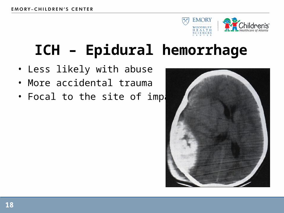
ICH – Epidural hemorrhage• Less likely with abuse• More accidental trauma• Focal to the site of impact
19
ICH – Subarachnoid hemorrhage
• Hard to detect• Not good correlation with abuse• Detected mostly at autopsy

20
ICH – Parenchymal Injury• Contact forces• Inertia forces with rotational deceleration
– Traumatic Axonal injury– Sub-cortical white matter, corpus collosum,
periventricular regions, dorsolateral aspect of the rostral brainstem
• Global Hypoxic Ischemic injury- May cause primary brainstem damage- Prolonged seizure- Secondary hypotension

21
ICH – Parenchymal Injury• Infarct, atrophy• Encephalomalacia with ventriculomegaly

22
Associated Injury – Retinal hemorrhage
• Numerous• Multi-layered• Extend beyond the posterior pole to the
peripheral retina
23
Associated Injury• Bone fractures• Blunt trauma to abdomen and pelvis

24
Skull Fractures• Most common parietal• Both accidental & non-accidental
• Common sites in abuse– Crossing suture lines– Multiple– Diastatic– Growing– Depressed – Complex– Bilateral

Skull Fractures
25

Skeletal Fractures• 20-50% of abused
children associated with extracranial skeletal fracture
• Ribs, long bone and metaphyseal
• Classic metaphyseal avulsion lesion of long bone caused by torsion and traction when extremities in twisted or pulled
26

27
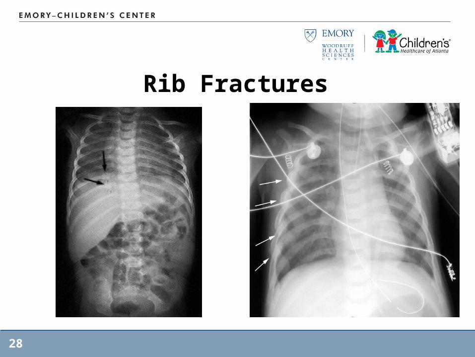
Rib Fractures• Most common posterior and lateral• 82% associated with abuse• 8% accidental• 8% bone fragility• 2% birth trauma
** Chest compression more commonly causes lateral and anterior rib fractures
Rib Fractures
28
29
Associated Injuries: Blunt Trauma
• Thoracic– Esophageal injury: can result from forced F.B. ingestion,
forced caustic ingestion, blunt external trauma, and penetrating trauma
– Sx: non specific, pain to the neck and shoulder, shortness of breath, dysphagia, abdominal pain
– Early signs: tachycardia, dyspnea, abdominal guarding, pneumothorax, mediastinal air, subcutaneous emphysema

30
Associated Injuries• Pulmonary Injury
– Pulmonary laceration, contusion or diffuse alveolar damage
• Chylothorax– Cause by rupture of thoracic duct from blunt trauma or
anteroposterior acceleration/deceleration forces– Signs; respiratory distress, nutritional deficiency,
electrolytes abnormality, immunosuppression from T-cell depletion
31
Associated Injuries• Cardiac Injury
– Dysrhythmias: commotiao cordis or cardiac concussion causes sudden cardiac arrest (blow at upstroke of the T wave associated with v-fib, blow at the peak of QRS results in asystole
– Direct trauma: impact of the heart against the sternum or crushing of the heart due to blunt trauma to the anterior chest
– Others: traumatic VSD, cardiac aneurysm, laceration or rupture
32
Associated Injuries• Abdominal Injury
– 1% of abused children suffered intra-abdominal injury with 50% mortality
– Sx: tenderness, distension, enlargement of the liver or spleen, and/or bruising of the abdominal wall
– Liver injury: most common organ injured; cause contusion, subcapsular hematoma, laceration and rupture
– Splenic injury: less common than liver– Pancreatic injury– GI tract
» Perforation more common in NAT» Hematoma: intramural hematomas occur most frequently in the
duodenum and can cause perforation or stricture

33
Associated Injuries• Urinary Tract Injury
– Renal injury: contusion or subcapsular hematoma, shattered kidney or vasculaar pedicle avulsion
– Hematuria is present in 41-68% of victims with renal trauma
– Ureteral injury– Bladder injury: bladder rupture (blunt force to a full
bladder). Rupture occurs at the dome of the bladder, fluid and blood extravasate into the peritoneum
34
Evaluation• History• Physical Examination• Laboratory studies• Radiographic studies

35
Evaluation: History• Who, what, when and where• Document your history• Document inconsistency of the story through
details• Help your memory at a later time (across a DA
and a defense lawyer)

36
Evaluation: History• Who was present?• Who had been taking care of the patient at least
4 hours prior to the event• When did the last time the child seem normal?
When was the event• Review the event after the child last seen to be
normal

37
Evaluation: History• Where did the event occur? Who was there with
the baby?• What would care provider consider normalcy in
the patient? (behavior, development)

38
Evaluation: History• Don’t forget details of family history
– Bleeding tendency in family– Bleeding at time of circumcision for boys– Easy bruising

39
Evaluation: Laboratory• CBC with Platelet• Coagulation study: DIC panel• Electrolytes, liver function test, and urinalysis
• * preliminary evidence of CSF and serum measuremenf of biomarkesr of brain injury – neuron-specifiec enolase, S100B(a calcium binding protein found in astrocytes), and myelin basic protein

40
Evaluation: Imaging• CT – brain and bone window is best as an initial
tool. • MRI – superior to CT for documenting the pattern,
extent, and timing • Skeletal survey
41
Evaluation: Opthalmologic Exam
• Need to have an opthalmologic exam to stand legally
42
Differential Diagnosis• Accidental injury• Birth trauma• Apparent Life-threatening event• Bleeding disorder• Others
43
Differential Diagnosis – Accidental Injury
• A history of traumatic event• Retinal hemorrhages are typically fewer in
number and less extensive• Subdural hematomas
44
Differential Diagnosis – Birth Trauma
• Commonly associated with instrumented deliveries
• Both retinal hemorrhage and subdural hemorrhage
45
Differential Diagnosis – Bleeding Disorder
• ICH can occur in severe bleeding disorer (hemophilia) spontaneously or following an injury
• Retinal hemorrhages are small in number and are typically confined to the posterior pole
• Boys with hemophilia, ICU occurs most often in the neonatal period
• ICH is uncommon in idiopathic thrombocytopenic purpura