non-episode-dependent assessment of paroxysmal atrial fibrillation through measurement of rr...
TRANSCRIPT
P1: IZO
Annals of Biomedical Engineering [AMBE] PP1181-ambe-485211 March 27, 2004 12:53 Style file version 14 Oct, 2003
Annals of Biomedical Engineering, Vol. 32, No. 5, May 2004 (©2004) pp. 677–687
Non-Episode-Dependent Assessment of Paroxysmal AtrialFibrillation through Measurement of RR Interval Dynamics
and Atrial Premature Contractions
BRIAN HICKEY, CONORHENEGHAN, and PHILIP DE CHAZAL
University College Dublin, Dublin, Ireland
(Received 11 July 2003; accepted 16 December 2003)
Abstract—A method is presented for classifying a single leadsurface electrocardiogram recording from a Holter monitor as be-ing from a subject with paroxysmal atrial fibrillation (PAF) or not.The technique is based on first assessing the likelihood of 30-minsegments of electrocardiogram (ECG) being from a subject withPAF, and then combining these per-segment likelihoods to forma per-subject classification. The per-segment assessment is basedon the output of a supervised linear discriminant classifier (LDC)which has been trained using known data from the Physionet AtrialFibrillation Prediction Database (which consists of two hundred30-min segments of Holter ECG, taken from 53 subjects with PAF,and 47 without). One of two LDCs is used depending on whetherthere is a significant correlation between observed low-frequencyand high-frequency spectral power in the RR power spectral den-sity over the 30-min segment. If there is high correlation, then theLDC uses spectral features calculated over a 10-min window; inthe low-correlation case, both spectral features and atrial prema-ture contractions are used as features. The classifier was tested forits ability to distinguish PAF and non-PAF segments using threeindependent data sets (representing a total of 1370 segments from50 subjects). The cumulative sensitivity, specificity, and accuracyon a per-segment basis were 43.0, 99.3, and 80.5%, respectively onthese independent test sets. By combining the results of segmentclassification, a per-subject classification into PAF and non-PAFclasses was performed. For the 50 subjects in the independent datasets, the sensitivity and specificity of the per-subject classifier were100%.
Keywords—Electrocardiogram, Atrial fibrillation, Classification,Linear discriminant classifier, Atrial premature contractions.
INTRODUCTION
Atrial Fibrillation (AF) is the most common abnormalheart rhythm encountered in clinical practice, and has seri-ous associated morbidity and mortality.3,12,27 AF is a sig-nificant risk factor for stroke; about 15% of strokes occurin people with AF. The prevalence of AF is 0.5% for thegroup aged 50–59 years, and rises to approximately 10% in
Address correspondence to Conor Heneghan, University CollegeDublin, Dublin, Ireland. Electronic mail: [email protected]
the group aged 80–89 years.8 AF can be classified as inter-mittent, persistent, or permanent; intermittent AF is also re-ferred to as Paroxysmal Atrial Fibrillation (PAF). Episodesof PAF can last from seconds to hours or even days, but willspontaneously revert to normal sinus rhythm. Persistent AFlasts for more than 7 days, but responds to cardioversion,and permanent AF does not respond to cardioversion.
In contrast to normal sinus rhythm, in which the sino-atrial node initiates a well-organized wave of electrical ac-tivation spreading throughout the atria, AF occurs when anumber of electrical activation waveforms travel through theatria, leading to uncoordinated contraction of atrial tissue.This electrical activity is seen at the atrio-ventricular (AV)node, and can be conducted through to the ventricles (if thefrequency of electrical activity is not too high), which leadsto an elevated (and irregular) heart rate (90–160 bpm). Inthe surface electrocardiogram, this uncoordinated atrial ac-tivity is reflected as a randomly variable baseline measure-ment in place of a well-defined P-wave, though in generalthe subsequent ventricular electrical activity is unaffectedin morphology. The progression from normal sinus rhythmto atrial fibrillation is not completely understood, thoughthis transition is often associated with either changes in au-tonomic tone, or the presence of very early Premature AtrialContractions (PAC), or atrial tachycardia.
At present, atrial fibrillation is definitively diagnosed bydetecting episodes of atrial fibrillation recorded using sur-face ECG techniques (using either 12-lead resting, Holter,or cardiac event recorders). In a 12-lead recording, the car-diologist can visually scan the entire record for AF since theduration of the recording is typically short. In Holter sys-tems, automated software will usually highlight episodesof supraventricular tachycardia, which an expert can thenexamine for evidence of AF. In cardiac event recorders,recording of suspect episodes is triggered by the patientwhen he or she has symptoms such as palpitations or dizzi-ness; however many episodes of AF are asymptomatic andnot captured by this approach. A recent innovation in cardiacevent recorders has been the introduction of auto-triggered
677
0090-6964/04/0500-0677/1C© 2004 Biomedical Engineering Society

P1: IZO
Annals of Biomedical Engineering [AMBE] PP1181-ambe-485211 March 27, 2004 12:53 Style file version 14 Oct, 2003
678 HICKEY et al.
recording in the presence of AF episodes as estimated by RRinterval dynamics (“King of Hearts,” Instromedix). Morecomplex algorithms for detection of episodes of AF arebased on extracting the atrial component from the ECG,by implementing some form of QRST cancellation. Cur-rent methods to accurately cancel the QRST componentinclude average beat subtraction (ABS)21; spatiotemporalcancellation22; and blind source separation (BSS).17 Fol-lowing removal of this large ventricular component, the re-maining atrial signal can be analyzed for the presence of AF,typically using spectral analysis. The main atrial componentduring AF occurs between 4 and 9 Hz.28
The systems described above are focused on detectionof AF episodes. From an electrical engineering point ofview, this is essentially a signal-to-noise ratio limited prob-lem, since the energy of the atrial fibrillation component isconsiderably less than that of the normal ventricular com-ponents. The performance of episode-detection algorithmscan be reasonably assessed by their sensitivity and posi-tive predictive value, where sensitivity (Se) is defined asthe percentage of AF episodes correctly detected, and posi-tive predictive value (PPV) is the percentage of AF-labeledepisodes which are in fact true AF episodes. A perfectepisode detector would have Se and PPV of 100%.
Alternatively, the performance of an AF diagnostic sys-tem could be measured on a per-subject basis. Sensitivitywill then be defined as the percentage of subjects with AFwho are correctly classified as having AF, and specificitywill be the percentage of non-AF subjects who are correctlyclassified. An AF episode-detection scheme can be incor-porated into such a system. In such a case, any subject inwhom an episode of AF is detected will be classified as anAF-subject; otherwise they will be classified as non-AF. In-formation in the ECG during non-AF periods is not used insuch a system. The performance of such a per-subject AFdiagnostic system will depend on two factors: (1) the relia-bility of the AF episode detection scheme and (b) whetheran episode actually occurs during the recording.
The focus of this paper is to provide an alternativeparadigm for per-subject classification of whether a per-son has paroxysmal AF. Unlike an episode-detection-basedsystem, our system does not depend on an episode of AFoccurring during the recording period, since it analyzes theentire ECG record for diagnostic information. As a designcriterion, our system should also function if an episode ofAF does actually occur, or indeed if the person has persistentor permanent AF. The overall performance of our systemshould be considered primarily in terms of its overall sen-sitivity and specificity in distinguishing AF from non-AFsubjects. It is primarily intended for providing a diagnosticassessment in subjects with paroxysmal AF who displayeither no episodes of AF, or a small number of episodesduring the recording period.
The motivation for such a system stems from the fact thata definitive diagnosis of paroxysmal atrial fibrillation from
surface ECG recordings can be challenging, since parox-ysmal atrial fibrillation is by definition intermittent, andepisodes can be hard to detect because of the low SNRof each episode. It is not uncommon for 24 or 48 h Holterrecording to yield a negative result, without definitively rul-ing out the presence of paroxysmal atrial fibrillation.20 Car-diac event recorders provide a partial solution to this prob-lem. Various reports have demonstrated that the diagnosticyield of event monitors is significantly higher than Holtermonitors in establishing the presence of paroxysmal atrialfibrillation due to their longer observation window.13,16,20
However, they still rely on the capture of actual episodes ofAF. Event recorders are also dependent on patient compli-ance, and are not suited to all clinical populations.29
Our system is motivated by recent observations that sub-jects with atrial fibrillation have dynamics present in theirECG which differ from non-AF subjects, even when AFis not actually occurring. In the last number of years, sev-eral studies have been conducted to monitor the ECG ofsubjects with PAF, in an effort to identify patterns associ-ated with atrial fibrillation.1,4–6,9–11,14,15,25,26 Two dominanttypes of analysis have been carried out: heart rate variabil-ity (HRV) analysis of the cardiac rhythm preceding the AFepisode1,4,6,9–11,25 and the role of atrial ectopic beats in ini-tiating the onset of AF.5,14,15,26 These studies are consis-tent with physiological mechanisms associated with PAF,namely (a) autonomic mediated AF in which excess adren-ergic (sympathetic) or vagal (parasympathetic) autonomictone leads to AF and (b) ectopic mediated mechanisms inwhich abnormal tissue in the atria leads to AF.
Our proposed system combines elements of these worksin order to provide a per-subject assessment for the presenceof AF. Earlier versions of this work have been presentedat the Computers in Cardiology Conferences in 2001 and20026,9 and the original motivation for this work was theComputers in Cardiology Challenge in 2001.18
METHODS
Study Databases
For the purpose of developing and testing our classifica-tion system, we have used a variety of databases:
1. The Physionet Atrial Fibrillation Prediction Database(AFPDB). This database consists of two hundred 30-min records, extracted from two-channel ECGs (leads Iand II), sampled at 128 Hz, and 12-bit resolution. Thedatabase contains records from 53 PAF subjects, and 47subjects who do not suffer from PAF. These 47 subjectsinclude healthy controls, subjects referred for long-termambulatory ECG monitoring, and subjects in intensivecare units. Each subject contributes two 30-min record-ings to the database. In the case of the PAF subjects,one recording was taken at a point distant (>45 min)from any episode of AF, and the second recording was

P1: IZO
Annals of Biomedical Engineering [AMBE] PP1181-ambe-485211 March 27, 2004 12:53 Style file version 14 Oct, 2003
Non-Episode-Dependent Assessment of PAF 679
taken over the 30 min immediately preceding an episodeof AF. For the non-PAF subjects, the 30-min segmentswere chosen randomly. This database is publicly avail-able at http://www.physionet.org.
2. The Physionet Normal Sinus Rhythm Database(NSRDB). This database consists of 18 records, eachapproximately 24 h long, recorded using lead II, with asampling frequency of 128 Hz. These subjects have nosignificant arrhythmias, and include 5 men, aged 26–45,and 13 women aged 20–50. This database is publiclyavailable at http://www.physionet.org.
3. The UCD Overnight Database (UCDODB). Thisdatabase was recorded using a two-channel Holter moni-tor (Lifecard CF, Reynolds Medical Ltd., Hertford, UK),with subsequent analysis of a single channel from amodified V5 lead position. The signals are sampled at128 Hz, with 12-bit resolution. The database consistsof nine overnight records of approximately 8-h durationfrom subjects with no known cardiac pathology, agedbetween 23 and 40, comprising of three women and sixmen. Informed consent was given for these recordingsas required by institutional guidelines. This database canbe supplied on CD by the authors to interested readers.
4. The MIT-BIH AF Database (AFDB). This database con-sists of 23 two lead (lead V1, II) ECG recordings ofhuman subjects with atrial fibrillation (mostly paroxys-mal). The individual recordings are each approximately10 h in duration, and contain two ECG signals each sam-pled at 250 Hz with 12-bit resolution. The number ofepisodes of AF for each subject varies from 2 to 39 withtwo subjects in persistent AF. This database is publiclyavailable at http://www.physionet.org.
The parameters of these databases are summarized inTable 1.
Preprocessing of the Electrocardiogram
Since our algorithm is based primarily on RR intervals,the first stage of processing is to perform reliable extrac-tion of QRS peaks. The raw ECG signals were processedusing a linear phase bandpass FIR filter windowed using aKaiser–Bessel window to reduce baseline wander and EMGartefact. The band stop frequencies were set at 0.25 and20 Hz. Following this, QRS detection was carried out us-ing a Hilbert Transform-based method.2 This QRS detec-tor was tested against the MIT-BIH Arrhythmia database
(http://www.physionet.org), and achieved a sensitivity of98.5%, and a positive predictive accuracy of 98.4%. Fromthis set of R peaks, the RR intervals were calculated. Plots ofthe RR intervals acquired in this manner showed that somerecords had physiologically unreasonable RR intervals (i.e.,RR intervals of excessively long or short duration). Thesespurious RR intervals were generated by dropped QRS de-tections (primarily due to temporary lead dropout) or incor-rect detections of P-waves, T-waves, or noise waveformsas QRS detections. To avoid the introduction of errors dueto these misdetections, a simple correction algorithm wasapplied to the RR series. A new R-wave was inserted if anRR interval of greater than or equal to twice the averageRR interval (over the record) was found. The number ofinserted R-waves was directly proportional to the length ofthe RR interval, i.e., two R-waves are inserted if an RRinterval three times the mean is found. If for example an in-terval equal to 2.5RR (whereRR is the mean RR interval)is found, only one beat will be inserted, resulting in two newintervals of lengthRR and 1.5RR. The new R-waves willbe inserted starting from the previous R-waves. For exam-ple, if the uncorrected R-wave detection times were [0.1,1.2, 3.4, 4.1, 4.9, 8.2,. . . ] with a mean record RR intervalof 0.9 s, then the corrected R-wave occurrence times wouldbe [0.1, 1.2, 2.1, 3.4, 4.1, 4.9, 5.8, 6.7, 8.2,. . . ]. More elabo-rate RR correction algorithms could be envisaged; howeverexperimentation showed that the impact of the exact schemefor RR correction was negligible.
Classification Philosophy
The overall goal of our research was to produce a sys-tem, which provides a clinical assessment of whether ornot a person was suffering from PAF given a reasonablylong segment of surface ECG (e.g.,>8 h). The outcome ofthis classification system is called a per-subject decision.This assessment is based on a collection of decisions madeon shorter duration segments of ECG (e.g., 15- or 30-minsegments). The outcome of a decision on an individual seg-ment is called a per-segment decision. It is important tobear in mind that the overall goal of our study is classifierperformance on a per-subject basis.
Since a variety of data sources were available to us,with excellent clinical annotation, we used a supervisedclassifier-training scheme. We made the following hypoth-esis. Thirty-minute segments immediately preceding an
TABLE 1. Data acquisition and demographic parameters of databases used in this study.
Database Leads No. of subjects (M/F) Age (range) Bit depth Sampling frequency (Hz)
AFPDB V1,II 100 (unknown) Not supplied 12 128NSRDB II 18 (5/13) 20—50 12 128UCDODB Modified V5 9 (6/3) 22—40 12 128AFDB V1,II 23 (unknown) Not supplied 12 250

P1: IZO
Annals of Biomedical Engineering [AMBE] PP1181-ambe-485211 March 27, 2004 12:53 Style file version 14 Oct, 2003
680 HICKEY et al.
episode of AF in subjects known to have PAF would def-initely contain ECG dynamics strongly associated withepisodes of AF. Conversely, 30-min segments from non-PAF subjects would contain characteristics not associatedwith PAF. We included non-PAF subjects who did demon-strate other cardiac arrhythmias or abnormalities. Accord-ingly, we decided to use the AFPDB for development ofthe classifier. An interesting question is whether to use the30-min segments from PAF subjects which were distantfrom the AF episode. We decided not to use these segmentsin training our classifier, as we believed that this would leadto a more complex three-way classification problem (non-PAF, pre-PAF, and distant-PAF). A further justification forour choice of only using “pre-PAF” segments in training isthat intuitively such segments are the most likely to containdefinite examples of PAF-related cardiac dynamics. Thisis analogous to training a system for disease classificationbased on using training data which only contains “severe”cases of the disease. Including ambiguous or intermediatecases in training is equivalent to including redundant ormeaningless features, and should have no significant im-pact on the final trained classifier. A much more impor-tant point to note is that the final system istestedon both“pre-PAF” and “distant-PAF” segments, and it is the perfor-mance on this data that is of most significance. Accordingly,the classifier was trained using fifty-three 30-min segments(henceforth simply referred to as PAF-classified), which im-mediately preceded a 5-min episode of AF (referred to asthe Group 1 records), against ninety-four 30-min segmentsfrom non-PAF subjects (referred to as the Group 2 records),and henceforth simply referred to as non-PAF-classified.Given the use of the AFPDB, to develop the classifier, wethen used the other three databases to provide independentmeasures of the classifier performance, as well as obtainingan estimate of the performance of the classifier on the train-ing data from the AFPDB. In assessing the performance ofthe classifier on a per-segment basis, we decided that we re-quire “distant-PAF” segments (i.e., 30-min segments fromPAF subjects distant from AF episodes) to be classified as“PAF.” In other words, we expect distant-PAF segments tocontain AF-related dynamics, but not in as pronounced afashion as the pre-PAF segments.
Identification of ECG Features Potentially Linkedwith Atrial Fibrillation
As discussed earlier, a variety of features visible in thesurface electrocardiogram have been associated with atrialfibrillation, and its onset. Our system is based on a combi-nation of features extracted from HRV analysis and atrialectopic beat detection.
Classification Stage 1: Assessment of Autonomic Tone
Since changes in autonomic tone have been implicatedin the initiation of AF, the first stage of our classifier pro-
vides a measure of such changes. In this first stage, wecalculate the correlation between the high frequency (HF)and low frequency (LF) spectral content of the RR inter-val sequence at 2-min intervals for the 30 min prior to theonset of AF. These components were calculated as follows.The HF and LF components were derived from the interval-based power spectral density estimates of nonoverlapping2-min RR interval sequences.24 The RR interval sequencecontains both normal and ectopic beats, i.e., we did not at-tempt to restrict our analysis to NN intervals. To obtain azero-mean sequence the mean RR interval was subtractedfrom each segment. The sequence was zero padded to thenearest integer multiple ofN = 98 exceeding the length ofthe sequence, and the Fast Fourier Transform (FFT) wastaken of the entire sequence. The original choice of 98 wasbased on a desire to have individual frequency bins in theinterval spectra be 0.01 Hz in width; the exact choice ofNis unimportant however, provided it is greater than 16. Theabsolute values of the FFT coefficients were then squaredto yield a periodogram estimate of the power spectral den-sity. Adjacent frequency bins were then combined to resultin a 98-point PSD estimate (of which only bins 0–49 arerelevant since 50–97 provide identical information as 1–48). The magnitudes of these PSD bins from 0.04 to 0.15cycles/interval were added to yield the LF component, andsimilarly the bins from 0.15 to 0.4 cycles/interval yieldedthe HF component (this is in line with the conventionalfrequency ranges used in defining LF and HF components).
Overall, for each 30-min segment, this process yieldstwo sets of 15 values, and the correlation coefficient be-tween these sets of numbers is taken, where the correlationcoefficient is calculated as follows:
cxy =115
∑15i=1 (xi − x)(yi − y)
σxσy(1)
wherexi andyi are the LF and HF powers, respectively;xandy are their mean values; andσx andσy are the standarddeviations. From this analysis, two subgroups of records canbe developed: those records with a correlation coefficientabove 0.75 (Group A) and those records with a correlationcoefficient below 0.75 (Group B). A physiological inter-pretation is that the subjects in Group A experience paral-lel changes in sympathetic and parasympathetic activation,whereas those in Group B experience some decoupling.This decoupling could be due to something as simple as apostural change, or may reflect an origin of unknown cause.
Classification Stage 2: Further Classification Using HeartRate Variability or Atrial Premature Contractions
As a result of Stage 1, we now have two subgroups toclassify, Groups A and B, both of which contain PAF andnon-PAF segments. To separate the PAF segments from thenon-PAF segments in each group, further analysis based

P1: IZO
Annals of Biomedical Engineering [AMBE] PP1181-ambe-485211 March 27, 2004 12:53 Style file version 14 Oct, 2003
Non-Episode-Dependent Assessment of PAF 681
on heart rate variability and atrial premature contractioncounting is carried out.
Group A: To classify the members of Group A, the PSDof the RR interval sequence was once again taken andcalculated as above. However, in this case only the RRsequence corresponding to the final 10 min prior to theonset of AF was used. The sequence was zero padded tothe nearest integer multiple of 64 exceeding the lengthof the sequence, and the Fast Fourier Transform (FFT)was taken of the entire sequence. The absolute valuesof the FFT coefficients were then squared to yield a pe-riodogram estimate of the power spectral density. Ad-jacent frequency bins were then combined to result ina 64-point PSD estimate (of which only bins 0–32 arerelevant since 33–63 provide identical information as 1–31). The logarithmic magnitudes of these PSD bins wereused as features in a linear discriminant classifier.
Group B: To classify the members of Group B, the sameanalysis as above was carried out, but this time over thefinal 20 min before AF onset. In addition, the number ofatrial premature contractions over this 20-min segmentwas calculated as described below.
Atrial Premature Contraction Detector
An ectopic beat occurs when a focus from somewhereother than the sinoatrial node stimulates contraction of themyocardium. When this focus is located within the ven-tricles, the electrocardiogram shows a beat of abnormalQRS morphology. This is known as a Ventricular Prema-ture Complex (VPC or PVC), Ventricular Ectopic (VE), ora Ventricular Extrasystole (VE). In the case where the focusis in the atria, atrial ectopic beats occur with normal ventric-ular activation, so that a beat with normal QRS morphologyis observed on the ECG. This type of beat is most com-monly referred to as an atrial premature contraction (APC),or an atrial ectopic beat. (Note that an APC is a specialcase of a supraventricular beat, which refers to any beat inwhich the focus occurs above the ventricles, and which alsoincludes so-called junctional beats, which are initiated inthe atrioventricular node.) In the case of all ectopic beats,the beat occurs earlier than a regular sinus beat. The degreeof this earliness is quite variable. Considerable expertise isrequired to distinguish between different types of ectopicbeats, and to determine the underlying physiology of theobserved surface ECG. Fortunately, it appears that a simpledetector of APCs (with relatively low accuracy) is useful inour assessment of atrial fibrillation.
The algorithm for atrial premature contraction detectionhas two stages. Firstly, a beat is flagged as a suspected APCif the RR interval preceding a QRS complex falls 15% shortof a local moving average of the RR intervals:
RR[n] = 1
10
n−1∑i=n−10
RR[i ] (2)
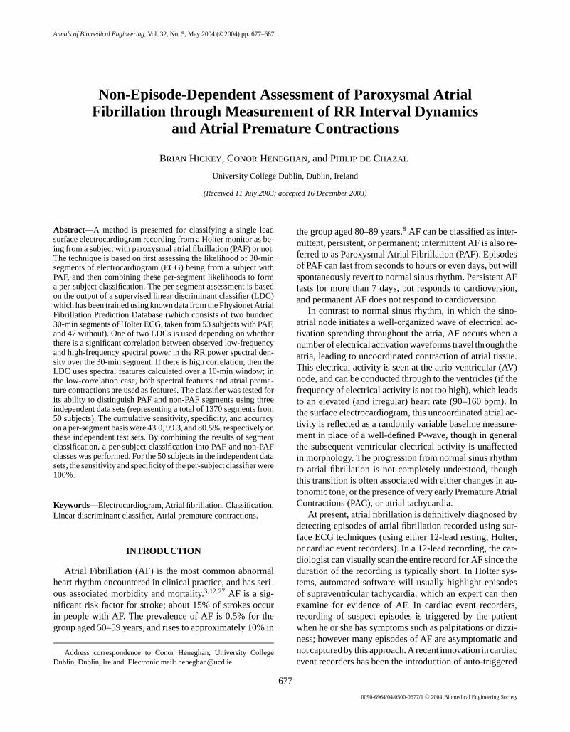
FIGURE 1. Definition of QRS parameters used in distinguish-ing atrial premature contractions from ectopic beats of ven-tricular origin.
The second stage of the detector calculates the width, am-plitude, and area of the QRS complex. If all three QRSparameters vary from those of a normal sinus beat by lessthan or equal to 10%, the beat is confirmed as an APC.The parameters of a normal sinus beat are determined byfinding the average parameters of the first 100 beats inthe segment that show a regular sinus rhythm. The reason-ing behind this is that a PVC will exhibit a QRS complexwhich differs quite significantly from that of a normal beat.Figure 1 shows the definitions of the QRS parameters. TheQRS amplitude is measured as the vertical distance betweenthe Q-wave and the R-wave. The QRS width is measuredas the horizontal distance between the Q-wave and the cor-responding value on the other side of the QRS complex,called Q2. The Q onset point is found by searching back-wards from the R peak until a local minimum is found; con-versely the Q2 point is found by a forward search from the Rpeak to the first local minimum. The QRS area is calculatedusing the trapezoidal method of area approximation, andis the area contained between Q, R, and Q2. All of theseparameters are calculated for each beat using the filteredECG signal. These parameters are used to characterize abeat into different classes, such as ventricular extrasystoles,atrial premature contractions, and normal sinus beats. ThisAPC detector was tested against the MIT SupraventricularArrhythmia Database. The algorithm achieved a sensitivityof 59% and a positive predictive value of 73% in detec-tion of APCs, which compares favourably with the resultsreported by Zonget al.(56 and 47, respectively).30 The out-put of this stage is the estimated number of APCs occurringin a 20-min segment.
Classification Techniques
A linear discriminant classifier (LDC), based on Fisher’srule, was used.19 For each 30-min segment of data fed in,there are two possible output classes—PAF or non-PAF.The classifier outputs a set of numbers representing the

P1: IZO
Annals of Biomedical Engineering [AMBE] PP1181-ambe-485211 March 27, 2004 12:53 Style file version 14 Oct, 2003
682 HICKEY et al.
probability estimate of each class, in response to a set of in-put features. Linear discriminants partition the feature spaceinto different classes using a set of hyperplanes. Optimiza-tion of the model is achieved through direct calculation andis extremely fast relative to other models such as neural net-works. The training of a LDC proceeds as follows. Letx bead × 1 column vector containing feature values calculatedfrom a data set (e.g., the mean RR interval, the standarddeviation of the RR intervals, etc.). We wish to assignx toone ofc possible classes (c = 2 in our case). A total ofNfeature vectors are available for training the classifier, withthe number of feature vectors representing classk equal toNk, i.e.,
N =∑
k
Nk (3)
Thenth training vector in classk is denoted asxk,n. Theclass-conditional mean vectorsµk are defined as
µk =1
Nk
Nk∑n=1
xk,n (4)
We now define a common covariance matrix defined overall classes (i.e., we assume that each class only differs inits mean value, and not in its higher order statistics). Thecommon covariance matrix is defined as
6 = 1
N − c
c∑k=1
Nk∑n=1
(xk,n − µk)(xk,n − µk)T (5)
Given these definitions, we can now calculate a discrim-inant valueyk for an arbitrary data vectorx:
yk = −1
2µT
k6−1µk + µT
k6−1x+ log(πk) (6)
whereπk is thea priori probability of the vectorx beingfrom classk. It is easy to convert the discriminant values toposterior probabilities using
p(k|x) = exp(yk)∑ck=1 exp(yk)
(7)
This formulation provides a mapping from discriminantvalue to posterior probabilities which tends to bias the prob-abilities toward the extremes (since linear discrimnant clas-sifiers generally tend to underestimate the class probabil-ities). The final class assigned tox is the class with thehighest posterior probability.
Two separate LDC models (with differing parameters)are used: one is used to separate the PAF and non-PAFmembers of Group A and a second to separate the PAF andnon-PAF members of Group B. Sensitivity and specificitycan be traded off by adding terms to Eq. (6) which weight thecost of assigning data to the wrong class, or equivalently byaltering the threshold at which we make a decision to assigna vector to a class based on posterior probability. For theresults reported here, the posterior probability of the PAF
class must exceed 0.6 (60%) for a segment to be classifiedas PAF.
For both LDC models, an exhaustive search was per-formed to find the best two spectral features that opti-mally separated PAF members from non-PAF members.To reduce computational complexity, only the best two-feature combination was calculated. For Group A, the twooptimal spectral measures corresponded approximately tospectral features at frequencies of 0.11 cycles/interval and0.48 cycles/interval, whereas for Group B the two opti-mal spectral measures corresponded approximately to spec-tral features at frequencies of 0.016 cycles/interval and0.375 cycles/interval.
The final classifier is depicted in Fig. 2. This flowchartcaptures the tree decision structure of our classifier model.
Per-Segment Classifier Performance Estimation
To estimate the classifier performance, theleave-one-outmethod was used (also referred to as a jack-knife paradigm).As the amount of data in this study was quite small, this vali-dation method was used to help improve the performance es-timate of the classifier. This method involves leaving out onerecord, training the classifier using the remaining records,and then testing the record that was left out. This procedurewas then carried out for each record, which resulted in anestimated classification performance. In the absence of anyindependent test data, the jack-knife validation method isused to provide anestimateof how a classifier will classifyunknown data. In our case, we also have true independenttest data (i.e., data that was never used in training the classi-fier). In such cases, performance estimation on the trainingdata is a useful cross-check with the actual performance onthe independent test data.
RESULTS
Table 2 shows the observed LF/HF correlation and theclassification results obtained on the one hundred and forty-seven 30-min segments used from the AFPDB using theclassifier portrayed in Fig. 2. The results are given as theaverage classification performance obtained using the jack-knife validation technique described above. We found thatthe correlation between the HF and LF components washigh in almost 60% of the Group 1 (PAF) recordings dur-ing the 30 min immediately prior to the onset of PAF. Incomparison, only approximately 30% of the Group 2 (non-PAF) recordings showed high correlation between HF andLF. The value of the LF/HF correlation determined the sub-sequent choice of linear classifier. The overall estimatedclassification performance using the jack-knife method isalso given in Table 2. The classification system achieved es-timated per-segment sensitivity of 85%, specificity of 81%,accuracy of 84%, and positive predictive value of 76%. Forcomparison, a na¨ıve classification scheme based on random

P1: IZO
Annals of Biomedical Engineering [AMBE] PP1181-ambe-485211 March 27, 2004 12:53 Style file version 14 Oct, 2003
Non-Episode-Dependent Assessment of PAF 683
FIGURE 2. Flowchart of overall classification process for asingle 30-min segment of data, resulting in a single valueof the posterior probability of that segment belonging to thePAF class. The following steps are performed (refer to text formore complete description). (1) Thirty power spectral density(PSD) estimates are formed, one for each minute of the data.(2) The correlation between the low-frequency (LF) and high-frequency (HF) components for each 1-min segment is calcu-lated over the 30-min segment. (3) If the HF/LF correlation isbelow a certain figure, we then move to steps (4) and (5). (4) APSD estimate of the data is taken over the last 10 min of the30-min segment. (5) A linear discriminant classifier is appliedusing features derived from step 4. (6) If the result of step (3)is that a high correlation exists, then we check for atrial pre-mature contractions (APCs). If one or less is present, then theposterior probability of this being a PAF segment is very low,and we set it to an arbitrary low value (0.01). (7) Otherwise,we calculate a power spectral density estimate over the last20 min of data. (8) A second linear discriminant classifier isapplied using features from steps (6) and (7).
guessing using the prior probabilities of PAF=1/3 and non-PAF= 2/3 will yield sensitivity of 1/2 (50%), specificity of2/3 (66%), accuracy of 5/9 (56%), and PPV of 1/3 (33%).
Per-Segment Classification Resultson Independent Test Sets
Further performance figures were obtained using therecords from the NSRDB, UCDOB, and AFDB databases.
TABLE 2. Summary statistics of per-segment classificationperformance on AFPDB database, using jack-knife validation.
Number of 30-min Cor(LF, HF)a Accuracysegments >0. 75 (%) (%)
Non-PAF 94 33 85.1PAF 53 57 81.1Total 147 — 83.7
aCorrelation coefficient between LF and HF.
The classifier tested was the optimal classifier trained usingall 147 records from the AFPDB. These databases are indi-vidually not ideal test sets, as they are composed of records,which exclusively represent just a single class per database.Perhaps more importantly, we are making an assumptionthat the 30-min segments from PAF subjects, which are dis-tant from episodes of AF, will be statistically similar to theknown pre-PAF segments used in training. There is no priorreason why this has to be the case. It is perfectly possiblethat the cardiac dynamics associated with episodes of AFare highly localized in time, but our subsequent results sug-gest that it is a reasonable assumption, particularly since ouroverall goal is to provide per-subject classification, ratherthan predict the exact onset of AF.
Given these caveats, the classification results were as fol-lows. For the NSRDB, there are seven hundred and forty-seven 30-min segments from 18 subjects, each of whichshould be assigned to the non-PAF category. One hundredand thirty-six (16.7%) showed high correlation between HFand LF. In total, 738 segments are correctly identified asnon-PAF and 9 are classified as PAF, giving a specificity of98.8% and accuracy of 98.8% (we cannot estimate sensi-tivity and PPV since there are no PAF segments).
For the AFDB, there are a total of four hundred and fifty-eight 30-min segments from 23 subjects. Two hundred andthirty-nine (52.2%) showed high correlation between HFand LF. If we use all segments from the database (i.e., askour classifier to identify distant PAF segments as PAF), weidentify 213 with posterior probabilities greater than 0.6.This yields a sensitivity of 46.5%, accuracy of 46.5%, andPPV of 100% on a per-segment basis (we cannot estimatespecificity since there are no non-PAF segments). The sen-sitivity figures are much lower than for the AFPDB. Weattribute this to the fact that the AFPDB was trained to rec-ognize segments close to the onset of AF. Segments fromAF subjects which are further away from an episode areperhaps less likely to appear “AF” in nature, and hence willbe classified as non-PAF. Note that we made no attempt toremove segments from the analysis, which contain actualepisodes of AF.
For the UCDODB, there are one hundred and sixty-five30-min segments from 9 subjects, of which 4 have poste-rior probabilities greater than 0.6, with 22 (13.3%) show-ing a high correlation between HF and LF. This yields aspecificity and accuracy of 97.6%. If we pool the three

P1: IZO
Annals of Biomedical Engineering [AMBE] PP1181-ambe-485211 March 27, 2004 12:53 Style file version 14 Oct, 2003
684 HICKEY et al.
TABLE 3. Classifier performance on a per-segment and per-subject basis on the three independentECG databases described in the text.
Database Data type Total Cor(LF, HF)a > 0. 75 (%) PAF-classified Accuracy (%)
NSRDB 30-min segments 747 16.7 9 98.8Subjects 18 — 0 100
AFDB 30-min segments 458 52.2 213 46.5Subjects 23 — 23 100
UCDODB 30-min segments 165 13.3 4 97.6Subjects 9 — 0 100
aCorrelation coefficient between LF and HF.
database results together, the performance figures on a per-segment basis are sensitivity 46.5%, specificity 98.6%, ac-curacy 81.2%, and PPV of 94.2%. These actual performancefigures can be compared with the corresponding perfor-mance estimates of Se= 85%, Sp= 81%, Acc= 84%,and PPV= 76% from the training data. The discrepanciescan be attributed to (a) the different prior probabilities ofPAF and non-PAF segments in the training and test datasets and (b) statistical differences between the test PAF seg-ments and the training PAF segments (since all trainingPAF segments were required to immediately precede an AFepisode).
Per-Subject Classification Results on Independent TestSets.We note from above, that the classifier provides amoderate estimate of whether a given 30-min segment isfrom a subject with PAF or not. In particular, its sensitiv-ity is quite modest, though its specificity is high. However,since the overall goal of our system is performance on a per-subject basis, we will now incorporate this classifier into amore general scheme to provide per-subject classificationof surface ECG recordings (Table 3). To obtain per-subjectrecording classifications, we adopt the following approach.The record is broken into nonoverlapping 30-min segments(e.g., a 24-h Holter recording provides 48 independent seg-ments). Each segment is independently classified using theclassifier trained on the AFPDB. For each segment, the LDCwill produce a discriminant value, which can be mapped toa probability that the corresponding segment is from a PAFsubject. Empirically, we then define a recording as PAF inorigin if
1. At least 10% of the classified segments are marked asPAF (where a segment is called PAF if the probability isgreater than or equal to 0.6), and
2. The average probability of PAF for the entire recordingis at least 0.35.
An implicit assumption is that it is possible for the ECG toshow characteristics of the “pre-PAF” state, even when anepisode of AF is not present. Using our decision rule, thethree databases were tested on a per-subject basis. For theNSRDB, all 18 subjects are classified as non-PAF. Forthe UCDDB, all nine records are classified as non-PAF,
and finally, for the AFDB, all 23 subjects are classified asPAF. The results of these classifications are summarized inFig. 3, which shows the average probability of PAF over thetotal number of segments included in a subject’s recording.For the particular set of 50 subjects represented here, it ispossible to draw a threshold, which distinguishes the PAFand non-PAF subjects, allowing 100% accuracy in classi-fication (100% sensitivity/100% specificity). Note that thisis not an unbiased estimate of the actual performance sinceour decision rule and threshold is based on these subjects.
It is also instructive to look at the temporal evolution ofthe PAF probability over the time course of a recording,for both PAF and non-PAF recordings. In Figs. 4 and 5, weshow the PAF probabilities for two subjects from the AFDBover the 10 h of recording. During these recordings, two andthree actual episodes of AF took place (denoted by arrows)respectively. In general, the AF episodes are associated withtimes when the classifier has labelled the corresponding 30-min segment as being likely to be AF related. However, itcan be seen that the episodes are not necessarily associatedwith the regions of highest probability. Several regions showhigh probability of PAF, but no actual episodes occur. These
FIGURE 3. Overall per-subject average probabilities for sub-ject being PAF for the three independent databases. Recordsfrom the AFDB are denoted with circles, those from NSRDB aremarked with squares, and those from the UCDODB are markedwith triangles.

P1: IZO
Annals of Biomedical Engineering [AMBE] PP1181-ambe-485211 March 27, 2004 12:53 Style file version 14 Oct, 2003
Non-Episode-Dependent Assessment of PAF 685
FIGURE 4. Temporal evolution of the segment PAF probabil-ity over 10 h of recording for subject “07879” from the AFDBdatabase. Two episodes of AF occur, and are highlighted bytwo arrows. The first episode persisted for 370 min, with thesecond episode lasting 2.74 s. The average probability of PAFover the 10 h is 0.57.
observations can be interpreted as follows. They are consis-tent with the hypothesis of a “pre-PAF” state, which doesnot always trigger actual AF episodes; and they support theconcept of detecting AF without any actual episodes of AFoccurring. In Figs. 6 and 7 we show the corresponding tem-poral evolution for subjects who do not suffer from PAF. Ingeneral, the PAF probability remains close to zero, thoughthere are occasional segments where the probability devi-ates significantly from 0 without exceeding the arbitrarythreshold of 0.6 which we have set.
It could be argued that since the AFDB consists of sub-jects whose records contain AF episodes, that it providesan optimistic estimate of the accuracy of our algorithm. Asa counter argument, we have noted for PAF subjects, ingeneral (as illustrated in Fig. 5), the overall subject clas-sification is not dominated by the segments where the AFepisode occurs. However, an ideal test set for our algorithmwould be Holter monitor records from subjects with knownPAF, but who presented with 24 h recordings which con-tain absolutely no episodes of AF. Unfortunately, we are notaware of the existence of such a database at present.
FIGURE 5. Temporal evolution of the segment PAF probabil-ity over 10 h of recording for subject “08434” from the AFDBdatabase. Three episodes of AF occur, and are highlighted bythree arrows. The three episodes lasted 5.2, 11.2, and 7.3 min,respectively. The average probability of PAF over the 10 h is0.52.
FIGURE 6. Temporal evolution of the segment PAF probabilityover 22 h of recording for subject “16483” from the NSRDBdatabase. The average probability of PAF over the 10 h is 0.10.
DISCUSSION AND CONCLUSIONS
We have presented a technique for assessing whether asubject is suffering from paroxysmal atrial fibrillation. Thetechnique is based on a combination of techniques whichassess autonomic tone (through heart rate variability) andatrial ectopic beat occurrence. A novel contribution of ourwork is that we have used the temporal correlation betweenLF and HF spectral components in the classification pro-cess. It appears that subjects with PAF are likely to haveLF and HF components, which are highly correlated. Aphysiological interpretation of this is that sympathetic andparasympathetic autonomic activity is coupled in these sub-jects. In these subjects, the presence of atrial premature con-tractions and HF spectral measures calculated over 10-mintime scales gave further classification information. How-ever, not all subjects with PAF showed highly correlatedLF and HF components. In such subjects, consideration ofboth LF and HF spectral features over a 20 min windowwas most useful. The choice of time scale over which tomeasure the HRV spectral content was empirical. The othertime periods evaluated were 30, 5, 2, and 1 min. prior tothe onset of AF. At the lower time intervals, it is hard toobtain reliable frequency component estimates. It appearsfrom this analysis that changes due to an imminent episode
FIGURE 7. Temporal evolution of the segment PAF probabilityover 21 h of recording for subject “19093” from the NSRDBdatabase. The average probability of PAF over the 10 h is 0.16.

P1: IZO
Annals of Biomedical Engineering [AMBE] PP1181-ambe-485211 March 27, 2004 12:53 Style file version 14 Oct, 2003
686 HICKEY et al.
of AF begin 10 to 20 min prior to AF onset. Vikmanet al.also noticed this time window was important, stating thatthere was an alteration in the complexity of the RR intervaldynamics in the 20-min period prior to AF onset.25
It is not surprising that a system which combines au-tonomic tone assessment with detection of atrial ectopicsis of utility in assessing for the presence of AF. Sev-eral recent studies have demonstrated that changes oc-cur in the autonomic tone before the onset of paroxysmalatrial fibrillation.1,4,10 For example, Bengtet al. observeda change in the high frequency spectral power of the RRinterval sequence in the moments preceding AF onset.1 Be-tonni et al. also observed this increase in high frequencycomponents, but also noted a progressive decrease in lowfrequency components.4 Other analyses have focused on theimportant role of atrial ectopic beats in initiating paroxys-mal atrial fibrillation. An atrial ectopic beat can be definedas a beat whose origin lies in the atria, but not at the sinoatrial node. Recent reports have shown that there is an in-crease in the frequency of ectopic beats before PAF.5,14,26
One study of atrial ectopic beats showed that the frequencyof ectopic beats increases before AF onset, compared withsections of data 30 min or more away preceding an episodeof AF10 and developed a predictive system based on calcu-lation of atrial premature beats.
Other nonconventional ECG analysis techniques havealso been proposed to identify either episodes of AF, or thepre-PAF state, and hence be of utility in screening Holterrecords for AF. Tateno and Glass proposed a system basedon characterization of the first difference in RR intervals,which yielded a hard-decision as to whether atrial fibril-lation was present or not.23 Vikman et al. suggested thatthere might be an alteration in the complexity of RR inter-val dynamics prior to the onset of PAF,25 as measured usingapproximate entropy.
A further interesting question is how our system mightperform as a screening tool for the presence of AF in com-parison with other clinical techniques. For example, patient-reported symptoms of palpitations and syncope togetherwith clinical history could be considered as a potential AFscreening tool. As part of the PAFAC (Prevention of AtrialFibrillation after Cardioversion) study, Fetschet al. how-ever found that patient symptoms were a relatively poorparameter for the reliable detection of AF, and concludedthat documentation of AF with the ECG was still required.7
Measurement of radial pulse has also been proposed as auseful screening tool for AF. The ongoing SAFE (Screeningfor Atrial Fibrillation in the Elderly) study at the Universityof Birmingham, United Kingdom, is currently investigat-ing the efficacy of simple primary care screening. However,given the relatively low cost and convenience of Holter mon-itoring, we believe that our approach will be competitivewith other simple screening methodologies.
To conclude, our results can be summarized as follows.The sensitivity and specificity of our system in recognizing a
given 30-min segment as being from a subject with PAF are46.5 and 98.6%, respectively, as assessed using independenttest data. However, despite the relatively low sensitivity on a30-min time scale, our system is 100% sensitive and specificin separating subjects with PAF from those without PAF onindependent databases representing 23 AF subjects and 27normal subjects. The system is robust in the presence of noepisodes, a small number of episodes of AF, or persistentAF. We conclude that it is possible to provide a reliableassessment of the likelihood of a subject suffering fromPAF without first detecting actual episodes of AF.
ACKNOWLEDGMENTS
This work was supported by Enterprise Ireland under aResearch Innovation Fund Grant (Grant no. IF/2001/006).We thank Paul Nolan, Joseph Galvin, Philip Nolan, andtwo anonymous reviewers for helpful suggestions in thepreparation of this manuscript.
REFERENCES
1Bengt, H., P. Dalal, B. Nagy, and P. Schweitzer. Power spectralanalysis of heart period variability of preceding sinus rhythmbefore the initiation of paroxysmal atrial fibrillation.Am. J.Cardiol. 82:869–874, 1998.
2Benitez, D., P. A. Gaydecki, A. Zaidi, and A. P. Fitzpatrick. Theuse of the Hilbert transform in ECG signal analysis.Comput.Biol. Med.31:399–406, 2001.
3Benjamin, E. J., P. A. Wolf, R. B. D’Agostino, H. Silbershatz,W. B. Kannel, and D. Levy. Impact of atrial fibrillation on therisk of death.Circulation98:946–952, 1998.
4Betonni, M., and M. Zimmermann. Autonomic tone variationsbefore the onset of paroxysmal atrial fibrillation.Circulation105:2753–2759, 2002.
5Capucci, A., A. Santarelli, G. Boriani, and B. Magnani. Atrialpremature beats coupling interval determines lone paroxysmalatrial fibrillation onset.Int. J. Cardiol.36:87–93, 1992.
6De Chazal, P., and C. Heneghan. Automated assessment of atrialfibrillation. Comput. Cardiol.28:117–120, 2001.
7Fetsch, T., R. Engberding, H. P. Koch, J. Lukl, M. Oeff, H.Trapper, N. Treese, and G. Breithardt. How reliable are symp-toms for detection of atrial fibrillation in clinical routine? Resultsof the PAFAC trial.Eur. Heart J.23(Suppl.):662, 2002.
8Go, A. S., E. M. Hylek, K. A. Phillips, Y.-C. Chang, L. E.Henault, J. V. Selby, and D. E. Singer. Prevalence of diagnosedatrial fibrillation in adults.J. Am. Med. Assoc.286:2370–2375,2001.
9Hickey, B., and C. Heneghan. Screening for paroxysmal atrialfibrillation using atrial premature contractions and spectral mea-sures.Comput. Cardiol.29:217–220, 2002.
10Hnatkova, K., and J. E. P. Waktare. Analysis of the cardiacrhythm preceding episodes of paroxysmal atrial fibrillation.Am.Heart J.135:1010–1019, 1998.
11Huang, J. L., Z. C. Wen, W. L. Lee, M. S. Chang, and S. A. Chen.Changes in autonomic tone before the onset of paroxysmal atrialfibrillation. Int. J. Cardiol.66:275–283, 1998.
12Kannel, W. B., R. D. Abbott, D. D. Savage, and O. M.McNamara. Epidemiologic features of chronic atrial fibrillation:The Framingham study.N. Engl. J. Med.306:1018–1022, 1982.

P1: IZO
Annals of Biomedical Engineering [AMBE] PP1181-ambe-485211 March 27, 2004 12:53 Style file version 14 Oct, 2003
Non-Episode-Dependent Assessment of PAF 687
13Kinlay, S., J. W. Leitch, A. Neil, B. L. Chapman, D. B. Hardy,and P. J. Fletcher. Cardiac event recorders yield more diagnosesand are more cost-effective than 48-hour holter monitoring insubjects with palpitations—A controlled clinical trial.Ann. In-tern. Med.124:16–20, 1996.
14Kolb, C., S. Nurnberger, G. Ndrepepa, B. Zrenner, A. Schomig,and C. Scmitt. Modes of initiation of paroxysmal atrial fibrilla-tion from analysis of spontaneously occurring episodes using a12-lead holter monitoring system.Am. J. Cardiol.88:853–857,2001.
15Lu, T., C. Tai, M. Hsieh, C. Tsai, Y. Lin, W. Yu, H. Tsao, S. Lee,Y. Ding, M. Chang, and S. Chen. Electrophysiological charac-teristics in initiation of paroxysmal atrial fibrillation from a focalarea.J. Am. Coll. Cardiol.37:1658–1664, 2001.
16McClennen, S., P. J. Zimetbaum, K. K. L. Ho, and A. L.Goldberger. Holter monitoring: Are two days better than one?Am. J. Cardiol.86:562–563, 2000.
17Millet-Roig, J., J. J. Rieta, V. Zarzoso, A. Cebrian, F. Castells,C. Sanchez, and R. Garcia-Civera. Surface ECG atrial activityextraction via blind source separation: Spectral validation.Com-put. Cardiol.29:605–608, 2002.
18Moody, G. B., A. L. Goldberger, S. McClennen, and S. P. Swiryn.Predicting the onset of paroxysmal atrial fibrillation: The com-puters in cardiology challenge 2001.Comput. Cardiol.28:113–117, 2002.
19Ripley, B. D. Pattern Recognition and Neural Networks.Cambridge, UK: Cambridge University Press, 1996.
20Roche, F. D. R., J. M. Gaspoz, A. Da Costa, K. Isaaz,D. Duverney, V. Pichot, F. Costes, J. R. Lacour, and J. C.Barthelemy. Frequent and prolonged asymptomatic episodesof paroxysmal atrial fibrillation revealed by automatic long-term event recorders in patients with a negative 24-hourHolter. Pacing Clin. Electrophysiol. (PACE)25:1587–1593,2002.
21Shkurovich, S., A. Sahakian, and S. Swiryn. Detection of atrialactivity from high voltage leads of implantable ventricular de-fibrillators using a cancellation technique.IEEE Trans. Biomed.Eng.45:229–234, 1998.
22Stridh, M., and L. Sornmo. Spatiotemporal QRST cancellationtechniques for analysis of atrial fibrillation.IEEE Trans. Biomed.Eng.48:105–111, 2001.
23Tateno, K., and L. Glass. A method for detection of atrial fibril-lation using RR intervals.Comput. Cardiol.27:391–394, 2000.
24Teich, M. C., S. B. Lowen, M. B. Jost, K. Vibe-Rheymer, andC. Heneghan. Heart rate variability: Measures and models. In:Nonlinear Biomedical Signal Processing, edited by M. Akay.Piscataway, NJ: IEEE Press, 2001.
25Vikman, S., T. Makikallio, S. Yli-Mayry, S. Pikkujamsa, A. M.Koivisto, P. Reinikainen, K. E. Johani Airaksinen, and H. V.Huikuri. Altered complexity and correlation properties of RRinterval dynamics before the spontaneous onset of paroxysmalatrial fibrillation.Circulation100:2079–2084, 1999.
26Waktare, J. E. P., K. Hnatkova, S. M. Sopher, F. D. Murgatroyd,X. Guo, A. J. Camm, and M. Malik. The role of atrial ectopicsin initiating paroxysmal atrial fibrillation.Eur. Heart J.22:333–339, 2001.
27Wheldon, N. M. Atrial fibrillation and anticoagulant therapy.Eur. Heart J.16:302–312, 1995.
28Xi, Q., A. V. Sahakian, and S. Swiryn. The influence of QRScancellation on signal characteristics of atrial fibrillation in thesurface electrocardiogram.Comput. Cardiol.29:13–16, 2002.
29Zimetbaum, P., K. Y. Kim, K. K. L. Ho, J. Zebede, M. E.Josephson, and A. L. Goldberger. Utility of patient-activated car-diac event recorders in general clinical practice.Am. J. Cardiol.79:371–372, 1997.
30Zong, W., R. Mukkamala, and R. G. Mark. A methodology forpredicting atrial fibrillation based on ECG arrhythmia featureanalysis.Comput. Cardiol.28:125–128, 2001.