non neoplastic hepatobiliary lecture
TRANSCRIPT

INHAND Nomenclature Non-Neoplastic – Hepatobiliary
BobMaronpot(Email:[email protected]) (Website:Focusontoxpath.com)
Modular Education Course: Toxicologic Pathology of the Hepatobiliary System
Society of Toxicologic Pathology
Embassy Suites Raleigh-Durham Raleigh, North Carolina
October 23–26, 2016
Course DirectorSunish Mohanan, PhD, DACVP, Eli Lilly and Company
Faculty Russell Cattley, VMD, PhD, Auburn UniversityJohn Cullen, VMD, PhD, DACVP, North Carolina State UniversityDaniela Ennulat, DVM, PhD, GlaxoSmithKlineDavid Malarkey, DVM, PhD, DACVP, National Institute
of Environmental Health Sciences (NIEHS)Bob Maronpot, DVM, Maronpot Consulting, LLCRichard Miller, DVM, PhD, GlaxoSmithKlineGregory Kedderis, PhD, ConsultantShashi Ramaiah, DVM, PhD, DACVP, DABT, PfizerJerry Ward, DVM, PhD, DACVP, Global Vet PathologyPaul Watkins, MD, University of North Carolina Chapel Hill
Course ObjectiveThe objective of this Society of Toxicologic Pathology (STP) Modular Education course is to educate individuals in the principles of toxicologic pathology of the hepatobiliary system.
Course DescriptionThe STP course will bring together course attendees and world renowned subject experts for didactic lectures and practical sessions including whole-slide digital images and data sets. The course will be held in an environment that facilitates an intensive learning experience. Practical evaluation and interpretation of toxicologic pathology data will be emphasized.
Who Should AttendThe STP Modular Education courses are designed with the novice toxicologic pathologist in mind; however, pathology residents/graduate students with an interest in toxicologic pathology or experienced pathologists who desire a more in-depth review in toxicologic pathology of the hepatobiliary system will also benefit from the course. In addition, nonpathologists with an interest in the histology, pathology, or toxicology of the hepatobiliary system will also benefit from the course.
Location and LodgingThe modular course will be held at the Embassy Suites RDU/Brier Creek in Raleigh, North Carolina. The facility is within easy access to Raleigh-Durham International Airport (RDU). Guests may walk or take the complimentary hotel shuttle to nearby restaurants and shopping. Fitness room and pool are available to all attendees.
Continuing Education This program will be submitted for continuing education credit in jurisdictions which recognize AAVSB RACE approval; however, participants should be aware that some regulatory boards have limitations on the number of hours accepted in certain categories and/or restrictions on certain methods of delivery of continuing education. More details to follow.
Course Registration (Includes lodging Sunday night to Tuesday night)
Early Registration(through August 31, 2016)
Registration(September 1–30, 2016)
STP Member (Single lodging) $1,350 $1,500Nonmember (Single lodging) $1,550 $1,700Student Member* (Single lodging) $750 $800
Local Registration (Lodging not included)Early Registration
(through August 31, 2016) ) Registration
(September 1–30, 2016)STP Member $750 $900Nonmember $950 $1,100Student Member* $150 $200
* A letter of verification by department chair must accompany student registration.The course registration fee is inclusive of breakfast and lunch from Monday (October 24, 2016) through Wednesday (October 26, 2016). Early registration is encouraged, as registration is limited.
Cancellation PolicyA written request for cancellation must be received by STP Headquarters no later than September 23, 2016. The registration fee will be refunded less a $50 processing fee. No refunds will be issued after September 23, 2016. STP reserves the right to cancel the course, in which case all registrants will receive a full refund..
STP Modular Course Student Travel AwardsThe Society of Toxicologic Pathology (STP) is pleased to offer the STP Modular Course Scholarship to eligible students and trainees with demonstrated interest in the field of toxicologic pathology. This scholarship is intended to cultivate those interests by exposing trainees to in-depth applicable knowledge in subspecialties of Toxicologic Pathology, while also facilitating interactions and networking opportunities for trainees interested in pursuing a career in Toxicologic Pathology.

Acknowledgements • Photomicrographs from NTP Archives • Photomicrographs and information from the NTP Non-
Neoplastic Lesion Atlas (http://ntp.niehs.nih.gov/nnl/) • Photomicrographs from important colleagues
• Dave Malarkey • Rick Hailey • Jerry Ward
Disclaimer Opinions expressed are mine and not necessarily in agreement with my INHAND colleagues or the NTP

Categorization of Hepatic Responses
• Cytoplasmic Alteration Cytologic Alteration
• Clear cell change • Hypertrophy - Enzyme
induction • Peroxisome proliferation • Fatty change • Cholestasis • Atrophy
• Infiltration/Inflammation
• Cystic Change • Cystic degeneration • Biliary cysts
• Vascular Effects • Angiectasis • Infarction
• Cell Death • Necrosis & Apoptosis
• Proliferative Responses • Foci, Hyperplasia • Cholangiofibrosis

Hepatodiaphragmatic Nodule

Hepatodiaphragmatic Nodule
are: body weight loss, blood flow, food intake, vascular andhemodynamic changes, timing and duration of exposure, with-drawal effects, and functional heterogeneity. Functional hetero-geneity expresses itself via differences in metabolism, oxygensupply, b-oxidation, amino acid metabolism, gluconeogenesis,glycolysis, ureagenesis, liponeogenesis, and bile acid and biliru-bin secretion. These factors can affect occurrence of nonproli-ferative as well as proliferative liver lesions in rodents.
V. LIVER NECROPSY AND TRIMMING PROTOCOL
At necropsy, rat and mouse liver may be weighed andindividual liver lobes examined carefully for gross lesions. Inconventional preclinical rodent studies, gross lesions must becorrelated with the histopathological findings. Liver-specifictrimming protocols (see Figure 1) according to standard oper-ating procedures (SOPs) are used (e.g., see Ruehl-Fehlertet al. 2003). Dissected lobes and trimmed liver pieces can befixed in 10% neutral buffered formalin (no more than 1 cmthick in 1:10 tissue: formalin).
VI. GRADING OF LIVER LESIONS
Interpretation of hepatic lesions in safety assessment studiesrequires consideration of gross and microscopic findings,hematology, clinical chemistry, and liver weights in the con-current control groups of animals and should take into accountspecies and strain, age, caging, diet, and tissue sampling.
Many pathologists use a grading system to document lesionseverity. In toxicological pathology, the generation of ordinaldata using a scoring system allows statistical analysis foreffects and trends (Gad and Rousseaux 2002). However, not allgrading systems are the same and may differ in how they incor-porate distribution, stage, and extent of lesions. The problem ofharmonization as it relates to lesion severity has been recog-nized and discussed in some detail (Hardisty and Eustis1990; World Health Organization 1978).
Most toxicologic pathologists use a common grading scalesuch as marginal or minimal, slight, moderate, marked, andsevere for inflammatory, necrotizing, or other degenerative andresponsive lesions. Tissue-specific locators are often used, suchas portal, periportal, midzonal, centrilobular, hilar, ductal, peri-ductal, peri-canalicular, or subcapsular to indicate the lesiondistribution within the liver. Focal, multifocal, and diffuse arecommonly used modifiers in the morphological diagnosis fordistribution parameters. Based on the formal definition, a focallesion refers to one specific area, or focus, whereas multifocalrefers to more than one focus (foci). However, some patholo-gists use focal for both focal and multifocal, referring to thenature of the lesion rather than its actual distribution and usinggrading to reflect the extent of the multifocality. Schemes forscoring lesion severity vary widely and no single system islikely to be accepted by all pathologists. While a sample grad-ing scheme for focal and multifocal liver lesions is provided inTable 3, this should not be regarded as a universal or specificINHAND-recommended grading scheme.
VII. NOMENCLATURE, DIAGNOSTIC CRITERIA, AND DIFFERENTIAL
DIAGNOSIS
A. Congenital Lesions
Introduction
Developmental anomalies occasionally occur in the liver ofrodents. These malformations might be expressed in differentforms and be of different origin. They mostly occur as isolatedeffects and are considered by the pathologist in distinguishingbackground hepatic lesions versus xenobiotic-induced lesionsthat occur in rodent preclinical toxicity studies.
Hepatodiaphragmatic Nodule (Figures 3 and 4)
Pathogenesis: Developmental alteration.
Diagnostic features:
! Visible grossly and tinctorially similar to normalhepatic parenchyma.
! Rounded extensions usually of the medial lobe(s).! Increased mitoses, cytological alterations, and
nuclear alterations may be present.! Linear chromatin structures with small lateral projec-
tions are pathognostic.
TABLE 3.—A sample grading scheme for focal and multifocalliver lesions (modified from Hardisty and Eustis 1990; World
Health Organization 1978; Derelanko 2000).
Severity
Proportion of
liver affected Grade
Quantifiable
finding
Marginal or minimal Very small amount 1 1-2 foci
Slight or few Small amount 2 3-6 foci
Moderate or several Medium amount 3 7-12 foci
Marked or many Large amount 4 >12 foci
Severe Very large amount 5 Diffuse
TABLE 2.—Selected immunohistochemical stains that have beenused to identify different cell types in liver sections.
Immunohistochemical stains of liver cells
Cell type Antibody
Hepatocytes CK8, CK18
Bile canaliculi Polyclonal CEA
Bile duct epithelium CK7, CK19, AE1/AE3
Endothelial cell Factor VIII, CD31, CD34
Exudate macrophages (monocytes) ED1
Kupffer cells CD68, F4/80, ED2, SRA-E5
Hepatic stellate cells (activated),
myofibroblasts and smooth muscle cells
a-SMA
Dendritic cells NLDC-145, OX-6
Oval cells a-fetoprotein (AFP), CK20
Apoptosis Bcl-2, Caspase 3 and 7
Proliferation markers Ki67/MIB-1, PCNA
Geller, Dahll, and Alsabeh (2008); Malhotra, Sakhuja, and Gondal (2004); Hurlimann
and Gardiol (1991); Davenport et al. (2001); Kashiwagi, Kaidoh, and Inoue (2001); Faa
et al. (1998).
8S THOOLEN ET AL. TOXICOLOGIC PATHOLOGY
at Society of Toxicologic Pathology on August 8, 2013tpx.sagepub.comDownloaded from

Hepatodiaphragmatic Nodule
This is probably pathognomonic.

Ectopic Pancreas

Subendothelial Hepatocytes

Subendothelial Hepatocytes

Subendothelial Hepatocytes
female B6C3F1 mice were 3/50, 1/50, 15/50, 47/50, and 0/49, 2/50, 20/50, 46/50, respectively. Although there was a high inci-dence in this chronic NTP Pulegone study, this lesion has notbeen recorded in previous NTP studies. A previous report indiethylnitrosamine-treated mice noted this lesion within baso-philic foci, which extended to incorporate a central vein (Gold-farb et al. 1983; Koen, Pugh, and Goldfarb 1983). In the presentcase, the subendothelial hepatocytes were not associated withbasophilic foci. In addition to this lesion, there were a numberof other significant findings in the livers in this Pulegone studyincluding clear cell, eosinophilic and mixed cell foci, focal anddiffuse fatty change, centrilobular hypertrophy, oval cellhyperplasia, bile duct hyperplasia, bile duct cysts, necrosis,
inflammation, pigmentation, hepatoblastomas, hepatocellularadenomas, and hepatocellular carcinomas. However, the lesionof subendothelial hepatocytes (i.e., intravascular hepatocytes)was not directly located within or associated with these othervarious lesions in the liver.
In summary, the important findings of this lesion are:(1) usually involves medium to large size hepatic veins,(2) hepatocytes protrude into the vein lumen and infiltrate thevein wall, (3) infiltrating hepatocytes are covered by anendothelial cell lining, (4) rarely seen in control or treatedmice, and (5) not necessarily within basophilic foci or associ-ated with other hepatic lesions. The significance and pathogen-esis of this lesion remains unknown.
FIGURE 7.—Continued
FIGURE 7 (Continued). ‘‘subendothelial hepatocytes’’ in the liver. G & H, Low-magnification images of the lesion illustrating hepatocytes protrudinginto the vessel lumen. (H&E). I & J, Higher-magnification images showing that the hepatocytes are covered with an endothelial lining (arrows).(H&E). K & L, Low- and high-magnification images of CD31 expression in endothelial cells overlying the hepatocytes lining the vessel wall(arrows). M & N, Factor VIII–related antigen expression in endothelial cells overlying the hepatocytes lining the vessel wall (arrows). O & P, Low-and high-magnification images of smooth muscle actin immunohistochemical staining of the fibromuscular portions of the hepatic vein wall (arrows).Internal positive control is an unaffected hepatic vein and portal triad vessels (arrowheads). Q–T, Trichrome stain of liver lesion with subendothelialhepatocytes. Q & R, Fibromuscular tissue of the vein wall stains positive (blue) for collagen. S & T, Migration of hepatocytes through the vein wallcauses separation and displacement of the fibromuscular tissue of the vein wall in some areas (arrows).
168 ELMORE ET AL. TOXICOLOGIC PATHOLOGY
at Society of Toxicologic Pathology on October 14, 2016tpx.sagepub.comDownloaded from
PITUITARY: LESIONS OF RATHKE’S CLEFT AND PARS DISTALIS
Dr. Deepa Rao (Integrated Laboratory Systems, Inc., RTP,NC) presented some diagnostically challenging lesions in thepituitary gland. This was a project conducted in collaboration
with Dr. Rodney Miller (Experimental Pathology Labora-tories, Inc., RTP, NC, USA). Dr. Rao first presented a briefoverview of the normal pituitary to indicate the location ofRathke’s cleft between the pars intermedia and the parsdistalis. This was followed by signalment and
FIGURE 7.—Continued
Vol. 41, No. 2, 2013 2012 NTP SATELLITE SYMPOSIUM 169
at Society of Toxicologic Pathology on October 14, 2016tpx.sagepub.comDownloaded from
photomicrographs (Figure 8A–C) from one high-dose groupfemale F344/N rat in a chronic 2-yr NTP bioassay at terminalsacrifice. Voting choices and results were Rathke’s cleft:hemorrhage (5%); Rathke’s cleft: cyst, hemorrhage (1%);Rathke’s cleft: dilatation, hemorrhage (9%); Rathke’s cleft:cyst, hemorrhage, sterol clefts (60%); and other (11%). Dur-ing her presentation, Dr. Rao clarified the terms ‘‘dilatation’’and ‘‘cyst’’ for the audience. Dilatation is defined as thewidening of the space between the pars distalis and the parsintermedia, whereas a true cyst is lined by ciliated cuboidalor columnar epithelium (Lansdown and Grasso 1971; Wata-nabe 1991). To confirm hemorrhage, a photomicrograph ofthe lesion under fluorescence was shown to demonstrate thepresence of autofluorescing red blood cells in Rathke’s cleft.Dr. Rao continued the presentation with signalment andphotomicrographs (Figure 8D–F) from another high-dosegroup female F344/N rat from the same study. Voting choicesand results for the second set of photomicrographs were parsdistalis: macrophage, pigment (15%); pars distalis: hypopla-sia (1%); pars distalis: atrophy (1%); pars distalis: hypoplasia;macrophage, pigment (13%); pars distalis: atrophy,
macrophage, pigment (70%); and other (1%). During her pre-sentation, Dr. Rao described the lesion for the second case assmall focal areas ‘‘capping’’ Rathke’s cleft and the pars inter-media, showing cell loss and stromal collapse, most likely dueto necrosis (photomicrographs of extensive necrosis in thepars distalis were shown). Often, these areas contained infil-trates of histiocytes, most likely filled with hemosiderin pig-ment. The lesions noted were treatment and dose related in thechronic studies with a,b-Thujone (NTP, 2011, TR570).
Dr. Rao continued her presentation with a brief overview ofRathke’s pouch, Rathke’s cleft, and the development of thepituitary gland. In humans, Rathke’s cleft is reduced to a smallseries of fluid-filled cysts, with no known functional signifi-cance. During development of the pituitary gland, an evagina-tion (Rathke’s pouch) from the dorsal surface of the oral cavitytoward the brain abuts a downward vesicle from the floor of thebrain toward the oral cavity. Eventually, the evagination formsthe adenohypophysis and the vesicle forms the neurohypophy-sis. The adenohypophysis consists of the pars distalis, parstuberalis, and the pars intermedia. The pars distalis and parstuberalis originate from the anterior wall of Rathke’s pouch,
FIGURE 7.—Continued
170 ELMORE ET AL. TOXICOLOGIC PATHOLOGY
at Society of Toxicologic Pathology on October 14, 2016tpx.sagepub.comDownloaded from
ToxicologicPathology41:151-180(2013)

Amyloidosis
indicative of more serious hepatic dysfunction but can alsoresult from nutritional disturbances (Greaves 2007).
Specific xenobiotics can induce either macrovesicular ormicrovesicular lipidosis in humans (Kanel and Korula 2005).In animal studies, it is common to see a mixture of macrovesi-cular and microvesicular lipidosis. In those situations one caneither diagnose the most prevalent form or record the findingsas mixed. Commentary in the pathology narrative report mightbe appropriate, especially if recording the most prevalent formof lipidosis. Liver with admixed presence of glycogen and fattychange can be observed (Figures 8 and 9).
Fatty change and necrosis may appear together althoughthey may differ in proportion. A number of causes other thanxenobiotic exposure, such as chronic hepatic injury, diet,metabolic and hormonal status, debilitation of animals, andfasting before necropsy, should be taken into considerationin reviewing these changes (Vollmar et al. 1999; Katoh andSugimoto 1982; Nagano et al. 2007; Denda et al. 2002). Thedistribution can be either diffuse (e.g., ethionine) or zonal(e.g., centrilobular in CCl4; periportal in phosphorus toxicity;midzonal in choline deficiency). Inadequate fixation proce-dures may sometimes give rise to artifacts with microvesicu-lar vacuolation, although mostly with less clear cytoplasm(Li et al. 2003).
Focal fatty change can sometimes be seen spontaneously andis usually described as such. A specific variation occurs near theattachment of the falciform ligament and gallbladder in mice andis referred to as ‘‘tension lipidosis’’ (Harada et al. 1999) (Figures10 and 11). Spontaneous fatty change can differ between strainsand is a normal finding in BALB mice. Livers of these mice aretypically paler than in other strains. Focal fatty change in theliver of rodents has previously been categorized as vacuolatedaltered hepatic foci (Eustis et al. 1990), but current practice isto diagnose this change as focal fatty change rather than as afocus of hepatic alteration (Figures 12 and 13).
Fatty change can also be observed in combination with otherhepatotoxic injuries (e.g., chronic liver toxicity, degeneration,inflammation, and necrosis) or nutritional disturbance (e.g.,diet, vitamin A excess) in both animals and man. Special stainson cryostat sections can demonstrate fat (e.g., Oil red O orSudan Black) (Jones 2002).
Phospholipidosis2
Synonym: Cytoplasmic vacuolation, foam cells.
Pathogenesis: Induced by xenobiotics with a cationic ampho-philic structure.
Diagnostic features:
! Multiple irregular to round clear membrane-boundvacuoles.
! Tends to be a diffuse change affecting hepatocytes.
Differential diagnosis:
! Fatty change—round clear vacuoles tend to be singleor multiple and discrete.
! Glycogen accumulation—irregular and poorlydefined clear spaces in the cytoplasm (rarefaction)usually with centrally located nuclei; positive stainedwith periodic acid-Schiff staining.
Comment: Definitive diagnosis of phospholipidosis is not pos-sible based strictly on H&E-stained liver sections. A diagnosisof cytoplasmic vacuolation of hepatocytes will typically be anacceptable descriptive diagnosis. Since the cytoplasmicvacuolation may mimic microvesicular fatty change, a descrip-tive diagnosis of cytoplasmic vacuolation is recommended inthe absence of electron microscopy or special immunostaining.
Phospholipidosis can be induced by xenobiotics with acationic amphophilic structure (Halliwell 1997; Anderson andBorlak 2006; Reasor, Hastings, and Ulrich 2006; Chatmanet al. 2009) (Figures 14 and 15). It is a lipid storage disorderseen when complexes between xenobiotics and phospholipidsaccumulate within lysosomes. Phospholipidosis refers to a spe-cific form of hepatic vacuolation with the occurrence of con-centric membrane bound lysosomal myeloid bodies/lamellarbodies that can be confirmed by specific staining and electronmicroscopy (Hruban, Slesers, and Hopkins 1972; Obert et al.2007) (Figure 16). Definitive diagnosis requires electronmicroscopy or positive immunostaining. Immunohistochem-ical staining for a lysosomal-associated protein and adipophilinmay be used to differentiate phospholipidosis from conven-tional fatty change (Obert et al. 2007). Both preexisting neutralfat and phospholipids can be observed in combination. Themacrovesicular and the microvesicular fatty change (vacuola-tion) generally located at the cell periphery stains positively forOil Red-O and the membranes surrounding these lipidvacuoles stain positively for adipophilin (a protein that formsthe membrane around non-lysosomal lipid droplets) but neg-ative for LAMP-2 (a lysosome-associated protein) by immu-nohistochemical techniques (Obert et al. 2007). This indicatesthat this vacuolation was due to accumulation of non-lysosomal neutral lipid. Cytoplasmic microvesiculationlocated centrally in hepatocytes that exhibit positive immuno-histochemical staining for LAMP-2 (Figure 17) but is nega-tive for Oil-Red-O and adipophilin is indicative ofphospholipid accumulation (Obert et al. 2007).
Amyloidosis (Figures 18 and 19)
Pathogenesis: Cellular process related to misfolding of protein.
Diagnostic features:
! Deposition of pale, homogeneous, amorphous eosi-nophilic material.
! Deposition often peri-sinusoidal, periportal, orinvolving blood vessel walls.
! Localization is extracellular.2 Electron microscopy or special staining needed for a definitive diagnosis.
10S THOOLEN ET AL. TOXICOLOGIC PATHOLOGY
at Society of Toxicologic Pathology on August 8, 2013tpx.sagepub.comDownloaded from

Pigmentation
Comment: This is a rare condition in rats but is a more com-mon age-related phenomenon in hamsters and mice(Greaves 2007; BSTP 2007). The basis of the pathologicalchange is the cell’s inability to prevent protein misfolding,to revert misfolded proteins to normal, or to eliminate mis-folded proteins by degradation. This can result in depositionof potentially cytotoxic protein aggregates of amyloid as inother protein aggregation diseases (Aigelsreiter et al. 2007).The amyloid is predominantly composed of protein in abeta-pleated sheet conformation.
The incidence of spontaneous amyloidosis usuallyincreases with age and is common in CD-1 mice (Haradaet al. 1996). Amyloid observed in the liver often is referredto as secondary amyloidosis (serum amyloid A protein) andis seen in the sinusoids and within the portal vessel walls.Hepatocytes adjacent to sinusoidal amyloid deposits are oftenatrophic. A number of factors (e.g., species, age, strain, gen-der, endocrine status, diet, stress, and parasitism) can influ-ence the occurrence of amyloidosis (Beregi et al. 1987; Coeand Ross 1990; Lipman et al. 1993; Harada et al. 1996; Liuet al. 2007). Other organs are often involved in the depositionof amyloid (e.g., kidney, nasal submucosa, lamina propriaintestines, heart, salivary gland, thyroid, adrenal cortex, lung,tongue, testis, ovary, and aorta).
Amyloidosis can be confirmed with additional histo-chemical staining (Congo red) where it shows pink-red stain-ing and apple green birefringence under polarized light(Vowles and Francis 2002; Kanel and Korula 2005) and byimmunohistochemistry.
Mineralization (Figure 20)
Pathogenesis: Hypercalcemia secondary to diet or abnormalcalcium metabolism; hepatocellular necrosis (dystrophicmineralization).
Diagnostic features:
! Intra- or extracellular basophilic deposits, sometimeswith calcification.
Differential diagnosis:
! Artifact—hematoxylin stain deposits in clearspaces.
! Pigment deposits—may be tinctorially different frommineralization and often seen within macrophages.
! Intrabiliary accumulation of test compound ormetabolite.
! May be associated with necrosis, inflammation, orneoplasia.
Comment: Mineralization is rarely seen in the liver and gall-bladder in rodents. Dietary factors (mineral content) and dis-turbance of calcium metabolism commonly influence theprocess of hepatic mineralization (Harada et al. 1999;
Spencer et al. 1997; Yasui, Yase, and Ota 1991; DePasset al. 1986). Mineralization can sometimes be observed incombination with inflammation or neoplasia (Harada et al.1999; Kanel and Karuda 2005). Mineral deposits can bedemonstrated by using additional stains (Alizarin Red, vonKossa) (Churukian 2002).
Pigmentation (Pigment Deposition) (Figures 21–25)
Pathogenesis: Incidental occurrence and secondary to cellularand erythryoid breakdown products; lipid peroxidation of cel-lular membranes; altered heme metabolism.
Diagnostic features:
Lipofuscin:
! Pigment can be seen in hepatocytes as well as inKupffer cells.
! May vary from pale yellow to deep granularbrown.
! May be sudanophilic with autofluorescence underultraviolet light.
! Often located adjacent to bile canaliculi.
Iron/hemosiderin:
! Can be yellow to brown.! May be finely granular.! Usually appears intracellularly in Kupffer cells and
hepatocytes.
Porphyrin:
! Pigment is dense dark brown to red-brown and whenviewed with polarization is bright red with a centrallylocated dark ‘‘Maltese cross.’’
! Brilliant red fluorescence when viewed in freshfrozen sections; fades with exposure to ultravio-let light.
! Most often located in bile ductules and bilecanaliculi.
Bile (cholestasis) (Figures 23–25):
! Appears as elongated pale green-brown plugs withinbile caniculi.
! Will appear in Kupffer cells following rupture ofcaniculi.
! Can appear as finely granular pigment within in hepa-tocytes, which is common in human liver but much lesscommon in rodents.
! Not a common xenobiotic response in rodents; morecommon in humans and monkeys.
Vol. 38, No. 7S, 2010 LESIONS OF THE HEPATOBILIARY SYSTEM 11S
at Society of Toxicologic Pathology on August 8, 2013tpx.sagepub.comDownloaded from

Pigmentation

Mineralization
Male B6C3F1 Mouse Tx: 2-Chloronitrobenzene Dx: Chronic inflammation with mineralization

Pigmentation - Bile
Comment: This is a rare condition in rats but is a more com-mon age-related phenomenon in hamsters and mice(Greaves 2007; BSTP 2007). The basis of the pathologicalchange is the cell’s inability to prevent protein misfolding,to revert misfolded proteins to normal, or to eliminate mis-folded proteins by degradation. This can result in depositionof potentially cytotoxic protein aggregates of amyloid as inother protein aggregation diseases (Aigelsreiter et al. 2007).The amyloid is predominantly composed of protein in abeta-pleated sheet conformation.
The incidence of spontaneous amyloidosis usuallyincreases with age and is common in CD-1 mice (Haradaet al. 1996). Amyloid observed in the liver often is referredto as secondary amyloidosis (serum amyloid A protein) andis seen in the sinusoids and within the portal vessel walls.Hepatocytes adjacent to sinusoidal amyloid deposits are oftenatrophic. A number of factors (e.g., species, age, strain, gen-der, endocrine status, diet, stress, and parasitism) can influ-ence the occurrence of amyloidosis (Beregi et al. 1987; Coeand Ross 1990; Lipman et al. 1993; Harada et al. 1996; Liuet al. 2007). Other organs are often involved in the depositionof amyloid (e.g., kidney, nasal submucosa, lamina propriaintestines, heart, salivary gland, thyroid, adrenal cortex, lung,tongue, testis, ovary, and aorta).
Amyloidosis can be confirmed with additional histo-chemical staining (Congo red) where it shows pink-red stain-ing and apple green birefringence under polarized light(Vowles and Francis 2002; Kanel and Korula 2005) and byimmunohistochemistry.
Mineralization (Figure 20)
Pathogenesis: Hypercalcemia secondary to diet or abnormalcalcium metabolism; hepatocellular necrosis (dystrophicmineralization).
Diagnostic features:
! Intra- or extracellular basophilic deposits, sometimeswith calcification.
Differential diagnosis:
! Artifact—hematoxylin stain deposits in clearspaces.
! Pigment deposits—may be tinctorially different frommineralization and often seen within macrophages.
! Intrabiliary accumulation of test compound ormetabolite.
! May be associated with necrosis, inflammation, orneoplasia.
Comment: Mineralization is rarely seen in the liver and gall-bladder in rodents. Dietary factors (mineral content) and dis-turbance of calcium metabolism commonly influence theprocess of hepatic mineralization (Harada et al. 1999;
Spencer et al. 1997; Yasui, Yase, and Ota 1991; DePasset al. 1986). Mineralization can sometimes be observed incombination with inflammation or neoplasia (Harada et al.1999; Kanel and Karuda 2005). Mineral deposits can bedemonstrated by using additional stains (Alizarin Red, vonKossa) (Churukian 2002).
Pigmentation (Pigment Deposition) (Figures 21–25)
Pathogenesis: Incidental occurrence and secondary to cellularand erythryoid breakdown products; lipid peroxidation of cel-lular membranes; altered heme metabolism.
Diagnostic features:
Lipofuscin:
! Pigment can be seen in hepatocytes as well as inKupffer cells.
! May vary from pale yellow to deep granularbrown.
! May be sudanophilic with autofluorescence underultraviolet light.
! Often located adjacent to bile canaliculi.
Iron/hemosiderin:
! Can be yellow to brown.! May be finely granular.! Usually appears intracellularly in Kupffer cells and
hepatocytes.
Porphyrin:
! Pigment is dense dark brown to red-brown and whenviewed with polarization is bright red with a centrallylocated dark ‘‘Maltese cross.’’
! Brilliant red fluorescence when viewed in freshfrozen sections; fades with exposure to ultravio-let light.
! Most often located in bile ductules and bilecanaliculi.
Bile (cholestasis) (Figures 23–25):
! Appears as elongated pale green-brown plugs withinbile caniculi.
! Will appear in Kupffer cells following rupture ofcaniculi.
! Can appear as finely granular pigment within in hepa-tocytes, which is common in human liver but much lesscommon in rodents.
! Not a common xenobiotic response in rodents; morecommon in humans and monkeys.
Vol. 38, No. 7S, 2010 LESIONS OF THE HEPATOBILIARY SYSTEM 11S
at Society of Toxicologic Pathology on August 8, 2013tpx.sagepub.comDownloaded from

Cholestasis

Mineralization
Comment: This is a rare condition in rats but is a more com-mon age-related phenomenon in hamsters and mice(Greaves 2007; BSTP 2007). The basis of the pathologicalchange is the cell’s inability to prevent protein misfolding,to revert misfolded proteins to normal, or to eliminate mis-folded proteins by degradation. This can result in depositionof potentially cytotoxic protein aggregates of amyloid as inother protein aggregation diseases (Aigelsreiter et al. 2007).The amyloid is predominantly composed of protein in abeta-pleated sheet conformation.
The incidence of spontaneous amyloidosis usuallyincreases with age and is common in CD-1 mice (Haradaet al. 1996). Amyloid observed in the liver often is referredto as secondary amyloidosis (serum amyloid A protein) andis seen in the sinusoids and within the portal vessel walls.Hepatocytes adjacent to sinusoidal amyloid deposits are oftenatrophic. A number of factors (e.g., species, age, strain, gen-der, endocrine status, diet, stress, and parasitism) can influ-ence the occurrence of amyloidosis (Beregi et al. 1987; Coeand Ross 1990; Lipman et al. 1993; Harada et al. 1996; Liuet al. 2007). Other organs are often involved in the depositionof amyloid (e.g., kidney, nasal submucosa, lamina propriaintestines, heart, salivary gland, thyroid, adrenal cortex, lung,tongue, testis, ovary, and aorta).
Amyloidosis can be confirmed with additional histo-chemical staining (Congo red) where it shows pink-red stain-ing and apple green birefringence under polarized light(Vowles and Francis 2002; Kanel and Korula 2005) and byimmunohistochemistry.
Mineralization (Figure 20)
Pathogenesis: Hypercalcemia secondary to diet or abnormalcalcium metabolism; hepatocellular necrosis (dystrophicmineralization).
Diagnostic features:
! Intra- or extracellular basophilic deposits, sometimeswith calcification.
Differential diagnosis:
! Artifact—hematoxylin stain deposits in clearspaces.
! Pigment deposits—may be tinctorially different frommineralization and often seen within macrophages.
! Intrabiliary accumulation of test compound ormetabolite.
! May be associated with necrosis, inflammation, orneoplasia.
Comment: Mineralization is rarely seen in the liver and gall-bladder in rodents. Dietary factors (mineral content) and dis-turbance of calcium metabolism commonly influence theprocess of hepatic mineralization (Harada et al. 1999;
Spencer et al. 1997; Yasui, Yase, and Ota 1991; DePasset al. 1986). Mineralization can sometimes be observed incombination with inflammation or neoplasia (Harada et al.1999; Kanel and Karuda 2005). Mineral deposits can bedemonstrated by using additional stains (Alizarin Red, vonKossa) (Churukian 2002).
Pigmentation (Pigment Deposition) (Figures 21–25)
Pathogenesis: Incidental occurrence and secondary to cellularand erythryoid breakdown products; lipid peroxidation of cel-lular membranes; altered heme metabolism.
Diagnostic features:
Lipofuscin:
! Pigment can be seen in hepatocytes as well as inKupffer cells.
! May vary from pale yellow to deep granularbrown.
! May be sudanophilic with autofluorescence underultraviolet light.
! Often located adjacent to bile canaliculi.
Iron/hemosiderin:
! Can be yellow to brown.! May be finely granular.! Usually appears intracellularly in Kupffer cells and
hepatocytes.
Porphyrin:
! Pigment is dense dark brown to red-brown and whenviewed with polarization is bright red with a centrallylocated dark ‘‘Maltese cross.’’
! Brilliant red fluorescence when viewed in freshfrozen sections; fades with exposure to ultravio-let light.
! Most often located in bile ductules and bilecanaliculi.
Bile (cholestasis) (Figures 23–25):
! Appears as elongated pale green-brown plugs withinbile caniculi.
! Will appear in Kupffer cells following rupture ofcaniculi.
! Can appear as finely granular pigment within in hepa-tocytes, which is common in human liver but much lesscommon in rodents.
! Not a common xenobiotic response in rodents; morecommon in humans and monkeys.
Vol. 38, No. 7S, 2010 LESIONS OF THE HEPATOBILIARY SYSTEM 11S
at Society of Toxicologic Pathology on August 8, 2013tpx.sagepub.comDownloaded from


Inclusions
Differential diagnosis:
! Artifact—wispy blue hematoxylin deposits in clearspaces.
Comment: In hyperlipidemia, cholesterol crystals can depositin the liver with or without granulomatous inflammation(Greaves 2007; Graewin et al. 2004; Handley, Chien, andArbeeny 1983). During gall stone formation, in addition toclassical rhomboid-shape monohydrate crystals, cholesterolcan also crystallize transiently as needle-, spiral-, and tubule-shaped crystals of anhydrous cholesterol (Dowling 2000). Eosi-nophilic crystals have been described in intrahepatic bile ductsand gallbladder of different laboratory mice strains, and someof these crystals have been shown to contain chitinase-like pro-teins confirmed by immunohistochemistry for Ym1 protein(now Chi313) (Ward et al. 2001; Harbord et al. 2002).
Crystal formation may be associated with inflammatoryand/or proliferative bile duct changes and fibrosis in mice andmay also occur spontaneously (Lewis 1984; Rabstein, Peters,and Spahn 1973; Enomoto et al. 1974). Numerous crystals canbe demonstrated using a simple system of polarizing micro-scopy. Crystals are capable of producing plane-polarized light,thus showing birefringence.
Inclusions, Intranuclear, and Cytoplasmic (Figures 29–32)
Synonyms: Inclusion bodies, intranuclear cytoplasmic invagi-nation, acidophilic inclusions, globular bodies.
Pathogenesis: Protrusion of cytoplasm into an invagination ofthe hepatocyte nuclear membrane without the actual protrusionnecessarily being present in the plane of section. Seen in spe-cific viral infections. Deposition of protein material withinhepatocyte cytoplasm.
Diagnostic features:
! Intranuclear inclusions are round, distinct, usuallyeccentrically located, and may partially or almostcompletely fill the nucleus.
! Contents of intranuclear inclusion bodies are ofteneosinophilic and may be granular or flocculent.
! Intracytoplasmic inclusions are round to oval, homo-genous, eosinophilic, and occur as single or multiplestructures in the cytoplasm.
Differential diagnosis:
! Enlarged nucleolus—one or more deeply basophilicstructures in normal size nuclei.
! Viral inclusion bodies (cytomegalic virus, experi-mental viral infections).
! Cytoplasmic vacuole artifact—postmortem plasmainflux (Li et al. 2003).
Comment: Both intranuclear and intracytoplasmic inclu-sions are common findings in the aging mouse liver andmay be seen in normal as well as neoplastic hepatocytes(Percy and Barthold 2001; Frith and Ward 1988; Irisarriand Hollander 1994). When the intranuclear inclusionsrepresent invaginations of the cytoplasm into the nucleus,they may contain cytoplasmic organelles in electron micro-graphs (van Zwieten and Hollander 1997). Ultrastructu-rally, three types of cytoplasmic inclusions have beendescribed: dense reticulated substance in the dilated cister-nae of rough endoplasmic reticulum, fine granular sub-stance in rough endoplasmic reticulum, and non–membranebound dense granulofibrillar in the cytoplasm (Helyer andPetrelli 1978).
Kakizoe, Goldfarb, and Pugh (1989) have correlated theincidences of cytoplasmic inclusion with hepatocellulartumors in different mice strains. C57BL/6 mice are rela-tively more resistant to hepatocarcinogens than C3H andC57BL/6 x C3H F1 mice. The tumors in the C57BL/6 micewere unique in their early focal development of cells con-taining inclusions. The authors suggested that the higherincidence of inclusions in liver might be related to slowingof the tumor growth leading to lower incidence of hepato-cellular tumors in C57BL/6 mice. Other types of intracyto-plasmic inclusions such as Mallory bodies, lamellated, andcrystalloid inclusions have been described in mice treatedwith different chemicals and in lysosomal storage diseases(Gebbia et al. 1985; Meierhenry et al. 1983; Rijhsinghaniet al. 1980; Shio et al. 1982).
Cytoplasmic vacuoles can occur in hepatocytes andendothelial cells in a postmortem time-dependent manner infasted and non-fasted rats (Li et al. 2003). This artifact is espe-cially common in rats that are not exsanguinated at necropsyand the cytoplasmic vacuoles represent plasma influx into toaffected cells (Figure 33). This artifact is more common inmales than in females.
Hypertrophy, Hepatocellular (Figures 34–41)
Synonyms: Hepatocytomegaly.
Pathogenesis: Metabolic enzyme induction causing increase inendoplasmic reticulum; increase in peroxisomes; increase inmitochondria.
Diagnostic features:
! Enlarged hepatocytes may be tinctorially distinct.! Cytoplasm may be homogeneous or granular.! Zonal pattern of distribution (centrilobular, peripor-
tal, midzonal) may be present.! Involving most or all lobules.! Loss of hepatocellular plate architecture is possible.! Sinusoidal compression.! Concurrent degeneration and/or single cell necrosis is
possible.
Vol. 38, No. 7S, 2010 LESIONS OF THE HEPATOBILIARY SYSTEM 13S
at Society of Toxicologic Pathology on August 8, 2013tpx.sagepub.comDownloaded from

Intrahepatocytic Erythrocytes

Intrahepatocytic Erythrocytes
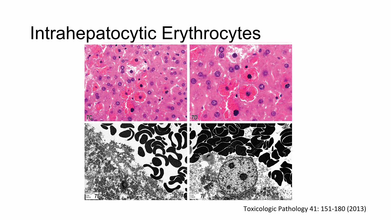
Intrahepatocytic Erythrocytes
FIGURE 7.—A–F, Series of images characterizing the lesion of ‘‘intrahepatocytic erythrocytes.’’ A, Low-magnification image showing the loca-tion of several areas containing intrahepatocytic erythrocytes near the edge of the liver (arrows; H&E). B, Higher magnification of (A) illus-trating intrahepatocellular erythrocytes with some adjacent hepatocytes containing a decreased cytoplasmic density. (H&E). C & D, Highermagnification images of hepatocytes containing erythrocytes within the cytoplasm. The hepatocyte nucleus is slightly condensed and hyper-chromatic and the cytoplasm is packed with erythrocytes. The remaining cytoplasm of the hepatocytes is marginated to the periphery of thecell. (H&E). E & F, Transmission electron micrographs illustrating (E) numerous erythrocytes within the hepatocellular cytoplasm and (F) thatthe erythrocytes are not contained within endothelial lined spaces or a lysosomal membrane. G-T, Series of images characterizing the lesion of
Vol. 41, No. 2, 2013 2012 NTP SATELLITE SYMPOSIUM 167
at Society of Toxicologic Pathology on October 14, 2016tpx.sagepub.comDownloaded from
ToxicologicPathology41:151-180(2013)

Hyaline Degeneration (Inclusions)
High-Dose Male B6C3F1 Mouse Tx: Ethylene glycol

Hyaline Degeneration (Inclusions)
High-Dose Male B6C3F1 Mouse Tx: Ethylene glycol

Hyaline Bodies

Hyaline Bodies

Hyaline Bodies

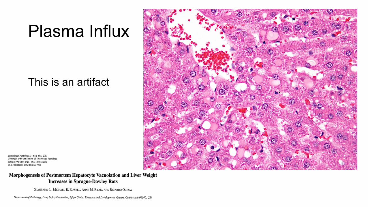
Plasma Influx
This is an artifact

Crystals
Differential diagnosis:
! Artifact—hematoxylin stain deposits in clear spaces.! Formalin precipitated pigment—extracellular granu-
lar yellow-brown deposits often associated witherythrocytes.
! Test compound/metabolite—may be distinctive forthe specific compound.
! Mineralization—basophilic deposits; may be associ-ated with calcification.
Comment: A number of different pigments may be seen as anincidental finding within hepatocytes and Kupffer cells inrodents. Some of them may increase and/or accumulate aftertreatment. Definitive diagnosis of a specific pigment typicallyrequires special stains.
Lipofuscin or ceroid is sometimes referred to as ‘‘wear andtear’’ or ‘‘aging’’ pigment and therefore is often observed inolder animals. It is considered to represent a breakdown of cellmembranes. Lipofuscin accumulates in postmitotic and agingcells. It has been shown to be a mixture of oxidized proteins andlipids, carbohydrates, and trace amount of metals (Seehafer andPearce 2006). A variety of stimuli can accelerate theaccumulation of this pigment, such as drug and chemical expo-sure, trauma and circulatory factors, and diet (Greaves 2007).Lipofuscin accumulation in the liver may be augmented by cer-tain chemicals (Kim and Kaminsky 1988; Marsman, 1995).Treatment of rats with PPAR alpha agonists such as fenofibrateand associated increased lipid peroxidation seen in rodentstreated with hypolipidemic agents can induce lipofuscin accu-mulation in liver after prolonged treatment (Nishimura et al.2007; Goel, Lalwani, and Reddy 1986; Reddy et al. 1982).Increased lipofuscin accumulation has also been observed in par-tially hepatectomized liver of rats (Sigal et al. 1999). Lipofuscinis insoluble in alcohols and xylene and other solvents normallyused in the preparation of slides. Special stains such as Smorl’scan be used to demonstrate the pigment. Storage granules appeargray with Sudan Black B, may be PAS-positive, and may stainwith Luxol fast blue and Ziehl-Neelsen (Jones 2002).
Porphyrin pigment, a precursor of heme protein, is seen withtreatment of some xenobiotics. Bile pigment is a common find-ing when there is cholestasis secondary to obstruction of bileflow or when there is perturbation in bile metabolism. Bile pig-ment stains green with Hall’s method.
Hemosiderin pigment represents precipitated iron that ismost frequently generated as a breakdown product of ery-throcytes and is derived from hemoglobin and accumulatesin the liver following local or systemic excess of iron.Deposition or iron may occur following excess dietaryintake or treatment by xenobiotics (Popp and Cattley1991; Greaves 2007; Travlos et al. 1996). Excess of ironfollowing injection may be stored as hemosiderin anddeposited in the reticuloendothelial component of the liver(and other organs such as spleen and bone marrow)(Bruguera 1999; Pitt et al. 1979). Intraperitoneal injectionof aflatoxin B1 can also induce hemosiderosis in hamsters
(Ungar, Sullman, and Zuckerman 1976). Endogenous irondeposition can be found following breakdown of blood cells(hemolytic event). Iron pigment can be found in Kupffercells, macrophages, and hepatocytes. In hepatocytes, theiron is stored in the form of ferritin (ferric iron bound toprotein apoferritin) (Popp and Cattley 1991). A spontaneousinherited predisposition for hepatic iron pigmentation hasbeen reported in Sprague-Dawley rats (Masson and Roome1997), and iron deposition can be found in the aging mouseliver (Harada et al. 1996). Iron can be demonstrated usingPerls’ Prussian blue stain in which iron stains blue.
Hemosiderin slowly dissolves in acids, especially oxalicacid. Non-aldehyde fixatives can remove hemosiderin oralter it in such a way that reactions for iron are (false) neg-ative (Churukian 2002). Malarial pigment is seen in hepato-cytes and Kupffer cells of Plasmodium sp experimentallyinfected mice. It is the pigment from the organism and nothemosiderin.
Porphyrin pigment normally occurs in tissues only insmall amounts and is a precursor of the heme portion ofhemoglobin (Churukian 2002). Porphyrin deposition in theliver of rodents is found after administration of a numberof compounds including griseofulvin where it can be seenin association with hepatocellular neoplasia (Stejskal et al.1975; Zatloukal et al. 2000; Knasmuller et al. 1997;Tschudy 1962). Griseofulvin administration in mice mayresult in inhibition of the mitochondrial enzyme ferrochela-tase and (compensatory) induction of ALA synthetase.Griseofulvin-induced accumulation of porphyrins in mouseliver is followed by cell damage and necrotic and inflamma-tory processes (Knasmuller et al. 1997). Proto-porphyrinpigment in liver of rats and mice is mainly found in the bileducts and leads to bile duct proliferation and portal inflam-mation, but can also occur in hepatocytes, Kupffer cells, andportal macrophages (Hurst and Paget 1963). The birefrin-gence of porphyrin appears to be associated with bilamellarcomponents within the pigment (Stejskal et al. 1975). Thispigment is also seen in combination with liver fibrosis andcirrhosis, bile duct proliferation, periportal inflammation,and hepatocarcinogenesis (Kanel and Korula 2005; Hurstand Paget 1963; Greaves 2007; Rank, Straka, and Bloomer1990).
Crystals (Figures 26–28)
Pathogenesis: Hyperlipidemia (cholesterol crystals), Chi313(Ym1) protein (eosinophilic biliary crystals).
Diagnostic features:
! Rhomboid or needle-like structures often birefringentunder polarized light.
! Needle-like crystals in the mouse can be intracellularor extracellular and may be associated with intenseeosinophilic epithelial cytoplasm and extracellularcrystals of various sizes.
12S THOOLEN ET AL. TOXICOLOGIC PATHOLOGY
at Society of Toxicologic Pathology on August 8, 2013tpx.sagepub.comDownloaded from

Crystals

Cytologic Alteration
cystic degeneration, that are more clearly established in tradi-tional pathology literature. The preferred diagnosis will beinfluenced by morphological features, conventional pathologypractice, and the experience of the pathologist.
Glycogen accumulation in hepatocytes is a type of cytoplas-mic alteration manifested on H&E-stained paraffin sections asclear spaces in the cytoplasm and a centrally located nucleus.Intracellular accumulation of glycogen is a normal physiologicalresponse following food ingestion. Since rodents eat primarily inthe evening hours, the largest amount of glycogen will be presentduring early morning hours. Intrahepatocyte glycogen is mobi-lized throughout the day, initially being removed from centrilob-ular hepatocytes. Consequently the amount present variesdepending upon whether the animals were fasted and on the timeof necropsy during the day. Failure to accumulate glycogenbecause of inanition or abnormal glycogen retention may resultfrom treatment-induced metabolic perturbations.
Cytoplasmic Alteration (Figure 44)
Synonyms: Cytoplasmic alteration, cytoplasmic change, granu-lar change, granular degeneration, hyaline degeneration, glyco-gen accumulation; ground glass change.
Pathogenesis: Often xenobiotic-induced and may be associatedwith other forms of liver damage.
Diagnostic features:
! Affected cells may show increased cytoplasmic gran-ularity, cell swelling, and eosinophilia.
Differential diagnosis:
! Artifact of fixation or processing—poorly stainedtissue with loss of normal structure.
! Hepatocyte hypertrophy—cytoplasmic volumeincreased with uniform finely granular texture; usu-ally associated with microsomal enzyme inductionor peroxisome proliferation.
! Cytoplasmic vacuole artifact—postmortem plasmainflux.
! Coagulation necrosis—loss of cytoplasmic andnuclear detail.
Comment: What has been described as granular degenerationcan be seen in combination with other forms of liver damage(e.g., necrosis, hydropic degeneration, inflammation) (Huanget al. 2007; Gokalp et al. 2003; Datta et al. 1998; Xu et al.1992; Aydin et al. 2003). Hepatocellular granularity may bedue to swelling of cell organelles or increase in the numbersof cell organelles including peroxisomes, mitochondria, andsmooth endoplasmic reticulum. Some pathologists do not con-sider granular degeneration to be a distinct entity and do notinclude it in their diagnostic lexicon.
Hyaline degeneration has been described by a number ofauthors, sometimes in combination with Mallory body
formation (Gonzalez-Quintela et al. 2000; NTP Toxicology andCaracinogenesis Studies Ethylene Glycol 1993; Peters et al.1983; Bruni 1960; Shea 1958; Omar, Elmesallamy, and Eassa2005; Lin et al. 1996), but is rarely used as a separate descriptionsince a combination of findings is often present. Cytoplasmicalteration reflecting plasma influx is an artifact seen in non-exsanguinated rats in a postmortem time-dependent manner(Li et al. 2003) (see Figure 33).
Degeneration, Hydropic (Figure 45)
Synonyms: Cytoplasmic alteration, cytoplasmic change, hydro-pic change, cloudy swelling.
Pathogenesis: Intracytoplasmic fluid accumulation secondaryto disturbance of cell membrane integrity.
Diagnostic features:
! Cytoplasmic vacuolation and ‘‘ballooning’’ with acentrally located nucleus.
! Lobular location may be centrilobular or periportalwith increased clear cell change and cell swelling.
Differential diagnosis:
! Cytoplasmic vacuole artifact—postmortem plasmainflux.
! Glycogen accumulation—hepatocytes not markedlyenlarged; cytoplasmic clear areas are irregular.
Comment: Because of disturbance of the cell membrane integ-rity, accumulation of intracytoplasmic fluid may occur. Thiscauses vacuolation and ‘‘ballooning’’ of cells. This change canbe caused by a number of xenobiotics with differing lobularlocalization and may be a precursor to hepatocyte necrosis(Gkretsi et al. 2007; Wang et al. 2007; Peichoto et al. 2006;Matsumoto et al. 2006; Chengelis 1988).
Degeneration, Cystic (Figure 46 and 47)
Synonyms: Spongiosis hepatis (traditional diagnostic term pre-ferred by many pathologists).
Pathogenesis: Cystic enlargement of perisinusoidal stellatecells (Ito cells) particularly observed in aging rats.
Diagnostic features:
! Multi-loculated cyst(s) lined by fine septa containingfine flocculent eosinophilic material (PAS-positive).
! The cysts are not lined by endothelial cells and do notcompress the surrounding liver parenchyma.
! May be accompanied by occasional erythrocytes orleukocytes.
! May be observed within altered hepatic foci and livertumors.
! Affected cells may be markedly enlarged.
Vol. 38, No. 7S, 2010 LESIONS OF THE HEPATOBILIARY SYSTEM 15S
at Society of Toxicologic Pathology on August 8, 2013tpx.sagepub.comDownloaded from

Clearcellchange-GlycogenaccumulaJon
In the INHAND document this change falls under the diagnostic category of Cytoplasmic Alteration
A proposed alternative has been suggested by SEND for consideration: Rarefaction
For example: Rarefaction, hepatocyte, increased, diffuse
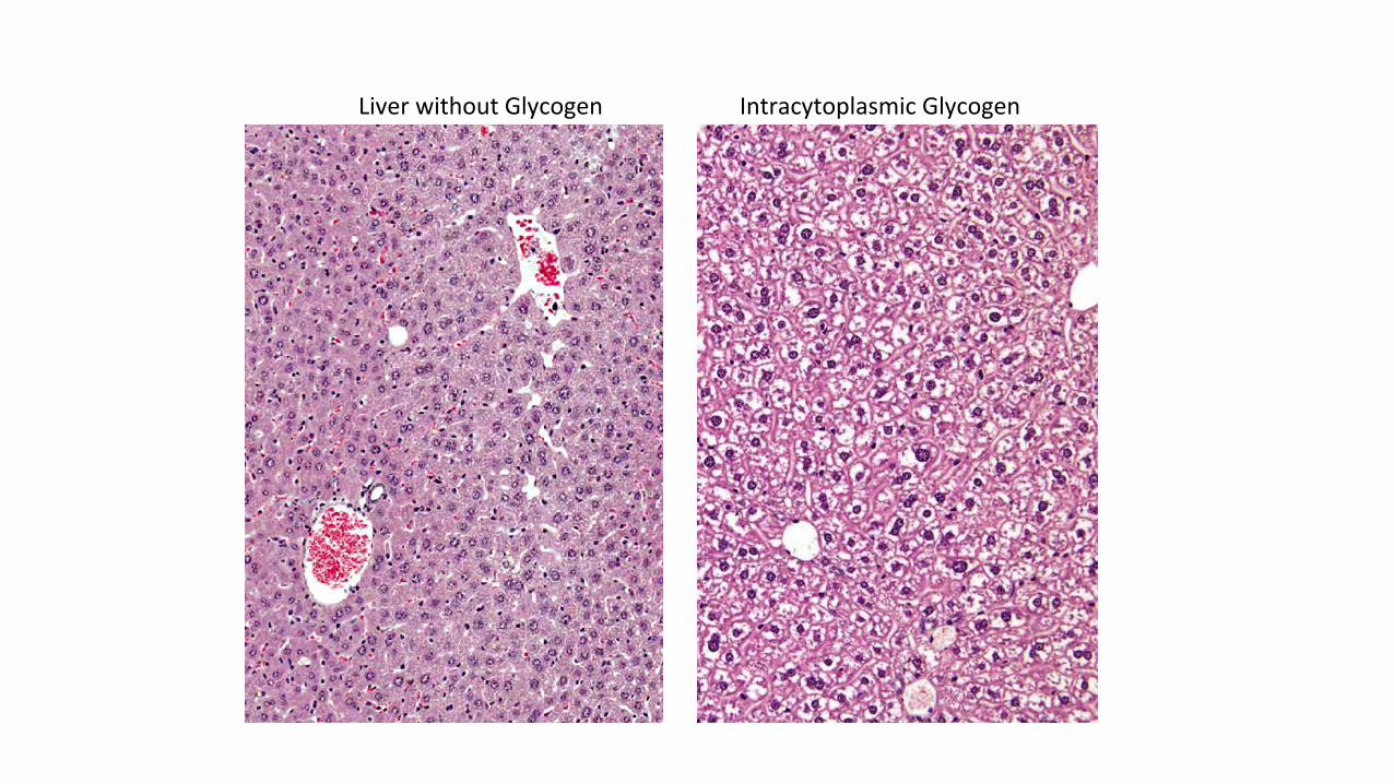
IntracytoplasmicGlycogenLiverwithoutGlycogen

PASwithdiastasePAS


N
M
G
L L
G
M
M
P
RER
Glycogen

Clearcellchange-GlycogenaccumulaJon


NormalglycogenaccumulaJon GlycogendepleJon
Glycogen

NormalglycogenaccumulaJon
Excessiveglycogen
Glycogen

Fatty Change
Differential diagnosis:
! Hepatocellular focus of cellular alteration—tinctorialvariation from normal parenchyma and does not pro-trude into the diaphragm.
! Hepatocellular neoplasia—when visible grossly doesnot protrude into the thoracic cavity.
! Regenerative hyperplastic nodule (nodularhyperplasia)—typically involves multiple nodules ofhyperplasia separated by proliferative bands of ovalcells or connective tissue.
Comment: Hepatodiaphragmatic nodules can be seen in rats atany age and their occurrence in fetuses is considered presump-tive evidence of a congenital origin. While they appear to beprotruding through the diaphragm and extending into the thor-acic cavity, they actually are attached to and covered by a thinfibrous portion of the diaphragm (Eustis et al. 1990).
An incidence ranging from 1% to 11% has been reported forhepatodiaphragmatic nodules in Fischer 344 rats (Eustis et al.1990), with few cases reported in other rat stocks and strains.Mice do not develop such nodules but may have focal lesionssimilar to those in rat hepatodiaphragmatic nodules and withlarge nuclei with large central nucleoli-like basophilic bodies.
B. Hepatocellular Responses, Cellular Degeneration,Injury, and Death
Introduction
The function and structure of most liver cells arerelatively constrained by their genetic programs of metabolism,differentiation, and specialization. While the cells of the hepaticparenchyma have the flexibility to adapt to changing physiologi-cal demands with reversible functional and morphological altera-tions, sufficient stress, or noxious stimuli may lead to inability tomaintain homeostasis and adverse cellular adaptations. The mor-phological response to injurious stimuli depends on the nature ofthe injury and its severity and duration. Often at high doses, tar-geted cells go through a sequence of cellular degeneration fol-lowed by cell death, but at lower doses degenerative changesdo not necessarily lead to cell death. Consequentially, cellularchanges that do not lead to cell death or death of the animal maybe called ‘‘adaptive’’ changes that can be considered eitheradverse or not adverse reactions, depending on the nature of thechange. There are cellular adaptations involving metabolic orfunctional alterations that lead to increases in cellular organellesand intracellular accumulations of a variety of endogenous andexogenous substances but allow the cell and animal to surviveand often live normally. Similar changes may occur in humanliver, such as cholestasis, a common lesion in human liverafter long-term drug therapy. However, in animals, when thelimits of adaptive responses are exceeded or do not occur inresponse to chemical exposure, irreversible cellular injury andcellular death occurs, with possible subsequent illness anddeath. Adaptive changes or doses of chemicals that induce
adaptive changes usually do not result in illness or death ofrodents. Often these processes are dose and chemical related.
Fatty Change
Synonyms/subtypes: Lipidosis, vacuolation, lipid, macrovesi-cular and/or microvesicular steatosis, phospholipidosis.1
Pathogenesis: Perturbations in lipid metabolism and disposition.
Diagnostic features:
Macrovesicular fatty change (Figures 5 and 6).
! Hepatocytes contain a large well-defined singlerounded vacuole within each cell.
! Nucleus and cytoplasm displaced to the periphery.! A few hepatocytes may contain one or more smaller
vacuoles.
Microvesicular fatty change (Figure 7).
! Hepatocytes partially or completely filled withnumerous small lipid vacuoles.
! Affected hepatocytes may have a ‘‘foamy’’ appearance.! Small vacuoles do not normally displace the nucleus to
the periphery in contrast to macrovesicular steatosis.
Differential diagnosis:
! Hydropic degeneration—clear cytoplasm withoutnuclear displacement.
! Glycogen accumulation—irregular and poorly definedlacy clear spaces in the cytoplasm (rarefaction) usuallywith centrally located nuclei.
Comment: There is a difference in preferred nomenclatureamong pathologists for this change. Based strictly on anH&E-stained section, a diagnosis of cytoplasmic vacuolationof hepatocytes is a universally acceptable descriptive diagno-sis. Based on the experience of the observer, the specific mor-phological features of the cytoplasmic vacuolation may besufficiently consistent with intracytoplasmic lipid accumula-tion to warrant a presumptive diagnosis of fatty change. Theunequivocal demonstration of intracytoplasmic fat, however,requires a special stain.
Fatty change can be induced by a number of different agentsand is usually divided into two main types, namely, microvesi-cular and macrovesicular, although mixed forms can frequentlybe observed (Greaves 2007; Gopinath, Prentice, and Lewis1987; Goodman and Ishak 2006; Kanel and Korula 2005).Macrovesicular lipidosis is a reaction to a wide variety ofinjuries and can also be regarded as a physiological adapta-tion demonstrated as an imbalance between uptake of lipidsfrom blood and secretion of lipoproteins by the hepatocyte(Goodman and Ishak 2006). Microvesicular lipidosis is usually
1 Electron microscopy or special staining needed for a definitive diagnosis.
Vol. 38, No. 7S, 2010 LESIONS OF THE HEPATOBILIARY SYSTEM 9S
at Society of Toxicologic Pathology on August 8, 2013tpx.sagepub.comDownloaded from
FIGURE 1.—Gross appearance and tissue trimming recommendations for a normal rodent liver. Ref. to http://reni.item.fraunhofer.de/reni/trimming/index.php. FIGURE 2.— Two-dimensional microarchitecture of the liver. FIGURE 3.—Rat liver. Hepatodiaphragmatic nodule. FIGURE 4.—Rat liver.Hepatodiaphragmatic nodule with intranuclear inclusions (chromatin). Higher magnification of Figure 3. FIGURE 5.—Rat liver. Macrovesicular fattychange. FIGURE 6.—Rat liver. Macrovesicular fatty change. Higher magnification of Figure 5.
Vol. 38, No. 7S, 2010 LESIONS OF THE HEPATOBILIARY SYSTEM 43S
at Society of Toxicologic Pathology on August 8, 2013tpx.sagepub.comDownloaded from
FIGURE 7.—Mouse liver. Fatty change, microvesicular. FIGURE 8.—Mouse liver. Mixture of fatty change and cytoplasmic glycogen. FIGURE 9.—Mouse liver. Mixture of fatty change and cytoplasmic glycogen. Higher magnification of Figure 8. FIGURE 10.—Mouse liver. Tension lipidosis.FIGURE 11.—Mouse liver. Tension lipidosis. Higher magnification of Figure 10. FIGURE 12.—Rat liver. Focal fatty change.
44S THOOLEN ET AL. TOXICOLOGIC PATHOLOGY
at Society of Toxicologic Pathology on August 8, 2013tpx.sagepub.comDownloaded from

Oil-Red-O Stain for Lipid Mouse Liver

Fatty Change
Zonal
Focal

Vacuolated Focus Focal Fatty Change

Vacuolated Focus Focal Fatty Change
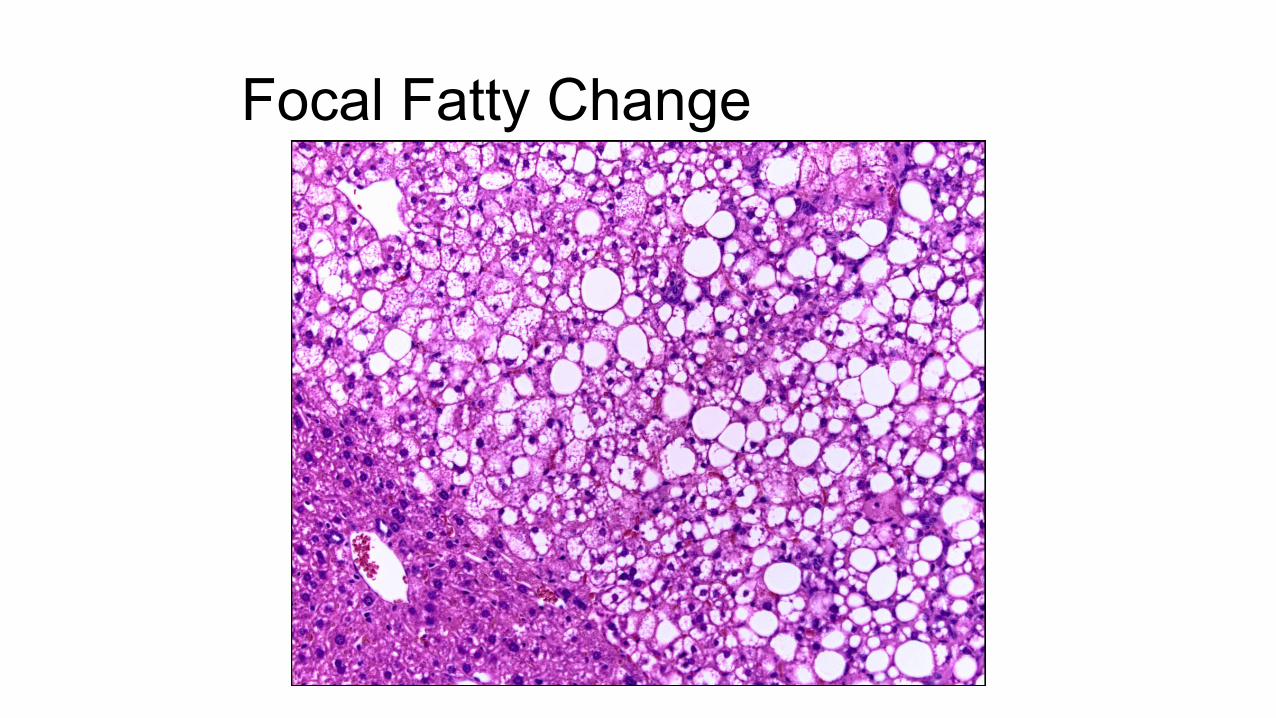
Focal Fatty Change

Differential diagnosis:
! Hepatocellular focus of cellular alteration—tinctorialvariation from normal parenchyma and does not pro-trude into the diaphragm.
! Hepatocellular neoplasia—when visible grossly doesnot protrude into the thoracic cavity.
! Regenerative hyperplastic nodule (nodularhyperplasia)—typically involves multiple nodules ofhyperplasia separated by proliferative bands of ovalcells or connective tissue.
Comment: Hepatodiaphragmatic nodules can be seen in rats atany age and their occurrence in fetuses is considered presump-tive evidence of a congenital origin. While they appear to beprotruding through the diaphragm and extending into the thor-acic cavity, they actually are attached to and covered by a thinfibrous portion of the diaphragm (Eustis et al. 1990).
An incidence ranging from 1% to 11% has been reported forhepatodiaphragmatic nodules in Fischer 344 rats (Eustis et al.1990), with few cases reported in other rat stocks and strains.Mice do not develop such nodules but may have focal lesionssimilar to those in rat hepatodiaphragmatic nodules and withlarge nuclei with large central nucleoli-like basophilic bodies.
B. Hepatocellular Responses, Cellular Degeneration,Injury, and Death
Introduction
The function and structure of most liver cells arerelatively constrained by their genetic programs of metabolism,differentiation, and specialization. While the cells of the hepaticparenchyma have the flexibility to adapt to changing physiologi-cal demands with reversible functional and morphological altera-tions, sufficient stress, or noxious stimuli may lead to inability tomaintain homeostasis and adverse cellular adaptations. The mor-phological response to injurious stimuli depends on the nature ofthe injury and its severity and duration. Often at high doses, tar-geted cells go through a sequence of cellular degeneration fol-lowed by cell death, but at lower doses degenerative changesdo not necessarily lead to cell death. Consequentially, cellularchanges that do not lead to cell death or death of the animal maybe called ‘‘adaptive’’ changes that can be considered eitheradverse or not adverse reactions, depending on the nature of thechange. There are cellular adaptations involving metabolic orfunctional alterations that lead to increases in cellular organellesand intracellular accumulations of a variety of endogenous andexogenous substances but allow the cell and animal to surviveand often live normally. Similar changes may occur in humanliver, such as cholestasis, a common lesion in human liverafter long-term drug therapy. However, in animals, when thelimits of adaptive responses are exceeded or do not occur inresponse to chemical exposure, irreversible cellular injury andcellular death occurs, with possible subsequent illness anddeath. Adaptive changes or doses of chemicals that induce
adaptive changes usually do not result in illness or death ofrodents. Often these processes are dose and chemical related.
Fatty Change
Synonyms/subtypes: Lipidosis, vacuolation, lipid, macrovesi-cular and/or microvesicular steatosis, phospholipidosis.1
Pathogenesis: Perturbations in lipid metabolism and disposition.
Diagnostic features:
Macrovesicular fatty change (Figures 5 and 6).
! Hepatocytes contain a large well-defined singlerounded vacuole within each cell.
! Nucleus and cytoplasm displaced to the periphery.! A few hepatocytes may contain one or more smaller
vacuoles.
Microvesicular fatty change (Figure 7).
! Hepatocytes partially or completely filled withnumerous small lipid vacuoles.
! Affected hepatocytes may have a ‘‘foamy’’ appearance.! Small vacuoles do not normally displace the nucleus to
the periphery in contrast to macrovesicular steatosis.
Differential diagnosis:
! Hydropic degeneration—clear cytoplasm withoutnuclear displacement.
! Glycogen accumulation—irregular and poorly definedlacy clear spaces in the cytoplasm (rarefaction) usuallywith centrally located nuclei.
Comment: There is a difference in preferred nomenclatureamong pathologists for this change. Based strictly on anH&E-stained section, a diagnosis of cytoplasmic vacuolationof hepatocytes is a universally acceptable descriptive diagno-sis. Based on the experience of the observer, the specific mor-phological features of the cytoplasmic vacuolation may besufficiently consistent with intracytoplasmic lipid accumula-tion to warrant a presumptive diagnosis of fatty change. Theunequivocal demonstration of intracytoplasmic fat, however,requires a special stain.
Fatty change can be induced by a number of different agentsand is usually divided into two main types, namely, microvesi-cular and macrovesicular, although mixed forms can frequentlybe observed (Greaves 2007; Gopinath, Prentice, and Lewis1987; Goodman and Ishak 2006; Kanel and Korula 2005).Macrovesicular lipidosis is a reaction to a wide variety ofinjuries and can also be regarded as a physiological adapta-tion demonstrated as an imbalance between uptake of lipidsfrom blood and secretion of lipoproteins by the hepatocyte(Goodman and Ishak 2006). Microvesicular lipidosis is usually
1 Electron microscopy or special staining needed for a definitive diagnosis.
Vol. 38, No. 7S, 2010 LESIONS OF THE HEPATOBILIARY SYSTEM 9S
at Society of Toxicologic Pathology on August 8, 2013tpx.sagepub.comDownloaded from
Macrovesicular Fatty Change (Diffuse)
Vehicle Male F344 Study: Kava kava extract Dx: Fatty change, diffuse +3

Differential diagnosis:
! Hepatocellular focus of cellular alteration—tinctorialvariation from normal parenchyma and does not pro-trude into the diaphragm.
! Hepatocellular neoplasia—when visible grossly doesnot protrude into the thoracic cavity.
! Regenerative hyperplastic nodule (nodularhyperplasia)—typically involves multiple nodules ofhyperplasia separated by proliferative bands of ovalcells or connective tissue.
Comment: Hepatodiaphragmatic nodules can be seen in rats atany age and their occurrence in fetuses is considered presump-tive evidence of a congenital origin. While they appear to beprotruding through the diaphragm and extending into the thor-acic cavity, they actually are attached to and covered by a thinfibrous portion of the diaphragm (Eustis et al. 1990).
An incidence ranging from 1% to 11% has been reported forhepatodiaphragmatic nodules in Fischer 344 rats (Eustis et al.1990), with few cases reported in other rat stocks and strains.Mice do not develop such nodules but may have focal lesionssimilar to those in rat hepatodiaphragmatic nodules and withlarge nuclei with large central nucleoli-like basophilic bodies.
B. Hepatocellular Responses, Cellular Degeneration,Injury, and Death
Introduction
The function and structure of most liver cells arerelatively constrained by their genetic programs of metabolism,differentiation, and specialization. While the cells of the hepaticparenchyma have the flexibility to adapt to changing physiologi-cal demands with reversible functional and morphological altera-tions, sufficient stress, or noxious stimuli may lead to inability tomaintain homeostasis and adverse cellular adaptations. The mor-phological response to injurious stimuli depends on the nature ofthe injury and its severity and duration. Often at high doses, tar-geted cells go through a sequence of cellular degeneration fol-lowed by cell death, but at lower doses degenerative changesdo not necessarily lead to cell death. Consequentially, cellularchanges that do not lead to cell death or death of the animal maybe called ‘‘adaptive’’ changes that can be considered eitheradverse or not adverse reactions, depending on the nature of thechange. There are cellular adaptations involving metabolic orfunctional alterations that lead to increases in cellular organellesand intracellular accumulations of a variety of endogenous andexogenous substances but allow the cell and animal to surviveand often live normally. Similar changes may occur in humanliver, such as cholestasis, a common lesion in human liverafter long-term drug therapy. However, in animals, when thelimits of adaptive responses are exceeded or do not occur inresponse to chemical exposure, irreversible cellular injury andcellular death occurs, with possible subsequent illness anddeath. Adaptive changes or doses of chemicals that induce
adaptive changes usually do not result in illness or death ofrodents. Often these processes are dose and chemical related.
Fatty Change
Synonyms/subtypes: Lipidosis, vacuolation, lipid, macrovesi-cular and/or microvesicular steatosis, phospholipidosis.1
Pathogenesis: Perturbations in lipid metabolism and disposition.
Diagnostic features:
Macrovesicular fatty change (Figures 5 and 6).
! Hepatocytes contain a large well-defined singlerounded vacuole within each cell.
! Nucleus and cytoplasm displaced to the periphery.! A few hepatocytes may contain one or more smaller
vacuoles.
Microvesicular fatty change (Figure 7).
! Hepatocytes partially or completely filled withnumerous small lipid vacuoles.
! Affected hepatocytes may have a ‘‘foamy’’ appearance.! Small vacuoles do not normally displace the nucleus to
the periphery in contrast to macrovesicular steatosis.
Differential diagnosis:
! Hydropic degeneration—clear cytoplasm withoutnuclear displacement.
! Glycogen accumulation—irregular and poorly definedlacy clear spaces in the cytoplasm (rarefaction) usuallywith centrally located nuclei.
Comment: There is a difference in preferred nomenclatureamong pathologists for this change. Based strictly on anH&E-stained section, a diagnosis of cytoplasmic vacuolationof hepatocytes is a universally acceptable descriptive diagno-sis. Based on the experience of the observer, the specific mor-phological features of the cytoplasmic vacuolation may besufficiently consistent with intracytoplasmic lipid accumula-tion to warrant a presumptive diagnosis of fatty change. Theunequivocal demonstration of intracytoplasmic fat, however,requires a special stain.
Fatty change can be induced by a number of different agentsand is usually divided into two main types, namely, microvesi-cular and macrovesicular, although mixed forms can frequentlybe observed (Greaves 2007; Gopinath, Prentice, and Lewis1987; Goodman and Ishak 2006; Kanel and Korula 2005).Macrovesicular lipidosis is a reaction to a wide variety ofinjuries and can also be regarded as a physiological adapta-tion demonstrated as an imbalance between uptake of lipidsfrom blood and secretion of lipoproteins by the hepatocyte(Goodman and Ishak 2006). Microvesicular lipidosis is usually
1 Electron microscopy or special staining needed for a definitive diagnosis.
Vol. 38, No. 7S, 2010 LESIONS OF THE HEPATOBILIARY SYSTEM 9S
at Society of Toxicologic Pathology on August 8, 2013tpx.sagepub.comDownloaded from
Vehicle Male F344 Study: Kava kava extract Dx: Fatty change, diffuse +3

Microvesicular – Zonal High-dose Male B6C3F1 Tx: Kava kava extract Dx: Centrilobular fatty change 4+ and necrosis

Microvesicular Fatty Change - Diffuse
Female B6C3F1 Tx: 3,3’,4,4’-Tetrachloroazobenzene

Microvesicular – Diffuse

Differential diagnosis:
! Hepatocellular focus of cellular alteration—tinctorialvariation from normal parenchyma and does not pro-trude into the diaphragm.
! Hepatocellular neoplasia—when visible grossly doesnot protrude into the thoracic cavity.
! Regenerative hyperplastic nodule (nodularhyperplasia)—typically involves multiple nodules ofhyperplasia separated by proliferative bands of ovalcells or connective tissue.
Comment: Hepatodiaphragmatic nodules can be seen in rats atany age and their occurrence in fetuses is considered presump-tive evidence of a congenital origin. While they appear to beprotruding through the diaphragm and extending into the thor-acic cavity, they actually are attached to and covered by a thinfibrous portion of the diaphragm (Eustis et al. 1990).
An incidence ranging from 1% to 11% has been reported forhepatodiaphragmatic nodules in Fischer 344 rats (Eustis et al.1990), with few cases reported in other rat stocks and strains.Mice do not develop such nodules but may have focal lesionssimilar to those in rat hepatodiaphragmatic nodules and withlarge nuclei with large central nucleoli-like basophilic bodies.
B. Hepatocellular Responses, Cellular Degeneration,Injury, and Death
Introduction
The function and structure of most liver cells arerelatively constrained by their genetic programs of metabolism,differentiation, and specialization. While the cells of the hepaticparenchyma have the flexibility to adapt to changing physiologi-cal demands with reversible functional and morphological altera-tions, sufficient stress, or noxious stimuli may lead to inability tomaintain homeostasis and adverse cellular adaptations. The mor-phological response to injurious stimuli depends on the nature ofthe injury and its severity and duration. Often at high doses, tar-geted cells go through a sequence of cellular degeneration fol-lowed by cell death, but at lower doses degenerative changesdo not necessarily lead to cell death. Consequentially, cellularchanges that do not lead to cell death or death of the animal maybe called ‘‘adaptive’’ changes that can be considered eitheradverse or not adverse reactions, depending on the nature of thechange. There are cellular adaptations involving metabolic orfunctional alterations that lead to increases in cellular organellesand intracellular accumulations of a variety of endogenous andexogenous substances but allow the cell and animal to surviveand often live normally. Similar changes may occur in humanliver, such as cholestasis, a common lesion in human liverafter long-term drug therapy. However, in animals, when thelimits of adaptive responses are exceeded or do not occur inresponse to chemical exposure, irreversible cellular injury andcellular death occurs, with possible subsequent illness anddeath. Adaptive changes or doses of chemicals that induce
adaptive changes usually do not result in illness or death ofrodents. Often these processes are dose and chemical related.
Fatty Change
Synonyms/subtypes: Lipidosis, vacuolation, lipid, macrovesi-cular and/or microvesicular steatosis, phospholipidosis.1
Pathogenesis: Perturbations in lipid metabolism and disposition.
Diagnostic features:
Macrovesicular fatty change (Figures 5 and 6).
! Hepatocytes contain a large well-defined singlerounded vacuole within each cell.
! Nucleus and cytoplasm displaced to the periphery.! A few hepatocytes may contain one or more smaller
vacuoles.
Microvesicular fatty change (Figure 7).
! Hepatocytes partially or completely filled withnumerous small lipid vacuoles.
! Affected hepatocytes may have a ‘‘foamy’’ appearance.! Small vacuoles do not normally displace the nucleus to
the periphery in contrast to macrovesicular steatosis.
Differential diagnosis:
! Hydropic degeneration—clear cytoplasm withoutnuclear displacement.
! Glycogen accumulation—irregular and poorly definedlacy clear spaces in the cytoplasm (rarefaction) usuallywith centrally located nuclei.
Comment: There is a difference in preferred nomenclatureamong pathologists for this change. Based strictly on anH&E-stained section, a diagnosis of cytoplasmic vacuolationof hepatocytes is a universally acceptable descriptive diagno-sis. Based on the experience of the observer, the specific mor-phological features of the cytoplasmic vacuolation may besufficiently consistent with intracytoplasmic lipid accumula-tion to warrant a presumptive diagnosis of fatty change. Theunequivocal demonstration of intracytoplasmic fat, however,requires a special stain.
Fatty change can be induced by a number of different agentsand is usually divided into two main types, namely, microvesi-cular and macrovesicular, although mixed forms can frequentlybe observed (Greaves 2007; Gopinath, Prentice, and Lewis1987; Goodman and Ishak 2006; Kanel and Korula 2005).Macrovesicular lipidosis is a reaction to a wide variety ofinjuries and can also be regarded as a physiological adapta-tion demonstrated as an imbalance between uptake of lipidsfrom blood and secretion of lipoproteins by the hepatocyte(Goodman and Ishak 2006). Microvesicular lipidosis is usually
1 Electron microscopy or special staining needed for a definitive diagnosis.
Vol. 38, No. 7S, 2010 LESIONS OF THE HEPATOBILIARY SYSTEM 9S
at Society of Toxicologic Pathology on August 8, 2013tpx.sagepub.comDownloaded from
Microvesicular Fatty Change - Focal

Phospholipidosis
indicative of more serious hepatic dysfunction but can alsoresult from nutritional disturbances (Greaves 2007).
Specific xenobiotics can induce either macrovesicular ormicrovesicular lipidosis in humans (Kanel and Korula 2005).In animal studies, it is common to see a mixture of macrovesi-cular and microvesicular lipidosis. In those situations one caneither diagnose the most prevalent form or record the findingsas mixed. Commentary in the pathology narrative report mightbe appropriate, especially if recording the most prevalent formof lipidosis. Liver with admixed presence of glycogen and fattychange can be observed (Figures 8 and 9).
Fatty change and necrosis may appear together althoughthey may differ in proportion. A number of causes other thanxenobiotic exposure, such as chronic hepatic injury, diet,metabolic and hormonal status, debilitation of animals, andfasting before necropsy, should be taken into considerationin reviewing these changes (Vollmar et al. 1999; Katoh andSugimoto 1982; Nagano et al. 2007; Denda et al. 2002). Thedistribution can be either diffuse (e.g., ethionine) or zonal(e.g., centrilobular in CCl4; periportal in phosphorus toxicity;midzonal in choline deficiency). Inadequate fixation proce-dures may sometimes give rise to artifacts with microvesicu-lar vacuolation, although mostly with less clear cytoplasm(Li et al. 2003).
Focal fatty change can sometimes be seen spontaneously andis usually described as such. A specific variation occurs near theattachment of the falciform ligament and gallbladder in mice andis referred to as ‘‘tension lipidosis’’ (Harada et al. 1999) (Figures10 and 11). Spontaneous fatty change can differ between strainsand is a normal finding in BALB mice. Livers of these mice aretypically paler than in other strains. Focal fatty change in theliver of rodents has previously been categorized as vacuolatedaltered hepatic foci (Eustis et al. 1990), but current practice isto diagnose this change as focal fatty change rather than as afocus of hepatic alteration (Figures 12 and 13).
Fatty change can also be observed in combination with otherhepatotoxic injuries (e.g., chronic liver toxicity, degeneration,inflammation, and necrosis) or nutritional disturbance (e.g.,diet, vitamin A excess) in both animals and man. Special stainson cryostat sections can demonstrate fat (e.g., Oil red O orSudan Black) (Jones 2002).
Phospholipidosis2
Synonym: Cytoplasmic vacuolation, foam cells.
Pathogenesis: Induced by xenobiotics with a cationic ampho-philic structure.
Diagnostic features:
! Multiple irregular to round clear membrane-boundvacuoles.
! Tends to be a diffuse change affecting hepatocytes.
Differential diagnosis:
! Fatty change—round clear vacuoles tend to be singleor multiple and discrete.
! Glycogen accumulation—irregular and poorlydefined clear spaces in the cytoplasm (rarefaction)usually with centrally located nuclei; positive stainedwith periodic acid-Schiff staining.
Comment: Definitive diagnosis of phospholipidosis is not pos-sible based strictly on H&E-stained liver sections. A diagnosisof cytoplasmic vacuolation of hepatocytes will typically be anacceptable descriptive diagnosis. Since the cytoplasmicvacuolation may mimic microvesicular fatty change, a descrip-tive diagnosis of cytoplasmic vacuolation is recommended inthe absence of electron microscopy or special immunostaining.
Phospholipidosis can be induced by xenobiotics with acationic amphophilic structure (Halliwell 1997; Anderson andBorlak 2006; Reasor, Hastings, and Ulrich 2006; Chatmanet al. 2009) (Figures 14 and 15). It is a lipid storage disorderseen when complexes between xenobiotics and phospholipidsaccumulate within lysosomes. Phospholipidosis refers to a spe-cific form of hepatic vacuolation with the occurrence of con-centric membrane bound lysosomal myeloid bodies/lamellarbodies that can be confirmed by specific staining and electronmicroscopy (Hruban, Slesers, and Hopkins 1972; Obert et al.2007) (Figure 16). Definitive diagnosis requires electronmicroscopy or positive immunostaining. Immunohistochem-ical staining for a lysosomal-associated protein and adipophilinmay be used to differentiate phospholipidosis from conven-tional fatty change (Obert et al. 2007). Both preexisting neutralfat and phospholipids can be observed in combination. Themacrovesicular and the microvesicular fatty change (vacuola-tion) generally located at the cell periphery stains positively forOil Red-O and the membranes surrounding these lipidvacuoles stain positively for adipophilin (a protein that formsthe membrane around non-lysosomal lipid droplets) but neg-ative for LAMP-2 (a lysosome-associated protein) by immu-nohistochemical techniques (Obert et al. 2007). This indicatesthat this vacuolation was due to accumulation of non-lysosomal neutral lipid. Cytoplasmic microvesiculationlocated centrally in hepatocytes that exhibit positive immuno-histochemical staining for LAMP-2 (Figure 17) but is nega-tive for Oil-Red-O and adipophilin is indicative ofphospholipid accumulation (Obert et al. 2007).
Amyloidosis (Figures 18 and 19)
Pathogenesis: Cellular process related to misfolding of protein.
Diagnostic features:
! Deposition of pale, homogeneous, amorphous eosi-nophilic material.
! Deposition often peri-sinusoidal, periportal, orinvolving blood vessel walls.
! Localization is extracellular.2 Electron microscopy or special staining needed for a definitive diagnosis.
10S THOOLEN ET AL. TOXICOLOGIC PATHOLOGY
at Society of Toxicologic Pathology on August 8, 2013tpx.sagepub.comDownloaded from
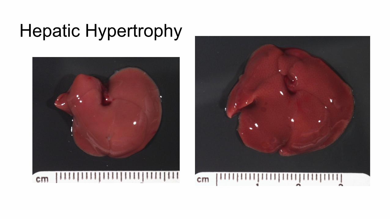
Hepatic Hypertrophy

Hypertrophy
Differential diagnosis:
! Artifact—wispy blue hematoxylin deposits in clearspaces.
Comment: In hyperlipidemia, cholesterol crystals can depositin the liver with or without granulomatous inflammation(Greaves 2007; Graewin et al. 2004; Handley, Chien, andArbeeny 1983). During gall stone formation, in addition toclassical rhomboid-shape monohydrate crystals, cholesterolcan also crystallize transiently as needle-, spiral-, and tubule-shaped crystals of anhydrous cholesterol (Dowling 2000). Eosi-nophilic crystals have been described in intrahepatic bile ductsand gallbladder of different laboratory mice strains, and someof these crystals have been shown to contain chitinase-like pro-teins confirmed by immunohistochemistry for Ym1 protein(now Chi313) (Ward et al. 2001; Harbord et al. 2002).
Crystal formation may be associated with inflammatoryand/or proliferative bile duct changes and fibrosis in mice andmay also occur spontaneously (Lewis 1984; Rabstein, Peters,and Spahn 1973; Enomoto et al. 1974). Numerous crystals canbe demonstrated using a simple system of polarizing micro-scopy. Crystals are capable of producing plane-polarized light,thus showing birefringence.
Inclusions, Intranuclear, and Cytoplasmic (Figures 29–32)
Synonyms: Inclusion bodies, intranuclear cytoplasmic invagi-nation, acidophilic inclusions, globular bodies.
Pathogenesis: Protrusion of cytoplasm into an invagination ofthe hepatocyte nuclear membrane without the actual protrusionnecessarily being present in the plane of section. Seen in spe-cific viral infections. Deposition of protein material withinhepatocyte cytoplasm.
Diagnostic features:
! Intranuclear inclusions are round, distinct, usuallyeccentrically located, and may partially or almostcompletely fill the nucleus.
! Contents of intranuclear inclusion bodies are ofteneosinophilic and may be granular or flocculent.
! Intracytoplasmic inclusions are round to oval, homo-genous, eosinophilic, and occur as single or multiplestructures in the cytoplasm.
Differential diagnosis:
! Enlarged nucleolus—one or more deeply basophilicstructures in normal size nuclei.
! Viral inclusion bodies (cytomegalic virus, experi-mental viral infections).
! Cytoplasmic vacuole artifact—postmortem plasmainflux (Li et al. 2003).
Comment: Both intranuclear and intracytoplasmic inclu-sions are common findings in the aging mouse liver andmay be seen in normal as well as neoplastic hepatocytes(Percy and Barthold 2001; Frith and Ward 1988; Irisarriand Hollander 1994). When the intranuclear inclusionsrepresent invaginations of the cytoplasm into the nucleus,they may contain cytoplasmic organelles in electron micro-graphs (van Zwieten and Hollander 1997). Ultrastructu-rally, three types of cytoplasmic inclusions have beendescribed: dense reticulated substance in the dilated cister-nae of rough endoplasmic reticulum, fine granular sub-stance in rough endoplasmic reticulum, and non–membranebound dense granulofibrillar in the cytoplasm (Helyer andPetrelli 1978).
Kakizoe, Goldfarb, and Pugh (1989) have correlated theincidences of cytoplasmic inclusion with hepatocellulartumors in different mice strains. C57BL/6 mice are rela-tively more resistant to hepatocarcinogens than C3H andC57BL/6 x C3H F1 mice. The tumors in the C57BL/6 micewere unique in their early focal development of cells con-taining inclusions. The authors suggested that the higherincidence of inclusions in liver might be related to slowingof the tumor growth leading to lower incidence of hepato-cellular tumors in C57BL/6 mice. Other types of intracyto-plasmic inclusions such as Mallory bodies, lamellated, andcrystalloid inclusions have been described in mice treatedwith different chemicals and in lysosomal storage diseases(Gebbia et al. 1985; Meierhenry et al. 1983; Rijhsinghaniet al. 1980; Shio et al. 1982).
Cytoplasmic vacuoles can occur in hepatocytes andendothelial cells in a postmortem time-dependent manner infasted and non-fasted rats (Li et al. 2003). This artifact is espe-cially common in rats that are not exsanguinated at necropsyand the cytoplasmic vacuoles represent plasma influx into toaffected cells (Figure 33). This artifact is more common inmales than in females.
Hypertrophy, Hepatocellular (Figures 34–41)
Synonyms: Hepatocytomegaly.
Pathogenesis: Metabolic enzyme induction causing increase inendoplasmic reticulum; increase in peroxisomes; increase inmitochondria.
Diagnostic features:
! Enlarged hepatocytes may be tinctorially distinct.! Cytoplasm may be homogeneous or granular.! Zonal pattern of distribution (centrilobular, peripor-
tal, midzonal) may be present.! Involving most or all lobules.! Loss of hepatocellular plate architecture is possible.! Sinusoidal compression.! Concurrent degeneration and/or single cell necrosis is
possible.
Vol. 38, No. 7S, 2010 LESIONS OF THE HEPATOBILIARY SYSTEM 13S
at Society of Toxicologic Pathology on August 8, 2013tpx.sagepub.comDownloaded from


Increased Smooth Endoplasmic Reticulum
Control Treated

Constitutive Enzyme Activity
CYP 3A1

Enzyme Induction


Persistent & Excessive Enzyme Induction

Persistent & Excessive Enzyme Induction

Hypertrophy – Peroxisome Proliferation
Differential diagnosis:
! Artifact—wispy blue hematoxylin deposits in clearspaces.
Comment: In hyperlipidemia, cholesterol crystals can depositin the liver with or without granulomatous inflammation(Greaves 2007; Graewin et al. 2004; Handley, Chien, andArbeeny 1983). During gall stone formation, in addition toclassical rhomboid-shape monohydrate crystals, cholesterolcan also crystallize transiently as needle-, spiral-, and tubule-shaped crystals of anhydrous cholesterol (Dowling 2000). Eosi-nophilic crystals have been described in intrahepatic bile ductsand gallbladder of different laboratory mice strains, and someof these crystals have been shown to contain chitinase-like pro-teins confirmed by immunohistochemistry for Ym1 protein(now Chi313) (Ward et al. 2001; Harbord et al. 2002).
Crystal formation may be associated with inflammatoryand/or proliferative bile duct changes and fibrosis in mice andmay also occur spontaneously (Lewis 1984; Rabstein, Peters,and Spahn 1973; Enomoto et al. 1974). Numerous crystals canbe demonstrated using a simple system of polarizing micro-scopy. Crystals are capable of producing plane-polarized light,thus showing birefringence.
Inclusions, Intranuclear, and Cytoplasmic (Figures 29–32)
Synonyms: Inclusion bodies, intranuclear cytoplasmic invagi-nation, acidophilic inclusions, globular bodies.
Pathogenesis: Protrusion of cytoplasm into an invagination ofthe hepatocyte nuclear membrane without the actual protrusionnecessarily being present in the plane of section. Seen in spe-cific viral infections. Deposition of protein material withinhepatocyte cytoplasm.
Diagnostic features:
! Intranuclear inclusions are round, distinct, usuallyeccentrically located, and may partially or almostcompletely fill the nucleus.
! Contents of intranuclear inclusion bodies are ofteneosinophilic and may be granular or flocculent.
! Intracytoplasmic inclusions are round to oval, homo-genous, eosinophilic, and occur as single or multiplestructures in the cytoplasm.
Differential diagnosis:
! Enlarged nucleolus—one or more deeply basophilicstructures in normal size nuclei.
! Viral inclusion bodies (cytomegalic virus, experi-mental viral infections).
! Cytoplasmic vacuole artifact—postmortem plasmainflux (Li et al. 2003).
Comment: Both intranuclear and intracytoplasmic inclu-sions are common findings in the aging mouse liver andmay be seen in normal as well as neoplastic hepatocytes(Percy and Barthold 2001; Frith and Ward 1988; Irisarriand Hollander 1994). When the intranuclear inclusionsrepresent invaginations of the cytoplasm into the nucleus,they may contain cytoplasmic organelles in electron micro-graphs (van Zwieten and Hollander 1997). Ultrastructu-rally, three types of cytoplasmic inclusions have beendescribed: dense reticulated substance in the dilated cister-nae of rough endoplasmic reticulum, fine granular sub-stance in rough endoplasmic reticulum, and non–membranebound dense granulofibrillar in the cytoplasm (Helyer andPetrelli 1978).
Kakizoe, Goldfarb, and Pugh (1989) have correlated theincidences of cytoplasmic inclusion with hepatocellulartumors in different mice strains. C57BL/6 mice are rela-tively more resistant to hepatocarcinogens than C3H andC57BL/6 x C3H F1 mice. The tumors in the C57BL/6 micewere unique in their early focal development of cells con-taining inclusions. The authors suggested that the higherincidence of inclusions in liver might be related to slowingof the tumor growth leading to lower incidence of hepato-cellular tumors in C57BL/6 mice. Other types of intracyto-plasmic inclusions such as Mallory bodies, lamellated, andcrystalloid inclusions have been described in mice treatedwith different chemicals and in lysosomal storage diseases(Gebbia et al. 1985; Meierhenry et al. 1983; Rijhsinghaniet al. 1980; Shio et al. 1982).
Cytoplasmic vacuoles can occur in hepatocytes andendothelial cells in a postmortem time-dependent manner infasted and non-fasted rats (Li et al. 2003). This artifact is espe-cially common in rats that are not exsanguinated at necropsyand the cytoplasmic vacuoles represent plasma influx into toaffected cells (Figure 33). This artifact is more common inmales than in females.
Hypertrophy, Hepatocellular (Figures 34–41)
Synonyms: Hepatocytomegaly.
Pathogenesis: Metabolic enzyme induction causing increase inendoplasmic reticulum; increase in peroxisomes; increase inmitochondria.
Diagnostic features:
! Enlarged hepatocytes may be tinctorially distinct.! Cytoplasm may be homogeneous or granular.! Zonal pattern of distribution (centrilobular, peripor-
tal, midzonal) may be present.! Involving most or all lobules.! Loss of hepatocellular plate architecture is possible.! Sinusoidal compression.! Concurrent degeneration and/or single cell necrosis is
possible.
Vol. 38, No. 7S, 2010 LESIONS OF THE HEPATOBILIARY SYSTEM 13S
at Society of Toxicologic Pathology on August 8, 2013tpx.sagepub.comDownloaded from

Peroxisome Proliferation

Peroxisome Proliferation Ultrastructure
Control Treated
• Hypolipidemics• Clofibrate• Gemfibrozil
• Methaphenilene• Ibuprofen• Diethylhexylphthalate

Persistent & Excessive Peroxisome Proliferation

Mitochondrial Hypertrophy

Atrophy
Differential diagnosis:
! Hepatocellular neoplasia—expansile mass withaltered growth pattern; loss of lobular structure.
! Regenerative hyperplastic nodules—altered growthpattern; distorted lobular pattern.
! Foci of cellular alteration—usually a discretecollection of cells within the hepatic parenchyma.
! Hepatocellular degeneration—affected cellshave increased cytoplasmic granularity andeosinophilia.
! Hepatocellular storage disease.! Polyploidy—enlarged nuclei and/or binuclear
hepatocytes are often associated with increased cyto-plasmic volume.
Comment: Hepatocellular hypertrophy, secondary to increasein microsomal enzymes often occurs with a zonal or specificlobular pattern and commonly occurs following exposure toenzyme inducing xenobiotics. There is enlargement of thehepatocyte cytoplasm secondary to increase in the cytosolicprotein or number of organelles (e.g., smooth endoplasmic reti-culum, peroxisomes, mitochondria). Classically hepatocytehypertrophy occurs without increase in hepatocyte numbersor DNA (i.e., hyperplasia or polyploidy), however, combina-tions with increased mitoses do occur (e.g., PPAR-alphaagonists).
Hepatocellular hypertrophy following enzyme inductionis considered an adaptive response to chemical stress. Straindifferences in responsiveness occur. While typically anadaptive response, excessive hypertrophy from enzymeinduction of hypertrophy can lead to hepatocellular degen-eration and necrosis. Hepatocellular hypertrophy may be asso-ciated with increase in absolute liver weights. Enzymeinduction leading to hepatocellular hypertrophy may beaccompanied by some evidence of transient mitogenesis.Hypertrophy that is panlobular may be difficult to appreciatehistologically because the contrast provided by a sublobularpattern is not evident. In some cases, hepatocyte hypertrophyrelated to metabolic enzyme induction may not be evident tothe pathologist when liver weight increase is small for a group,for example, less than 20%.
Hepatocellular enlargement or swelling may occur fromaccumulation of glycogen, fat, or other substances and mayalso be a feature of degeneration and some forms of hepa-tocellular necrosis. To avoid confusion with the more com-mon usage of hepatocyte hypertrophy for physiologicalenzyme induction, it is recommended that alternative formsof hepatocyte enlargement not be diagnosed as hepatocellu-lar hypertrophy.
Hepatocellular Atrophy (Figures 42 and 43)
Pathogenesis: Inanition, starvation, hemodynamic changes, orpressure atrophy from neoplasia.
Diagnostic features:
! Decreased size of hepatocytes.! Small liver trabeculae with decreased cytoplasmic
volume, close proximity of hepatocyte nuclei, closeproximity of portal tracts, and increased basophilia.
! Hepatocyte nuclei may be smaller than normal.! May have a zonal distribution.! May be associated with hepatocellular degeneration
and/or single cell necrosis.! May be associated with increased sinusoidal size.! Depletion of cytoplasmic glycogen.
Differential diagnosis:
! Shrinkage artifact—retraction of softer tissue fromfirmer tissue.
! Artifact of fixation or processing—poorly stained tis-sue with loss of normal structure.
Comment: Hepatocellular atrophy can be caused by a number offactors such as inanition, starvation, hemodynamic changes, orpressure atrophy from neoplasia (Yu et al. 1994; Gruttadauriaet al. 2007; Belloni et al. 1988). Hepatocyte atrophy may beassociated with decrease in absolute liver weights in rats(Belloni et al. 1988). Ultrastructurally atrophic hepatocyteshave reduced amounts of glycogen and decreased numbers ofmitochondria.
Degeneration
Introduction: In the diagnostic lexicon, degeneration is a non-specific diagnosis that provides limited useful informationunless qualified to reflect the dominant morphological features.It is often at the borderline between adaptation with resolutionback to normal structure and function and inability to adaptleading to cell death. In human clinical medicine degenerativedisease most often refers to chronic debility involving organsand tissues that slowly accumulate damage over time. In rodentstudies, degeneration may also be applicable to chronicdebility, but more often it is used to reflect acute or chroniccytological alterations with characteristic morphological fea-tures. Combinations of different degenerative features mayoccur with or without inflammation and/or necrosis.
Based on H&E-stained sections, distinction betweendifferent forms of degeneration, hepatocellular hypertrophysecondary to enzyme induction, other forms of hepatocellularenlargement such as glycogen accumulation/retention, andeven early necrosis (a.k.a. onconosis) may be difficult. In somecases special stains may be required to more clearly delineatethe nature of the cytologic alteration. Based strictly on H&Estaining, a descriptive diagnosis of cytoplasmic alteration isrecommended in lieu of interpretative diagnosis such as gran-ular degeneration and hyaline degeneration. However, there aresome degenerative lesions, such as hydropic degeneration and
14S THOOLEN ET AL. TOXICOLOGIC PATHOLOGY
at Society of Toxicologic Pathology on August 8, 2013tpx.sagepub.comDownloaded from Differential diagnosis:
! Hepatocellular neoplasia—expansile mass withaltered growth pattern; loss of lobular structure.
! Regenerative hyperplastic nodules—altered growthpattern; distorted lobular pattern.
! Foci of cellular alteration—usually a discretecollection of cells within the hepatic parenchyma.
! Hepatocellular degeneration—affected cellshave increased cytoplasmic granularity andeosinophilia.
! Hepatocellular storage disease.! Polyploidy—enlarged nuclei and/or binuclear
hepatocytes are often associated with increased cyto-plasmic volume.
Comment: Hepatocellular hypertrophy, secondary to increasein microsomal enzymes often occurs with a zonal or specificlobular pattern and commonly occurs following exposure toenzyme inducing xenobiotics. There is enlargement of thehepatocyte cytoplasm secondary to increase in the cytosolicprotein or number of organelles (e.g., smooth endoplasmic reti-culum, peroxisomes, mitochondria). Classically hepatocytehypertrophy occurs without increase in hepatocyte numbersor DNA (i.e., hyperplasia or polyploidy), however, combina-tions with increased mitoses do occur (e.g., PPAR-alphaagonists).
Hepatocellular hypertrophy following enzyme inductionis considered an adaptive response to chemical stress. Straindifferences in responsiveness occur. While typically anadaptive response, excessive hypertrophy from enzymeinduction of hypertrophy can lead to hepatocellular degen-eration and necrosis. Hepatocellular hypertrophy may be asso-ciated with increase in absolute liver weights. Enzymeinduction leading to hepatocellular hypertrophy may beaccompanied by some evidence of transient mitogenesis.Hypertrophy that is panlobular may be difficult to appreciatehistologically because the contrast provided by a sublobularpattern is not evident. In some cases, hepatocyte hypertrophyrelated to metabolic enzyme induction may not be evident tothe pathologist when liver weight increase is small for a group,for example, less than 20%.
Hepatocellular enlargement or swelling may occur fromaccumulation of glycogen, fat, or other substances and mayalso be a feature of degeneration and some forms of hepa-tocellular necrosis. To avoid confusion with the more com-mon usage of hepatocyte hypertrophy for physiologicalenzyme induction, it is recommended that alternative formsof hepatocyte enlargement not be diagnosed as hepatocellu-lar hypertrophy.
Hepatocellular Atrophy (Figures 42 and 43)
Pathogenesis: Inanition, starvation, hemodynamic changes, orpressure atrophy from neoplasia.
Diagnostic features:
! Decreased size of hepatocytes.! Small liver trabeculae with decreased cytoplasmic
volume, close proximity of hepatocyte nuclei, closeproximity of portal tracts, and increased basophilia.
! Hepatocyte nuclei may be smaller than normal.! May have a zonal distribution.! May be associated with hepatocellular degeneration
and/or single cell necrosis.! May be associated with increased sinusoidal size.! Depletion of cytoplasmic glycogen.
Differential diagnosis:
! Shrinkage artifact—retraction of softer tissue fromfirmer tissue.
! Artifact of fixation or processing—poorly stained tis-sue with loss of normal structure.
Comment: Hepatocellular atrophy can be caused by a number offactors such as inanition, starvation, hemodynamic changes, orpressure atrophy from neoplasia (Yu et al. 1994; Gruttadauriaet al. 2007; Belloni et al. 1988). Hepatocyte atrophy may beassociated with decrease in absolute liver weights in rats(Belloni et al. 1988). Ultrastructurally atrophic hepatocyteshave reduced amounts of glycogen and decreased numbers ofmitochondria.
Degeneration
Introduction: In the diagnostic lexicon, degeneration is a non-specific diagnosis that provides limited useful informationunless qualified to reflect the dominant morphological features.It is often at the borderline between adaptation with resolutionback to normal structure and function and inability to adaptleading to cell death. In human clinical medicine degenerativedisease most often refers to chronic debility involving organsand tissues that slowly accumulate damage over time. In rodentstudies, degeneration may also be applicable to chronicdebility, but more often it is used to reflect acute or chroniccytological alterations with characteristic morphological fea-tures. Combinations of different degenerative features mayoccur with or without inflammation and/or necrosis.
Based on H&E-stained sections, distinction betweendifferent forms of degeneration, hepatocellular hypertrophysecondary to enzyme induction, other forms of hepatocellularenlargement such as glycogen accumulation/retention, andeven early necrosis (a.k.a. onconosis) may be difficult. In somecases special stains may be required to more clearly delineatethe nature of the cytologic alteration. Based strictly on H&Estaining, a descriptive diagnosis of cytoplasmic alteration isrecommended in lieu of interpretative diagnosis such as gran-ular degeneration and hyaline degeneration. However, there aresome degenerative lesions, such as hydropic degeneration and
14S THOOLEN ET AL. TOXICOLOGIC PATHOLOGY
at Society of Toxicologic Pathology on August 8, 2013tpx.sagepub.comDownloaded from

Atrophy

Atrophy

Atrophy

Part 2 - Hepatobiliary Non-Neoplastic
• Cystic Changes • Cystic degeneration (spongiosis hepatis) • Biliary cysts
• Cell Death • Necrosis • Apoptosis
• Ploidy

Cystic Degeneration
cystic degeneration, that are more clearly established in tradi-tional pathology literature. The preferred diagnosis will beinfluenced by morphological features, conventional pathologypractice, and the experience of the pathologist.
Glycogen accumulation in hepatocytes is a type of cytoplas-mic alteration manifested on H&E-stained paraffin sections asclear spaces in the cytoplasm and a centrally located nucleus.Intracellular accumulation of glycogen is a normal physiologicalresponse following food ingestion. Since rodents eat primarily inthe evening hours, the largest amount of glycogen will be presentduring early morning hours. Intrahepatocyte glycogen is mobi-lized throughout the day, initially being removed from centrilob-ular hepatocytes. Consequently the amount present variesdepending upon whether the animals were fasted and on the timeof necropsy during the day. Failure to accumulate glycogenbecause of inanition or abnormal glycogen retention may resultfrom treatment-induced metabolic perturbations.
Cytoplasmic Alteration (Figure 44)
Synonyms: Cytoplasmic alteration, cytoplasmic change, granu-lar change, granular degeneration, hyaline degeneration, glyco-gen accumulation; ground glass change.
Pathogenesis: Often xenobiotic-induced and may be associatedwith other forms of liver damage.
Diagnostic features:
! Affected cells may show increased cytoplasmic gran-ularity, cell swelling, and eosinophilia.
Differential diagnosis:
! Artifact of fixation or processing—poorly stainedtissue with loss of normal structure.
! Hepatocyte hypertrophy—cytoplasmic volumeincreased with uniform finely granular texture; usu-ally associated with microsomal enzyme inductionor peroxisome proliferation.
! Cytoplasmic vacuole artifact—postmortem plasmainflux.
! Coagulation necrosis—loss of cytoplasmic andnuclear detail.
Comment: What has been described as granular degenerationcan be seen in combination with other forms of liver damage(e.g., necrosis, hydropic degeneration, inflammation) (Huanget al. 2007; Gokalp et al. 2003; Datta et al. 1998; Xu et al.1992; Aydin et al. 2003). Hepatocellular granularity may bedue to swelling of cell organelles or increase in the numbersof cell organelles including peroxisomes, mitochondria, andsmooth endoplasmic reticulum. Some pathologists do not con-sider granular degeneration to be a distinct entity and do notinclude it in their diagnostic lexicon.
Hyaline degeneration has been described by a number ofauthors, sometimes in combination with Mallory body
formation (Gonzalez-Quintela et al. 2000; NTP Toxicology andCaracinogenesis Studies Ethylene Glycol 1993; Peters et al.1983; Bruni 1960; Shea 1958; Omar, Elmesallamy, and Eassa2005; Lin et al. 1996), but is rarely used as a separate descriptionsince a combination of findings is often present. Cytoplasmicalteration reflecting plasma influx is an artifact seen in non-exsanguinated rats in a postmortem time-dependent manner(Li et al. 2003) (see Figure 33).
Degeneration, Hydropic (Figure 45)
Synonyms: Cytoplasmic alteration, cytoplasmic change, hydro-pic change, cloudy swelling.
Pathogenesis: Intracytoplasmic fluid accumulation secondaryto disturbance of cell membrane integrity.
Diagnostic features:
! Cytoplasmic vacuolation and ‘‘ballooning’’ with acentrally located nucleus.
! Lobular location may be centrilobular or periportalwith increased clear cell change and cell swelling.
Differential diagnosis:
! Cytoplasmic vacuole artifact—postmortem plasmainflux.
! Glycogen accumulation—hepatocytes not markedlyenlarged; cytoplasmic clear areas are irregular.
Comment: Because of disturbance of the cell membrane integ-rity, accumulation of intracytoplasmic fluid may occur. Thiscauses vacuolation and ‘‘ballooning’’ of cells. This change canbe caused by a number of xenobiotics with differing lobularlocalization and may be a precursor to hepatocyte necrosis(Gkretsi et al. 2007; Wang et al. 2007; Peichoto et al. 2006;Matsumoto et al. 2006; Chengelis 1988).
Degeneration, Cystic (Figure 46 and 47)
Synonyms: Spongiosis hepatis (traditional diagnostic term pre-ferred by many pathologists).
Pathogenesis: Cystic enlargement of perisinusoidal stellatecells (Ito cells) particularly observed in aging rats.
Diagnostic features:
! Multi-loculated cyst(s) lined by fine septa containingfine flocculent eosinophilic material (PAS-positive).
! The cysts are not lined by endothelial cells and do notcompress the surrounding liver parenchyma.
! May be accompanied by occasional erythrocytes orleukocytes.
! May be observed within altered hepatic foci and livertumors.
! Affected cells may be markedly enlarged.
Vol. 38, No. 7S, 2010 LESIONS OF THE HEPATOBILIARY SYSTEM 15S
at Society of Toxicologic Pathology on August 8, 2013tpx.sagepub.comDownloaded from

Cystic Degeneration (Spongiosis hepatis)

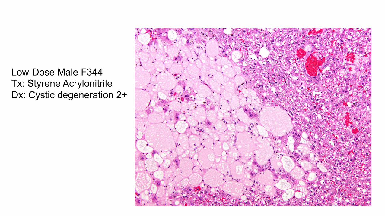
Low-Dose Male F344 Tx: Styrene Acrylonitrile Dx: Cystic degeneration 2+
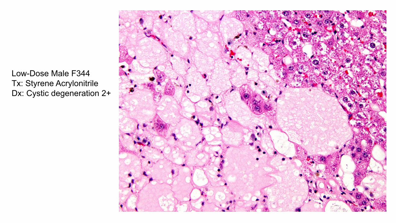
Low-Dose Male F344 Tx: Styrene Acrylonitrile Dx: Cystic degeneration 2+


Low Dose Female Sprague-Dawley Tx: Tetrachloroazobenzene Dx: Angiectasis

Low Dose Female Sprague-Dawley Tx: Tetrachloroazobenzene Dx: Angiectasis

Biliary Cysts
can also be induced by xenobiotics (Schoental and Magee1959; Jones and Butler 1975; Singh et al. 2007; Nyska et al.2002; Guzman and Solter 2002; Lalwani et al. 1997; Travloset al. 1996; Kari et al. 1995; Herman et al. 2002). In addition,multinucleated cells (formed by cell fusion rather than divi-sion) can be formed in rats after administration of 2, 3, 7, 8-tetrachloro-dibenzo-p-dioxin (Gopinath, Prentice, and Lewis1987; Jones and Butler 1975). Eosinophilic cytoplasmic inclu-sions may be seen in affected hepatocyte nuclei because of cellmembrane invaginations.
Cysts, Biliary (Hepatic Cysts) (Figures 67–69)
Pathogenesis: More common in aging animals occurs as adilation of biliary structures.
Diagnostic features:
! Range in size from small to very large.! Single or multiple cysts.! Macroscopically may contain clear to pale yellow
fluid.! May occur anywhere in the liver and may be unilocu-
lar or multilocular.! Multilocular cysts are divided into variably sized
compartments by partial or complete connectivetissue septa.
! Cyst walls are characteristically lined by flattened tocuboidal epithelium.
! May be mild compression of adjacent hepaticparenchyma.
Differential diagnosis:
! Cystic degeneration—consists of markedly enlargedcells with finely flocculent pale eosinophilic cytoplasm.
! Angiectasis (Peliosis hepatis)—dilated vascular spaceslined by endothelial cells; may contain blood cells.
! Bile duct dilation—dilated bile ducts lined by cuboi-dal epithelium; not multiloculated.
! Parasitic cyst—may have thickened wall and containparasite tissue.
! Cholangioma—may cause compression of adjacentparenchyma and spaces lined by more endothelialcells than in biliary cysts.
Comment: Biliary cysts are commonly seen in older rats (Burek1978; Greaves 2007; Harada et al. 1999). Solitary cysts can beobserved without major adjacent morphology changes of the sur-rounding tissue. However, depending on the location, adjacentparenchyma may contain pressure atrophy of the hepatic cordsof the liver, fibrosis, hemosiderin deposition, proliferation of bileducts, or periportal lymphocytic infiltration. Single cysts areoften caused by cystic dilatation of the intrahepatic bile ducts(Sato et al. 2005). Multiple cysts are observed also in hepaticpolycystic disease, where they occur alone or in combinationwith polycystic kidney disease (Masyuk et al. 2004, 2007). They
are often referred to as simple or multiloculated biliary cysts(Goodman et al. 1994). Polycystic liver can be observed inthe rat (Muff et al. 2006; Sato et al. 2006) and hamster (Percyand Barthold 2001), resembling Caroli’s disease in humans(Clemens et al. 1980; Numan et al. 1986; Serra, Recalde, andMartellotto 1987). The cysts seen in polycystic disease are mul-tiple and seen diffusely throughout the liver and are of variablesize but generally large compared to the smaller biliary cysts.
C. Inflammatory Cell Infiltrates and Hepatic Inflammation(Hepatitis)
Introduction
A variety of focal, multifocal, and more generalized infiltra-tions of inflammatory cells are frequently present in liver tis-sue. Changes range from acute inflammatory cell infiltrate(s)or occasional aggregates of lymphocytes/lymphohistiocyticcells/foci of mononuclear cells without associated alterationsof adjacent hepatocytes, to large panlobular patches of distincthepatocyte necrosis accompanied by polymorphonuclear andmononuclear (lymphocytes, plasma cells, macrophages) cellu-lar infiltrates. ‘‘Mononuclear cell’’ can be used when there is amixture of cell types (lymphocytes; less often macrophages andplasma cells) or the cell type is mononuclear but cannot beunequivocally identified in H&E stain. If a cell type predomi-nates, then the infiltrate should be classified as lymphocytic,plasmacytic, or histiocytic. While etiological agents (e.g., bac-teria, virus, parasite) may be present, in most safety assessmentstudies the causes of significant inflammation are either crypticor are attributed to a specific treatment regimen. Inflammatoryreactions in the liver may be accompanied by oval cell andfibroblast proliferation and the propensity for hepatocellularproliferative responses to replace lost parenchyma.
It is recommended that use of the diagnostic term‘‘inflammation’’ for the liver should be used sparingly. Liverinflammation (hepatitis) is operationally defined as a constella-tion of changes that represent a severe and generalized liverreaction and would require multiple diagnostic terms to ade-quately characterize (Figure 70). This type of reaction is nottypically encountered in conventional rodent toxicity studies.
Traditionally, hepatic inflammatory responses have beenclassified as acute, subacute, chronic, granulomatous, and soon. These terms are somewhat interpretative, lack precise def-initions, vary depending upon study duration, usually do notconsist of a singular cell type, and do not have exclusivepathognomic features. A more descriptive approach is recom-mended and can be qualified by lesion distribution or use ofsubclassification and discretionary qualifiers (Figure 71).
Infiltration, Inflammatory Cell
Pathogenesis: Infiltration of different inflammatory cells istypically a response to parenchymal cell death with causesranging from infectious agents, exposure to toxicants, genera-tion of toxic metabolites, and tissue anoxia.
Vol. 38, No. 7S, 2010 LESIONS OF THE HEPATOBILIARY SYSTEM 19S
at Society of Toxicologic Pathology on August 8, 2013tpx.sagepub.comDownloaded from

Biliary Cysts


BiliaryCysts
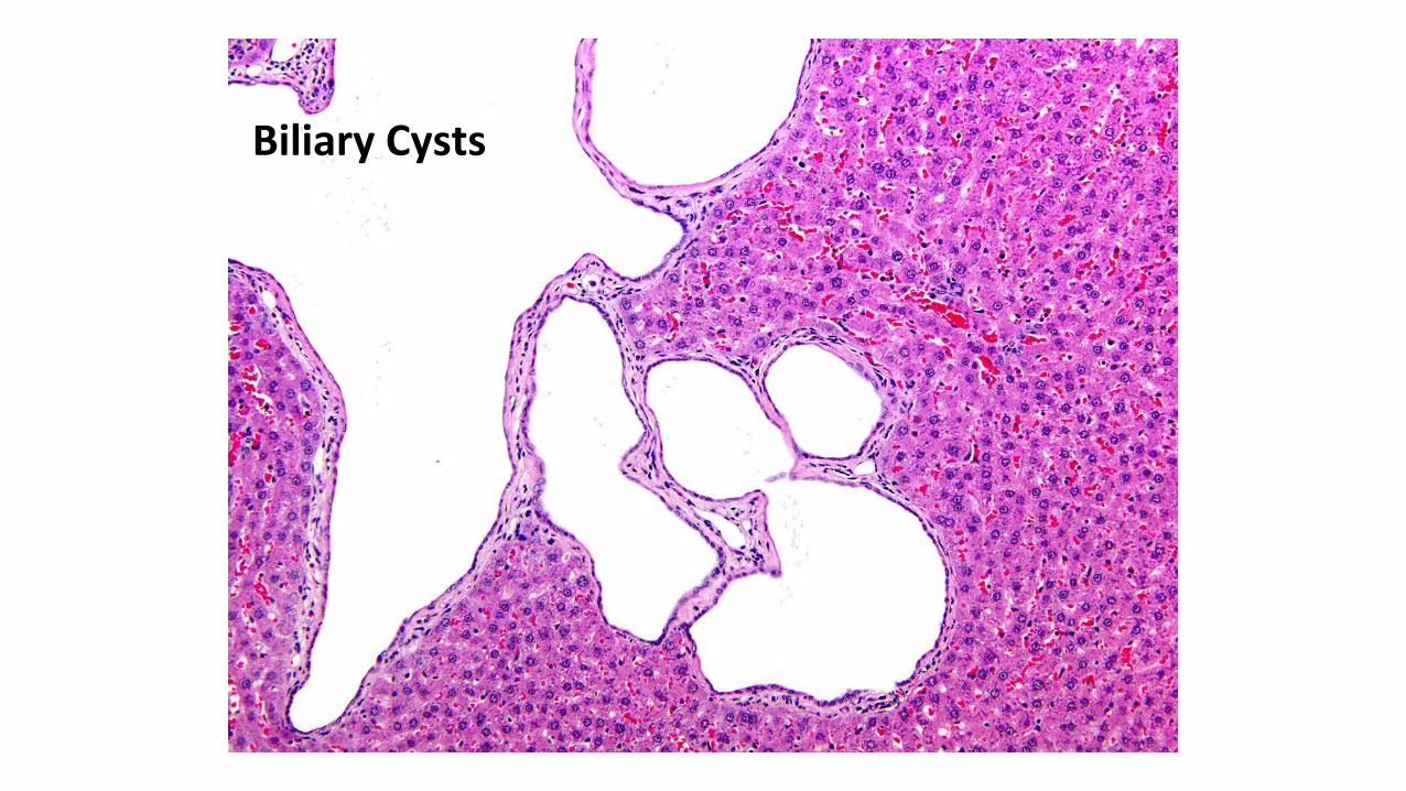
BiliaryCysts
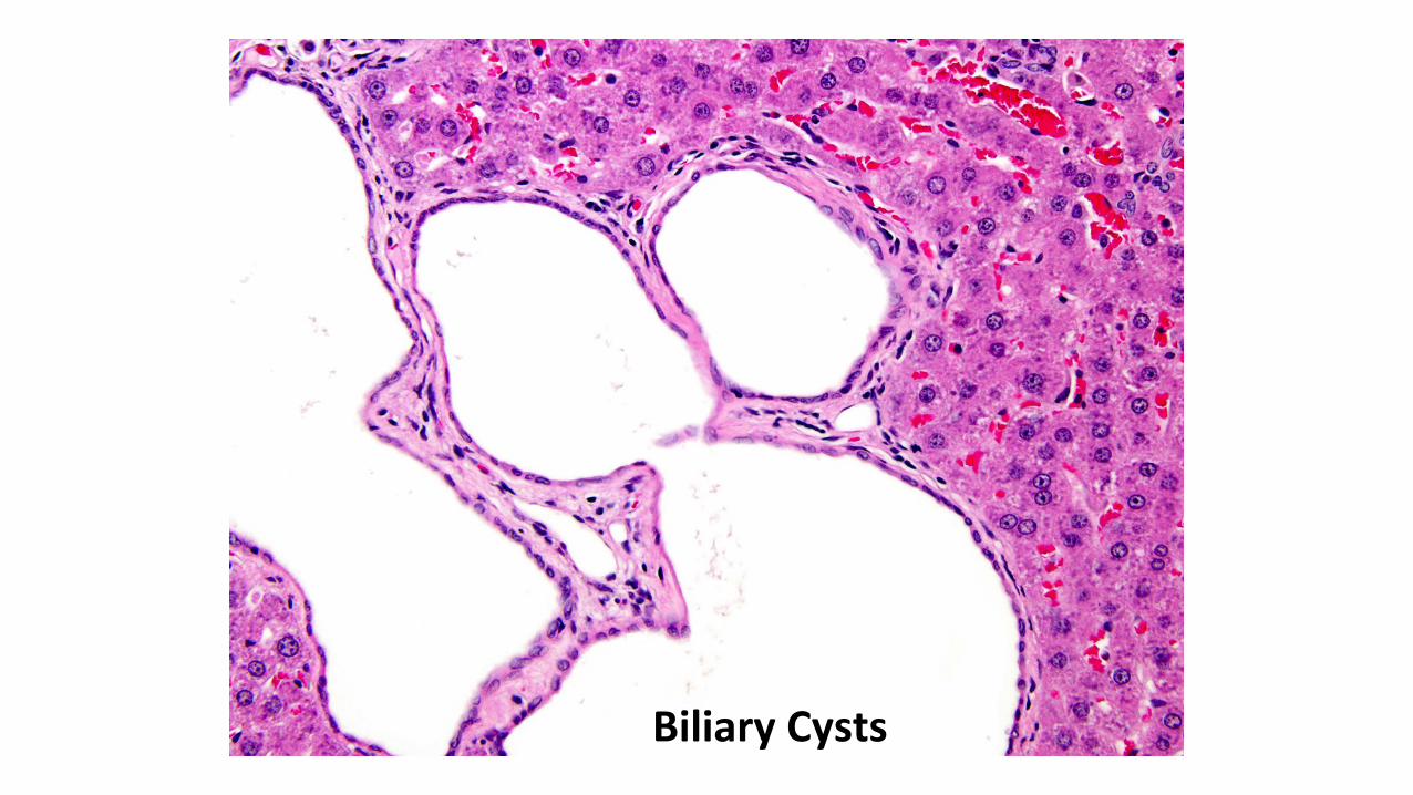
BiliaryCysts

Biliary Cysts

Apoptosis Differential diagnosis:
! Angiectasis (Peliosis hepatis; vascular ectasia)—dilated endothelial lined vascular spaces that oftencontain blood cells.
! Sinusoidal dilatation—sinusoidal structure evidentand spaces lined by endothelial cells.
! Necrosis—loss of nuclear and cytoplasmic detail andloss of stain affinity.
! Hemangioma—expansile structure lined by flattenedendothelial cells; may be associated with parenchy-mal compression.
! Hemangiosarcoma—expansile mass lined by plumpendothelial cells and/or layers of endothelial cellsand associated with destruction of hepaticparenchyma.
Comment: Spontaneous and xenobiotic-induced cystic degen-eration/spongiosis hepatis may occur in rats (Karbe and Kerlin2002; Bannasch 2003; Babich et al. 2004; Newton et al. 2001).It is more common in aging rats with some male predilection. Itis less common in mice. This lesion may be seen in or associ-ated with other hepatic lesions (necrosis, regeneration; foci ofcellular alteration; hepatocellular neoplasms). The pathogen-esis is not fully understood (Bannasch, Block, and Zerban1981; Karbe and Kerlin 2002).
Cell Death (Necrosis, Apoptosis)
Introduction: In the fully developed organism, cell death is theultimate result of irreversible cellular injury. Cellular death inthe liver is manifested by a spectrum of morphological pat-terns that can occur alone or in combinations. However, thereare two primary manifestations of cell death: necrosis andapoptosis. For decades a form of necrosis involving individualisolated hepatocytes has been diagnosed as ‘‘single cell necro-sis.’’ This particular change is now regarded as ‘‘apoptosis’’by most pathologists (Levin 1999; Levin et al. 1999; Elmore2007) when the majority of injured cells have the typicalapoptotic morphology. Provided that there is no accompany-ing inflammatory reaction, the two terms are synonymous.However, since a diagnosis of ‘‘apoptosis’’ implies a specificsequence of biochemical and morphological events andshould ideally be supported by electron microscopy, it maybe more prudent to diagnose single cell necrosis unless thereis definitive proof of apoptosis provided by electron micro-scopy (Levin et al. 1999). It can be mentioned in the pathol-ogy narrative that the observed ‘‘single cell necrosis’’ ismorphologically consistent with ‘‘apoptosis.’’
Pathogenesis: Direct or indirect cellular damage, includinganoxia. Apoptosis (a.k.a., single cell necrosis) can occur spon-taneously in liver and may also be exacerbated or induced bytreatment.
Single Cell Necrosis (Apoptosis) (Figures 48–50)
Diagnostic features:
! Affected hepatocytes may have condensed hyper-eosinophilic cytoplasm and a somewhat angular outline.
! Not associated with an inflammatory response unlessthere is simultaneous necrosis.
! May occur spontaneously with one or two affectedhepatocytes present in an occasional hepatic lobule.
! May be exacerbated by treatment.! In standard H&E-stained sections, apoptotic hepato-
cytes (apoptotic bodies) are usually rounded withcondensed cytoplasm.
! Rounded apoptotic bodies are typically surroundedby a clear halo.
! Fragments of nuclear material may be present withinaffected cells.
! Apoptotic bodies are frequently phagocytosized byadjacent normal cells including hepatocytes andmacrophages.
Differential diagnosis:
! Small foci of necrosis—typically cells are swollenand there is loss of membrane integrity; usually notrounded; less intensely stained than apoptotic bodies;may be accompanied by inflammatory cells.
Comment: Apoptosis is a form of genetically controlled‘‘programmed cell death.’’ Microscopically in H&E-stained tissue sections, apoptosis appears as dense eosino-philic shrunken cell bodies with maintenance of membraneintegrity, nuclear fragmentation and cytoplasmic budding,and without an inflammatory response. Definitive diagnosisof apoptosis can be made by histological findings andconfirmed by distinctive electron microscopic features. Con-sequently, use of a diagnosis of ‘‘single cell necrosis’’ isappropriate based strictly on H&E staining. The use ofTUNEL kits or caspase immunostaining may assist in diag-nosing apoptosis and enumerating affected cells, but necro-sis may also be immunopositive. Inhibition of apoptosisalso plays a key role in the process of carcinogenesis (Fos-ter 2000). Although apoptosis can be observed sponta-neously in the liver, certain chemicals may be able totrigger direct stimulation of pro-apoptotic pathways in hepa-tocytes (Feldmann 1997; Reed 1998). Apoptosis can alsoaccompany treatment-related zonal necrosis in the liver,especially in situations where there may be a xenobiotic-induced effect (Cullen 2005; Greaves et al. 2001).
While apoptosis represents a specific genetically pro-grammed form of cell death unaccompanied by an inflamma-tory response, there are situations where small numbers ofcells and even occasional single cells characterized by cellswelling can undergo necrosis without an inflammatoryresponse. This represents an early stage of conventional
16S THOOLEN ET AL. TOXICOLOGIC PATHOLOGY
at Society of Toxicologic Pathology on August 8, 2013tpx.sagepub.comDownloaded from

Single Cell Necrosis - Apoptosis
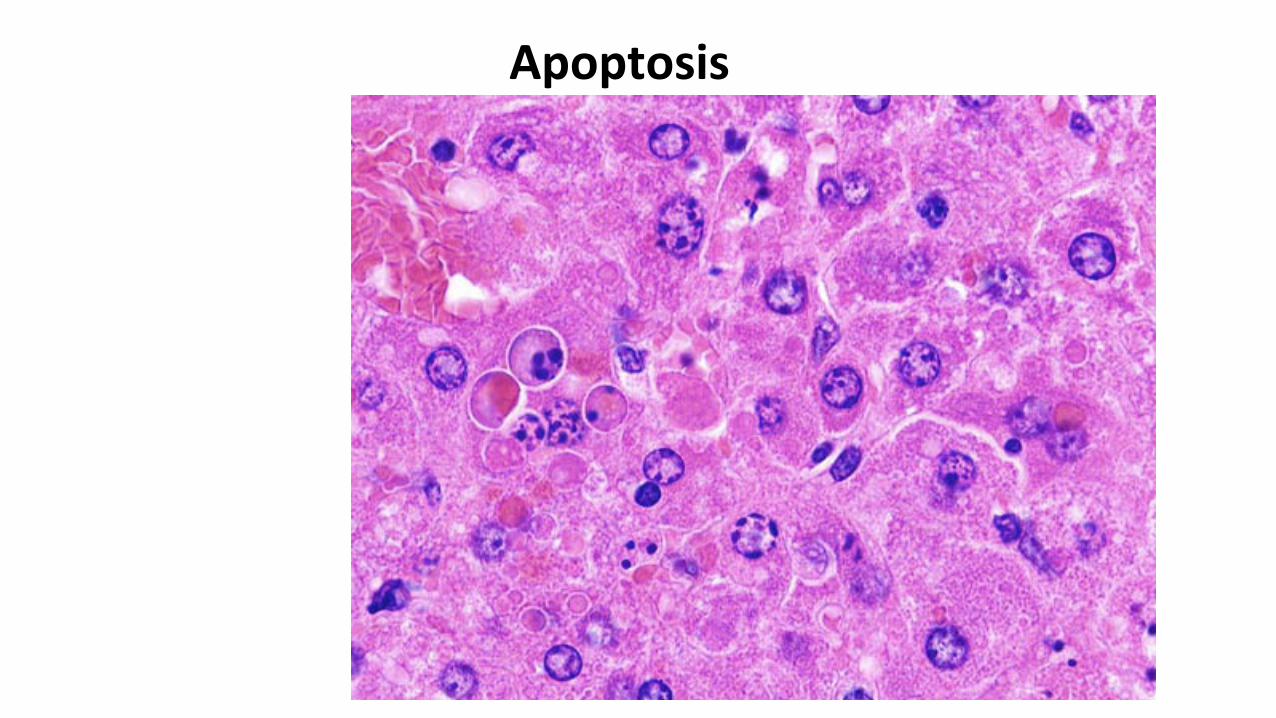
Apoptosis

Apoptosis in a Liver with Increased Peroxisomes in Hepatocytes


Necrosis necrosis, may occur within hours after exposure to a xenobio-tic, and should not be diagnosed as single cell necrosis (apop-tosis). A more appropriate diagnosis for this situation is focalnecrosis (see the following).
Necrosis, Focal/Multifocal (Figures 51–53)
Diagnostic features:
! Single or multiple foci of a few pale staininghepatocytes.
! Usually retain normal morphological outline.! May be associated with inflammation.! May have an irregular distribution but can also occur in
the subcapsular areas with minimal or no inflammation.! Early lesions typically consist of three or four hepa-
tocytes, but as the lesions progress more hepatocytesmay be involved.
! Subcapsular necrosis may sometimes be observed incombination with hypertrophy.
Differential diagnosis:
! Foci of extramedullary hematopoiesis—mature and/or immature erythroid and myeloid cell aggregateswithout accompanying hepatocyte necrosis.
! Foci of inflammatory cell infiltrate—aggregates ofcells, usually mononuclear cells, in absence of obvi-ous hepatocellular necrosis.
! Infectious disease (MHV, Ectromelia, Clostridiumpilliforme, Helicobacter hepaticus, Parvo virus,Noro virus)—a spectrum of acute to chronic activeinflammation, degenerative, and proliferative changesspecific for the infectious disease entity.
Comment: Some pathologists use focal for both focal and mul-tifocal, referring to the nature of the lesion rather than its actualdistribution. A severity grade can be used to reflect the multi-focal nature of the lesion. Focal, multifocal, and subcapsularnecrosis is occasionally seen in untreated rodents and may bea terminal event potentially due to hypoxic change secondaryto impaired blood flow. Subcapsular necrosis has also beenreported from direct pressure secondary to gastric distentionand from some types of restraint (Parker and Gibson 1995;Nyska et al. 1992)
Necrosis, Zonal (Centrilobular, Midzonal, Periportal,Diffuse)
Pathogenesis: Secondary to direct or indirect damage fromxenobiotic exposure; tissue anoxia.
Centrilobular (Figures 54–59)
Sometimes referred to as periacinar necrosis, it consists ofirreversible cell death of centrilobular hepatocytes and is often
seen after anoxia, or exposure to tannic acid, chloroform, orother hepatotoxic agents (Gopinath, Prentice, and Lewis1987). This zone (Rappaport zone 3) is particularly vulnerableto ischemic damage because of its low oxygen gradient andgeneration of toxic metabolites due to high content of xenobio-tic metabolizing enzymes (Comporti 1985; Walker, Racz, andMcElligott 1985).
Diagnostic features:
! Early necrotic hepatocytes are swollen.! Cytoplasm has increased eosinophilia.! Nucleus undergoing lysis, not pyknosis.! May have a minimal associated inflammatory
reaction.! Can be accompanied by glycogen depletion, hydro-
pic degeneration, fatty change, hemorrhage, and‘‘ballooning’’ of hepatocytes.
Midzonal (Figures 60–61)
This necrosis is the least common form of zonal necrosis andis mediated by specific toxicants (e.g., furan, concavalin-A,beryllium) (Wilson et al. 1992; Boyd 1981; Seawright 1972;Satoh et al. 1996; Cheng 1956). The location is consideredspecific and has a metabolic basis.
Diagnostic features:
! Seen as a band of swollen and eosinophilic cellsintermediate to the central vein (zone III) and the por-tal triad (zone I).
! Nucleus undergoing lysis.! Two to three cells in thickness in the middle of the
lobule.
Periportal (Figure 62)
Hepatic necrosis in the periportal zone is observed follow-ing a variety of agents (e.g., phosphorus, ferrous sulphate,allyl alcohol) (Kanel and Korula 2005; Atzori and Congiu1996; Sasse and Maly 1991). Affected cells may encircle theportal tract (Popp and Cattley 1991) and may be associatedwith inflammatory and other changes (Ward, Anver, et al.1994; Ward, Fox, et al. 1994; NTP Technical Report on theToxicology and Carcinogenesis Studies of a Binary Mixture2006; NTP Technical Report on the Toxicology and Carcino-genesis Studies of 2, 3, 7, 8-tetracholorodibenzo-p-dioxin2006).
Diagnostic features:
! Swollen and/or eosinophilic hepatocytes may com-pletely encircle the portal tract.
! Nucleus undergoing lysis.! May be accompanied by periportal inflammation, fibro-
sis, bile duct proliferation, and oval cell hyperplasia.
Vol. 38, No. 7S, 2010 LESIONS OF THE HEPATOBILIARY SYSTEM 17S
at Society of Toxicologic Pathology on August 8, 2013tpx.sagepub.comDownloaded from
Midzonal
necrosis, may occur within hours after exposure to a xenobio-tic, and should not be diagnosed as single cell necrosis (apop-tosis). A more appropriate diagnosis for this situation is focalnecrosis (see the following).
Necrosis, Focal/Multifocal (Figures 51–53)
Diagnostic features:
! Single or multiple foci of a few pale staininghepatocytes.
! Usually retain normal morphological outline.! May be associated with inflammation.! May have an irregular distribution but can also occur in
the subcapsular areas with minimal or no inflammation.! Early lesions typically consist of three or four hepa-
tocytes, but as the lesions progress more hepatocytesmay be involved.
! Subcapsular necrosis may sometimes be observed incombination with hypertrophy.
Differential diagnosis:
! Foci of extramedullary hematopoiesis—mature and/or immature erythroid and myeloid cell aggregateswithout accompanying hepatocyte necrosis.
! Foci of inflammatory cell infiltrate—aggregates ofcells, usually mononuclear cells, in absence of obvi-ous hepatocellular necrosis.
! Infectious disease (MHV, Ectromelia, Clostridiumpilliforme, Helicobacter hepaticus, Parvo virus,Noro virus)—a spectrum of acute to chronic activeinflammation, degenerative, and proliferative changesspecific for the infectious disease entity.
Comment: Some pathologists use focal for both focal and mul-tifocal, referring to the nature of the lesion rather than its actualdistribution. A severity grade can be used to reflect the multi-focal nature of the lesion. Focal, multifocal, and subcapsularnecrosis is occasionally seen in untreated rodents and may bea terminal event potentially due to hypoxic change secondaryto impaired blood flow. Subcapsular necrosis has also beenreported from direct pressure secondary to gastric distentionand from some types of restraint (Parker and Gibson 1995;Nyska et al. 1992)
Necrosis, Zonal (Centrilobular, Midzonal, Periportal,Diffuse)
Pathogenesis: Secondary to direct or indirect damage fromxenobiotic exposure; tissue anoxia.
Centrilobular (Figures 54–59)
Sometimes referred to as periacinar necrosis, it consists ofirreversible cell death of centrilobular hepatocytes and is often
seen after anoxia, or exposure to tannic acid, chloroform, orother hepatotoxic agents (Gopinath, Prentice, and Lewis1987). This zone (Rappaport zone 3) is particularly vulnerableto ischemic damage because of its low oxygen gradient andgeneration of toxic metabolites due to high content of xenobio-tic metabolizing enzymes (Comporti 1985; Walker, Racz, andMcElligott 1985).
Diagnostic features:
! Early necrotic hepatocytes are swollen.! Cytoplasm has increased eosinophilia.! Nucleus undergoing lysis, not pyknosis.! May have a minimal associated inflammatory
reaction.! Can be accompanied by glycogen depletion, hydro-
pic degeneration, fatty change, hemorrhage, and‘‘ballooning’’ of hepatocytes.
Midzonal (Figures 60–61)
This necrosis is the least common form of zonal necrosis andis mediated by specific toxicants (e.g., furan, concavalin-A,beryllium) (Wilson et al. 1992; Boyd 1981; Seawright 1972;Satoh et al. 1996; Cheng 1956). The location is consideredspecific and has a metabolic basis.
Diagnostic features:
! Seen as a band of swollen and eosinophilic cellsintermediate to the central vein (zone III) and the por-tal triad (zone I).
! Nucleus undergoing lysis.! Two to three cells in thickness in the middle of the
lobule.
Periportal (Figure 62)
Hepatic necrosis in the periportal zone is observed follow-ing a variety of agents (e.g., phosphorus, ferrous sulphate,allyl alcohol) (Kanel and Korula 2005; Atzori and Congiu1996; Sasse and Maly 1991). Affected cells may encircle theportal tract (Popp and Cattley 1991) and may be associatedwith inflammatory and other changes (Ward, Anver, et al.1994; Ward, Fox, et al. 1994; NTP Technical Report on theToxicology and Carcinogenesis Studies of a Binary Mixture2006; NTP Technical Report on the Toxicology and Carcino-genesis Studies of 2, 3, 7, 8-tetracholorodibenzo-p-dioxin2006).
Diagnostic features:
! Swollen and/or eosinophilic hepatocytes may com-pletely encircle the portal tract.
! Nucleus undergoing lysis.! May be accompanied by periportal inflammation, fibro-
sis, bile duct proliferation, and oval cell hyperplasia.
Vol. 38, No. 7S, 2010 LESIONS OF THE HEPATOBILIARY SYSTEM 17S
at Society of Toxicologic Pathology on August 8, 2013tpx.sagepub.comDownloaded from
Focal,MulJfocal
necrosis, may occur within hours after exposure to a xenobio-tic, and should not be diagnosed as single cell necrosis (apop-tosis). A more appropriate diagnosis for this situation is focalnecrosis (see the following).
Necrosis, Focal/Multifocal (Figures 51–53)
Diagnostic features:
! Single or multiple foci of a few pale staininghepatocytes.
! Usually retain normal morphological outline.! May be associated with inflammation.! May have an irregular distribution but can also occur in
the subcapsular areas with minimal or no inflammation.! Early lesions typically consist of three or four hepa-
tocytes, but as the lesions progress more hepatocytesmay be involved.
! Subcapsular necrosis may sometimes be observed incombination with hypertrophy.
Differential diagnosis:
! Foci of extramedullary hematopoiesis—mature and/or immature erythroid and myeloid cell aggregateswithout accompanying hepatocyte necrosis.
! Foci of inflammatory cell infiltrate—aggregates ofcells, usually mononuclear cells, in absence of obvi-ous hepatocellular necrosis.
! Infectious disease (MHV, Ectromelia, Clostridiumpilliforme, Helicobacter hepaticus, Parvo virus,Noro virus)—a spectrum of acute to chronic activeinflammation, degenerative, and proliferative changesspecific for the infectious disease entity.
Comment: Some pathologists use focal for both focal and mul-tifocal, referring to the nature of the lesion rather than its actualdistribution. A severity grade can be used to reflect the multi-focal nature of the lesion. Focal, multifocal, and subcapsularnecrosis is occasionally seen in untreated rodents and may bea terminal event potentially due to hypoxic change secondaryto impaired blood flow. Subcapsular necrosis has also beenreported from direct pressure secondary to gastric distentionand from some types of restraint (Parker and Gibson 1995;Nyska et al. 1992)
Necrosis, Zonal (Centrilobular, Midzonal, Periportal,Diffuse)
Pathogenesis: Secondary to direct or indirect damage fromxenobiotic exposure; tissue anoxia.
Centrilobular (Figures 54–59)
Sometimes referred to as periacinar necrosis, it consists ofirreversible cell death of centrilobular hepatocytes and is often
seen after anoxia, or exposure to tannic acid, chloroform, orother hepatotoxic agents (Gopinath, Prentice, and Lewis1987). This zone (Rappaport zone 3) is particularly vulnerableto ischemic damage because of its low oxygen gradient andgeneration of toxic metabolites due to high content of xenobio-tic metabolizing enzymes (Comporti 1985; Walker, Racz, andMcElligott 1985).
Diagnostic features:
! Early necrotic hepatocytes are swollen.! Cytoplasm has increased eosinophilia.! Nucleus undergoing lysis, not pyknosis.! May have a minimal associated inflammatory
reaction.! Can be accompanied by glycogen depletion, hydro-
pic degeneration, fatty change, hemorrhage, and‘‘ballooning’’ of hepatocytes.
Midzonal (Figures 60–61)
This necrosis is the least common form of zonal necrosis andis mediated by specific toxicants (e.g., furan, concavalin-A,beryllium) (Wilson et al. 1992; Boyd 1981; Seawright 1972;Satoh et al. 1996; Cheng 1956). The location is consideredspecific and has a metabolic basis.
Diagnostic features:
! Seen as a band of swollen and eosinophilic cellsintermediate to the central vein (zone III) and the por-tal triad (zone I).
! Nucleus undergoing lysis.! Two to three cells in thickness in the middle of the
lobule.
Periportal (Figure 62)
Hepatic necrosis in the periportal zone is observed follow-ing a variety of agents (e.g., phosphorus, ferrous sulphate,allyl alcohol) (Kanel and Korula 2005; Atzori and Congiu1996; Sasse and Maly 1991). Affected cells may encircle theportal tract (Popp and Cattley 1991) and may be associatedwith inflammatory and other changes (Ward, Anver, et al.1994; Ward, Fox, et al. 1994; NTP Technical Report on theToxicology and Carcinogenesis Studies of a Binary Mixture2006; NTP Technical Report on the Toxicology and Carcino-genesis Studies of 2, 3, 7, 8-tetracholorodibenzo-p-dioxin2006).
Diagnostic features:
! Swollen and/or eosinophilic hepatocytes may com-pletely encircle the portal tract.
! Nucleus undergoing lysis.! May be accompanied by periportal inflammation, fibro-
sis, bile duct proliferation, and oval cell hyperplasia.
Vol. 38, No. 7S, 2010 LESIONS OF THE HEPATOBILIARY SYSTEM 17S
at Society of Toxicologic Pathology on August 8, 2013tpx.sagepub.comDownloaded from
Centrilobular
necrosis, may occur within hours after exposure to a xenobio-tic, and should not be diagnosed as single cell necrosis (apop-tosis). A more appropriate diagnosis for this situation is focalnecrosis (see the following).
Necrosis, Focal/Multifocal (Figures 51–53)
Diagnostic features:
! Single or multiple foci of a few pale staininghepatocytes.
! Usually retain normal morphological outline.! May be associated with inflammation.! May have an irregular distribution but can also occur in
the subcapsular areas with minimal or no inflammation.! Early lesions typically consist of three or four hepa-
tocytes, but as the lesions progress more hepatocytesmay be involved.
! Subcapsular necrosis may sometimes be observed incombination with hypertrophy.
Differential diagnosis:
! Foci of extramedullary hematopoiesis—mature and/or immature erythroid and myeloid cell aggregateswithout accompanying hepatocyte necrosis.
! Foci of inflammatory cell infiltrate—aggregates ofcells, usually mononuclear cells, in absence of obvi-ous hepatocellular necrosis.
! Infectious disease (MHV, Ectromelia, Clostridiumpilliforme, Helicobacter hepaticus, Parvo virus,Noro virus)—a spectrum of acute to chronic activeinflammation, degenerative, and proliferative changesspecific for the infectious disease entity.
Comment: Some pathologists use focal for both focal and mul-tifocal, referring to the nature of the lesion rather than its actualdistribution. A severity grade can be used to reflect the multi-focal nature of the lesion. Focal, multifocal, and subcapsularnecrosis is occasionally seen in untreated rodents and may bea terminal event potentially due to hypoxic change secondaryto impaired blood flow. Subcapsular necrosis has also beenreported from direct pressure secondary to gastric distentionand from some types of restraint (Parker and Gibson 1995;Nyska et al. 1992)
Necrosis, Zonal (Centrilobular, Midzonal, Periportal,Diffuse)
Pathogenesis: Secondary to direct or indirect damage fromxenobiotic exposure; tissue anoxia.
Centrilobular (Figures 54–59)
Sometimes referred to as periacinar necrosis, it consists ofirreversible cell death of centrilobular hepatocytes and is often
seen after anoxia, or exposure to tannic acid, chloroform, orother hepatotoxic agents (Gopinath, Prentice, and Lewis1987). This zone (Rappaport zone 3) is particularly vulnerableto ischemic damage because of its low oxygen gradient andgeneration of toxic metabolites due to high content of xenobio-tic metabolizing enzymes (Comporti 1985; Walker, Racz, andMcElligott 1985).
Diagnostic features:
! Early necrotic hepatocytes are swollen.! Cytoplasm has increased eosinophilia.! Nucleus undergoing lysis, not pyknosis.! May have a minimal associated inflammatory
reaction.! Can be accompanied by glycogen depletion, hydro-
pic degeneration, fatty change, hemorrhage, and‘‘ballooning’’ of hepatocytes.
Midzonal (Figures 60–61)
This necrosis is the least common form of zonal necrosis andis mediated by specific toxicants (e.g., furan, concavalin-A,beryllium) (Wilson et al. 1992; Boyd 1981; Seawright 1972;Satoh et al. 1996; Cheng 1956). The location is consideredspecific and has a metabolic basis.
Diagnostic features:
! Seen as a band of swollen and eosinophilic cellsintermediate to the central vein (zone III) and the por-tal triad (zone I).
! Nucleus undergoing lysis.! Two to three cells in thickness in the middle of the
lobule.
Periportal (Figure 62)
Hepatic necrosis in the periportal zone is observed follow-ing a variety of agents (e.g., phosphorus, ferrous sulphate,allyl alcohol) (Kanel and Korula 2005; Atzori and Congiu1996; Sasse and Maly 1991). Affected cells may encircle theportal tract (Popp and Cattley 1991) and may be associatedwith inflammatory and other changes (Ward, Anver, et al.1994; Ward, Fox, et al. 1994; NTP Technical Report on theToxicology and Carcinogenesis Studies of a Binary Mixture2006; NTP Technical Report on the Toxicology and Carcino-genesis Studies of 2, 3, 7, 8-tetracholorodibenzo-p-dioxin2006).
Diagnostic features:
! Swollen and/or eosinophilic hepatocytes may com-pletely encircle the portal tract.
! Nucleus undergoing lysis.! May be accompanied by periportal inflammation, fibro-
sis, bile duct proliferation, and oval cell hyperplasia.
Vol. 38, No. 7S, 2010 LESIONS OF THE HEPATOBILIARY SYSTEM 17S
at Society of Toxicologic Pathology on August 8, 2013tpx.sagepub.comDownloaded from
PeriportalDiffuse (Figures 63 and 64)
Synonym: Massive necrosis, panlobular necrosis.
Diagnostic features:
! Necrosis involving a large portion of a liver lobe.! May be associated with torsion of a liver lobe.! May be randomly distributed throughout the liver
without a specific lobular localization.
Differential diagnosis:
! Autolysis—loss of microscopic tissue structure andstain affinity; pale eosinophilic staining and absenceof nuclear detail.
! Torsion of a liver lobe—Affects an entire liver lobe,loss of microscopic structure.
! Infarction—usually an angularly shaped wedge orarea of tissue necrosis; may be associated with anearby thrombus.
Comment: Zonal necrosis is typically associated with expo-sure to xenobiotics that either directly damage hepatocytesor cause damage following metabolic activation by endo-genous or induced enzymes. There is often a concentrationgradient within the hepatic lobule with more extensive lob-ular involvement associated with higher doses of the toxicagent.
Hepatocellular necrosis can occur spontaneously in rodentsor be induced by xenobiotics, toxins, or following treatment athigh dosages with associated tissue anoxia, circulatoryderangements, and biliary stasis. Necrosis (centrilobular, mid-zonal, periportal) might be accompanied by other histologicalchanges (fatty change, congestion, hemorrhage, inflammation,bile stasis, fibrosis, etc.) to form a myriad of pathologicalchanges. The distribution might also cross certain zones andmay manifest as ‘‘bridging necrosis’’ showing confluence ofthe lesions (e.g., central to central veins, portal tract to portaltract, or portal tract to central zones). Bridging necrosis mayultimately give rise to bridging fibrosis.
A specific form of necrosis, ‘‘piecemeal necrosis,’’ is char-acterized by necrosis of the limiting plate of the portal tract atthe interface of hepatocytes and connective tissue of the portaltract, usually accompanied by inflammation, can be immune-mediated, and is seen in mice with resemblance to chronicactive hepatitis in man (Kitamura et al. 1992; Nonomuraet al. 1991; Kuriki et al. 1983).
Karyocytomegaly and/or Multinucleated Hepatocytes(Figures 65 and 66)
Synonyms: Karyocytomegaly, multinucleated hepato-cytes, binucleated hepatocytes, karyomegaly, nuclearhypertrophy, hepatocytomegaly, polyploidy, anisonucleosis,anisokaryosis.
Pathogenesis: Duplication of nuclear material in absence ofcytokinesis. Variations in nuclear size and ploidy (karyome-galy and/or anisokaryosis) are common in aging rodents.
Diagnostic features:
! Hepatocytes with either two or more nuclei or with asingle enlarged nucleus which may be tetraploid oroctaploid.
! Polyploid hepatocytes are frequently larger than adja-cent diploid hepatocytes.
! Anisokaryosis is randomly distributed in the hepaticlobule with more affected hepatocytes in the centri-lobular region.
Differential diagnosis:
! Hepatocellular neoplasia—mass or expansile prolif-eration of hepatocytes with distortion or loss of lobu-lar architecture.
! Hepatocellular hypertrophy (enzyme induction)—increase in cytoplasmic volume not typically associ-ated with increased nuclear size or number.
Comment: Karyocytomegaly is a reflection of hepatocytepolyploidy that occurs when there is duplication of nuclearmaterial in the absence of cytokinesis. The result is an increasein the number of diploid nuclei per hepatocyte or an increase inthe ploidy level of a single hepatocyte nucleus. Polyploidyincreases with age in some strains of mice as well as followingsome treatment regimens resulting in hepatocytomegaly aswell as karyomegaly (Harada et al. 1996). Variations in cellsize as well as in nuclei and polyploidy are also common inaging rats of different strains. Karyomegaly and anisokaryosisare normal incidental findings, especially in older mice (Percyand Barthold 2001). Increase in cell size (cytomegaly) mayaccompany the increase in hepatocyte ploidy. Anisokaryosis(inequality in size of nuclei) is more common and dramaticin mice than in rats.
The development of polyploidy and its pattern vary amongstrains. C3H and DBA mice more commonly have octaploidcells with two tetraploid nuclei in adult liver while NZB and theout-bred strain NMRI at the corresponding age show a higherproportion of diploid cells with strikingly low proportions oftetraploid cells. Polyploidy has been observed in the early life(three weeks) in Ercc1 null mice. This premature polyploidy inErcc1-deficient liver is most likely caused by increased levelsof p21 in response to accumulating DNA damage (Chipchase,O’Neill, and Melton 2003). Toxic injury caused by chemicalssuch as phenobarbitone (Martin et al. 2001) and partial hepa-tectomy also induce an increase in ploidy, usually associatedwith extensive but transient hepatocyte proliferation (Gerlynget al. 1993).
Although anisonucleosis (polyploidy) is known to occur asan age-related phenomenon, the nuclear and cellular changes
18S THOOLEN ET AL. TOXICOLOGIC PATHOLOGY
at Society of Toxicologic Pathology on August 8, 2013tpx.sagepub.comDownloaded from
Diffuse

Necrosis

Necrosis, Focal

Necrosis

CentrilobularHepatocyteNecrosis
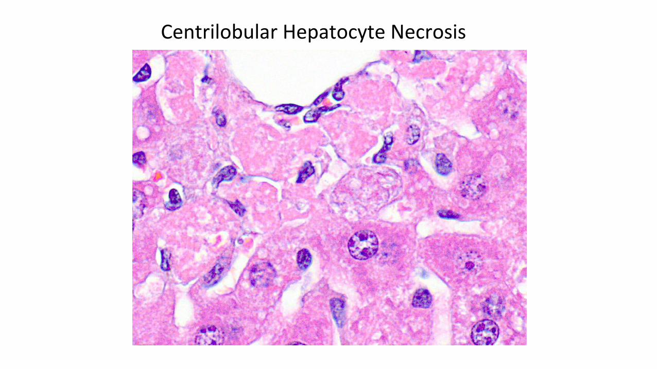
CentrilobularHepatocyteNecrosis

CentrilobularHepatocyteNecrosis
Subchronic study B6C3F1 Tx: Divinyl benzene
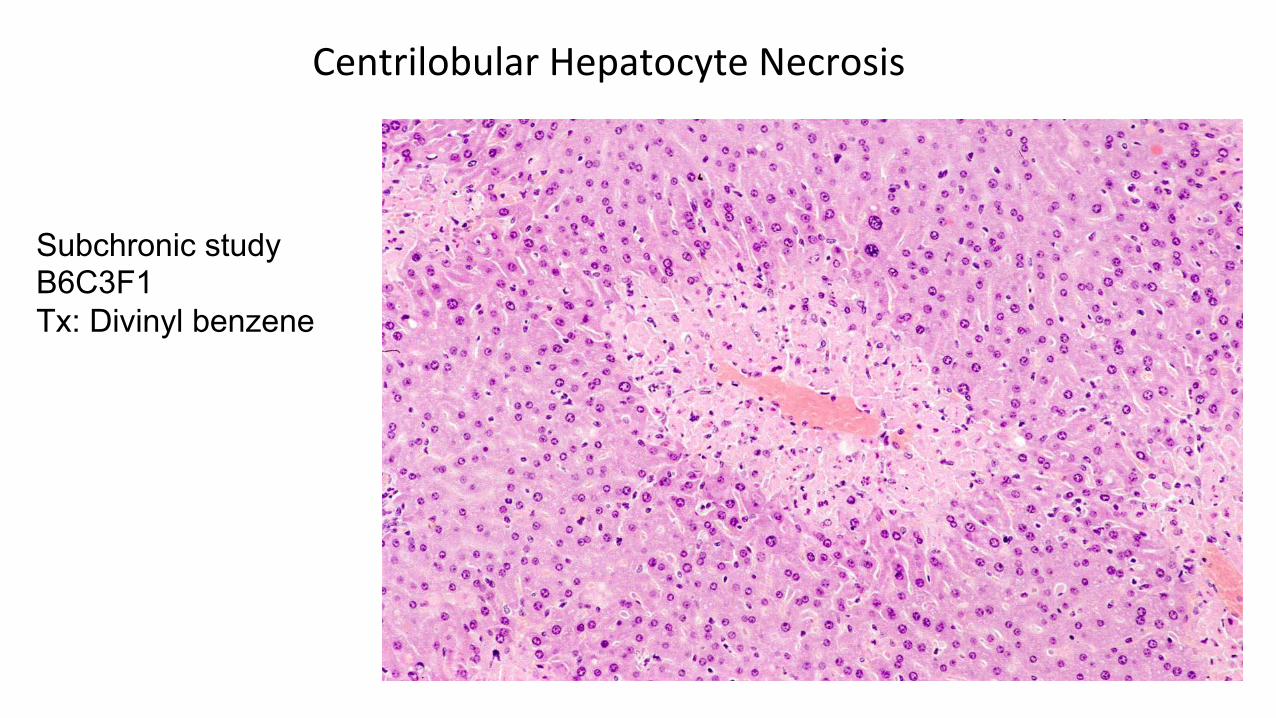
CentrilobularHepatocyteNecrosis
Subchronic study B6C3F1 Tx: Divinyl benzene

Periportal Necrosis

Mid-lobular Necrosis

Severe Necrosis with Bridging
Male F344 Rat High-dose Tx: Coumarin Dx: Coagulation necrosis

Centrilobular Necrosis with Hemorrhage and Mineralization
Acute necrosis Tx: Bromobenzene

Centrilobular Necrosis with Hemorrhage and Mineralization
Acute necrosis Tx: Bromobenzene

Necrosis


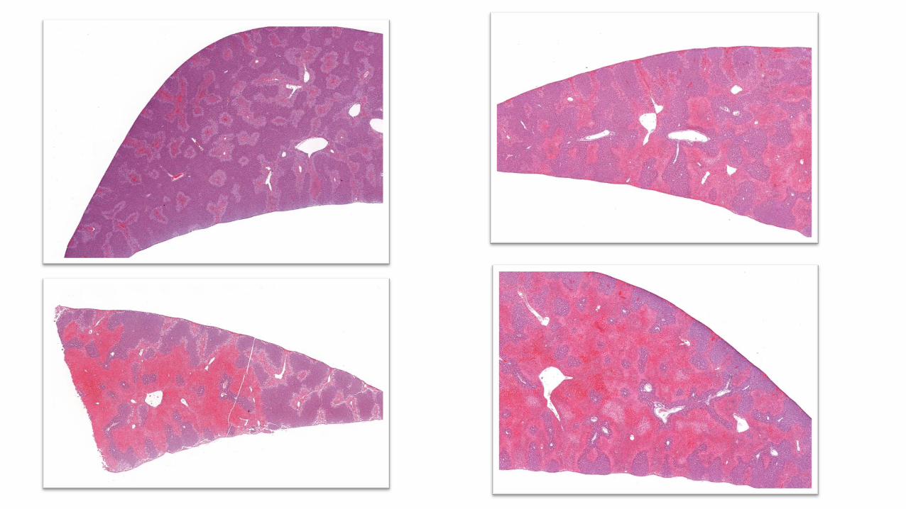


Teutsch, et al. 1999. Hepatology 29:494-505

Thrombosis
liver after treatment with nitrosamines, pyrrolizidine alkaloidsor glucocorticoids (Greaves 2007; Ruebner, Watanabe, andWand 1970; Ungar 1986; Wolstenholme and Gardner 1950).Altered hemodynamic and changes in the hepatic microcircula-tion have been proposed to be of importance in the pathogen-esis of sinusoidal dilatation (Slehria et al. 2002). Subcapsularsinusoidal dilatation can also be found a postmortem findingin rats (Kimura and Abe 1994).
Angiectasis is defined by multiple blood-filled cystic spacesof different size and shape occurs after suspected loss or weaken-ing of sinusoidal walls and/or supporting tissue. The two subtypesmay occur in combination. The cystic spaces are devoid ofendothelial lining in the cavernous subtype (‘‘parenchymal’’)although controversy still exists (Greaves 2007; Edwards,Colombo, and Greaves 2002). Angiectasis can be found in agedrats (Lee 1983). However, these lesions can also be induced inboth rats and mice after viral infection (Bergs and Scotti 1967)or exposure to certain drugs or chemicals (Mendenhall andChedid 1980; Husztik, Lazar, and Szabo 1984).
Angiectasis has also been reported in animals or humansinfected with Bartonella spp. (Wong et al. 2001; Breitschwerdtand Kordick 2000) and it has been associated with a number ofdiseases in humans as well as administration of anabolic steroidsand oral contraceptives (Naeim, Cooper, and Semion 1973;Zimmerman 1998; Tsokos and Erbersdobler 2005).
Angiectasis may refer to a vascular lesion formed by dilatationof a group of small blood vessels. It can be observed in transgenicmouse models (Srinivasan et al. 2003; Bourdeau et al. 2001) orrelated to xenobiotic administration in rats and mice (Robisonet al. 1984; Kim et al. 2004). It can be found as an incidental find-ing in aging mice and is sometimes associated with hepatocellu-lar neoplasms (Harada et al. 1996, 1999). Angiectasis can bechemically induced (Bannasch, Wayss, and Zerban 1997) andhas been suggested to be preneoplastic in some animal models.
Thrombosis (Figure 88)
Pathogenesis: Activation of the coagulation system associatedwith arteritis or phlebitis or secondary to atrial thrombosis.
Diagnostic features:
! It is characterized by the formation of a thrombuswithin the lumen of a blood vessel, like sinusoids andcentral veins.
! Amorphous mass attached to the endothelium or freewithin the blood vessel lumen (due to plane of section).
! Contains fibrin, platelets, and entrapped blood cells.! Damaged endothelium can be seen.! May occur with histiocytic sarcoma in rats or mice.
Differential diagnosis:
! Postmortem clot.! Necrotic area of tissue—loss of microscopic tissue
structure and stain affinity; pale eosinophilic stain-ing, and absence of nuclear detail.
Comment: There are several potential mechanisms leading toliver thrombosis. Activation of the coagulation system associ-ated with fibrin deposits and hypoxia located in the centrilob-ular sinusoids was reported to occur in the livers of ratsexposed to monocrotaline (MCT) (Copple et al. 2002). It wassuggested that the fibrin thrombi were formed followingchemical-induced hepatic endothelial cell damage. In studyingan endotoxin-exposure model, it was suggested that the notedfocal and random hepatocellular necrosis was caused by circu-latory disturbances due to fibrin thrombi in clusters of adjacentsinusoids. Using a rat model of 2-butoxyethanol induced hemo-lytic anemia associated with systemic thrombosis, fibrinthrombi were noted in the central vein and sinusoids of theliver, in addition to the presence of thrombi seen in severalother organs (Ramot et al. 2007).
Infarction (Figures 89 and 90)
Pathogenesis: Interruption of blood flow in a major vessel.Torsion of hepatic lobe.
Diagnostic features:
! Extensive area of necrosis may be associated withinflammation.
! The necrosis does not have acinar pattern.
Differential diagnosis:
! Hepatocellular necrosis resulting from direct toxicitymay have a diffuse or lobular distribution but also maybe accompanied by infarction; not mutually exclusive.
Comment: Aside from torsion of liver lobes, which can occurspontaneously in rodents, infarction is a very rare lesion thatcan be induced only under very specific experimental condi-tions. The combined injection in mice of NG-monomethyl-L-arginine and aspirin after lipopolysaccharide exposure resultedin significant hepatocellular enzyme release, characterized his-tologically by intravascular thrombosis with diffuse infarctionand necrosis (Harbrecht et al. 1994). Intraperitoneal injectionsin rats of vasoconstrictor xenobiotics such as phenylephrineproduced infarcts of the spleen regularly and infarcts of theliver occasionally (Levine and Sowinski 1985). Isolated perfu-sion with 1.0 g/kg of the cytotoxic xenobiotic 5-FU orhyperthermia of 41 degrees C " 10 min resulted in 90% to100% mortality in rats, with extensive, patchy necrosis, andinfarction on histologic examination (Miyazaki et al. 1983).
Endothelial Cell Hypertrophy/Karyomegaly3 (Figure 91)
Synonyms: Endothelial cell enlargement, cytomegaly.
Introduction: This is a relatively new diagnostic entity.Because of the difficulty of identifying specific sinusoidal cell
3 The examples provided were confirmed with special stains (not shown).
However, based solely on H&E staining, a diagnosis of ‘‘sinusoidal cell
hypertrophy/karyomegaly’’ is appropriate.
Vol. 38, No. 7S, 2010 LESIONS OF THE HEPATOBILIARY SYSTEM 25S
at Society of Toxicologic Pathology on August 8, 2013tpx.sagepub.comDownloaded from
Infarction


Karyocytomegaly and/or Multinucleated Hepatocytes (Ploidy) Diffuse (Figures 63 and 64)
Synonym: Massive necrosis, panlobular necrosis.
Diagnostic features:
! Necrosis involving a large portion of a liver lobe.! May be associated with torsion of a liver lobe.! May be randomly distributed throughout the liver
without a specific lobular localization.
Differential diagnosis:
! Autolysis—loss of microscopic tissue structure andstain affinity; pale eosinophilic staining and absenceof nuclear detail.
! Torsion of a liver lobe—Affects an entire liver lobe,loss of microscopic structure.
! Infarction—usually an angularly shaped wedge orarea of tissue necrosis; may be associated with anearby thrombus.
Comment: Zonal necrosis is typically associated with expo-sure to xenobiotics that either directly damage hepatocytesor cause damage following metabolic activation by endo-genous or induced enzymes. There is often a concentrationgradient within the hepatic lobule with more extensive lob-ular involvement associated with higher doses of the toxicagent.
Hepatocellular necrosis can occur spontaneously in rodentsor be induced by xenobiotics, toxins, or following treatment athigh dosages with associated tissue anoxia, circulatoryderangements, and biliary stasis. Necrosis (centrilobular, mid-zonal, periportal) might be accompanied by other histologicalchanges (fatty change, congestion, hemorrhage, inflammation,bile stasis, fibrosis, etc.) to form a myriad of pathologicalchanges. The distribution might also cross certain zones andmay manifest as ‘‘bridging necrosis’’ showing confluence ofthe lesions (e.g., central to central veins, portal tract to portaltract, or portal tract to central zones). Bridging necrosis mayultimately give rise to bridging fibrosis.
A specific form of necrosis, ‘‘piecemeal necrosis,’’ is char-acterized by necrosis of the limiting plate of the portal tract atthe interface of hepatocytes and connective tissue of the portaltract, usually accompanied by inflammation, can be immune-mediated, and is seen in mice with resemblance to chronicactive hepatitis in man (Kitamura et al. 1992; Nonomuraet al. 1991; Kuriki et al. 1983).
Karyocytomegaly and/or Multinucleated Hepatocytes(Figures 65 and 66)
Synonyms: Karyocytomegaly, multinucleated hepato-cytes, binucleated hepatocytes, karyomegaly, nuclearhypertrophy, hepatocytomegaly, polyploidy, anisonucleosis,anisokaryosis.
Pathogenesis: Duplication of nuclear material in absence ofcytokinesis. Variations in nuclear size and ploidy (karyome-galy and/or anisokaryosis) are common in aging rodents.
Diagnostic features:
! Hepatocytes with either two or more nuclei or with asingle enlarged nucleus which may be tetraploid oroctaploid.
! Polyploid hepatocytes are frequently larger than adja-cent diploid hepatocytes.
! Anisokaryosis is randomly distributed in the hepaticlobule with more affected hepatocytes in the centri-lobular region.
Differential diagnosis:
! Hepatocellular neoplasia—mass or expansile prolif-eration of hepatocytes with distortion or loss of lobu-lar architecture.
! Hepatocellular hypertrophy (enzyme induction)—increase in cytoplasmic volume not typically associ-ated with increased nuclear size or number.
Comment: Karyocytomegaly is a reflection of hepatocytepolyploidy that occurs when there is duplication of nuclearmaterial in the absence of cytokinesis. The result is an increasein the number of diploid nuclei per hepatocyte or an increase inthe ploidy level of a single hepatocyte nucleus. Polyploidyincreases with age in some strains of mice as well as followingsome treatment regimens resulting in hepatocytomegaly aswell as karyomegaly (Harada et al. 1996). Variations in cellsize as well as in nuclei and polyploidy are also common inaging rats of different strains. Karyomegaly and anisokaryosisare normal incidental findings, especially in older mice (Percyand Barthold 2001). Increase in cell size (cytomegaly) mayaccompany the increase in hepatocyte ploidy. Anisokaryosis(inequality in size of nuclei) is more common and dramaticin mice than in rats.
The development of polyploidy and its pattern vary amongstrains. C3H and DBA mice more commonly have octaploidcells with two tetraploid nuclei in adult liver while NZB and theout-bred strain NMRI at the corresponding age show a higherproportion of diploid cells with strikingly low proportions oftetraploid cells. Polyploidy has been observed in the early life(three weeks) in Ercc1 null mice. This premature polyploidy inErcc1-deficient liver is most likely caused by increased levelsof p21 in response to accumulating DNA damage (Chipchase,O’Neill, and Melton 2003). Toxic injury caused by chemicalssuch as phenobarbitone (Martin et al. 2001) and partial hepa-tectomy also induce an increase in ploidy, usually associatedwith extensive but transient hepatocyte proliferation (Gerlynget al. 1993).
Although anisonucleosis (polyploidy) is known to occur asan age-related phenomenon, the nuclear and cellular changes
18S THOOLEN ET AL. TOXICOLOGIC PATHOLOGY
at Society of Toxicologic Pathology on August 8, 2013tpx.sagepub.comDownloaded from

ExtremeHepatocytomegaly

Multinucleated Hepatocytes

Part 3- Hepatobiliary Non-Neoplastic
• Infiltration – Inflammation • Toxic Hepatopathy • Vascular Changes • Increased Mitosis • Foci of Cellular Alteration • Hyperplasia • Cholangiofibrosis

Infiltration - Inflammation

Small Focal Lesions
Female Swiss Mouse Subchronic Study Tx: Oxazepam

Small Focal Lesions
Thisisacommonlesionseeninrodenttoxicitystudies.
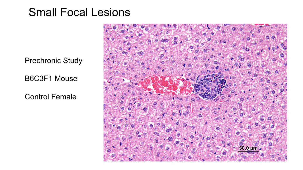
Small Focal Lesions
Prechronic Study B6C3F1 Mouse Control Female

Small Focal Lesions
Female B6C3F1 Untreated Prechronic Study Dx: Within normal limits
Female B6C3F1 Untreated Prechronic Study Dx: Inflammation

Small Focal Lesions

Small Focal Lesions

Small Focal Lesions
Focal mononuclear cell aggregates? Microgranulomas? Focal cellular infiltrates? Hematopoietic cell proliferation?

Small Focal Lesions Focal mononuclear cell aggregates? Microgranulomas? Focal cellular infiltrates? Hematopoietic cell proliferation?
ControlFemaleSpragueDawleyPrechronicStudyDx:InflammaJon,chronic1+
Focal neutrophilic infiltrate associated with hepatocyte necrosis

Hematopoietic Cell Proliferation

Hematopoietic Cell Proliferation

Hematopoietic Cell Proliferation

Hematopoietic Cell Proliferation

Granulomatous Inflammation

Chronic Inflammation
High-dose Male (#434) F344 Rat Tx: Coumarin Dx: Fibrosis
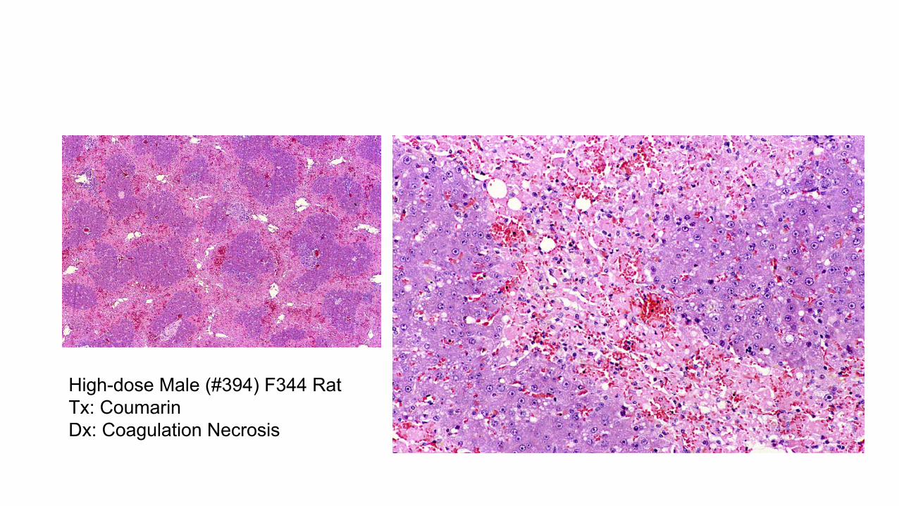
High-dose Male (#394) F344 Rat Tx: Coumarin Dx: Coagulation Necrosis

Cirrhosis – Multiple Nodules of Hepatocyte Hyperplasia Separated by Scarring and Chronic Inflammation and Oval Cell Proliferation
Treated Female Sprague Dawley Hyperplasia (Regeneration)
In a chronic inflammatory situation, there will probably be a constellations of changes.

Chronic Inflammation
F344 Rat Tx: Direct Blue 6 for 90 days Dx: Cirrhosis (fibrosis) & regenerative nodules

Toxic Hepatopathy Toxic hepatopathy is a general diagnostic term used when a constellation of microscopic changes secondary to chronic toxicity is present. Changes include hepatocyte degeneration, bile duct and oval cell hyperplasia, pigment deposition, fibrosis, and nodular hyperplasia.
Female Sprague Dawlely High-dose Tx: PCB Mixture Dx: Toxic hepatopathy

Toxic Hepatopathy
Female Sprague Dawlely High-dose Tx: PCB Mixture Dx: Toxic hepatopathy Portal fibrosis
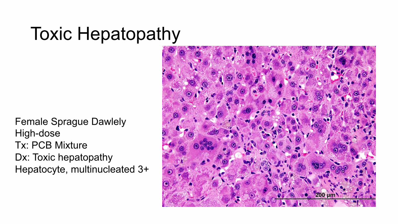
Toxic Hepatopathy
Female Sprague Dawlely High-dose Tx: PCB Mixture Dx: Toxic hepatopathy Hepatocyte, multinucleated 3+

Congestion vs Hemorrhage

Passive Congestion
vein, which carries venous blood that is largely depleted ofoxygen, and relatively minor (about 25%) arterial blood sup-ply, via the hepatic artery. The portal blood contains toxicmaterials absorbed in the intestine, and therefore the liver is thefirst tissue to be exposed to toxic substances that have beenabsorbed through the gastro-intestinal tract.
Within the hepatic parenchyma, the hepatocytes are inintimate contact with the sinusoidal capillaries, which are car-rying the mixture of blood originating from ramifications ofthe portal vein and hepatic artery to the central vein. Thesinusoids are lined by modified endothelial cells containingfenestrations, which allow passage of lipoproteins and otherlarge molecules but provide a barrier to blood cells. Kupffercells reside in the lumen of the sinusoids and are anchoredto their wall.
The morphological aspect of the hepatic pathology duringcirculatory disorders depends on the location of the vascularstructure being affected (i.e., lobular sinusoids, the outflowhepatic vein, or the inflow portal vein).
Congestion (Figure 85)
Synonym: Chronic passive congestion.
Pathogenesis: Circulatory failure, typically right-sided heartfailure.
Diagnostic features:
! Increase prominence (number) of erythrocytes in thecapillary bed or larger vessels of an organ.
! No appreciable distention (angiectasis) of the vesselwall.
! Diagnosis often correlates with gross observation(e.g., reddened, darkened focus).
! May be associated with centrilobular necrosis.
Differential diagnosis:
! Angiectasis—dilated vascular spaces lined byendothelial cells; dilated vascular channels andspaces frequently contain erythrocytes.
! Massive necrosis.! Hemorrhage-irregular patchy lakes of blood not con-
tained within defined vascular channels.! Autolysis-altered cellular texture and loss of staining
intensity.
Comment: Congestion may result from circulatory disturbancesuch as right-sided heart failure and is usually associated withnecrosis of the centrilobular areas (Burt, Portmann, andMacSween 2002). Presence of blood in hepatic sinusoids seenin animals that die or in situations where there is incompleteexsanguination should not be diagnosed as congestion. If it isabsolutely necessary to record such findings, it is importantto qualify the blood stasis in the liver as passive congestion.
Angiectasis (Figures 86 and 87)
Synonyms: Peliosis hepatis, telangiectasis, sinusoidal dilation.
Pathogenesis: Perturbations in blood flow and/or drainage;weakening of sinusoidal walls.
Diagnostic features:
! Macroscopically—seen on the surface as blood-filled, thin walled cavities projecting above the sur-face (Bannasch et al. 1997).
! Microscopically—There are two morphologicaltypes, as follows:1. Cystic (‘‘Phlebectatic’’)—Focal dilation (disten-
sion) of endothelial lined channels (photographicpresentation contributed by Hardisty et al. 2007).Can be an isolated lesion or have a multicentricform. The lacunae are densely packed with bloodand are coated by a single layer of endotheliumand separated from one another by cords of liverparenchyma (Bannasch, Wayss, and Zerban1997). The endothelial cells appear to be unal-tered, and there is no increase of mitotic figures.The tissue adjacent to the dilated sinusoids iswell preserved and free of necrotic cells.
2. Cavernous (‘‘Parenchymal’’)—The peliotic cystsare not, or are only partially, lined by endothelium.Thus, the cysts involve not only the sinusoidallumen, but also the space of Disse, and the bloodcomes in direct contact with the neighboring par-enchymal cells. The parenchyma undergoes focalnecrosis without a zonal distribution. This lesion ismuch less likely to be preneoplastic and a few tox-ins have been shown to induce the lesion. Whenendothelial lining is absent, the term PeliosisHepatis instead of Angiectasis is indicated.
Differential diagnosis:
! Hemangioma—expansile structure lined by flattenedendothelial cells; may be associated with parenchy-mal compression.
! Cystic degeneration (Spongiosis hepatis)—cavitiesare not filled with blood but with a finely flocculentacidophilic material.
Comment: Angiectasis is a cystic or cavernous widening of theliver sinusoids that can occur in a variety of pathologicalinsults. In human, sinusoidal dilatation has been reported fol-lowing hypoxia or hyperperfusion as a result of right-sidedheart failure, thrombosis of hepatic veins, amyloid deposition,granulomatous, or neoplastic disease (Greaves and Faccini1992; Bruguera et al. 1978). Although these lesions can alsooccur spontaneously in different rodents with different diseasesor with certain neoplasms causing hemodynamic changes, itcan also be induced by different compounds. Focal sinusoidaldilatation and peliosis hepatis have been observed in the rodent
24S THOOLEN ET AL. TOXICOLOGIC PATHOLOGY
at Society of Toxicologic Pathology on August 8, 2013tpx.sagepub.comDownloaded from

Angiectasis vein, which carries venous blood that is largely depleted ofoxygen, and relatively minor (about 25%) arterial blood sup-ply, via the hepatic artery. The portal blood contains toxicmaterials absorbed in the intestine, and therefore the liver is thefirst tissue to be exposed to toxic substances that have beenabsorbed through the gastro-intestinal tract.
Within the hepatic parenchyma, the hepatocytes are inintimate contact with the sinusoidal capillaries, which are car-rying the mixture of blood originating from ramifications ofthe portal vein and hepatic artery to the central vein. Thesinusoids are lined by modified endothelial cells containingfenestrations, which allow passage of lipoproteins and otherlarge molecules but provide a barrier to blood cells. Kupffercells reside in the lumen of the sinusoids and are anchoredto their wall.
The morphological aspect of the hepatic pathology duringcirculatory disorders depends on the location of the vascularstructure being affected (i.e., lobular sinusoids, the outflowhepatic vein, or the inflow portal vein).
Congestion (Figure 85)
Synonym: Chronic passive congestion.
Pathogenesis: Circulatory failure, typically right-sided heartfailure.
Diagnostic features:
! Increase prominence (number) of erythrocytes in thecapillary bed or larger vessels of an organ.
! No appreciable distention (angiectasis) of the vesselwall.
! Diagnosis often correlates with gross observation(e.g., reddened, darkened focus).
! May be associated with centrilobular necrosis.
Differential diagnosis:
! Angiectasis—dilated vascular spaces lined byendothelial cells; dilated vascular channels andspaces frequently contain erythrocytes.
! Massive necrosis.! Hemorrhage-irregular patchy lakes of blood not con-
tained within defined vascular channels.! Autolysis-altered cellular texture and loss of staining
intensity.
Comment: Congestion may result from circulatory disturbancesuch as right-sided heart failure and is usually associated withnecrosis of the centrilobular areas (Burt, Portmann, andMacSween 2002). Presence of blood in hepatic sinusoids seenin animals that die or in situations where there is incompleteexsanguination should not be diagnosed as congestion. If it isabsolutely necessary to record such findings, it is importantto qualify the blood stasis in the liver as passive congestion.
Angiectasis (Figures 86 and 87)
Synonyms: Peliosis hepatis, telangiectasis, sinusoidal dilation.
Pathogenesis: Perturbations in blood flow and/or drainage;weakening of sinusoidal walls.
Diagnostic features:
! Macroscopically—seen on the surface as blood-filled, thin walled cavities projecting above the sur-face (Bannasch et al. 1997).
! Microscopically—There are two morphologicaltypes, as follows:1. Cystic (‘‘Phlebectatic’’)—Focal dilation (disten-
sion) of endothelial lined channels (photographicpresentation contributed by Hardisty et al. 2007).Can be an isolated lesion or have a multicentricform. The lacunae are densely packed with bloodand are coated by a single layer of endotheliumand separated from one another by cords of liverparenchyma (Bannasch, Wayss, and Zerban1997). The endothelial cells appear to be unal-tered, and there is no increase of mitotic figures.The tissue adjacent to the dilated sinusoids iswell preserved and free of necrotic cells.
2. Cavernous (‘‘Parenchymal’’)—The peliotic cystsare not, or are only partially, lined by endothelium.Thus, the cysts involve not only the sinusoidallumen, but also the space of Disse, and the bloodcomes in direct contact with the neighboring par-enchymal cells. The parenchyma undergoes focalnecrosis without a zonal distribution. This lesion ismuch less likely to be preneoplastic and a few tox-ins have been shown to induce the lesion. Whenendothelial lining is absent, the term PeliosisHepatis instead of Angiectasis is indicated.
Differential diagnosis:
! Hemangioma—expansile structure lined by flattenedendothelial cells; may be associated with parenchy-mal compression.
! Cystic degeneration (Spongiosis hepatis)—cavitiesare not filled with blood but with a finely flocculentacidophilic material.
Comment: Angiectasis is a cystic or cavernous widening of theliver sinusoids that can occur in a variety of pathologicalinsults. In human, sinusoidal dilatation has been reported fol-lowing hypoxia or hyperperfusion as a result of right-sidedheart failure, thrombosis of hepatic veins, amyloid deposition,granulomatous, or neoplastic disease (Greaves and Faccini1992; Bruguera et al. 1978). Although these lesions can alsooccur spontaneously in different rodents with different diseasesor with certain neoplasms causing hemodynamic changes, itcan also be induced by different compounds. Focal sinusoidaldilatation and peliosis hepatis have been observed in the rodent
24S THOOLEN ET AL. TOXICOLOGIC PATHOLOGY
at Society of Toxicologic Pathology on August 8, 2013tpx.sagepub.comDownloaded from

Angiectasis
Female control F344 Rat Diet restriction study Dx: Angiectasis

Female control F344 Rat Diet restriction study Dx: Angiectasis

Female control F344 Rat Diet restriction study Dx: Angiectasis

Treated Male B6C3F1 Tx: Tetrafluoroethylene Dx: Angiectasis
Angiectasis

Angiectasis
Treated Male B6C3F1Tx: TetrafluoroethyleneDx: Angiectasis

Increased Mitoses

Male F344 Tx: acetaminophen for 14 days

Male F344 Tx: acetaminophen for 14 days

Foci of Cellular Alteration
alteration with a description in the pathology narrative.In some laboratories diagnosis of ‘‘increased mitoses’’ isused.
Focus of Cellular Alteration
Introduction: Foci of cellular alteration are common in rodentstudies greater than duration of twelve months and may beseen in short duration toxicity studies following exposureto certain xenobiotics. Foci of cellular alteration can be iden-tified by special stains. In H&E-stained slides they may besubclassified based on the predominant cell type present.Diagnosis of the mixed cell subtype of altered hepatic focusvaries among different laboratories. One viewpoint defines amixed focus of cellular alteration as consisting of a combina-tion of basophilic, vacuolated, eosinophilic, and/or clear cellhepatocytes without a predominant cell type. An alternativeviewpoint defines a mixed cell focus as containing any twophenotypes of cells in approximately a 50%/50% proportion.Others regard a ‘‘true’’ mixed focus as one that containsclearly identified basophilic and eosinophilic cells regardlessof the proportions of each. Because of this diverse set ofdiagnostic opinions regarding focus subtypes, the pathologistis encouraged to describe the morphological featuresof documented foci in detail, especially if they are alteredby treatment.
Synonyms: Areas of cellular alteration; focus of alteredhepatocytes; hyperplastic focus; preneoplastic focus; enzymealtered focus; phenotypically altered focus.
Pathogenesis: A localized proliferation of hepatocytes pheno-typically different from surrounding hepatocyte parenchyma.
Diagnostic features:
! May occasionally be observed grossly as small whitefoci on the liver surface, but not round nodules.
! Size may range from less than one lobule to multiplelobules in diameter.
! Circular or ovoid shape; irregular formed foci mayoccur.
! Distinguished into types of foci by virtue of tinctorialvariation, size of hepatocytes, and textural appear-ances from surrounding parenchyma.
! May be subclassified based on predominant cell type.The fact that 80% of the focus is composed of onemorphologic cell type (basophilic, eosinophilic, etc.)or a mixed cell type.
! Normally no or only minimal compression of the sur-rounding liver tissue.
! Liver plates merge imperceptible with surroundinghepatic parenchyma; nevertheless foci are sharplydemarcated from the adjacent normal hepatocytesby the appearance and staining reaction of its cells.
! Lobular architecture preserved.
! Portal areas and central veins not present in small focibut can be seen in larger foci.
! Sinusoids within focus may be compressed, sotypical parenchymal plates are difficult to detect.
! Tortuous hepatic cords may occur due to increasednumber of cells.
! Size of cells and cytoplasmic tinctorial variationdepend on type of focus.
! Normally absence of cellular atypia.! Cystic degeneration (spongiosis hepatis) and
angiectasis (peliosis hepatis) may occur within fociof altered cells.
! Cytoplasmic lipid may be present.! Intracytoplasmic inclusions of various types may be
present.
Basophilic (Figures 93–97)
Basophilic, diffuse (Figures 93 and 94).
! Hepatocytes of normal size or slightly enlarged withhomogeneously staining cytoplasmic basophilia dueto abundant free ribosomes.
! Cells may be pleomorphic with enlarged vesiculatednuclei and prominent nucleoli.
! Dissociation of cells may occur.! Mitotic figures may occur.
Basophilic, tigroid (Figure 95).
! Cells are usually smaller; enlarged cells mayoccur.
! Cells are often arranged as tortuous cords.! Cells display large abundant basophilic bodies often
arranged in clumps or long bands with striped patternin paranuclear or peripheral regions of cytoplasm(due to increased rough endoplasmic reticulum).
! Mitotic rate may be increased.
Basophilic, NOS (Figure 96).
! Foci that are not clearly tigroid or diffuselybasophilic.
! Peliosis or spongiosis may occur within these foci.
Basophilic (no further classification in mice) (Figure 97).
! Consist of cells larger or smaller than normal hepato-cytes, in general they are smaller.
! Cytoplasm exhibits distinct basophilia due to freeribosomes or rough endoplasic reticulum.
! Often cells contain obvious glycogen.! Intracytoplasmic basophilic clumps with relatively
clear intervening cytoplasm or the cytoplasmic baso-philia may be distributed homogeneously.
! Vascular pseudo-invasion may be present.! Eosinophilic cytoplasmic inclusions may be found
occasionally within hepatocytes.
Vol. 38, No. 7S, 2010 LESIONS OF THE HEPATOBILIARY SYSTEM 27S
at Society of Toxicologic Pathology on August 8, 2013tpx.sagepub.comDownloaded from
Basophilic focus Eosinophilic focus Clear cell focus Mixed focus Vacuolated focus Amphophilic focus Tigroid focus

Foci of cellular alteration occur spontaneously in older rodents and when induced by treatment may occur in younger rodents. They are regarded as preneoplastic lesions by many because they are induced by known hepatocarcinogens and they temporally occur before liver tumors develop.

Basophilic Foci
B6C3F1 Mouse Tx: Diethylnitrosamine

Basophilic Focus

BasophilicFocus

BasophilicFocus
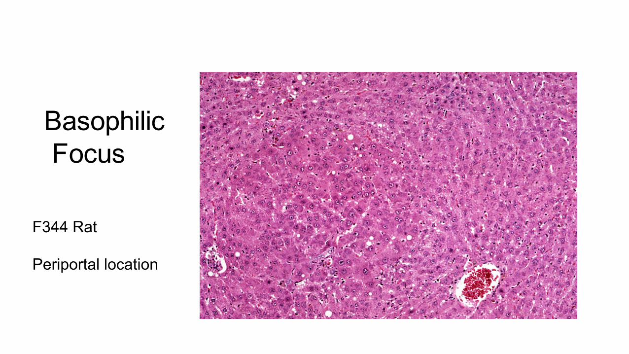
BasophilicFocus
F344 Rat Periportal location

BasophilicFocus
Control F344 Rat Focus surrounds a central vein & with extramedullary hematopoiesis

BasophilicFocus

BasophilicFocus

BasophilicFocus

Eosinophilic Focus
F344 Rat Rat had LGL leukemia

EosinophilicFocusofCellularAlteraJon
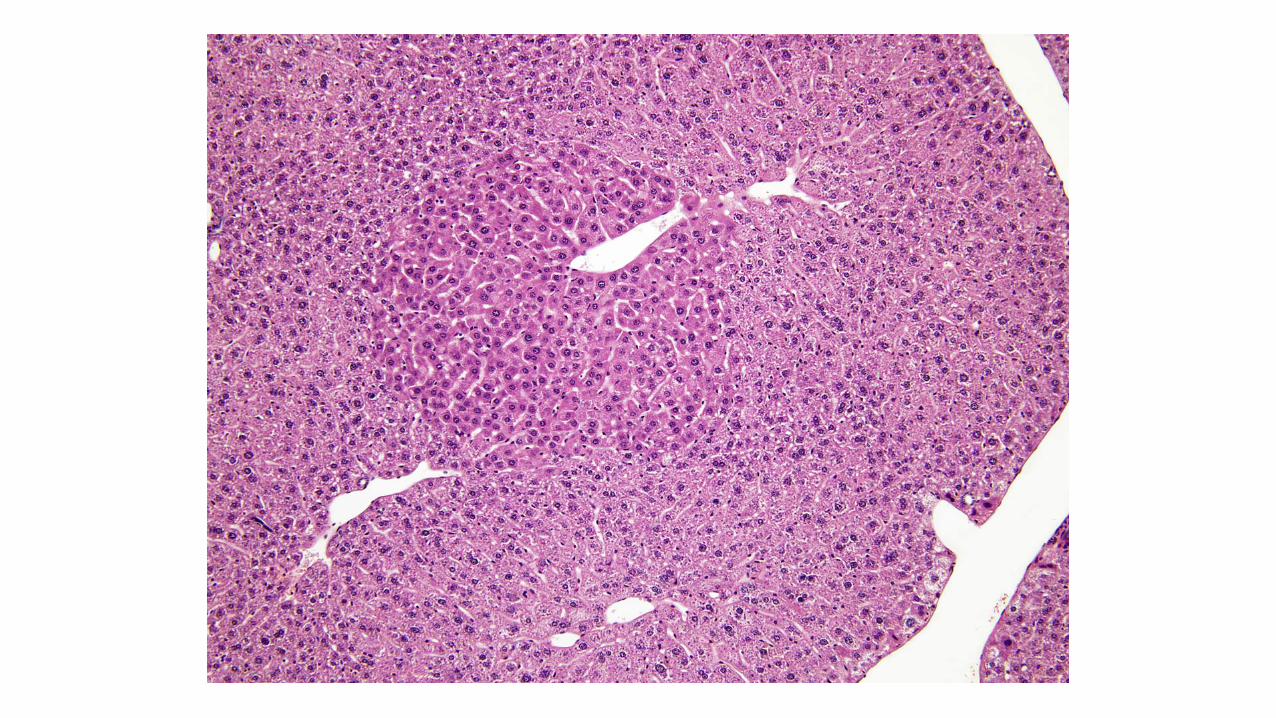


Eosinophilic Focus

Eosinophilic Foci
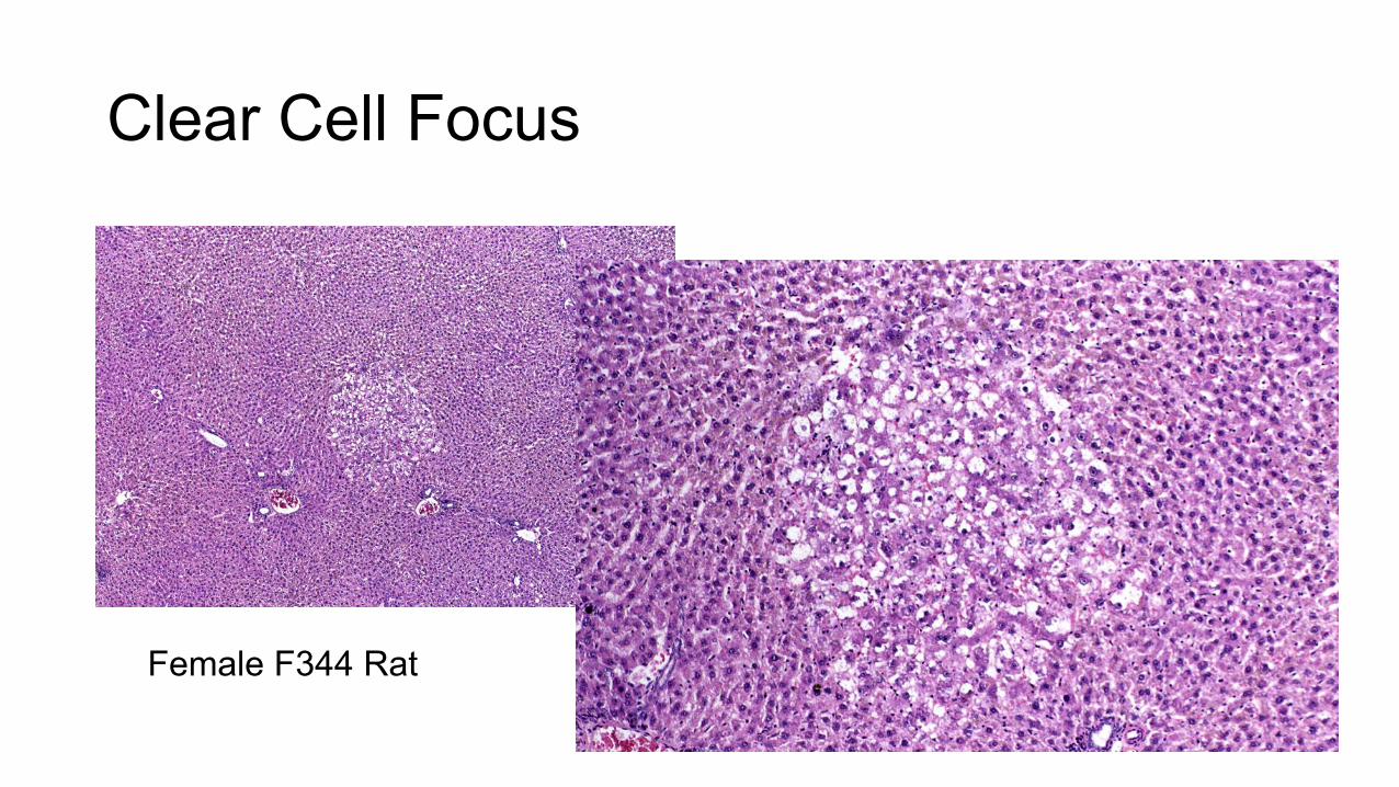
Clear Cell Focus
Female F344 Rat

Clear Cell Focus
Female F344 Rat

Clear Cell Focus

Mixed Cell Foci

Mixed Cell Focus
Treated Male F344 Rat

Mixed Cell Focus
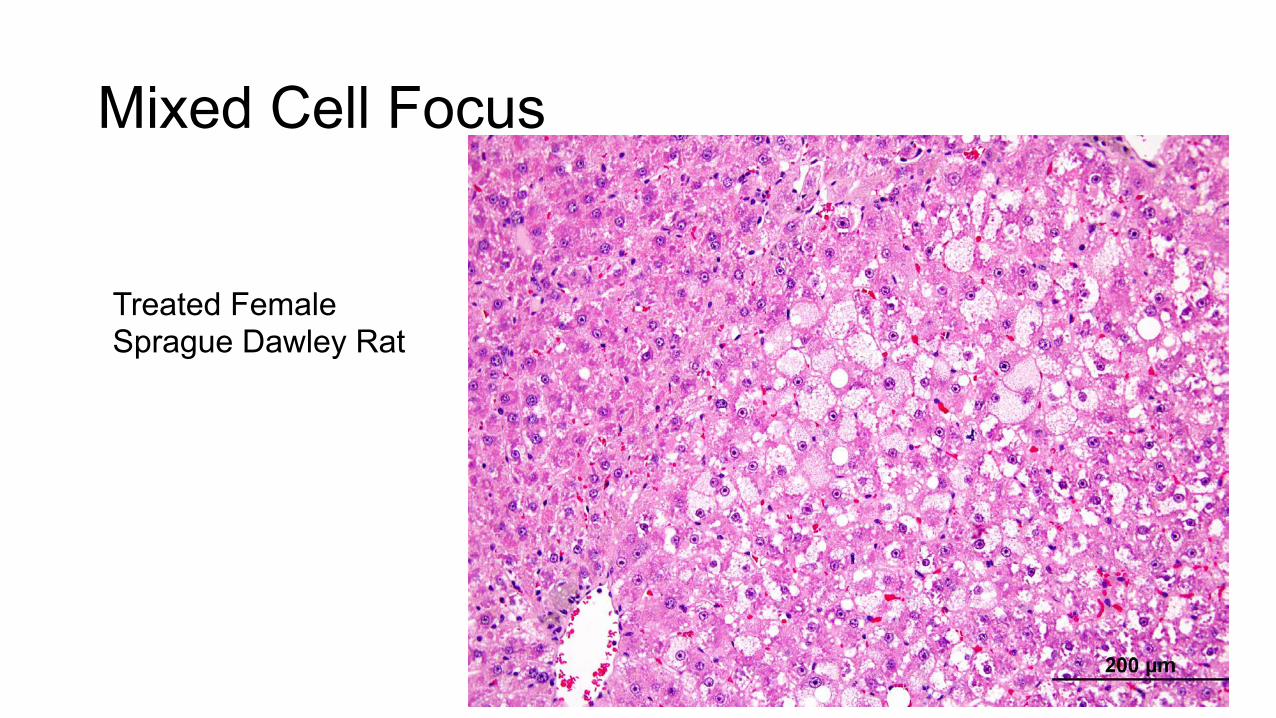
Mixed Cell Focus
Treated Female Sprague Dawley Rat

Mixed Cell Focus
Treated FemaleSprague Dawley Rat

Basophilic Focus with Fatty Change
Untreated Male F344 Rat

Basophilic Focus with Fatty Change
Untreated Male F344 Rat

Vacuolated Focus?

FocusofCellularAlteraJon

Basophilic Focus of Cellular Alteration
Vehicle Female Sprague Dawley

Basophilic Focus - Homogeneous Pattern

Basophilic Focus - Tigroid Pattern

Basophilic Focus - Cresyl Violet Stain

Amphophilic Focus

Glutathione S-Transferase pi


Tension Lipidosis
FIGURE 7.—Mouse liver. Fatty change, microvesicular. FIGURE 8.—Mouse liver. Mixture of fatty change and cytoplasmic glycogen. FIGURE 9.—Mouse liver. Mixture of fatty change and cytoplasmic glycogen. Higher magnification of Figure 8. FIGURE 10.—Mouse liver. Tension lipidosis.FIGURE 11.—Mouse liver. Tension lipidosis. Higher magnification of Figure 10. FIGURE 12.—Rat liver. Focal fatty change.
44S THOOLEN ET AL. TOXICOLOGIC PATHOLOGY
at Society of Toxicologic Pathology on August 8, 2013tpx.sagepub.comDownloaded from
Associated with attachment of falciform ligament

Tension Lipidosis
Associated with attachment of falciform ligament

Regenerative Hyperplasia
Harada, Maronpot, Morris, and Boorman, 1989; Squire 1989;Schulte-Hermann et al. 1989). Most importantly, types offoci in controls should be compared to those found in treated ani-mals. Some pathologists regard vacuolated foci as focal fattychange and do not consider them a subtype of focus of cellularalteration.
Hyperplasia, Hepatocellular, Non-Regenerative (Figures109 and 110)
Synonyms: Hyperplasia, hepatocellular, focal hepatocellularhyperplasia.
Pathogenesis: A spontaneous or treatment-associated prolif-erative collection of hepatocytes spanning several lobules andwithout evidence of prior hepatic damage.
Diagnostic features:
! A relatively large lesion that is often greater than sev-eral adjacent lobules and is occasionally accompa-nied by angiectasis and/or spongiosis hepatis (cysticdegeneration).
! Comprised of slightly enlarged hepatocytes.! Hepatocytes are tinctorially similar to surrounding
parenchyma.! The liver plates in the lesion tend to merge with the
adjacent hepatic parenchyma.! May be minimal to mild compression of adjacent
hepatic parenchyma.! Lobular architecture is maintained.! Portal triads and central veins are present.! When accompanied by angiectasis/spongiosis hepa-
tis, hepatic cords may be distorted.
Differential diagnosis:
! Focus of cellular alteration—phenotypical or tinctor-ial variation is present. Generally not associated withchronic liver damage, though foci of cellularalteration may occur in damaged liver.
! Adenoma, hepatocellular—loss of normal lobulararchitecture with irregular growth pattern. Liver platesoften impinge perpendicular or obliquely on the sur-rounding parenchyma. Distinct compression is present.
! Carcinoma, hepatocellular—distinct trabecular, ade-noid, or poorly differentiated growth is present.Lobular and plate architecture is not maintained.Cellular atypia and invasive growth may be present.
! Hyperplasia, hepatocellular (regenerative)—evidenceof ongoing or prior hepatocellular damage, fibrosis,or a history of exposure to a known hepatotoxicant.Lobular architecture usually distorted.
Comment: Non-regenerative hepatocellular hyperplasiais rare in rodents. It may occur spontaneously or be
treatment-associated and consists of a proliferative collectionof hepatocytes spanning several lobules and without evidenceof prior hepatic damage. This lesion is not associated with anyevidence of existing or prior hepatocellular injury. Diagnosticdifficulties occur when preneoplastic foci and hepatocellularadenomas occur in the same liver sections of older rodents.This lesion may be similar to that of hepatic nodular hyperpla-sia in dogs.
There are basically two variations of non-regenerativehepatocellular hyperplasia. One is relatively smaller and isaccompanied by angiectasis and/or spongiosis hepatis and theother tends to be larger than several lobules. The formeroccurs in both sexes and the latter predominantly in untreatedfemale control F344 rats but occasionally reported in treatedrats (Tasaki et al. 2008; Hailey et al. 2005; Bach et al.2010). When present near the capsular surface, this type ofnodular hyperplasia may be evident grossly as a raised area.The proliferating cell nuclear antigen (PCNA) labeling indexin these nodules is increased in comparison with surroundingparenchyma and the lesion is glutathione S-transferase pla-cental form (GSTP) immunonegative.
Very early non-regenerative hyperplasia may be the size ofsmall foci of cellular alteration and are identified by theiraltered growth pattern and tinctorial similarity to surroundingparenchyma (see Figure 109).
Hyperplasia, Hepatocellular, Regenerative (Figures 111–113)
Synonyms: Hyperplasia, hepatocellular; hyperplasia, regenera-tive; hyperplasia, nodular; regeneration, nodular.
Pathogenesis: A nodular regenerative response to prior or con-tinuous hepatocellular damage.
Diagnostic features:
! Focal or multifocal (nodular) appearance.! Lesion may reach several millimeters in diameter.! Spherical proliferation may be accompanied by slight
encapsulation.! Compression of surrounding liver parenchyma often
occurs.! Normal lobular architecture usually present but may
be distorted.! Portal triads and central veins may be present.! Bile duct and oval cell proliferation may be
present.! Hepatocytes appear slightly altered, but may have
slightly basophilic cytoplasm or prominent nucleoli.! Increased mitotic index may be observed.! Evidence of prior or ongoing hepatocellular damage,
such as apoptosis/necrosis, chronic inflammation,chronic congestion, fibrosis, cirrhosis, or a knowncause of toxicity.
! Lesions in rats tend to be more nodular than in mice.
Vol. 38, No. 7S, 2010 LESIONS OF THE HEPATOBILIARY SYSTEM 29S
at Society of Toxicologic Pathology on August 8, 2013tpx.sagepub.comDownloaded from

Non-Regenerative Hyperplasia
Harada, Maronpot, Morris, and Boorman, 1989; Squire 1989;Schulte-Hermann et al. 1989). Most importantly, types offoci in controls should be compared to those found in treated ani-mals. Some pathologists regard vacuolated foci as focal fattychange and do not consider them a subtype of focus of cellularalteration.
Hyperplasia, Hepatocellular, Non-Regenerative (Figures109 and 110)
Synonyms: Hyperplasia, hepatocellular, focal hepatocellularhyperplasia.
Pathogenesis: A spontaneous or treatment-associated prolif-erative collection of hepatocytes spanning several lobules andwithout evidence of prior hepatic damage.
Diagnostic features:
! A relatively large lesion that is often greater than sev-eral adjacent lobules and is occasionally accompa-nied by angiectasis and/or spongiosis hepatis (cysticdegeneration).
! Comprised of slightly enlarged hepatocytes.! Hepatocytes are tinctorially similar to surrounding
parenchyma.! The liver plates in the lesion tend to merge with the
adjacent hepatic parenchyma.! May be minimal to mild compression of adjacent
hepatic parenchyma.! Lobular architecture is maintained.! Portal triads and central veins are present.! When accompanied by angiectasis/spongiosis hepa-
tis, hepatic cords may be distorted.
Differential diagnosis:
! Focus of cellular alteration—phenotypical or tinctor-ial variation is present. Generally not associated withchronic liver damage, though foci of cellularalteration may occur in damaged liver.
! Adenoma, hepatocellular—loss of normal lobulararchitecture with irregular growth pattern. Liver platesoften impinge perpendicular or obliquely on the sur-rounding parenchyma. Distinct compression is present.
! Carcinoma, hepatocellular—distinct trabecular, ade-noid, or poorly differentiated growth is present.Lobular and plate architecture is not maintained.Cellular atypia and invasive growth may be present.
! Hyperplasia, hepatocellular (regenerative)—evidenceof ongoing or prior hepatocellular damage, fibrosis,or a history of exposure to a known hepatotoxicant.Lobular architecture usually distorted.
Comment: Non-regenerative hepatocellular hyperplasiais rare in rodents. It may occur spontaneously or be
treatment-associated and consists of a proliferative collectionof hepatocytes spanning several lobules and without evidenceof prior hepatic damage. This lesion is not associated with anyevidence of existing or prior hepatocellular injury. Diagnosticdifficulties occur when preneoplastic foci and hepatocellularadenomas occur in the same liver sections of older rodents.This lesion may be similar to that of hepatic nodular hyperpla-sia in dogs.
There are basically two variations of non-regenerativehepatocellular hyperplasia. One is relatively smaller and isaccompanied by angiectasis and/or spongiosis hepatis and theother tends to be larger than several lobules. The formeroccurs in both sexes and the latter predominantly in untreatedfemale control F344 rats but occasionally reported in treatedrats (Tasaki et al. 2008; Hailey et al. 2005; Bach et al.2010). When present near the capsular surface, this type ofnodular hyperplasia may be evident grossly as a raised area.The proliferating cell nuclear antigen (PCNA) labeling indexin these nodules is increased in comparison with surroundingparenchyma and the lesion is glutathione S-transferase pla-cental form (GSTP) immunonegative.
Very early non-regenerative hyperplasia may be the size ofsmall foci of cellular alteration and are identified by theiraltered growth pattern and tinctorial similarity to surroundingparenchyma (see Figure 109).
Hyperplasia, Hepatocellular, Regenerative (Figures 111–113)
Synonyms: Hyperplasia, hepatocellular; hyperplasia, regenera-tive; hyperplasia, nodular; regeneration, nodular.
Pathogenesis: A nodular regenerative response to prior or con-tinuous hepatocellular damage.
Diagnostic features:
! Focal or multifocal (nodular) appearance.! Lesion may reach several millimeters in diameter.! Spherical proliferation may be accompanied by slight
encapsulation.! Compression of surrounding liver parenchyma often
occurs.! Normal lobular architecture usually present but may
be distorted.! Portal triads and central veins may be present.! Bile duct and oval cell proliferation may be
present.! Hepatocytes appear slightly altered, but may have
slightly basophilic cytoplasm or prominent nucleoli.! Increased mitotic index may be observed.! Evidence of prior or ongoing hepatocellular damage,
such as apoptosis/necrosis, chronic inflammation,chronic congestion, fibrosis, cirrhosis, or a knowncause of toxicity.
! Lesions in rats tend to be more nodular than in mice.
Vol. 38, No. 7S, 2010 LESIONS OF THE HEPATOBILIARY SYSTEM 29S
at Society of Toxicologic Pathology on August 8, 2013tpx.sagepub.comDownloaded from

The described malignant tumor therefore seems to differ fromits benign counterpart only by its morphology. Moreover, itshows the same biological behavior without any regional ordistant metastasis. As a nonproliferative lesion, the granularcell aggregates present as small cell clusters with at least threeto five cells without any disturbance of the normal tissuearchitecture (Figure 11E). At the end of her talk, Dr. Bachstated that in the RITA database, among all granular celllesions in the female genital tract, the benign granularcell tumor is the most frequent finding. However, in general,all granular cell lesions are rather rare occurrences (<0.6%) inthe female genital tract.
NODULAR HYPERPLASIA REVISITED
Dr. Robert Maronpot introduced the topic of proliferativeliver lesions in rodents, with a brief review of the nomenclatureterms from the 1960s to the present time (Maronpot et al. 1986;Narama et al. 2003; Squire and Levitt 1975; Stewart et al.1980). Primary diagnostic features of rat hepatoproliferativelesions show considerable overlap, especially for regenerativehyperplasia secondary to hepatotoxicity (synonym: nodularhyperplasia) and the newly recognized large hyperplasticlesion not associated with obvious hepatotoxicity. The latterwas presented at the NTP Satellite Symposium in 2008, and
FIGURE 12.—(A) This image was projected at the 2008 NTP Satellite Symposium by Dr. Taki Harada and was presented as a representative exam-ple of a hyperplasia not associated with hepatotoxicity. The arrows show the edges of the hyperplastic lesion, which is tinctorially similar to theadjacent hepatic parenchyma. Multiple portal triads are present within this hyperplastic lesion. (B) An enlargement of the lower right quadrant ofFigure 12A showing a portal triad just above the center of the image. Adjacent uninvolved hepatic parenchyma is present in the lower right cornerof the photomicrograph. (C) This early example of focal hyperplasia was provided by Dr. Harada. It is phenotypically similar to surroundinghepatic parenchyma and evident only because of its altered growth pattern and sinusoidal congestion. (D) Another example of a large area ofhyperplasia similar to Figure 12A. Macrovesicular fat vacuoles are present within this lesion.
Vol. 38, No. 1, 2010 2009 NTP SATELLITE SYMPOSIUM 33
at Society of Toxicologic Pathology on October 14, 2016tpx.sagepub.comDownloaded from
Toxicologic Pathology 38: 9-36 (2010)
Non-Regenerative Hyperplasia

Kupffer Cell Hyperplasia/Hypertrophy
Differential diagnosis:
! Focus of cellular alteration—tinctorial variation ispresent. Generally not associated with chronic liverdamage, though foci of cellular alteration may occurin damaged liver.
! Adenoma, hepatocellular—loss of normal lobulararchitecture with irregular growth pattern. Liverplates often impinge perpendicular or obliquely onthe surrounding parenchyma. Distinct compressionis present.
! Carcinoma, hepatocellular—distinct trabecular,adenoid, or poorly differentiated growth is present.Lobular and plate architecture is not maintained. Cel-lular atypia and invasive growth may be present.
! Hyperplasia, hepatocellular (non-regenerative)—nohistory or evidence of hepatocellular damage.
Comment: These lesions are considered to represent a regen-erative response to prior or continuous hepatocellular dam-age. A history of exposure to a hepatotoxicant and thepresence of multiple nodules of regeneration that maintain alobular but usually distorted architecture makes diagnosismore convincing. Diagnostic difficulties occur when preneo-plastic foci and hepatocellular adenomas occur in the sameliver sections of older rodents or in livers with many inducedfoci and tumors.
In livers with partial hepatectomy (PH), the pattern ofhyperplasia at twenty-four to seventy-two hours post surgeryis diffuse hepatocyte hyperplasia with many mitotic figures andno evidence of liver toxicity. Although this response is alsohyperplasia (hepatocellular, regenerative), it is not to be con-fused with the nodular hepatic response to toxic damage tohepatocytes. After ninety-six hours, the liver may be almostnormal histologically. In mice however, chronic biliary lesionsmay be seen in the liver after PH.
Hypertrophy/Hyperplasia, Kupffer Cell (Figures 114 and 115)
Synonyms: Kupffer cell proliferation; histiocytosis, focal ordiffuse.
Pathogenesis: Following phagocytosis of foreign material,estrogen treatment, inflammatory conditions, and response tocytokines. A rare spontaneous finding.
Diagnostic features:
! Diffuse to multifocal proliferation of oval to spinde-loid cells lining sinusoids.
! Cells resemble histiocytes and often contain phago-cytic material.
! May form as sheets or nodules.! Hypertrophy can occur without hyperplasia and vice
versa.
Differential diagnosis:
! Infiltration, mononuclear—well demarcated; usuallysolitary; associated with cell necrosis.
! Histiocytic sarcoma—diffuse and irregular prolifera-tion of histiocytic cells throughout the liver. Usuallyassociated with destruction of hepatic parenchyma.Occasional multinucleated giant cells may bepresent.
! Hyperplasia, oval cell—consists of a single or doublerow of oval cells sometimes forming incomplete(pseudo-) duct-like structure. Cells are usually uni-form in size and shape and have scant pale basophiliccytoplasm and round to oval nuclei.
Comment: Rare as a spontaneous finding, hyperplasia ofKupffer cells may be seen following phagocytosis of foreignmaterial and as a consequence of estrogen treatment and ininflammatory conditions. It can be induced by cytokines.Hypertrophy and hyperplasia often accompany each other. Thehypertrophy of normal Kupffer cells gives the impression ofpresence of more Kupffer cells since they are difficult to visua-lize in normal livers.
Hyperplasia, Ito Cell (Figures 116–118)
Synonyms: Stellate cell; perisinusoidal cell; fat-storing perisi-nusoidal cell.
Pathogenesis: Proliferation of fat-storing perisinusoidal cells.
Diagnostic features:
! Focal or diffuse proliferation of Ito cells.! May grow in sheets, clusters, or along cords of
hepatocytes.! Cells vary in size and shape and are vacuolated.! Multiple cytoplasmic fat droplets of different size
occur.! The nuclei are ovoid or round and may be indented by
cytoplasmic lipid droplets.! Modest amount of collagenous matrix may be present.
Differential diagnosis:
! Fatty change/Lipidosis—the cytoplasm of fat cellsmay be clearer than that of Ito cells.
! Ito cell tumor—larger and more extensive thanhyperplasia. Partially distinct compression of adja-cent hepatic parenchyma.
Comment: Ito cell hyperplasia is extremely rare and occurspredominantly in mice. It arises from fat-storing perisinusoi-dal cells, better known as Ito cells (Dixon et al. 1994; Enzan1985; Tillmann et al. 1997). The biological behavior of the
30S THOOLEN ET AL. TOXICOLOGIC PATHOLOGY
at Society of Toxicologic Pathology on August 8, 2013tpx.sagepub.comDownloaded from

Kupffer Cell Hyperplasia

Kupffer Cell Hyperplasia

Ito Cell Hyperplasia
Differential diagnosis:
! Focus of cellular alteration—tinctorial variation ispresent. Generally not associated with chronic liverdamage, though foci of cellular alteration may occurin damaged liver.
! Adenoma, hepatocellular—loss of normal lobulararchitecture with irregular growth pattern. Liverplates often impinge perpendicular or obliquely onthe surrounding parenchyma. Distinct compressionis present.
! Carcinoma, hepatocellular—distinct trabecular,adenoid, or poorly differentiated growth is present.Lobular and plate architecture is not maintained. Cel-lular atypia and invasive growth may be present.
! Hyperplasia, hepatocellular (non-regenerative)—nohistory or evidence of hepatocellular damage.
Comment: These lesions are considered to represent a regen-erative response to prior or continuous hepatocellular dam-age. A history of exposure to a hepatotoxicant and thepresence of multiple nodules of regeneration that maintain alobular but usually distorted architecture makes diagnosismore convincing. Diagnostic difficulties occur when preneo-plastic foci and hepatocellular adenomas occur in the sameliver sections of older rodents or in livers with many inducedfoci and tumors.
In livers with partial hepatectomy (PH), the pattern ofhyperplasia at twenty-four to seventy-two hours post surgeryis diffuse hepatocyte hyperplasia with many mitotic figures andno evidence of liver toxicity. Although this response is alsohyperplasia (hepatocellular, regenerative), it is not to be con-fused with the nodular hepatic response to toxic damage tohepatocytes. After ninety-six hours, the liver may be almostnormal histologically. In mice however, chronic biliary lesionsmay be seen in the liver after PH.
Hypertrophy/Hyperplasia, Kupffer Cell (Figures 114 and 115)
Synonyms: Kupffer cell proliferation; histiocytosis, focal ordiffuse.
Pathogenesis: Following phagocytosis of foreign material,estrogen treatment, inflammatory conditions, and response tocytokines. A rare spontaneous finding.
Diagnostic features:
! Diffuse to multifocal proliferation of oval to spinde-loid cells lining sinusoids.
! Cells resemble histiocytes and often contain phago-cytic material.
! May form as sheets or nodules.! Hypertrophy can occur without hyperplasia and vice
versa.
Differential diagnosis:
! Infiltration, mononuclear—well demarcated; usuallysolitary; associated with cell necrosis.
! Histiocytic sarcoma—diffuse and irregular prolifera-tion of histiocytic cells throughout the liver. Usuallyassociated with destruction of hepatic parenchyma.Occasional multinucleated giant cells may bepresent.
! Hyperplasia, oval cell—consists of a single or doublerow of oval cells sometimes forming incomplete(pseudo-) duct-like structure. Cells are usually uni-form in size and shape and have scant pale basophiliccytoplasm and round to oval nuclei.
Comment: Rare as a spontaneous finding, hyperplasia ofKupffer cells may be seen following phagocytosis of foreignmaterial and as a consequence of estrogen treatment and ininflammatory conditions. It can be induced by cytokines.Hypertrophy and hyperplasia often accompany each other. Thehypertrophy of normal Kupffer cells gives the impression ofpresence of more Kupffer cells since they are difficult to visua-lize in normal livers.
Hyperplasia, Ito Cell (Figures 116–118)
Synonyms: Stellate cell; perisinusoidal cell; fat-storing perisi-nusoidal cell.
Pathogenesis: Proliferation of fat-storing perisinusoidal cells.
Diagnostic features:
! Focal or diffuse proliferation of Ito cells.! May grow in sheets, clusters, or along cords of
hepatocytes.! Cells vary in size and shape and are vacuolated.! Multiple cytoplasmic fat droplets of different size
occur.! The nuclei are ovoid or round and may be indented by
cytoplasmic lipid droplets.! Modest amount of collagenous matrix may be present.
Differential diagnosis:
! Fatty change/Lipidosis—the cytoplasm of fat cellsmay be clearer than that of Ito cells.
! Ito cell tumor—larger and more extensive thanhyperplasia. Partially distinct compression of adja-cent hepatic parenchyma.
Comment: Ito cell hyperplasia is extremely rare and occurspredominantly in mice. It arises from fat-storing perisinusoi-dal cells, better known as Ito cells (Dixon et al. 1994; Enzan1985; Tillmann et al. 1997). The biological behavior of the
30S THOOLEN ET AL. TOXICOLOGIC PATHOLOGY
at Society of Toxicologic Pathology on August 8, 2013tpx.sagepub.comDownloaded from

Bile Duct Hyperplasia
lesion is not well established. There appears to be a continuumwith Ito cell tumor (see the following), which may be just anexaggerated and sometimes more localized form of Ito cellhyperplasia.
Bile Duct Hyperplasia (Figures 119–122)
Pathogenesis: A spontaneous change in portal areas of olderanimals; may be induced or exacerbated by treatment.
Diagnostic feature:
! Increased number of small bile ducts arising in portalregion.
! May not involve all portal areas.! May be associated with periductular fibrosis and
periductular cell infiltration.! Biliary epithelium is well differentiated, forming
normal ducts.! Biliary epithelium may show degenerative or
atrophic changes.! May be associated with oval cell hyperplasia.! May contain mucous metaplasia or hyalinosis in
specific situations in mice.! Cystic form.! Focal bile duct proliferation with cystic dilation of
ducts may occur.! Usually acini lined by flattened epithelium.
Differential diagnosis:
! Cholangioma—well demarcated; usually solitary;acini lined by cuboidal epithelium.
! Cholangiofibrosis—central portions becomeatrophic, collagenized, and avascular whereas theactive proliferating portion is at the periphery; mucusproduction is common.
! Cholangiocarcinoma—invasive growth into sur-rounding hepatic tissue or vessels; cell are pleo-morphic; mucus production.
! Oval cell hyperplasia—consists of a single or doublerow of oval cells forming incomplete duct-like struc-tures. Cells usually uniform with scant basophiliccytoplasm and round or oval nuclei.
Comment: Often associated with evidence of hepatic injury andrepair and obstruction of bile flow. Dilation of intrahepatic bileducts is a spontaneous, age-associated lesion that is more com-mon in rats than in mice.
Cholangiofibrosis (Figures 123–126)
Synonyms: Bile duct adenomatosis; intestinal cell metaplasia;adenofibrosis.
Pathogenesis: Originates from an initial oval cell hyperplasiain response to pronounced hepatic parenchymal necrosis.
Diagnostic features:
! Consists of dilated to cystic bile ducts filledwith mucus and cellular debris and surroundedby inflammatory cell infiltrates and connectivetissue.
! Glandular epithelium is typically a single layer andvaries from flattened to tall columnar hyperbasophi-lic and pleomorphic cells along with goblet cells andoccasional Paneth cells.
! Glandular epithelium, particularly in cystic glands,may be partially lost through degeneration resultingin crescent shaped structures.
! Central portions of large lesions may become sclero-tic with only remnants of biliary epithelium suggest-ing regression.
! Lesions may be limited to small foci but may occupylarge interconnecting areas of a lobe without mark-edly disturbing the lobe outline.
! Lesion growth typically involves contraction withretraction of surrounding parenchyma. Markedlydilated or cystic mucus-filled glands along the livercapsule may protrude above the lobe outline.
! Older lesions may be shrunken from the liver surfaceand appear as scars.
! Regenerative hepatocellular hyperplasia maybe present when there is extensive parenchymalinvolvement.
Differential diagnosis:
! Cholangiocarcinoma—solid sheets, trabeculae, ornests of closely packed biliary cells largely displacinghepatic parenchyma. Intestinal metaplasia is not aprominent feature. Glandular dilation is absent orminimal.
! Hyperplasia, bile duct—multiple small bile ductsarising in portal region. Biliary epithelium is welldifferentiated, forming more normal appearingducts, with minimal glandular dilation in somecases.
Comment: This lesion is an inflammatory, proliferative, andmetaplastic reaction involving bile duct epithelium and isseen with hepatocellular toxicity caused by xenobiotics suchas dioxins, furans, and related chemicals in rats (Bannaschand Zerban 1990; Deschl et al. 1997; Eustis et al. 1990;Kimbrough et al. 1973; Kimbrough and Linder 1974; Sirica1992; Hailey et al. 2005). The initial reaction following pro-nounced hepatic parenchymal necrosis is oval cell hyperplasia(Engelhardt 1997).
Cholangiofibrosis is a controversial lesion that has beendiagnosed as cholangiocarcinoma especially when there isextensive involvement of the liver. Cholangiofibrosis is notseen as a spontaneous lesion but occurs primarily in rats treatedwith a variety of xenobiotic agents that are hepatotoxic at high
Vol. 38, No. 7S, 2010 LESIONS OF THE HEPATOBILIARY SYSTEM 31S
at Society of Toxicologic Pathology on August 8, 2013tpx.sagepub.comDownloaded from

Bile Duct Hyperplasia
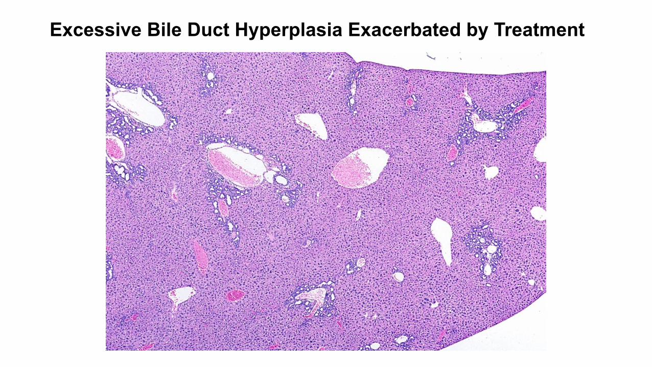
Excessive Bile Duct Hyperplasia Exacerbated by Treatment


Bile Duct Hyperplasia and Inflammation
Treated Female Sprague Dawley Rat Tx: PCB118 Dx: Bile duct hyperplasia

Treated Female Sprague Dawley Rat Tx: PCB118 Dx: Bile duct hyperplasia

Bileducthyperplasiaisacommonagingchangeinrodents&maybe
accompaniedbychronicinflammaDonVehicle Female
F344 Rat Study: Beta Thujone Dx: Bile duct hyperplasia


Sprague Dawley Female High-dose Tx: TCDD Dx: Bile duct fibrosis

Mdr2 Mouse

Mdr2 Mouse

Mdr2 Mouse

Oval Cell Hyperplasia
dose. The intestinal cell metaplasia in cholangiofibrosisincludes goblet and Paneth cells and columnar cells similarto chief cells identified in H&E-stained paraffin sections andenterochromaffin cells identified by special staining. Themetaplastic cells have been confirmed by electron micro-scopy in some cases. While cholangiofibrosis has beenreported to progress to cholangiocarcinoma with malignantfeatures (Bannasch and Zerban, 1990; Sirica 1992), unequivo-cal metastases have not been confirmed in most cases. Thislesion is not observed in humans.
Oval Cell Hyperplasia (Figures 127–129)
Synonyms: Oval cell proliferation; bile ductule cell hyperplasia.
Pathogenesis: Arises from terminal ductule epithelial cells(canal of Hering cells) spontaneously, following liver infec-tions, and secondary to hepatotoxic injury.
Diagnostic features:
! Generally originates from portal areas and is oftenmultifocal.
! Consists of a single or double row of oval to roundcells along sinusoids in linear arrays.
! May form a few or many small ductules with stream-ing into the hepatic parenchyma.
! Formation of incomplete duct-like structures maybe present.
! Cells are usually uniform in size and shape and maybe fusiform.
! Cells have scant pale basophilic cytoplasm and roundor oval nuclei.
! Oval cells express keratin.
Differential diagnosis:
! Hyperplasia, bile duct—several small bile ductsarising in portal region. Biliary epithelium is welldifferentiated, forming normal ducts.
! Early or mild fibrosis—collagen matrix is evident.! Inflammation—presence of mononuclear, polymor-
phonuclear, or connective tissue cells will be presentin inflammation.
Comment: Oval cell proliferation is considered to arise fromterminal ductule epithelial cells (canal of Hering cells). It isa rare spontaneous lesion in rats. Oval cell hyperplasia canbe observed following severe hepatotoxic injury and treat-ment with hepatocarcinogens. There is often a close relationto the portal tract, although more scattered groups of prolif-erating oval cells can be seen diffusely throughout the liverfollowing xenobiotic-induced hepatic injury (Engelhardt1997).
In mice oval cell hyperplasia is a feature of chronic activehepatitis caused by H. hepaticus and H. bilis and is seen fol-lowing treatment with various hepatocarcinogens. Hyperplas-tic oval cells occur in association with a high incidence of
hepatocellular neoplasms and may play an important role inhepatocarcinogenesis. Some authors support the concept thatoval cells may participate in the lineage of hepatocellular andcholangiocellular carcinomas and may serve as hepatic stemcells. Oval cell hyperplasia diagnosis including grade is rec-ommended, even when it is part of a complex set of hepaticchanges.
G. Neoplasms
Introduction
The rodent liver is the most common target site of chemicalcarcinogens (Maronpot et al. 1986; Evans and Lake 1998), per-haps due to its major function as a metabolizing and detoxifyingorgan for xenobiotics. Rodent hepatocarcinogens are usuallyhepatotoxins. The chronic toxicity of these toxins maycontribute to hepatocarcinogenesis although genotoxic livercarcinogens are often also hepatotoxins. There is over a thirty-year history of experimental induction (Frith and Wiley 1982;Malarkey et al. 1995; Evans et al. 1992; Ward et al. 1983,1986; Ward, Lynch, and Riggs 1988; Popp 1984) and classifica-tion of preneoplastic and neoplastic lesions of the rat and mouseliver in book chapters (Bannasch and Zerban 1990; Brooksand Roe 1985; Greaves and Faccini 1984; Jones and Butler1978; Ward 1981; Harada et al. 1999; Eustis et al. 1990) andby committee (ILAR 1980) or toxicologic pathologysocieties (Standardized System of Nomenclature and Diagnos-tic Criteria [SSNDC] Guides, http://www.toxpath.org/ssndc.asp). Terminology has evolved to the present nomencla-ture that is also based on many publications on livercarcinogenesis.
There is evidence from experimental studies documentingthe regression of proliferative hepatocellular lesions includingfoci of cellular alteration, hepatocellular adenomas, and hepato-cellular carcinomas following cessation of treatment (Maronpot2009). A dramatic example was reported in mice followingcessation of chronic chlordane exposure (Malarkey et al.1995). Similar experience has been reported in rats and micein other studies (Lipsky et al. 1984; Greaves, Irisarri, andMonro 1986; Marsman and Popp 1994) as well as in humans(Fremond et al. 1987; McCaughan, Bilous, and Gallagher1985; Emerson et al. 1980; Steinbrecher et al. 1981). Agents thatrequire continual administration for the stable presence andgrowth of preneoplastic and neoplastic rodent liver lesions canbe categorized as conditional hepatocarcinogens (Maronpot2009).
Hepatocellular Adenoma (Figures 130-134)
Synonyms: Adenoma, hepatic; adenoma, liver parenchymalcell; hepatoma, benign; tumor, liver cell, benign; hepatoma,benign; type A nodule.
Pathogenesis: Spontaneous and following treatment withhepatotoxins that are carcinogenic xenobiotics; with genealterations in genetically engineered mice.
32S THOOLEN ET AL. TOXICOLOGIC PATHOLOGY
at Society of Toxicologic Pathology on August 8, 2013tpx.sagepub.comDownloaded from

Oval Cell Hyperplasia
Treated Female Sprague Dawley Tx: PCB Mixture Dx: Oval cell proliferation

Oval Cell Hyperplasia
Treated Female Sprague Dawley Tx: PCB Mixture Dx: Oval cell proliferation

Cholangiofibrosis lesion is not well established. There appears to be a continuumwith Ito cell tumor (see the following), which may be just anexaggerated and sometimes more localized form of Ito cellhyperplasia.
Bile Duct Hyperplasia (Figures 119–122)
Pathogenesis: A spontaneous change in portal areas of olderanimals; may be induced or exacerbated by treatment.
Diagnostic feature:
! Increased number of small bile ducts arising in portalregion.
! May not involve all portal areas.! May be associated with periductular fibrosis and
periductular cell infiltration.! Biliary epithelium is well differentiated, forming
normal ducts.! Biliary epithelium may show degenerative or
atrophic changes.! May be associated with oval cell hyperplasia.! May contain mucous metaplasia or hyalinosis in
specific situations in mice.! Cystic form.! Focal bile duct proliferation with cystic dilation of
ducts may occur.! Usually acini lined by flattened epithelium.
Differential diagnosis:
! Cholangioma—well demarcated; usually solitary;acini lined by cuboidal epithelium.
! Cholangiofibrosis—central portions becomeatrophic, collagenized, and avascular whereas theactive proliferating portion is at the periphery; mucusproduction is common.
! Cholangiocarcinoma—invasive growth into sur-rounding hepatic tissue or vessels; cell are pleo-morphic; mucus production.
! Oval cell hyperplasia—consists of a single or doublerow of oval cells forming incomplete duct-like struc-tures. Cells usually uniform with scant basophiliccytoplasm and round or oval nuclei.
Comment: Often associated with evidence of hepatic injury andrepair and obstruction of bile flow. Dilation of intrahepatic bileducts is a spontaneous, age-associated lesion that is more com-mon in rats than in mice.
Cholangiofibrosis (Figures 123–126)
Synonyms: Bile duct adenomatosis; intestinal cell metaplasia;adenofibrosis.
Pathogenesis: Originates from an initial oval cell hyperplasiain response to pronounced hepatic parenchymal necrosis.
Diagnostic features:
! Consists of dilated to cystic bile ducts filledwith mucus and cellular debris and surroundedby inflammatory cell infiltrates and connectivetissue.
! Glandular epithelium is typically a single layer andvaries from flattened to tall columnar hyperbasophi-lic and pleomorphic cells along with goblet cells andoccasional Paneth cells.
! Glandular epithelium, particularly in cystic glands,may be partially lost through degeneration resultingin crescent shaped structures.
! Central portions of large lesions may become sclero-tic with only remnants of biliary epithelium suggest-ing regression.
! Lesions may be limited to small foci but may occupylarge interconnecting areas of a lobe without mark-edly disturbing the lobe outline.
! Lesion growth typically involves contraction withretraction of surrounding parenchyma. Markedlydilated or cystic mucus-filled glands along the livercapsule may protrude above the lobe outline.
! Older lesions may be shrunken from the liver surfaceand appear as scars.
! Regenerative hepatocellular hyperplasia maybe present when there is extensive parenchymalinvolvement.
Differential diagnosis:
! Cholangiocarcinoma—solid sheets, trabeculae, ornests of closely packed biliary cells largely displacinghepatic parenchyma. Intestinal metaplasia is not aprominent feature. Glandular dilation is absent orminimal.
! Hyperplasia, bile duct—multiple small bile ductsarising in portal region. Biliary epithelium is welldifferentiated, forming more normal appearingducts, with minimal glandular dilation in somecases.
Comment: This lesion is an inflammatory, proliferative, andmetaplastic reaction involving bile duct epithelium and isseen with hepatocellular toxicity caused by xenobiotics suchas dioxins, furans, and related chemicals in rats (Bannaschand Zerban 1990; Deschl et al. 1997; Eustis et al. 1990;Kimbrough et al. 1973; Kimbrough and Linder 1974; Sirica1992; Hailey et al. 2005). The initial reaction following pro-nounced hepatic parenchymal necrosis is oval cell hyperplasia(Engelhardt 1997).
Cholangiofibrosis is a controversial lesion that has beendiagnosed as cholangiocarcinoma especially when there isextensive involvement of the liver. Cholangiofibrosis is notseen as a spontaneous lesion but occurs primarily in rats treatedwith a variety of xenobiotic agents that are hepatotoxic at high
Vol. 38, No. 7S, 2010 LESIONS OF THE HEPATOBILIARY SYSTEM 31S
at Society of Toxicologic Pathology on August 8, 2013tpx.sagepub.comDownloaded from

Cholangiofibrosis
Female Sprague Dawley Treated Tx: PCB Mixture Ki-67 Immunopositivity

Cholangiofibrosis
Treated Female Sprague Dawley Tx: PCB Mixture Dx: Bile duct hyperplasia

Cholangiofibrosis
Treated Female Sprague Dawley Tx: PCB Mixture Dx: Cholangiofibrosis

Cholangiofibrosis

Cholangiofibrosis
Treated Female Sprague Dawley Tx: PCB Mixture Dx: Cholangiofibrosis

Cholangiofibrosis
Has been called intestinal metaplasia

Cholangiofibrosis Male F344 Treated Rat Tx: Estragole subchronic study Dx: Cholangiofibrosis

Cholangiofibrosis

Cholangiofibrosis vs. Cholangiocarcinoma

Gallbladder

Normal Gallbladder

Normal Gallbladder

Normal Gallbladder

Normal Gallbladder

Submucosal Edema

Submucosal Edema

Inflammation & Cytoplasmic Alteration


Inflammation & Cytoplasmic Alteration


Cholecystitis

Cholecystitis

Cholecystitis

Cholecystitis

Epithelial Hyperplasia

Epithelial Hyperplasia

Epithelial Hyperplasia

Epithelial Hyperplasia

Epithelial Hyperplasis vs. Papilloma


Epithelial Hyperplasis vs. Papilloma ?

Some Liver Artifacts


Hematoxylin Crystals (Need to filter the hematoxylin)

Another Example


Multiple Surface Nodules


Cytoplasmic Vacuoles

Cytoplasmic Vacuoles


• Rats remained in CO2 chamber• after death for a few minutes• Time-dependent postmortem change• A result of plasma influx• Associated with congestion and• increased liver weight• Males more sensitive than females

Large Clear Vacuoles (Some Small Vacuoles are in Hepatocytes)

TOXICOLOGY AND APPLIED PHARMACOLOGY 36,31-39 (1976)
Hepatocyte Vacuolation and Increased Liver Weight Occurring in Anoxic Rats’
BARBARA I. SYKES, ELIZABETH PENNY AND I. F. H. PURCHASE
Central Toxicology Laboratories, Itnperial Chemical Industries Limited, Alderley Park, Cheshire SKI0 4TJ, England
Received May 14, 1975, accepted December 2,1975
Hepatocyte Vacuolation and Increased Liver Weight Occurring in Anoxic Rats. SYKES, BARBARA I., PENNY, ELIZABETH AND PURCHASE, I. F. H. (1976). Toxicol. Appl. Pharmacol. 36, 31-39. Hepatocyte vacuoles are often observed histologically in rats used in toxicology tests. They are regular in outline, contain eosinophilic material, and several may be present in individual hepatocytes, particularly those in the centrilobular areas. This vacuolation has been shown to result from anoxia. It occurs in rats that are not bled immediately after death and if there is a slight delay of only a few minutes before removal of the liver. It is enhanced if the animals are killed using volatile anesthetics in anoxic conditions. The vaculoes con- tain blood plasma which is thought to move through the disrupted spaces of Disse and hepatocyte plasma membranes into the cells. Anoxic vacuolation is associated with an increase in liver weight. It is important, therefore, both from a morphological viewpoint and with regard to the liver/body weight ratio, that this lesion be avoided in toxicology tests by ensuring that the necropsy is performed and that the animals are bled immediately after cessation of breathing.
At the termination of toxicology studies in rats carried out in our laboratory, the animals are killed with halothane vapor in a closed jar. The amount of anesthetic, the composi- tion of the air in the jar, and the time during which the rats are left either in the jar or on the bench before postmortem examination, may vary considerably. It was observed by light microscopy that in some rats from these studies there was extensive vacuolation of liver cells and corresponding ultrastructural changes. Vacuoles of various sizes, up to 15 pm in diameter, were distributed throughout the cytoplasm of some hepatocytes and the sinusoidal area was disrupted.
In a report on “watery vacuolation of liver cells,” Trowel1 (1946) described a similar change. He indicated that two factors were necessary for vacuolation to occur: (1) liver anoxia; and (2) intrasinusoidal pressure must be maintained at least at its normal value; if it is greater, vacuolation is enhanced. He distinguished between irz uiuo vacuolation due to severe anoxia and postmortem vacuolation in the highly anoxic liver after cessation of breathing which arises if the animal is not bled promptly.
As preliminary inquiries showed that there may be an association between the occurrence of vacuolation and the time the animals are left in the killing jar, an investi- gation into postmortem conditions which gave rise to this lesion was initiated. The study was then extended to include other volatile anesthetics used in similar conditions,
1 Address reprint requests and correspondence to I. F. H. Purchase. Copyright 0 1976 by Academic Press, Inc. All rights of reproduction in any form reserved. 31 Printed in Great Britain
• Anoxicvacuoles• Animalsnotbledimmediately• a\erdeath• Livernotremovedpromptly• Associatedwithincreased• liverweight• Bloodplasmamovesthrough• disruptedspaceofDisseand• hepatocytemembranes


