non-surgical vs. surgical treatment of meniscus tears … · non-surgical vs. surgical treatment of...
TRANSCRIPT
Non-Surgical vs. Surgical Treatment of
Meniscus Tears of the Knee
Greg I. Nakamoto, MD FACP
Section of Orthopedics and Sports Medicine
Virginia Mason Medical Center
CASE 1
• 45 y/o construction worker sent to you in consultation for
gradual onset right anterior knee pain for 3 months. Has tried PT
without significant improvement. Pain is starting to interfere
with work.
• PE: Small effusion. Patellar facet tenderness. No joint line
tenderness. McMurray’s reproduces pain behind the patella.
• Imaging: Patient has an MRI that shows a medial meniscus tear.
a) Send directly for surgical consult for meniscal tear.
b) Cortisone injection. If no improvement, then surgical
consult for meniscal tear.
c) More physical therapy.
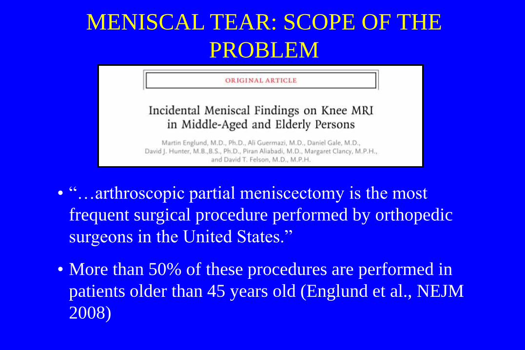
MENISCAL TEAR: SCOPE OF THE
PROBLEM
• “…arthroscopic partial meniscectomy is the most
frequent surgical procedure performed by orthopedic
surgeons in the United States.”
• More than 50% of these procedures are performed in
patients older than 45 years old (Englund et al., NEJM
2008)
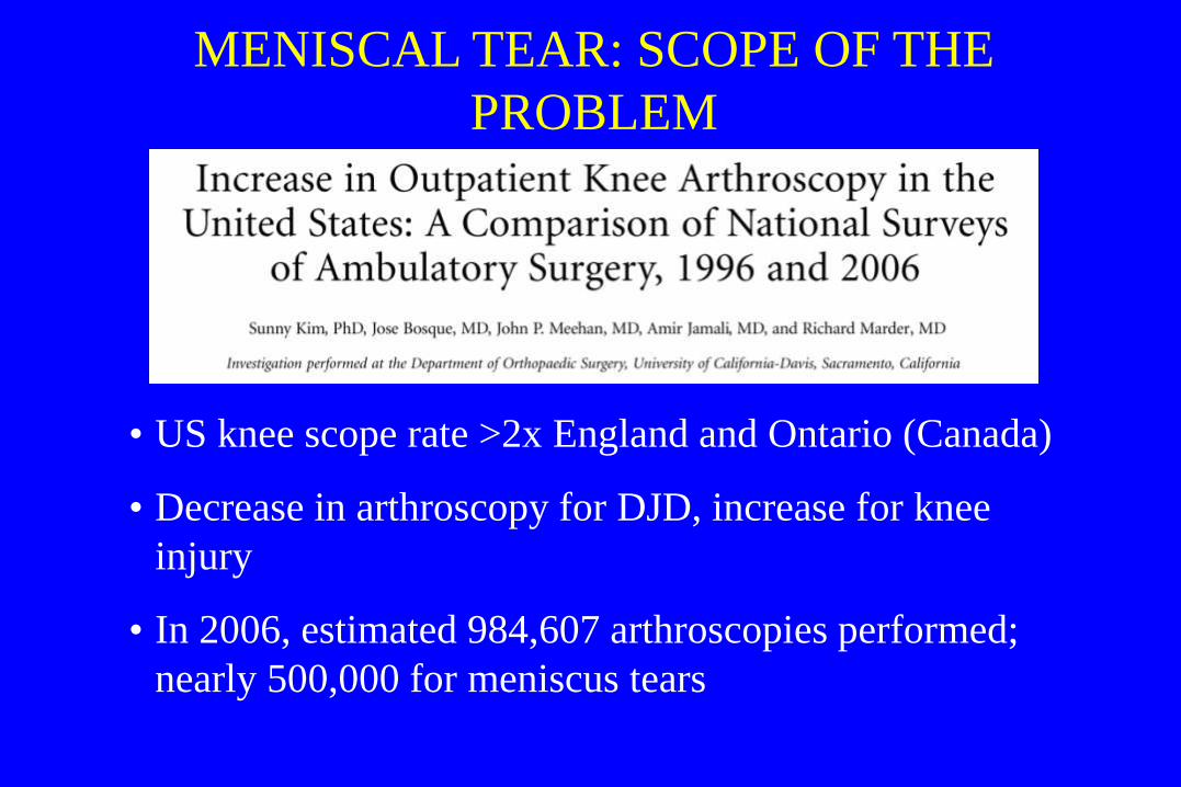
MENISCAL TEAR: SCOPE OF THE
PROBLEM
• US knee scope rate >2x England and Ontario (Canada)
• Decrease in arthroscopy for DJD, increase for knee
injury
• In 2006, estimated 984,607 arthroscopies performed;
nearly 500,000 for meniscus tears
MENISCAL TEAR: SCOPE OF THE
PROBLEM
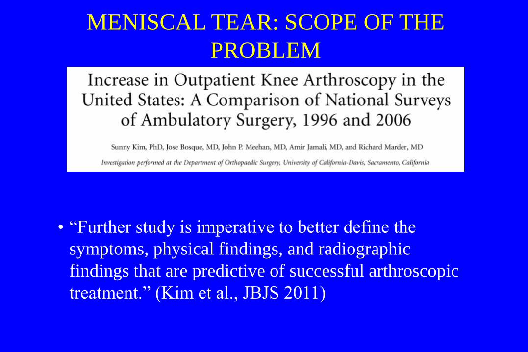
• “Further study is imperative to better define the
symptoms, physical findings, and radiographic
findings that are predictive of successful arthroscopic
treatment.” (Kim et al., JBJS 2011)
MENISCAL TEAR: ANATOMY
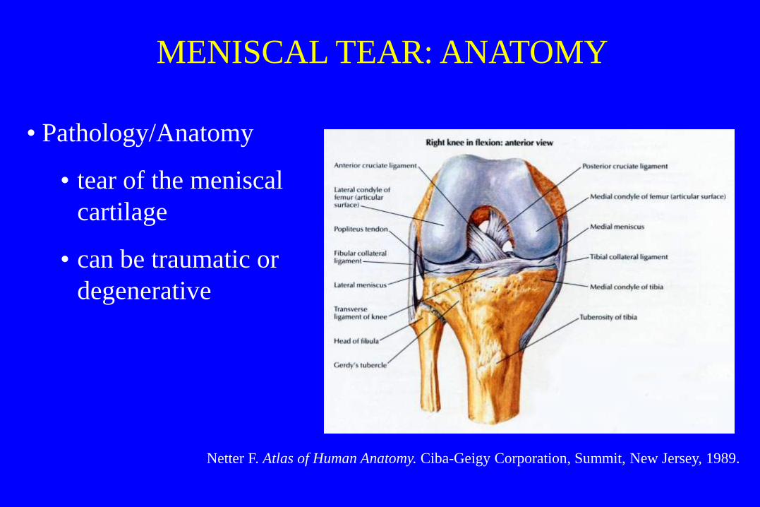
• Pathology/Anatomy
• tear of the meniscal
cartilage
• can be traumatic or
degenerative
Netter F. Atlas of Human Anatomy. Ciba-Geigy Corporation, Summit, New Jersey, 1989.
MENISCAL TEAR: ANATOMY
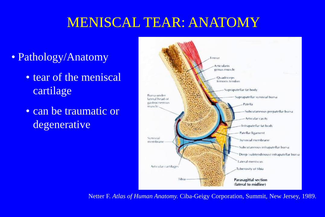
• Pathology/Anatomy
• tear of the meniscal
cartilage
• can be traumatic or
degenerative
Netter F. Atlas of Human Anatomy. Ciba-Geigy Corporation, Summit, New Jersey, 1989.
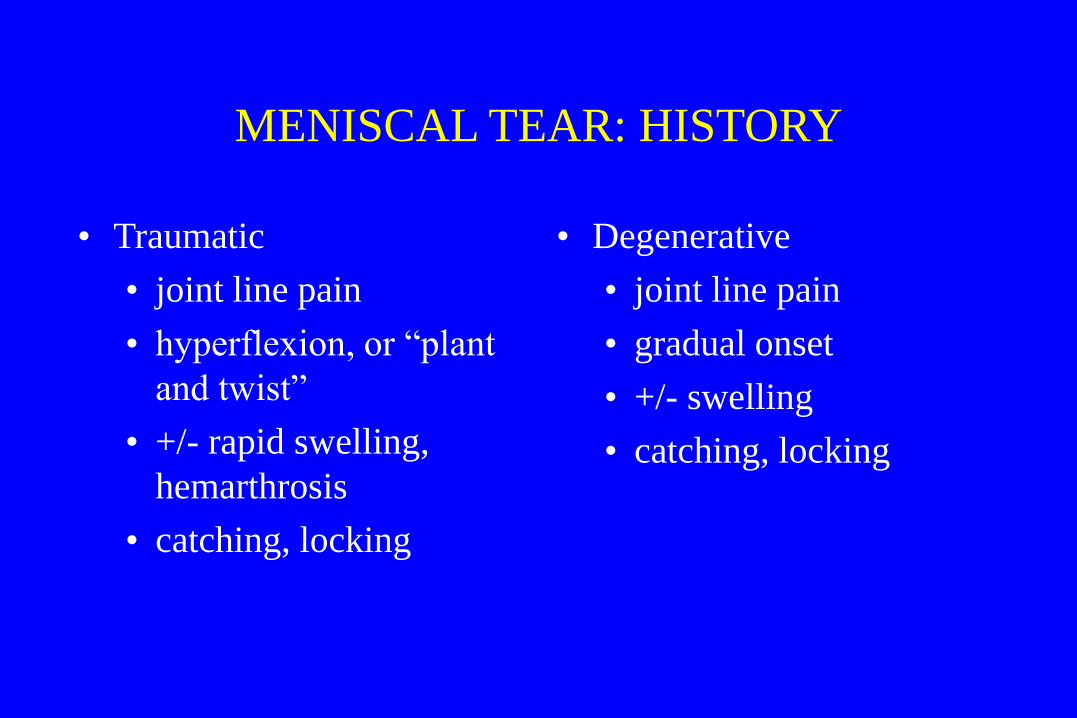
MENISCAL TEAR: HISTORY
• Traumatic
• joint line pain
• hyperflexion, or “plant
and twist”
• +/- rapid swelling,
hemarthrosis
• catching, locking
• Degenerative
• joint line pain
• gradual onset
• +/- swelling
• catching, locking
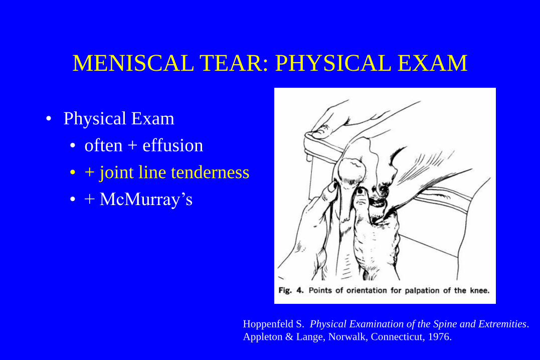
MENISCAL TEAR: PHYSICAL EXAM
• Physical Exam
• often + effusion
• + joint line tenderness
• + McMurray’s
Hoppenfeld S. Physical Examination of the Spine and Extremities.
Appleton & Lange, Norwalk, Connecticut, 1976.
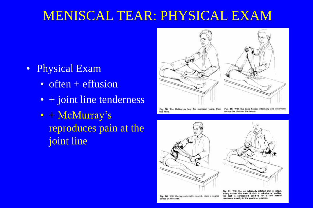
• Physical Exam
• often + effusion
• + joint line tenderness
• + McMurray’s
reproduces pain at the
joint line
MENISCAL TEAR: PHYSICAL EXAM
MENISCAL TEAR: EPIDEMIOLOGY
• Undiagnosed meniscal tears are common in the community, with
an incidence that increases with age (Englund et al, NEJM 2008):
• incidence of undiagnosed meniscus tears was :
• 32% and 19% of men and women 50-59 y/o
• 56% and 51% in men and women 70-90 y/o
• 61% of these tears were in patients who were asymptomatic in
the last month

CASE 1
• 45 y/o construction worker sent to you in consultation for
gradual onset right anterior knee pain for 3 months. Has tried PT
without significant improvement. Pain is starting to interfere
with work.
• PE: Small effusion. Patellar facet tenderness. No joint line
tenderness. McMurray’s reproduces pain behind the patella.
• Imaging: Patient has an MRI that shows a medial meniscus tear.
a) Send directly for surgical consult for meniscal tear.
b) Cortisone injection. If no improvement, then surgical
consult for meniscal tear.
c) More physical therapy.
CASE 1
Key points:
• Meniscus tears cause pain that is well localized to the
joint line.
• Many meniscal tears are asymptomatic.
CASE 2
• “Right knee meniscus tear, f/u ER”
• Per ER notes, “60 y/o male accountant with right
medial knee pain after twisting injury yesterday on a
wet floor at work. Difficulty walking after the
injury. Swollen today.”

CASE 2
• PE: Limping. Right knee with small effusion.
Tenderness along the medial joint line.
McMurray’s test reproduces medial pain.
• X-rays: 2 non-weightbearing views of the knees
“normal.”
• “Assessment and Plan: Acute medial meniscus tear.
Ice, ibuprofen, knee immobilizer, F/U with primary
care doctor in 1 week.”
CASE 2
a) Refer to physical therapy.
b) Order a 4 view weight bearing series of knee
x-rays.
c) Order MRI to evaluate for a meniscal tear.
d) Send directly for surgical consult.

MENISCAL TEAR: IMAGING
X-ray
• To rule out
fracture: 2 view
non-weight
bearing
• To rule out
significant
osteoarthritis: 4
view weight
bearing
MRI
McKinnis L. Fundamentals of Orthopedic Radiology. F A Davis Co, Philadelphia, 1997.

OSTEOARTHRITIS: XRAYS
4 weight bearing
views of the knee
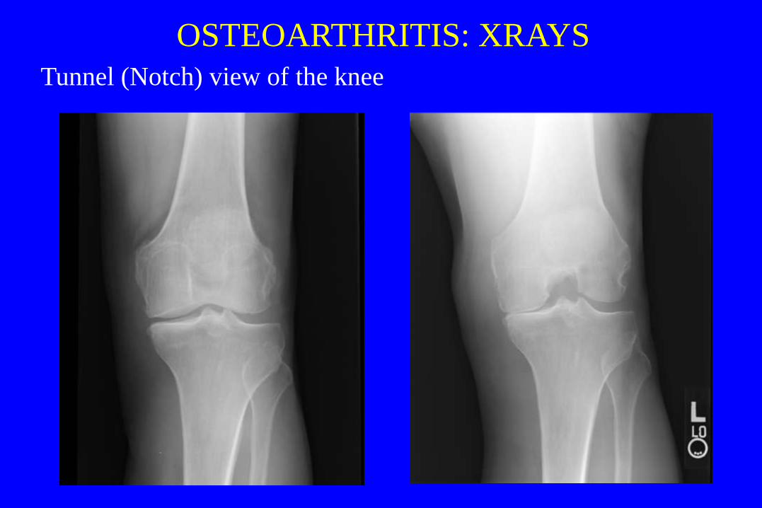
OSTEOARTHRITIS: XRAYS
Tunnel (Notch) view of the knee
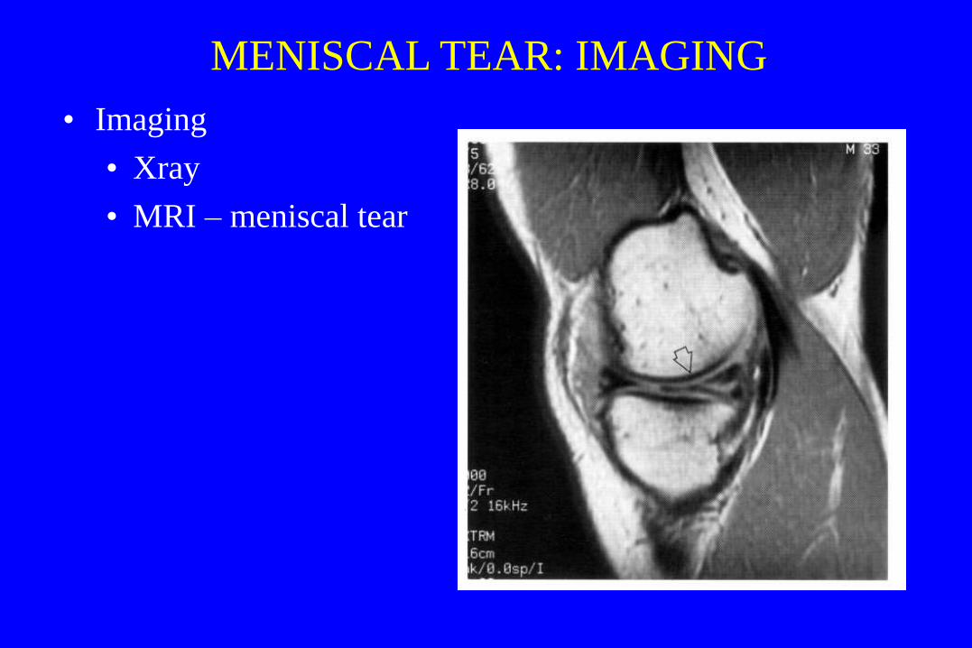
MENISCAL TEAR: IMAGING
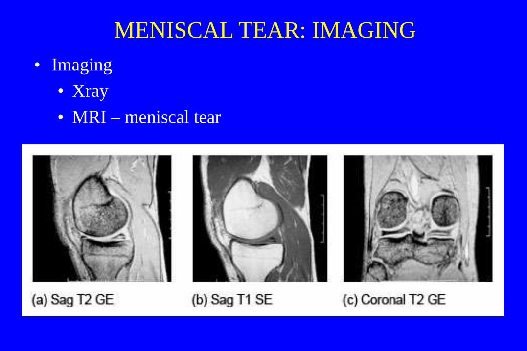
• Imaging
• Xray
• MRI – meniscal tear
MENISCAL TEAR: IMAGING
• Imaging
• Xray
• MRI – meniscal tear
MENISCAL TEAR: EPIDEMIOLOGY
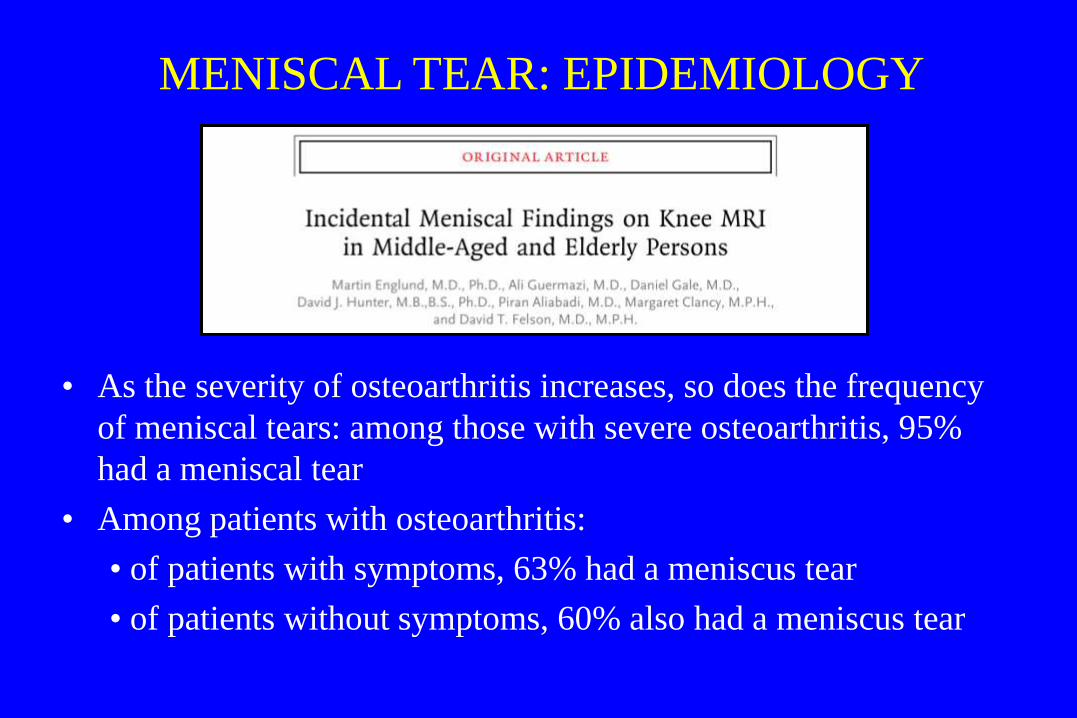
• As the severity of osteoarthritis increases, so does the frequency
of meniscal tears: among those with severe osteoarthritis, 95%
had a meniscal tear
• Among patients with osteoarthritis:
• of patients with symptoms, 63% had a meniscus tear
• of patients without symptoms, 60% also had a meniscus tear
MENISCAL TEAR: IMAGING
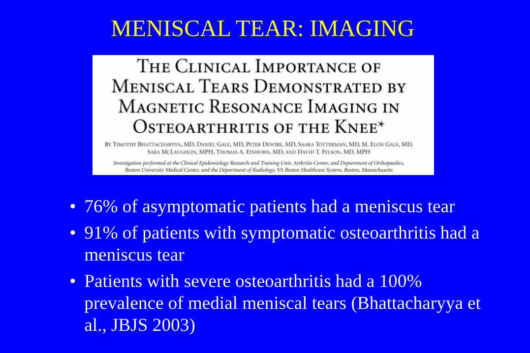
• 76% of asymptomatic patients had a meniscus tear
• 91% of patients with symptomatic osteoarthritis had a
meniscus tear
• Patients with severe osteoarthritis had a 100%
prevalence of medial meniscal tears (Bhattacharyya et
al., JBJS 2003)
MENISCAL TEAR: IMAGING
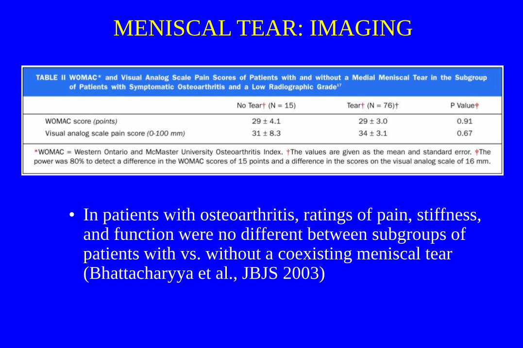
• In patients with osteoarthritis, ratings of pain, stiffness, and function were no different between subgroups of patients with vs. without a coexisting meniscal tear (Bhattacharyya et al., JBJS 2003)
CASE 2
• “60 y/o male, right knee meniscus tear, f/u ER”
• X-rays: 2 non-weightbearing views of the knees “normal.”
• “Assessment and Plan: Acute medial meniscus tear. Ice, ibuprofen, knee immobilizer, F/U with primary care doctor in 1 week.”
a) Refer to physical therapy. b) Order a 4 view weight bearing series of knee x-rays. c) Order MRI to evaluate for a meniscal tear. d) Send directly for surgical consult.
CASE 2
Key points:
• Meniscus tears are increasingly common in knees with
osteoarthritis.
• As a group, osteoarthritic patients with a meniscus tear are
no more symptomatic than osteoarthritic patients without a
meniscus tear .
• Consequently, review x-rays (4 views, weight bearing) to
assess for osteoarthritis prior to obtaining MRI.
CASE 3
• 60 y/o male with gradual onset right medial knee pain for 3 weeks. Worsened yesterday when twisted it in the workplace parking lot.
• PE: Small effusion. + medial joint line tenderness. McMurray’s reproduces medial joint line pain.
• X-rays show trace osteoarthritic changes.
• MRI confirms minimal medial articular cartilage changes without associated bone marrow edema, as well as a medial meniscus tear.
a) Trial of conservative therapy including some combination of rest, ice, NSAIDS, cortisone injections, viscosupplementation, and/or physical therapy.
b) Send directly for surgical consult.
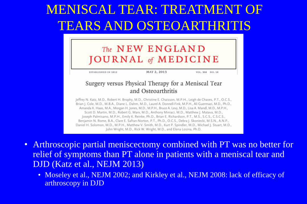
MENISCAL TEAR: TREATMENT OF
TEARS AND OSTEOARTHRITIS
• Arthroscopic partial meniscectomy combined with PT was no better for relief of symptoms than PT alone in patients with a meniscal tear and DJD (Katz et al., NEJM 2013)
• Moseley et al., NEJM 2002; and Kirkley et al., NEJM 2008: lack of efficacy of arthroscopy in DJD
MENISCAL TEAR: TREATMENT OF
DEGENERATIVE TEARS WITHOUT DJD
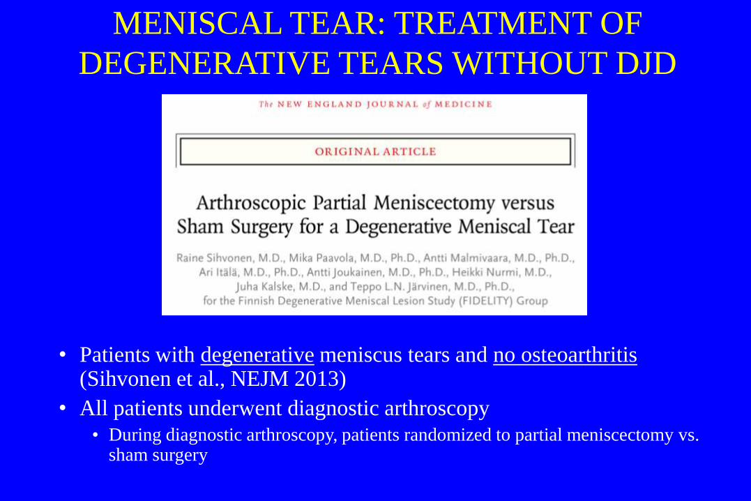
• Patients with degenerative meniscus tears and no osteoarthritis (Sihvonen et al., NEJM 2013)
• All patients underwent diagnostic arthroscopy
• During diagnostic arthroscopy, patients randomized to partial meniscectomy vs. sham surgery
MENISCAL TEAR:
TREATMENT OF
DEGENERATIVE TEARS
WITHOUT DJD
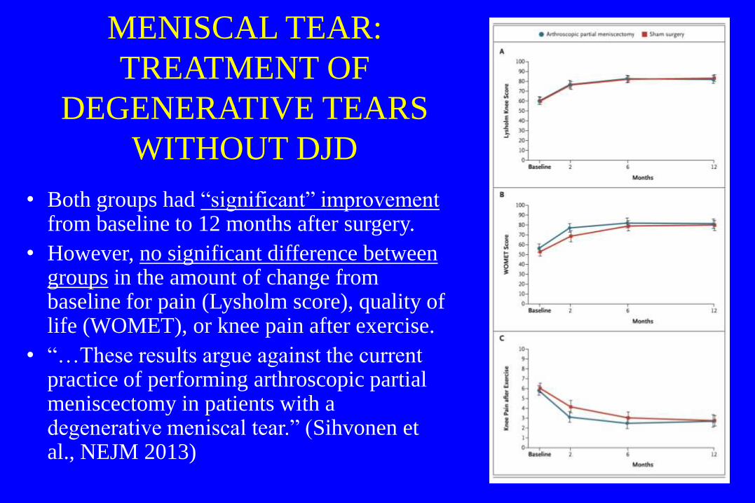
• Both groups had “significant” improvement from baseline to 12 months after surgery.
• However, no significant difference between groups in the amount of change from baseline for pain (Lysholm score), quality of life (WOMET), or knee pain after exercise.
• “…These results argue against the current practice of performing arthroscopic partial meniscectomy in patients with a degenerative meniscal tear.” (Sihvonen et al., NEJM 2013)

MENISCAL TEAR: TREATMENT OF
DEGENERATIVE TEARS WITHOUT DJD
Limitations and critiques of the study
• Factors such as subchondral edema or chondromalacia on MRI were not used to exclude or stratify patients
• Patients with mechanical symptoms were excluded, “yet this is probably the group that would benefit most from arthroscopic partial meniscectomy.” (Krych et al., NEJM 2014)
• “Mechanical symptoms are an important primary problem that arthroscopic meniscectomy can alleviate. Such symptoms were reported by less than half the patients in this study, and a locked knee was an exclusion criterion….” (Jevsevar et al., NEJM 2014)
MENISCAL TEAR: TREATMENT OF
DEGENERATIVE TEARS WITH
MECHANICAL SYMPTOMS
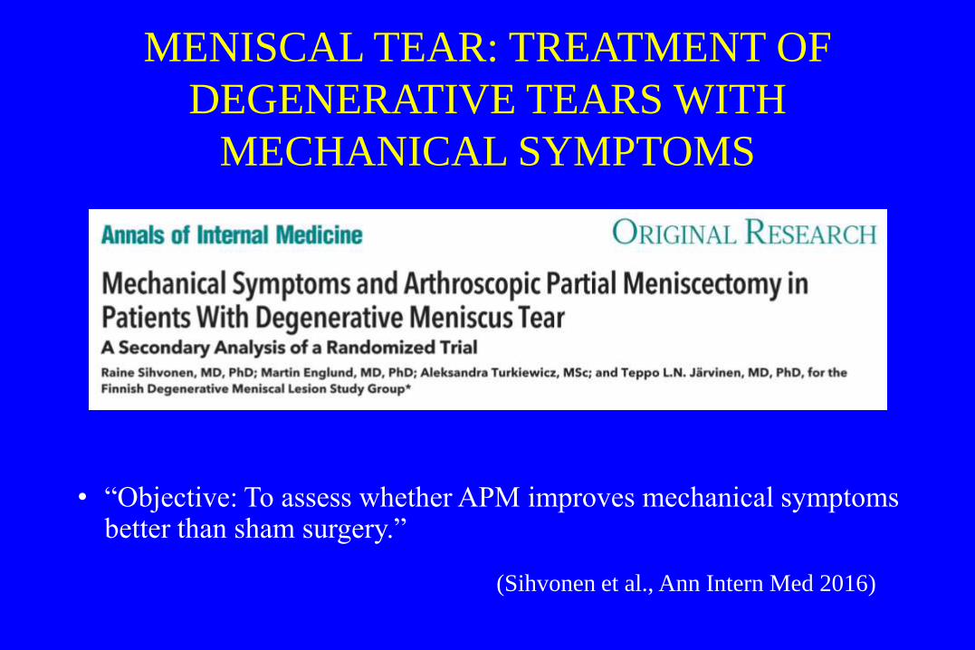
• “Objective: To assess whether APM improves mechanical symptoms better than sham surgery.”
(Sihvonen et al., Ann Intern Med 2016)
MENISCAL TEAR: TREATMENT OF
DEGENERATIVE TEARS WITH
MECHANICAL SYMPTOMS
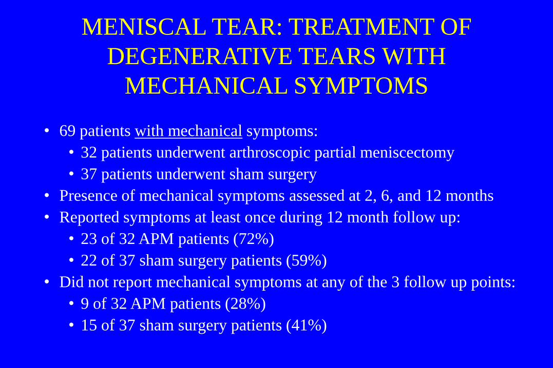
• 69 patients with mechanical symptoms:
• 32 patients underwent arthroscopic partial meniscectomy
• 37 patients underwent sham surgery
• Presence of mechanical symptoms assessed at 2, 6, and 12 months
• Reported symptoms at least once during 12 month follow up:
• 23 of 32 APM patients (72%)
• 22 of 37 sham surgery patients (59%)
• Did not report mechanical symptoms at any of the 3 follow up points:
• 9 of 32 APM patients (28%)
• 15 of 37 sham surgery patients (41%)
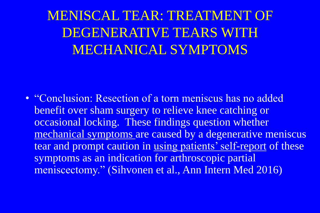
MENISCAL TEAR: TREATMENT OF
DEGENERATIVE TEARS WITH
MECHANICAL SYMPTOMS
• “Conclusion: Resection of a torn meniscus has no added benefit over sham surgery to relieve knee catching or occasional locking. These findings question whether mechanical symptoms are caused by a degenerative meniscus tear and prompt caution in using patients’ self-report of these symptoms as an indication for arthroscopic partial meniscectomy.” (Sihvonen et al., Ann Intern Med 2016)

MENISCAL TEAR: SURGICAL DECISION
MAKING
• Arthroscopy for any meniscal tear with significant osteoarthritis: no benefit
• Arthroscopy for degenerative meniscal tear without osteoarthritis: no benefit
• Arthroscopy for degenerative meniscal tear with “mechanical” symptoms: no benefit
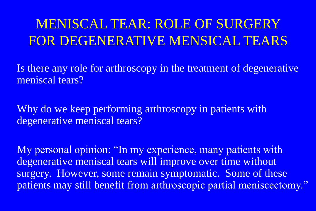
MENISCAL TEAR: ROLE OF SURGERY
FOR DEGENERATIVE MENSICAL TEARS
Is there any role for arthroscopy in the treatment of degenerative meniscal tears?
Why do we keep performing arthroscopy in patients with degenerative meniscal tears?
My personal opinion: “In my experience, many patients with degenerative meniscal tears will improve over time without surgery. However, some remain symptomatic. Some of these patients may still benefit from arthroscopic partial meniscectomy.”

MENISCAL TEAR: SURGICAL DECISION
MAKING
• “Meniscal tears and osteoarthritis frequently coexist, but to our knowledge, no data exist to identify who will benefit from arthroscopic partial meniscectomy versus non-operative management. Our objective was to evaluate the capability of preoperative information to predict arthroscopic partial meniscectomy outcomes in osteoarthritis.” (Suter et al., Arth Rheum 2009)
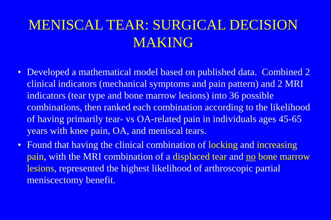
MENISCAL TEAR: SURGICAL DECISION
MAKING
• Developed a mathematical model based on published data. Combined 2
clinical indicators (mechanical symptoms and pain pattern) and 2 MRI
indicators (tear type and bone marrow lesions) into 36 possible
combinations, then ranked each combination according to the likelihood
of having primarily tear- vs OA-related pain in individuals ages 45-65
years with knee pain, OA, and meniscal tears.
• Found that having the clinical combination of locking and increasing
pain, with the MRI combination of a displaced tear and no bone marrow
lesions, represented the highest likelihood of arthroscopic partial
meniscectomy benefit.
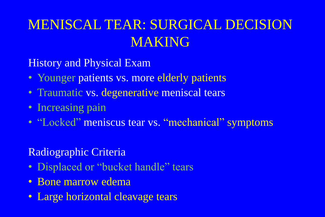
MENISCAL TEAR: SURGICAL DECISION
MAKING
History and Physical Exam
• Younger patients vs. more elderly patients
• Traumatic vs. degenerative meniscal tears
• Increasing pain
• “Locked” meniscus tear vs. “mechanical” symptoms
Radiographic Criteria
• Displaced or “bucket handle” tears
• Bone marrow edema
• Large horizontal cleavage tears

MENISCAL TEAR: ROLE OF SURGERY
FOR DEGENERATIVE MENSICAL TEARS
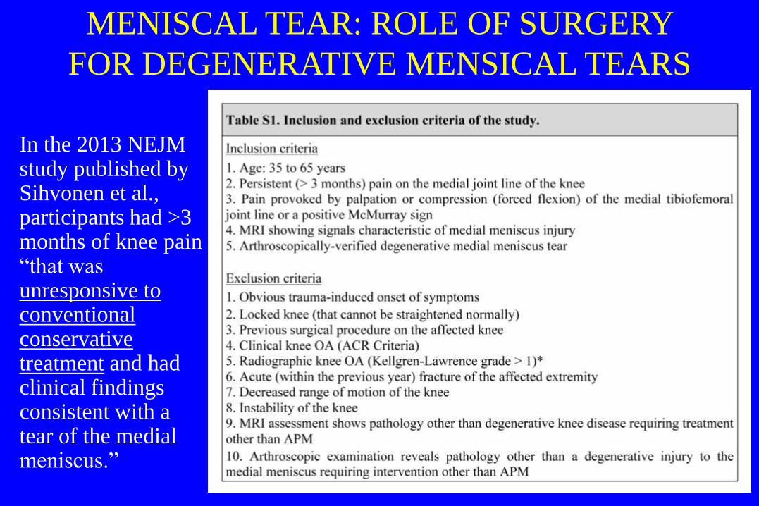
In the published study by Sihvonen et al., participants had >3 months of knee pain “that was unresponsive to conventional conservative treatment and had clinical findings consistent with a tear of the medial meniscus.”
MENISCAL TEAR: ROLE OF SURGERY
FOR DEGENERATIVE MENSICAL TEARS
In the 2013 NEJM study published by Sihvonen et al., participants had >3 months of knee pain “that was unresponsive to conventional conservative treatment and had clinical findings consistent with a tear of the medial meniscus.”
MENISCAL TEAR: ROLE OF SURGERY
FOR DEGENERATIVE MENSICAL TEARS
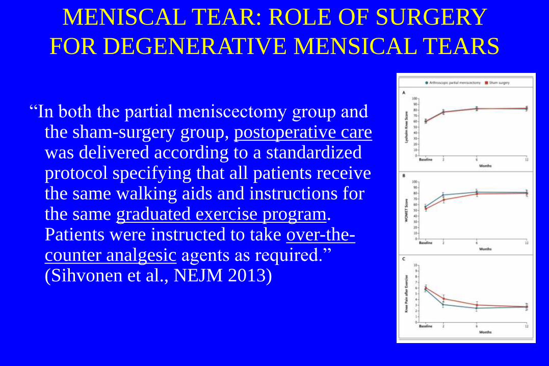
“In both the partial meniscectomy group and the sham-surgery group, postoperative care was delivered according to a standardized protocol specifying that all patients receive the same walking aids and instructions for the same graduated exercise program. Patients were instructed to take over-the-counter analgesic agents as required.” (Sihvonen et al., NEJM 2013)
MENISCAL TEAR: ROLE OF SURGERY
FOR DEGENERATIVE MENSICAL TEARS
Possible approach to the patient with symptoms suggestive of a degenerative meniscal tear
• If “locked,” then early MRI. If MRI confirms displaced tear plausible for patient’s disability, then early surgical referral.
• Otherwise, optimize a period of relative rest, oral NSAIDS, cortisone injections, and physical therapy. May also consider viscosupplementation for patients with mild osteoarthritis.
• If still not improved, then MRI for consideration of arthroscopy.
MENISCAL TEAR: ROLE OF SURGERY
FOR DEGENERATIVE MENSICAL TEARS
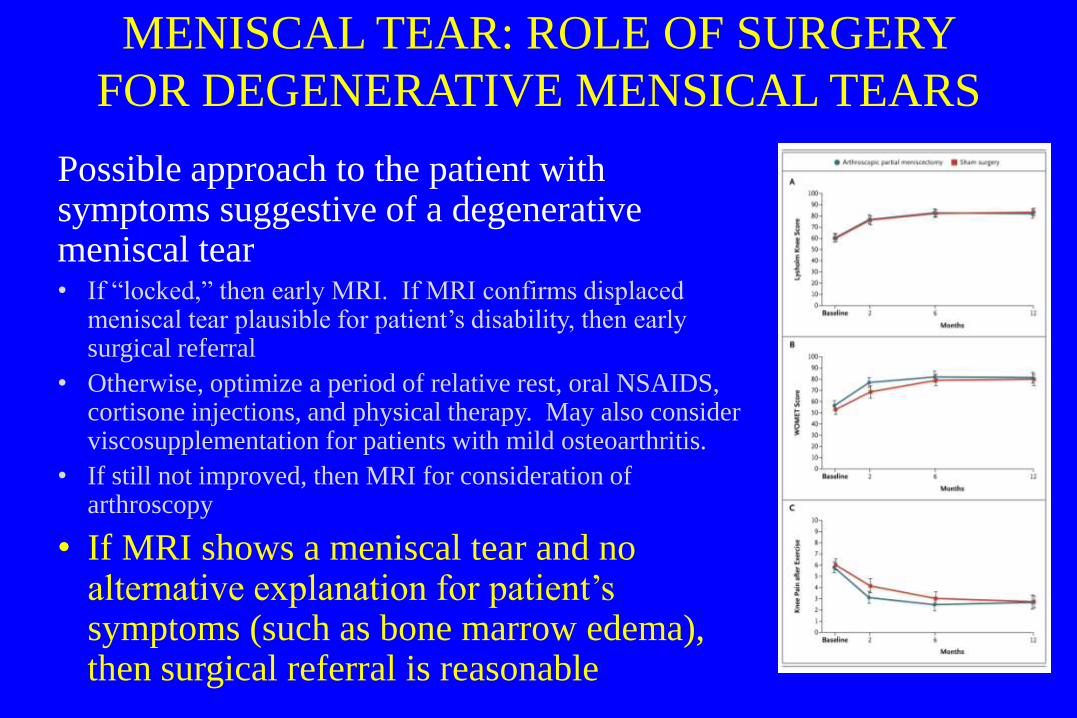
Possible approach to the patient with symptoms suggestive of a degenerative meniscal tear • If “locked,” then early MRI. If MRI confirms displaced
meniscal tear plausible for patient’s disability, then early surgical referral
• Otherwise, optimize a period of relative rest, oral NSAIDS, cortisone injections, and physical therapy. May also consider viscosupplementation for patients with mild osteoarthritis.
• If still not improved, then MRI for consideration of arthroscopy
• If MRI shows a meniscal tear and no alternative explanation for patient’s symptoms (such as bone marrow edema), then surgical referral is reasonable

CASE 3
• 60 y/o male with gradual onset right medial knee pain for 3 weeks. Worsened yesterday when twisted it in the workplace parking lot.
• PE: Small effusion. + medial joint line tenderness. McMurray’s reproduces medial joint line pain.
• X-rays show trace osteoarthritic changes.
• MRI confirms minimal medial articular cartilage changes without associated bone marrow edema, as well as a medial meniscus tear.
a) Trial of conservative therapy including some combination of rest, ice, NSAIDS, cortisone injections, viscosupplementation, and/or physical therapy.
b) Send directly for surgical consult.

THE END