non-traditional sources for isolation of lactic acid bacteria
TRANSCRIPT
ORIGINAL ARTICLE
Non-traditional sources for isolation of lactic acid bacteria
Tsvetanka Teneva-Angelova1 & Dora Beshkova1
Received: 7 April 2015 /Accepted: 26 June 2015 /Published online: 28 July 2015# Springer-Verlag Berlin Heidelberg and the University of Milan 2015
Abstract In recent years there has been increasing interestin lactic acid bacteria isolated from non-dairy products,due to their diverse metabolic profile, unique flavor-forming activities, and potential for use as starters or start-er adjuncts for the dairy industry. Screening of 400 mi-crobial isolates obtained from the herbs Geraniumsanguineum L., Hypericum perforatum L., and Panaxginseng C.A. Meyer was performed, and 64 isolates wereselected based on milk coagulation and gas formationability and non-specific odour. Using tests involving mul-tiple transfer and growth in selective and differential me-dia, 258 single colonies were isolated, of which 98 wereaffiliated with the lactic acid bacteria group. These bacte-ria are homofermentative cocci and rods with a wide pH(5.0–9.6) and temperature range (15–45 °C), high salttolerance (3.0–10.0 % NaCl), and high acid-producingactivity (3.50–12.00 g/L). With the use of genotype-based methods, the plant isolates were identified at thespecies level as Enterococcus faecium, Streptococcusthermophilus and Lactobacillus rhamnosus.
Keywords Lactic acid bacteria .Medicinal plants . Isolation .
16S rDNA
Introduction
Lactic acid bacteria (LAB) are predominantly represented as anon-taxonomic heterogeneous group of Gram-positive andnon-spore-forming facultative anaerobic bacteria. This groupincludes a wide variety of cell types and physiological andbiochemical characteristics. LAB are designated "generallyrecognized as safe" (GRAS status), and have long played animportant role in food technology—they are responsible forlactic fermentation in several products, including dairy, meat,and silage. LAB can be isolated from traditional sources suchas raw milk, dairy products and fermented foods (Wouterset al. 2002 ; Kimoto et al. 2004; Tamang et al. 2005;Nomura et al. 2006; Kostinek et al. 2007; Tanasupawat et al.2007; Kivanc et al. 2011; Venturi et al. 2012; Abegaz 2014),and from alternative sources including fecal samples, soil andplants (Hartnett et al. 2002; Magnusson et al. 2003; Cock andde Stouvenel 2006; Siezen et al. 2008; Trias et al. 2008; DiCagno et al. 2009; Cakir 2010; Chen et al. 2010; Venugopalanet al. 2010; Baradaran et al. 2012; Emerenini et al. 2013;Fhoula et al. 2013; Nguyen et al. 2013; Alemayehu et al.2014). In recent years, there has been increasing interest inLAB isolated from non-dairy sources due to their diverse met-abolic profile and unique flavor-forming activities. Plant-de-rived strains of lactobacteria have demonstrated toleranceto high pH values and salt concentrations, an ability toferment more types of carbohydrates, and a high level ofstress resistance compared to those of dairy origin.Furthermore, studies have noted no significant differencesin fermentation characteristics and profiles of enzymes(lipases, peptidases and phosphatases) required forobtaining various fermented dairy products with plant-derived and commercial strains of lactobacteria (Nomuraet al. 2006; Michaylova et al. 2007; Siezen et al. 2008;Venugopalan et al. 2010).
* Dora [email protected]
1 The Stephan Angeloff Institute of Microbiology, BAS, 26 GeorgiBonchev St., 1113 Sofia, Bulgaria
Ann Microbiol (2016) 66:449–459DOI 10.1007/s13213-015-1127-9

The search for new solutions to improve starter systems forfermented health foods and for opportunities to realize thebiological potential of LAB has given rise to initiatives forexploiting biodiversity in unique natural systems (medicinalplants). These plants are an important ecosystem for isolationof LAB (Siezen et al. 2008; Cakir 2010; Venugopalan et al.2010; Baradaran et al. 2012), and each individual plant speciesprovides a unique environment in terms of competing micro-organisms and natural plant antagonists, as well as accessibil-ity, type and concentration of the substrate in the various phys-ical factors. When the plant material is collected and preparedfor fermentation, these conditions allow for growth of typicalepiphytic flora from which a population and chain of fermen-tation processes is derived.
Current available information on the use of medicinalplants as a source for isolation of LAB and their subsequentpotential application as components to form starters for milkfermentation is scarce (Venugopalan et al. 2010). There are nodata in the scientific literature regarding the microbial pres-ence of different types of bacteria—and LAB in particular—isolated from herbs of the genera Geranium, Panax orHypericum. Extracts from various parts of Geraniumsanguineum L., known in Bulgaria by the popular name"bloody geranium," have significant antiviral, antibacterial,anti-inflammatory and antioxidant activity (Serkedjieva andManolova 1992; Hammami et al. 2011). Extracts ofHypericum perforatum L., commonly known in Bulgaria asByellow wort^ and as BSt John’s wort^ in other countries andon other continents, also possess antibacterial and antiviralproperties. St John’s wort has traditionally been used in theform of homeopathic medicines as an alternative treatment forvarious depressive disorders of mild to moderate severity(Barnes et al. 2001). The incorporation of plant extracts inpreparations for topical wound treatment has also been suc-cessful (Newall et al. 1996). Husain et al. (2011) demonstratedsignificant hypolipidemic activity for the Hypericumperforatum L. extract in a rat model. In addition, the plant isclassified by the Council of Europe (2000) as a natural sea-soning for various foods (Barnes et al. 2001). There have beenreports of high levels of antioxidant activity and pharmaco-logical effects for extracts of Panax ginseng C.A. Meyer,known worldwide as Bginseng,^ including stimulation ofbrain function, pain relief, activation of antitumor immunityand improvement in total immunity, regulation of blood pres-sure and blood sugar levels in diabetic patients, tonic effectson physical fatigue and mental stress, a positive influence onliver function, male potency and side effects of menopause inwomen, and the inhibition of growth of the AIDS virus (Choi2008). A new direction in scientific research that has recentlybeen explored involves obtaining bioactive or biogenic sub-stances extracted from different plants or synthesized duringfood fermentation, and the subsequent creation of novel foods(defined as healthy and functional) by further introduction of
such exogenous functional components into their technologi-cal schemes or through the use of microorganisms as pro-ducers of biogenic substances or microorganisms with probi-otic characteristics (Gobbetti et al. 2010). In this context,Servili et al. (2011) reported the production of a functionalmilk beverage through fermentation of cow's milk that hadbeen enriched with phenolic extracts from olive vegetablewater. For this purpose, the authors used starter cultures in-cluding Lactobacillus delbrueckii subsp. bulgaricusDPPMALDb5 and S t rep tococcus thermoph i l u sDPPMAST1, and used the probiotic strain Lactobacillusparacasei 15 N and γ-amino butyric acid-producing-strainLactobacillus plantarum C48 as adjunct cultures.
The present study was a preliminary evaluation of LABisolated from the herbs Geranium sanguineum L.,Hypericum perforatum L. and Panax ginseng C.A. Meyer aspotential starters or adjunct starters for fermented dairy prod-ucts. The main objective of this work was to evaluate thepresence of LAB within the context of microbial diversity innatural biosystems (medicinal plants Geranium sanguineumL.,Hypericum perforatum L. and Panax ginsengC.A.Meyer)with desired metabolic activities in order to include them asstarter or non-starter components during in situ cultivation inmilk, and to identify the isolates at the species level usinggenotype-based methods including 16S rDNA and phyloge-netic tree construction.
Materials and methods
Sample collection
Plant material was collected from following locations:Geranium sanguineum L., Eastern Rhodopes (Ivaylovgrad),the Sofia region (experimental field of the Institute ofBiodiversity and Ecosystem Research [IBER] at theBulgarian Academy of Sciences [BAS]) and the Vitosha re-gion (Iskar Dam); Hypericum perforatum L., WesternRhodopes (Persenk Chalet); Panax ginseng C.A. Meyer,Botanical Garden of the Technical University of Dresden,Germany. All plant samples were collected aseptically in ster-ile poly-bags, kept under refrigeration, and transported to thelaboratory for analyses.
Lactic acid bacteria isolation procedure
Individual parts (flower, leaf and stem) of each plant specieswere carefully washed in sterile water, then transferred to testtubes with sterile 10 % reconstituted skim milk (RSM;HiMedia Laboratories, Mumbai, India) and incubated at 30and 37 °C until coagulation of milk occurred. After assess-ment of milk coagulation, gas formation and non-specificodour, samples were selected for subsequent transfer to M17
450 Ann Microbiol (2016) 66:449–459

broth (pH 6.6; Merck Millipore, Darmstadt, Germany) andMRS broth (pH 5.7; Merck Millipore), with the addition of100 μg/mL of cycloheximide (Sigma-Aldrich, St. Louis. MOUSA) (Hartnett et al. 2002) in order to prevent fungal growthand to select for LAB. Selected samples were cultured inanaerobic jars using the Anaerocult A mini system (MerckMillipore) for a period of 72 h at the above-referenced tem-peratures. Serial decimal dilutions of broth cultures in 0.85 %(w/v) sterilized NaCl solution were plated by spreading0.1 mL on MRS and M17 agar plates (Merck Millipore),which were incubated anaerobically at 30 and 37 °C, respec-tively, for 3–5 days to obtain single bacterial colonies. Thesecolonies were then randomly selected and purified by streak-ing again and subculturing on fresh MRS and M17 agarplates. The purity of isolated single colonies was evaluatedmicroscopically (MICROS Pink MC50; MICROSProduktions- und HandelsgesmbH St. Veit an der Glan,Austria).
The abbreviations of isolated lactic acid strains from therelevant plant samples include the initials of the relevant plantspecies, an index indicating the location of the plant species oran initial of a nearby location to the region in question inBulgaria and, respectively, the abbreviated spelling ofGermany (e.g., GsfIV123: Gs [Geranium sanguineum], f[flower], IV [Ivaylovgrad], 123 [number of colony]).
Identification of lactic acid bacteria
Phenotypic and biochemical characteristics
The initial study of purified bacterial isolates involved micro-scopic characterization of the morphology of cells (MICROSPink MC50) and colonies (shape, color and size) (CETI DigiSteddy II, Medline Scientific, Chalgrove, Oxfordshire, UK)and subsequent differentiating Gram staining. Biochemicaltests (catalase test, oxidase test, reaction to indole) were car-ried out in selected Gram (+) isolates in order to establish theirLAB group affiliation.
Gram-positive, catalase-negative, indole-negative andoxidase-negative presumptive LAB were stored in M17 andMRS broth containing 15 % glycerol (Merck Millipore,Darmstadt, Germany) at −80 °C for use in subsequent exam-inations. For cultivation, bacterial isolates were reactivated inM17 and MRS broth at 37 °C for 24–48 h.
Production of CO2 from glucose was determined in tubesof MRS broth containing inverted Durham tubes, incubatedfor 3 days at 37 °C.
The configuration of lactic acid (LA) produced from glu-cose was determined enzymatically using the D-lactate and L-lactate dehydrogenase test kit K-DLATE 12/12 (MegazymeInternational Ireland, Bray, Co. Wicklow, Ireland), with incu-bation for 72 h at 37 °C.
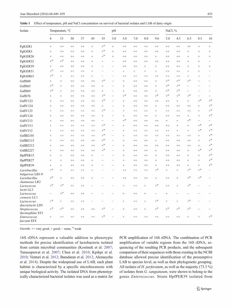
The presumptive LAB that manifested high acid-producingactivity were tested for growth at different temperatures (4, 15,30, 37, 45 and 55 °C) and different pH values (3.0, 5.0, 7.0,8.0 and 9.6) inM17 broth and MRS broth for 72 h, with initialpH values of 6.6 and 5.7 for the two media, respectively, at atemperature 37 °C. The level of salt tolerance was determinedafter growth inM17 broth (pH 6.6) andMRS broth (pH 5.7) inthe presence of various concentrations of NaCl (3.0, 4.5, 6.5,8.5 and 10.0 %) at 37 °C for 72 h.
For comparative characterization of the presumptive LABisolated from G. sanguineum, H. perforatum and P. ginseng,with regard to the above-mentioned properties, these werecultivated in parallel with LAB belonging to the laboratorycollection, which were isolated from various dairy products,and were used in our previous studies: Lactococcus lactis ssp.lactis biovar. diacetylactis LD5 (Lactococcus diacetylactisLD5), Streptococcus thermophilus ST3, Lactococcus lactisssp. lactis LL3, Lactococcus lactis ssp. cremoris LC1(Lactococcus cremoris LC1), Enterococcus faecium EF4,Lactobacil lus delbrueckii ssp. bulgaricus LB1-9(Lactobacillus bulgaricus LB1-9) and Lactobacillusrhamnosus LR2.
The preliminary identification of selected presumptiveLAB was investigated using the API 50 CHL and API 20STREP (bioMérieux SA, Marcy-l'Étoile, France) galleries.The tests were conducted according to manufacturer instruc-tions, and the results were read after incubation of the strain at37 °C for 2–3 days.
Genotypic characterization
Each isolate was grown anaerobically on M17 and MRS agarfor 48 h at 37 °C. A single colony was suspended in 1 mLMilli-Q water and centrifuged for 1 min at 12,000 g. GenomicDNAwas isolated from the pellet using the NucleoSpin® SoilKit (Macherey-Nagel GmbH & Co. KG, Düren, Germany),following the manufacturer's instructions. The concentrationof the resultant genomic DNA was measured with aNanoDrop 2000 UV-Vis spectrophotometer (Thermo FisherScientific Inc., Waltham, MA, USA).
U n i v e r s a l p r i m e r s 2 7 F ( 5 ′ -AGAGTTTGATCCTGGCTCAG-3′) and 1492R (5 ′-TACGGТTACCTTGTTACGACTT-3′) were used to amplifythe fragments of the 16S ribosomal gene. Each PCR mixture(50 μL) contained a reaction mix of 25 μL HotStarTaq PlusMaster Mix Kit, 2× (Qiagen GmbH, Düsseldorf, Germany),1 μL of each primer (10 μM), 100 ng of DNA template andautoclavedMilli-Q water. The amplification was performed ina Mastercycler® pro (Eppendorf North America, Hauppauge,NY, USA), and the following program was used: initial dena-turation at 95 °C for 1 min, followed by 33 cycles of denatur-ation at 95 °C for 15 s, annealing at 50 °C for 30 s, elongationat 72 °C for 100 s, and a final extension for 3 min at 72 °C. The
Ann Microbiol (2016) 66:449–459 451
amplified product was cooled at 4 °C. The quality of the iso-lated total DNA and amplification fragments of 16S rDNA(approximately 1500 base pairs [bp]) was analyzed by elec-trophoresis on a 1% (w/v) agarose gel in 1XTAE buffer (stock50X TAE: 242 g/L Tris base, 57.1 mL/L acetic acid, 100 mL0.5 M EDTA, pH 8.5) at 80 V for 45 min. Staining wasperformed in GelRed (Biotium Inc., Hayward, CA, USA)fluorescent dye (0.05 μg/mL), and the bands were visualizedunder an ULTima 10si (Hoefer Inc., Holliston, MA, USA).The size of DNA fragments was estimated using a standard100-bp DNA ladder (Invitrogen, Carlsbad, CA, USA).Amplification fragments from 16S rDNAwere purified usingthe NucleoSpin® Gel and PCR Clean-up kit (Macherey-NagelGmbH & Co. KG, Düren, Germany), according to the manu-facturer’s instructions.
Sequencing was performed by Eurofins MWGOperon LLC(Ebersberg, Germany). Sequence assembly was performedusing the BioEdit software program. The sequence homologieswere examined by comparing the sequences obtained with thosein the National Center for Biotechnology Information (NCBI)database using BLAST (http://www.ncbi.nlm.nih.gov/BLAST/). The results obtained were used to identify isolatesto genus or species level. Phylogenetic trees were constructedusing MEGA 4.1 software (Tamura et al. 2007).
Statistical analyses
Data represent the mean values of three independent experi-ments. The errors of experimental data from the mean valueswere expressed as standard deviations using the MicrosoftExcel 2010 program. Standard deviations were illustrated aserror bars.
Nucleotide sequence accession numbers
The nucleotide sequences obtained in this study were depos-ited in NCBI GenBank under the following accession num-bers: KR054662, KR054663, KR054664, KR054665,KR054666, KR054667, KR054668, KR054669, KR054670,KR054671, KR054672, KR054673, KR054674, KR054675,KR054676, KR054677, KR054678, KR054679, KR054680,KR054681, KR054682, KR054683, KR054684, KR054685and KR054686.
Results
Bacterial isolates obtained fromGeranium sanguineumL.,Hypericum perforatum L. and Panax ginseng C.A. Meyer,and their preliminary identification
As a result of large-scale screening of 400 microbial isolates(derived from flower, leaf or stem of G. sanguineum,
H. perforatum and P. ginseng) based on visual assessment ofmilk coagulation, gas formation and non-specific odour, 64microbial isolates (16.0 %) were selected. After subsequentmultiple transfer and growth on selective media, 258 singlebacterial colonies were isolated, which were further identifiedbymeans of classical techniques to determine their phenotypiccharacteristics and their LAB group affiliation. Based on therequired and confirmatory tests performed, 98 isolates (38 %)showed phenotypic identity with the lactobacteria group,which were Gram-positive, catalase-negative, oxidase-negative and indole-negative. Representatives of LAB werenot isolated from the stem of G. sanguineum, but the flowersproved to be the most preferred site for habitation bylactobacteria (37 single colonies), followed by the leaves (25single colonies). Presumptive LAB were isolated only fromthe flowers ofH. perforatum (11 single colonies) and from theleaves of P. ginseng (25 single colonies).
The presumptive LAB were morphologically defined ascocci and rods. Cocci were isolated from G. sanguineumand H. perforatum, and rods from P. ginseng. Cocci werearranged as single cells and cell pairs, in short and long chains,with cell sizes from 0.8 to 2.9 μm. The relevant colonies wereshiny, with white and light beige colors, circular and convex inshape, with entire or undulate margins, and ranging in sizefrom 1.0 to 2.5 mm. Rods were arranged in pairs, in short orlong chains, with cells 1.4–2.8 × 0.3–0.8 μm in size. Thecolonies were shiny, with white and grayish-white colors, cir-cular, convex shape with entire margins, and ranging in sizefrom 1.0 to 2.1 mm.
All examined isolates were related to the homofermentativeLAB group, as determined from testing of their ability toproduce CO2 from glucose indicating the absence of suchmetabolic activity.
Determination of isomeric forms of lactic acid, levelof halotolerance, and temperature and pH rangesfor growth of the bacteria
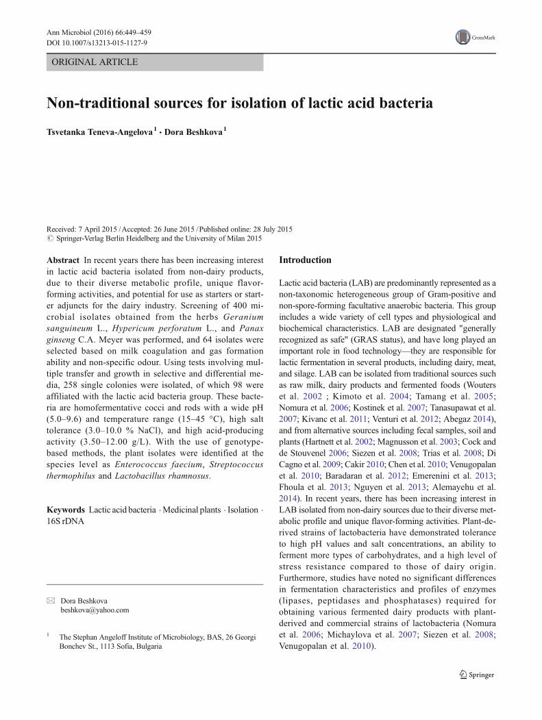
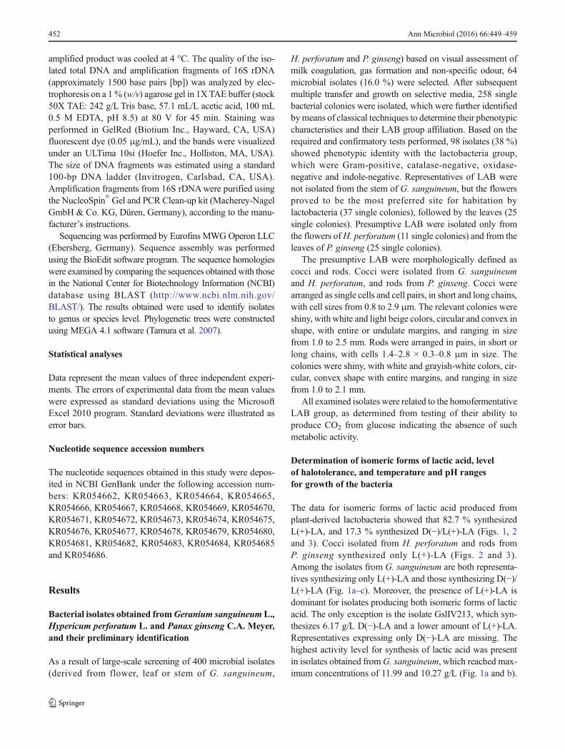
The data for isomeric forms of lactic acid produced fromplant-derived lactobacteria showed that 82.7 % synthesizedL(+)-LA, and 17.3 % synthesized D(−)/L(+)-LA (Figs. 1, 2and 3). Cocci isolated from H. perforatum and rods fromP. ginseng synthesized only L(+)-LA (Figs. 2 and 3).Among the isolates from G. sanguineum are both representa-tives synthesizing only L(+)-LA and those synthesizing D(−)/L(+)-LA (Fig. 1а–c). Moreover, the presence of L(+)-LA isdominant for isolates producing both isomeric forms of lacticacid. The only exception is the isolate GslIV213, which syn-thesizes 6.17 g/L D(−)-LA and a lower amount of L(+)-LA.Representatives expressing only D(−)-LA are missing. Thehighest activity level for synthesis of lactic acid was presentin isolates obtained fromG. sanguineum, which reached max-imum concentrations of 11.99 and 10.27 g/L (Fig. 1a and b).
452 Ann Microbiol (2016) 66:449–459
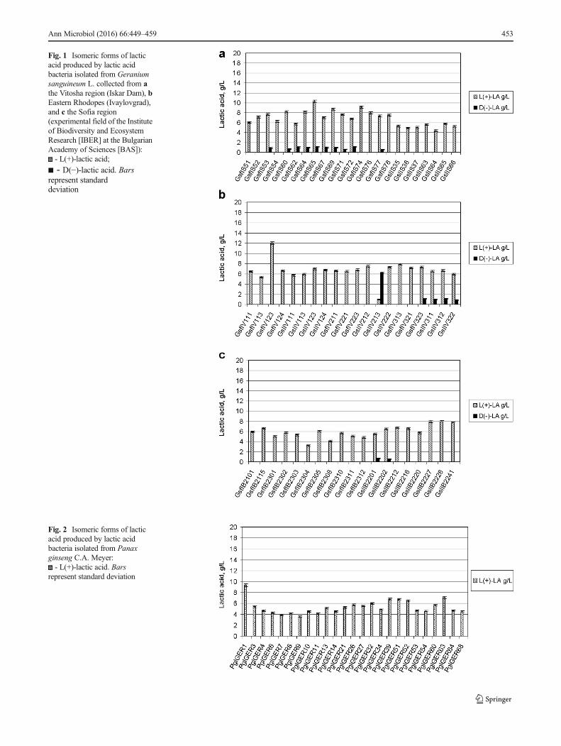
Fig. 1 Isomeric forms of lacticacid produced by lactic acidbacteria isolated from Geraniumsanguineum L. collected from athe Vitosha region (Iskar Dam), bEastern Rhodopes (Ivaylovgrad),and c the Sofia region(experimental field of the Instituteof Biodiversity and EcosystemResearch [IBER] at the BulgarianAcademy of Sciences [BAS]):- L(+)-lactic acid;
■ - D(−)-lactic acid. Barsrepresent standarddeviation
Fig. 2 Isomeric forms of lacticacid produced by lactic acidbacteria isolated from Panaxginseng C.A. Meyer:- L(+)-lactic acid. Bars
represent standard deviation
Ann Microbiol (2016) 66:449–459 453
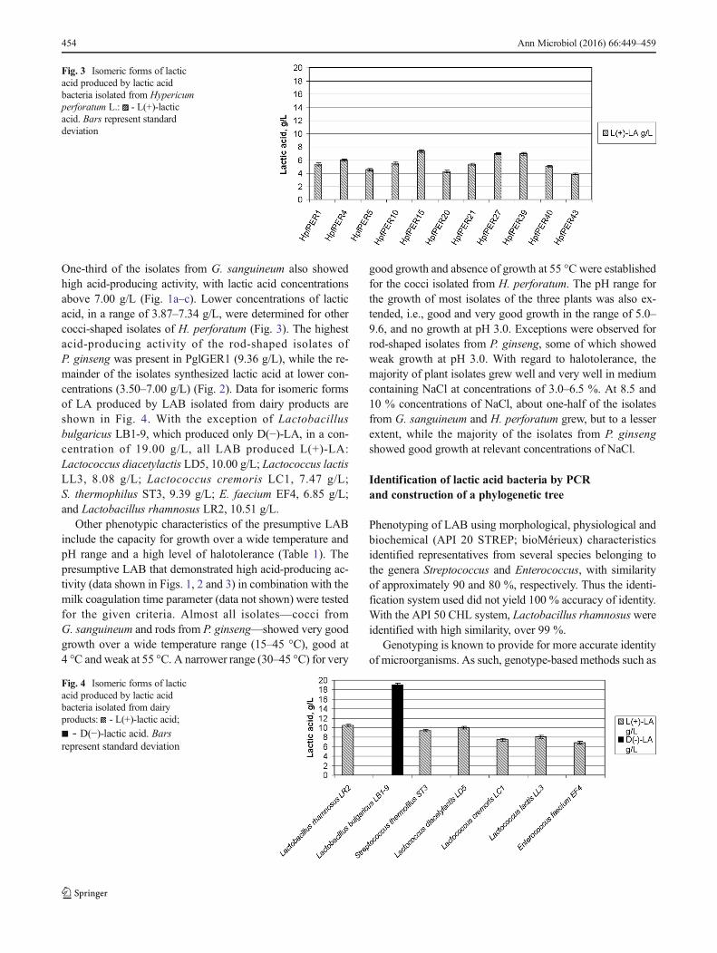
One-third of the isolates from G. sanguineum also showedhigh acid-producing activity, with lactic acid concentrationsabove 7.00 g/L (Fig. 1a–c). Lower concentrations of lacticacid, in a range of 3.87–7.34 g/L, were determined for othercocci-shaped isolates of H. perforatum (Fig. 3). The highestacid-producing activity of the rod-shaped isolates ofP. ginseng was present in PglGER1 (9.36 g/L), while the re-mainder of the isolates synthesized lactic acid at lower con-centrations (3.50–7.00 g/L) (Fig. 2). Data for isomeric formsof LA produced by LAB isolated from dairy products areshown in Fig. 4. With the exception of Lactobacillusbulgaricus LB1-9, which produced only D(−)-LA, in a con-centration of 19.00 g/L, all LAB produced L(+)-LA:Lactococcus diacetylactis LD5, 10.00 g/L; Lactococcus lactisLL3, 8.08 g/L; Lactococcus cremoris LC1, 7.47 g/L;S. thermophilus ST3, 9.39 g/L; E. faecium EF4, 6.85 g/L;and Lactobacillus rhamnosus LR2, 10.51 g/L.
Other phenotypic characteristics of the presumptive LABinclude the capacity for growth over a wide temperature andpH range and a high level of halotolerance (Table 1). Thepresumptive LAB that demonstrated high acid-producing ac-tivity (data shown in Figs. 1, 2 and 3) in combination with themilk coagulation time parameter (data not shown) were testedfor the given criteria. Almost all isolates—cocci fromG. sanguineum and rods from P. ginseng—showed very goodgrowth over a wide temperature range (15–45 °C), good at4 °C and weak at 55 °C. A narrower range (30–45 °C) for very
good growth and absence of growth at 55 °C were establishedfor the cocci isolated from H. perforatum. The pH range forthe growth of most isolates of the three plants was also ex-tended, i.e., good and very good growth in the range of 5.0–9.6, and no growth at pH 3.0. Exceptions were observed forrod-shaped isolates from P. ginseng, some of which showedweak growth at pH 3.0. With regard to halotolerance, themajority of plant isolates grew well and very well in mediumcontaining NaCl at concentrations of 3.0–6.5 %. At 8.5 and10 % concentrations of NaCl, about one-half of the isolatesfrom G. sanguineum and H. perforatum grew, but to a lesserextent, while the majority of the isolates from P. ginsengshowed good growth at relevant concentrations of NaCl.
Identification of lactic acid bacteria by PCRand construction of a phylogenetic tree
Phenotyping of LAB using morphological, physiological andbiochemical (API 20 STREP; bioMérieux) characteristicsidentified representatives from several species belonging tothe genera Streptococcus and Enterococcus, with similarityof approximately 90 and 80 %, respectively. Thus the identi-fication system used did not yield 100 % accuracy of identity.With the API 50 CHL system, Lactobacillus rhamnosus wereidentified with high similarity, over 99 %.
Genotyping is known to provide for more accurate identityof microorganisms. As such, genotype-based methods such as
Fig. 3 Isomeric forms of lacticacid produced by lactic acidbacteria isolated from Hypericumperforatum L.: - L(+)-lacticacid. Bars represent standarddeviation
Fig. 4 Isomeric forms of lacticacid produced by lactic acidbacteria isolated from dairyproducts: - L(+)-lactic acid;
■ - D(−)-lactic acid. Barsrepresent standard deviation
454 Ann Microbiol (2016) 66:449–459
16S rDNA represent a valuable addition to phenotypicmethods for precise identification of lactobacteria isolatedfrom certain microbial communities (Kostinek et al. 2007;Tanasupawat et al. 2007; Chen et al. 2010; Kpikpi et al.2010; Venturi et al. 2012; Baradaran et al. 2012; Alemayehuet al. 2014). Despite the widespread use of LAB, each planthabitat is characterized by a specific microbocenosis withunique biological activity. The isolated DNA from phenotyp-ically characterized bacterial isolates was used as a matrix for
PCR amplification of 16S rDNA. The combination of PCRamplification of variable regions from the 16S rDNA, se-quencing of the resulting PCR products, and the subsequentcomparison of their sequences with those existing in the NCBIdatabase allowed precise identification of the presumptiveLAB to species level, as well as their phylogenetic grouping.All isolates ofH. perforatum, as well as the majority (73.3 %)of isolates from G. sanguineum, were shown to belong to thegenus Enterococcus. Strain HpfPER39 isolated from
Table 1 Effect of temperature, pH and NaCl concentration on survival of bacterial isolates and LAB of dairy origin
Isolate Temperature, °C pH NaCl, %
4 15 30 37 45 55 3.0 5.0 7.0 8.0 9.6 3.0 4.5 6.5 8.5 10
PglGER1 + ++ ++ ++ + +w + ++ ++ ++ ++ ++ ++ ++ + +
PglGER3 + ++ ++ ++ + +w + ++ ++ ++ ++ ++ ++ + + +
PglGER26 + ++ ++ ++ + +w + ++ ++ ++ + ++ ++ + + +
PglGER32 +w +w ++ ++ + − − ++ ++ ++ ++ ++ ++ + + −PglGER39 + ++ ++ ++ + − − ++ ++ + + ++ ++ + + +
PglGER51 +w ++ ++ ++ + − − + + + + ++ ++ + + −PglGER63 +w + ++ ++ + − − ++ ++ ++ ++ ++ ++ + + −GsfIS60 + + ++ ++ ++ +w − + ++ ++ + +w +w +w − −GsfIS65 +w + ++ ++ ++ + − + ++ ++ + +w +w − − −GsfIS69 +w + ++ ++ ++ + − + ++ ++ + +w +w − − −GsfIS76 + + ++ ++ ++ +w − +w ++ ++ +w +w +w +w − −GsfIV123 + ++ ++ ++ ++ +w − + ++ ++ ++ ++ + + +w +w
GsfIV124 + ++ ++ ++ ++ + − + ++ ++ + ++ ++ ++ + +w
GslIV123 + ++ ++ ++ ++ +w − + ++ ++ + ++ ++ ++ + +w
GslIV124 + ++ ++ ++ ++ + − + ++ ++ + ++ ++ + + +w
GslIV213 + ++ ++ ++ ++ − − +w ++ ++ ++ + + +w − −GsfIV313 + ++ ++ ++ ++ +w − + ++ ++ ++ ++ + + +w +w
GslIV312 + ++ ++ ++ ++ +w − + ++ ++ ++ ++ + + +w +w
GsfIB2101 + ++ ++ ++ ++ +w − + ++ ++ ++ ++ ++ ++ + +w
GsfIB2115 + ++ ++ ++ ++ +w − + ++ ++ + ++ ++ ++ + +w
GslIB2212 + ++ ++ ++ ++ +w − + ++ ++ ++ ++ ++ ++ + +w
GslIB2227 + ++ ++ ++ ++ +w − + ++ ++ + ++ ++ + +w +w
HpfPER15 + + ++ ++ + − − + ++ ++ + ++ ++ + + +w
HpfPER27 + + ++ ++ + − − + ++ ++ + ++ ++ + + +w
HpfPER39 + + ++ ++ + − − + ++ ++ + ++ ++ + + +w
Lactobacillusbulgaricus LB1-9
+w + ++ ++ + − − ++ ++ ++ +w + + +w +w −
Lactobacillusrhamnosus LR2
+w + ++ ++ + − − ++ ++ ++ + ++ + +w +w −
Lactococcuslactis LL3
+w +w ++ ++ − − − + ++ + +w ++ + +w − −
Lactococcuscremoris LC1
− +w ++ ++ − − − + ++ + − + − − − −
Lactococcusdiacetylactis LD5
+w + ++ ++ − − − + ++ + +w + + +w − −
Streptococcusthermophilus ST3
+w +w ++ ++ ++ +w − + ++ + +w +w +w +w − −
Enterococcusfaecium EF4
+ ++ ++ ++ ++ +w − + ++ ++ ++ ++ ++ + + +w
Growth: ++ very good, + good, − none, wweak
Ann Microbiol (2016) 66:449–459 455
H. perforatum showed 99 % similarity to the speciesE. faecium, while the other two isolates (HpfPER15 andHpfPER27) showed 97 % similarity with this species. Thestrains GsfIB2115, GslIB2227, GsfIV123, GslIV123 andGslIV213 were related to the species E. faecium, based on fullconformity of phenotypic characteristics and the results ofPCR analysis, reaching 100 % homology of nucleotide se-quences. The strains GsfIB2101, GslIB2212, GsfIV124,GsfIV313, GslIV124 and GslIV312 were also found to berelated to the same species, indicating 99 % homology. Asmaller portion (26.7 %) of isolates fromG. sanguineumwereidentified as representatives of the genus Streptococcus. Highsimilarity (99.0 %) with S. thermophiluswas registered for thestrains GsfIS60, GsfIS65, GsfIS69 and GsfIS76. The isolatesfrom P. ginseng were identified as Lactobacillus rhamnosus.The strains PglGER3, PglGER26, PglGER32, PglGER39 andPglGER63 showed 99.0 % homology, and 100 % similaritywas reached with strains PglGER1 and PglGER51.
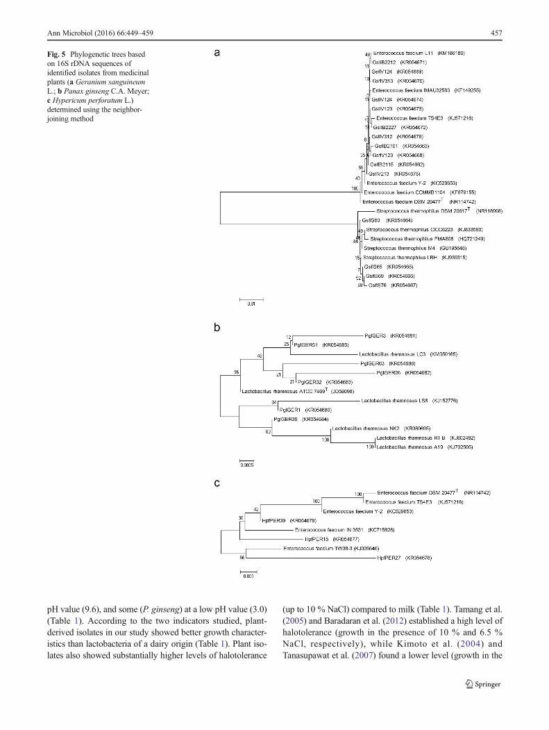
Based on the data from 16S rDNA sequence analysis, aphylogenetic tree was constructed, using the computer pro-gram MEGA 4.1, in order to determine the phylogenetic po-sition of the isolated sequences of the strains of plant origin(Fig. 5a–c).
Discussion
Each plant species provides unique conditions for differentspecies of lactobacteria. Among the medicinal plants that westudied, only cocci were isolated from G. sanguineum andH. perforatum, while only rods were isolated fromP. ginseng. Cocci have been isolated from the leaves of 14different plant species, and rods have been identified fromfour plant species (Michaylova et al. 2007). In naturallyfermented herbs used in traditional herb cheese in Turkey,lactobacilli (76.2 % vs. 23.8 % cocci) have predominantlybeen isolated (Cakir 2010), and only rods have been isolatedfrom the herbal surface of Phyllanthus niruri (Venugopalanet al. 2010). Cocci and rods have been isolated from the sur-face of Polygonum minus leaves, a local Malaysian herb (in aratio of 2:1) and from various plants (clover, grass, dandelion,lilac flowers, chestnut flowers, Hepatica flowers, coltsfootflowers and rowan leaves) (Magnusson et al. 2003;Baradaran et al. 2012). Cocci have been isolated from grassvarieties and vegetables (Alemayehu et al. 2014).
The defined isomeric forms of lactic acid synthesized fromselected LAB from plant species G. sanguineum ,H. perforatum and P. ginseng (Figs. 1, 2 and 3), with a dom-inant presence of L(+) form and concentrations of 3.50–11.99 g/L, are comparable to those produced by lactobacteriafrom the research team's collection that were isolated fromdairy products: Lactococcus diacetylactis LD5, Lactococcuslactis LL3, Lactococcus cremoris LC1, S. thermophilus ST3,
E. faecium EF4 and Lactobacillus rhamnosus LR2 (Fig. 4).The exception is Lactobacillus bulgaricus LB1-9, which syn-thesizes only D(−)-LA (Fig. 4). Our results are similar to thosereported by Kimoto et al. (2004), who obtained 20 bacterialisolates of raw grass (Napier grass) in Japan, which were sub-sequently morphologically defined and tested ashomofermentative cocci producing only L(+)-LA. Kostineket al. (2007) isolated homofermentative cocci and rods fromfermented cassava that produced L(+)-LA. Homofermentativerods producing the same isomer have been isolated fromfermented tea leaves (miang) in Thailand (Tanasupawat et al.2007), and the same authors isolated homofermentative cocciproducing D(−)/L(+)-LA, just as was seen in our study, andwhich was also proven by Tamang et al. (2005) for pediococcifrom traditionally fermented vegetable products of the EasternHimalayas. In contrast to our data, Tanasupawat et al. (2007)and Tamang et al. (2005) isolated homofermentative rods pro-ducing D(−)/L(+)-LA, and Kostinek et al. (2007) reportedhomofermentative rods producing L(+)-, D(−)- and D(−)/L(+)-LA. From these plant sources, heterofermentative cocciproducing only D(−)-LA (Tamang et al. 2005; Kostinek et al.2007) and heterofermentative rods producing D(−)/L(+)-LA(Kostinek et al. 2007; Tanasupawat et al. 2007) have also beenisolated. In addition to the determination of isomeric forms oflactic acid synthesized from presumptive lactobacteria presentin certain bacterial isolates, as an element of preliminary iden-tification, some authors also report data for concentrations oflactic acid. The results reported by Cock and de Stouvenel(2006) are interesting in this regard. Based on screening of20 bacterial isolates obtained from the leaves of sugar molas-ses, the authors found only one strain (with index CC 85–92)with a high potential for synthesis of L(+)-LA, in concentra-tions of 12.4 and 13.7 g/L and at cultivation temperatures of36 and 32 °C, respectively—concentrations comparable tothose for our isolates (Fig. 1a and b). With this strain, grownwithin parameters suitable for targeted synthesis of lactic acid,the authors achieved a maximum yield of 35.0 g/L.
Data indicating a wide temperature range for bacterialgrowth have been reported by Baradaran et al. (2012) andTanasupawat et al. (2007) for lactococci and lactobacilli iso-lated from the herb Polygonum minus (10–45 °C) and forlactococci from fermented tea leaves (15–45 °C), respectively,which is consistent with our results revealing a temperaturerange of 15–45 °C for growth of isolated lactobacteria fromthe three plant species studied (Table 1). Tanasupawat et al.(2007) reported the absence of growth of lactobacilli at 45 °C.The same authors established that these isolates grew well in awide pH range, 3.0–7.0 and 4.0–8.5, which is consistent withresults reported by Tamang et al. (2005) for lactococci of otherplant origins. Alemayehu et al. (2014) demonstrated goodgrowth at pH 9.5 for lactococci isolated from grass varietiesand vegetables. All isolates obtained from G. sanguineum,H. perforatum and P. ginseng showed good growth at a high
456 Ann Microbiol (2016) 66:449–459
pH value (9.6), and some (P. ginseng) at a low pH value (3.0)(Table 1). According to the two indicators studied, plant-derived isolates in our study showed better growth character-istics than lactobacteria of a dairy origin (Table 1). Plant iso-lates also showed substantially higher levels of halotolerance
(up to 10 % NaCl) compared to milk (Table 1). Tamang et al.(2005) and Baradaran et al. (2012) established a high level ofhalotolerance (growth in the presence of 10 % and 6.5 %NaCl, respectively), while Kimoto et al. (2004) andTanasupawat et al. (2007) found a lower level (growth in the
Fig. 5 Phylogenetic trees basedon 16S rDNA sequences ofidentified isolates from medicinalplants (a Geranium sanguineumL.; b Panax ginseng C.A. Meyer;c Hypericum perforatum L.)determined using the neighbor-joining method
Ann Microbiol (2016) 66:449–459 457

presence of 4.0 % NaCl) for lactococci of different plant ori-gins. Baradaran et al. (2012) demonstrated good growth oflactobacilli isolated from Polygonum minus, and Alemayehuet al. (2014) of lactococci from grass and vegetables, in thepresence of 6.5 % NaCl.
All isolates of P. ginseng and H. perforatum were geneti-cally identified as Lactobacillus rhamnosus and E. faecium,respectively, with the majority of G. sanguineum identified asE. faecium and a small minority as S. thermophilus. The pres-ence of genotypically identified representatives of species ofthe genera Enterococcus, Streptococcus and Lactobacillus inisolates from plants of different origins have been reported byother authors (Hartnett et al. 2002; Magnusson et al. 2003;Michaylova et al. 2007; Chen et al. 2010; Baradaran et al.2012). Hartnett et al. (2002) identified E. faecium in isolatesfrom raw barley; in isolates from sorghum, defined speciesEnterococcus mundtii and E. faecalis were found—whichwere undetected in plant isolates that we examined—as wellas Lactococcus lactis in isolates from raw barley.Streptococcus thermophilus has been isolated from leavesfrom the plant species Capsel la bursa-pastoris ,Chrysanthemum, Cichorium intybus, Colchicum, Dianthus,Hedera, Nerium oleander, Plantago lanceolata, Rosa,Tropaeolum, Calendula officinalis, Cornus mas, Galanthusnivalis and Prunus spinosa, and Lactobacillus bulgaricuswas also identified in some of the plant isolates (Michaylovaet al. 2007). Several groups have isolated species of LAB thatwere not detected in plants that we tested. For instance,Baradaran et al. (2012) identified Lactococcus lactis,Pediococcus pentosaceus and Lactobacillus curvatus in iso-lates from Polygonum minus (a Malaysian herb). Magnussonet al. (2003) isolated representatives of the generaLactobacillus (L. plantarum, L. coryniformis, L. acidophilusand L. sakei) and Pediococcus and of Enterococcus hiraefrom different parts of plants (grass, dandelion, lilac, chestnut,Hepatica, coltsfoot and rowan). The species Lactobacillusp l a n t a r um , We i s s e l l a c i b a r i a , L e u c o n o s t o cpseudomesenteroides and Lactococcus lactis subsp. lactiswere isolated from ripe mulberries from five countries inTaiwan (Chen et al. 2010). In the present study, it should benoted that each of the plant habitats represents a single eco-logical niche for the growth of specific lactic acid microflora.
Conclusions
The results of 16S rDNA sequence analysis and phylogeneticposition determination of the sequences of strains isolatedfrom the herbs Geranium sanguineum L., Hypericumperforatum L. and Panax ginseng C.A. Meyer indicate thatthey are appropriate natural ecological niches for the isolationof the lactobacteria Enterococcus faecium, Streptococcusthermophilus and Lactobacillus rhamnosus. The better
growth characteristics of isolated and identified lactococciand lactobacilli in a wide pH and temperature range, aswell as their high acid-producing activity, compared toLAB isolated from dairy sources indicates the excellentpotential for in situ cultivation of these newly isolatedstrains in milk, with subsequent creation of starters fornew fermented dairy products.
References
Abegaz K (2014) Isolation, characterization and identification of lacticacid bacteria involved in traditional fermentation of borde, anEthiopian cereal beverage. Int J Food Nutr Sci 1:7–15
Alemayehu D, Hannon JA, McAuliffe O, Ross RP (2014)Characterization of plant-derived lactococci on the basis of theirvolatile compounds profile when grown in milk. Int J FoodMicrobiol 172:57–61. doi:10.1016/j.ijfoodmicro.2013.11.024
Baradaran A, Foo HL, Sieo CC, Rahim RA (2012) Isolation, identifica-tion and characterization of lactic acid bacteria from Polygonumminus. Roum Biotechnol Lett 17:7245–7252
Barnes J, Anderson LA, Phillipson JD (2001) St John’s wort (Hypericumperforatum L.): a review of its chemistry, pharmacology and clinicalproperties. J Pharm Pharmacol 53:583–600
Cakir I (2010) Antibacterial and antifungal activities of some lactic acidbacteria isolated from naturally fermented herbs. J Food AgricEnviron 8:223–226
Chen Y-S,WuH, Yanagida F (2010) Isolation and characteristics of lacticacid bacteria isolated from ripe mulberries in Taiwan. Braz JMicrobiol 41:916–921
Choi K-T (2008) Botanical characteristics, pharmacological effects and me-dicinal components of Korean Panax ginseng C A Meyer. ActaPharmacol Sin 29:1109–1118. doi:10.1111/j.1745-7254.2008.00869.x
Cock LS, de Stouvenel AR (2006) Lactic acid production by a strain ofLactococcus lactis subs lactis isolated from sugar cane plants.Electron J Biotechnol 9:40–45. doi:10.2225/vol9-issue1-fulltext-10
Council of Europe (2000) Natural sources of flavourings. Council ofEurope, Strasbourg, Report No. 1
Di Cagno R, Rizzello CG, Gagliardi F, Ricciuti P, Ndagijimana M,Francavilla R, Guerzoni ME, Crecchio C, Gobbetti M, De AngelisM (2009) Different fecal microbiotas and volatile organic com-pounds in treated and untreated children with celiac disease. ApplEnviron Microbiol 75:3963–3971. doi:10.1128/AEM.02793-08
Emerenini EC, Afolabi OR, Okolie PI, Akintokun AK (2013) Isolationand molecular characterization of lactic acid bacteria isolated fromfresh fruits and vegetables using nested PCR analysis. Br MicrobiolRes J 3:368–377
Fhoula I, Najjari A, Turki Y, Jaballah S, Boudabous A, Ouzari H (2013)Diversity and antimicrobial properties of lactic acid bacteria isolatedfrom rhizosphere of olive trees and desert truffles of Tunisia.Biomed Res Int 2013:405708. doi:10.1155/2013/405708
Gobbetti M, Di Cagno R, De Angelis M (2010) Functional microorgan-isms for functional food quality. Crit Rev Food Sci Nutr 50:716–727
Hammami I, Triki MA, Rebai A (2011) Chemical compositions, antibac-terial and antioxidant activities of essential oil and various extractsofGeranium sanguineum L. flowers. Arch Appl Sci Res 3:135–144
Hartnett DJ, Vaughan A, van SinderenD (2002)Antimicrobial-producinglactic acid bacteria isolated from raw barley and sorghum. J InstBrew 108:169–177
Husain GM, Chatterjee SS, Singh PN, Kumar V (2011) Hypolipidemic andantiobesity-like activity of standardised extract of Hypericum
458 Ann Microbiol (2016) 66:449–459

perforatum L. In rats. ISRN Pharmacol 2011:505247. doi:10.5402/2011/505247
Kimoto H, Nomura M, Kobayashi M, Okamoto T, Ohmomo S (2004)Identification and probiotic characteristics of Lactococcus strainsfrom plant materials. JARQ 38:111–117
KivancM, YilmazM, Cakir E (2011) Isolation and identification of lacticacid bacteria from boza, and their microbial activity against severalreporter strains. Turk J Biol 35:313–324. doi:10.3906/biy-0906-67
Kostinek M, Specht I, Edward VA, Pinto C, Egounlety M, Sossa C,Mbugua S, Dortu C, Thonart P, Taljaard L, Mengu M, FranzCMAP, Holzapfel WH (2007) Characterisation and biochemicalproperties of predominant lactic acid bacteria from fermenting cas-sava for selection as starter cultures. Int J Food Microbiol 114:342–351. doi:10.1016/j.ijfoodmicro.2006.09.029
Kpikpi EN, Glover RLK, Dzogbefia VP, Nielsen DS, JakobsenM (2010)Isolation of lactic acid bacteria from kantong, a condiment producedfrom the fermentation of kapok (Ceiba pentandra) seeds and cassa-va (Manihot esculentum) flour. Rep Opin 2:1–7
Magnusson J, Str m K, Roos S, Sjögren J, Schnürer J (2003) Broad andcomplex antifungal activity among environmental isolates of lacticacid bacteria. FEMS Microbiol Lett 219:129–135. doi:10.1016/S0378-1097(02)01207-7
Michaylova M, Minkova S, Kimura K, Sasaki T, Isawa K (2007)Isolation and characterization of Lactobacillus delbrueckii ssp.bulgaricus and Streptococcus thermophilus from plants inBulgaria. FEMS Microbiol Lett 269:160–169. doi:10.1111/j.1574-6968.2007.00631.x
Newall CA, Anderson LA, Phillipson JD (1996) Herbal medicines. aguide for health-care professionals, 1st edn. Pharmaceutical Press,London
Nguyen THK, Doan VTT, Ha LD, Nguyen HN (2013) Molecular clon-ing, expression of minD gene from Lactobacillus acidophilusVTCC-B-871 and analyses to identify Lactobacillus rhamnosusPN04 from Vietnam Houttuynia cordata Thunb. Indian JMicrobiol 53:385–390. doi:10.1007/s12088-013-0384-1
Nomura M, Kobayashi M, Narita T, Kimoto-Nira H, Okamoto T (2006)Phenotypic and molecular characterization of Lactococcus lactisfrom milk and plants. J Appl Microbiol 101:396–405. doi:10.1111/j.1365-2672.2006.02949.x
Serkedjieva J, Manolova N (1992) A plant polyphenolic complex inhibitsthe reproduction of influenza and herpes simplex viruses. In:
Hemingway RW, Laks PE (eds) Plant polyphenols, basic life sci.Plenum Press, New York, pp 705–715
ServiliM, Rizzello CG, Taticchi A, Esposto S, Urbani S,Mazzacane F, DiMaio I, Selvaggini R, Gobbetti M, Di Cagno R (2011) Functionalmilk beverage fortified with phenolic compounds extracted fromolive vegetation water, and fermented with functional lactic acidbacteria. Int J Food Microbiol 147:45–52. doi:10.1016/j.ijfoodmicro.2011.03.006
Siezen RJ, Starrenburg MJC, Boekhorst J, Renckens B, MolenaarD, van Hylckama Vlieg JET (2008) Genome-scale genotype-phenotype matching of two Lactococcus lactis isolates fromplants identifies mechanisms of adaptation to the plant niche.Appl Environ Microbiol 74:424–436. doi:10.1128/AEM.01850-07
Tamang JP, Tamang B, Schillinger U, Franz CM, Gores M, HolzapfelWH (2005) Identification of predominant lactic acid bacteria isolat-ed from traditionally fermented vegetable products of the EasternHimalayas. Int J Food Microbiol 105:347–356. doi:10.1016/j.ijfoodmicro.2005.04.024
Tamura K, Dudley J, Nei M, Kumar S (2007) MEGA4: molecular evo-lutionary genetics analysis (MEGA) software version 4.0. Mol BiolEvol 24:1596–1599, Publication PDF at http://www.kumarlab.net/publications
Tanasupawat S, Pakdeeto A, Thawai C, Yukphan P, Okada S (2007)Identification of lactic acid bacteria from fermented tea leaves(miang) in Thailand and proposals of Lactobacillus thailandensissp. nov., Lactobacillus camelliae sp. nov., and Pediococcussiamensis sp. nov. J Gen Appl Microbiol 53:7–15
Trias R, Bañeras L, Montesinos E, Badosa E (2008) Lactic acid bacteriafrom fresh fruit and vegetables as biocontrol agents of phytopatho-genic bacteria and fungi. Int Microbiol 11:231–236. doi:10.2436/20.1501.01.66
Venturi M, Guerrini S, Granchi L, Vincenzini M (2012) Typing ofLactobacillus sanfranciscensis isolates from traditional sourdoughsby combining conventional andmultiplex RAPD-PCR profiles. Int JFood Microbiol 156:122–126
Venugopalan V, Dinesh MS, Geetha KS (2010) Enhancement of antimi-crobial potential of phyllanthus niruri by fermentation. J Herb MedToxicol 4:167–175
Wouters JTM, Ayad EHE, Hugenholtz J, Smit G (2002) Microbes fromraw milk for fermented dairy products. Int Dairy J 12:91–109. doi:10.1016/S0958-6946(01)00151-0
Ann Microbiol (2016) 66:449–459 459