normal intraoral radiographic anatomy
TRANSCRIPT
NORMAL INTRAORAL RADIOGRAPHIC ANATOMY IN MAXILLA AND MANDIBLE
Dr. ULLAS SAXENA PG – IInd yearORAL MEDICINE AND RADIOLOGY

INRODUCTION• The radiographic recognition of disease requires
knowledge of radiographic appearance of normal anatomical structures.
• The radiographic appearance of various structures which can be visualized on intra oral periapical radiographs can be classified under:
• Teeth• Supporting structures• Maxilla• Mandible• Restorative materials

It's important for your doctor to have accurate and complete information to plan your treatment.

RADIOPAQUE LANDMARKS• MAXILLA• Enamel dentin• Cementum• Lamina dura • Alveolar crest• Cancellous bone• Nasal septum • Anterior nasal spine• Floor of nasal cavity• Inferior nasal conchae• Nasolabial fold• Floor of maxillary sinus
• Septa in maxillary sinus• Inverted Yin maxillary sinus• Zygomatic process of maxilla• Zygoma ( malar bone )• Pterygoid plates• Hamular process• Maxillary tuberosity• Coronoid process• Internal oblique ridge

RADIOPAQUE LANDMARKS• MANDIBLE• Enamel• Dentin• Cementum• Lamina dura• Alveolar crest• Cancellous bone• Genial tubercles• Mental ridge• Mylohyoid ridge• External oblique ridge• Inferior border of mandible
RADIOLUCENT LANDMARKS• MAXILLA• Pulp• Periodontal ligament space• Nutrient canals• Intermaxillary suture• Nasal fossa ( nasal cavity )• Incisive foramen• Superior foramina of nasolacrimal canal• Incisive fossa• Maxillary sinus• nose

RADIOLUCENT LANDMARKS• MANDIBLE• Pulp• Periodontal ligament space• Nutrient canals • Lingual foramen• Symphysis• Mental fossa• Mental foramen• Mandibular canal• Submandibular fossa

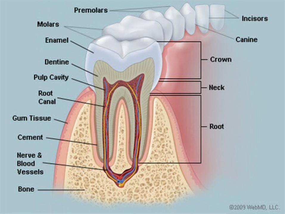
• Teeth are composed primarily of dentin, with an enamel
cap over the coronal portion and a thin layer of cementum
over the root surface.
• The enamel cap characteristically appears more
radiopaque than the other tissues because it is the most
dense, naturally occurring substance in the body.
• Because it is 90% mineral, it causes the greatest
attenuation of x-ray photons.
• Its radiographic appearance is uniformly opaque and
without evidence of the fine structure.
TEETH
• The dentin is about 75% mineralized, and because of its lower
mineral content, its radiographic appearance is roughly
comparable to that of bone.
• Dentin is smooth and homogeneous on radiographs because of
its uniform morphologic features.
• The junction between enamel and dentin appears as a distinct
interface that separates these two structures.
• The thin layer of cementum on the root surface has a mineral
content (50%) comparable to that of dentin.
• Cementum is not usually apparent radiographically because the
contrast between it and dentin is so low and the cementum layer
is so thin.
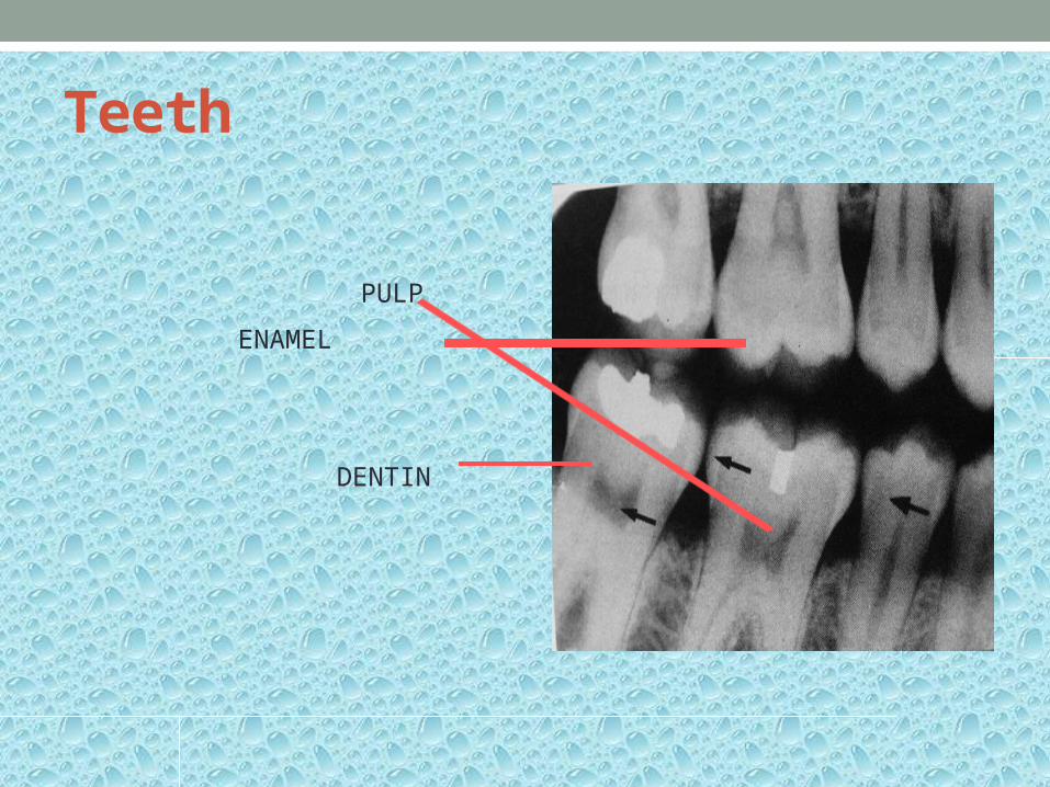
Teeth
DENTIN
ENAMEL
PULP

SUPPORTING STUCTURES• Lamina dura• Alveolar crest• Periodontal ligament space• Cancellous bone

LAMINA DURA• It is wall of tooth socket that surrounds
the tooth.
• It is made of dense cortical bone.
• It appears as thin radiopaque line that
surrounds root of the tooth.
• It is continuous with shadow of cortical
bone at alveolar crest.
• Double lamina dura appears if mesial
or distal surfaces of root present two
elevations in path of x ray beam.
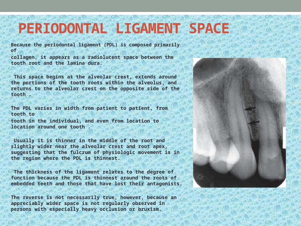
PERIODONTAL LIGAMENT SPACE• Because the periodontal ligament (PDL) is composed primarily
of• collagen, it appears as a radiolucent space between the
tooth root and the lamina dura.
• This space begins at the alveolar crest, extends around the portions of the tooth roots within the alveolus, and returns to the alveolar crest on the opposite side of the tooth .
• The PDL varies in width from patient to patient, from tooth to
• tooth in the individual, and even from location to location around one tooth .
• Usually it is thinner in the middle of the root and slightly wider near the alveolar crest and root apex, suggesting that the fulcrum of physiologic movement is in the region where the PDL is thinnest.
• The thickness of the ligament relates to the degree of function because the PDL is thinnest around the roots of embedded teeth and those that have lost their antagonists.
• The reverse is not necessarily true, however, because an appreciably wider space is not regularly observed in persons with especially heavy occlusion or bruxism.

Double periodontal ligament space is seen when there is convexity in proximal surface of root.
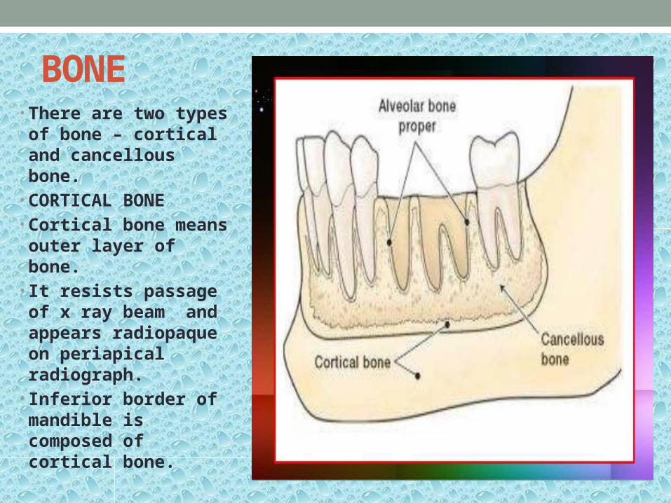
BONE• There are two types
of bone – cortical and cancellous bone.
• CORTICAL BONE• Cortical bone means
outer layer of bone.• It resists passage of
x ray beam and appears radiopaque on periapical radiograph.
• Inferior border of mandible is composed of cortical bone.
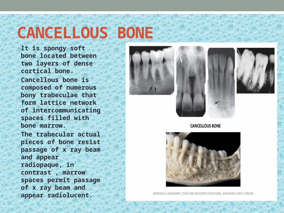
CANCELLOUS BONE• It is spongy soft bone
located between two layers of dense cortical bone.
• Cancellous bone is composed of numerous bony trabeculae that form lattice network of intercommunicating spaces filled with bone marrow.
• The trabecular actual pieces of bone resist passage of x ray beam and appear radiopaque, in contrast , marrow spaces permit passage of x ray beam and appear radiolucent.

TRABECULAR PATTERN• In anterior
maxilla : fine, granular & dense pattern, the marrow spaces are small and numerous.
• In posterior maxilla the trabecular is similar to anterior maxilla, slightly larger in marrow spaces.
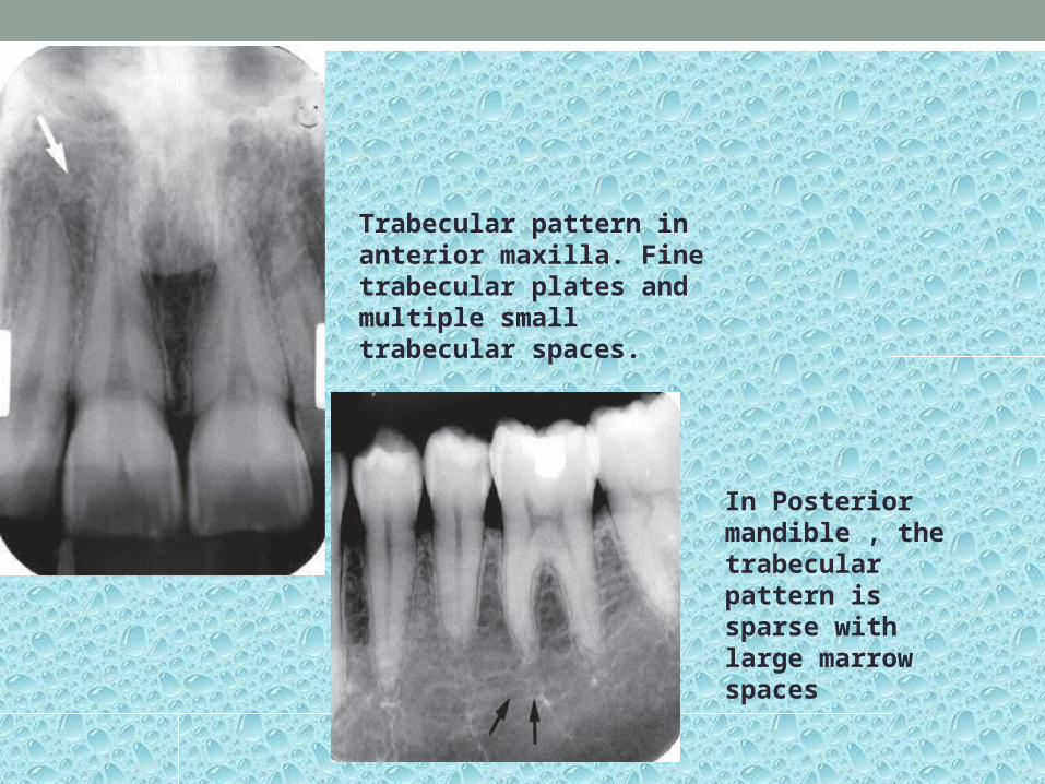
Trabecular pattern in anterior maxilla. Fine trabecular plates and multiple small trabecular spaces.
In Posterior mandible , the trabecular pattern is sparse with large marrow spaces
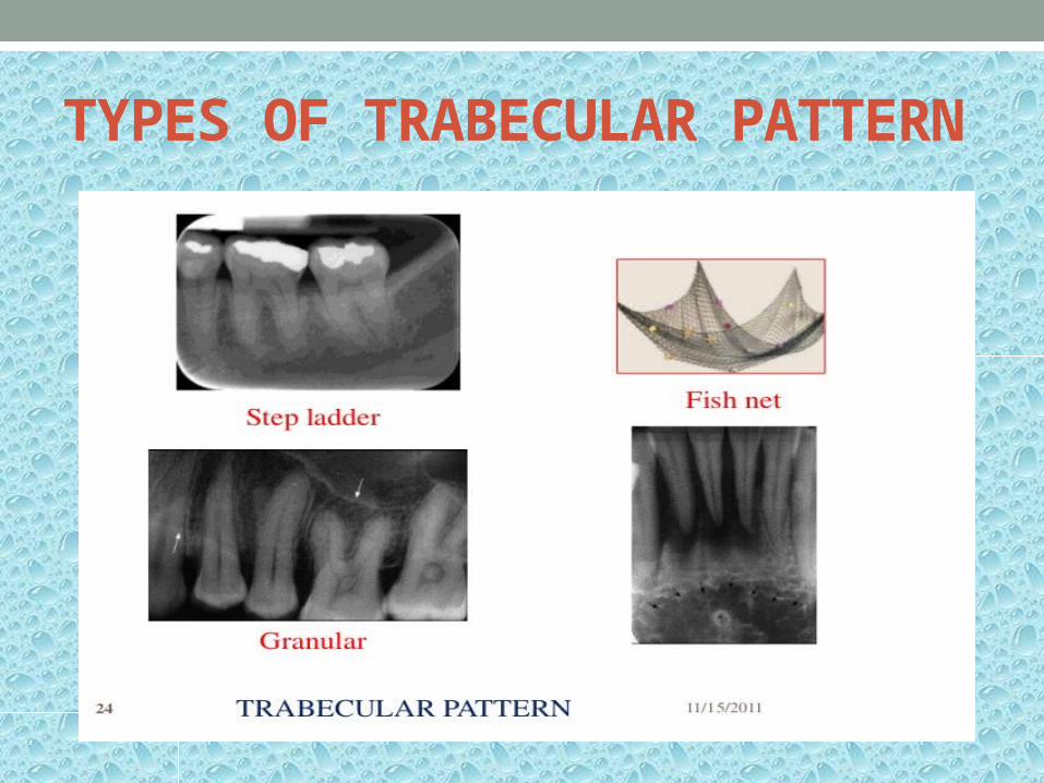
TYPES OF TRABECULAR PATTERN
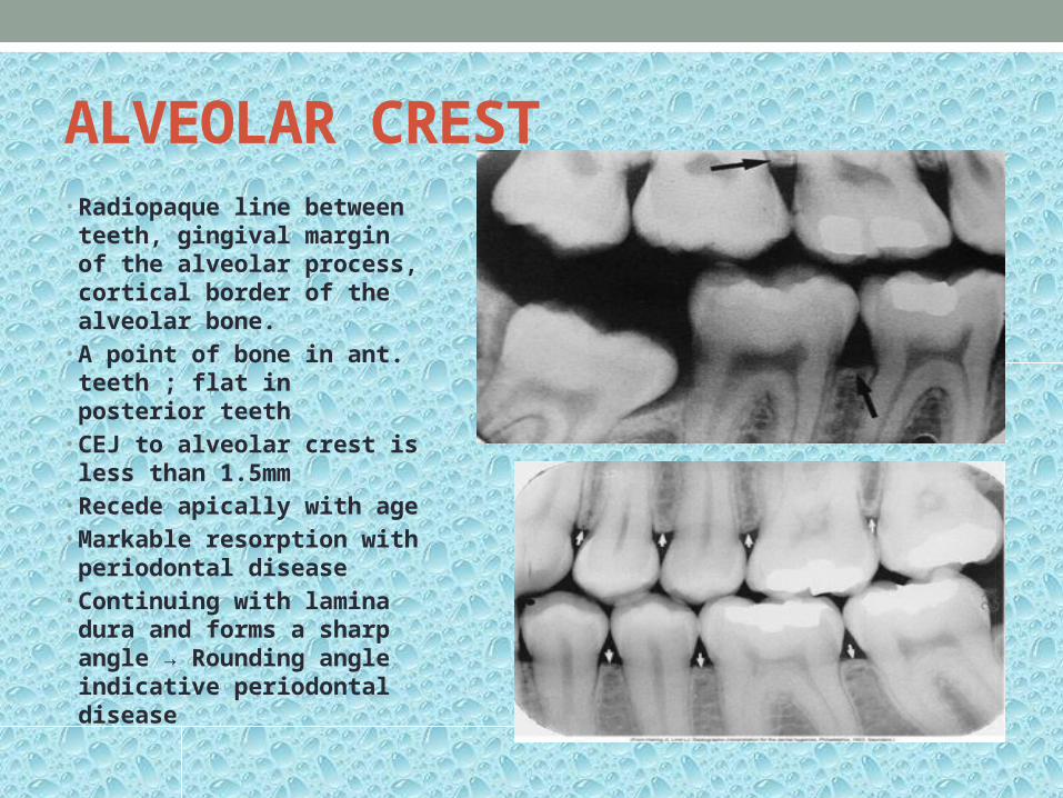
ALVEOLAR CREST• Radiopaque line between
teeth, gingival margin of the alveolar process, cortical border of the alveolar bone.
• A point of bone in ant. teeth ; flat in posterior teeth
• CEJ to alveolar crest is less than 1.5mm
• Recede apically with age • Markable resorption with
periodontal disease• Continuing with lamina
dura and forms a sharp angle → Rounding angle indicative periodontal disease

ANATOMIC LANDMARKS OF MAXILLA
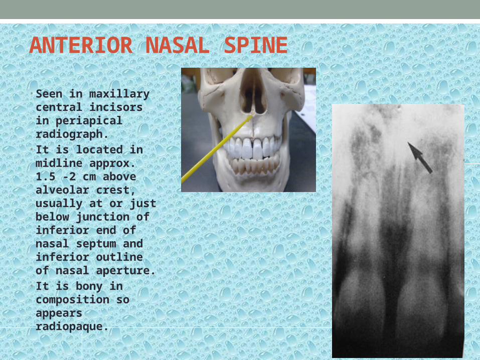
ANTERIOR NASAL SPINE
• Seen in maxillary central incisors in periapical radiograph.
• It is located in midline approx. 1.5 -2 cm above alveolar crest, usually at or just below junction of inferior end of nasal septum and inferior outline of nasal aperture.
• It is bony in composition so appears radiopaque.

Anterior nasal spine appears as V shape radiolucency from floor of nasal aperture.
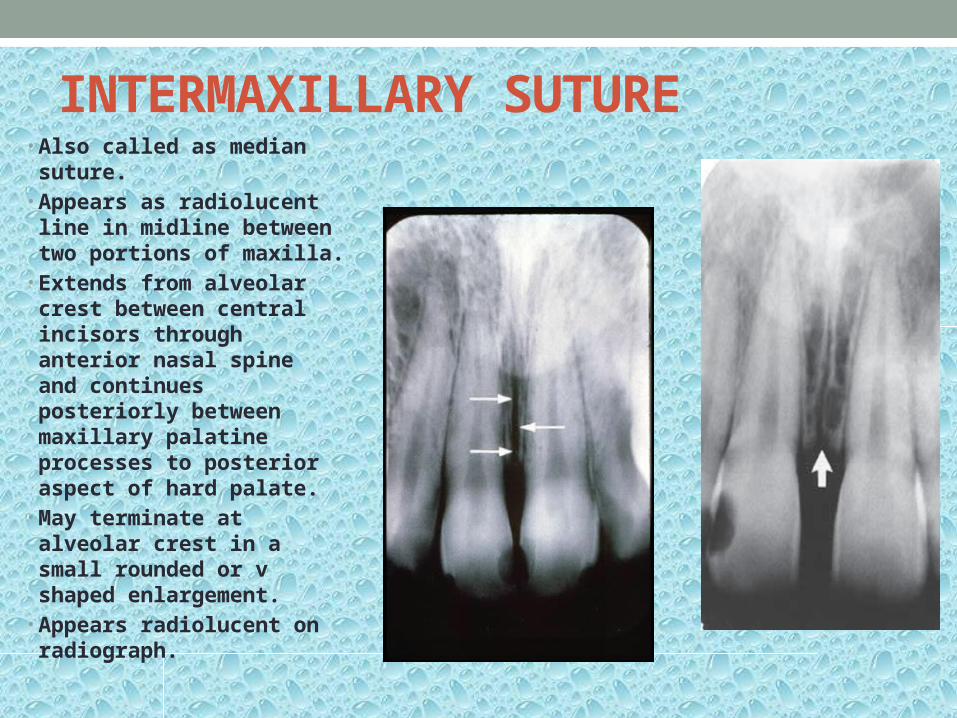
INTERMAXILLARY SUTURE• Also called as median
suture.• Appears as radiolucent
line in midline between two portions of maxilla.
• Extends from alveolar crest between central incisors through anterior nasal spine and continues posteriorly between maxillary palatine processes to posterior aspect of hard palate.
• May terminate at alveolar crest in a small rounded or v shaped enlargement.
• Appears radiolucent on radiograph.
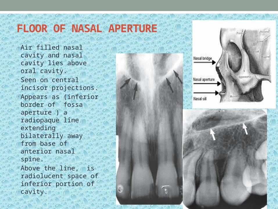
FLOOR OF NASAL APERTURE• Air filled nasal cavity
and nasal cavity lies above oral cavity.
• Seen on central incisor projections.
• Appears as (inferior border of fossa aperture ) a radiopaque line extending bilaterally away from base of anterior nasal spine.
• Above the line, is radiolucent space of inferior portion of cavity.

INCISIVE FORAMEN• Also called as
anterior palatine foramen.
• Radiographic image is projected between roots and in region of midline and apical thirds of maxillary central incisors.
• Transmits nasopalatine nerves and vessels.

INCISIVE FORAMEN

SUPERIOR FORAMINA OF NASOPALATINE CANAL• Nasopalatine foramina
originates at two foramina in floor of nasal cavity.
• The openings are on each side of nasal septum close to anteroinferior border of nasal cavity and each canal passes downward anteriorly and medially to unite with canal from other side in a common opening nasopalatine foramen i. e. incisive foramen.
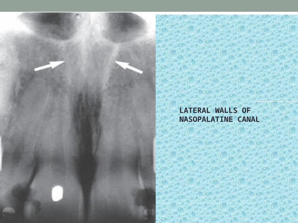
LATERAL WALLS OF NASOPALATINE CANAL

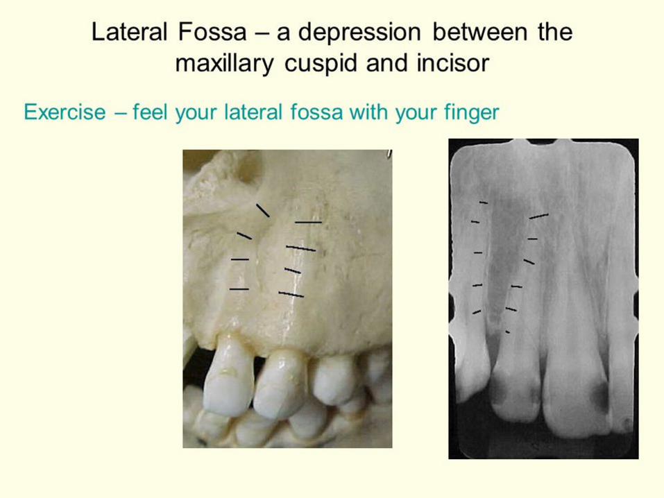
LATERAL FOSSA
• Called as incisive fossa.• Gentle depression in
maxilla near apex of lateral incisor.
• Appears as diffuse radiolucent area on periapical radiograph.

NASAL SEPTUM• Appears
radiopaque on radiograph.
• Composed of bone.
• Seen in midline of central incisors

NOSE• Tip of nose is seen in projection of maxillary central
incisors superimposed over roots of teeth.

INFERIOR NASAL CONCHAE• They are wafer
thin curved plates of bone that form lateral walls of nasal cavity.
• Concha means shell or scroll shaped.
• Appear as diffuse radiopaque mass on periapical radiograph.

MAXILLARY SINUS• It is air containing cavity lined with mucous membrane.• Also called antrum of highmore.• Anteriorly, each sinus is restricted by canine fossa.• Appears as three sided pyramid.• On periapical radiographs, floor of maxillary sinus and
nasal cavity are superimposed, cross each other and form a inverted Y shape in the area.
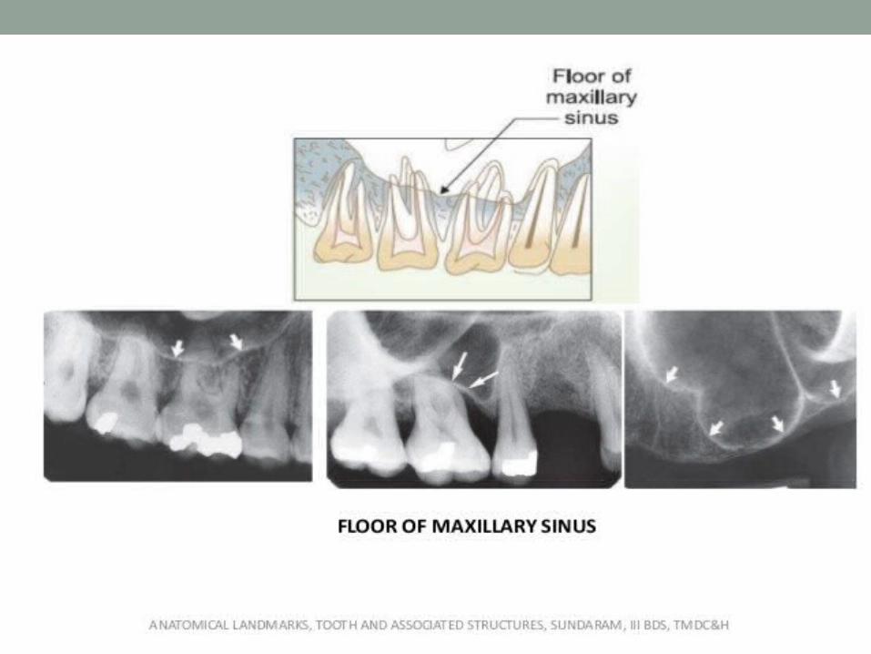
• Roots of maxillary molars lie close to maxillary sinus.• Floor of sinus – radiopaque• Maxillary sinus - radiolucent

FLOOR OF NASAL APERTURE EXTENDS TO MAXILLARY SINUS

ANTERIOR BORDER OF MAXILLARY SINUS
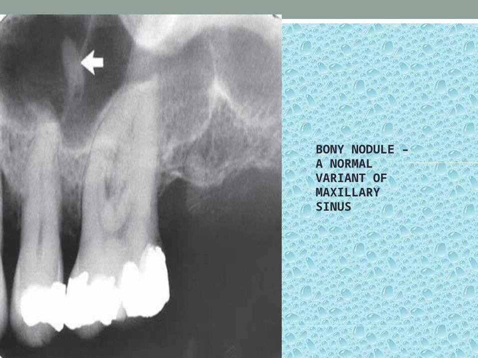
BONY NODULE – A NORMAL VARIANT OF MAXILLARY SINUS


MAXILLARY SINUS SEPTA• They are bony wall projections that divide maxillary sinus
into compartments.• Appear as radiopaque lines within the sinus.

NASOLACRIMAL CANAL• The nasal and maxillary bones form
the nasolacrimal canal. • It runs from the medial aspect of the
anteroinferior border of the orbit inferiorly to drain under the inferior concha into the nasal cavity.
• Occasionally , it can be visualized on periapical radiographs in the region above the apex of the canine, especially when steep vertical angulation is used .
• The nasolacrimal canals are routinely seen on maxillary occlusal projection in the region of the molars .
• Appear as ovoid radiolucency on radiograph.

MAXILLARY TUBEROSITY• It is rounded protuberance of bone that extends posterior
to third molar.• Appear as radiopaque bulge distal to third molar.
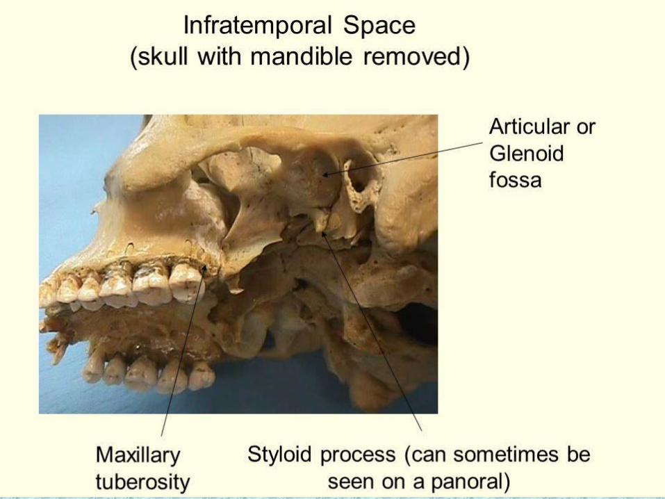
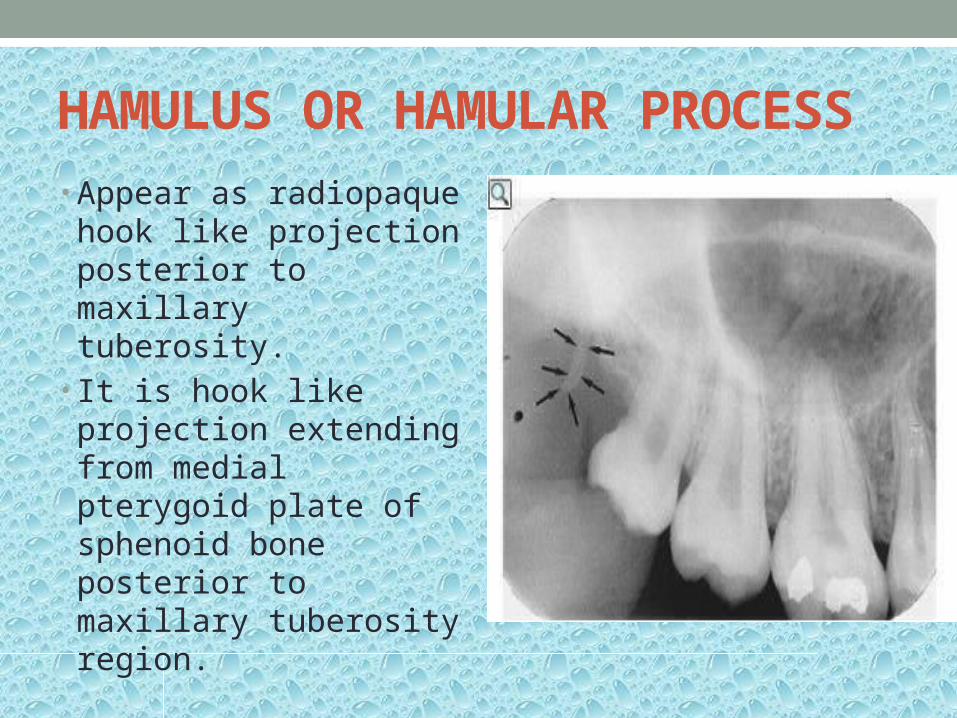
HAMULUS OR HAMULAR PROCESS• Appear as radiopaque hook
like projection posterior to maxillary tuberosity.
• It is hook like projection extending from medial pterygoid plate of sphenoid bone posterior to maxillary tuberosity region.

PTERYGOID PLATES• Lie posteriorly to
tuberosity of maxilla.( medial and lateral pterygoid plates ).
• They cast a radiopaque shadow which is homogenous without any evidence of trabecular pattern.
• Seen in maxillary third molar area posterior to maxillary tuberosity.

ZYGOMATIC PROCESS OF MAXILLA• It is bony projection of maxilla that articulates with zygoma
or malar bone.• Seen as J or U shape radiopacity posterior to first molar
region.


ZYGOMA• Zygoma or cheek
bone articulates with zygomatic process of maxilla.
• Composed of dense cortical bone.
• Appears as radiopaque band extending posterior from zygomatic process of maxilla .
• Appears superior to maxillary molars.
• AMATOMIC LANDMARKS OF MANDIBLE

GENIAL TUBERCLES• Tiny bumps of bone
that serve as attachment for genioglossus muscle.
• Present on lingual aspect of mandible.
• Appear as ring shape radiolucency below apices of mandibular incisors.

GENIAL TUBERCLES

LINGUAL FORAMEN• It is a tiny opening
or hole in bone located on internal surface of mandible.
• It is located near midline and surrounded by genial tubercles.
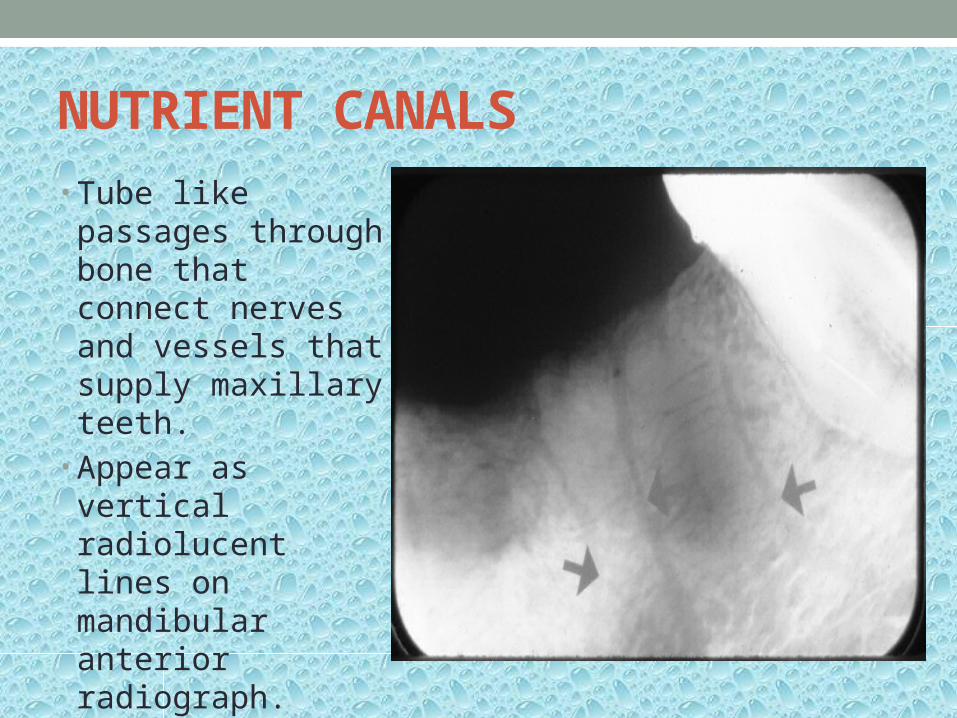
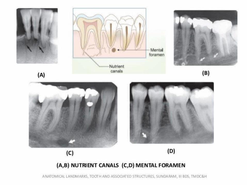
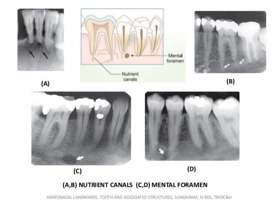
NUTRIENT CANALS• Tube like passages
through bone that connect nerves and vessels that supply maxillary teeth.
• Appear as vertical radiolucent lines on mandibular anterior radiograph.

MENTAL RIDGE• It is linear prominence
of cortical bone located on external surface of anterior portion of mandible.
• Extends from premolar region to midline .
• Appears as thick radiopaque band that extends from premolar region to incisor region.

MENTAL FOSSA• It is scooped out
depressed area of bone located on external surface of anterior mandible.
• Located above mental ridge in mandibular incisor region.
• Appears radiolucent on periapical radiograph.

MENTAL FORAMEN• It is a hole or
opening in bone in mandibular premolar region.
• Appears as ovoid radiolucency in apical portion of mandibular premolars.
• Can be misdiagnosed as cyst or abscess.

MYLOHYOID RIDGE• It is linear
prominence of bone located on internal surface of mandible.
• Appears as dense radiopaque band that extends downward and forward from mandibular molar region.
• It may be continuous with internal oblique ridge.
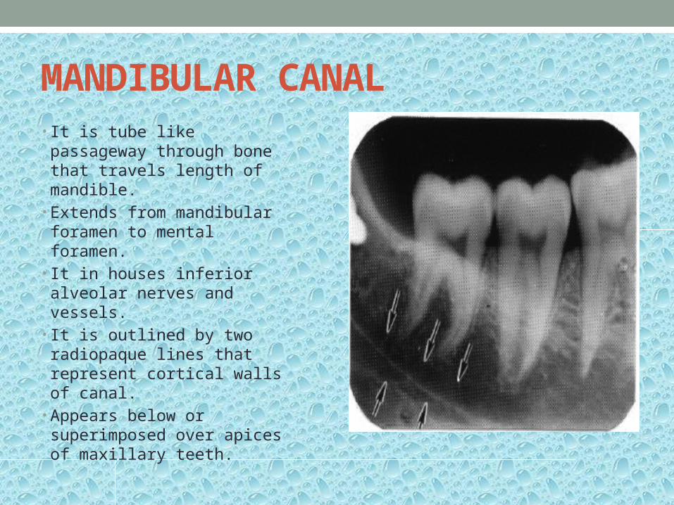
MANDIBULAR CANAL• It is tube like passageway
through bone that travels length of mandible.
• Extends from mandibular foramen to mental foramen.
• It in houses inferior alveolar nerves and vessels.
• It is outlined by two radiopaque lines that represent cortical walls of canal.
• Appears below or superimposed over apices of maxillary teeth.
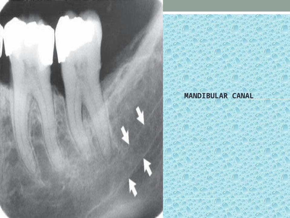
MANDIBULAR CANAL

INTERNAL OBLIQUE RIDGE• It is a linear prominence of bone
located on internal surfaces of mandible and extends from ramus.
• May extend in region of mandibular third molar or may continue as mylohyoid ridge.
• Appears as radiopaque band on radiograph.
• External and internal oblique ridge may superimpose depending upon technique used.
• When ridges appear separate, superior radiopaque band is external oblique ridge and inferior radiopaque band is internal oblique ridge.


EXTERNAL OBLIQUE RIDGE• Anterior border of
ramus ends in external oblique ridge.
• Appears as radiopaque band extending from anterior border of ramus .
• Usually ends in mandibular third molar region.

SUBMANDIBULAR GLAND FOSSA• Also known as mandibular
fossa or submaxillary fossa.
• It is scooped out depressed area of bone located on internal surface of mandible inferior to mylohyoid ridge.
• Submandibular salivary gland resides in this region.
• Appears as radiolucent area in mandibular molar region below mylohyoid ridge.

CORONOID PROCESS• It is marked prominence of
bone on anterior ramus of mandible.
• It is not seen on periapical radiograph.
• On maxillary radiograph , it is seen as triangular radiopacity superimposed over or inferior to maxillary tuberosity region.
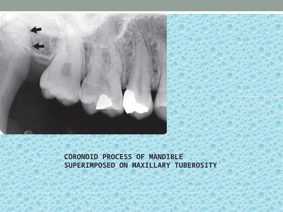
CORONOID PROCESS OF MANDIBLE SUPERIMPOSED ON MAXILLARY TUBEROSITY
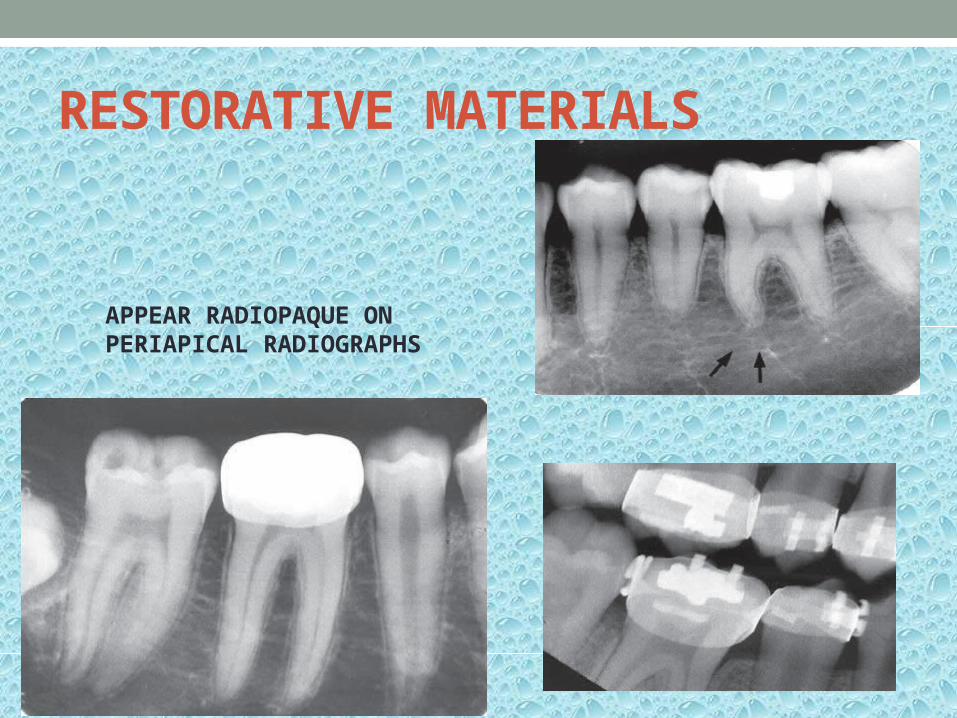
RESTORATIVE MATERIALS
APPEAR RADIOPAQUE ON PERIAPICAL RADIOGRAPHS

THANK YOU