normal tension glaucoma: who needs neuroimaging? julie falardeau, md, frcsc casey eye institute...
TRANSCRIPT
Normal Tension Glaucoma:Who Needs Neuroimaging?
Julie Falardeau, MD, FRCSC
Casey Eye Institute
Devers Eye Institute
Portland, Oregon
Background Normal tension glaucoma (NTG) is
characterized by: Cupping of the optic nerve head Visual field loss Intraocular pressure (IOP) 21 mmHg No obvious or apparent cause for these
changes
Nonglaucomatous optic disc cupping
Following an ischemic optic neuropathy (anterior or posterior - AION or PION) Temporal arteritis
Quigley and Anderson found that 50% of patient with arteritic -AION developed cupping, compared to 10% after non-arteritic-AION
Severe hypotensive/hypovolemic event
Demyelinating optic neuritis
Quigley et Anderson. Cupping of the optic disc in ischemic optic neuropathy. Trans Am Acad Ophthalmol Otol. 1977;83:755-762
Nonglaucomatous optic disc cupping
Hereditary optic neuropathy Leber’s hereditary optic neuropathy Autosomal dominant optic atrophy
Temporal disc excavation and pallor
Traumatic optic neuropathy Infectious
Syphilis Toxic
Methanol

Nonglaucomatous optic disc cupping
Compressive lesion Meningioma Aneurysm Dolichoectasia of the internal carotid
artery Suprasellar mass
Glaucomatous VS Nonglaucomatous cupping
Distinguishing glaucomatous from non-glaucomatous disc cupping is often difficult
A detailed history is crucial Presence of neurological symptoms Chronicity and pattern of visual loss History of head trauma History of shock or severe low blood pressure
Glaucomatous VS Nonglaucomatous cupping
Systematic approach recommended
Demographic characteristics Visual acuity Optic disc characteristics Visual field findings
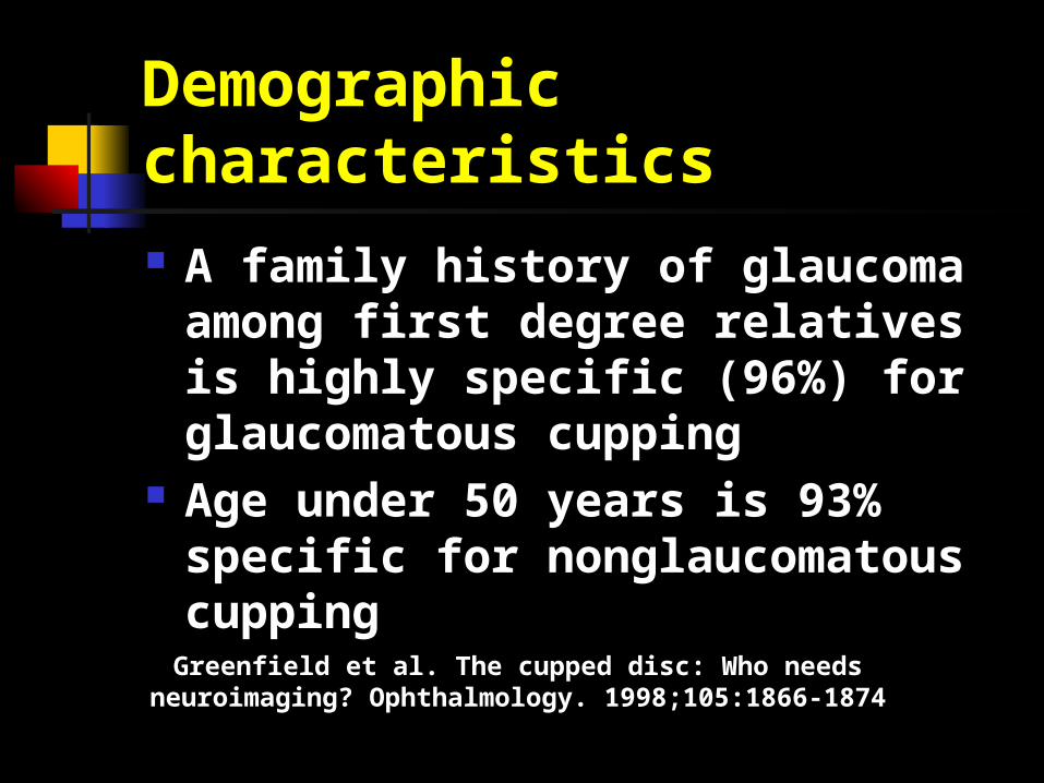
Demographic characteristics A family history of glaucoma among
first degree relatives is highly specific (96%) for glaucomatous cupping
Age under 50 years is 93% specific for nonglaucomatous cupping
Greenfield et al. The cupped disc: Who needs neuroimaging? Ophthalmology. 1998;105:1866-1874
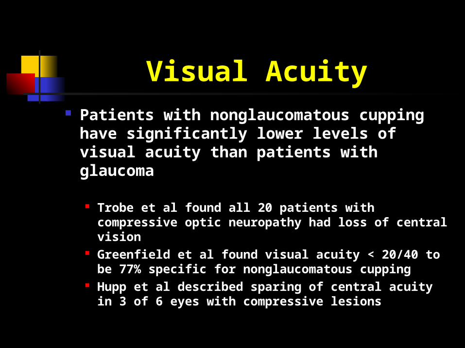
Visual Acuity Patients with nonglaucomatous cupping
have significantly lower levels of visual acuity than patients with glaucoma
Trobe et al found all 20 patients with compressive optic neuropathy had loss of central vision
Greenfield et al found visual acuity < 20/40 to be 77% specific for nonglaucomatous cupping
Hupp et al described sparing of central acuity in 3 of 6 eyes with compressive lesions
Optic disc characteristics Glaucomatous cupping:
Vertical elongation Cupping more than pallor Greater frequency of peripapillary atrophy Disc hemorrhage
Highly specific
Nonglaucomatous cupping: Pallor of the neuroretinal rim
Highly specific sign but relatively insensitive The absence of disc pallor does not exclude compressive
lesions
Optic nerve appearance Baring of the circumlinear vessels
and temporal saucerization Common in glaucoma Can also be seen in compressive optic
neuropathy
Kupersmith and Krohn. Cupping of the optic disc with compressive lesions of the anterior visual pathway. Ann Ophthalmol 1984;16:948-53
Visual field findings Glaucoma
Nerve-fiber-layer (arcuate) defects, bordering horizontal midline
Arcuate scotoma Nasal step
Compressive lesion Central scotoma Temporal hemianopia Incongruous hemianopia respecting the vertical
meridian Glaucomatous types of VF defects can occur
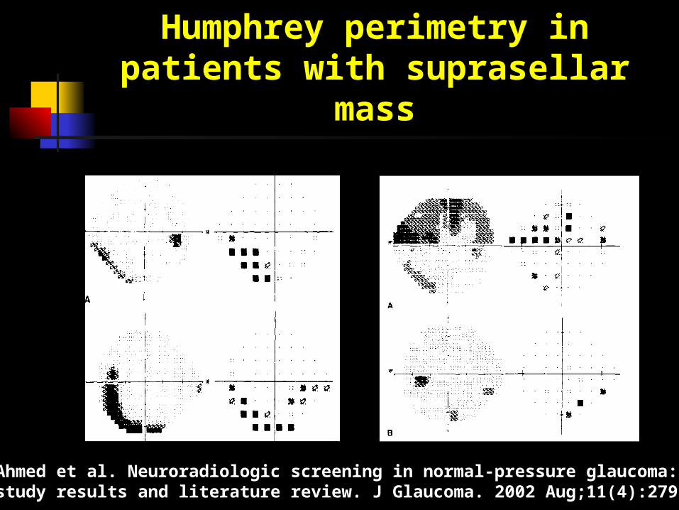
Humphrey perimetry in patients with suprasellar mass
Ahmed et al. Neuroradiologic screening in normal-pressure glaucoma: study results and literature review. J Glaucoma. 2002 Aug;11(4):279-86

NTG and Neuroimaging Some physicians routinely obtain
neuroimaging studies in patients with NTG
Cost-to-benefit ratio of performing such studies is unknown
NTG and Neuroimaging
Ahmed et al found that routine neuroimaging of NTG patients was cost-effective 6.5% of 62 consecutive patients with NTG had
clinically significant intracranial lesions associated with optic neuropathy and visual field loss typical of glaucoma
Ahmed et al. Neuroradiologic screening in normal-pressure glaucoma:
study results and literature review. J Glaucoma. 2002 Aug;11(4):279-86
NTG and Neuroimaging Steward and Reid reported
compressive lesions in 2 of 53 patients (3.8%) referred for evaluation of NTG
In the series by Greenfield et al, none of the patients diagnosed with glaucoma had neuroradiological evidence of compressive lesion
NTG and Neuroimaging In Bianchi-Marzoli at al’s series of 29
patients with cupping from unilateral compressive lesion, only one had cupping and field loss as an isolated manifestation of their optic neuropathy
All others had: Reduced acuity Decreased color vision RAPD
Bianchi-Marzoli et al. Quantitative analysis of optic disc cupping in compressive optic neuropathy. Ophthalmology 1995;102:436-440.
NTG: Who needs neuroimaging? Presence of headache or other
neurological symptoms
Symptoms of decreased vision, fluctuating vision, or visual field loss
Atypical visual field for glaucoma Visual field defect respecting the vertical meridian Junctional scotoma Central or cecocentral scotoma
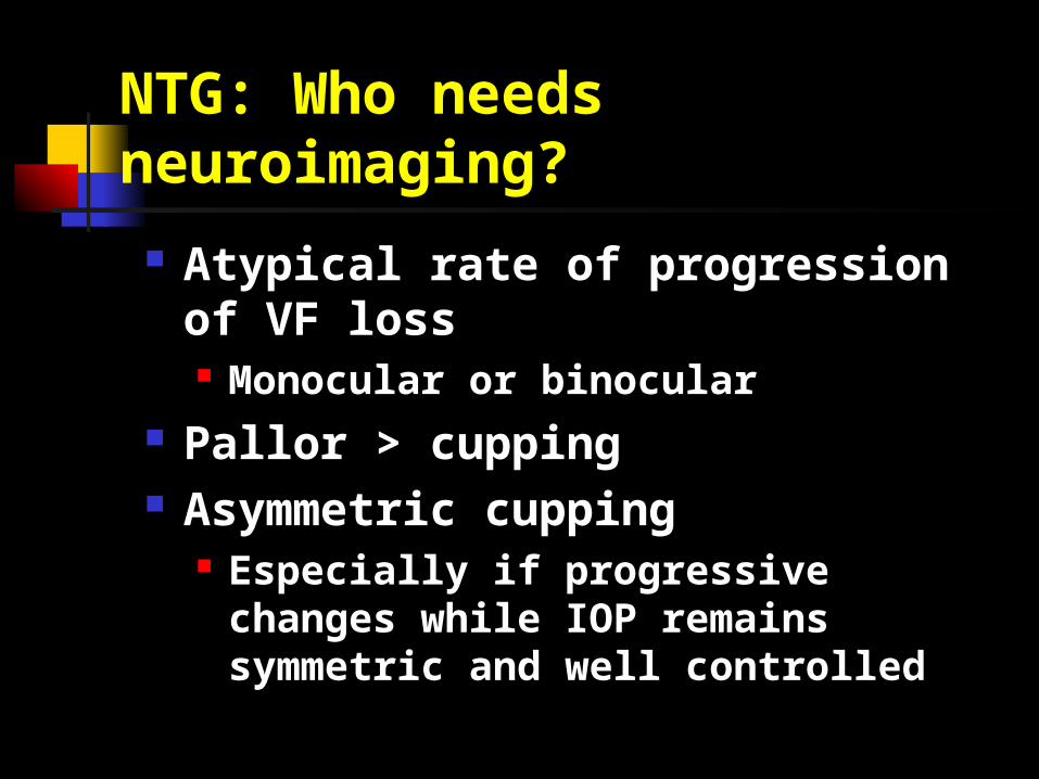
NTG: Who needs neuroimaging?
Atypical rate of progression of VF loss Monocular or binocular
Pallor > cupping Asymmetric cupping
Especially if progressive changes while IOP remains symmetric and well controlled
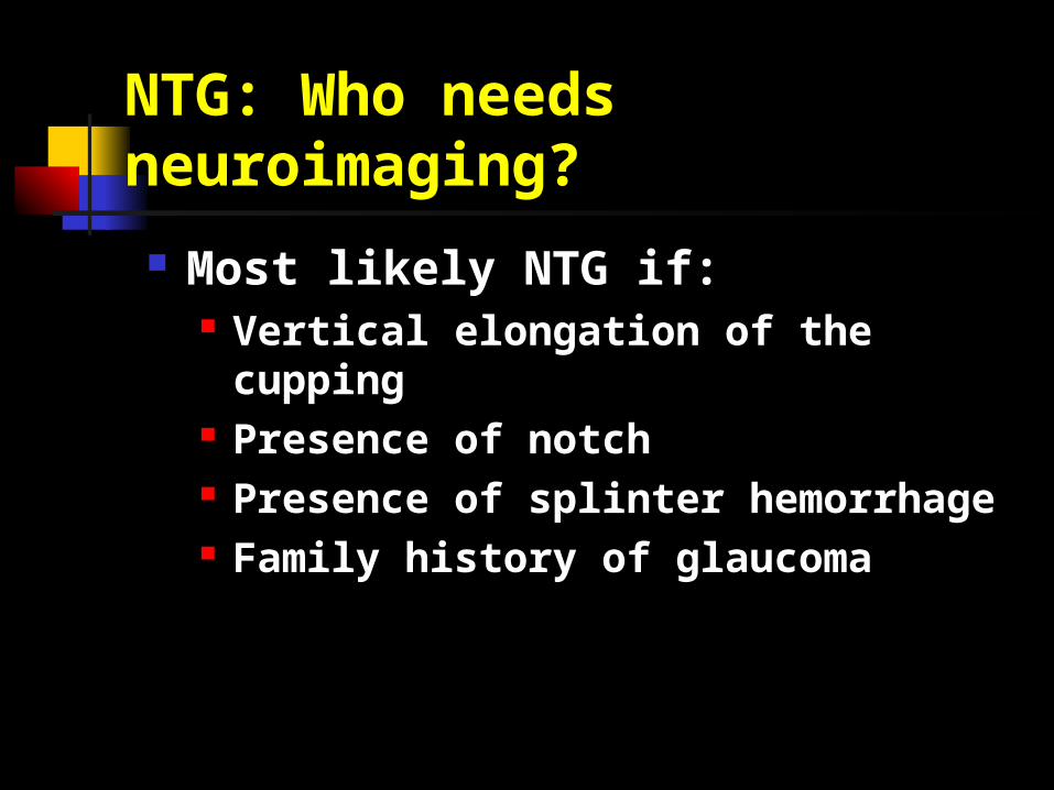
NTG: Who needs neuroimaging?
Most likely NTG if: Vertical elongation of the cupping Presence of notch Presence of splinter hemorrhage Family history of glaucoma