not so atypical pediatric cases - sunyopt.edu · not so atypical pediatric cases steven brooks md,...
TRANSCRIPT

6/8/2016
1
Not So Atypical Pediatric Cases
Steven Brooks MD, Jenelle Mallios OD, FAAO
Daniella Rutner OD, MS, FAAO, FCOVD Lauren Yeager MD
• 5 y.o AM referred in for RXT eval and amblyopia – Parents interested in VT for XT – Parents noticed the eye turn 1.5 years ago – Has worn glasses q1year – H/o patching 5-60min/day OS daily for 1.5 years
• BHx: Unremarkable • DHx: Unremarkable • Ehx: Unremarkable • Meds: None • FHx: Unremarkable
Lensometry: OD: +0.75-2.75x175 OS: plano-3.00x180
VAcc: D: 20/50 OD, 20/50 OS with HOTV , poor attention
Vacc: N: 20/50 OD,OS
Pupils, EOMS – normal
CVF- pt uncooperative
Nystagmus: unilateral (OD), high frequency, low amplitude horizontal nystagmus; dampens in left gaze and convergence
Color – pt uncooperative
CTcc: Primary gaze: 40 XT, 8RHT @ D (comitant), except no hyper on L tilt
NPC: poor
Stereo: RDS: none
Dry ret:
OD: +1.50-1.50x180 OS: plano-2.00x180
• SLE- unremarkable
• IOPs: soft and equal : digital
Cyclo ret:
OD: +3.00-2.50x180 OS: +1.50-2.75x180
DFE: Retinoschisis OD>OS
A: X linked juvenile retinoschisis
P: Consult with Dr. Brooks, Pedi OMD Stat

6/8/2016
2
• Typically males
• VA 20/60-20/120
• Requires Retinal consult
• Low Vision Referral
• Avoid Contact Sports
• Tx: topical dorzolamide or oral azetazolamide
• In the pipeline gene replacement therapy
Anisometropic Amblyopia ?
• 11 y-o male
• BCVA 20/20 OD and 20/50 OS
• Rx: +3.50 -3.50 X 175 OD
-5.00 -2.00 X 180 OS

6/8/2016
3
OD OS
Another Anisometropic Amblyope?
• 12 year-old male
• BCVA 20/40 OD and 20/20 OS
• Rx: -6.00 OD, Plano OS
OD OS
6/8/2016
4
Myelinated Nerve Fibers, Dysplastic Discs and Myopia
Developmental anomaly
Affected macular integrity from birth
Resulting myopia?
Degree of myelination directly proportional to poorer
prognosis of visual improvement with therapy Kee C, Hwand JM. Visual prognosis of amblyopia associated with myelinated retinal nerve
fibers. Am J Ophthalmol 2005;139:259-65.
Nystamgus
• Nystagmus occurs in 0.4% of the clinical population.
• frequency range of 2-5 Hertz (Hz) with an amplitude of 1-5 degrees. Usually horizontal in direction, although they may include a small vertical or rotary component.
• They cause sensory deficits such as reduced contrast sensitivity, visual acuity, and stereo acuity can have profound psychological effects resulting from the unusual cosmetic
• Nystagmus present at birth or prior to age
2 months is more likely to be idiopathic in
nature or due to neurologic dysfunction.
• Sensory deficit nystagmus most commonly
presents at age 2-3 months. Further
investigation of the visual system is
warranted in these cases.
Case 1 Nystagmus
• 7 month old
• F&F
• OD +1.25-0.50x180
• OS +1.25
• Pendular Nystagmus
• CAXT (Right eye preferred fixation)
• 3YO
• VA Cardiff OU 20/50
• EOM full OA IO
• Intermittent LXT
• 5yo
• 20/60 OD 20/32 OS
• EOM full
• NO tropia

6/8/2016
5
Infantile/Congenital
• F – fixation
• U-upgaze
• N-null point
• B- bilateral
• L-latent
• O-OKN inverse response
• C-convergence
• S-symptomless
THE 5
• Aniridia
• Achromatopsia
• Lebers Amarosis
• Albinism
• Optic Atrophy
Treatment options
• Glasses
• Prism
• Contact lenses
• Biofeedback
– Visual
– Auditory
• Surgery
Nystagmus
Rutner Treatment of Choice • less optical aberrations
• enlarged retinal image (in refractive myopes)
• increased peripheral visual field.
• Any or all of the above would improve the quality and/or extent of the retinal image, and hence provide a visual input of higher fidelity for fusion and subsequent visual information processing.
Nystagmus Case 2
• An 18-year-old female with non-PAN, congenital jerk nystagmus, who had never worn contact lenses, was evaluated. Her refraction and best corrected visual acuity with spectacles was OD -4.00D/-2.75D x 170 (20/120), and OS -4.00D/-3.00D x 025 (20/200). She had strabismus surgery two years earlier to correct a constant esotropia, with a residual 15 prism diopters of intermittent alternating esotropia. Ocular health examination revealed ocular albinism; all else was within normal limits
• After a comprehensive eye examination to assess refractive, binocular, and ocular health factors, the patient was fit with Coopervision Preference Toric soft contact lenses (SCL). Lens parameters were: 8.7mm base curve, 14.4mm diameter, 0.09mm center thickness, OD -3.00D/-2.25Dx180, and OS -2.00D/-2.25Dx010.
Nystagmus
Condition Amplitude
(degrees
)
Frequency (Hz) High Contrast Visual
Acuity
Low Contrast
Visual Acuity
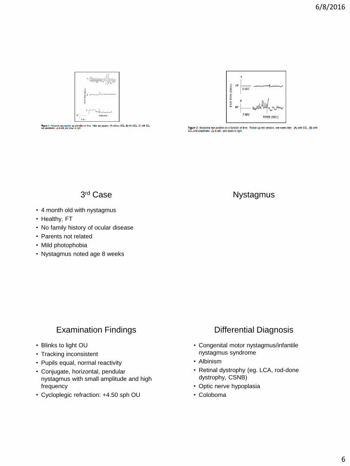
Test Session One:
Spectacles (baseline)
9.00 1.25 20/120 OD 20/200 OS - - - -
SCL 1.75 1.05 20/100 OD 20/125
OS, 20/100 OU
20/125 OD unable
to perform
OS at 1 meter
test distance
20/125 OU
SCL with anesthetic 2.70 1.40 - - - - - - - -
Test Session Two:
SCL one week later
0.72 1.80 20/80 OD 20/125 OS
20/80 OU
20/100 OD 20/125
OS 20/100
OU
SCL with anesthetic
1week later
3.30 1.95 - - - - - - - -
6/8/2016
6
3rd Case
• 4 month old with nystagmus
• Healthy, FT
• No family history of ocular disease
• Parents not related
• Mild photophobia
• Nystagmus noted age 8 weeks
Nystagmus
Examination Findings
• Blinks to light OU
• Tracking inconsistent
• Pupils equal, normal reactivity
• Conjugate, horizontal, pendular
nystagmus with small amplitude and high
frequency
• Cycloplegic refraction: +4.50 sph OU
Differential Diagnosis
• Congenital motor nystagmus/infantile
nystagmus syndrome
• Albinism
• Retinal dystrophy (eg. LCA, rod-done
dystrophy, CSNB)
• Optic nerve hypoplasia
• Coloboma
6/8/2016
7
Key Examination Points
• Assess visual function
• Iris transillumination
• Check for paradoxical pupil response
• Funduscopic examination
New Onset Nystagmus in a 2 year
old
• Healthy 2 yo F
• No prior illness
• Sudden onset of ataxia and nystagmus
• Lethargy, irritability
• Myoclonic jerks
Examination Findings
• Rapid, conjugate, multi-directional jerky
eye movements
• Central fixation, disrupted by jerky eye
movements
• Normal pupils
Examination Findings
• Normal anterior segment and funduscopic
examination
• Full range of EOM’s
• Cycloplegic refraction +2.00sph OU
Diagnosis
• OMA (opsoclonus-myoclonus-ataxia)
– Post-encephalitic (viral)
– Para-neoplastic (50%)
– Idiopathic
• This is NOT nystagmus
Opsoclonus-Myoclonus-Ataxia
• Rare
• Requires urgent neurological evaluation
• Neuroblastoma most frequently implicated
neoplasm
• Opsoclonus secondary to autoimmune
attack of purkinje cells in the cerebellum
• Recovery often incomplete

6/8/2016
8
5 year old with R face turn
• Healthy 5 yo M
• Noted by parents to turn habitually turn
face to the right when watching TV
• Nystagmus noted age 4 months
• Good vision, asymptomatic
• Family history negative
Examination Findings
• Va 20/60 OD, 20/70 OS, 20/30 OU open,
20/25 OU open with face turn to R
• Conjugate, horizontal, nystagmus with fast
phase to L in primary position
• Normal pupils and EOM’s
Examination Findings
• No iris transillumination
• No photophobia
• No strabismus
• Nystagmus decreases in right gaze,
increases in left gaze
• Normal anterior segment and fundus exam
• Cycloplegic refraction: plano OU
Differential Diagnosis
• Congenital motor nystagmus/infantile
nystagmus syndrome
• Manifest-latent nystagmus/fusion
maldevelopment nystagmus syndrome
• Periodic alternating nystagmus
• Cerebellar lesion
• Vestibular lesion
• Retinal dystrophy
Interpretation of Examination
• Congenital motor nystagmus with null
point in left gaze (causing right face turn)
• Visual acuity improved with OU open
(latent component to nystagmus)
• Visual acuity improved in left gaze (null
point)
Management
• Surgery to shift null point
• Surgery to reduce (dampen) nystagmus
• Prism glasses
• Imaging?? Neurology consultation??
• Follow-up

6/8/2016
9
6 month old with OS “shaking”
• Healthy, FT
• Parents note that left eye seems to
intermittently shake or jiggle
• Normal development
• No FH of ocular disease
Examination Findings
• Normal appearing baby
• Central, steady fixation with each eye
• Normal pupils
• OS intermittently shows very low
amplitude, high frequency, pendular
nystagmus
• Child seems to tilt head to left and shake
head intermittently
Examination Findings
• Normal anterior segment OU
• Normal funduscopic examination OU
• Full EOM’s
• Cycloplegic refraction
OD: +1.75+1.75x50
OS: +1.75+1.50x130
Differential Diagnosis
• Spasmus nutans
• Sensory-loss nystagmus
• Retinal dystrophy
• Idiopathic
• Leukoencephalopathy/Leukomalacia
Interpretation of Exam
• Likely spasmus nutans
Management
• Neuro-imaging
• Close observation
• Correction of refractive error?
• Patching to prevent amblyopia?

6/8/2016
10
Peds Ophth and Some Red
Flags
8 month old with esotropia
• Parents have noted ET “since birth”
• Healthy, FT
• Family history positive for ET in mother
and a cousin
• Slower to crawl than older sibling
Examination Findings
• Normal appearing baby
• Constant esotropia 40 PD by Krimsky
testing
• Holds fixation well with either eye
• Mild limitation of abduction OU
Examination Findings
• Normal pupils
• Normal anterior segment and fundus
• No nystagmus
• Normal versions
• Cycloplegic refraction
OD: +1.75 sph
OS: +1.75 sph
Differential Diagnosis
• Congenital esotropia
• Infantile accommodative esotropia
• Sensory loss
• EOM fibrosis syndrome
• Duane syndrome
• Nystagmus blockage syndrome
Interpretation of Examination
• Large angle constant esotropia
• No amblyopia
• Normal vision for age, no evidence of
ocular defects
• Essentially full ductions
• Minimal hyperopia, normal for age

6/8/2016
11
Interpretation of Examination
• Likely congenital esotropia without
amblyopia
• Accommodative component unlikely
Management
• Glasses??
• Patching??
• Vision therapy??
• Surgery?? Timing??
5 yo M with new onset Left ET
• Healthy
• No FH of ocular disease
• Normal development
• Occassional headaches recently
Examination Findings
• Va 20/25 OD, 20/25 OS
• Pupils normal
• Normal orbits
• Anterior segment: OD palpebral fissure
narrower than OS
Examination Findings
• EOM: Full OD, mod limitation of Abduction
OS
• No nystagmus
5 ET 20ET 30ET
Interpretation of Findings
• Incomitant esotropia
• Limited ductions for OS
• No amblyopia
• Palpebral fissure asymmetry

6/8/2016
12
Differential Diagnosis
• EOM fibrosis syndrome
• Duane Syndrome
• Myasthenia gravis
• Orbital lesion
• Sixth n. palsy (multiple possible causes)
3 year old with red eye OD
• Healthy
• Redness and photophobia OD for 10 days
• Not responsive to topical antibiotics
• Similar episode 6 months ago
• Tearing
Examination Findings
• Va 20/50 OD, 20/25 OS (allen pics)
• OD mild ptosis
• OD 2+injection, small epi defect and
opacity paracentrally with pannus
• No adenopathy
Examination Findings
• No hypopyon
• No FB
• Eyelid margins clear
• Unable to measure IOP
• Fundus normal

6/8/2016
13
Interpretation of Findings
• Keratitis OD with corneal
neovascularization
• Unresponsive to topical antibiotics
• Prior episode in same eye
Differential Diagnosis
• Recurrent corneal erosions
• Occult FB
• Staph marginal keratitis
• HSV
3 year old with head tilt
• Healthy
• Head tilt to R noted age 10 months
• Normal development
• Family history negative for ocular disease
Examination Findings
• Va Fixes and Follows well each eye
• Tilts head approx. 10 degrees to the right
• Resists having head tilted to the left
• Normal pupils
• Normal anterior segment and optic disc
OU
Examination Findings
• Left inferior oblique overaction
• Full ductions OU
8LHT/5XT
15 LHT 8 LHT 4LHT
8LHT/5ET
Tilt R: ortho Tilt L: 12 LHT
Three Step Test
• Measure hypertropia in primary gaze
• Measure hypertropia in R and L gaze
• Measure hypertropia in R and L head tilt

6/8/2016
14
Example: LHT secondary to LSO palsy
Step 1 (LHT in primary)
RSR RIO LIO LSR
RIR RSO LSO LIR
Elevators
Depressors
OD OS
Example: LHT secondary to LSO palsy
Step 2 (LHT worse in right gaze)
RSR RIO LIO LSR
RIR RSO LSO LIR
Elevators
Depressors
OD OS
Example: LHT secondary to LSO palsy
Step 3 (LHT worse in left tilt)
RSR RIO LIO LSR
RIR RSO LSO LIR
Elevators
Depressors
OD OS
Additional Testing
• Patch test
• Fundus exam
• Review old photos

6/8/2016
15
Diagnosis
• Left superior oblique palsy
8 month old with R ET
• Healthy, FT
• Normal development
• Family history of strabismus in a 1st cousin
Examination Findings
• Va Blinks to light, poor fixation OD, Fixes
well OS
• Pupils appear normal
• Anterior segment and red reflex clear OU
Examination Findings
• Possible nystagmus OD
• 25 RET
• Full ductions
Interpretation of Findings
• Esotropia
• Decreased vision OD
• Normal ocular motility
• Concern for vision loss OD leading to ET

6/8/2016
16
Examination Findings
• Blinks to light OU
• Tracking inconsistent
• Pupils equal, normal reactivity
• Conjugate, horizontal, pendular
nystagmus with small amplitude and high
frequency
• Cycloplegic refraction: +4.50 sph OU
Differential Diagnosis
• Congenital motor nystagmus/infantile
nystagmus syndrome
• Albinism
• Retinal dystrophy (eg. LCA, rod-done
dystrophy, CSNB)
• Optic nerve hypoplasia
• Coloboma
Key Examination Points
• Assess visual function
• Iris transillumination
• Check for paradoxical pupil response
• Funduscopic examination
COATS
• Abnormal development of Blood Vessels
in the Retina: dilated tortuous and leaky
• Males to females 3:1

6/8/2016
17
5 STAGES
• 1. abnormal blood vessels are seen in the
retina but these vessels are not yet leaky.
• 2. leakage from the vessels into the retina.
Effect on VA variable
• 3. Retinal detachment
• 4. Complicated with glaucoma (raised
• 5. End stage blind painful eye.
Treatment
• Referral for cyro
• Laser
• Surgical
Not So Simple Tropia
• 3 year old referred by pediatrician for
second opinion for strabismus surgery
Previous Hx
• H/O strabismus surgery secondary to
double elevator palsy
• Previous diagnosis of bilateral ambylopia
• Born at 34 wks
• 4lbs 8 oz at birth
• +developmental delays with +ST, OT, PT
• Long standing ptosis of right eye
Monocular elevation Deficiency
• Monocular Elevation Deficiency, also known by the older term Double Elevator Palsy, is an inability to elevate one eye, usually resulting in one eye that is pointed downward relative to the other eye
• The apparent paralysis of both elevators (superior rectus and inferior oblique) of one eye that results in a rather large hypotropia on the affected side is uncommon. The levator palpebrae may or may not be involved, and Bell’s phenomenon may be present but is usually absent; if it is present, a supranuclear lesion is implied. The pupil is normal, as are horizontal rotations.
• The eyelid on the involved side is droopy (ptosis) 25% of the time while 75% of cases have pseudoptosis. In this case, the pseudoptosis is the appearance of ptosis caused by the eye being hypotropic (downward deviation).
• 25% of those with Monocular Elevation Deficiency and Congenital Ptosis have a phenomenon called Marcus Gunn jaw-winking. This a condition in which the cranial nerve that usually controls eyelid movement is mis-wired with the cranial nerve that controls chewing or sucking thus creating a "wink" when chewing or sucking
6/8/2016
18
• VA
– OD 20/70 current Rx pl-0.75x5
– OS 20/60 current Rx +0.25-0.75x158
Cycloplegic Refraction
• OD +1.00-1.00x180
• OS +1.00-2.00x150
Cover Test
• With correction
• 15CLHT, 15 CLXT equal distance and near
• With a V pattern
• EOM full with over action of the IO of the left eye
Slit Lamp
• Normal findings
• No ptosis noted
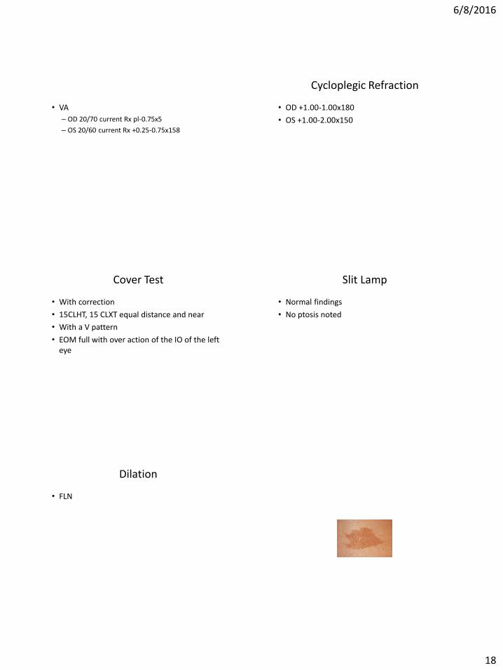
Dilation
• FLN
6/8/2016
19
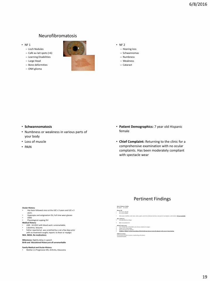
Neurofibromatosis
• NF 1
– Lisch Nodules
– Café au lait spots (>6)
– Learning Disabilities
– Large Head
– Bone deformities
– ONH glioma
• NF 2
– Hearing loss
– Schwannomas
– Numbness
– Weakness
– Cataract
• Schwannomatosis
• Numbness or weakness in various parts of your body
• Loss of muscle
• PAIN
• Patient Demographics: 7 year old Hispanic female
• Chief Complaint: Returning to the clinic for a comprehensive examination with no ocular complaints. Has been moderately compliant with spectacle wear
Ocular History: • Has been followed since at the UEC x 5 years and LEE x 3
years. • Hyperopia and astigmatism OU, full time wear glasses • OMD • Physiological cupping OU Medical History: • LME: 10-2013 with blood work: unremarkable; • (-)Asthma, Seizures • Father reported pt. was scratched by a cat a few days prior
with no treatment sought; reports no fever or myalgia NKA. NKDA. No medications Milestones: Slightly delay in speech Birth and Educational History are all unremarkable Family Medical and Ocular History: • Mother (+) Progressive MS, Arthritis, Glaucoma
Pertinent Findings
VAcc in Distance: Snellen OD: 20/20 OS: 20/20 Glasses RX:
• OD: +0.25-1.00x180 • OS: +0.50-1.50x065
• Extra-ocular motilities, color vision, stereo, pupils, cover test at distance and near, near point of convergence, confrontations All unremarkable
IOPs: Goldmann
12mmHg OD,OS at 5:20pm
SLE: Unremarkable OU
Dilated fundus exam: C/D: 0.55V/0.50H; 0.55V/0.60H, pink, distinct, healthy rim margins Vessels: 2/3, normal caliber Macula: flat and clear (+)FR OU Periphery: Bilateral vitreal snow balling I/IT/IN OS>OD and vitreous heme OS adjacent to the area of snow banking
Additional testing: Fundus photos (unable to capture snowbanking with photos) (-)lymphadenopathy

6/8/2016
20
Optos image of inferior snowbanking Oceanoptometry.blogspot.com
• Intermediate uveitis, or pars planitis, is an insidious, chronic,
relapsing inflammation of the anterior vitreous and pars plana. While uveitis occurs in 5-10% of the pediatric population, pars planitis accounts for 8-33% of all uveitis in pediatric patients.
• Clinical presentation includes snowballs, snowbanking, vitritis, and peripheral retinal vasculitis. Vitreous hemorrhages occur in 16.7% of pediatric patients. It is typically bilateral and asymmetric and patients often remain asymptomatic.
• Although pars planitis is often idiopathic (96%) in pediatric patients, inflammatory diseases must be ruled out as a possible etiology.
Systemic Associations
• Idiopathic: Most common • Sarcoidosis: relatively uncommon, can precede systemic disease • Lyme disease associated with severe anterior uveitis. • Tuberculosis: uncommon • Toxoplasmosis • EBV or HTLV-1 infections • Neurological concomitants: Vogt–Koyanagi–Harada syndrome, • Behcet syndrome, AIDS, primary CNS lymphoma, herpes virus infections, • syphilis, acute posterior multifocal placoid pigment epitheliopathy • and Whipple disease. • Multiple sclerosis: 15% of the patients with pars planitis developed • MS after five years of follow-up
3 year old with red eye OD
• Healthy
• Redness and photophobia OD for 10 days
• Not responsive to topical antibiotics
• Similar episode 6 months ago
• Tearing
Examination Findings
• Va 20/50 OD, 20/25 OS (allen pics)
• OD mild ptosis
• OD 2+injection, small epi defect paracentrally with pannus
• No adenopathy
Examination Findings
• No hypopyon
• No FB
• Eyelid margins clear
• Unable to measure IOP
• Fundus normal

6/8/2016
21
Interpretation of Findings
• Keratitis OD with corneal neovascularization
• Unresponsive to topical antibiotics
• Prior episode in same eye
Differential Diagnosis
• Recurrent corneal erosions
• Occult FB
• Staph marginal keratitis
• HSV
3 year old with head tilt
• Healthy
• Head tilt to R noted age 10 months
• Normal development
• Family history negative for ocular disease
Examination Findings
• Va Fixes and Follows well each eye
• Tilts head approx. 10 degrees to the right
• Resists having head tilted to the left
• Normal pupils
• Normal anterior segment and optic disc OU
Examination Findings
• Left inferior oblique overaction
• Full ductions OU
8LHT/5XT
15 LHT 8 LHT 4LHT
8LHT/5ET
Tilt R: ortho Tilt L: 12 LHT
Additional Testing
• Patch test
• Fundus exam
• Review old photos

6/8/2016
22
Diagnosis
• Left superior oblique palsy
Three Step Test
• Measure hypertropia in primary gaze
• Measure hypertropia in R and L gaze
• Measure hypertropia in R and L head tilt
Example: LHT secondary to LSO palsy Step 1 (LHT in primary)
RSR RIO LIO LSR
RIR RSO LSO LIR
Elevators
Depressors
OD OS
Example: LHT secondary to LSO palsy Step 2 (LHT worse in right gaze)
RSR RIO LIO LSR
RIR RSO LSO LIR
Elevators
Depressors
OD OS
Example: LHT secondary to LSO palsy Step 3 (LHT worse in left tilt)
RSR RIO LIO LSR
RIR RSO LSO LIR
Elevators
Depressors
OD OS
8 month old with R ET
• Healthy, FT
• Normal development
• Family history of strabismus in a 1st cousin

6/8/2016
23
Examination Findings
• Va Blinks to light, poor fixation OD, Fixes well OS
• Pupils appear normal
• Anterior segment and red reflex clear OU
Examination Findings
• Possible nystagmus OD
• 25 RET
• Full ductions
Interpretation of Findings
• Esotropia
• Decreased vision OD
• Normal ocular motility
• Concern for vision loss OD leading to ET