novel biochemical regulatory mechanisms of...
TRANSCRIPT

Novel Biochemical Regulatory Mechanisms of Developmental Signaling Pathways
By
Tony Wayne Chen
Dissertation
Submitted to the Faculty of the
Graduate School of Vanderbilt University
in partial fulfillment of the requirements
for the degree of
DOCTOR OF PHILOSOPHY
in
Cell and Developmental Biology
May, 2015
Nashville, Tennessee
Approved:
Ethan Lee, M.D., Ph.D.
Chin Chiang, Ph.D.
Stacey S. Huppert, Ph.D.
David Bader, Ph.D.
Sandra S. Zinkel, M.D., Ph.D.

ii
DEDICATION
To my family

iii
ACKNOWLEDGMENTS
None of this work would have been possible without unwavering support and
guidance from my mentor, Dr. Ethan Lee. His excitement and passion for science
continue to amaze me every single day. I marvel at his ability to always come up with
brilliant and creative scientific ideas and I truly appreciate the freedom he lets his
students operate with to tackle scary tough scientific questions. I will always be grateful
to Ethan for taking a chance on me and allowing me to join his lab and attempt to do
interesting, groundbreaking science. His enthusiasm makes the lab a place that I look
forward to going every single day for 6+ years and that enthusiasm has spread to all his
lab personnel. If I can be half of the scientist that Ethan is, then I will have considered
my career to be wildly successful.
I would also like to thank Dr. Laurie Lee. Her ability to clearly think about science
and dispense scientific advice was critical to my maturation as a researcher. Her ability
to keep our lab running smoothly cannot be overstated and I am eternally grateful to her
for her contributions to all aspects of the lab. Her partnership with Ethan really lended
itself to a unique and great lab environment which I would not trade for any other lab
environment in the world. The collaborative lab environment that the Lees cultivated and
nurtured was critical in during my Ph.D. studies.
I also need to thank Dr. Matt Broadus. Even though he was only in the lab for
about 18 months, those 18 months were the most productive of my entire career by a
significant margin. I would not be where I am without Matt. His scientific abilities dwarf
mine and I can only hope to approach them over time. Some of the things he taught me
I will carry with me the rest of my life.

iv
I would also like to thank the other members of the Lee lab. One often
underrated aspect of graduate school is the people whom you work with. I joined the
Lee lab with 3 other grad students, Brian Hang in Ethan’s lab, Poojitha Sitaram and
Sarah Hainline in Laurie’s lab. Poojitha was my long-time baymate and we often
engaged in spirited conversations about a variety of interesting subjects, including but
not limited to science, Game of Thrones, and tennis. I greatly appreciate her tolerating
my ridiculousness for many years. Her presence has been missed. Brian is a great
labmate who always adds great scientific insight and his presence in the lab can only be
described as fantastic. Whenever he spoke I knew he was saying something profound
and insightful and I would go out of my way to listen. Sarah was always tremendous to
have in the lab and I always enjoyed having someones to commiserate with when our
experiments were not working (which happened all too often). Our interactions will be
missed. Joining the lab and learning together with these 3 people has made my
graduate school career a pleasant experience even in the face of failed experiments
and changed projects and I thank them for joining me in this long and eventful journey.
The next people I want to thank are the two graduate students who joined the Lee Lab
the year after I did, Kenyi Saito-Diaz in Ethan’s lab and Jeanne Jodoin in Laurie’s lab.
Kenyi has been a great source of both scientific and personal advice for many years
and the lab would not be the same without him. Jeanne is one of the most entertaining
labmates I have ever had and is a wonderful scientist. She added a spark to the lab that
is irreplaceable. I would be remiss if I did not thank the previous lab personnel: graduate
students Curtis Thorne, Chris Cselenyi, Kristin Jernigan, Ali Hanson, Julie Merkle,
Jamie Rickmyre, Michael Anderson, postdoctoral fellow Dr. Emilios Tahinci and

v
technician Kelly Meyers. Without their guidance and support early in my career I would
likely not be where I am. Next, I have to thank the newest additions to the lab, graduate
student Leif Neitzel, lab manager Leah Sawyer, postdoctoral fellows Dr. Aja Hyde and
Dr. Amanda Hansen, and undergraduate Eddie Ross. Leif’s zebrafish expertise has had
a major impact on this work and in his short time in the lab he has proven to be both a
great scientist and a great labmate. I have not worked with Leah, Aja, or Amanda for
that long but in the short time they’ve been in the lab they’ve already greatly enhanced
my graduate school career. Each brings their own unique expertise and my own limited
knowledge and skillset is greatly enhanced by their presence and I am truly excited to
see where they can take the lab after I have graduated. Eddie also deserves special
mention as an undergraduate because the natural aptitude and scientific curiositiy he
exhibits is very vey rare at his stage of schooling and his ability to perform all the taks
assigned to him quickly and efficiently and correctly has been invaluable to the lab.
I also need to express a heartfelt thanks to my thesis committee: Chin Chiang,
Stacey Huppert, David Bader, and Sandy Zinkel. I really felt the support and
encouragement from my committee even as my project kept changing. In addition, they
were instrumental in guiding my project into what it is today.
I also want to thank our collaborators for their kindness. Their countless
contributions (both intellectual and in terms of reagents) greatly impacted this work
presented in this thesis.
I owe a large debt of gratitude to my parents, Thomas and Caroline Chen, and
my brother Michael Chen who have molded me into the person I am today. They have
always supported me and they inspire me to strive for perfection every single day.

vi
TABLE OF CONTENTS
Page
DEDICATION ...................................................................................................................ii ACKNOWLEDGMENTS .................................................................................................. iii LIST OF TABLES .......................................................................................................... viii LIST OF FIGURES ..........................................................................................................ix Chapter I. INTRODUCTION TO DEVELOPMENTAL SIGNALING PATHWAYS .......................... 1
Introduction .................................................................................................................. 1 Cell Signaling and Signal Transduction of Developmental Pathways .......................... 1 Historical Significance: Wnt Signaling in Development and Disease ........................... 4 The Current Model of the Wnt/β-catenin Signaling Pathway ........................................ 7 Wnt/β-catenin Signaling: Surface Receptor Activation ................................................. 9 Wnt/β-catenin Signaling: The β-catenin destruction complex .................................... 16 Wnt/β-catenin signaling: Transcriptional activation .................................................... 25 Historical Significance: Notch Signaling in Development and Disease ...................... 29 The Current Model of the Notch Signaling pathway ................................................... 33 Notch Signaling: Surface Receptor Activation............................................................ 36 Notch Signaling: Regulation of the NICD ................................................................... 45 Notch Signaling: Activation of a NICD-CSL-MAML transcriptional complex .............. 54 Notch and Wnt Signaling: Evidence for Wntch Signaling? ......................................... 58
II. MATERIALS AND METHODS: PROTEIN DEGRADATION IN XENOPUS EGG EXTRACT ................................................................................................................. 67
Introduction ............................................................................................................. 67 Protocol .................................................................................................................. 70 Representative Results ........................................................................................... 80
III.IDENTIFICATION OF A NOTCH1 INTRACELLULAR DOMAIN DEGRONG THAT REGULATES SIGNALING IN A PARALOG-SPECIFIC MANNER ........................... 90
Materials and Methods ............................................................................................. 90 Preparation of Xenopus egg extract ....................................................................... 90 [S35]-radiolabeled degradation assay in Xenopus egg extract ................................ 90 Luciferase-tagged fusion protein degradation assay in Xenopus egg extract ......... 91 High-throughput degradation assay in Xenopus egg extract .................................. 92

vii
Plasmids/Cloning .................................................................................................... 92 Site-directed Mutagenesis ...................................................................................... 96 siRNA constructs .................................................................................................. 105 Mammalian Cell Culture ....................................................................................... 105 Hes-1 Reporter Assays ......................................................................................... 105 Zebrafish Somite Formation Assay ....................................................................... 106 Immunoblot analysis ............................................................................................. 106 Live-Cell Imaging .................................................................................................. 107
Introduction .............................................................................................................. 108 Results ..................................................................................................................... 110
NICD1 degrades robustly in Xenopus egg extract ................................................ 110 NICD degradation within Xenopus egg extract is restricted to the NICD1 paralog 112 NICD1 degradation in Xenopus egg extract does not require its PEST domain or Fbxw7 ................................................................................................................... 115 The N-terminal end of hNICD1 encodes a degron required for degradation in Xenopus egg extract ............................................................................................. 118 The N1-Box controls hNICD1 stability and activity in vitro and in vivo .................. 126 The N1-Box is not regulated by Fbxw7 or Itch ...................................................... 133 Binding of CSL to hNICD1 regulates its stability ................................................... 135 Mutations within the N1-Box are potential drivers of Notch-mediated tumorigenesis .............................................................................................................................. 139
Discussion................................................................................................................ 141 IV. DISCUSSION AND FUTURE DIRECTIONS ......................................................... 146
Introduction .............................................................................................................. 146 Part I ........................................................................................................................ 146
Discussion ............................................................................................................ 146 Future Directions .................................................................................................. 152
Identifying the complete regulatory mechanism of the N1-Box….……………….152 In vivo disease models of the N1-Box................…..………………….……….….155
Part II ...................................................................................................................... .157 Discussion ............................................................................................................ 157 Identifying Novel Therapeutic Targets of Notch Signaling .................................... 158
REFERENCES ............................................................................................................ 162
viii
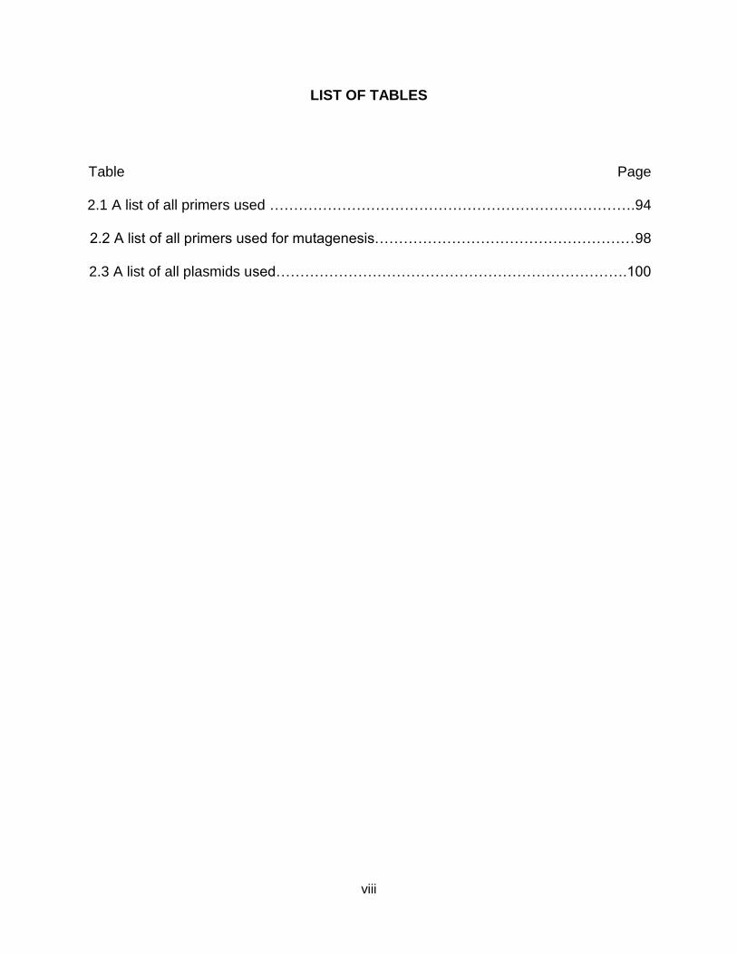
LIST OF TABLES
Table Page
2.1 A list of all primers used ………………………………………………………………….94
2.2 A list of all primers used for mutagenesis………………………………………………98
2.3 A list of all plasmids used……………………………………………………………….100

ix
LIST OF FIGURES
Figure Page
1.1 The current model of Wnt/β-catenin signaling…….…………………………………….8
1.2 Synthesis and export of Wnt ligand …………………………………...………………..11
1.3 Nuclear TCF/β-catenin transcriptional complexes………………………..…………...27
1.4 The core Notch pathway contains a limited set of components that form the signal
transmitting chain in the pathway………………………………………………………35
1.5 The structural conservation of mammalian Notch receptors…..…..…………………37
1.6 Domain organization of mammalian Notch ligands……………….…………………...41
1.7 The NICD undergoes multiple post-translational modifications………………………50
1.8 Structure and function of Wntch…………………………………………………………63
2.1 A schematic representation of the preparation of concentrated Xenopus egg
extract…………………………………………………………………………………….86
2.2 β-catenin degrades robustly when incubated in Xenopus egg extract………………87
2.3 Luciferase-tagged β-catenin degrades when incubated in Xenopus egg extract….88
2.4 Regulated degradation of -catenin-luciferase in Xenopus extract can be adapted to
a high-throughput format…………………………………………………..…………….89
3.1 hNICD1 is degraded in Xenopus egg extract…………………………………………111
3.2 hNICD1 degradation in Xenopus egg extract occurs independent of GSK3…...…113
3.3 NICD1 is degraded in Xenopus egg extract, in contrast to other NICD paralogs,
and degradation is not affected by C-terminal fusions……………………………114
3.4 PEST domain mutants of hNICD1 degrade in Xenopus egg extract…..……..……117
3.5 The N-terminal half of hNICD1 promotes hNICD1 degradation in
Xenopus egg extract……………………………………………………………………...….119

x
3.6 Mutations within the 35 amino acid N-terminal region of hNICD1 inhibit its
degradation in Xenopus egg extract………………………………………………….120
3.7 The first 10 amino acids of hNICD1 are critical for N1-Box-mediated degradation,
whereas the first 50 amino acids of hNICD1 are required to promote degradation
of heterologous proteins………………………………………………………………..123
3.8 hNICD1 N1-Box mutants have elevated steady-state levels and increased
activity in cultured human cells and zebrafish embryos.……………………………128
3.9 hNICD1 degron mutants have decreased rates of degradation in cultured
human cells……………………………………………………………………………….132
3.10 N1-Box mutants do not affect the capacities of the E3 ligases, Fbxw7 and Itch,
to regulate hNICD1 levels in cultured human cells……..…………………………134
3.11 N1-Box-mediated hNICD1 degradation in Xenopus egg extract and cultured
human cells is inhibited by its binding to CSL………………………………………137
3.12 Mutations within the N1-Box of hNotch1 in human cancers have enhanced
signaling activity………………………………….……………………………………140
3.13 Model of N1-Box-mediated regulation of hNotch1 signaling………………………143

1
CHAPTER I
INTRODUCTION TO DEVELOPMENTAL SIGNALING PATHWAYS
Introduction
The fundamental processes of metazoan development are widely conserved
throughout the animal kingdom. Among the conserved components are two critical
developmental pathways, the Wnt signaling pathway and the Notch signaling pathway.
These pathways are similar in many ways, including ligand-dependent activation of the
pathway in wild-type conditions, key transcriptional co-activators which form
transcriptional activation complexes, and a downstream effector protein in which the
stability of the protein is tightly regulated in order to regulate the transcriptional activity
of the pathway. Because these developmental pathways regulate organismal growth
and development, they can potentially be co-opted when key regulatory genes are
mutated. Both the Wnt pathway and the Notch pathway are very often misregulated in
human cancers. In this work, Chapter I sets the historical and scientific foundation for
key mechanistic questions in both the Wnt and Notch pathways which are explored in
greater mechanistic detail in Chapters III and IV. Chapters V and VI conclude with
discussion and future directions from the findings presented in this document.
Cell Signaling and Signal Transduction of Developmental Pathways
Cell signaling is a form of cellular communication in which cells interact with their
surroundings, process this information, and provided an appropriate response to these

2
signals. The process in which a cell recognizes, processes, and responds to a signal is
called signal transduction.
The process of signal transduction regulates and coordinates all metazoan
developmental processes. Metazoan organismal development is a highly regulated and
coordinated series of signal transduction events between cells and other cells which
cooperate to form a fully functional reproducing animal.
One of the earliest studies involving signal transduction and development comes
from the work of Hans Spemann in the early 20th century. In this seminal work,
Spemann, along with Hilde Mangold, showed that transplantation of the dorsal lip of the
blastopore of an amphibian embryo beginning gastrulation onto the other side of
another developmentally staged embryo resulted in the formation of two body axes in
the grafted embryo, producing a mirror image twin. One of these axes was formed by
the endogenous dorsal lip of the blastopore while the other was formed by the
transplanted dorsal lip tissue. The transplanted dorsal lip induced the formation of a
complete dorsal axis in a location that normally forms the ventral side of the embryo.
Due to its inductive potential, the dorsal lip of the amphibian blastopore was termed the
organizer (Spemann and Mangold, 1938; Spemann and Mangold, 2001). This landmark
discovery provided major evidence that cells signal to each other and that these signals
can induce cooperative growth and development. Because of this work, Spemann was
awarded the Nobel Prize in Medicine or Physiology in 1935.
Currently, 18 signal transduction pathways have been identified (Gerhart, 1999).
These signaling pathways typically have conserved structural mechanisms in order to
transduce the signal. Some type of ligand is released into the extracellular environment

3
or expressed at the surface of the siganal-sending cell and binds to a cell surface
receptor. This ligand-bound receptor gets activated. Then, the activated receptor
transduces the signal intracellularly. Finally, intracellular signal transduction occurs
through secondary messengers which send the signal through the cell via other
messengers to ultimately induce a physiological response.
Current evidence suggests that 5 of the 18 currently known signaling pathways
control developmental processes (Gerhart, 1999). As metazoans evolved multi-
cellularity, a means of signaling between these multiple cells also evolved in order to
facilitate communication between these cells. These cell-cell signaling pathways that
evolved in response to multicellularity are the developmental signaling pathways that
are conserved throughout all metazoans. Two of these developmental pathways, the
Wnt/β-catenin signaling pathway and the Notch signaling pathway, will be described in
more detail later in this document. This dissertation will focus primarily on Notch
signaling.
The description of these signaling processes as “pathways” paints an inaccurate
picture of the diversity and complexity of metazoan development. These signaling
processes, rather than being discrete, independent “pathways”, are interconnected,
forming a signaling “network”. Abundant evidence exists that there is crosstalk between
these “pathways”, in which the activation or non-activation of a receptor of one pathway
affects another pathway (van Amerongen and Nusse, 2009). In this chapter I will
describe the history and importance of Wnt/β-catenin signaling, the history and
importance of Notch signaling, and the evidence of cross-talk between the two that links
them into a signaling network.

4
Historical Significance: Wnt Signaling in Development and Disease
The canonical Wnt/β-catenin signaling pathway plays a critical role in cell fate
determination, cell proliferation, cell polarity, and cell death during embryonic
development and in tissue homeostasis in adults. The Wnt pathway is named for its
ligands, the Wnt family of secreted glycoproteins, was discovered nearly 40 years ago.
The history of Wnt/β-catenin signaling highlights the roles of this pathway in both
development and disease. Many of the details of Wnt/β-catenin signaling can be found
in other reviews [reviewed in (MacDonald et al., 2009; Saito-Diaz et al., 2013)].
In 1976, Sharma and Chopra described a Drosophila melanogaster mutant which
had absent or reduced wings and halteres, which they named wingless (wg). Based on
the mutant phenotype, they hypothesized that the wingless locus played a critical role in
development (Sharma and Chopra, 1976). This hypothesis was confirmed in 1980 when
Wieschaus and Nusslein-Volhard identified wg as a segmentation gene in a Drosophila
mutagenesis screen for gene required in segmentation(Nusslein-Volhard and
Wieschaus, 1980). For this landmark discovery in developmental biology, Nusslein-
Volhard and Wieschaus were awarded the Nobel Prize in Physiology or Medicine in
1995.
Several years later, Nusse and Varmus conducted a forward genetic screen to
identify genes which could lead to tumorigenesis. They used mouse mammary tumor
virus (MMTV) insertion sites and identified a locus termed int-1, short for integration-1,
which induced mouse mammary tumors (Nusse et al., 1984; Nusse and Varmus, 1982).
Later, comparative genomic studies identified wg and int-1 as homologs, and the name

5
was merged into the mnemonic Wnt (Nusse et al., 1991). The injection of int-1 in
Xenopus embryos induced the formation of a secondary body axis, confirming the role
of int-1 as both an oncogene and a critical component of vertebrate early axis formation
(McMahon and Moon, 1989a; McMahon and Moon, 1989b). These studies take
together suggest that the Wnt proteins play a critical role in normal development as well
as a critical role in carcinogenesis.
Drosophila mutagenesis screens (similar to the one described earlier from
Nusslein-Volhard and Wieschaus) played an important role in identifying components of
the Wnt/β-catenin signaling pathway (Nusslein-Volhard and Wieschaus, 1980). In the 15
years after that initial publication, key Wnt pathway components such as armadillo (the
Drosophila homolog of β-catenin), dishevelled (Dsh), shaggy (the Drosophila homolog
of glycogen synthase kinase 3, GSK3), frizzled (Fz), and arrow (the Drosophila homolog
of LRP5/6) (Bhanot et al., 1996; Klingensmith et al., 1994; Riggleman et al., 1990;
Riggleman et al., 1989; Siegfried et al., 1992; Wehrli et al., 2000) were identified.
The Wnt/β-catenin pathway was then linked to the formation of the Spemann-
Mangold organizer referenced earlier in this chapter (Spemann and Mangold, 1938).
Injection of Wnt-1 and XWnt8 into Xenopus blastomeres induces a secondary axis due
to a second organizer (Smith and Harland, 1991; Sokol et al., 1991). This secondary
axis formation was also phenocopied using other Wnt/β-catenin pathway components
(Dominguez et al., 1995; Fagotto et al., 1999; Guger and Gumbiner, 1995; He et al.,
1995; Sokol et al., 1995). Many of these other components were identified by their
effects on vertebrate development, such as Axin (Zeng et al., 1997), APC (Munemitsu
et al., 1995; Rubinfeld et al., 1993), and the co-receptor LRP5/6 (Pinson et al., 2000;

6
Tamai et al., 2000; Wehrli et al., 2000). All of these major Wnt components can induce
secondary axis formation in Xenopus embryos, and the axis duplication assay has
emerged as a powerful validation tool to identify bona fide regulators of Wnt/β-catenin
signaling.
Many developmental signaling pathways are also critical drivers of cell growth
and cell-cell signaling in cancer. The Wnt/β-catenin signaling pathway is no exception.
Perturbations in Wnt/β-catenin signaling lead to a large number of diseases, varying
from congenital birth defects to multiple types of cancer [reviewed in (MacDonald et al.,
2009)]. Perhaps the most well-known connection between Wnt/β-catenin signaling and
cancer is a genetic lesion in the Wnt pathway component APC that occurs colorectal
cancer. In familial adenomatous polyposis (FAP), a form of hereditary colorectal cancer
(Kinzler et al., 1991; Nishisho et al., 1991), patients missing one copy of APC lose their
second copy of APC and develop benign polyps at an early age. These polyps then
develop other mutations and lead to invasive colon carcinoma. Later, loss of both APC
alleles was linked to over 80% of sporadic, nonhereditary colorectal cancers (Kinzler
and Vogelstein, 1996). Misregulated Wnt/β-catenin signaling was then found in many
other types of cancers, including liver cancer, skin cancer, lung cancer, Wilms’ Tumor,
breast cancer, prostate cancer, and others [reviewed in (Klaus and Birchmeier, 2008)
and (Saito-Diaz et al., 2013)]. Developmental genetic defects can also result from
misregulated Wnt/β-catenin signaling (Boyden et al., 2002; Gong et al., 2001; Lammi et
al., 2004; Niemann et al., 2004; Toomes et al., 2004; Xu et al., 2004). Understanding
the molecular mechanisms governing Wnt/β-catenin signaling is critical towards both

7
understanding the pathophysiological effects of Wnt/β-catenin misregulation and
designing therapeutics against the Wnt/β-catenin signaling pathway.
The Current Model of the Wnt/β-catenin Signaling Pathway
Wnt signaling promotes a variety of cellular responses in development,
physiology, and disease. The original hypothesis was that Wnt signaling promotes these
responses by activating different transcriptional target genes in different cellular
contexts. This pathway, in which Wnt signaling activates specific transcriptional target
genes, was previously referred to as “canonical” Wnt signaling. I have referred to it as
Wnt/β-catenin signaling to distinguish it from other Wnt-mediated pathways. Other Wnt-
mediated pathways signal cytoplasmic changes involving the action cytoskeleton
(Wnt/PCP pathway) and intracellular calcium stores (Wnt/Ca2+ pathway). These other
pathways may be regulated by the tyrosine kinase receptors ROR and RYK (Nusse,
2008). In recent years, even the simplicity of the two pathway model has been
questioned (van Amerongen et al., 2008). These other pathways are outside the scope
of this document. For all intents and purposes, every reference to Wnt signaling refers
to Wnt/β-catenin signaling.
The Wnt/β-catenin signaling pathway, fundamentally, results in the cytoplasmic
protein β-catenin entering the nucleus to modulate transcription. When Wnt ligand is not
bound, β-catenin is continually degraded by the β-catenin destruction complex. The
destruction complex consists of the scaffold proteins Axin and APC and the protein
kinases GSK3 and Casein Kinase 1 (CK1) [Figure 1.1 (Saito-Diaz et al., 2013)].

8
Figure 1.1. The current model of Wnt/β-catenin signaling. (Left panel) In the absence of Wnt, cytoplasmic β-catenin forms a complex with APC, Axin, GSK3, and CK1α. β-Catenin is phosphorylated by CK1α and subsequently phosphorylated by GSK3. The phosphorylated form of β-catenin is recognized by the E3 ubiquitin ligase SCFβ-TRCP, which targets β-catenin for proteasomal degradation. In the absence of nuclear β-catenin, Wnt target genes are repressed. APC, adenomatous polyposis coli; GSK3, glycogen synthase kinase 3; CK1α, casein kinase 1 alpha. (Right panel) In the presence of Wnt ligand, a receptor complex forms between Fz, LRP5/6, and Wnt. The recruitment of Dsh by Fz leads to LRP5/6 phosphorylation by CK1α and GSK3 followed by recruitment of Axin to LRP5/6. The latter disrupts Axin-mediated phosphorylation/degradation of β-catenin, leading to accumulation of β-catenin in the cytoplasm and its translocation to the nucleus, where it acts as a transcriptional co-activator with TCF to activate Wnt-responsive target genes. Fz, Frizzled; Dsh, Dishevelled; TCF, T-cell factor [Figure from (Saito-Diaz et al., 2013)].

9
Activation of Wnt/β-catenin signaling removes APC from the complex and
relocalizes the other components to the plasma membrane via the adaptor Dsh, thus
stabilizing β-catenin which enters the nucleus to mediate transcription (Figure 1). Thus,
Wnt/β-catenin signaling can be divided into three general molecular events: (1) surface
receptor activation, (2) inhibition of the β-catenin destruction complex, and (3) activation
of a Wnt-specific nuclear transcriptional complex. The next sections of this document
consider each of these steps more closely.
Wnt/β-catenin Signaling: Surface Receptor Activation
The secreted Wnt proteins are cysteine-rich morphogens between approximately
350-400 amino acids which can act in both short-range and long-range signaling. There
are at least 19 vertebrate Wnt proteins and are capable of activating the pathway. Wnt
ligands bind to their receptor Frizzled (Fz). The structural basis of Wnt-receptor
interactions has been characterized (Janda et al., 2012). All of the Wnt ligands contain
an N-terminal signal peptide for secretion and are N-linked glycosylated (Smolich et al.,
1993; Takada et al., 2006; Willert et al., 2003). The N-glycosylation of the Drosophila
Wnt homolog Wg is stimulated by lipid modifications (Tanaka et al., 2002). Early studies
suggested that glycosylation of Wnt was dispensable for Wnt activity (Mason et al.,
1992), but more recent studies have demonstrated the requirement of glycosylation for
Wnt secretion (Komekado et al., 2007; Kurayoshi et al., 2007).
The Wnt proteins contain multiple charged amino acids and undergo lipid
modifications which are required for activity (Bradley and Brown, 1990). Wnt3a protein
(and by extension Wnts in general) is acylated with a palmitate at Cys77 and a
palmitoleate at Ser209 (Takada et al., 2006; Willert et al., 2003). Interestingly, the

10
crystal structure of the Wnt/receptor complex shows Cys77 engaged in disulfide
bonding and the palmitoleate at Ser209 docked inside a hydrophobic groove on a
cysteine-rich domain (CRD) of the receptor, playing a direct role in Wnt-receptor
interaction (Janda et al., 2012).
These lipid modifications of Wnt are mediated by an endoplasmic reticulum (ER)-
embedded, multi-pass transmembrane O-acetyl transferase known as Porcupine (Porc)
[reviewed in (MacDonald et al., 2009; Port and Basler, 2010; Saito-Diaz et al., 2013)].
Porc was initially identified in Drosophila as a segment polarity gene was the first gene
shown to be required in Wnt-secreting cells. Loss-of-function of Porc leads to
accumulation of Wnt in the ER (Kadowaki et al., 1996; van den Heuvel et al., 1993) and
overexpression of Porc results in a high percentage of Wnts that are lipid-modified (Galli
et al., 2007). The p24 family of proteins is required for modified Wnts to get transported
from the ER to the Golgi (Buechling et al., 2011; Port et al., 2011). Once in the Golgi,
the trans-Golgi seven-pass transmembrane protein Wntless (Wls transports Wnt from
the Golgi to the plasma membrane. Wls binds to the palmitoylated Ser209 which is
mediated by Porc (Herr and Basler, 2011). WIs is recycled back to the plasma
membrane via a protein complex known as the retromer. The retromer complex routes
WIs back from endosomes into trans-Golgi in a retrograde manner (Coudreuse et al.,
2006; Port and Basler, 2010). WIs gets degraded in the endosome in the absence of the
retromer complex (Yang et al., 2008). The addition of exogenous WIs bypasses the
requirement of the retromer (Franch-Marro et al., 2008; Port et al., 2008) Together,
Porc, Wls, and indirectly, the retromer complex, form a pathway critical for secretion of
Wnt ligands (Figure 1.2).

11
Figure 1.2. Synthesis and export of Wnt ligand. Wnt ligand undergoes multiple posttranslational modifications in the ER. Glycosylation and palmitoylation of Wnt ligand (the latter mediated by the transmembrane protein Porc) are required for its translocation to the Golgi apparatus. Palmitoylation of Wnt allows it to bind Wls, which provides a mechanism for transportation to the plasma membrane. The retromer complex recycles Wls from the plasma membrane back to the Golgi [Figure from (Saito-Diaz et al., 2013)].

12
The soluble Wnt ligands bind to the Frizzled (Fz) family of seven transmembrane
domain receptors, which share structural features with G-protein coupled receptors
(GPCRs). Biochemical experiments showed that Wnt binds to the CRD domain of Fz
with a binding affinity in the low nanomolar range (Bhanot et al., 1996; Hsieh et al.,
1999). Because of Fz’s topological similarity to classical GPCRs, heterotrimeric G-
protein signaling has been hypothesized as critical in transducing Wnt signaling. A link
between G proteins and Wnt signaling has been suggested in several studies. First, in
Drosophila, studies suggest that Gαo transduces signaling through Fz and interacts with
the scaffold protein Axin to promote its localization to the plasma membrane (Egger-
Adam and Katanaev, 2009; Katanaev et al., 2005). In mammalian cell culture, depletion
of Gαo and Gαq inhibited Wnt/β-catenin (Liu et al., 2005). Reconstitution experiments in
Xenopus egg extract show that Gαo, Gαq. Gαi2, and Gβγ can inhibit β-catenin
phosphorylation and turnover. Gβγ was proposed to promote GSK3 recruitment to the
membrane that enhanced low-density lipoprotein receptor-related protein 6 (LRP6)
phosphorylation and activation (Jernigan et al., 2010).
LRP5 and LRP6 are functionally redundant single pass transmembrane
receptors which serve as co-receptors of Wnt/β-catenin signaling (Pinson et al., 2000;
Tamai et al., 2000; Wehrli et al., 2000). In Drosophila wingless signaling, the lone LRP
family member is known as Arrow. Although there have been some differences in
potency, LRP5 and LRP6 were shown to be mechanistically nearly identical in the
Wnt/β-catenin signaling pathway, despite some differences during development (He et
al., 2004; Mi and Johnson, 2005). Biochemical and structural studies have shown that

13
different Wnt ligands bind to different extracellular domains of LRP5/6 (Ahn et al., 2011;
Bourhis et al., 2010; Chen et al., 2011; Cheng et al., 2011). Wnt binding to Fz and
LRP5/6 leads to the production of phosphatidylinositol 4,5-biphosphate (PIP2) (Pan et
al., 2008). The production of PIP2 has been hypothesized to promote the
oligomerization and clustering of Fz and LRP6 into “signalosomes” upon activation of
Wnt/β-catenin signaling. The in vivo physiological significance of signalosome formation
is still being investigated (Bilić et al., 2007; Cong et al., 2004b). PIP2 production also
promotes recruitment of destruction complex components to LRP5/6 on the plasma
membrane, possibly through Amer1/WTX (APC membrane recruitment 1 or Wilms
tumor gene on the X chromosome), a tumor suppressor in Wilms’ tumor which binds to
Axin and GSK3. Amer1/WTX’s recruitment to the plasma membrane is PIP2- dependent
(Major et al., 2007; Tanneberger et al., 2011). The recruitment of the destruction
complex to the plasma membrane upon Wnt binding leads to the phosphorylation of
LRP5/6 in an event known as the “initiation step” of Wnt/β-catenin signaling (Baig-Lewis
et al., 2007). LRP5/6 is phosphorylated by the destruction complex kinases GSK3 and
CK1 at PPPSPxS motifs on LRP5/6 which are both necessary and sufficient to activate
Wnt/β-catenin signaling (Davidson et al., 2005; MacDonald et al., 2009; MacDonald et
al., 2008; Tamai et al., 2004; Wolf et al., 2008; Zeng et al., 2005). The recruitment of the
concentration-limiting scaffold protein Axin (Lee et al., 2003) brings additional GSK3
and CK1 molecules to the plasma membrane during the “amplification step” (Baig-Lewis
et al., 2007). Subsequently, the activated and phosphorylated LRP6 intracellular domain
inhibits further GSK3 activity by directly binding to it (Cselenyi et al., 2008; Piao et al.,
2008; Wu et al., 2009). This GSK3 inhibition by phosphorylated LRP6 frees up β-catenin

14
from getting phosphorylated by GSK3 and targeted for ubiquitin-mediated degradation,
thus transducing the signal further downstream. The mechanistic relationship between
LRP6 and GSK3 amplification and then inhibition requires further study.
Other molecules linked to agonizing or antagonizing Wnt/β-catenin signaling
have been identified. There are two classes of secreted Wnt/β-catenin antagonists. One
class, consisting of secreted Fz-related proteins (sFRPs) and Wnt inhibitory factors
(WIFs), bind and sequester Wnt ligands and prevent their interaction with Wnt receptors
(Bovolenta et al., 2008). The other class, made up of Dkk1 and Wise/SOST members,
binds to LRP5/6 and blocks its interaction with Wnt ligands (Mao et al., 2002; Semenov
et al., 2001). Other Wnt/β-catenin agonists include Norrin and R-Spondin (Kazanskaya
et al., 2004; Kim et al., 2006; Nam et al., 2006; Wei et al., 2007; Xu et al., 2004). R-
spondin may be a driver of colorectal cancer (Seshagiri et al., 2012) and has been
shown to bind to leucine-rich repeat-containing GPCRs 4,5 and 6 (LGR4/5/6), which are
intestinal stem cell markers , but how this binding agonizes Wnt/β-catenin signaling is
still unclear (Barker et al., 2007; Carmon et al., 2011; de Lau et al., 2011; Glinka et al.,
2011; Snippert et al., 2010). Recent studies suggest that R-spondin stabilizes the
Wnt/β-catenin receptors Fz and LRP5/6 by inhibiting the activity of two E3 Ubiquitin
Ligases, RNF43 and ZNRF3, which target Fz and LRP6 for degradation (Hao et al.,
2012; Koo et al., 2012). This stabilization of Fz and LRP5/6 potentiates Wnt/β-catenin
signaling. Very recently, the type 1 transmembrane protein Tiki was identified in an
expression cloning screen that perturbed axis formation in X. laevis embryos (Zhang et
al., 2012). Tiki was identified as a novel metalloprotease that cleaves the N-terminal 8
amino acids of mature Wnt proteins which results in the formation of large, soluble

15
oligomeric Wnt complexes due to oxidation and the formation of disulfide bonds in vitro.
Whether the formation of these inactive Wnt complexes is how Tiki affects the Wnt/β-
catenin pathway in vivo is still unclear.
Another critical component of the Wnt/β-catenin pathway is the cytoplasmic
effector protein Dishevelled (Dsh). Dsh is required genetically in Drosophila wingless
signaling (Klingensmith et al., 1994) and there are 3 vertebrate paralogs encoded by 3
distinct genes (Dvl1-3) (Semenov and Snyder, 1997; Sussman et al., 1994; Yang et al.,
1996). Dsh gets phosphorylated and recruited to the cytoplasmic portion of the receptor
upon Wnt-receptor binding (Rothbacher et al., 2000; Semenov and Snyder, 1997;
Yanagawa et al., 1995). This Dsh phosphorylation is independent of LRP6 activation
(Gonzalez-Sancho et al., 2004). Dsh contains 3 known structural domains, the DEP, the
PDZ, and the DIX domains. The PDZ and DIX domains have been shown to be
important in Dsh binding to Fz (Tauriello et al., 2012; Wong et al., 2003; Wong et al.,
2000). The DIX domain is thought to polymerize and promote receptor clustering
(Schwarz-Romond et al., 2007). Though Dsh is thought to be upstream of LRP6 in the
Wnt/β-catenin pathway (Tolwinski et al., 2003) and can stimulate PIP2 production (Pan
et al., 2008), in Drosophila and Xenopus egg extracts Dsh activates Wnt/β-catenin
independently of Arrow/LRP6 (Salic et al., 2000b; Wehrli et al., 2000). In another
invertebrate species, Caenorhabditis elegans, there is a Dsh homolog but no LRP5/6
homolog, suggesting that Dsh might play a more critical role in different phyla (Phillips
and Kimble, 2009). Dsh is likely regulated by ubiquitin-mediated proteasomal
degradation through at least 3 known E3 ubiquitin ligases, the HECT-type ligases
NEDL1 and ITCH and the SCF-type ligase KLHL12 (Angers et al., 2006; Miyazaki et al.,

16
2004; Wei et al., 2012). This degradation is also mediated by the Naked2 protein as a
co-factor (Hu et al., 2010). Additionally, the deubiquitinase CYLD (encoded by the
familial cylindromatosis tumor suppressor gene) negatively regulated Wnt/β-catenin
signaling (Tauriello et al., 2010).
These Wnt/β-catenin pathway components (Wnt, Fz, LRP5/6, and Dsh) and their
regulators (Porc, WIs, sFRPs, WIFs, PIP2, ITCH, NEDL1, KLHL12, and others) combine
to form a highly regulated network of plasma membrane surface proteins that are critical
for Wnt/β-catenin signal transduction. After surface receptor activation and transduction
of the signal, the cytoplasmic β-catenin destruction complex comes into play.
Wnt/β-catenin Signaling: The β-catenin destruction complex
The β-catenin destruction complex is a macromolecular machine that efficiently
phosphorylates β-catenin and targets it for degradation. I will first describe the
molecules involved in the formation of the β-catenin destruction complex (Figure 1.1)
and follow with the current model of Wnt/β-catenin signaling pathway upon receptor
activation [reviewed in (Chen et al., 2014b; Saito-Diaz et al., 2013)].
The transcriptional regulator β-catenin, as mentioned earlier, is the primary
effector of the Wnt/β-catenin signaling pathway. In the absence of Wnt/β-catenin
signaling, the destruction complex targets β-catenin for degradation by SCFβ-TRCP, a
Skp1-Cullin-Fbox (SCF) E3 Ubiquitin Ligase complex family member. When Wnt/β-
catenin signaling is active, β-catenin degradation is inhibited and translocates from the
cytoplasm to the nucleus to activate Wnt/β-catenin signaling. There are other substrates
of the destruction complex but their physiological relevance is still unclear. β-catenin
was originally identified in Drosophila as the segment polarity armadillo as a component

17
of the adherens junction in Xenopus (McCrea et al., 1991; Nusslein-Volhard and
Wieschaus, 1980). Structurally, β-catenin contains a central core consisting of 12 helical
42 amino acid armadillo repeats which form a superhelix (Huber et al., 1997). The
unstructured N-terminal and C-terminal ends of β-catenin form dynamic interactions with
the armadillo repeats (Xing et al., 2008). These armadillo repeats form a positively
charged groove which regulates β-catenin’s interaction with other Wnt/β-catenin
pathway components (i.e. APC, Axin, TCF/Lef) as well as E-cadherin (Graham et al.,
2000; Huber et al., 1997; Huber and Weis, 2001; Xing et al., 2003; Xing et al., 2004).
The cellular signals that regulate whether newly synthesized β-catenin mediates gene
transcription or maintains the adherens junction are not well-understood. There is
substantial evidence that overexpression of cadherins inhibits Wnt/β-catenin gene
transcription and promotes localization of β-catenin to the membrane (Gottardi et al.,
2001; Heasman et al., 1994; Sadot et al., 1998; Sanson et al., 1996; Shtutman et al.,
1999; Stockinger et al., 2001). Further evidence of the interplay between cadherins and
Wnt/β-catenin signaling occurs when the proteolytic cleavage of cadherins by ADAM1-
and presinilin-1 (a subunit of γ-secretase) activates Wnt/β-catenin target gene
expression (Marambaud et al., 2002; Maretzky et al., 2005; Reiss et al., 2005; Uemura
et al., 2006). Evidence for direct crosstalk between cadherins and Wnt/β-catenin
signaling has been elusive, as E-cadherin knockdowns did not activate Wnt/β-catenin
signaling (Herzig et al., 2007; Kuphal and Behrens, 2006). These results combine to
suggest that there are two distinct pools of β-catenin, which is further supported by a
study demonstrating that β-catenin can exist as a monomer and a dimer bound to α-
catenin (Gottardi and Gumbiner, 2004). The monomeric form preferentially activates

18
Wnt/β-catenin signaling and the dimeric form preferentially binds cadherins.
Surprisingly, β-catenin’s mechanism of nuclear translocation is still unclear.
The scaffold protein Axin is a critical, concentration-limiting negative regulator of
Wnt/β-catenin signaling (Lee et al., 2003). Axin is encoded by the fused gene locus in
mice (Zeng et al., 1997). Its primary function is to serve as a scaffold for the destruction
complex by binding to the other components and bringing them into close proximity with
each other (Figure 1.1). Structural analysis has visualized the interactions between Axin
and APC (Spink et al., 2000), Axin and β-catenin (Xing et al., 2003), and Axin and
GSK3β (Dajani et al., 2003). Studies in Drosophila embryos suggest that Axin forms
oligomers in vivo, and can potentially act as a cytoplasmic anchor of Armadillo/β-
catenin and prevent nuclear translocation, thus inhibiting Wnt/β-catenin signaling
(Peterson-Nedry et al., 2008; Tolwinski and Wieschaus, 2001). Axin is found at low
concentrations and serves as the concentration-limiting component of destruction
complex formation in Xenopus (Lee et al., 2003). The concentration of Axin plays a
critical role in creating specificity for Wnt/β-catenin signaling as many component of the
destruction complex play roles in other signaling pathway (i.e. GSK3) (Forde and Dale,
2007; Lee et al., 2003). Due to the critical nature of Axin concentration on Wnt/β-catenin
signaling, Axin protein levels are very highly regulated. GSK3 phosphorylation inhibits
Axin degradation (Yamamoto et al., 1999), and studies in Xenopus egg extract and in
Drosophila show that APC is required for Axin turnover, likely due to compensatory
regulation due to fluctuation in APC protein levels (Lee et al., 2003). Axin stability is
regulated by the E3 Smad ubiquitin regulatory factor 2 (Smurf2) (Kim and Jho, 2010)
and the poly(ADP-Ribose) Polymerase (PARP) Tankyrase, which poly(ADP-ribosy)lates

19
(PARsylates) Axin through the addition of poly(ADP-Ribose) moieties to promote the
ubiquitination and degradation of Axin through its poly(ADP-Ribose) moieties (Huang et
al., 2009a). The discovery of two distinct Tankyrase inhibitors, IWR-1 and XAV939,
which stabilize Axin and inhibit Wnt/β-catenin signaling further confirms the importance
of Axin protein levels (Chen et al., 2009; Huang et al., 2009a). These tankyrase
inhibitors act by inhibiting Axin PARsylation and thus inhibiting Axin turnover. Recently,
two separate groups have identified RNF146 as the poly(ADP-Ribose)-directed E3
ubiquitin ligase that ubiquitinates and targets Axin for degradation (Callow et al., 2011;
Zhang et al., 2011). RNF146 directly binds to poly(ADP-Ribose) and maintains low
steady-state levels of Axin. In addition, the deubiquitinase ubiquitin-specific protease 34
(USP34), catalyzed the deubiquitination of Axin and increases its steady-state levels in
cells (Lui et al., 2011). Axin can also be stabilized by SUMOylation at its C-terminus
which inhibits ubiquitination (Kim et al., 2008). Very recently, quantitative measurements
of Axin protein levels in a large panel of mammalian cells suggest that Axin protein
levels dynamically regulate the dynamics of Wnt/β-catenin signaling (Tan et al., 2012).
These results combine to strongly suggest that regulating Axin protein levels is likely a
major mechanism for regulating Wnt/β-catenin signaling.
The serine/threonine kinase GSK3 is a critical regulator of β-catenin degradation.
GSK3 is widely expressed and plays a role in many different cellular processes (Forde
and Dale, 2007) and inhibition of GSK3 activity is critical for activation of Wnt/β-catenin
signaling in all paradigms. The Drosophila homolog of GSK3 is called shaggy, or zeste
white 3 (Siegfried et al., 1992). Mammals have two distinct GSK3 genes, α and β, which
are functionally redundant in Wnt/β-catenin signaling (Doble et al., 2007). GSK3 gets its

20
name from its initial discovery in glucose metabolism as a kinase for glycogen synthase
(Embi et al., 1980). GSK3 usually requires its substrates to be phosphorylated (or
primed), and thus often acts in concert with other kinases. GSK3 phosphorylates β-
catenin at Ser33, Ser37, and Thr41 and this phosphorylation is required for β-catenin
degradation (Peifer et al., 1994; Yost et al., 1996). The structure of GSK3β contains an
activation loop which gives it its priming mechanism and a bilobed topology including a
β-sheet domain linked to a C-terminal α-helix domain (Dajani et al., 2001; Haar et al.,
2001). GSK3 activity is also regulated by an auto-inhibitory phosphorylation at Ser9
which blocks access to the catalytic site (Cross et al., 1995; Dajani et al., 2001). GSK3
phosphorylates other components of the Wnt/β-catenin pathway in addition to β-catenin
(Rubinfeld et al., 1996; Willert et al., 1999; Zeng et al., 2005).
The priming kinase that acts in concert with GSK3 to regulate Wnt/β-catenin
signaling is CK1α. The CK1 family of kinases has seven different paralogs encoded by
7 distinct genes (α, β, γ1, γ2, γ3, δ, and ε) (Knippschild et al., 2005; Price, 2006).
Similar to GSK3, CK1 is widely expressed and plays important roles in multiple cellular
processes. All the CK1 family members have highly similar catalytic domains, but the
length and sequence of their C-terminal non-catalytic domains differ significantly. CK1α,
which contains a short (~24 amino acid) C-terminal domain, appears to be an outlier
compared with the other family members, which contain longer C-terminal tails (~200
amino acids). CK1α, γ, δ, and ε are thought to be positive regulators of the Wnt/β-
catenin pathway through phosphorylation of pathway components (Cong et al., 2004a;
Gao et al., 2002; Kishida et al., 2001; Lee et al., 2001; Peters et al., 1999; Sakanaka et
al., 1999; Swiatek et al., 2004; Yanagawa et al., 1995; Zeng et al., 2005; Zhang et al.,

21
2006). Some CK1 paralogs are also thought to negatively regulate the Wnt/β-catenin
pathway (Gao et al., 2002; Hammerlein et al., 2005; Kishida et al., 2001; Liu et al.,
2002; Rubinfeld et al., 2001). CK1α phosphorylates β-catenin at Ser45 and serves as
the priming kinase for GSK3 at the destruction complex (Liu et al., 2002). Two separate
genome-wide S2 Drosophila RNAi screens identified CKIα as critical to suppress Wnt/β-
catenin signaling, which is consistent with the dual kinase (priming kinase followed by
processive kinase) model at the destruction complex (DasGupta et al., 2005; Lum et al.,
2003). Consistent with this model, CK1α activation by the antihelminthic drug pyrvinium
strongly inhibited Wnt/β-catenin signaling by enhancing β-catenin phosphorylation and
subsequent degradation (Thorne et al., 2010).
The 2843 amino acid scaffold protein APC, which is 310 kDa, acts as a negative
regulator of Wnt/β-catenin signaling. The gene was first identified as a mutation site in
FAP, a familial form of colon cancer (Kinzler et al., 1991). Like β-catenin, APC plays
many cellular roles and which likely occur due to different subpopulations of protein
(Faux et al., 2008). The C-terminal region of APC regulates microtubule dynamics in
mitosis and cell migration through binding to EB1 and Discs large (Matsumine et al.,
1996; Su et al., 1995), though this function is independent of Wnt/β-catenin signaling
(Nathke, 2006). APC binds to β-catenin and mutations in APC increased β-catenin
protein levels in cancer cells (Rubinfeld et al., 1993; Su et al., 1993). APC also binds to
GSK3 and Axin (Fagotto et al., 1999; Ikeda et al., 1998; Itoh et al., 1998; Rubinfeld et
al., 1996). In fact, overexpression of the concentration-limiting protein Axin can
compensate for the loss of APC (Lee et al., 2003). Additionally, a mutant form of Axin
which can’t bind to APC can still inhibit Wnt/β-catenin signaling similarly to wild-type

22
Axin, suggesting that APC isn’t strictly required for Wnt/β-catenin inhibition in the
presence of Axin (Hart et al., 1998). Unfortunately, APC’s precise mechanistic role in
regulating Wnt/β-catenin signaling is still unclear and several different models have
been proposed [reviewed in (Cadigan and Peifer, 2009; Chen et al., 2014b; MacDonald
et al., 2009; Saito-Diaz et al., 2013)]. None of the proposed models are mutually
exclusive and the strongest evidence supports APC’s role in regulating the steady state
levels of cytoplasmic β-catenin, but it is very likely that APC plays multiple roles in the
Wnt/β-catenin signaling pathway similar to several other components of the pathway
(Chen et al., 2014b; Saito-Diaz et al., 2013). APC is regulated by post-translational
modifications such as phosphorylation (Morin et al., 1997; Rubinfeld et al., 1996; Salic
et al., 2000b) and ubiquitination. The E3 ubiquitin ligase that targets APC for
degradation is still unknown. APC has, however, been linked to two deubiquitinases: the
COP9 signalosome-associated deubiquitinase, USP15, which stabilizes APC and binds
the destruction complex(Huang et al., 2009b), and Trabid, which removes K63-linked
ubiquitin chains from APC and acts as a positive regulator of Wnt/β-catenin signaling
(consistent with APC being a negative regulator of Wnt/β-catenin signaling) (Tran et al.,
2008). The mechanism of K63-linked ubiquitin chains regulating APC is still unknown.
All the previous destruction complex components mentioned have been
reconstituted biochemically and are considered “core” components of the destruction
complex. Some molecular studies have identified other components which may also be
a part of the destruction complex which have not been confirmed biochemically. One of
these is the heterotrimeric phosphatase PP2A. Multiple studies in multiple systems have
implicated PP2A as both an activator of Wnt/β-catenin signaling (Hsu et al., 1999;

23
Ratcliffe et al., 2000; Willert et al., 1999) but also as an inhibitor of Wnt/β-catenin
signaling (Gao et al., 2002; Li et al., 2001; Seeling et al., 1999). It is likely that PP2A,
similar to GSK3 and CK1, can both activate and inhibit the Wnt/β-catenin in a context-
dependent manner. Presenilin 1 (PS1), the catalytic subunit of γ-secretase, a protease
critical in both Notch signaling and Alzheimer’s disease, has been shown to inhibit
Wnt/β-catenin signaling (Kang et al., 2002; Killick et al., 2001). PS1 appears to function
as an alternative scaffold to Axin to promote GSK3 phosphorylation of β-catenin and
uses Protein Kinase A (PKA) as a priming kinase instead of CK1 (Kang et al., 2002).
Interestingly, PS1’s ability to promote β-catenin degradation is dependent on E-
cadherin, possibly linking the hypothesized two pools of β-catenin described earlier
(Serban et al., 2005). Other proteins that have been implicated in regulating Wnt/β-
catenin signaling through the destruction complex include PP2C, PP1, Amer1/WTX, and
the ankyrin protein Diversin (Itoh et al., 2009; Luo et al., 2007; Major et al., 2007;
Schwarz-Romond et al., 2002; Strovel et al., 2000).
The β-catenin destruction complex is evolutionarily conserved from metazoans to
humans. Even though it is traditionally considered a cytoplasmic complex, it has also
been found functional in the nucleus (Bienz, 2002; Cong and Varmus, 2004; Sierra et
al., 2006; Wiechens et al., 2004). The complex is constitutively active, with cells
constantly cycling between synthesis and degradation of β-catenin. On the surface, this
appears to be a futile cycle of synthesis and degradation. However, the existence of
these futile cycles in signaling is thought to be critical for more diverse modulation of
these signals, allowing for complex behaviors such as stochastic bistability (Samoilov et
al., 2005). Axin nucleates the formation of the complex by binding to GSK3, CK1, and

24
APC. These interactions have already been mapped (Dajani et al., 2003; Sobrado et al.,
2005; Spink et al., 2000). β-catenin then binds to APC and Axin and enters the
assembled complex. The kinetics of complex formation and whether it is stochastic or
ordered are still unclear (Lee et al., 2003). The phosphorylation of Axin by GSK3 and of
APC by CK1 and GSK3 increases their respective affinities for β-catenin (Ha et al.,
2004; Willert et al., 1999). The N-terminal region of β-catenin, upon Axin binding,
becomes positioned for phosphorylation by CK1 at Ser45. This priming phosphorylation
leads to subsequence successive phosphorylation at Thr41, Ser37, and Ser33 (Amit et
al., 2002; Liu et al., 2002). Phosphorylated APC competes β-catenin off of Axin and thus
allows for a new β-catenin molecule to bind Axin, continuing the cycle (Kimelman and
Xu, 2006). APC phosphorylation may also prevent the action of PP2A on β-catenin (Su
et al., 2008). GSK3 phosphorylation of β-catenin causes recognition of β-catenin by β-
TRCP, a recognition subunit of SCF complex E3 ubiquitin ligases (Jiang and Struhl,
1998; Kitagawa et al., 1999; Lagna et al., 1999; Liu et al., 1999; Marikawa and Elinson,
1998). SCFβ-TRCP directly catalyzes the polyubiquitination of β-catenin (via K48 linkages)
and its subsequent proteasome-mediated degradation. This degradation ensures low
steady state levels of β-catenin are maintained to prevent aberrant signal transduction.
One recent study shows that the HECT domain E3 Ligase EDD ubiquitinates β-catenin
and prevents its degradation (Hay-Koren et al., 2010). The full physiological significance
of EDD ubiquitination still needs to be elucidated.
The constitutively active β-catenin destruction complex becomes inhibited upon
Wnt binding and receptor activation and β-catenin protein levels increase. The actual
mechanistic details are still being investigated but the central unifying principle in all

25
proposed models is the inhibition of GSK3 enzymatic activity. These models are as
follows: 1) Dissociation of the destruction complex upon Wnt activation, 2) Inhibitory
phosphorylation of GSK3 at Ser9, 3) LRP6 Binding and direct inhibition of GSK3, 4)
Axin degradation upon Wnt activation which prevents formation of the complex and 5)
global inhibition of GSK3 through sequestration into multi-vesicular bodies (MVBs). The
details of each individual model are reviewed in [(Saito-Diaz et al., 2013) and (Chen et
al., 2014b)]. The inhibition of GSK3 and thus the β-catenin destruction complex allows
for the accumulation of β-catenin in the cytoplasm where it eventually translocates to
the nucleus and activates a Wnt/β-catenin specific transcriptional response. As
mentioned earlier, β-catenin serves as the main effector of the pathway and transduces
the signal into the nucleus.
Wnt/β-catenin signaling: Transcriptional activation
β-catenin accumulation in the cytoplasm, and subsequently the nucleus, was
widely considered the driving force of Wnt/βcatenin signaling. Contrary to the prevailing
model, recent studies have shown that the fold change, rather than the absolute
concentration increase, of β-catenin activates Wnt/β-catenin signaling (Goentoro and
Kirschner, 2009). The same group also showed that an approximately 2 fold change in
β-catenin levels is sufficient to activate Wnt/β-catenin signaling (Goentoro et al., 2009).
β-catenin does not contain any classical nuclear localization signals (NLS) or nuclear
export signals (NES) and how its localization is regulated is still under intense
investigation. Once it gets into the nucleus, β-catenin interacts and acts as a co-factor
with the TCF/LEF family of transcription factors which are critical for Wnt/β-catenin
signal transduction (Behrens et al., 1996; Molenaar et al., 1996). TCF, in the absence of

26
β-catenin, interacts with the co-repressor Groucho/transduction-like enhancer
(Gro/TLE1-3) to repress gene transcription. TCF binds to the DNA at a Wnt-responsive
element (WRE), of which there are over 6000 in a colon cancer cell line that regulate
the transcription of 300-400 genes (Hatzis et al., 2008). These TCF proteins are
regulated by post-translational modifications such as phosphorylation (Hammerlein et
al., 2005; Hikasa et al., 2010; Hikasa and Sokol, 2011; Ishitani et al., 2003; Ishitani et
al., 1999; Lee et al., 2001; Lo et al., 2004; Smit et al., 2004) and ubiquitination (Yamada
et al., 2006). There is also evidence of a deubiquitinase USP4 regulating TCF4 (Zhao et
al., 2009). In the classical model of Wnt/β-catenin signaling, the displacement of
Gro/TLE by β-catenin causes TCF/LEF to switch from a repressor to a transcriptional
activator. This was originally thought to be due to direct displacement (Daniels and
Weis, 2005), but recent studies show that the X-linked inhibitor of apoptosis (XIAP)
monoubiquitinates Gro/TLE and decreases its affinity for TCF/LEF, thus allowing β-
catenin to bind TCF/LEF (Hanson et al., 2012).

27
Figure 1.3. Nuclear TCF/β-catenin transcriptional complexes. Upon Wnt/β-catenin signaling, DNA-bound TCF/β-catenin recruits many other transcriptional complexes to Wnt target genes. Dotted lines represent interactions between the transcriptional complexes and β-catenin. During active Wnt target gene transcription, the co-repressor Gro/TLE cycles on and off of β-catenin in an XIAP-dependent manner with the other transcriptional complexes. Gro/TLE, Groucho/transducin-like enhancer of split [Figure from (Saito-Diaz et al., 2013)].

28
β-catenin binds to the nuclear transcriptional co-factors BCL9 and Pygopus
(Pygo) to mediate Wnt/β-catenin pathway-specific transcription (Figure 1.3) (Belenkaya
et al., 2002; Parker et al., 2002; Thompson et al., 2002). Pygo, BCL9, TCF, and β-
catenin represent a core transcriptional complex required for Wnt/β-catenin transcription
(Fiedler et al., 2008; Schwab et al., 2007; Sustmann et al., 2008). In addition, β-catenin
also interacts with multiple proteins involved in chromatin remodeling (Mosimann et al.,
2009; Willert and Jones, 2006). Wnt/β-catenin signaling requires responses at the
plasma membrane, in the cytoplasm via the destruction complex, and in the nucleus via
the β-catenin transcriptional complex. The Notch signaling pathway, the other pathway I
will be describing, shares many features in common with the Wnt/β-catenin pathway,
including a role for ligand-receptor interactions at the membrane, cytoplasmic regulatory
events on the primary effector of the pathway, and required transcriptional complex
formation in the nucleus leading to transcriptional activation. In fact, the Wnt/β-catenin
and the Notch pathway have extensive cross-talk and there is evidence that activation
of one pathway can regulate the activation of the other pathway. These will be
discussed later on in this chapter.
29
Historical Significance: Notch Signaling in Development and Disease
The canonical Notch signaling pathway is a highly conserved developmental
signaling pathway critical in cell fate determination through lateral inhibition,
differentiation, proliferation, cell death, and neuronal development in developing
embryos and stem cell and tissue maintenance in adults. The Notch pathway is named
for its family of single transmembrane Notch receptors. The Notch gene was first
identified by John Dexter in the lab of Thomas Hunt Morgan who noticed a notched
wing phenotype in Drosophila melanogaster (Dexter, 1914). A few short years later,
Morgan identified the mutant alleles (Mohr, 1919; Morgan, 1917; Morgan and Bridges,
1916). Details about the Notch pathway can be found in several excellent reviews
[reviewed in (Fortini, 2012; Kopan, 2010)].
The following decades yielded genetic data indicating that the Notch locus was
X-linked and had extremely complex allelic interactions [reviewed in (Artavanis-
Tsakonas and Muskavitch, 2010)] leading to multiple speculative hypotheses on its
biochemical nature (Foster, 1973; Thorig et al., 1981a; Thorig et al., 1981b). The Notch
gene was identified as a “neurogenic” mutation in Drosophila in the 1980s, linking the
mutation to developmental phenotypes (Lehmann et al., 1983). Further confirming the
importance of Notch signaling in development, Nusslein-Volhard and Weischaus
conducted a series of Drosophila mutagenesis screens for embryonic phenotypes
yielded six loci that were later identified as core components in the Notch signaling
pathway (Nüsslein-Volhard et al., 1984; Nusslein-Volhard and Wieschaus, 1980). In the
mid-1980s, Spyros Artavanis-Tsakanos and Michael Young independently cloned the
Notch receptor and identified it as a single-pass transmembrane receptor and attributed

30
its wing-notching phenotype to gene haploinsufficiency (Artavanis-Tsakonas et al.,
1983; Kidd et al., 1986; Kidd et al., 1983; Wharton et al., 1985). Notch was
subsequently cloned in other organisms, including C. elegans and Xenopus (Austin and
Kimble, 1987; Coffman et al., 1990; Greenwald et al., 1987). These initial studies
provided insight into the role of Notch signaling in multiple fields of biology, including
developmental and stem cell biology, neuroscience, and cancer biology (Fortini et al.,
1993).
The Notch locus in Drosophila and was shown to be both pleiotropic and
haploinsufficient. Notch loss-of-function mutations lead to a change in cell fate from
dermoblasts to neuroblasts in Drosophila embryos. One of the major insights from the
experiments in Drosophila embryos was the apparent necessity for the Notch-sending
and Notch-receiving cells to be adjacent to each other (Doe and Goodman, 1985;
Greenspan, 1990). Further studies in other tissues and other animals confirmed Notch’s
broad pleiotropic effect and its requirement in signaling between neighboring cells.
Proper regulation of Notch pathway is critical in nearly all cell fate decisions made
between neighboring cells and this pleiotropic effect can be extended to multiple
developmental processes including differentiation, proliferation, and apoptosis. The
particular developmental process affected by Notch affects in a specific tissue is likely
context-dependent, but it is clear that very tight regulation of Notch activity is critical for
determining cell fates in adjacent cells. The Notch pathway is very sensitive to dosage
effects, as loss-of-function and gain-of function mutations in Notch can often lead to the
“same phenotype”. Notch signaling has also been associated with stem cell
maintenance and proliferation (Austin and Kimble, 1987). Stem cell maintenance and

31
differentiation is dependent on cell-cell communication between stem cells and their
surrounding environment, or niche, and as mentioned above, Notch is critical for
processes that require cell-cell communication. The list of tissue-specific stem cells
regulated by proper Notch signaling is expanding rapidly (Liu et al., 2010). In fact, many
Notch reviewers have termed Notch a “stem cell pathway” because of its extensive
involvement in stem cell biology (Brack et al., 2008; Casali and Batlle, 2009; Dreesen
and Brivanlou, 2007; Farnie and Clarke, 2006).
Developmental pathways are often misregulated in cancers due to their critical
roles in cellular growth, differentiation, proliferation, and cell-cell signaling. The Notch
pathway, similar to the Wnt pathway, is misregulated in many types of cancers. Perhaps
the most well-characterized link between the Notch signaling pathway and
tumorigenesis is from studies on the molecular mechanisms underlying T-cell acute
lymphoblastic leukemia (T-ALL). Gain-of-function mutations in the Notch pathway were
first identified in cancer in the early 1990s (Ellisen et al., 1991; Gallahan and Callahan,
1997; Gallahan et al., 1987; Jhappan et al., 1992; Reynolds et al., 1987). These were
the first human homologs of the Drosophila Notch gene and they were identified as a
chromosomal translocation within T-ALL patients (Ellisen et al., 1991; Reynolds et al.,
1987). In the original study, four out of the 40 T-ALL patients had this mutation, that
results in a dominant active, ligand-independent NOTCH1 receptor, which was termed
TAN1 for translocation-associated Notch homolog. This discovery was the first direct
link between Notch signaling and human cancer. A few years later, experiments using
murine bone marrow (BM) reconstitution showed that TAN1 was causative for disease
development. Mice transplanted with TAN1-expressing BM progenitors developed T cell

32
neoplasms two weeks after BM transplantation (Pear et al., 1996). This evidence was
supported by in vitro studies and in vivo studies (Capobianco et al., 1997; Girard et al.,
1996). It wasn’t until the early 2000s, however, when Aster and colleagues identified
activating mutations in NOTCH1 were present in over 50% of all T-ALL patient cases
(Weng et al., 2004). NOTCH1 mutations were later identified in many other types of
hematopoietic tumors as well as solid tumors [reviewed in (Ntziachristos et al., 2014;
South et al., 2012). Interestingly, the Notch pathway has both oncogenic and tumor
suppressive roles in human cancers in a context-dependent manner , including breast
cancer, lung cancer, skin cancer, liver cancer, colorectal cancer, glioblastoma, AML,
CLL, and others (Balint et al., 2005; Fabbri et al., 2011; Klinakis et al., 2011; Licciulli et
al., 2013; Qi et al., 2003; Sun et al., 2014; Villanueva et al., 2012; Wang et al., 2011;
Weng et al., 2004). Unsurprisingly, Notch signaling is also often misregulated in
congenital developmental diseases consistent with its role in progenitor cell regulation
(Eldadah et al., 2001; Garg et al., 2005; Joutel et al., 1996; Li et al., 1997; McDaniell et
al., 2006; Oda et al., 1997; Simpson et al., 2011; Sparrow et al., 2006). Due to the
Notch pathway’s critical roles in both development and disease, understanding the
molecular mechanisms governing Notch signaling is critical for our understanding of the
pathophysiological effects of Notch pathway misregulation and for designing
therapeutics against the Notch signaling pathway. Unfortunately, many of the molecular
mechanisms of the Notch pathway have not been fully elucidated biochemically and
many of the present therapeutics for the Notch pathway have proven unsuccessful
when taken to clinical trials due to non-therapeutic Notch-mediated effects in the GI
tract and the formation of skin cancers.

33
The Current Model of the Notch Signaling pathway
The most widely characterized pathway initiated by the classical Notch-ligand
interaction is generally referred to as “canonical” Notch signaling. Other Notch-
dependent signaling pathways can occur independently of the processes and molecules
required for the classical Notch pathway or through cross-talk with other pathways (such
as the Wnt/β-catenin pathway). These other Notch pathways are referred to as “non-
canonical” Notch signaling. Details on non-canonical Notch signaling has been reviewed
elsewhere [reviewed in (D'Souza et al., 2010; Heitzler, 2010)] and is outside the scope
of this document. Unless specifically referred to as non-canonical, all references to
Notch signaling refer to canonical Notch signaling.
The Notch signaling pathway, at its core, results in the generation and
translocation of the Notch Intracellular Domain (NICD) into the nucleus to activate a
Notch-specific transcriptional program. In the core Notch signaling pathway, the Notch
transmembrane receptor (existing as a heterodimer) on a signal-receiving cell interacts
extracellularly with the canonical Notch pathway ligands Delta/Serrate/Lag-2 (DSL) on a
neighboring signal-sending cell. This ligand-receptor interaction initiates an ADAM 10
metalloprotease proteolysis (S2) which allows the remaining Notch receptor to be
proteolyzed by the ubiquitously expressed protease γ-secretase (S3). This γ-secretase
proteolysis generates the release of NICD. The S3 proteolysis can occur at the plasma
membrane or in the early endosome, as γ-secretase is present at both cellular
compartments. Recent studies suggest that γ-secretase is more active at the low PH of
the early endosome. The stability of the NICD is regulated but very little is known about
how NICD stability is regulated. Finally, the NICD translocates to the nucleus and binds

34
to the transcriptional co-activator C-promoter binding factor1 [CBF1 (also known as
recombination signal binding protein for immunoglobulin kappa J region (RBPJ-κ)]. In
Drosophila, CBF1 is known as Suppressor of Hairless (Su(H)) and in C. elegans,
Longevity-assurance gene-1 (LAG-1). Collectively, this transcriptional co-factor is called
CSL (for CBF1/Su(H)/LAG-1). The transcriptional complex, consisting of NICD, MAM,
and CSL is thought to activate a canonical Notch-mediated transcriptional program
[Figure 1.4, adapted from (Andersson et al., 2011)].

35
Figure 1.4. The core Notch pathway contains a limited set of components that form the signal transmitting chain in the pathway: a ligand (light blue), a Notch receptor (green and red) and the transcription factor CSL (green). In addition, some components (Furin (not shown), ADAM 10 secretase (green lightning bolt), γ-secretase (yellow lightning bolt) and MAML (blue oval)) are not part of conveying the signal but are nevertheless crucial for allowing the signal to be transmitted from one step to the next in the pathway. Briefly, the Notch receptor is synthesized as a single transmembrane receptor that is Furin cleaved to yield a bipartite heterodimeric Notch receptor, which is presented on the cell surface of a ‘receptor-expressing’ cell. This receptor can be activated at the plasma membrane by binding to Notch ligands on ‘ligand-expressing’ cells. This leads to the removal of the extracellular domain of Notch, which is then targeted for lysosomal degradation. The remaining portion of the receptor, termed the Notch extracellular truncated (NEXT) domain, undergoes sequential cleavage by ADAM secretases and γ-secretase as it becomes endocytosed, yielding the Notch intracellular domain (NICD). NICD then translocates to the nucleus where it binds the DNA-binding protein CSL (CBF1/Suppressor of Hairless/LAG-1) and activates the transcription of Notch target genes [adapted from (Andersson et al., 2011)].

36
The Notch pathway is very unusual among signal transduction pathways
because there is no evidence of an amplification step in between receptor activation
and transcriptional activation. The Notch receptor also serves as the transcriptional co-
activator rather than acting through another effector protein. Interestingly, each
activated Notch receptor generates one NICD and is consumed during the signal
transduction process, making the Notch pathway particularly sensitive to gene dosage.
The core Notch signal transduction pathway can be broken down into three distinct
molecular events: 1) Notch surface receptor activation, 2) Regulation of the NICD, and
3) Activation of a Notch-specific nuclear transcriptional complex by NICD. The next
sections of this document will describe each of these steps in greater detail as well as
how the non-core components regulate the Notch pathway.
Notch Signaling: Surface Receptor Activation
The Notch receptor is a family of single pass trans-membrane receptors that is
critical in cell-cell signaling. In mammals, there are 4 Notch receptors (called Notch1-4)
[reviewed in (D'Souza et al., 2010)]. All of the mammalian Notch receptors share
structural similarities (Figure 1.5). These receptors contain many of the same structural
motifs and are likely to be regulated in a similar fashion. These Notch receptors bind to
the cell surface expressed Delta-Serrate-LAG-2 ligands (Jagged1, Jagged 2, Delta-like
1 (Dll1), Delta-like 3 (Dll3), and Delta-like 4 (Dll4)) (Figure 1.6).

37
Figure 1.5. The structural conservation of mammalian Notch receptors. Diagrammatic representation of the four known mammalian receptors. EGF: epidermal growth factor; HD: heterodimerization domain; ICN: intracellular domain; LNR: cysteine-rich LNR repeats; TM: transmembrane domain; RAM: RAM domain; NLS: nuclear localizing signals; ANK: ankyrin repeat domain; NCR: cysteine response region; TAD: transactivation domain; PEST: region rich in proline (P), glutamine (E), serine (S) and threonine (T) residues [adapted from (Pancewicz and Nicot, 2011)].

38
The Notch receptors are translated as pre-proproteins and are cleaved by a
Furin-like protease (S1) in the trans-Golgi network and then trafficked to the plasma
membrane. This cleaved heterodimer is prsented on the cell surface composed of a
large extracellular domain non-covalently linked to the intracellular domain (Blaumueller
et al., 1997; Logeat et al., 1998). The extracellular domain of all Notch receptors
contains different numbers of epidermal growth-factor-like repeats (EGF-like) (36 for
Notch1 and Notch2, 34 for Notch3, and 29 for Notch4) which are critical in ligand-
receptor binding (Fig 1.5). A subset of the EGF-like repeats are calcium-binding EGF-
like repeats (cbEGF), that are required for ligand binding. Genetic experiments in
Drosophila and cell aggregation assays have identified EGF-like repeats 11 and 12 on
Notch as the major ligand-binding site. This region binds in a calcium-dependent
manner but does not have full functionality, suggesting that other EGF-like repeats likely
also contribute to ligand binding (Rebay et al., 1993; Rebay et al., 1991). Structural
studies later indicated that EGF-like 12, via an interaction between an aromatic residue
(Y/F/W) and a both a hydrophobic residue (I/L/V/P) with a glycine is the critical binding
pocket for the Notch ligand in a calcium-dependent manner (Cordle et al., 2008;
Hambleton et al., 2004). Additionally, the relative strength of receptor-ligand binding can
be modulated by post-translational modifications of these EGF-like repeats. O-
glycosylation of the EGF-like repeats, including the addition of O-fucose and O-glucose
on EGF-like repeat 12, primes the Notch receptor for further modification (Stanley and
Okajima, 2010). The addition of O-fucose is mediated by O-fucosyltransferase 1
(Pofut1) and is not directly required for Notch signaling (Okajima et al., 2008) but is
required for subsequent glycosylation of Notch receptors by the glycosyltransferase

39
Fringe proteins (Lunatic Fringe, Manic Fringe, and Radical Fringe in mammals). The
Fringe proteins add N-acetylglucosamine (GlcNAc) sugars to the O-fucose moiety and
modulate ligand-receptor binding by increasing the affinity for Delta-like and decreasing
affinity for Serrate-like ligands (Hicks et al., 2000; Kato et al., 2010; Okajima et al.,
2003). The expression domains of Fringe genes often coincides with either Delta or
Jagged but not both and this expression often leads to active Notch signaling only at the
margins in between the Delta and Jagged expressed regions (Irvine and Wieschaus,
1994; Marklund et al., 2010; Wu and Rao, 1999). A substitution in the O-fucose site on
EGF-like repeat 12 led to decreased Notch signaling (Ge and Stanley, 2008; Lei et al.,
2003). Notch can also be glycosylated by the glycosyltransferase Rumi (Poglut1) and
this glycosylation is required for Notch signaling (Acar et al., 2008; Fernandez-Valdivia
et al., 2011). Two enzymes of the glycosyltransferase 8 family also glycosylate the
Notch receptor (Sethi et al., 2010). Recently, a secreted Fringe protein, chondroitin
sulfate synthase 1(CHSY1) was identified as a negative regulator of Notch signaling
(Tian et al., 2010). It is clear that glycosylation plays a critical role in regulating Notch
signaling through regulation of receptor-ligand binding, but further structural, molecular,
and biochemical studies are still needed to identify the individual contributions of each
glycosylation event.
The extracellular domain of the Notch receptor also contains three LIN-12-Notch
(LNR) repeats and a hydrophobic region known to mediate heterodimerization (HD).
Those two regions, along with the S1 cleavage site (Furin-mediated) and the S2
cleavage site (ADAM 10/17 metalloprotease-mediated) comprise the negative
regulatory region (NRR) of the Notch receptor. The NRR normally autoinhibits the S2

40
cleavage by sterically blocking access to the S2 cleavage site through a hydrophobic
core (Gordon et al., 2009). Mutations in the NRR which mutate the hydrophobic core
and cause ligand-independent Notch receptor activation are the most common mutation
in T-ALL, a Notch-mediated cancer (Malecki et al., 2006; Weng et al., 2004). The
autoinhibitory conformation of the NRR is calcium-dependent, as depletion of calcium
activates the receptor (Rand et al., 2000). The autoinhibitory effect of the NRR is
relieved by a conformational change in the Notch heterodimer, through ligand
endocytosis in response to ligand-receptor binding.
The 5 canonical mammalian Notch ligands (Jagged1, Jagged2, Dll1, Dll3, Dll4)
can be divided into two general classes, a Delta/Delta-like family and a Serrate/Jagged
family. All of the ligands have a Notch-binding site within a DSL domain as well as N-
terminal domain and EGF-like repeats. The Jagged family of ligands also contains a
cysteine rich domain (CRD) and Jagged1, Jagged2, and Dll1 all contain two Delta and
OSM-11-like proteins (DOS) domains (Figure 1.6).

41
Figure 1.6. Domain organization of mammalian Notch ligands. Five mammalian ligands are classified into two categories, Delta-like (Dll1, Dll3, Dll4) and Serrate-like (Jagged1, Jagged2), based on structural homology to the two Drosophila ligands, Delta and Serrate. All Notch ligands have an N-terminal domain, a DSL (Delta/Serrate/LAG-2) domain and EGF-like repeats. Jagged1 and Jagged2 contain a cysteine-rich domain, whereas Jagged1, Jagged2 and Dll1 have two DOS (Delta and OSM-11-like proteins) domains located immediately following the DSL domain. DSL ligands are transmembrane proteins of which the extracellular domain contains a characteristic number of EGF-like repeats and a cysteine rich N-terminal DSL domain. The DSL domain is a conserved motif found in all DSL ligands and required for their interaction with Notch [adapted from (Kume, 2012)].
42
To date, there is little evidence of differences in signaling output between
different receptor-ligand combinations with the exception of Dll3, which lacks a DOS
domain and is the most structurally divergent of the Notch ligands (Dunwoodie et al.,
1997). Dll3 is incapable of activating Notch receptors from a neighboring cell (Ladi et al.,
2005) and is rarely present on the cell surface (Chapman et al., 2011; Geffers et al.,
2007). Because there are differences in signaling output between different receptor-
ligand combinations, there must be some mechanism for establishing the specificity of
Notch signaling. One way that Notch provides specificity is through restricting the
distribution of specific ligands and receptors to specific intracellular compartments. An
example of this phenomenon is in Drosophila sensory organ development, in which
Delta is specifically recycled during asymmetric cell division and sorted exclusively into
cells adjacent to Notch-expressing cells (Emery et al., 2005; Jafar-Nejad et al., 2005).
Notch ligands can also be specifically localized to cellular extensions such as filopodia,
which can activate signaling at distances larger than typical cell-cell distances (Cohen et
al., 2010b; De Joussineau et al., 2003). There is also some evidence that cell motility
dynamics can affect the specificity of Notch signaling (Del Bene et al., 2008).
Because most cells express both canonical Notch pathway ligands and Notch
receptors at their cell surface, the proper directionality of Notch signaling must be firmly
established. One way that directionality of Notch signaling is established is through cis-
inhibition, in which ligands that activate receptors on neighboring cells (trans-activation),
inhibit receptors expressed on the same cell surface (de Celis and Bray, 1997; del
Alamo et al., 2011; Micchelli et al., 1997; Miller et al., 2009; Sprinzak et al., 2010). Cis-
inhibition downregulates the Notch receptor at the cell surface (Matsuda and Chitnis,

43
2009) but this does not always occur (Fiuza et al., 2010), and cis- inhibition also cell-
autonomously downregulates Notch target genes. As mentioned earlier, Dll3 likely
serves exclusively as a cis-inhibiting ligand and cannot activate receptors in trans (Ladi
et al., 2005). Some recent reports have started to unravel how the individual ligands can
affect trans-activation and cis-inhibition. The extracellular DSL-EGF-like repeat 3
domain of Serrate is critical in both trans-activation and cis-inhibition (Cordle et al.,
2008) and mutations in the intracellular domain of Serrate affect trans-activation but not
cis-inhibition (Glittenberg et al., 2006). Additionally, reports have shown that Notch
ligand and receptor intracellular domains (ICDs) display competitive interactions. NICD
can suppress the antiproliferative effect of Delta ICD in endothelial cells (Kolev et al.,
2005). Conversely, Jag1 ICD suppresses NICD-induced transcription in COS cells
(LaVoie and Selkoe, 2003). Interestingly, the signal-sending cell can also undergo cis-
inhibition in which the Notch receptor inhibits the ligand on the same cell surface
(Becam et al., 2010). Also, many of the non-canonical ligands of Notch signaling are
capable of cis-inhibition. One specific example is Delta-like homolog 1/2 (Dlk1/2), which
competes with trans-presented canonical ligands to bind Notch receptors (Baladron et
al., 2005). A model for trans-activation vs. cis-inhibition was proposed in which trans-
activation occurs in a graded manner in response to increasing concentrations of ligand,
while cis-inactivation occurs with a sharp threshold of Notch ligand co-expression,
potentially leading to a bistable switch which generates mutually exclusive sending and
receiving states (Sprinzak et al., 2010) .This model still needs to be further tested in
vivo.
44
Upon ligand-receptor binding, the ADAM metalloproteases cleave the Notch
receptor (whose autoinhibitory NRR has been removed) in the extracellular space
between the two cells at the S2 cleavage site. Different ADAM proteases have been
implicated in Notch S2 cleavage (Brou et al., 2000; Canault et al., 2010; Tian et al.,
2008; Tousseyn et al., 2009; van Tetering et al., 2009), and one recent study claims that
specific ADAM proteases cleave Notch in a ligand-dependent or –independent manner
(Bozkulak and Weinmaster, 2009). The structural aspects of S2 cleavage were
reviewed recently [reviewed in (Kovall and Blacklow, 2010)]. The S2 cleavage is often
considered the limiting regulatory step in Notch receptor activation.
The S2 cleavage creates the membrane-tethered Notch extracellular truncation
(NEXT) region. The NEXT immediately becomes a substrate for regulated
transmembrane cleavage by the γ-secretase complex at the S3 cleavage site. γ-
secretase is a multi-subunit protease complex containing presenilin, nicastrin, presenilin
enhancer 2 (Pen2) and anterior pharynx-defective 1 (Aph1) (Jorissen and De Strooper,
2010). γ-secretase is ubiquitously expressed and cleaves transmembrane proteins at
residues within the transmembrane domain. Although the original model for S3
cleavage suggested that it follows constitutively after the S2 cleavage, recent studies
suggest that the activity of γ-secretase is regulated, both with regard to cleavage
efficacy and the position of the cleavage site in the receptor. γ-secretase complexes
containing different presenilin subunits (PS1 or PS2) have different cleavage
preferences for amyloid precursor protein (APP), although how this difference affects
Notch signaling is yet to be determined (Jorissen and De Strooper, 2010). One report
suggests that nicastrin is not required for γ-secretase-mediated processing of Notch, but

45
important for the stability of the γ-secretase complex (Zhao et al., 2010). Other proteins
that regulate the function of the γ-secretase complex include CD147 (also known as
BSG), transmembrane protein 21 (Tm21, also known as Tmed10) and γ-secretase
activating protein (GSAP also known as Pion) (Chen et al., 2006; He et al., 2010; Zhou
et al., 2006). The actual effect of these regulatory proteins on Notch receptor processing
still needs to be investigated. The S3 cleavage has been shown heterogeneous in terms
of the cleavage site. NICD fragments generated by S3 cleavage can have either an N-
terminal valine (Val) or an N-terminal serine/leucine (Ser/Leu) and the Ser/Leu-NICD
fragments are less stable than Val-NICD fragments (Tagami et al., 2008). Notch
processing is also regulated by the estrogen receptor (ER), and inhibition of ER activity
by tamoxifen increases Notch activity (Rizzo et al., 2008). Neuronal activity can also
enhance Notch processing through the protein activity-regulated cytoskeleton-
associated protein (Arc)/activity-regulated gene 3.1 protein homolog (Arg3.1) (Alberi et
al., 2011), giving another way to modulate Notch signaling.
Notch Signaling: Regulation of the NICD
It is not clear whether the S3 cleavage occurs at the cell surface or in the early
endosome upon endocytosis of the receptor. Endocytosis of the Notch receptor is
thought to be a critical step in transduction of the Notch signal. Notch initially binds to
the γ-secretase complex at the cell surface (Hansson et al., 2005), but there is evidence
that the majority of cleavage occurs after internalization of the receptor by endocytosis
(Vaccari et al., 2008) as well as evidence of cleavage at the membrane (Kaether et al.,
2006; Sorensen and Conner, 2010; Tarassishin et al., 2004). It is likely that the
localization of Notch cleavage is context-dependent and serves as another method of

46
modulating Notch signaling (Tagami et al., 2008). Notch receptor endocytosis requires
monoubiquitination of the receptor at lysine 1749 (Gupta-Rossi et al., 2004). This
monoubiquitination is followed by deubiquitination by eIF3f, a subunit of translation
initiation factor E74-like factor 3 (EIf3), which is required for Notch to be processed by γ-
secretase (Moretti et al., 2010). The Notch receptor is regulated by the putative E3
ubiquitin ligase Deltex, which has been implicated in the regulation of Notch processing
and internalization (Diederich et al., 1994; Hori et al., 2004; Matsuno et al., 1995; Wilkin
et al., 2008; Yamada et al., 2011). Deltex also may serve as a bridge between eIF3f and
Notch in early endosomes (Moretti et al., 2010). The exact role of Deltex in the Notch
pathway is still in question. It has mostly been described as a positive regulator of Notch
signaling (Fuwa et al., 2006; Matsuno et al., 1995; Matsuno et al., 2002; Wilkin et al.,
2008), but several reports have also described it as a negative regulator of Notch
signaling (Mukherjee et al., 2005; Sestan et al., 1999). Additionally, Deltex may not be
required for Notch signaling in all developmental contexts (Fuwa et al., 2006). Loss of
Deltex function does not seem to severely affect T-cell development, a Notch-
dependent process, in the mouse (Lehar and Bevan, 2006). Recently, it has been
hypothesized that canonical Notch signaling and Deltex-activated Notch signaling are
two distinct events activated in different endocytic compartments (Yamada et al., 2011).
Another regulator of Notch signaling is the endocytic adaptor protein Numb.
Numb, which is found in both Drosophila and vertebrates, and Numb-like, the
mammalian homolog of Numb, act as suppressors of Notch signaling (Rhyu et al., 1994;
Uemura et al., 1989; Zhong et al., 1997). Numb acts mechanistically by recruiting the
E3 ubiquitin ligase itchy (Itch), the mammalian homolog of Drosophila Suppressor of

47
deltex [Su(dx)], to promote degradation of the Notch receptor (Beres et al., 2011) and to
regulate post-endocytic sorting for Notch1 (McGill et al., 2009). Numb differentially
regulates the different Notch receptors and a recent report shows that Numb can
negatively regulate Notch1 and Notch2 but not Notch3 during myogenic differentiation
(Beres et al., 2011). There are 6 alternatively spliced NUMB isoforms in humans. The
two most recently identified ones, NUMB5 and NUMB6, are less potent Notch
antagonists than the others (Karaczyn et al., 2010), though it is possible that the
observed difference is due to Numb’s interaction with other signaling pathway
components such as p53 and Gli1, a Hedgehog pathway effector (Colaluca et al., 2008;
Di Marcotullio et al., 2006). Sanpodo, a Drosophila transmembrane protein with no
known vertebrate homolog, also regulates Notch signaling by associating with Notch
and Numb during asymmetric cell division (O'Connor-Giles and Skeath, 2003). Sanpodo
agonizes Notch signaling in the absence of Numb but inhibits Notch signaling in the
presence of Numb (Babaoglan et al., 2009). These data suggest that the relationship
between Notch and Numb may not be unidirectional as Notch may regulate Numb as
well. For example, high levels of Notch reduce Numb and Numb-like protein levels in
the developing chick CNs and in cultured cells (Chapman et al., 2006). In addition,
Notch controls the expression of Numb by upregulating it in cells that not did inherit
Numb during cell division but require Numb for Notch repression (Rebeiz et al., 2011).
Intracellular trafficking has been shown to regulate Notch signaling after its
internalization. Ectopic ligand-independent Notch signaling is activated when the sorting
of Notch from early endocytic vesicle to multivesicular bodies (MVBs) or lysosomal
compartments is defective. This is shown in endosomal sorting complex required for

48
transport (ESCRT) and lethal giant discs [lgd; also known as l(2)gdl] mutants (Childress
et al., 2006; Jaekel and Klein, 2006; Vaccari et al., 2008). Drosophila sensory organ
precursors (SOPs) traffic Notch via a specialized endocytic route which generates
differential Notch signaling in the resulting daughter cells. This trafficking is mediated by
Smad anchor for receptor activation (SARA) endosomes, which segregate into one of
the two daughter cells during asymmetric SOP cell division. Delta and Notch are both
internalized into SARA endosomes and asymmetrically localized to one of the cells
during SOP mitosis, resulting in the ligand-dependent appearance of NICD in only that
cell (Coumailleau et al., 2009). SARA itself is not required for this process.
The Notch intracellular domain serves as the major effector of the canonical
Notch signaling pathway. All Notch receptors contain the RAM23 domain (which
mediate interactions with CSL) and seven Ankyrin/CDC10 repeats (ANK), necessary for
protein-protein interactions. In addition, Notch receptors 1-3 contain two nuclear
localization signals (NLS) compared to one NLS in Notch4. The NLS is necessary to
target the intracellular domain to the nucleus where the transcriptional activation domain
(TAD) activates downstream events. Notch3 and Notch4 contain no identifiable TAD
domain and weakly activate transcription. All four Notch receptors contain a C-terminal
Pro-Glu-Ser-Thr rich domain (PEST) for degradation (Kovall and Blacklow, 2010). It has
become increasingly clear in recent years that the NICD is subject to a large number of
post-translational modifications which have the potential to modulate Notch signaling.
NICD undergoes multiple post-translational modifications, including
phosphorylation, ubiquitination, hydroxylation, and acetylation [Figure 1.7; adapted from

49
(Andersson et al., 2011)]. Below, I will describe, separately, the regulation of each type
of post-translational modification on NICD and their impact on Notch signaling.

50
Figure 1.7. The NICD undergoes multiple post-translational modifications. The NICD is composed of several domains (JM, RAM, ANK, TAD and PEST), two nuclear localization signals and several ankyrin repeats. These various domains and motifs can be modified by phosphorylation, hydroxylation, ubiquitination or acetylation to alter signaling through NICD. The specific proteins that mediate these modifications are described in the text [from (Andersson et al., 2011)].

51
Phosphorylation: The NICD is modified extensively by phosphorylation. One of
the key kinases in developmental signaling pathways is GSK3β. The literature is
conflicted about GSK3’s role in Notch signaling. One report suggests that GSK3β
phosphorylates NICD C-terminally to the ANK repeats and inhibits NICD2-mediated
induction of Notch target genes such as hairy and enhancer of split 1 (Hes1) (Espinosa
et al., 2003) but another suggests that GSK3 stabilizes NICD1 (Foltz et al., 2002). Other
reports have been published which suggest GSK3 as a positive regulator of Notch
signaling through stability or localization (Guha et al., 2011; Han et al., 2012). Other
reports suggest that GSK3 is in fact a negative regulator of Notch signaling (Jin et al.,
2009b; Kim et al., 2009). Cyclin C/CDK8 phosphorylates NICD and has been shown to
be critical in regulating the activity and stability of the NICD (Fryer et al., 2002; Fryer et
al., 2004). Granulocyte colony stimulating factor (Csf) also phosphorylates NICD2 at Ser
2078, leading to its transcriptional activation (Ingles-Esteve et al., 2001). Notch has also
been shown to be phosphorylated by Akt (Song et al., 2008), Calcium/Calmodulin-
dependent kinase IV (CaMKIV) (Choi et al., 2013), CK2 (Ranganathan et al., 2011a),
Nemo-like kinase (NLK) (Ishitani et al., 2010), Down syndrome associated kinase
DYRK1A (Fernandez-Martinez et al., 2009), Abelson tyrosine kinase (Abl) (Xiong et al.,
2013), CaMKII (Ann et al., 2012; Mamaeva et al., 2009), Disabled-1 (Dab-1) tyrosine
kinase (Keilani et al., 2012), Protein Kinase Cδ (PKCδ) (Kim et al., 2012), Adaptor-
Associated Kinase 1 (AAK1) (Gupta-Rossi et al., 2011), and DDR1 tyrosine kinase (Kim
et al., 2011).
Ubiquitination: The NICD can also be modified by ubiquitination. The
ubiquitination of NICD has been shown to modulate its half-life [reviewed in (Le Bras et

52
al., 2011)]. As mentioned earlier, the putative E3 ubiquitin ligase Deltex likely serves as
a positive regulator of Notch signaling, possibly through a ligand-independent pathway
distinct from canonical Notch signaling (Yamada et al., 2011). The most well-
characterized E3 ubiquitin ligase for NICD is F-box and WD-40 domain protein 7
(Fbxw7; also known as Cdc4 and SEL-10), which ubiquitinates NICD within the PEST
domain and promotes its rapid degradation (Fryer et al., 2004; Gupta-Rossi et al., 2001;
Oberg et al., 2001; Wu et al., 2001). Functionally, Fbxw7 is critical in controlling
stemness and neuronal fate versus glial differentiation in the developing brain
(Matsumoto et al., 2011). The transcriptional activity of NICD1, but not NICD4, was
enhanced in by a dominant negative form of Fbxw7 (Wu et al., 2001). In contrast to
those results, Fbxw7-/- mice showed that Notch4 ICD, but not Notch1, 2, and 3 ICDs
was elevated following Fbxw7 knockout (Tsunematsu et al., 2004). These results also
strongly suggest that the individual Notch receptors are regulated differently. Recently, it
has been shown serum- and glucocorticoid-inducible kinase (SGK1) forms a trimeric
complex with NICD and Fbxw7 and enhances Fbxw7-mediated NICD degradation (Mo
et al., 2011). The importance of NICD stability is supported by the fact that NOTCH1
and FBXW7 mutations are found in T-cell acute lymphoblastic leukemia (T-ALL)
(Erbilgin et al., 2010; Malyukova et al., 2007). Gain-of-function mutations in NOTCH1
are found in over 50% of T-ALL patients (Weng et al., 2004) and loss-of-function
mutations in FBXW7 have also been found (Malyukova et al., 2007; Mansour et al.,
2009; O'Neil et al., 2007). Interestingly, there is some evidence that NICD1 stability is
still modulated even in the absence of FBXW7/PEST domain-mediated ubiquitination
(O'Neil et al., 2007; Tsunematsu et al., 2004). These results suggest that there is

53
another region of NICD1 that regulates its stability. These NOTCH1 mutations are
concentrated at the HD domain and the PEST domain; the HD domain mutants promote
ligand-independent activation and the PEST domain mutants confers resistance to
ubiquitination and subsequent degradation (Weng et al., 2004). Additionally, T-ALL cell
lines lacking functional FBXW7 have extended NICD1 half-lives (Malyukova et al., 2007;
Mansour et al., 2009; O'Neil et al., 2007). Mutations in the PEST domain of NOTCH1
have also been found in non-small-cell lung cancer (Westhoff et al., 2009), suggesting
that NICD stability, and consequently Notch signaling, can lead to cancer. One other E3
ubiquitin ligase that ubiquitinates Notch is Itch (Cornell et al., 1999; Qiu et al., 2000)
which is required for Notch1 degradation in the absence of ligand (Chastagner et al.,
2008). An extensive list of NICD interacting proteins can be found in (Andersson et al.,
2011). Much is still not known about many of these interactions, which have only been
observed in overexpression studies. Whether they interact with NICD under
physiological conditions in cultured cells or in vivo, with free NICD in the cytoplasm or
nucleoplasm of the signal-receiving cell, or with the NICD-CSL-MAML transactivating
complex inside the nucleus could inform future studies on NICD regulation.
Hydroxylation and Acetylation: NICD is also affected by two other types of post-
translational modification, hydroxylation and acetylation. NICD is hydroxylated by the
asparagines hydroxylase factor-inhibiting HIF1α (FIH1l also known as HIF1AN), which
hydroxylates NICD at N1945 and N2012 (Coleman et al., 2007; Zheng et al., 2008).
This might contribute to signaling specificity because Notch2 and Notch3 are
hydroxylated whereas Notch4 is not. The in vitro studies suggest that HIF1α a negative
regulator of Notch, but HIF1α targeted mice do not display a Notch gain-of-function

54
phenotype (Zhang et al., 2010). Another recent study has recently identified the
deacetylase sirtuin 1 (Sirt1) as critical to the acetylation/deacetylation of NICD in
endothelial cells, affecting NICD half-life (Guarani et al., 2011).
These results clearly indicate that the NICD is extensively modified by post-
translational modifications, which serve to regulate the NICD. Most of these
modifications affect the stability or half-life of NICD, implicating NICD protein turnover as
a critical step in regulating the canonical Notch signaling pathway. Further studies on
the regulation of NICD protein turnover would be extremely critical for understanding the
mechanisms of the pathway between receptor activation and transcriptional activation.
Not much is known about how these mechanisms that affect NICD turnover alter
canonical Notch signaling.
Notch Signaling: Activation of a NICD-CSL-MAML transcriptional complex
Upon Notch activation, the liberated NICD translocates to the nucleus where it
binds to CSL and the scaffold protein MAML; this NICD-CSL-MAML complex
subsequently activates downstream target genes. This transcriptional activation is
central to canonical Notch signaling (Kovall and Blacklow, 2010). The Notch-mediated
transcriptome appears to be very diverse in different cell types, contributing to the
specificity of Notch signaling. Genome-wide Notch transcriptome studies in healthy or
mutated T-cells (Chadwick et al., 2009; Dohda et al., 2007; Palomero et al., 2006;
Weerkamp et al., 2006), mouse embryonic stem (ES) cells (Main et al., 2010; Meier-
Stiegen et al., 2010), alveolar epithelial cells (Aoyagi-Ikeda et al., 2011), endometrial
stromal cells (Mikhailik et al., 2009), C2C12 mouse myoblast cells (Buas et al., 2009),
and Drosophila myogenic cells (Krejci et al., 2009) yield a vast array of diversity in their

55
target gene transcriptomes. In addition to the diversity in target gene activation, Notch
signaling is also affected by cell cycle stage [reviewed in (Kageyama et al., 2009)], and
through cell lineage progression(e.g. in T-cell development [reviewed in (Radtke et al.,
2010)]), and during neural differentiation of ES cells in vitro, when cyclin D1 is activated
in a specific temporal window during ES cell neural differentiation (Das et al., 2010).
The canonical Notch signaling pathway directly activates the downstream hairy
and enhancer of split-related (HESR) genes, a family of basic helix-loop-helix (bHLH)
transcriptional repressors. The HESR genes have been shown to be activated by Notch
during tumor progression (Sethi et al., 2011; Wendorff et al., 2010). Surprisingly, in the
five cell types listed above which have done genome-wide transcriptome analysis,
hairy/enhancer-of-split related with YRPQ motif 1 (Hey1) was upregulated in four of the
five. Additionally, Hes5 was upregulated in only the ES cells. These data suggest that
even though there are some common upregulated target genes, there is no universal
target gene upregulated in all cases of Notch signaling. The list of immediate Notch
target genes which are upregulated in parallel with the HESR genes is quite extensive;
including c-Myc (Rao and Kadesch, 2003; Satoh et al., 2004; Weng et al., 2006), cyclin
D1 (Cohen et al., 2010a; Ronchini and Capobianco, 2001; Satoh et al., 2004), cyclin D3
(Joshi et al., 2009), cyclin-dependent kinase 5 (CDK5) (Palomero et al., 2006), p21
(Rangarajan et al., 2001), Snail (Sahlgren et al., 2008), and platelet-derived growth
factor receptor beta (PDGFRβ) (Jin et al., 2008; Morimoto et al., 2010). The differences
in Notch-mediated transcriptome response after receptor activation is only partially
understood. The prevailing model is that CSL binds DNA via conserved CGTGGGAA
motifs to target promoters and in the absence of NICD represses transcription. Upon
56
Notch pathway activation, NICD, along with MAML, is thought to displace co-repressors
and recruit co-activators to the NICD-CSL complex, leading to transcriptional activation
of target genes. Certain genes, at least in Drosophila, appear repressed in the absence
of Notch signaling (Bardin et al., 2010; Castro et al., 2005; Koelzer and Klein, 2006).
However, other studies in Drosophila suggest that Su(H), the Drosophila homolog of
CSL, is actively recruited to its binding sites by NICD rather than being placed there
during the “Notch-off” state (Krejci and Bray, 2007). The binding coefficient between
CSL and DNA is weaker than previously thought (Friedmann and Kovall, 2010), and the
affinity of CSL for the RAM domain of NICD is unchanged by DNA binding (Friedmann
et al., 2008), suggesting that the interaction between CSL and DNA is likely to be
dynamic.
Each different Notch receptor has distinct tissue-specific expression patterns,
and this distinction between the Notch receptors can lead to transcriptional specificity.
The configuration of CSL-binding sites influences the likelihood of recruiting NICD1 or
NICD3 (Ong et al., 2006). NICD1 is highly active on paired CSL binding sites while
NICD3 is highly active when binding to CSL motifs adjacent to zinc-finger transcription
factor binding sites (Ong et al., 2006). The dimerization potential of NICD can also
influence the target gene repertoire by restricting the response to dimeric CSL binding
sites (Cave et al., 2005). Structural analysis of the NICD dimeric complex suggests
flexibility in spacer length is tolerable (Arnett et al., 2010). There is evidence that NICD
multimerization is an initial step in forming the active transcriptional complex (Vasquez-
Del Carpio et al., 2011). Additionally, because there is some difference in target gene
expression, it is likely that there might be distinct biological functions for each Notch

57
receptor. NICD2, but not NICD1, promotes tumor growth in xenografts in a
medulloblastoma model (Fan et al., 2004). NICD1 and NICD3 signaling generate
distinct phenotypes in the pancreas (Apelqvist et al., 1999; Hald et al., 2003) but have
similar phenotypes in adult CNS progenitor cells (Tanigaki et al., 2001). NICD3, but not
NICD1 or NICD2, can drive the formation of invasive gliomas during embryonic CNS
development (Pierfelice et al., 2011). One recent study suggests that the difference in
signaling between the Notch receptors comes primarily from its extracellular domain
(Liu et al., 2013).
Another way to generate signaling specificity is through feedback loops with
downstream target genes. One example is c-Myc, which activates genes in concert with
NICD-CSL that NICD-CSL does not activate alone (Palomero et al., 2006). In smooth
muscle cells, Hey1 and Hey2 are activated by Notch and dampen Notch-mediated
transcription by blocking NICD-CSL binding to DNA (Tang et al., 2008) that may affect
the duration of the Notch signal. Another example of Notch feedback is the Notch target
gene Notch-regulated ankyrin repeat protein (Nrarp), which feeds back to modulate
Notch signaling while also potentiating Wnt signaling (Ishitani et al., 2005; Phng et al.,
2009).
The cooperation of NICD-CSL and other transcription factors may also play a
role in generating signal specificity. Proneural bHLH proteins cooperate with NICD-CSL
to regulate HESR gene expression (Holmberg et al., 2008) and synergistic
transcriptional responses between NICD-CSL and GATA factors (Neves et al., 2007),
NF-κB (Vilimas et al., 2007), and Twist (Bernard et al., 2010) has also been
demonstrated. In addition, the spacing between the binding sites has been shown to be

58
important (Swanson et al., 2010). The binding of CSL to the N-terminal region of NICD
likely also affects the regulation of the NICD itself, as described earlier. As mentioned
previously, SCFFbxw7 then serves as an “off” switch to turn off Notch transcription by
binding and degrading the NICD. This degradation also serves as a mechanism to
continually refresh the Notch signal by degrading a post-transcriptionally active NICD
and freeing up CSL and MAML to bind another NICD. Much remains unknown about
why CSL is sometimes bound to the DNA in the absence of NICD but in other cases is
recruited to the DNA by NICD.
The Notch signaling pathway has many key features in common with other
developmental signaling pathways, including the Wnt/β-catenin pathway. However, very
little is known about the cytoplasmic mechanism of the Notch pathway relative to other
pathways. Notch is unique in that an obvious amplification step in the cytoplasm has yet
to be identified, and the receptor itself serves as the downstream effector and
transcriptional co-activator. Due to the importance of the NICD and the critical role that
the downstream effector plays in other pathways, it is very likely that the NICD is very
highly regulated at the protein level, possibly through cross-talk with other pathways but
also through distinct mechanisms that affect only the NICD. The next section of the
introduction focuses on regulation of Notch signaling through crosstalk with other
developmental pathways, specifically the Wnt/β-catenin pathway.
Notch and Wnt Signaling: Evidence for Wntch Signaling?
Due to the small number of developmentally critical signaling pathways and the
diverse functions required for embryonic development, it has become increasingly clear

59
that many of these pathways operate cooperatively to form a signaling network.
Interestingly, Notch cross-talk has been shown to occur at all three levels of pathway
interaction; epistasis of one pathway to another, convergence of two pathways to the
same target genes, and direct interaction between components of each pathway.. The
most well-characterized pathway that has been shown to cross-talk with the Notch
pathway is the Wnt/β-catenin pathway and in this document I will focus on the cross-talk
between canonical Notch signaling and canonical Wnt/β-catenin signaling.
Wnt/β-catenin signaling and Notch signaling are oftentimes required for similar
developmental processes (Arias and Hayward, 2006) and multiple instances of Wnt-
Notch crosstalk have been observed. Wnt/β-catenin signaling upregulates Jag1 as a
target gene of β-catenin in the hair follicle (Estrach et al., 2006), upregulates Dll4 during
vascular remodeling (Corada et al., 2010) and induced Notch2 expression in colorectal
cancer cells (Ungerback et al., 2011). These studies suggest that Wnt/β-catenin
signaling promotes Notch signaling. These two pathways can also converge
downstream of the receptor; β-catenin can bind to NICD in neural precursor cells
(Shimizu et al., 2008) and form complexes with NICD-CSL in arterial cells but not
venous endothelial cells (Yamamizu et al., 2010). Intriguingly, the Dll1 ICD induces Wnt
reporter activity and upregulates the expression of connective tissue growth factor
(CTGF) (Bordonaro et al., 2011). Additionally, the scaffold protein MAML, which is
critical for Notch transcriptional activation, binds to both GSK3β (Saint Just Ribeiro et
al., 2009) and β-catenin (Alves-Guerra et al., 2007). MAML binding to GSK3β (which is
inhibited by Wnt/β-catenin signaling) decreases MAML-mediated transcriptional activity
(Saint Just Ribeiro et al., 2009) and MAML can act as a transcriptional co-activator for

60
β-catenin to increase expression of cyclin D1 and c-myc, target genes of Wnt/β-catenin
signaling (Alves-Guerra et al., 2007). These data further supports that Notch signaling
and Wnt/β-catenin signaling can positively regulate each other. Another unexpected
level of crosstalk occurs at the receptor level, where the soluble Frizzled-related
proteins (sFRPs), Wnt/β-catenin antagonists, bind to ADAM10 metalloprotease and
downregulate its activity, inhibiting Notch signaling. This regulation affects the Notch-
dependent process of retinal neurogenesis, which is also Wnt-independent (Esteve et
al., 2011). All of these suggest that Notch signaling and Wnt/β-catenin signaling affect
each other positively. However, other studies have actually identified another method of
crosstalk in which Notch signaling inhibits Wnt/β-catenin signaling (Hayward et al.,
2008; Munoz-Descalzo et al., 2012).
Notch loss of function has been shown to result in ligand independent activation
of Wnt/β-catenin signaling (Brennan et al., 1999a; Demehri and Kopan, 2009; Hanlon et
al., 2010; Kwon et al., 2011; Kwon et al., 2009; Lawrence et al., 2001; Lin et al., 2008;
Nicolas et al., 2003; Pan et al., 2004). This regulation is likely a post-translational effect
of Notch on Wnt signaling. In some systems, gain of function of Notch downregulates
the activity of β-catenin (Acosta et al., 2011; Deregowski et al., 2006; Hayward et al.,
2005; Kwon et al., 2011; Langdon et al., 2006; Nicolas et al., 2003; Sanders et al.,
2009). Additionally, Notch’s regulation of Wnt/β-catenin signaling is independent of its
transcriptional activity as it does not depend on CSL binding or generation of the NICD
(Acosta et al., 2011; Hayward et al., 2005; Kwon et al., 2011; Sanders et al., 2009).
Genetic experiments in Drosophila have also identified alleles of Notch that affect
interactions with Wnt/β-catenin signaling (Brennan et al., 1997; Brennan et al., 1999c;

61
Langdon et al., 2006; Ramain et al., 2001; Ruel et al., 1993). These data all strongly
suggest a role for Notch in inhibiting Wnt/β-catenin signaling in vivo and in vitro.
Structure function analysis has identified functionally distinct domains in Notch
receptors for Wnt/β-catenin signaling and for canonical Notch signaling (Arias, 2002;
Brennan et al., 1999b; Brennan et al., 1999c; Heitzler, 2010; Langdon et al., 2006). This
suggests that Notch may interact with components of Wnt/β-catenin signaling directly. In
support of this possibility, multiple reports have shown that Notch interacts either
genetically or molecularly with key components of the Wnt/β-catenin signaling pathway:
Dishevelled, APC, Axin, TCF/LEF, GSK3 and β-catenin itself (Axelrod et al., 1996;
Espinosa et al., 2003; Foltz et al., 2002; Hayward et al., 2006; Hayward et al., 2005;
Herranz et al., 2008; Jin et al., 2009a; Kwon et al., 2011; Langdon et al., 2006; Lee et
al., 2009; Munoz-Descalzo et al., 2010; Munoz-Descalzo et al., 2011; Sanders et al.,
2009; Shimizu et al., 2008; Strutt et al., 2002). Furthermore, NICD and β-catenin can be
found in the same endocytic vesicles.
There is strong evidence that Notch signaling and Wnt/β-catenin signaling can be
described as two arms of a signaling network rather than as two distinct, discrete
signaling pathways. In cell culture studies, the Wnt/β-catenin and Notch pathways
cooperate to maintain adult stem cells in the skin (Blanpain and Fuchs, 2009; Blanpain
et al., 2006; Lowry et al., 2005), the intestine (Robine et al., 2005; Sancho et al., 2004;
van Es et al., 2005), and skeletal muscle (Brack et al., 2008). In the intestine, treatment
with gamma secretase inhibitors (GSIs) to inhibit Notch signaling also inhibits Wnt/β-
catenin signaling (van Es et al., 2005) but loss of function of CSL does not suppress
Wnt/β-catenin signaling (Peignon et al., 2011). These results strongly favor a model in

62
which Notch and Wnt signaling are functionally interconnected and can be integrated
into a single functional signaling unit which some have termed “Wntch” signaling
(Hayward et al., 2008; Munoz-Descalzo et al., 2010; Munoz-Descalzo et al., 2011;
Munoz Descalzo and Martinez Arias, 2012; Sanders et al., 2009). These interactions
can be summarized as one interconnected molecular network in Figure 1.8 [Figure 1.8,
from (Munoz Descalzo and Martinez Arias, 2012)].

63
Figure 1.8. Structure and function of Wntch. (A) Summary of the interactions between elements of Wnt and Notch signalling and outline of the network that configures Wntch signalling. For details see text. The transcriptional interactions are labelled in yellow. (1) Effects of GSK3 activity that destabilized β-catenin and NICD. (2) Wnt signalling inhibits GSK3 and thus stimulates β-catenin and CSL/NICD function. These effects are likely to be cell type specific and depend on basal levels of GSK3 activity. (3) Notch, in a CSL independent manner, inactivates the transcriptional activity of β-catenin. (4) Wnt signalling inhibits the CSL independent activity of Notch. (B) Activity of the network outlined in A in different conditions. Notice that activation of Wnt signalling can lead to DSL-Notch-CSL signalling [from (Munoz Descalzo and Martinez Arias, 2012)].

64
The simplest and most obvious explanation for “Wntch” signaling is that the
transcriptional effectors somehow converge to then regulate both pathways. However,
the results have that been observed cannot be easily explained by that hypothesis
alone. One key observation is that ligand independent Notch trafficking inhibits Wnt/β-
catenin signaling. Experiments in Drosophila imaginal discs, mammalian cells, and
embryos show that Notch inhibits β-catenin activity even when the Notch receptors
cannot interact with DSL ligands nor bind to CSL for transcriptional activation (Hayward
et al., 2005; Kwon et al., 2011; Sanders et al., 2009). Even though these Notch
receptors cannot activate canonical Notch signaling, they still get endocytosed and
trafficked, which allows them to specifically target the transcriptionally competent form
of Arm/β-catenin but does not significantly affect the overall cytosolic pool of β-catenin
(Hayward et al., 2005; Kwon et al., 2011; Sanders et al., 2009). There is evidence that
the dephosphorylated transcriptionally competent form of β-catenin is recruited to the
plasma membrane upon Wnt activation and might have different regulators than the
cytosolic pool (Hendriksen et al., 2008). Notch’s regulatory effect on β-catenin might be
through Axin, which is thought to be membrane-anchored and has been shown to
interact with Notch and APC to regulate Armadillo in Drosophila (Hayward et al., 2006;
Munoz-Descalzo et al., 2011; Tolwinski et al., 2003; Tolwinski and Wieschaus, 2001). It
is still unclear whether Axin’s interactions with Notch are related to its function as a
membrane anchor.
The effect of Notch on dephosphorylated β-catenin likely occurs through other
adaptor proteins. In Drosophila, ligand independent trafficking of Notch requires Dsh, a
positive regulator of Wnt/β-catenin signaling (Munoz-Descalzo et al., 2010; Munoz-
65
Descalzo et al., 2011). In mammalian cells, the Notch regulation of β-catenin is likely
through the adaptor protein Numb (Cheng et al., 2008; Kwon et al., 2011). In both
Drosophila and mammalian cells, the putative E3 and adaptor protein Deltex
participates in CSL independent Notch signaling to regulate β-catenin (Langdon et al.,
2006; Ordentlich et al., 1998; Ramain et al., 2001). Taken together, a model for Notch-
mediated inhibition of Wnt/β-catenin signaling emerges where Notch interacts with
dephosphorylated β-catenin at the membrane and degrades or sequesters β-catenin.
This likely requires some combination of Dsh, Numb, and Deltex (Gupta-Rossi et al.,
2004). Validation of such a model will require further biochemical studies.
There is also evidence that Wnt signaling can modulate Notch signaling.
Wingless, the Drosophila homolog of Wnt, has been shown to regulate Notch signaling
(Munoz-Descalzo et al., 2010; Wesley, 1999). Multiple reports have also shown that the
classical DSL ligand Jagged1 is a target gene of β-catenin-mediated transcription
(Amoyel et al., 2005; Estrach et al., 2006; Galceran et al., 2004; Rodilla et al., 2009).
Interestingly, MAML is also a transcriptional co-activator for β-catenin (Alves-Guerra et
al., 2007; Kankel et al., 2007).
The idea of a single functional Wntch signaling module is a compelling one, as
evidence shows that each of these individual pathways can influence the output of the
other one. This strongly suggests that these two pathways are functionally
interconnected, forming a signaling network to regulate cell growth and proliferation and
cell fate decisions. This is likely the first step towards integrating signal transduction into
one large network where the activation of one pathway regulates the activation of other
pathways. More studies to elucidate the mechanistic underpinnings of “Wntch” signaling

66
will critical towards an integration of Notch and Wnt signaling into a single functional
paradigm.

67
CHAPTER II
MATERIALS AND METHODS: PROTEIN DEGRADATION IN XENOPUS EGG EXTRACT
One critical aspect of studying developmental signaling networks is the use of
proper systems to study them in. I used Xenopus egg extract as one of the primary
systems to study degradation of Notch. Details about the use of Xenopus egg extract
can be found in this chapter and in Chen et al. 2014a (Chen et al., 2014a). This system
can be used to study both Wnt signaling and Notch signaling.
Reconstitution of β-catenin degradation in Xenopus egg extract
Introduction
Xenopus laevis egg extract has been used extensively to study many cell
biological processes including cytoskeletal dynamics, nuclear assembly and import,
apoptosis, ubiquitin metabolism, cell cycle progression, signal transduction, and protein
turnover(Blow and Laskey, 1986; Chan and Forbes, 2006; Dabauvalle and Scheer,
1991; Forbes et al., 1983; Glotzer et al., 1991; Kornbluth et al., 2006; Lohka and Masui,
1983; Maresca and Heald, 2006; Masui and Markert, 1971; Mitchison and Kirschner,
1984; Murray, 1991; Newport and Kirschner, 1984; Salic et al., 2000b; Shennan, 2006;
Theriot et al., 1994; Tutter and Walter, 2006; Verma et al., 2004). The Xenopus egg
extract system is amenable to the biochemical analysis of a legion of cellular processes
because egg extract represents essentially undiluted cytoplasm that contains all the
essential cytoplasmic components necessary to execute these processes and enable
68
investigation. Large quantities of egg extract can be prepared at one time for
biochemical manipulations that require large amounts of material (e.g. protein
purification or high-throughput screening)(Thorne et al., 2010; Thorne et al., 2011; Yu et
al., 1996). Another advantage is that the concentration of specific proteins in Xenopus
egg extract can be precisely adjusted by addition of recombinant protein and/or
immunodepletion of endogenous proteins in contrast to transfection of plasmid DNA
where expression of the protein of interest is difficult to control. In addition, the lack of
available recombinant proteins can be overcome by the addition of transcripts encoding
the protein of interest, taking advantage of the freshly prepared Xenopus egg extract’s
high capacity to translate exogenously added mRNA.
The regulation of protein degradation is critical for the control of many cellular
pathways and processes(Hinkson and Elias, 2011). Xenopus egg extract has been
used extensively to study protein degradation as the system allows for multiple ways to
monitor protein turnover without confounding influences of transcription and translation.
The Wnt signaling pathway is a highly conserved signaling pathway that plays critical
roles in development and disease. The turnover of β-catenin, the major effector of the
Wnt pathway, is highly regulated, and an increased steady-state level of -catenin is
critical for the activation of Wnt target genes. The importance of -catenin degradation
is highlighted by the fact that mutations in the Wnt pathway that inhibit β-catenin
degradation found in ~90% of all sporadic cases of colorectal cancer(Kinzler and
Vogelstein, 1996). -catenin degradation by components of the Wnt pathway can be
faithfully recapitulated in Xenopus egg extract to study the mechanism of its turnover as
well as to identify novel small molecule modulators of its degradation (Cselenyi et al.,

69
2008; Guger and Gumbiner, 1995; Jernigan et al., 2010; Lee et al., 2001; Lee et al.,
2003; Major et al., 2007; Salic et al., 2000b; Seeling et al., 1999; Thorne et al., 2010;
Thorne et al., 2011).
Methods for the preparation of Xenopus egg extract for studying the cell cycle
have been described in previous JoVE publications (Cross and Powers, 2008a; Cross
and Powers, 2008b; Willis et al., 2012). The current protocol describes a modification of
these methods and is optimized for the degradation of [35S]-radiolabeled -catenin and
luciferase-tagged -catenin in Xenopus egg extract. The radiolabeled degradation
assay allows for direct visualization of protein levels via autoradiography.
[35S]methionine is incorporated into the protein of interest using an in vitro translation
reaction that can then be directly added to a degradation reaction. In addition, the
radiolabeled protein turnover assay does not require an antibody against the protein of
interest or an epitope tag, which can influence protein stability. Because even small
changes in protein levels, as reflected in changes in the intensity of the radiolabeled
protein band, are readily visualized by autoradiography, the [35S]-radiolabeled
degradation assay represents a very useful method for visualization of protein
turnover(Salic et al., 2000b).
Fusion of -catenin to firefly luciferase (hereafter referred to as
simply”luciferase”) allows for more precise and quantitative measurements of protein
level, and the capacity to more readily determine the kinetic properties of -catenin
turnover(Thorne et al., 2010; Thorne et al., 2011). A major advantage of the luciferase
assay is that it provides a strong quantitative system that is easily scaled up. The
following protocol provides simple methods for assaying -catenin degradation and a

70
robust, efficient, and effective method for high-throughput screening of novel -catenin
modulators.
Protocol
1. Preparation of Xenopus egg extract
Each frog yields approximately 1 mL of usable egg extract. Extracts from 10
frogs are typically prepared at one time, and the volume of buffer described below is for
performing a 10-frog Xenopus egg extract prep. The buffer volume can be adjusted
accordingly for larger or smaller preparations of egg extract. The process of collecting
eggs and processing them into extract is most efficient when conducted by two people.
1.1) Egg collection
1.1.1) To prime the frogs, inject each female frog with 100 U of Pregnant Mare Serum
Gonadotropin (PMSG) from a freshly made 250 U/mL stock. Use a 3 mL tuberculin
syringe with a 27g needle to inject subcutaneously, with the bevel of the needle up, into
the dorsal lymph sac. This is approximately 1 cm towards the midline from the notched
discolorations along the length of the legs of the frog.
1.1.2) Store primed frogs in water at 18°C for 5-10 days. For standing water tank
systems, the animal density is approximately 4 liters of water per female frog. The
minimum time required for priming to take effect is 5 days, and the effects of priming
wear off after 10 days.
1.1.3) Prepare 0.5 x Marc’s Modified Ringers (MMR) solution from a 20 x MMR stock.
20 x MMR consists of 2 M sodium chloride, 40 mM potassium chloride, 40 mM calcium

71
chloride, 20 mM magnesium chloride, and 100 mM 4-(2-hydroxyethyl)-1-
piperazineethanesulfonic acid (HEPES), pH 7.4.
1.1.4) Set up buckets for all injected frogs (one frog per 4 L bucket). Although more than
one frog can be placed in the same bucket for egg collection, if one of the frogs lays
predominantly poor quality eggs, a substantial amount of effort will be required to
separate the poor quality eggs from those that are suitable for making extract. Thus,
maximizing the number of frogs in the same tank to minimize the amount of buffer used
for egg collect is not worth the risk.
1.1.5) Inject 750 U Human Chorionic Gonadotropin (HCG) into the dorsal lymph sac of
each frog using a 27g needle as described in 1.1.1.
1.1.6) Place each of the HCG-injected frogs into individual 4 L buckets containing 0.5 x
MMR cooled to 16°C.
1.1.7) Place the containers with the frogs in a 16°C incubator to collect eggs overnight
(15-16 hours). Maintaining the proper temperature is critical for the entire procedure,
from collecting eggs to preparing egg extract.
1.2 Dejellying eggs
Eggs are covered with a jelly coat that must be removed prior to making extract. The
likelihood of spontaneous lysis of the eggs increases as the time between egg laying
and extract preparation increases. Thus, it is important to proceed through the following
steps as rapidly as possible.
1.2.1) Prepare 4 L of 1 x MMR, 50 mL of 0.1 x MMR, and 400 mL of 2% cysteine, pH
7.7, made in distilled water. All solutions should be maintained at 16°C.

72
1.2.2) To expel additional eggs, one can gently squeeze the lower back and abdomen
of the frog.
1.2.3) Remove the frogs and the bulk of the MMR, to leave the eggs in approximately 1-
200 mL of MMR in each bucket.
1.2.4) Remove debris with a transfer pipet, and assess the quality of the eggs. High
quality eggs are generally marked by a clear separation between the darkly pigmented
animal hemisphere and the lightly colored vegetal hemisphere and have the highest
dark-to-light contrast. Discard with a transfer pipet any eggs that appear stringy,
mottled, or lysed (white and puffy) as they will decrease the overall quality of the extract.
If >10% of the eggs are of poor quality, the entire batch should be discarded.
1.2.5) Combine eggs into a 500 mL glass beaker and pour out as much MMR as
possible while keeping eggs submerged.
1.2.6) Rinse eggs by gentle swirling with twice the egg volume of MMR. Repeat 2X, and
remove any debris or obviously poor quality eggs.
1.2.7) Add approximately 100 mL of 2% cysteine to the glass beaker, swirl gently to
mix, and allow eggs to settle for 5 minutes at 16°C. Pour off the cysteine. Dejellying is
marked by the gradual appearance of jelly coats floating above the eggs and the more
compact packing of the eggs as they now occupy a smaller volume without the jelly
coat.
1.2.8) Add another 100 mL of 2% cysteine, gently swirl, wait 5 minutes, and then slowly
pour off the cysteine. Repeat until eggs have become tightly compacted (usually by the
third cysteine treatment). Note that if eggs are left too long in cysteine, they are prone to
lysis. Similarly, dejellied eggs are fragile and prone to mechanical lysis if they are

73
swirled too vigorously or if they are exposed to air. Once the eggs have been dejellied, it
is important to rapidly proceed to the centrifugation steps.
1.2.9) Pour off cysteine, and rinse away the jelly coat and other debris by gently
washing eggs in 1 x MMR. Pour off buffer carefully along the side of the beaker. Repeat
2X or until MMR solution is no longer cloudy. While rinsing with 1 x MMR continue to
remove the bad eggs using a transfer pipet.
1.2.10) Perform a final gentle rinse with 30 mL 0.1 x MMR, and gently pour off as much
of the buffer as possible. Again, remove any obviously bad eggs.
1.3) Packing and crushing eggs by centrifugation
The extract described below that is used for -catenin degradation is a variant of
the cytostatic factor extract (metaphase II-arrested). In contrast to the low-speed and
high-speed extract used for cell cycle studies, intermediate speed extract works best for
-catenin degradation. Interphase extract similarly promote robust -catenin
degradation although is more labor intensive to prepare.
1.3.1) Add Leupeptin, Pepstatin, Aprotinin mixture (LPA, a protease inhibitor) at 10
μg/mL (diluted from a 10 mg/mL stock solution in DMSO) and Cytochalasin D at 20
μg/mL (diluted from a 10 mg/mL stock solution in DMSO) into the remaining 20 mL of
0.1 x MMR.
1.3.2) Add 0.1 x MMR containing LPA and Cytochalasin D to the washed eggs, swirl
gently, and incubate for 5 minutes at 16°C.
1.3.3) Transfer eggs into 16°C pre-chilled 50 mL centrifuge tubes, allow the eggs to
settle, and remove residual buffer from the top. To prevent exposure to air, withdraw a
small amount of buffer into the transfer pipet prior to withdrawing eggs for transfer.

74
Continue to transfer additional eggs into the centrifuge tubes and remove residual buffer
from the top until the eggs fill the centrifuge tube to the top (maximize the yield of
extract).
1.3.4) To pack the eggs, spin centrifuge tubes at 400 x g for 60 seconds at 4°C.
Remove residual buffer from the top of the centrifuge tubes.
1.3.5) For the crushing spin, spin tubes at 15,000 x g for 5 minutes at 4°C.
1.4) Collecting cytoplasmic layer of extract
(At this point extract should be kept cold during throughout the process, and all steps
should be performed at 4°C.)
1.4.1) Clear a hole in the lipid layer using P1000 pipet tip.
1.4.2) Collect the cytoplasmic layer (between the dark pigmented layer and the light lipid
layer) using a new P1000 pipet tip (See Figure 1) into clean pre-chilled centrifuge tubes.
For high-quality extract that robustly degrades -catenin, the amount of pigmented and
lipid layer that is withdrawn with the cytoplasmic layer should be minimized.
1.4.3) Spin extracted cytoplasmic layer at 15,000 x g for 10 minutes at 4°C and again
collect the cytoplasmic layer. Repeat the spin and extraction 1X. The extract should be
“straw colored.” If there is substantial contamination with the pigmented and lipid layers
at this point, one can repeat the spin one more time (although excessive spins will
decrease the capacity of the extract to degrade -catenin).
1.4.4) Add LPA and Cytochalasin D to the extract at final concentrations of 10 μg/mL
each.
1.4.5) (optional) Freshly prepared extract has a high capacity to translated exogenously
added mRNA. Unfortunately, this capacity is lost once the extract is frozen. For

75
translation, add capped mRNA (0.1 mg/ml), RNAsin (1.5 U/L), and energy
regeneration mix (2.1.1) and incubate the reaction at RT for 2 hr. Use translated extract
immediately for -catenin degradation assays or snap-freeze in liquid nitrogen for later
use. Capped mRNA can be readily prepared using commercially available kits.
1.4.6) Snap-freeze extract in liquid nitrogen. Extracts are stored in small (200 μL)
aliquots for single use because they rapidly lose their capacity to degrade -catenin if
refrozen. For long-term storage, extract can be stored in liquid nitrogen. For short-term
storage, extract can be stored at -80°C, although the capacity of extract to degrade -
catenin can be dramatically reduced with extended storage at -80°C (longer than 2
months).
2. Preparing extract for -catenin degradation assay
2.1) Depletion from Xenopus extract
A major advantage of Xenopus extract is the capacity to readily deplete components of
a pathway and precisely add back a defined amount of a protein in order to determine
its dose-dependent effects.
2.1.1) Use freshly prepared Xenopus egg extract or quickly thaw frozen extract and
place on ice. All manipulations should be performed in the cold.
2.1.2) Add extract to 1/10 the volume of pelleted antibody or affinity beads (e.g. 20 L
pelleted beads to 200 L extract). In order to minimize dilution of the extract, care
should be taken to withdraw as much liquid from the beads as possible before addition
of extract. Gel loading tips with long, tapered tips used for gel loading work well for this
purpose.
2.1.3) Rotate extract-bead mix at 4°C for 1 hour.

76
2.1.4) Spin extract-bead mix at 12,600 x g in microfuge at 4 °C for 30 seconds.
Alternatively, if magnetic beads are used, apply magnetic field to collect beads.
2.1.5) Transfer depleted extract to a fresh microfuge tube on ice. Be careful not to
transfer any beads with the extract.
2.1.6) Prepare extract for -catenin degradation assay as described in 2.2.
2.1.7) Confirm efficiency of depletion by immunoblotting both depleted extract and
beads.
2.2) Optimizing Xenopus extract for -catenin degradation
-catenin degradation in Xenopus egg extract is an energy-dependent process that
quickly depletes the endogenous ATP stores. Consequently, an energy regeneration
system is required to maintain robust -catenin degradation.
2.2.1) Prepare a 20 x energy regeneration (ER) mix consisting of 150 mM creatine
phosphate, 20 mM ATP, 600 μg/mL creatine phosphokinase, and 20 mM MgCl2. ER
should be aliquoted and stored at -80°C. Repeated freeze/thaw cycles should be
avoided, so small frozen aliquots are preferable.
2.2.2) Quickly thaw Xenopus egg extract by rubbing the frozen tube between your
hands. Place the tube on ice just before all of the extract has melted.
2.2.3) Add 10 μL of energy regeneration mix (20 x ER) into an aliquot (200 uL) of
Xenopus egg extract. Mix thoroughly by quickly flicking the tube and vortexing. Pulse-
spin and immediately place on ice.
2.2.4) (optional) The turnover of -catenin can be slightly enhanced in Xenopus egg
extract by addition of ubiquitin (1.25 mg/ml final). Cycloheximide (0.1 mg/ml final) can
also be added to minimize translation of endogenous transcripts.

77
2.2.5) Aliquot the appropriate volumes for degradation assay into pre-chilled microfuge
tubes on ice. For radiolabeled -catenin degradation assays, withdraw 2-5 l extract for
each time point.
3. Radiolabeled -catenin degradation assay in Xenopus egg extract
(All steps should be performed on ice unless otherwise indicated).
3.1) Preparing radiolabeled -catenin
3.1.1) Prepare freshly in vitro-synthesized [35S]methionine-radiolabeled protein using
commercially available kits. Generating 35S-labeled proteins are easily and efficiently
produced using commercially available in vitro-coupled transcription-translation kits. It is
important that the translated protein is sufficiently labeled such that changes in protein
turnover can be readily visualized.
3.1.2) To confirm successful radiolabeling, perform SDS-PAGE/autoradiography with
0.5 μL of the translated protein. The radiolabeled -catenin protein band should be
clearly visible on film within a few hours (4-6 hours). The intensity of the radiolabeled -
catenin band can be quantified using ImageJ, ImageQuant, or an alternative
quantitative software program.
3.1.3) Snap-freeze the radiolabeled protein in liquid nitrogen for storage until use.
Prolonged storage (>2 months) and multiple freeze/thaw (greater than 2) can severely
impact the capacity of the radiolabeled -catenin to degrade robustly in Xenopus egg
extract.
3.2) Performing -catenin degradation assay
3.2.1) Add 1-3 μL (depending on the strength of the radiolabeled band signal) of in vitro-
translated -catenin (and other proteins, small molecules, etc. that are being tested) into

78
20 μL of Xenopus reaction mix on ice. Mix thoroughly by quickly flicking the tube and
vortexing. This is an important step as Xenopus egg extract is very viscous, and
incomplete mixing will affect the consistency of the results. Pulse spin and place on ice.
3.2.2) Start the -catenin degradation reaction by shifting the tubes to room
temperature.
3.2.3) At the designated time point, remove 1-5 L of the sample and mix immediately
with SDS sample buffer (5 x volume) to stop the reaction. To make sure the degradation
reaction is completely terminated, flick tube several times and vortex vigorously.
3.2.4) Perform SDS-PAGE/autoradiography. Run 1 L equivalents (~ 50 g of protein)
of the extract for each time point/lane. Degradation of -catenin in Xenopus egg extract
should be evidenced by the time-dependent decrease in intensity of the radiolabeled -
catenin band (see Figure 2). Results can be quantified using ImageJ, ImageQuant, or
other preferred imaging software if necessary.
3.2.5) (optional) Soak SDS-polyacrylamide gel in fixing solution (10% acetic acid and
30% methanol in distilled water) prior to drying to decrease background radioactivity
and increase the quality of the image.
4. -catenin-luciferase degradation assay in Xenopus egg extract
Perform all steps on ice unless otherwise indicated.
4.1) Preparing -catenin-luciferase
4.1.1) Non-radiolabeled, luciferase-tagged -catenin can be synthesized using the
transcription-translation coupled system with complete amino acid mix.
4.1.2) Confirm production of the luciferase-tagged -catenin by measuring luciferase
activity from 0.5-1 μL of the reaction. Background luminescence can be assessed by

79
measuring luminescence from an untranslated reaction mix. Multiple commercial kits
are available for measuring luciferase activity. Long-lived luminescence, however,
works particularly well for the degradation assay.
4.2) Performing -catenin-luciferase degradation assay
4.2.1) Thaw and prepare and Xenopus egg extract as in 2.2.
4.2.2) Add in vitro-translated -catenin-luciferase fusion (from 4.1) into prepared
Xenopus reaction mix (from 2.2) on ice and mix well as in 3.2.1. The activity of the -
catenin luciferase that is added to the extract is typically between 20-50,000 relative
luminescence units (RLU)/L of extract (based on measurements obtained from 4.1.2).
Starting signal should be approximately 100,000 RLU (2-5 L of the in vitro-translated -
catenin-luciferase fusion).
4.2.3) Shift the extract to room temperature to start the degradation reaction.
4.2.4) Remove an aliquot of the reaction at the indicated time and snap-freeze in liquid
nitrogen. Triplicate samples are typically removed for analysis for each time point.
Frozen extract can be stored at -80°C until they are ready to be analyzed.
4.2.5) Thaw samples ice, transfer samples to standard white 96 well plates on ice, and
process for luciferase activity.
80
Representative Results
Xenopus egg extract is a robust biochemical system for investigating -catenin
turnover. The concentration of -catenin in Xenopus egg extract is ~25 nM (Salic et al.,
2000b). Under optimal conditions, the egg extract is capable of degrading -catenin at a
rate of 50-100 nM/hr and is half-maximal at 200 nM (Lee et al., 2003). There are several
critical steps for successful reconstitution of -catenin degradation using Xenopus egg
extract. These include 1) generating high quality Xenopus egg extract and the manner
by which egg extract is prepared, 2) generating quality radiolabeled -catenin and -
catenin-luciferase protein, 3) optimizing reaction conditions to support -catenin
degradation, and 4) proper processing of reaction time points.
A high-quality preparation of Xenopus egg extract in which -catenin is robustly
degraded depends on both the quality of the eggs and how the eggs are handled prior
to the crushing step as well how the extract is subsequently centrifuged to obtain the
final extract. As mentioned in the protocol, it is important to ensure that only high quality
eggs (evidenced by sharp contrast between the dark animal hemisphere and light
vegetal hemisphere) are used, that poor quality eggs (stringy, mottled, or white puffs)
are removed throughout the process, that eggs are maintained at a cool temperature
(16°C) throughout the procedure, and that the procedure is carried out as quickly as
possible. It is important not to sacrifice quality for quantity of extract. Additionally, as
previously mentioned, eggs from a frog that lays poor quality eggs (>10%) should be
entirely discarded.
The extract described above is a modification of meiosis II-arrested CSF extract
(Masui and Markert, 1971). The preparation of Xenopus egg extract for -catenin

81
degradation (intermediate speed extract) differs from classic extracts prepared for
studying cell cycle (Cross and Powers, 2008a; Cross and Powers, 2008b; Willis et al.,
2012), which are low-speed or high-speed extracts (Salic and King, 2005). Adapting
Xenopus egg extract for optimal degradation of other proteins may require altering the
centrifugation speed and number of spins. Low-speed extract contains intact organelles
and other large cellular components. Thus, higher speed spins will alter the composition
of the extract and potentially remove inhibitory and/or essential components that will
affect degradation of the protein of interest. The preparation of intermediate speed
extract herein described is optimized for degradation of -catenin by components of the
Wnt pathway. Spinning the extract greater than 4X can significantly decrease the
capacity of the extract to degrade -catenin (although it has minimal effect on the
degradation of another Wnt component, Axin). -catenin is susceptible to caspase-
mediated proteolysis in low-speed spin extract and does not noticeably degrade in high-
speed spin extract. Thus, it is likely that different speed extracts will be optimal for
degradation of components of other signaling pathways.
The generation of 35S--catenin and -catenin-luciferase can be performed using
a number of commercially available kits. Protein production using transcription-
translation coupled systems is highly dependent on the quality of the plasmid: highly
pure midi-preparations of plasmids work better and are more reliable compared to DNA
mini-preparations. For radiolabeling -catenin, freshly obtained [35S]methionine or
translabel ([35S]methionine plus [35S]cysteine) is preferred. Note that methionine and
cysteine both have a tendency to oxidize with prolonged storage thereby decreasing
their incorporation into -catenin in the in vitro transcription-translation coupled reaction.

82
Because prolonged storage and repeated freeze-thawing cycles can inhibit degradation,
small amounts of the radiolabeled -catenin are prepared at a time and used soon after.
The amount of protein translated and the number of methionines in the protein
determine the strength of the radiolabeled signal. For -catenin, there should be no
problems obtaining strong radioactive signals for degradation assays. For proteins with
few methionines and/or that do not translate well, a [35S]methionine and [35S]cysteine
mix can be used instead of [35S]methionine and/or added an epitope tag (Myc) that
contains multiple methionines. Although success with various expression plasmids
containing the appropriate phage promoters (T7, T3, and SP6) can be obtained for in
vitro transcription-translation reactions, pCS2-based plasmid generally gives the best
results. In addition, the signal of the translated protein can sometimes be dramatically
increased by deleting the endogenous 5’ UTR. Finally, for large-scale experiments (e.g.
high-throughput screening), a large quantity of recombinant -catenin-luciferase may be
required. -catenin-luciferase from the Sf9/baculovirus system degrades with similar
kinetics as the wild-type -catenin in Xenopus extract and embryos (Salic et al., 2000b).
Alternatively, a high-yield in vitro expression system (e.g. wheat germ-based) has been
successfully used for high-throughput screening (Thorne et al., 2010; Thorne et al.,
2011).
In order to reliably measure the degradation of -catenin in the extract system, it
is important that the initial signal from either the radiolabeled -catenin or the luciferase
-catenin fusion is sufficiently robust. Thus, some amount of optimization by the
experimenter on the size of the degradation assay, the number of time points needed,
and the efficiency of the in vitro translation reaction will be required. When assembling

83
the degradation reaction, there are several steps one can take to enhance the chance
of obtaining robust degradation. Firstly, all reagents should be thawed quickly and
placed on ice prior to their complete thaw. Because the degradation of -catenin is
highly energy dependent, it is important that the ER is relatively fresh. Secondly,
Xenopus egg extract is viscous, and it is critical that the reaction is thoroughly mixed
after addition of radiolabeled -catenin/-catenin-luciferase fusion, ER, protein, small
molecule modulators, etc. Thirdly, pre-incubating the reaction mix for 20-30 minutes on
ice gives more reproducible results when testing effects of small molecule modulators.
The capacity to deplete proteins from Xenopus egg extract represents a powerful
tool to assess protein function and their concentration-dependent effects. As an
example, we demonstrate that depleting GSK3 from Xenopus egg extract blocks the
degradation of -catenin, indicating the important role of GSK3 in -catenin turnover.
Different depletion conditions will need to be empirically determined for different
proteins. For example, abundant proteins will require an increase in the amount of
antibody or affinity ligand beads used to achieve full depletion. A good starting point is
to use packed beads at 1/10 the volume of the extract in order to minimize extract
dilution. In addition to the strength of binding of the antibody/affinity ligand to the target
protein, the type of beads (e.g. sepharose, agarose, or magnetic) used may impact the
efficiency of protein depletion from Xenopus egg extract(Trinkle-Mulcahy et al., 2008).
Finally, it would be ideal to analyze the extent of depletion (typically by immunoblotting).
Once the -catenin reaction is completed, the manner by which the samples are
processed can greatly affect the results of the experiment. The concentration of
Xenopus egg extract preps prepared in the manner described is 52.41 mg/mL ± 5.40

84
mg/mL, and it is important not to overload the capacity of the SDS-PAGE gel; this is not
a problem with the luciferase--catenin assay. Thus, for each time point, no more than 1
L equivalent of extract should be loaded into each lane of an SDS-PAGE gel. To
further enhance the quality of the results, the gel fixation step (optional) will decrease
background radioactivity and increase the signal-to-noise ratio. For the luciferase assay,
a decrease from an initial signal of 100,000 RLU in Xenopus egg extract faithfully
reflects the change in the protein levels of the -catenin-luciferase fusion protein.
The Xenopus egg extract system represents an attractive system to study the
regulation of -catenin degradation. The extract system allows for the precise control of
the concentration of individual proteins via depletion and reconstitution. The capacity to
monitor their effects on the kinetics of -catenin turnover over time allows for a better
understanding the biochemical mechanism of this crucial step in Wnt signal
transduction. Such manipulations have provided deep insight into the complex
molecular interactions between components of the Wnt pathway and were critical for the
development of the first mathematical model of the Wnt pathway (Lee et al., 2003). One
caveat is that the concentrations of Wnt pathway components in Xenopus egg extract
and mammalian cell lysates have been found to differ, possibly reflecting differences in
the way the Wnt pathway is regulated during embryogenesis versus the adult situation
(Tan et al., 2012). The capacity to perform both radioactive and enzymatic, luciferase-
based assays using Xenopus egg extract provides added powerful complementary
qualitative and qualitative tools for studying the biochemical regulation of -catenin
degradation.

85
In addition to monitoring the turnover of -catenin, degradation of the negative
Wnt regulator, Axin, can be monitored in Xenopus egg extract either as a radiolabeled
protein or fusion to luciferase. Using a variant of luciferase, Renilla, fused to Axin, was
previously adapted for the luciferase degradation assay into a high-throughput format to
simultaneously screen for modulators of two Wnt pathway proteins, β-catenin and Axin
(Thorne et al., 2010; Thorne et al., 2011). This biochemical screen identified the FDA-
approved drug, pyrvinium that increased and decreased the degradation rate of β-
catenin and Axin, respectively, in Xenopus egg extract (Thorne et al., 2010). Pyrvinium
was subsequently validated in cultured human cells and in various model organisms
(e.g. Xenopus and C. elegans) as a small molecule inhibitor of the canonical Wnt
pathway. In summary, the Xenopus egg extract system is a versatile biochemical
system that can be exploited in a multitude of ways to study the mechanism of Wnt
signaling as well as for identification of small molecule modulators of the Wnt pathway.
The biochemical method described herein can be applied to other signaling pathways in
which protein degradation may play a critical role.

86
Figure 2.1. A schematic representation of the preparation of concentrated Xenopus egg extract. Eggs are collected from HCG-injected Xenopus laevis females, compacted with a low-speed (400 x g) spin, and crushed and separated with a medium-speed (15,000 x g) spin. Take care to inject subcutaneously into the dorsal lymph sac, remove the bad eggs carefully, and carefully separate the high-quality cytoplasmic extract from the lower-quality darker extract [Figure from (Chen et al., 2014a)].

87
Figure 2.2. β-catenin degrades robustly when incubated in Xenopus egg extract. (A) A schematic of a degradation assay. (B) [35S]-labeled β-catenin prepared by an IVT reaction degrades robustly when incubated in Xenopus egg extract. Addition of MG132 (200 μM) to Xenopus egg extract inhibits β-catenin degradation. Mutation to alanines of the GSK3 phosphosites within β-catenin (β-catenin SA) or addition of LiCl (25 mM) (C) inhibits β-catenin degradation. [Figure adapted from (Chen et al., 2014a)].

88
Figure 2.3. Luciferase-tagged β-catenin degrades when incubated in Xenopus egg extract. (A) Radiolabeled β-catenin-luciferase degrades at a rate similar to that of untagged β-catenin
protein. As with the wild-type protein, addition of LiCl (25 mM) inhibits -catenin-luciferase degradation. Luciferase alone does not noticeably turn over in Xenopus egg extract. Changes in
luciferase activity of the -catenin-luciferase fusion parallel the changes in its protein levels. (B)
Addition of LiCl (25 mM) or depletion of GSK3 (C) inhibits -catenin-luciferase turnover as assessed by measuring luciferase activity. (D) Immunoblotting for in vitro translated β-catenin-luciferase (using an anti-luciferase antibody) revealed significant amounts of free luciferase protein in certain preps that likely contributes to the higher background when compared to the radiolabeled degradation assay. [Figure from (Chen et al., 2014a)].

89
Figure 2.4. Regulated degradation of -catenin-luciferase in Xenopus extract can be adapted to
a high-throughput format. A representative checkerboard analysis for -catenin-luciferase using MG132 (450 μM) as an inhibitor of β-catenin degradation. Shaded columns indicate MG132-treated wells, and lighter columns represent vehicle (DMSO)-treated wells. Quantification and Z-factor calculation result in a score of 0.3 indicating an acceptable assay for potential use in high-throughput screening. [Figure from (Chen et al., 2014a)].

90
CHAPTER III
IDENTIFICATION OF A NOTCH1 INTRACELLULAR DOMAIN DEGRON THAT REGULATES SIGNALING IN A PARALOG-SPECIFIC MANNER
Materials and Methods
Preparation of Xenopus egg extract
Meiosis II-arrested cytostatic factor (CSF) Xenopus egg extract was prepared as
described in Chen et al. 2014 (Chen et al., 2014a). The Xenopus egg extract generated
was used for all [S35]-methionine radiolabeled degradation assays and luciferase-tagged
fusion protein degradation assays. A schematic showing the general process of
preparing Xenopus egg extract is shown in Figure 2.1 (Figure 2.1).
[S35]-radiolabeled degradation assay in Xenopus egg extract
Xenopus egg extract was collected as previously described (Chen et al., 2014a).
Either [S35] radiolabeled protein or Luciferase-tagged protein was in vitro transcribed
and translated using rabbit reticulocyte lysate using the Promega protocol (Rabbit
reticulocyte lysate, nuclease treated, Promega). In vitro transcribed and translated
protein was incubated in Xenopus egg extract and samples were taken at specific time
points and placed into SDS-PAGE sample buffer. The [S35] radiolabeled samples at
each time point were subjected to SDS-PAGE and were assessed by autoradiography
as previously described (Chen et al., 2014a). The difference in radioactivity over time
directly corresponds to the protein levels at those specific time points. A decrease in

91
radioactivity is directly proportional to a decrease in protein levels also. An example of a
[S35]-radiolabeled degradation assay in Xenopus egg extract is shown in Figure 2.2
[Figure 2.2 (Chen et al., 2014a)].
Luciferase-tagged fusion protein degradation assay in Xenopus egg extract
Additionally, Luciferase-tagged proteins were also used to assess protein
degradation. Luciferase-tagged proteins are valid tools for assessing protein
degradation because Luciferase-tagged proteins still exhibits the same kinetics as
untagged proteins [Figure 2.3; (Chen et al., 2014a)]. Because SDS Sample Buffer
quenches the enzymatic activity of Luciferase, the Luciferase-tagged in vitro transcribed
and translated proteins were snap-frozen in liquid nitrogen immediately for each time
point. After all time points have been taken, the Luciferase-tagged samples for each
time point were placed into a 96-well plate and assessed by luminescence readings
taken by a luminometer as previously described (Chen et al., 2014a). Just as for the
[S35]-radiolabeled samples, a change in the luminescence over time is directly
proportional to a change in protein levels. An example of this is shown in Figure 2.3
[Figure 2.3; (Chen et al., 2014a)]. This change can be quantified because the
luminescence readout gives specific numbers for each time point. Since NICD1 has
similar degradation kinetics with [S35]-radiolabeling and Luciferase fusions, both of these
readouts were used to assess NICD degradation. His-CSL-Flag was purified from SF21
cells as previously described (Vasquez-Del Carpio et al., 2011). GST-β-catenin (gift
from Wenqing Xu) was expressed and purified from bacterial cell lysates with
glutathione beads (Merck Millipore) according to the manufacturer’s suggestions.

92
High-throughput degradation assay in Xenopus egg extract
One advantage that studying protein degradation in Xenopus egg extract has
over cell culture is the ability to adapt the egg extract system to high-throughput assays,
such as screening for modulators of Notch signaling (Chen et al., 2014a; Thorne et al.,
2011). An example of data from the high-throughput luciferase assay is shown in figure
2.4 (Figure 2.4). We used the high-throughput assay to perform a preliminary small
molecule screen for kinase inhibitors that modulate NICD turnover. Please see future
directions for more about this screen.
Plasmids/Cloning
The AIP4 and AIP4 C830A plasmids were generous gifts from Dr. Adriano
Marchese at Loyola University-Chicago. The full-length mNotch1 and the Hes-1 reporter
plasmid were gifts from Stacey Huppert. The Fbxw7 clone came from DNASU
(HsCD00404060, DNASU).
The hNICD(NT1-CT2), (NT1-CT3), (NT1-CT4), (CT2-NT1), (CT3-NT1), and
(CT4-NT1) fusion plasmids were generated by NdeI digestion at an internal NdeI site at
position 1016 of NICD1 and position 1034 of NICD2 (NdeI, NEB). NdeI sites were
created using single site directed mutagenesis inside NICD3 and NICD4 at the
conserved residues that corresponded to the NdeI sites in NICD1 and NICD2. These
mutagenesis reactions did not change amino acid sequences and only changed the
nucleotide sequence. The NdeI sites were near regions conserved in all NICD paralogs.
The two distinct regions of each individual NICD paralog were then ligated together

93
using traditional ligation protocols (T4 DNA Ligase, NEB) and sequenced to confirm
correct insertions.
Other DNA plasmids were generated by traditional PCR-based subcloning
methods. Standard PCR amplification of template DNA containing the 8 base-cutting
restriction endonuclease FseI at the 5’ end and the 8 base-cutting restriction
endonuclease AscI at the 3’ end (FseI, AscI, both from NEB) was performed to generate
PCR amplicons. These PCR amplicons were then ligated into their respective vectors
using standard ligation protocols (T4 DNA Ligase, NEB). The complete list of primers
used for this subcloning is listed in Table 2.1.

94
Primer Name Primer sequence
hNICD1 FseI Start extra A Fwd
CGCGGGCCGGCCAATGGTGCTGCTGTCCCGCAAGCGC
hNICD1 Stop AscI Rev CAATGGCGCGCCTTACTTGAAGGCCTCCGGAATGCGGGC
hNICD1 no stop AscI R CAATGGCGCGCCCTTGAAGGCCTCCGGAATGCG
hNICD2 FseI extra A F CGGCCGGCCAATGGTAATCATGGCAAAACGAAAGCGT
hNICD2 AscI R CAATGGCGCGCCTCACGCATAAACCTGCATGTT
hNICD2 no stop R CAATGGCGCGCCCGCATAAACCTGCATGTTGTT
hNICD3 FseI extra A F CGGCCGGCCAATGGTCATGGTGGCCCGGCGCAAG
hNICD3 AscI R CAATGGCGCGCCTCAGGCCAACACTTGCCTCTT
hNICD3 no stop R CAATGGCGCGCCGGCCAACACTTGCCTCTTGGG
hNICD4 FseI extra A F CGGCCGGCCAATGGTCCTCCAGCTCATCCGGCGT
hNICD4 AscI R CAATGGCGCGCCCTATTTTTTACCCTCTCCTCCTTGGTT
hNICD4 no stop R CAATGGCGCGCCTTTTTTACCCTCTCCTCCTTGGTT
hNICD1 ΔPEST R CAATGGCGCGCCCTAGGAGCTGTCCAGCAGGCAGCC
hNICD1 ΔPEST no stop R
CAATGGCGCGCCGGAGCTGTCCAGCAGGCAGCC
hNICD1 2342* AscI R CAATGGCGCGCCTCAGGGGGCCTGTGTGCTCAG
hNICD1 2342* no stop R CAATGGCGCGCCGGGGGCCTGTGTGCTCAGGGG
hNICD1 2399* AscI R CAATGGCGCGCCTCACAGGTTCTGCTGCTGCATCTGTAA
hNICD1 2399* no stop R CAATGGCGCGCCCAGGTTCTGCTGCTGCATCTGTAACAG
hNICD1 2493* AscI R CAATGGCGCGCCTCAGGAGTAGCTGTGCTGCGAGGGGGGCGTCAG
hNICD1 2493* no stop R CAATGGCGCGCCGGAGTAGCTGTGCTGCGAGGGGGGCGTCAGGAA
hNICD1 2518* AscI R CAATGGCGCGCCTCAAGGAACACGGGACGGGGTGAGGAAGGG
hNICD1 2518* no stop R CAATGGCGCGCCAGGAACACGGGACGGGGTGAGGAAGGGGTG
hNICD1- NICD2 1 Fwd CGGCGGGAGCCCCTCGGCCAGGATGCTGTGGGGCTG
hNICD1- NICD2 1 Rev CAGCCCCACAGCATCCTGGCCGAGGGGCTCCCGCCG
hNICD1- NICD2 2 Fwd GACGTCAATGTCCGCGGGCCAGATGGCTGCACCCCA
hNICD1- NICD2 2 Rev TGGGGTGCAGCCATCTGGCCCGCGGACATTGACGTC
hNICD1- NICD2 3 Fwd ACAGACCGCACGGGCGAGATGGCCCTGCACCTTGCA
hNICD1- NICD2 3 Rev TGCAAGGTGCAGGGCCATCTCGCCCGTGCGGTCTGT
hNICD1 NT-10aa Fwd GAATGGCCGGCCAATGCATGGCCAGCTCTGGTTCCCT
hNICD1 NT50 Rev ATTGGGCGCGCCACCGTCTGAAGCGTTCTTCAG
hNICD2 NT51 Rev ATTGGGCGCGCCAGCTTCTGAGACTTGCAC
hNICD3 NT50 Rev ATTGGGCGCGCCCTCACCCTTGGCCATGTT
hNICD4 NT50 Rev ATTGGGCGCGCCTGCCTTTGGCTTCAGTGC
mNICD1 FseI F CGGCCGGCCGATGGTGCTGCTGTCCCGCAAGCGC

95
mNICD1 AscI R CGGCGCGCCCTAGTTTATTTTCTTGGAACAG
mNICD4 FseI F GAATGGCCGGCCAATGGTCCTCCAGCTCATTCGGCGA
mNICD4 AscI R CAATGGCGCGCCCTAGTTCAGATTTCTTACAAC
dNICD FseI Fwd TATAGGCCGGCCAATGGTGAAATACGTAATTACT
dNICD AscI Rev ATATGGCGCGCCTCAAATGTAGATGGCCTC
Fbxw7 FseI extra A F CGGCCGGCCAATGAATCAGGAACTGCTCTCT
Fbxw7 ΔFbox extra A CGGCCGGCCAATGAACAAATTACGGCAAAGT
Fbxw7 AscI R CAATGGCGCGCCTCACTTCATGTCCACATCAAA
NICD1 CT NdeI F GGATCATATGGACCGCCTGCCGCGCGACATC
NICD2 CT NdeI F AGACCATATGGATCGTCTTCCCCGGGATGTG
NICD3 CT NdeI F AGACCATATGGACAGGCTGCCGCGGGACGTA
NICD4 CT NdeI F AGACCATATGGGGCTAGCGCCGGCGGACGTC
NICD3 Add NdeI F AACCGTGAGATCACCGACCATATGGACAGGCTGCCGCGGGAC
NICD3 Add NdeI R GTCCCGCGGCAGCCTGTCCATATGGTCGGTGATCTCACGGTT
NICD4 Add NdeI F GCCCGAGAGCTGCGGGACCATATGGGGCTAGCGCCGGCGGACGT
NICD4 Add NdeI R ACGTCCGCCGGCGCTAGCCCCATATGGTCCCGCAGCTCTCGGGC
Table 2.1. List of all primers used in this thesis. All sequences are read from left to right and 5’ to 3’.

96
Site-directed Mutagenesis
Another large portion of the DNA plasmids generated in this study were
generated through either single site directed mutagenesis or multi site directed
mutagenesis. Multi site directed mutagenesis was performed according to original
Agilent Technologies protocol (QuikChange Lightning Multi Site Directed Mutagenesis
Kit, Agilent Technologies) and using the Agilent Technologies PCR program. Single site
directed mutagenesis was performed using the following protocol adapted from Agilent
Technologies:
Step 1
PCR reaction containing:
35.4 μL H20
5.0 μL 10X PFU Turbo Polymerase Buffer
5.0 μL 2.5 mM dNTPs
1.0 μL template DNA (30 ng/μL)
1.3 μL Forward primer (100 ng/μL)
1.3 μL Reverse primer (100 ng/μL)
1.0 μL PFU turbo (Agilent Technologies)
for a total volume of 50 μL
Step 2
PCR the reaction mix with the following program:
Lid 95°C
1) 95°C 50 seconds (can use 98°C if needed)
97
2) 95°C 50 seconds (can use 98°C if needed)
3) 55°C 50 seconds
4) 68°C 24 minutes
Repeat Steps 2-4 17 times
5) 68°C 24 minutes
6) Hold at 12°C indefinitely
Step 3
Add 1 μL of DpnI (DpnI, NEB) into each PCR reaction mix and incubate at 37°C for 1
hour. DpnI digests methylated DNA so should digest template DNA but not amplified
DNA.
Step 3
Transform into bacteria (10 μL of DpnI-digested PCR product into 100 μL of DH5α
competent cells) and plate onto agar plates containing antibiotic for plasmid.
Step 4
Pick bacterial colonies and miniprep to isolate plasmid DNA.
Step 5
Sequence colonies to identify the correctly mutagenized clones.
The complete list of mutagenesis primers used in this study is provided in Table 2.2.

98
Primer Name Primer Sequence hNICD1 S2538A F ACTGGTCCGAGGGCGTCTCCGCCCCTCCCACCAGCATGCAGTC
hNICD1 S2538A R GACTGCATGCTGGTGGGAGGGGCGGAGACGCCCTCGGACCAGT
hNICD1
S2514A/S2517A F
AGCACCCCTTCCTCACCCCGGCCCCTGAGGCCCCTGACCAGTGGTCCAG
CTC
hNICD1
S2514A/S2517A R
GAGCTGGACCACTGGTCAGGGGCCTCAGGGGCCGGGGTGAGGAAGGG
GTGCT
hNICD1 L2X F CATCGATGGCCGGCCAATGGTGCTGTCCCGCAAGCGCCGGCGGC
hNICD1 L2X R GCCGCCGGCGCTTGCGGGACAGCACCATTGGCCGGCCATCGATG
hNICD1 S4M F GGCCGGCCAATGGTGCTGCTGATGCGCAAGCGCCGGCGGCAGCATG
hNICD1 S4M R CATGCTGCCGCCGGCGCTTGCGCATCAGCAGCACCATTGGCCGGCC
hNICD1 R5A F CGGCCAATGGTGCTGCTGTCCGCCAAGCGCCGGCGGCAGCATGGCC
hNICD1 R5A R GGCCATGCTGCCGCCGGCGCTTGGCGGACAGCAGCACCATTGGCCG
hNICD1 Q10K F GCTGTCCCGCAAGCGCCGGCGGAAGCATGGCCAGCTCTGGTTCCCTG
hNICD1 Q10K R CAGGGAACCAGAGCTGGCCATGCTTCCGCCGGCGCTTGCGGGACAGC
hNICD1 Q13S F CAAGCGCCGGCGGCAGCATGGCTCGCTCTGGTTCCCTGAGGGCTTC
hNICD1 Q13S R GAAGCCCTCAGGGAACCAGAGCGAGCCATGCTGCCGCCGGCGCTTG
hNICD1 Bas Reg 1
F
GCTCTGGTTCCCTGAGGGCTTCACTCTTCGCCGAGAGGCCAGCAAGAAG
AAGCGG
hNICD1 Bas Reg 1
R
CCGCTTCTTCTTGCTGGCCTCTCGGCGAAGAGTGAAGCCCTCAGGGAAC
CAGAGC
hNICD1 Bas Reg 2
F
GAGGGCTTCACTCTTCGCCGAGATGCCAGCAATCACAAGCGGCGGGAG
CCCCTCGGC
hNICD1 Bas Reg 2
R
GCCGAGGGGCTCCCGCCGCTTGTGATTGCTGGCATCTCGGCGAAGAGT
GAAGCCCTC
hNICD1 L2X/S4M
F
CATCGATGGCCGGCCAATGGTGCTGATGCGCAAGCGCCGGCGGCAGC
hNICD1 L2X/S4M
R
GCTGCCGCCGGCGCTTGCGCATCAGCACCATTGGCCGGCCATCGATG
hNICD1 L2X/S4M
R5A F
CATCGATGGCCGGCCAATGGTGCTGATGGCCAAGCGCCGGCGGCAGCA
TG
hNICD1 L2X/S4M
R5A R
CATGCTGCCGCCGGCGCTTGGCCATCAGCACCATTGGCCGGCCATCGAT
G
hNICD1 L2X/S4M
Q10K F
CTGATGCGCAAGCGCCGGCGGAAGCATGGCCAGCTCTGGTTCCC
hNICD1 L2X/S4M
Q10K R
GGGAACCAGAGCTGGCCATGCTTCCGCCGGCGCTTGCGCATCAG
hNICD1 R1758S F CGGCCAATGGTGCTGCTGTCCTCCAAGCGCCGGCGGCAGCATGGCC
hNICD1 R1758S R GGCCATGCTGCCGCCGGCGCTTGGAGGACAGCAGCACCATTGGCCG
hNICD1 R1761W
F
GTGCTGCTGTCCCGCAAGCGCTGGCGGCAGCATGGCCAGCTCTGC
hNICD1 R1761W
R
GCAGAGCTGGCCATGCTGCCGCCAGCGCTTGCGGGACAGCAGCAC
hNICD1 S1776C F GTTCCCTGAGGGCTTCAAAGTGTGTGAGGCCAGCAAGAAGAAGCGG
hNICD1 S1776C R CCGCTTCTTCTTGCTGGCCTCACACACTTTGAAGCCCTCAGGGAAC
hNICD1 R1783W
F
GTCTGAGGCCAGCAAGAAGAAGTGGCGGGAGCCCCTCGGCGAGGAC
hNICD1 R1783W
R
GTCCTCGCCGAGGGGCTCCCGCCACTTCTTCTTGCTGGCCTCAGAC

99
hNICD1 R1784L F GAGGCCAGCAAGAAGAAGCGGCTGGAGCCCCTCGGCGAGGACTCC
hNICD1 R1784L R GGAGTCCTCGCCGAGGGGCTCCAGCCGCTTCTTCTTGCTGGCCTC
hNICD1 P1796H F GAGGACTCCGTGGGCCTCAAGCACCTGAAGAACGCTTCAGACGGT
hNICD1 P1796H R ACCGTCTGAAGCGTTCTTCAGGTGCTTGAGGCCCACGGAGTCCTC
hNICD1 WAAAAP
F
CCTGAGTCCCCTGACCAGTGGGCCGCCGCGGCCCCGCATTCCAACGTCT
CCGAC
hNICD1 WAAAAP
R
GTCGGAGACGTTGGAATGCGGGGCCGCGGCGGCCCACTGGTCAGGGGA
CTCAGG
hNICD1 1771-
1774A F
CAGCATGGCCAGCTCTGGTTCGCTGCGGCCGCCAAAGTGTCTGAGGCC
AGCAAG
hNICD1 1771-
1774A R
CTTGCTGGCCTCAGACACTTTGGCGGCCGCAGCGAACCAGAGCTGGCC
ATGCTG
hNICD1
D1839N/R1841Q F
CCTGACCTGGACGACCAGACAAACCACCAGCAGTGGACTCAGCAGCAC
CTG
hNICD1
D1839N/R1841Q R
CAGGTGCTGCTGAGTCCACTGCTGGTGGTTTGTCTGGTCGTCCAGGTCA
GG
hNICD2 NT2-5 F CATCGATGGCCGGCCAATGGTACTGCTGTCCCGCAAACGAAAGCGTAA
GCATGGC
hNICD2 NT2-5 R GCCATGCTTACGCTTTCGTTTGCGGGACAGCAGTACCATTGGCCGGCCA
TCGATG
hNICD2 NT2-5
K9Q F
CTGTCCCGCAAACGAAAGCGTCAGCATGGCTCTCTCTGGCTGCCTG
hNICD2 NT2-5
K9Q R
CAGGCAGCCAGAGAGAGCCATGCTGACGCTTTCGTTTGCGGGACAG
hNICD2 N1 Bas
Reg 1 F
CTCTGGCTGCCTGAAGGTTTCAAAGTGTCTGAGGATGCAAGCAATCACA
AGCGT
hNICD2 N1 Bas
Reg 1 R
ACGCTTGTGATTGCTTGCATCCTCAGACACTTTGAAACCTTCAGGCAGC
CAGAG
hNICD2 N1 Bas
Reg 2 F
GAAGGTTTCAAAGTGTCTGAGGCCAGCAAGAAGAAGCGTCGTGAGCCA
GTGGG
hNICD2 N1 Bas
Reg 2 R
CCCACTGGCTCACGACGCTTCTTCTTGCTGGCCTCAGACACTTTGAAAC
CTTC
mN1 FL
L1744X/S1747M F
CTTTGTGGGCTGTGGGGTGCTGATGCGCAAGCGCCGGCGGCAGCA
mN1 FL
L1744X/S1747M R
TGCTGCCGCCGGCGCTTGCGCATCAGCACCCCACAGCCCACAAAG
mN1 FL
L1744X/S1747M/Q
1753K F
CTGATGCGCAAGCGCCGGCGGAAGCATGGCCAGCTCTGGTTCCCT
mN1 FL
L1744X/S1747M/Q
1753K R
AGGGAACCAGAGCTGGCCATGCTTCCGCCGGCGCTTGCGCATCAG
mN1 FL 2467* F CCTTCCCAGCACAGTTACTCCTAGTCCCCTGTGGACAACACCCCC
mN1 FL 2467* R GGGGGTGTTGTCCACAGGGGACTAGGAGTAACTGTGCTGGGAAGG
Table 2.2. List of all primers used for mutagenesis. Sequences are read left to right and
5’ to 3’.

100
Gene Vector Collection #
hNICD1 CS2 EL1868
hNICD1-Luciferase CS2 EL1869
hNICD1-MYC CS2 EL1870
hNICD1-GFP CS2 EL1871
hNICD2 CS2 EL1872
hNICD2-Luciferase CS2 EL1873
hNICD2-MYC CS2 EL1874
hNICD3 CS2 EL1875
hNICD3-Luciferase CS2 EL1876
hNICD3-MYC CS2 EL1877
hNICD4 CS2 EL1878
hNICD4-Luciferase CS2 EL1879
hNICD4-MYC CS2 EL1880
hNICD1S3A CS2 EL1881
hNICD1S3A-Luciferase CS2 EL1882
hNICD1S3A-MYC CS2 EL1883
hNICD1S2493Δ CS2 EL1891
hNICD1S2493Δ -Luciferase CS2 EL1892
hNICD1S2493Δ -MYC CS2 EL1893
hNICD1/2(NT1-CT2) CS2 EL1896
hNICD1/2(NT1-CT2)-Luciferase CS2 EL1897
hNICD1/3(NT1-CT3) CS2 EL1898
hNICD1/3(NT1-CT3)-Luciferase CS2 EL1899
hNICD1/4(NT1-CT4) CS2 EL1900

101
hNICD1/4(NT1-CT4)-Luciferase CS2 EL1901
hNICD2/1(NT2-CT1) CS2 EL1902
hNICD2/1(NT2-CT1)-Luciferase CS2 EL1903
hNICD3/1(NT3-CT1) CS2 EL1904
hNICD3/1(NT3-CT1)-Luciferase CS2 EL1905
hNICD4/1(NT4-CT1) CS2 EL1906
hNICD4/1(NT4-CT1)-Luciferase CS2 EL1907
hNICD1/2(NT35) CS2 EL1908
hNICD1/2(NT35)-Luciferase CS2 EL1909
hNICD1/2(NT125) CS2 EL1910
hNICD1/2(NT125)-Luciferase CS2 EL1911
hNICD1/2(NT176) CS2 EL1912
hNICD1/2(NT176)-Luciferase CS2 EL1913
hNICD1L1755Δ CS2 EL1914
hNICD1L1755Δ-Luciferase CS2 EL1915
hNICD1L1755Δ-MYC CS2 EL1916
hNICD1S1757M CS2 EL1917
hNICD1S1757M-MYC CS2 EL1918
hNICD1Q1763K CS2 EL1920
hNICD1Q1763S CS2 EL1921
hNICD1KRR CS2 EL1922
hNICD1NTΔ10 CS2 EL1851
hNICD1NTΔ10-Luciferase CS2 EL1852
hNICD1NTΔ10-MYC CS2 EL1923
hNICD1L1755Δ-GFP CS2 EL1925

102
hNICD1S1757M-GFP CS2 EL1926
hNICD1L1755Δ / S1757M-GFP CS2 EL1927
hNICD1LSQ CS2 EL1931
hNICD1LSQ-MYC CS2 EL1932
hNICD1(1-50)-Luciferase CS2 EL1933
hNICD1(1-50)-GFP CS2 EL1934
hNICD1(1-50)L1755Δ-Luciferase CS2 EL1935
hNICD1(1-50)S1757M-Luciferase CS2 EL1937
hNICD1(1-50)S1757M-GFP CS2 EL1938
hNICD1(1-50)L1755Δ / S1757M-Luciferase CS2 EL1947
hNICD1(1-50)L1755Δ / S1757M-GFP CS2 EL1948
hNICD1(1-50)LS-Luciferase CS2 EL1950
hNICD1(1-50)LS-GFP CS2 EL1951
hNICD2(1-50)-GFP CS2 EL1952
hNICD2(1-50)-Luciferase CS2 EL1953
hNICD3(1-50)-GFP CS2 EL1954
hNICD3(1-50)-Luciferase CS2 EL1955
hNICD4(1-50)-GFP CS2 EL1956
hNICD4(1-50)-Luciferase CS2 EL1957
hNICD1NTΔ10 / S2493Δ-MYC CS2 EL1958
hNICD1NTΔ10 / S2493Δ CS2 EL1959
hNICD1LSQ / S2493Δ CS2 EL1962
hNICD1LSQ / S2493Δ-MYC CS2 EL1963
hNICD1R1758S CS2 EL1964
hNICD1R1758S-MYC CS2 EL1965

103
hNICD1R1761W CS2 EL1966
hNICD1R1761W-MYC CS2 EL1967
hNICD1S1776C CS2 EL1968
hNICD1S1776C-MYC CS2 EL1969
hNICD1R1783W CS2 EL1970
hNICD1R1783W-MYC CS2 EL1971
hNICD1R1784L CS2 EL1972
hNICD1R1784L-MYC CS2 EL1973
hNICD1LSQ / S2A CS2 EL1976
hNICD1LSQ / S2A CS2 EL1977
hNICD1NTΔ10 / S2A CS2 EL1978
hNICD1NTΔ10 / S2A-MYC CS2 EL1979
hNICD1W4AP CS2 EL1980
hNICD1W4AP-MYC CS2 EL1981
hNICD1S2A / W4AP CS2 EL1982
hNICD1S2A / W4AP-MYC CS2 EL1983
hNICD1NTΔ10 / W4AP CS2 EL1984
hNICD1NTΔ10 / W4AP-MYC CS2 EL1985
hNICD1NTΔ10 / S2A / W4AP CS2 EL1986
hNICD1NTΔ10 / S2A / W4AP-MYC CS2 EL1987
hNICD11771-74A CS2 EL1988
hNICD11771-74A-MYC CS2 EL1989
hNICD1/2KRR-Luciferase CS2 EL1994
hNICD1/2NT10 / KRR-Luciferase CS2 EL1996
hNICD1NTΔ10 / 1771-74A-MYC CS2 EL1997

104
hNICD11771-74A / S2493Δ-MYC CS2 EL1998
hNICDNTΔ10 / 1771-74A / S2493Δ-MYC CS2 EL1999
mNotch1S2467Δ pcDNA3.1 EL2001
mNotch1LSQ pcDNA3.1 EL2003
mNotch1LSQ / S2467Δ pcDNA3.1 EL2004
mNICD1 CS2 EL2007
mNICD4 CS2 EL2008
dNICD1 CS2 EL2009
Fbxw7DN CS2 EL2013
Fbxw7 CS2 EL2014
mNotch1 pcDNA3.1 Gift from Stacey Huppert
FLAG-Itch pCMV-10 Gift from Adriano
Marchese
FLAG-ItchC380A pCMV-10 Gift from Adriano
Marchese
Renilla luciferase CS2 EL986
Hes1-Luciferase pGL2-
Basic
Gift from Stacey Huppert
GST-β-catenin pGEX Gift from Wenqing Xu
GST pGEX GE Healthcare Life
Sciences
Table 2.3 List of all plasmids generated in this thesis

105
siRNA constructs
Two distinct siRNA sequences for RBPj knockdown (Dharmacon) were transfected into HEK293 cells. The sequences are as follows: RBPj 1 sense 5’-GGAAAUAGUGACCAAGAAAUGUU-3’ RBPj 1 antisense 5’-CAUUUCUUGGUCACUAUUUCCUU-3’ RBPj 2 sense 5’-GGUGAGUGCUUCAGUUAUAGUUU-3’ RBPj 2 antisense 5’-ACUAUAACUGAAGCACUCACCUU-3’
Mammalian Cell Culture
HEK293 cells were maintained in Dulbecco’s Modified Eagle Medium
(Corning) supplemented with 1% L-Glutamine, 10% (v/v) FBS, 100 µg/ml streptomycin,
and 100 U/ml penicillin at 37oC with 5% CO2. Transient transfections were performed
using Fugene HD (Promega) according to the manufacturer’s suggestions. For
cycloheximide (Sigma-Aldrich) chase experiments, media supplemented with 100 µg/ml
cyclohexamide was added to cells at the 0 minute time point. Cell were then incubated
in the presence of cyclohexamide for the duration of the experiment. For microscopy,
cell were plated on glass bottom MatTek dishes (MatTek Corporation) for imaging.
Hes-1 Reporter Assays
HEK293 cells were transiently transfected with 1 µg Hes1-Luciferase, 0.5 µg
Renilla luciferase, and 1 µg of the indicated Notch construct. Luciferase and Renilla
luciferase activities were assessed after 24 hrs using the Dual-Glo Luciferase Assay
System (Promega) according to the manufacturer’s instructions. Experiments were
performed in triplicate and replicated at least two times, and significance was
determined using a two-tailed student t-test. The luciferase levels are read on a
luminometer (Optima Fluostar, as described by Promega (Dual-Glo Luciferase Assay

106
System, Promega). Renilla is used as a control for background luciferase and for cell
death. The readings are normalized to wild-type NICD or full-length Notch1.
Zebrafish Somite Formation Assay
mRNAs were synthesized using the SP6 or T7 mRNA Message Machine
(Ambion). Zygotes were either injected with 25 pg/nl or 100 pg/nl mRNA mixed with
phenol red using the MPPI-2 injection system (Applied Scientific Instruments). Injected
embryos were incubated at 28oC until the 10-13 somite stage and scored for proper
somitogenesis using a Zeiss stemi 2000-CS scope. Live embryos were embedded in
0.8% agarose and images taken with an Olympus DP72 camera at 4X magnification.
For immunostaining, embryos were permeabilized with 10 μg/ml Proteinase K and
stained with α-EphA4 (Tyr-602) (ECM Biosciences) overnight at 40C. Primary antibodies
were then detected with Alexa-488 secondary antibodies (Life Technologies) and
imaged on a Nikon Eclipse 80I equipped with a photometrics CoolSNAP ES camera.
Significance was assessed using Fisher's exact test.
Immunoblot analysis
Cells were incubated in non-denaturing lysis buffer (50 mM Tris-HCl
pH7.4, 300 mM NaCl, 5 mM EDTA, 1% (w/v) Trition X-100, 1 mM PMSF) for 30 min on
ice. Lysates were rigorously vortexed once at 15 min during the incubation. At the end
of the incubation, lysates were cleared by centrifugation. Total protein was assessed
using the Bio-Rad protein assay dye reagent (Bio-Rad). SDS-PAGE and
immunoblotting were performed using standard techinques. To assesses changes in

107
steady-state protein levels, 1 ug of each DNA construct was transfected into an
equivalent number of HEK293 cells. 48 hr post-transfection, cells were lysed and 50 µg
of total protein processed for SDS-PAGE and immunoblottting. The following antibodies
were used: α-Fbxw7 (Bethyl Laboratories, Cat# A301-720A), α-MYC (9E10), α-Flag
(Sigma-Aldrich), α-β-Tubulin (Clone E7, Developmental Studies Hybridoma Bank,
University of Iowa, IA). Secondary antibodies conjugated to horseradish peroxidase
were purchased from Bethyl Laboratories. Blots were analyzed using ImageJ software.
Live-Cell Imaging
MatTek dishes were maintained at 37°C by heated stage (Warner Instruments).
Single-plane confocal videos were taken using a Yokogawa QLC-100/CSU-10 spinning
disk head (Visitec assembled by Vashaw) attached to a Nikon TE2000E microscope
using a CFI PLAN APO VC 100× oil lens, NA 1.4, with or without 1.5× intermediate
magnification, and a back-illuminated EM-CCD camera Cascade 512B (Photometrics)
driven by IPLab software (Scanalytics). A krypton-argon laser (75 mW 488; Melles
Griot) with AOTF was used for color excitation. Custom double dichroic mirror and filters
(Chroma) in a filter wheel (Ludl) were used in the emission light path. Intensity in live-
cells expressing various GFP constructs was measured using ImageJ software. Cells of
interest were outlined with a selection tool, and measurements were set (area,
integrated density, and mean gray value). Data points are plotted as the percent of
initial intensity.

108
Introduction
The Notch pathway is a highly conserved, metazoan signaling pathway critical for
organismal development (Koch et al., 2013; Kopan and Ilagan, 2009). Improper
regulation of the Notch pathway has been shown to contribute to numerous human
diseases including cancer (Koch and Radtke, 2010; Louvi and Artavanis-Tsakonas,
2012; Ntziachristos et al., 2014; Ranganathan et al., 2011b; South et al., 2012). The
Notch pathway communicates transcriptional decisions between adjacent cells through
the direct interaction of a Delta/Serrate/Lag-2 (DSL) type-1 transmembrane ligand on
the signaling cell and a Notch type-1 transmembrane receptor on a receiving cell
(D'Souza et al., 2010). The interaction between ligand and receptor promotes a series
of proteolytic events resulting in liberation of the Notch intracellular domain (NICD) from
its membrane tether. Once NICD is liberated into the cytoplasm, it enters the nucleus
where it forms a complex consisting of CSL (CBF1/RBPjk/Su(H)/Lag-1), MAML
(Mastermind-like), and CoA (coactivators) (Kovall and Blacklow, 2010). Formation of
this multimeric complex drives transcription of Notch target genes to promote
differentiation, stem cell maintenance, proliferation, or apoptosis. In the prevailing
model, transcriptional termination is mediated, in part, by the E3 ubiquitin ligase
complex, SCFFbxw7, which promotes its ubiquitin-mediated degradation (Moretti and
Brou, 2013).
Much of our understanding of Notch pathway regulation comes from studies of
Notch mutations in human leukemias. Notch1 is a major driver of T-Cell Acute
Lymphoblastic Leukemia (T-ALL), and mutations of Notch1 in T-ALL have been studied
in detail (Ellisen et al., 1991; South et al., 2012; Sulis et al., 2008; Weng et al., 2004).

109
Most mutations occur in the heterodimerization domain (HD) that lead to constitutive
cleavage and liberation of NICD1 from the holoprotein as well as mutations that truncate
the C-terminal PEST domain, thereby leading to increased NICD1 stability. Enhanced
stability of these truncated forms of NICD1 is due to loss of the Fbxw7 recognition
sequence, LTPSPE, and a poorly characterized phosphoregulated domain (WSSSSP)
(Chiang et al., 2006; Ellisen et al., 1991; Sulis et al., 2008; Weng et al., 2004). The
importance of NICD1 turnover in limiting Notch signaling is further highlighted by
findings, which implicate loss-of-function mutations in Fbxw7 to drive T-ALL (Malyukova
et al., 2007; O'Neil et al., 2007; Thompson et al., 2007).
Multiple studies indicate that SCFFbxw7-mediated turnover of NICD1 is not the
sole mechanism for regulating its steady-state levels, and, thus, its transcriptional
activity. Mutants of NICD1 that disrupt its interaction with SCFFbxw7 are still ubiquitinated
and degraded (O'Neil et al., 2007; Thompson et al., 2007). Similarly, NICD1 is degraded
in Fbxw7-/- mouse embryonic fibroblasts (Tsunematsu et al., 2004). Non-SCFFbxw7-
mediated degradation has been shown to occur via the HECT type E3 ligase, Itch,
although this mechanism remains unclear (Moretti and Brou, 2013).
We have previously used the Xenopus egg extract system to study the
cytoplasmic regulation of β-catenin turnover (Chen et al., 2014a; Lee et al., 2001; Salic
et al., 2000a). We now apply the Xenopus egg extract system to study cytoplasmic
human NICD1 (hNICD1) protein turnover and have identified a novel hNICD1-specific
degron at its N-terminal end distinct from its C-terminal PEST domain degradation
elements. We show that mutations in this degron stabilize hNICD1 and potentiate
hNICD1 activity in vitro and in vivo. Degradation mediated by the N-terminal degron is

110
inhibited by hNICD1 binding to CSL. Finally, we present evidence that mutations within
the N-terminal degron may function as a driver of Notch1 signaling in human cancers.
Results
NICD1 degrades robustly in Xenopus egg extract
To recapitulate cytoplasmic NICD turnover, we utilized the Xenopus egg extract
system that has been previously shown to support β-catenin degradation via
components of the Wnt pathway (Chen et al., 2014a; Lee et al., 2003; Salic et al.,
2000b). We found that radiolabeled in vitro-translated (IVT) hNICD1 degrades robustly
in Xenopus egg extract. The addition of MG132, a proteasome inhibitor, inhibited the
degradation of both hNICD1 and β-catenin (Figure 3.1A). Recombinant GST-β-catenin,
however, potently inhibited the turnover of radiolabeled IVT -catenin but had no effect
on the turnover of hNICD1 (Figure 3.1B). β-catenin degradation by Wnt pathway
components requires its phosphorylation by Glycogen synthase kinase 3 (GSK3), and
addition of inhibitors of GSK3 (e.g. LiCl or BIO) blocks -catenin turnover in Xenopus
egg extract (Figure 3.2; (Chen et al., 2014a; Salic et al., 2000b)); in contrast, addition of
LiCl or BIO to extract does not observably decrease the half-life of hNICD1 (Figure 3.2).
These results show that hNICD1 degradation in Xenopus egg extract occurs in a
proteasome-dependent manner distinct from that of -catenin.

111
Figure 3.1. hNICD1 is degraded in Xenopus egg extract. (A) In vitro-translated [35S]hNICD1 and [35S]β-catenin were incubated in Xenopus egg extract in the presence of DMSO (-) or MG132 (+). Samples were removed at indicated times and analyzed by SDS-PAGE/autoradiography. (B) Same as in (A) except extract was supplemented with MG132 and recombinant GST or GST-β-catenin. (C) Schematic of the four human NICD Notch paralogs. RAM is RBP-Jκ Associated Module domain, ANK is Ankyrin repeats domain, TAD is Transcriptional Activation Domain, and PEST is Proline, Glutamic acid, Serine, Threonine rich region. (D) Radiolabeled hNICD paralogs were incubated in extract. Samples were removed at the indicated times for analysis by SDS-PAGE/autoradiography. In contrast to hNICD1, hNICD2, 3, and 4 did not noticeably degrade in Xenopus egg extract. (E) Graph of densitometry measurements from (D). (F) Degradation of hNICD Luciferase fusions (hNICD1, 2, 3, and 4-Luc) parallels degradation of their radiolabeled, untagged versions in Xenopus egg extract. In-vitro translated NICD Luciferase fusions were incubated in extract and samples removed at the indicated times for luminescence measurement. Graph represents mean Luciferase signal for two independent experiments (performed in triplicate) normalized to the value of the initial time point (100%).

112
NICD degradation within Xenopus egg extract is restricted to the NICD1 paralog
To determine whether turnover of NICD is paralog-specific, we tested the
capacity of extract to degrade human NICD2, NICD3, and NICD4 in Xenopus egg
extract (Figure 3.1C). In contrast to hNICD1, we find that hNICD2, hNICD3 and hNICD4
are stable throughout the time course of the experiment (Figure 3.1D-E). In order to
more readily quantify the degradation of NICD proteins in Xenopus egg extract, we
generated hNICD paralogs fused at their C-terminal ends to firefly Luciferase
(Luciferase), normally a stable protein in extract (Chen et al., 2014a). We find that the
hNICD1 Luciferase fusion has a similar half-life in extract as radiolabeled hNICD1 (Fig
3.1F and Fig 3.3B). The half-lives of hNICD2, 3, and 4 Luciferase fusions were also
similar to the half-lives of the radiolabeled non-fusion proteins (Figure 3.1F). Residual
Luciferase activities of the fusion proteins likely reflect the presence of background
Luciferase protein produced by internal translational start sites in the IVT reaction (Chen
et al., 2014a). The differential degradation of NICD paralogs is evolutionarily conserved:
both mouse NICD1 and mouse NICD4 degraded with similar kinetics as their human
orthologs (Figure 3.3B). Interestingly, we find that the single Drosophila NICD ortholog
is stable in Xenopus egg extract (Figure 3.3B).

113
Figure 3.2. hNICD1 degradation in Xenopus egg extract occurs independent of GSK3. Radiolabeled in vitro-translated [35S]β-catenin or [35S]hNICD1 were incubated in the presence or absence of the GSK3 inhibitors LiCl (25 mM) or BIO (375 µM). Samples were removed at the indicated times and processed for SDS-PAGE/autoradiography. Water and DMSO were controls for LiCl and BIO, respectively.

114
Figure 3.3. NICD1 is degraded in Xenopus egg extract, in contrast to other NICD paralogs, and degradation is not affected by C-terminal fusions. (A) [35S]Met-labeled hNICD1 and hNICD1-Luc (Luciferase) were incubated in Xenopus egg extract, and samples were removed at the indicated times for analysis by SDS-PAGE/autoradiography. (B) Mouse NICD1 and NICD4 paralogs (mNICD1 and mNICD4) degrade similarly to their human counterparts in Xenopus egg extract. Drosophila dNICD does not degrade in extract. Mouse NICD1 (mNICD1), mouse NICD4 (mNICD4), and Drosophila NICD (dNICD1) were incubated in Xenopus egg extract and analyzed as in (A). (C) Degradation rates of radiolabeled hNICD paralogs fused at the C-terminal end to a MYC epitope are indistinguishable from their non-tagged versions. Radiolabeled hNICD paralogs fused at their C-terminal ends with the MYC epitope were incubated in Xenopus egg extract and analyzed by SDS-PAGE/autoradiography. (D) In contrast to Xenopus egg extract, C-terminal MYC fusions of all hNICD paralogs degrade when expressed in HEK293 cells. Cells expressing hNICD-MYC paralogs were collected at the indicated times after addition of cycloheximide (100 μg/ml) and immunoblotting was performed. Tubulin is loading control.

115
NICD1 degradation in Xenopus egg extract does not require its PEST domain or
Fbxw7
We next sought to assess differences between the turnover of NICD proteins in
Xenopus egg extract and in cultured human cells. To facilitate detection by
immunoblotting, NICD paralogs were fused at their C-terminal ends with a MYC epitope.
When NICD MYC fusions were added to Xenopus egg extract, we observed essentially
identical turnover rates as their non-tagged versions (Figure 3.4C). We next assessed
protein turnover in HEK293 cells by cycloheximide chase. In contrast to our Xenopus
egg extract experiments, we find that all NICD paralogs degrade with similar half-lives
comparable to those that have been previously reported in cultured human cells (Figure
3.4D) (Choi et al., 2013; Fryer et al., 2004; Malyukova et al., 2007; Mo et al., 2007;
Palermo et al., 2012; Tsunematsu et al., 2004).
All NICD paralogs contain a conserved C-terminal PEST domain that regulates
its turnover and is recognized, in part, by the SCFFbxw7 ubiquitin ligase complex (Gupta-
Rossi et al., 2001; Moretti and Brou, 2013; Oberg et al., 2001; Wu et al., 2001).
Because our Xenopus egg extract preparation is predominantly cytoplasmic in
character, whereas the Fbxw7 E3 ligase that mediates NICD turnover is predominantly
nuclear (O'Neil et al., 2007), it is possible the inability of extract to support degradation
of NICD2, 3, and 4 reflects the absence of a component of the SCFFbxw7 complex.
Consistent with this possibility, we found that, in contrast to HEK293 cell lysates, we
were unable to detect Fbxw7 in our extract using an antibody that recognizes a highly
conserved region of Fbxw7 present in the Xenopus protein (Figure 3.4A).

116
If Fbxw7 is absent or present at a very low concentration in Xenopus egg extract,
we hypothesize that hNICD1 degradation is occurring independently of the SCFFbxw7
complex. hNICD1S3A is a mutant in which three phosphorylated serine residues at
positions 2514, 2517, and 2539 necessary for recognition by Fbxw7 have been
changed to alanines (Figure 3.4B) (Fryer et al., 2004; O'Neil et al., 2007; Thompson et
al., 2007). The hNICDS2493Δ mutant (found in T-ALL patients) encodes a truncation at
residue S2493 that removes its Fbxw7 recognition site (Figure 2B) (Weng et al., 2004).
We found that both mutants degrade at rates indistinguishable from that of wild-type
hNICD1 in Xenopus egg extract (Figure 3.4C-E). Essentially identical results were
obtained with other mutants encoding truncations within the PEST domain found in T-
ALL patients and with a truncation that completely abolishes the C-terminal PEST
domain (data not shown).

117
Figure 3.4. PEST domain mutants of hNICD1 degrade in Xenopus egg extract. (A) Fbxw7 is not detectable in Xenopus egg extract. Equivalent amounts of HEK293 cell lysates and Xenopus egg extract (30 ug each) were processed for immunoblotting. Fbxw7 was detected using an antibody that recognizes a conserved region of Fbxw7 present in the Xenopus and human proteins. Asterisks indicates non-specific band. (B) Schematic of hNICD1 PEST domain mutants. (C) The Fbwx7 binding mutant, hNICDS3A, and the PEST domain truncation mutant hNICDS2493Δ degrade at similar rates as wild-type hNICD1. (D) Graph of densitometry measurements in (C). (E) Turnover rates of hNICD1 PEST mutants fused to Luciferase are similar to their untagged, radiolabeled forms in Xenopus extract. In-vitro translated hNICD1S3A
and hNICD1S2493Δ Luciferase fusions were added to Xenopus extract and sample removed at the indicated times for Luciferase activity measurements. Graph represents mean of two independent experiments (performed in triplicate) and normalized to value of the initial time point (100%).

118
The N-terminal end of hNICD1 encodes a degron required for degradation in
Xenopus egg extract
To identify the PEST-independent degron of hNICD1, we generated chimeric
proteins in which the N- and C-terminal portions of hNICD1 (just upstream of the ANK
repeats) were swapped for the corresponding regions of other hNICD paralogs (Figure
3.5A and E). We found that all swaps containing the N-terminal ends of hNICD1
degraded at a similar rate as wild-type hNICD1 (Figure 3.5B-D). In contrast, constructs
with C-terminal fragments of hNICD1 were stable throughout the time course of the
experiment (Figure 3.5F-H). These data suggest that the regulatory region controlling
hNICD1 degradation in Xenopus egg extract is located within its N-terminal end. We
performed further swapping analysis to delineate the N-terminal region of hNICD1
responsible for promoting its degradation in Xenopus egg extract (Figure 3.6A). We
found that the N-terminal 35 amino acid fragment of hNICD1 is sufficient to confer
robust degradation of hNICD2 (Figure 3.6B). Collectively, these results demonstrate
that the amino terminus of hNICD1 contains a Notch1-specific degron (N1-Box) that is
both necessary and sufficient to degrade hNICD1 in Xenopus egg extract.

119
Figure 3.5. The N-terminal half of hNICD1 promotes hNICD1 degradation in Xenopus egg extract. (A) Schematic of hNICD chimeras containing N-terminal hNICD1 fusions. (B) Chimeric mutants of NICD paralogs containing the N-terminal half of hNICD1 all degrade in Xenopus egg extract. Radiolabeled chimeric mutants were incubated in extract, and samples removed at the indicated time points for analysis by SDS-PAGE/autoradiography. (C) Graph of densitometry measurements in (B). (D) N-terminal hNICD1 chimeras fused to Luciferase degrade at a similar rate as the non-tagged proteins. In vitro-translated proteins were incubated in Xenopus egg extract, samples were removed at the indicated times, and Luciferase activity determined. Experiments were performed in triplicate. Graphs show mean SD of Luciferase signal normalized to the value of the initial time point. (E) Schematic of hNICD chimeras encoding C-terminal hNICD1 fusions. (F) Chimeric mutants of NICD paralogs containing the C-terminal half of hNICD1 are stable in Xenopus egg extract. (G) Graph of densitometry measurements in (F). (H) Similar to their radiolabeled, untagged versions, C-terminal hNICD1 chimeras fused to Luciferase are stable in Xenopus egg extract.

120
Figure 3.6 Mutations within the 35 amino acid N-terminal region of hNICD1 inhibit its degradation in Xenopus egg extract. (A) Schematic of hNICD1 and hNICD2 chimeras. Parenthesis indicates the number of N-terminal amino acids of hNICD1 present in each chimera. (B) Radiolabeled hNICD1/2 chimeras containing at least the N-terminal 35 amino acids of hNICD1 degrade robustly in Xenopus extract. (C) Alignment of the N-terminal regions of the human NICD paralogs. (D) Mutation of hNICD1 at position L1755, S1757, or Q1763 potently inhibits degradation of hNICD1. Radiolabeled hNICD1 point mutants of non-conserved residues within the N-terminal 35 amino acids were incubated in extract and their stability analyzed by SDS-PAGE/autoradiography. Δ indicates amino acid deletion. KRR is Lysine rich region. (E) Quantification of densitometry from (D). (F) N-terminal mutants of hNICD1 are transcriptionally active. hNICD1 point mutants were co-transfected with a HES1-Luciferase Notch reporter into
HEK293 cells and Luciferase activity assessed after 24 hr. Graph represents S.D. of Luciferase signal normalized to Renilla luciferase (transfection control) of at least 3 independent experiments (performed in triplicate).. ***p-value<0.0001, **p-value<0.05 relative to wild-type hNCID1. ns is not significant.

121
We next sought to identify the essential N1-Box residues that facilitate hNICD1
turnover in Xenopus egg extract. Alignment of the N-terminal end of all four human
NICD paralogs (Figure 3.6C) revealed non-conserved residues within the first 35 amino
acids of hNICD1 (corresponding to amino acids 1754-1788 of full-length Notch1) that
may contribute to hNICD1-specific degradation in extract. To test this possibility, non-
conserved residues within this region of hNICD1 were mutated to their corresponding
hNICD2 residues, and their stability in extract was assessed (Figure 3.6D-E). We find
that, in contrast to wild-type hNICD1, the L1755Δ (deletion of residue 2), S1757M, and
Q1763K mutants of hNICD1 are all stable in Xenopus egg extract as is the triple mutant
(hNICD1LSQ; Figure 3.7A). Further confirmation of the importance of the first 10 amino
acids in hNICD1 comes from our demonstration that the hNICD1NTΔ10 mutant (lacking
the N-terminal 10 amino acids) is stable in extract (Figure 3.7A).
To confirm that the increased stability of point mutants is not due to protein
misfolding, we performed Notch transcription assays (Figure 3.6F). All five mutants
tested retained the capacity to stimulate HES1-Luciferase reporter activity, indicating
that the proteins were not grossly misfolded. Furthermore, the three stabilized mutants
demonstrate increased reporter activity (Figure 3.6F). Based on these results, we tested
whether replacing the N-terminal 10 amino acids of hNICD2 with those of hNICD1 was
sufficient to promote degradation of the predominantly hNICD2 chimera (Figure 3.7B-
C). Whereas hNICD1/2 chimeras that exchanged the first 10 residues (hNICD1/2NT10) or
the Lysine rich region (hNICD1/2KRR) did not promote degradation, a combination of
these swaps (hNICD1/2NT10/KRR) showed enhanced turnover (Figure 3.7B). Substantial
degradation was observed for the hNICD1/2 chimera that replaced the N-terminal 35

122
amino acids of hNICD2 with those of hNICD1 (Figure 3.7B). These results suggest that,
although the N-terminal 10 amino acids of hNICD1 are critical for mediating its
degradation, the N-terminal 35 amino acids of hNICD1 represents the minimal unit that
is required to impart hNICD2 with the capacity to degrade in extract (the N1-Box).

123

124
Figure 3.7. The first 10 amino acids of hNICD1 are critical for N1-Box-mediated degradation, whereas the first 50 amino acids of hNICD1 are required to promote degradation of heterologous proteins. (A) The N-terminal ten amino acids of hNICD1 contain amino acids critical for its degradation. Radiolabeled hNICD1NTΔ10 and hNICD1LSQ were incubated in Xenopus egg extract and samples removed at the indicated times for analysis by SDS-PAGE/autoradiography. (B) Degradation of hNICD1/2 chimera-Luciferase fusions in Xenopus egg extract parallels degradation of the radiolabeled, untagged versions in extract. In vitro-translated hNICD1/2 chimeras fused to Luciferase were incubated in extract, samples
removed at the indicated times, and Luciferase activity determined. Graph represents mean s.e.m. from three independent experiments (performed in triplicate). (C) Schematic showing the first 35 residues of the hNICD1/2 chimeras used in (B). (D) The N-terminal 50 amino acids of hNICD1 and their mutants were fused to Luciferase and incubated in Xenopus egg extract. Samples were removed at the indicated times, and Luciferase activity measured. Graph
represents mean s.e.m. of two independent experiments (performed in triplicate) and normalized to the value of the initial time point. (E) Fusion of the N-terminal 50 amino acids of hNICD1 to GFP promotes its degradation in human cultured cells. GFP, hNICD1-GFP and hNICD1(1-50)-GFP were expressed in HEK293 cells. At time 0 min, cycloheximide (100 µg/ml) was added and live cell imaging performed. Representative images are shown. (F) In contrast to hNICD1, the N-terminal 50 amino acids of hNICD2 and 3 fused to GFP were stable in HEK293
cells. Graph represents mean s.e.m. of GFP fluorescence normalized to value of the initial time point. Experiments were performed at least twice with >10 cells quantified for each construct.
125
To determine whether the N-terminal region of hNICD1 can act in an
autonomous fashion to promote turnover of a stable heterologous protein, we fused the
N-terminal 50 residues of hNICD1 to Luciferase. We chose 50 amino acids because,
based on secondary structure predictions, it represents a discrete structural unit, and
we sought to minimize protein misfolding. We found that hNICD1(1-50)-Luc degraded in
extract in contrast to Luciferase alone (Figure 3.7D). To further demonstrate that
mutants identified in our hNICD1/2 chimera studies represent critical amino acids of the
N1-Box (rather than altering accessibility or conformation of a degradation signal
elsewhere on the protein), we generated the analogous L1755Δ, S1757M, and Q1763K
mutants in the hNICD1(1-50)-Luc fusion (Figure 3.7D). As expected, all the mutants
were stable in Xenopus extract, indicating that residues L1755Δ, S1757M, and Q1763K
are critical for N1-Box activity.
We tested if the hNICD1 N1-Box was functional in human cells by fusing the N1-
Box to GFP and monitoring its turnover by live-cell imaging upon cycloheximide
treatment. In contrast to GFP alone, hNICD1(1-50)-GFP showed a loss of fluorescence
similar to the rate for the full-length hNICD1-GFP fusion (Figure 3.7E,F). As controls, we
showed that GFP fusions of the first 50 amino acids of hNICD2 and 3, and the
hNICD1(1-50) triple mutant (hNICD1(1-50)LSQ) did not exhbibit substantial GFP turnover
over the time course of our measurements (Figure 3.7F). These results indicate that the
degradation machinery that recognizes the N1-Box of hNICD1 is evolutionarily
conserved in both Xenopus egg extract and cultured human cells.

126
The N1-Box controls hNICD1 stability and activity in vitro and in vivo
Our initial studies with the L1755Δ, S1757M, and Q1763K mutants of hNICD1,
which were all more stable than the wild-type protein in Xenopus egg extract, indicate
that they stimulated higher levels of Notch reporter activity (Figure 3.6F). We next
examined whether N1-Box-regulated degradation of hNICD1 correlates with Notch
transcriptional activity. The N1-Box mutants, hNICD1NTΔ10 and hNICD1LSQ, were tagged
at their C-terminal end with the MYC epitope to facilitate their detection, equivalent
amounts of DNA were transfected into HEK293 cells, and protein levels were assessed
by immunoblotting (Figure 3.8A). As expected, both N1-Box mutants display higher
steady-state levels of protein compared to wild-type hNICD1, consistent with a
decreased rate of degradation of the mutants (Figure 3.8A and 3.9). Consistent with
their increased stability, both hNICD1NTΔ10 and hNICD1LSQ have greater transcriptional
activity than wild-type hNICD1 (Figure 3.8B). To ensure that the increased transcription
we observe is not an artifact of transiently expressing the intracellular domain fragment,
and to demonstrate evolutionary conservation, we generated the analogous N1-Box
mutant in the full-length mouse Notch1 receptor (mNotch1LSQ). Consistent with our
hNICD1 studies, full-length mNotch1LSQ demonstrated elevated Notch transcriptional
activity compared to full-length wild-type mNotch1 (Figure 3.8C). A mutation in the HD
domain of human Notch1 (L1601P) identified in T-ALL patients has been shown to
result in a constitutively activated “leaky” Notch1 receptor due to its constitutive
cleavage from the plasma membrane (Chiang et al., 2006; Thompson et al., 2007;
Weng et al., 2004). When the L1601P mutant is combined with mNotch1LSQ, an
additional increase in transcriptional activation is observed (Figure 3.8C). These studies

127
indicate that regulation of steady-state levels of NICD1 by N1-Box alters Notch1
transcriptional activation.
In order to demonstrate that the N1-Box regulates Notch1 function in an in vivo
context, we assessed its effects on somite formation in the developing embryo of Danio
rerio. The Notch1 receptor has an established role in somitogenesis in mice and in D.
rerio (Harima and Kageyama, 2013; Lewis et al., 2009), and misregulation of Notch
signaling during development results in readily observable disruption of the symmetric,
bilaterally formed somites (Figure 3.8D). Conveniently, this biological readout is highly
dose-sensitive: injecting 100 pg mNotch1 mRNA results in defects in somitogenesis in
90% of the embryos, whereas only 25% of embryos are affected with injection of 25 pg
mNotch1 mRNA (Figure 3.8D). Consistent with a role for N1-Box in vivo, injecting
embryos with 25 pg mRNA of the mouse Notch1 N1-Box mutant, mNotch1LSQ, resulted
in 47% of embryos with defective somite formation, nearly double that of wild-type
mNotch1 mRNA (Figure 3.8E). A similar effect on somitogenesis is observed in the
background of the constitutively activated L1601P mutation (Figure 3.8E). These results
support a role for N1-Box in regulating Notch1 signaling in vivo.

128

129
Figure 3.8. hNICD1 N1-Box mutants have elevated steady-state levels and increased activity in cultured human cells and zebrafish embryos. (A) N1-Box mutants (hNICDNTΔ10
and hNICDLSQ), the Fbwx7 binding mutant (hNICD1S2A), the WSSSSP mutant (hNICD1W4AP), or combinations of these mutants were expressed in HEK293 cells and immunoblotting of lysates performed. Tubulin is control. (B) Stabilizing hNICD1 mutants of the N1-Box, Fbwx7, and WSSSP exhibit higher transcriptional activity compared to wild-type hNICD1. hNICD1 mutants alone or in combination were transfected with a HES1-Luciferase Notch reporter into HEK293
cells and luciferase activity assessed after 24 hr. Graphs show mean S.D.. of Luciferase signal normalized to Renilla Luciferase (transfection control) of at least 2 independent experiments (performed in triplicate). p-value is <0.002 relative to wild-type hNICD1 for all mutants. (C) The full-length Notch1 N1-Box mutant exhibits increased Notch transcriptional activity. HEK293 cells were transfected with plasmids encoding full-length mNotch1 receptor, mNotch1LSQ, mNotch1L1601P, or mNICD1L1601P/LSQ plus the HES1-Luciferase Notch reporter. mNICD1L1601p is an HD mutant that is constitutively cleaved from the holoreceptor to release the intracellular domain. Graphs shows the mean + S.D. Luciferase activity normalized to Renilla luciferase (transfection control) and is representative of four independent experiments performed in triplicate. *p-value<0.007 relative to wild-type. (D) The N1-Box Notch1 mutant exhibit enhanced capacity in zebrafish embryos to disrupt somitogenesis. (Top) Representative images taken at the 10-13 somite stage of an uninjected D. rerio embryo or injected with 100 pg of mNotch1 mRNA. (Bottom). Graph is percentage of D. rerio embryos at the 10-13 somite stage with defective somites. All data is from 3 clutches collected from 3 unique breeding pairs per clutch. n=78-200 embryos per injection. Compared to 25 pg Notch1 mRNA injection: P<0.05. Compared to 25 pg injection of hNICD1LSQ; S2467Δ mRNA injection: P<0.005, except for comparison to the Notch1 100 pg injection which is insignificant.

130
Two cis elements have been identified within the PEST domain that facilitate
NICD turnover - the conserved LTPSPE sequence recognized by the SCFFbxw7 complex
(Fryer et al., 2002; Fryer et al., 2004; O'Neil et al., 2007; Thompson et al., 2007) and the
WSSSSP sequence (Chiang et al., 2006). In order to determine the contributions of
these two degradation signals and the N1-Box on NICD1 protein stability, we made
mutants that disrupted their functions both singly and in combination. Consistent with
previous studies, we find that mutation within the LTPSPE (S2514A/S2517A;
hNICD1S2A), WSSSSP (hNICD1W4AP), both LTPSPE and WSSSSP (hNICD1S2A/W4AP), or
truncation of the PEST region (hNICD1S2493Δ), results in increased protein levels when
expressed in cultured cells (Figure 3.8A) (Chiang et al., 2006; Fryer et al., 2004; O'Neil
et al., 2007; Thompson et al., 2007; Weng et al., 2004). Increased steady-state levels of
hNICD1S2A and hNICD1W4AP mutants are also observed in combination with the NTΔ10
mutation when compared to wild-type hNICD1. Interestingly, there is an observable
enhancement in protein levels when the hNICD1NTΔ10 mutation is combined with W4AP
and/or S2493Δ, but not with the S2A mutation. All stabilizing mutants activate the HES1
reporter to a greater extent than wild-type hNICD1, and differences in protein levels of
individual hNICD1 mutants roughly correlate with the capacity of the mutant to activate
Notch transcription in reporter assays (Figure 3.8B). Consistent with their enhanced
stability, hNotch1W4AP and hNotch1S2493Δ, when combined with NTΔ10, exhibit increased
transcriptional activation (in contrast to hNICD1S2A) (Figure 3.8B). These results suggest
the WSSSSP sequence and the N1-Box act independently of each other. The absence
of further activation by hNICD1NTΔ10 or hNICD1W4AP upon mutation in the Fbwx7 binding

131
site may indicate that stabilization by NTΔ10 or WSSSSP fully saturates the SCFFbxw7
complex, which may be limiting.
This pattern of interaction between the NTΔ10 and S2493Δ mutations in hNotch1
was also observed in zebrafish embryos in our somitogenesis assay. The number of
embryos with somitogenesis defects following injection of mNotch1S2467Δ mRNA was
statistically greater when compared to injection of wild-type mNotch1 mRNA (Figure
3.8E). Similarly, the N1-Box mutant (mNotchLSQ) was more potent than the PEST
truncation mutant, and the effect of the N1-Box and PEST truncation double mutant on
somitogenesis was greater than either one alone (Figure 3.8E). A similar pattern was
observed in zebrafish somitogenesis in the L1601P mutant background.

132
Figure 3.9. hNICD1 degron mutants have decreased rates of degradation in cultured human cells. MYC-tagged hNICD1, N1-Box mutants (hNICDNTΔ10 and hNICDLSQ), PEST mutants (hNotch1S2A, hNotch1W4AP, and hNICD1S2493Δ) and a combination mutant (hNICD1NTΔ10/S2493Δ) were expressed in HEK293 cells. Cycloheximide (100 µg/ml) was added at 0 min and samples collected at the indicated times for immunoblotting. Tubulin is loading control.

133
The N1-Box is not regulated by Fbxw7 or Itch
It is possible that there are Fbxw7 sites in hNICD1 other than its PEST region. To
more carefully assess whether Fbxw7 regulates the N1-Box, we overexpressed Fbxw7
and a dominant-negative form (Fbxw7DN) that lacks the Fbox domain necessary for
interaction within the SCF complex (Skaar et al., 2013; Wu et al., 2001). As previously
reported, overexpression of Fbxw7 decreases, whereas overexpression of Fbxw7DN
increases, steady-state levels of hNICD1 in HEK293 cells (Figure 3.10A) (Gupta-Rossi
et al., 2001; Wu et al., 2001). In contrast, overexpressing Fbxw7 or Fbxw7DN did not
affect levels of the Fbxw7 binding site mutant, hNICD1S2A (Figure 3.10A). The effects of
overexpressing Fbxw7 and Fbxw7DN on wild-type hNICD1 were similarly observed for
hNICD1W4AP (Figure 3.10A), consistent with a previous study suggesting that the
WSSSSP site acts independently of the SCFFbxw7 complex (Chiang et al., 2006). A
similar effect was observed with NICD1NTΔ10, indicating that the N1-Box-mediated
degradation of NICD1 is independent of the SCFFbxw7 complex.
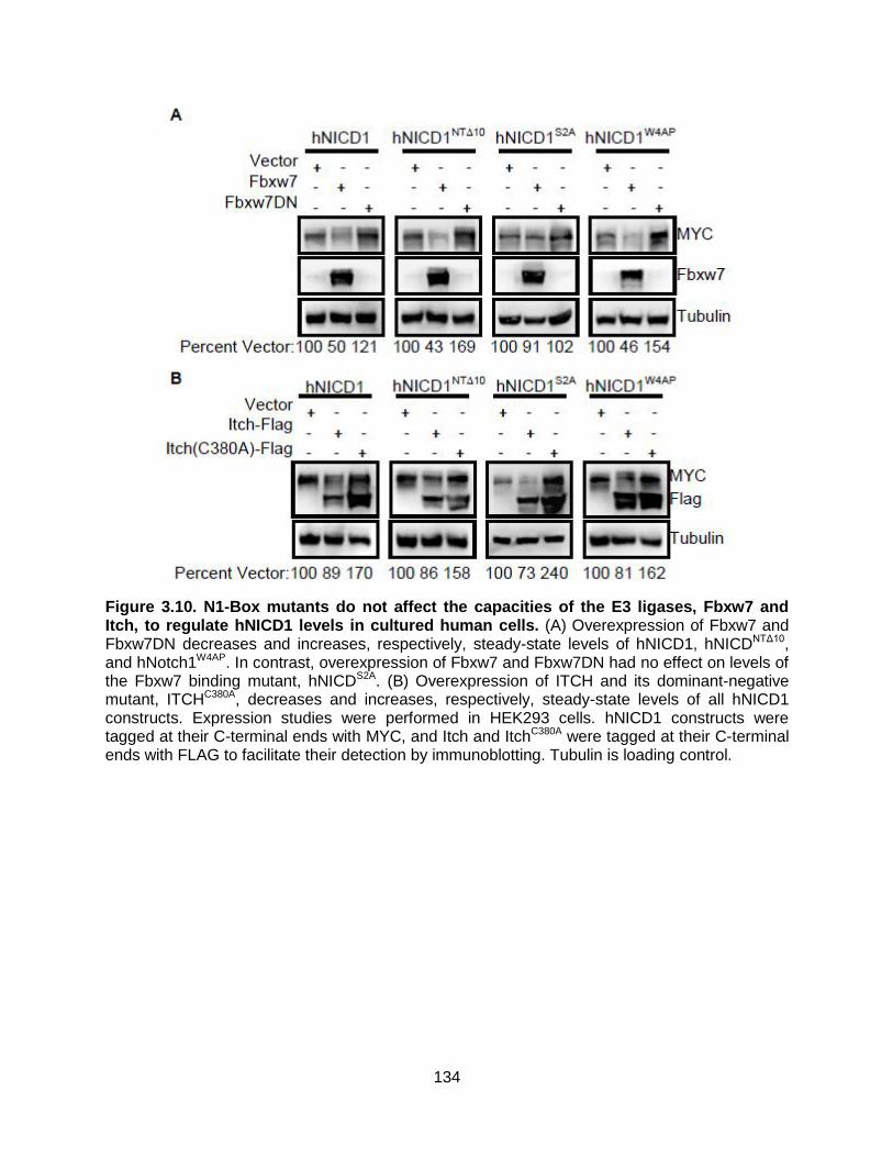
134
Figure 3.10. N1-Box mutants do not affect the capacities of the E3 ligases, Fbxw7 and Itch, to regulate hNICD1 levels in cultured human cells. (A) Overexpression of Fbxw7 and Fbxw7DN decreases and increases, respectively, steady-state levels of hNICD1, hNICDNTΔ10, and hNotch1W4AP. In contrast, overexpression of Fbxw7 and Fbxw7DN had no effect on levels of the Fbxw7 binding mutant, hNICDS2A. (B) Overexpression of ITCH and its dominant-negative mutant, ITCHC380A, decreases and increases, respectively, steady-state levels of all hNICD1 constructs. Expression studies were performed in HEK293 cells. hNICD1 constructs were tagged at their C-terminal ends with MYC, and Itch and ItchC380A were tagged at their C-terminal ends with FLAG to facilitate their detection by immunoblotting. Tubulin is loading control.

135
The E3 ubiquitin ligase, ITCH, has also been shown to promote PEST domain-
independent NICD1 degradation, although its binding site has not been well
characterized (Mazaleyrat et al., 2003; McGill and McGlade, 2003; Qiu et al., 2000). As
previously shown, overexpression of ITCH decreases, whereas overexpression of a
dominant-negative form of ITCH, ITCHC380A, increases, the steady-state levels of
hNICD1 in HEK293 cells (Figure 3.10B). We find that similar results are obtained for all
the mutants, indicating that ITCH does not mediate NICD1 degradation through the N1-
Box, WSSSSP, or LTPSPE.
Binding of CSL to hNICD1 regulates its stability
Because of the sequence overlap between the N1-Box and the RAM domain (the
major cis factor involved in CSL binding (Nam et al., 2003; Tamura et al., 1995)) (Figure
3.11A), we tested whether the binding of CSL to hNICD1 could influence hNICD1
stability. To examine this possibility, we incubated recombinant CSL (expressed and
purified from the Sf21/baculovirus system) with Xenopus egg extract and assessed its
effect on hNICD1 turnover. We find that CSL inhibits degradation of hNICD1 in extract
in a dose-dependent manner (Figure 3.11B-C). If the regulation of NICD1 stability by
CSL occurs via direct binding, mutants of NICD1 that prevent it from binding to CSL
should no longer be inhibited by CSL. This appears to be the case as the degradation of
the hNICD11771-74A mutant, which lacks the capacity to interact with CSL (Chu and
Bresnick, 2004; Vasquez-Del Carpio et al., 2011), is not inhibited by recombinant CSL
(Figure 3.11B-C). We speculate that the predominantly cytoplasmic character of
Xenopus egg extract contains low levels of CSL in contrast to human cells; thus,
hNICD11771-74A should turn over at a faster rate than wild-type hNICD1. Consistent with

136
this, we observe lower steady-state level of hNICD11771-74A than wild-type hNICD1 when
expressed in HEK293 cells (Figure 3.11D-E).
Because degradation of the NICD via the SCFFbxw7/PEST domain is dependent
on assembly of a transcriptional complex involving CSL (Fryer et al., 2002; Fryer et al.,
2004), we predict that turnover of the hNICD11771-74A mutant is primarily mediated by the
N1-Box. If this is the case, an N1-Box/CSL-binding double mutant should be more
stable than the N1-Box mutant. This increased stability is due to incapacity of the CSL-
binding mutant to assemble into a transcriptional complex (in contrast to the N1-Box
mutant) and, thus, would not be degraded via the SCFFbxw7 complex. Consistent with
this idea, we find that the steady-state level of hNICD1NTΔ10/1771-74A is greater than that of
hNICD1NTΔ10 (Figure 3.11D-E).
Conversely, in an hNICD1 PEST mutant, the inability to bind CSL should lead to
decreased protein stability due to enhanced N1-Box-mediated degradation. Consistent
with this model, we find that the steady-state level of hNICD11771-74A / S2493Δ is lower than
that of hNICD1S2493Δ (Figure 3.11D-E). These results provide strong evidence that CSL
binding to NICD1 inhibits its turnover by blocking N1-Box-mediated degradation.

137

138
Figure 3.11. N1-Box-mediated hNICD1 degradation in Xenopus egg extract and cultured human cells is inhibited by its binding to CSL. (A) Schematic showing overlap of the N1-Box (yellow) and RAM domains (blue). Overlapping sequence is displayed in green. The ΦWΦP motif is outlined by a black box. (B) Recombinant CSL inhibits degradation of hNICD1 (but not a CSL binding mutant of hNICD) in a dose-dependent manner in Xenopus egg extract. Radiolabeled hNICD1 or hNICD11771-74A were incubated in Xenopus egg extract in the presence of increasing amounts of recombinant CSL. Samples were removed after 1 hr and subjected to SDS-PAGE/autoradiography. (C) Graph of densitometry measurements in (B) normalized to Tubulin. (D) Steady-state levels of the CSL binding mutant, hNICD111771-74A, are lower than the wild-type protein in cultured human cells. Plasmids encoding the indicated N1-Box and CSL binding mutants tagged with the MYC epitope were transfected into HEK293 cells and immunoblotting performed. Tubulin is loading control. (E) Graph of densitometry measurements
in (D). Graph shows mean SD of MYC intensities normalized to Tubulin for 4 independent experiments. ***p-value<0.01, *p-value<0.1.

139
Mutations within the N1-Box are potential drivers of Notch-mediated
tumorigenesis
Multiple studies have identified mutations in the Notch1 receptor as a major
cancer driver (Koch et al., 2013; Ntziachristos et al., 2014; South et al., 2012). Because
our studies indicate that loss of N1-Box mutation promotes Notch signaling to an equal
or greater extent than mutations found in T-ALL patients, we sought to determine
whether mutations within the N1-Box also occur in human cancers. Utilizing the NIH
Catalogue of Somatic Mutations in Cancer (COSMIC) database, we identified two gain-
of-function mutations in patient tumors that are located within the first 35 residues of
hNICD1 (Figure 3.12A). We find that both mutants (hNICD1R1758S and hNICD1S1776C)
exhibit greater Notch transcriptional activity than wild-type hNICD1 and have elevated
steady state protein levels when compared to wild-type hNICD1 (Figure 3.12B-C).
Furthermore, we demonstrate that these mutants have enhanced activity in vivo, and
injection of D. rerio embryos with mRNA from either mutant show significantly increased
defects in somitogenesis compared to injection of wild-type hNICD1 mRNA (Figure
3.12D). These studies are consistent with mutations within the N1-Box as potential
cancer drivers that act by stabilizing NICD1.

140
Figure 3.12. Mutations within the N1-Box of hNotch1 in human cancers have enhanced signaling activity. (A) Table of somatic mutations found within residues 1754-1788 of hNotch1 (residues 1-35 of hNICD1) from the COSMIC database. (B) Both mutants in the N1-Box demonstrate elevated Notch reporter activity. HEK293 cells were transfected with plasmids encoding hNICD1 mutants found in human cancers in (A) and the HES1-Luciferase Notch reporter. Graph is representative of at least three independent experiments performed in
triplicate and show S.D. of Luciferase signal normalized to Renilla luciferase (transfection control). **p-value<0.007 for all mutants versus hNICD1. (C) The R1758S and S1776C mutants of hNotch1 have elevated steady-state protein levels when expressed in cultured cells. HEK293 cells were transfected with MYC epitope-tagged hNICD1 encoding the human N1-Box cancer mutants and immunoblotting performed. Tubulin is loading control. Intervening lanes were removed. (D) Graph is percentage of D. rerio embryos at the 10-13 somite stage with defective somites injected with 50 pg of the indicated hNICD1 mRNA. All data is from at least 3 clutches collected from 3 different breeding pairs. n=30-320 embryos per injection. ***p-value<0.0005 versus hNICD1.

141
Discussion
In T-ALL, mutations of Notch1 that truncate the C-terminal PEST domain remove
key cis factors necessary for its proteasomal-mediated degradation, highlighting the
importance of regulating NICD1 protein levels upon its liberation from the plasma
membrane (Ntziachristos et al., 2014; South et al., 2012). Prior studies indicated that
degradation of the intracellular domain of Notch1 (the most ubiquitously expressed of
the four Notch paralogs) differs from those of the other three Notch paralogs (Chiang et
al., 2006; Tsunematsu et al., 2004; Wu et al., 2001). Herein, we identify the N1-Box, a
novel degron of NICD1 that we identified using Xenopus egg extract. We show that the
N1-Box is transferable as an autonomous degron and that its activity is inhibited by
NICD1 binding to CSL. We show that the N1-Box is conserved in vertebrates and that
mutations in the N1-Box lead to increased Notch1 activity in vivo in zebrafish. Finally,
we demonstrate that mutations in the N1-box of Notch1 found in human cancers have
increased steady-state levels and enhanced activity. Further evidence for an in vivo role
for the N1-Box comes from a previous report demonstrating that a chimeric receptor
encoding the Notch2 extracellular domain and the Notch1 intracellular domain
(Notch21) is two-fold more active compared to wild-type Notch1 when expressed at
similar levels in mice (Liu et al., 2013). Interestingly, the Notch21 chimeric fusion
deleted the N1-Box of NICD1. Thus, it is possible that the increased activity of this
chimera is due to increased stability of its intracellular domain. Based on our results, we
propose a model (Figure 3.13) in which the liberated NICD1 (upon cleavage from the
Notch1 receptor by γ-secretase) has two fates: (1) rapid degradation and inactivation via
its N1-Box or (2) binding to CSL and Notch transcriptional complex components to drive
142
transcription of Notch target genes (Figure 3.13). Termination of Notch1 signaling
occurs upon ubiquitin-mediated degradation by the SCFFbxw7/proteasome.

143
Figure 3.13. Model of N1-Box-mediated regulation of hNotch1 signaling. See text for details.

144
Previous studies have shown that the NICD has the capacity to assemble into
higher order (intramolecular or intermolecular) forms, and disruption of these higher
order forms are required for NICD binding to CSL (Kelly et al., 2007; Vasquez-Del
Carpio et al., 2011). It is possible that these higher order complexes may promote N1-
box-mediated degradation by blocking NICD1 binding to CSL. Alternatively, these
higher order structures may inhibit NICD1 turnover as well as transcriptional activation
by blocking the activity of the N1-box and its overlapping RAM domain. Further studies
are needed to resolve these two possibilities.
Although it is obvious that there are advantages for a mechanism to terminate
transcription by proteolysis, it is not as clear why a cell would have a mechanism for
degrading cytoplasmic NICD1. Why is the cytoplasmic level of NICD1 not limited
exclusively by its release from the holoprotein upon receptor activation? One possibility
is that this system dampens the stochastic flux in the system, thereby minimizing noise.
Thus, a threshold level of Notch receptor activation needs to occur in order for
transcription to be activated. This may explain why the Drosophila NICD does not
encode an N1-Box as stochastic flux in Notch signaling plays an important role during
neuroblast differentiation via the mechanism of lateral inhibition (Artavanis-Tsakonas et
al., 1999). Conversely, once the transcriptional complex is fully saturated (i.e. all CSL is
occupied by NICD1), unbound NICD1 could be degraded to limit the activation window.
This model is consistent with the digital response model proposed for Notch1 pathway
activation (Ilagan et al., 2011). Regardless, the importance of NICD1-mediated
degradation in maintaining normal Notch1 signaling in humans is evidenced by our

145
findings that mutations of the N1-Box that lead to hNICD1 stabilization are found in
human cancers.
Of the four Notch paralogs, why is the N1-Box only present in the Notch1
paralog? It is possible that differences in regulation between the four Notch paralogs
may also simply reflect differences in their transcriptional behavior. One example is the
requirement for Notch2 over Notch1 in liver development (Sparks et al., 2010), in which
decreased levels of the Notch2 receptor, but not Notch1, leads to Alagille syndrome
(McDaniell et al., 2006). Also consistent with differences in the transcriptional behavior
between different Notch paralogs is the observation that certain Notch-driven cancers
are associated with particular Notch paralogs (Bellavia et al., 2000; Callahan and
Raafat, 2001; Capobianco et al., 1997; Kiaris et al., 2004; Pancewicz and Nicot, 2011).
Currently, γ-secretase inhibitors (GSIs) are the best characterized Notch pathway
therapeutics. Unfortunately, because of their pan-Notch inhibitory activities, there are
many associated adverse effects that limit their long-term use (Espinoza and Miele,
2013; Extance, 2010; Milano et al., 2004; Morell and Strazzabosco, 2013; van Es et al.,
2005; Wong et al., 2004). Thus, one long-term goal for treating Notch-driven cancers is
to develop paralog-specific drugs in order to minimize adverse effects associated with
pan-Notch inhibition. Our identification of the NICD1-specific N1-Box provides a
potential path to develop Notch1-selective inhibitors. Compounds that disrupt NICD1-
CSL interaction may be particularly potent as they would not only block NICD1-
mediated transcription, but also promote NICD1 degradation. The development of such
compounds would be greatly facilitated by the identification of the E3 ubiquitin ligase for
the N1-Box.

146
CHAPTER IV
DISCUSSION AND FUTURE DIRECTIONS
Introduction
The work presented in this dissertation represents mostly biochemical, cell-
based, and in vivo experiments identifying a novel regulatory region of NICD1 which
stabilizes the NICD and hyperactivates Notch-mediated transcription which elucidates a
novel mechanism that modulates the Notch signaling pathway. In the current Chapter, I
discuss and provide future directions for the results described in the previous two
Chapters. I have divided this chapter into parts I and II. Part I focuses exclusively on the
regulation of the NICD via its novel N1-Box and the implications for Notch signaling.
Part II focuses on the future of the Notch-Wnt signaling network and the implications for
“Wntch” signaling.
Part I
Discussion
Utilizing Xenopus egg extract, cell-based assays, and zebrafish embryos we
identified and characterized the N1-Box region of NICD1 as a regulatory domain of
Notch signaling, which has not been previously characterized. This novel region is very
likely a driver of Notch-mediated cancers in which Notch acts as an oncogene.
Additionally, we have identified cis-acting factors of NICD1 that regulate the capacity of
the N1-Box to promote degradation.

147
Our initial observation was that human NICD1 degrades robustly in Xenopus egg
extract, and NICD2, 3, and 4 do not (Figure 3.1). NICD1 degradation is proteasome-
dependent, as the addition of MG132 inhibits its turnover (Figure 3.1). NICD1’s
degradation occurs distinctly from the previously characterized β-catenin degradation in
Xenopus egg extract (Figure 3.1). Additionally, degradation of NICD1 is GSK3-
independent, contradicting previous reports about Notch1 degradation (Espinosa et al.,
2003; Foltz et al., 2002; Jin et al., 2009b) (Figure 3.2). These results were validated
using mouse NICD1 and NICD4 and tagged versions of the human NICDs, which also
degrade differentially (Figure 3.2). Interestingly, all 4 Myc-tagged NICD constructs
degrade in HEK293 cells as assessed by cycloheximide chase assay, even though only
NICD1-Myc degrades in Xenopus egg extract (Figure 3.3). The Xenopus egg extract
system allows us to separate distinct dynamic degradation pathways, which is much
more difficult in cell culture. The degradation of NICD1 in Xenopus egg extract is
independent of previously-identified PEST/Fbwx7-dependent degradation of NICD1 in
cell culture (Figure 3.4). So which domain regulates NICD1 turnover in Xenopus egg
extract?
We generated N-terminal and C-terminal fragments of NICD1 fused with the
respective N-terminal and C-terminal fragments of the other NICD paralogs to address
that question. We were able to identify that NICD1 degradation is N-terminal dependent
(Figure 3.5). We narrowed the N-terminal regulatory region of NICD1 down to the first
35 amino acids of NICD1 (Figure 3.6). The NICD1 and the Notch 2, 3, 4 ICDS have
conserved N-terminal domains within those 35 amino acids but the very N-terminus is
not conserved among the Notch paralogs. We found that three key residues at the very

148
N-terminal region of NICD1 (L2, S4, and Q10 in human NICD1) stabilize the NICD1
when they are individually mutated to resemble NICD2 in Xenopus egg extract (Figure
3.6). These stable mutated forms of NICD1 also hyperactivate Notch signaling in
HEK293 cells (Figure 3.6). When these three residues are collectively mutated to
resemble NICD2, the NICD1 is stable in HEK293 cells (Figure 3.6). These results
indicate that the N-terminal region of NICD1 is necessary for its degradation. But is this
N-terminal region sufficient to promote degradation?
The heterologous addition of the N-terminal region of N1ICD onto the stable
proteins NICD2, GFP, and Luciferase was sufficient to promote their degradation in
Xenopus egg extract and in HEK293 cells (Figure 3.6; Figure 3.7). Our results show that
the N-terminal region of NICD1 is both necessary and sufficient to promote degradation
of proteins, fully validating it as a novel degron. We have designated this new regulatory
region of NICD1 as the N1-Box. Our results show that the N1-Box is both necessary
and sufficient to promote degradation of proteins in Xenopus egg extract and HEK293
cells.
Next, we investigated the potential cooperation between the newly identified N1-
Box and previously established regulatory domains of NICD1, particularly the PEST
domain and the WSSSSP domains (Chiang et al., 2006; Fryer et al., 2004). The N1-Box
hyperactivates Notch signaling to a higher extent than the other previously established
domains and this hyperactivation is additive with the WSSSSP domain but none of the
others (Figure 3.8), strongly suggesting that the WSSSSP domain acts through a
parallel pathway with the N1-Box or acts upon the N1-Box in some way. Up until this
point, all of our experiments were performed with the NICD1 rather than the full-length

149
canonical Notch receptor. Do these stabilizing N1-Box mutations affect canonical Notch
signaling in the context of full-length Notch receptors?
The stabilized N1-Box mutant isoform of full-length mouse Notch1 hyperactivates
Notch signaling in HEK293 cells and in zebrafish embryos (Figure 3.8). These results
were validated using a constitutively active form of mouse Notch1 (Figure 3.8).
Interestingly, the N1-Box mutant isoform of Notch1 hyperactivates Notch signaling to a
larger extent than the PEST domain mutant of Notch1, supporting our cell culture
transcriptional reporter assay results (Figure 3.8). These results, taken collectively,
show that the N1-Box of Notch1 both stabilizes the NICD1 and hyperactivates canonical
Notch signaling in mammalian cell culture and in vivo in zebrafish embryos. This
transcriptional hyperactivation is higher for the N1-Box mutants than for the previously
characterized PEST domain mutants. We next ask whether mutations in the N1-Box are
found in human cancers and whether these cancers have elevated Notch signaling.
We found that mutations in the N1-Box identified from the COSMIC database in
Notch-driven cancers also confer stability to NICD1 in cell-based assays, which strongly
suggests that the N1-Box mutations are correlated with Notch-driven cancers (Figure
3.12). These results show that mutations in the N1-Box of Notch1 are associated with
human diseases, marking the novel N1-Box regulatory region as a potential therapeutic
for Notch-mediated cancers. Curiously, there are not a large number of mutations in the
N1-Box that are linked to cancer. Our hypothesis is that cis-binding factors on Notch1
affect the N1-Box’s ability to promote degradation.
Next, we investigated the role of known cis-binding factors on regulation of the
N1-Box-dependent degradation. Based on structural analysis, the CSL/Su(H)/Lag-1

150
binding site is located in the RAM domain, adjacent to the N1-Box. The addition of
recombinant CSL inhibits NICD1 degradation in a dose-dependent manner in Xenopus
egg extract and this effect is reversed in a CSL-binding mutant that cannot bind to CSL
(1771-1774A) (Figure 3.11). These results are validated in HEK293 cells, where the
steady state level of the CSL-binding mutant is significantly lower than wild-type NICD1
because it can no longer be shielded from degradation by CSL (Figure 3.11).
Interestingly, an N1-Box and CSL-binding double mutant appears to be even more
stable than the N1-Box mutant alone (based on the quantification), further supporting
the idea that CSL binding affects the N1-Box. These results partially elucidate a
mechanism for regulation of the N1-Box which is dependent on CSL binding and
subsequent CSL-mediated transcription.
Based on our results, a model emerges in which the N1-Box is actively degrading
NICD1 upon cleavage by δ-secretase. This degradation is independent of ligand
activation. Then, upon recruitment of CSL to the N1ICD, the N1-Box domain no longer
promotes degradation as it is sterically inhibited by CSL binding to the RAM domain
(Figure 3.13). But why does Notch1 specifically have this extra level of regulation that is
not present in the other paralogs? Notch1 is the most widely expressed paralog of
Notch and there is some evidence of ligand-independent signaling through Deltex
(Fuwa et al., 2006; Yamada et al., 2011). It is likely that this mechanism of regulating
protein levels is used to modulate the natural variability of signaling between cells as
Notch signaling requires a very precise amount to undergo proper cell-cell signaling.
One emerging idea about signal transduction pathways is the idea of bistability
within the system, where a threshold of signal must be reached within each individual

151
cell which then acts in a binary fashion to either activate signaling maximally or not
activate signaling. Our data suggest that the N1-Box may be the switch that imparts
Notch1 signaling bistability in an organismal setting. Because Notch1 is so highly
expressed in comparison to other Notch paralogs, it is likely that Notch1 requires an
additional level of signal modulation in order to prevent ectopic signaling, and the N1-
Box is likely at least a partial contributor to Notch1’s potential bistability. So how does
our model for the regulation of the Notch1 intracellular domain fit into the prevailing
models of how Notch signaling is regulated?
The previous prevailing model for the regulation of NICD stability was that it was
regulated by the N-end rule of protein stability. In the N-end rule, the N-terminal amino
acid of a protein determines its half-life (Bachmair et al., 1986). NICDs which start with
valine or methioninie were thought to escape the N-end rule which made them stable
enough to activate transcription (Tagami et al., 2008). Our results showed that the N-
end rule is not a likely explanation for the stability of the Notch1 mutants, as the wild-
type NICD1 starts with a valine but is significantly less stable than the NICD1 (NTΔ10)
that starts with an histidine and should be subject to the N-end rule. In addition, point
mutations in the N1-Box which do nto affect the initial valine can also greatly affect
stability of the NICD1. Our results show that NICD1’s stability is regulated by additional
factors other than the traditional N-end rule.
One other recently proposed model is that the Notch extracellular domain is the
only domain critical for Notch signaling. One recent report showed that a chimeric Notch
receptor containing the Notch2 extracellular domain and the Notch1 intracellular domain
(Notch21) is two-fold more active than wild-type Notch1 when expressed at similar

152
levels in mice. Their conclusion was that the Notch extracellular domain was the critical
region in the regulation of Notch signaling and that the intracellular domains are actually
interchangeable to transduce the signal (Liu et al., 2013). Ostensibly, this conflicts with
our conclusions that the N1-Box of the intracellular domain is likely the most important
region for regulation of Notch signaling. Interestingly, the Notch21 chimeric fusion
deletes the N1-Box of NICD1, which according to our results leads to a more stable
intracellular domain. Thus, it is possible that the increased activity of this chimera is due
to increased stability of its intracellular domain. A series of experiments to fully combine
these two models would start by comparing Notch signaling from the Notch21 chimeric
receptor to the Notch1 receptor in which the N1-Box is mutated. Our data suggests that
the Notch21 chimera and either the N1(LSQ) or the N1(NTΔ10) would have similar
levels of Notch signaling, due to an increase in the stability of the NICD1.
Future Directions
Identifying the complete regulatory mechanism of the N1-Box
This dissertation identifies a novel regulatory region of Notch1 termed the N1-
Box which serves as a major regulator of Notch1 protein stability. It also elucidates a
mechanism for how the N1-Box can be regulated in vivo upon CSL binding to inhibit N1-
Box-mediated degradation. However, the full mechanism of N1-Box regulation still
needs to be elucidated. Proteins are often regulated by post-translational modifications.
The degradation of NICD1 is proteasome-dependent in Xenopus egg extract.
Proteasome-mediated degradation requires the involvement of an E3 ubiquitin ligase
which adds ubiquitin moieties to lysine residues and targets the protein to the

153
proteasome. The candidate E3s which have been previously linked to Notch1 stability
(ITCH, SCFFbxw7) degraded NICD1 in HEK293 cells in the N1-Box stabilized mutant
similarly to wild-type NICD1 (Figure 3.10). In addition, there is a basic region in NICD1
which contains a string of lysines that is not found in the other NICD paralogs, strongly
suggesting that NICD1 is being regulated by an E3 distinct from those we tested. These
residues are very likely to be targets of ubiquitination and degradation.
One way to identify the E3 ubiquitin ligase for the N1-Box of NICD1 is to perform
a screen for mammalian E3s that increase NICD1 turnover. The Xenopus egg extract
system would be very amenable to this type of screen and using luciferase-tagged
proteins we could do a high throughput screen with a quantifiable readout. Validation of
any hits from the screen would then be performed in cell culture using a transcriptional
reporter assay also using luciferase levels as the readout for protein levels. Any hits
identified can be validated in cultured cells and Xenopus egg extract in a low throughput
manner. The effect of any E3s that alter NICD1 turnover via the N1-Box of NICD1
needs to be investigated thoroughly. If it is directly affecting the N1-Box, then the
degradation of NICD1 would be inhibited in the N1-Box mutant. In Chapter I, I discussed
that NICD1 is modulated by other post-translational modifications, and these can affect
the stability of NICD1 and are worth investigating. Oftentimes, ubiquitination is regulated
by phosphorylation at nearby residues.
The other major post-translational modification that could potentially regulate the
stability of the NICD1 is phosphorylation. Previous literature has suggested that
phosphoregulation of NICD1 can regulate its stability (Choi et al., 2013; Foltz et al.,
2002; Fryer et al., 2004). Because the regulation of Notch is so tightly regulated in vivo,

154
modulation of Notch1 in either direction could be critical in affecting canonical Notch
signaling. We have performed a small molecule kinase inhibitor screen for modulators
of NICD1 turnover in Xenopus egg extract using luciferase-tagged NICD1 and
assessing for changes in NICD1 degradation via luciferase signal. The library we used
was the TOCRIS kinase inhibitor toolbox [used in (Jester et al., 2010)]
(http://www.tocris.com/dispprod.php?ItemId=224690#.U-L8TM90zwo).
Our screen identified Ataxia telangiectasia mutated (ATM) and Ataxia
telangiectasia and Rad3-related protein (ATR), two well-characterized DNA damage
responding serine/threonine kinases that phosphorylate key tumor suppressors such as
p53 and CHK2, as modulators of NICD1 turnover in Xenopus egg extract. Further
characterization of the role of ATM and ATR on NICD1’s stability should be pursued.
These studies should include structure-function analysis, functional assays in cell
culture, and then potentially even in vivo studies. Follow up validation on any other hits
from the screen should also be investigated. If validated, the screen can begin to
identify other novel targets for therapeutics for treatment on Notch-mediated cancers.
Additionally, it would be interesting to identify whether any of these kinases affects the
capacity of the N1-Box to promote degradation. The N1-Box contains multiple serines
and threonines, making it highly likely to be phosphorylated.
If an E3 and a kinase that act on the N1-Box can both be identified, the next step
would be to identify the relationship between these two post-translational modifications.
Phosphorylation can both promote and inhibit ubiquitination and subsequent
proteasome-mediated degradation in other signaling pathways (Orford et al., 1997;
Yamamoto et al., 1999). Would the phosphorylation of the N1-Box promote or inhibit its

155
ubiquitination? Is it context-dependent or tissue-specific? These are the key questions
that still need to be answered about the regulation of the N1-Box.
We have also already shown that CSL binding to the N1-Box affects its ability to
get degraded. Previous literature showed that CSL binds to the RAM and Ankyrin
domains of NICD with high affinity (Hsieh et al., 1996). But is CSL binding to NICD1
regulated? Previous literature has shown that in the nucleus, the scaffold protein MAML
binds to the CSL/NICD complex and is required to activate transcription of the
CSL/NICD transcriptional complex (Fryer et al., 2002). Potentially the post-translational
modifications are actually affecting CSL binding to the RAM domain in the N1-Box,
which would then promote degradation of NICD1. The role of ubiquitination and/or
phosphorylation on CSL binding is still unclear, and this would be another interesting
question to address.
How about the role of the prevailing models in the regulation of Notch signaling?
One key experiment would be to assess the stability of the Notch21 chimera vs. wild-
type Notch1 and vs. stabilized Notch1. It is likely that the differential signaling levels for
the Notch21 chimera vs wild-type Notch1 is due to a difference in the stability of each
respective ICD, as we have shown that the stable mutants of NICD1 lead to
hyperactivation of Notch signaling in mammalian cell culture and strongly suggests
Notch hyperactivation in D. rerio embryos.
In vivo disease models of the N1-Box
Although we have identified a novel degron, called the N1-Box, in NICD1, and
shown that it is functional in canonical Notch signaling in cell culture and in zebrafish

156
embryos, there are still many unanswered questions about the in vivo importance of the
N1-Box in Notch-mediated disease. The first step is to do tumorigenic assays in cancer
cell lines using our N1-Box mutants, such as colony formation assays to assess for
tumorigenesis. If N1-Box mutants can induce tumorigenesis in cell culture, one very
obvious next step is to make a mouse model of the stabilized N1-Box mutant of NICD1.
Because T-ALL and other leukemias are primarily Notch-driven in humans, we propose
to express an inducible N1-Box mutant Notch1 under the control of a leukocyte specific
promoter in an inducible Cre-LoxP background mouse. Upon tamoxifen induction, N1-
Box-mutant NICD1 expression will potentially induce tumorigenesis in the adult mouse,
which would demonstrate that the N1-Box can in fact induce Notch-mediated tumors in
vivo. Alternatively, we could generate tissue-specific mouse models for the other tissues
that are susceptible to Notch-driven cancers, such as mammary glands, liver, skin,
lungs and others. In addition, we could make a stabilized N1-Box mouse that is not
inducible which express mutant N1 throughout development and look for other Notch-
driven phenotypes. Generating tissue-specific mutant lines for the specific mutations
that stabilized human NICD1 (Figure 3.5) and assessing their ability to induce
tumorigenesis would also be quite interesting and important. Are these stable mutants
identified from a human database actually able to induce tumorigenesis, or is the
Notch1 mutation a secondary mutation? The mouse model is an ideal model to test
many of these hypotheses.
The generation of a mouse model will allow us to assess whether the differential
signaling observed in the Notch21 chimeric mice can be explained by the increased
stability of the N1-Box. Based on our results in Xenopus egg extract, mammalian cell

157
culture, and zebrafish embryos we would expect the N1-Box mutant mice to have an
increased level of Notch signaling, similar to the Notch21 chimeric mice. Further
experiments should also be performed to fully elucidate the relationship between the
chimeric receptors and the stabilized N1-Box mutant receptors.
Additionally, Notch mutations have been linked to multiple developmental defects
in humans, including Alagille syndrome (McDaniell et al., 2006), aortic valve disease
(Garg, 2006; Garg et al., 2005), Hajdu-Cheney syndrome (Simpson et al., 2011),
CADASIL (Joutel et al., 1996), and T-ALL (Weng et al., 2004). The N1-Box mutant
mouse models in which the mutant form of NICD is expressed at birth would be very
useful in studies involving developmental defects caused by misregulated Notch
signaling. In summary, a mouse model of the N1-Box mutant would be very beneficial to
helping fully elucidate the mechanisms regulating Notch signaling and also the
mechanisms regulating the N1-Box.
Part II
Discussion
As discussed in Chapter I, there appears to be extensive crosstalk between the
Notch signaling pathway and the Wnt signaling pathway (Hayward et al., 2008; Munoz
Descalzo and Martinez Arias, 2012), and some refer to the Wnt/Notch signaling network
as the “Wntch” signaling network. Many aspects of these two pathways converge and
studying how one pathway affects the other would be quite interesting and the
identification of a novel regulatory region of NICD1 opens up new avenues for exploring
the interplay between Wnt and Notch signaling.

158
One example of convergence into the “Wntch” pathway is the observation that
Presenilin 1, a core subunit of the δ-secretase complex which cleaves Notch, negatively
regulates β-catenin stability and transcriptional activity and can promote
phosphorylation of β-catenin independent of Axin (Kang et al., 2002; Killick et al., 2001).
Other examples of Wnt/Notch signaling convergence have been described extensively
in Chapter I and include convergence on Dsh (Ramain et al., 2001; Sokol, 1996),
TCF/LEF (Galceran et al., 2004; Ross and Kadesch, 2001; Shimizu et al., 2008), and
even the core components themselves (Corada et al., 2010; Hayward et al., 2006;
Hayward et al., 2005; Jin et al., 2009a). Based on previous literature, regulating one of
these two pathways can also regulate the other one, allowing for combinatorial
treatment for both Wnt and Notch-mediated diseases.
Identifying Novel Therapeutic Targets of Notch Signaling
Due to the importance of Notch signaling in many types of diseases, both
developmental defects and cancer, studying how this signaling network is properly
regulated would be critical to addressing Notch-mediated disease. How would the
discovery of the novel N1-Box of Notch1 help address that question? This allows
researchers to identify novel targets for development of therapeutics against Notch-
mediated cancers. Most of the current treatments for Notch focus on δ-secretase
inhibitors (GSIs), most of which are unable to pass clinical trials due to toxicity issues
and off-target side effects due to δ-secretase’s promiscuity and effect on multiple cell
types and tissues. Our research has uncovered both a novel regulatory region of the

159
NICD1, the N1-Box, as well as a novel regulatory mechanism involving the cis-binding
factors CSL and the WSSSSP motif of NICD1.
There are two novel approaches to take for development of therapeutics based
on our research. The first is to target the N1-Box directly by activating the E3
responsible for promoting the degradation of NICD1. E3 activation would decrease
NICD1 levels and reduce Notch signaling, which would be critical in treating Notch-
dependent cancers which have ectopic Notch signaling. Identifying small molecules
which target NICD1 degradation via the N1-Box would be one way to specifically target
Notch1 as opposed to the other Notch paralogs and other proteins that are dependent
on δ-secretase. This would reduce potential off-target effects and likely reduce toxicity
to the patients. One caveat with targeting an E3 is that most E3s have multiple
substrates and it is possible that there will be other off-target effects distinct from the
GSIs. Regardless, our studies open up a significant opportunity for the development of
anti-cancer therapeutics against Notch1-driven cancers by increasing the specificity of
the therapeutic and decreasing off-target effects.
In Chapter III, we showed that CSL binding can inhibit N1-Box-mediated
degradation and stabilize the NICD1 in Xenopus egg extract and HEK293 cells. A
second novel approach for therapeutics would be to target CSL binding to NICD1.
Previous literature has suggested that CSL is a nuclear specific protein (Chiang et al.,
2006; Ong et al., 2006) which binds to DNA and activates transcription in a complex
with NICD and MAML inside the nucleus. Inhibiting CSL binding would clearly affect
CSL-dependent Notch signaling. Previous literature provides us evidence that CSL-
independent Notch signaling is mediated by Deltex (Hori et al., 2004). However, in

160
Xenopus egg extract (a cell-free and nucleus-free system), CSL binding disrupts NICD1
degradation through the N1-Box. Interestingly, this inhibition is independent of the
transcriptional inhibition due to loss of CSL binding in cell culture as a CSL-binding
mutant further changes the stability of N1-Box mutants in Xenopus egg extract (which
does not undergo CSL-mediated transcriptional activity). Identifying small molecules
which disrupt CSL binding to NICD1 would allow for constitutive activation of N1-Box-
mediated degradation and modulation of Notch signaling. Although CSL binds to all the
Notch paralogs to activate signaling, NICD2, 3, and 4 do not contain the N1-Box, and,
thus, their stability is not likely to be affected by CSL binding.
In addition, the disruption of the WSSSSP motif likely affects N1-Box-mediated
degradation. The WSSSSP motif is heavily phosphorylated and is shown to regulate
NICD stability. Identifying small molecules which could affect the phosphorylation of the
WSSSSP motif could also serve as a potential new target for therapeutics. We showed
that mutation of the WSSSSP motif cooperates with the N1-Box to hyperactivate Notch
signaling, which strongly suggests that the WSSSSP motif is actually inhibiting Notch
signaling in some fashion, likely through promoting its degradation. Being able to
stabilize the phosphorylation of the WSSSSP motif through a potential therapeutic
would also help be useful in the treatment of Notch-mediated cancers.
Collectively, our data identifies novel regulators of Notch signaling which have
not been previously reported. WE show that NICD1 degrades in a cell-free system
giving researchers additional tools for identifying modulators of the Notch signaling
network (specifically the Notch pathway) that were not available previously. Because
the N1-Box is specific to Notch1, inhibiting the N1-Box should not lead to global Notch

161
phenotypes but only Notch1-specific phenotypes, allowing researchers to parse out the
involvement of Notch1 specifically vs. the other Notch paralogs in vivo. This work will
help us take a small step forward towards identifying effective and usable new
therapeutics for Notch signaling. Any potential therapeutics for Notch signaling may also
have the potential to potentiate or dampen Wnt/β-catenin signaling because of the
extensive crosstalk between the Notch and Wnt/β-catenin signaling pathways.

162
REFERENCES
Acar, M., H. Jafar-Nejad, H. Takeuchi, A. Rajan, D. Ibrani, N.A. Rana, H. Pan, R.S. Haltiwanger, and H.J. Bellen. 2008. Rumi is a CAP10 domain glycosyltransferase that modifies Notch and is required for Notch signaling. Cell. 132:247-258.
Acosta, H., S.L. Lopez, D.R. Revinski, and A.E. Carrasco. 2011. Notch destabilises maternal beta-catenin and restricts dorsal-anterior development in Xenopus. Development. 138:2567-2579.
Ahn, Victoria E., Matthew L.-H. Chu, H.-J. Choi, D. Tran, A. Abo, and William I. Weis. 2011. Structural Basis of Wnt Signaling Inhibition by Dickkopf Binding to LRP5/6. Developmental Cell. 21:862-873.
Alberi, L., S. Liu, Y. Wang, R. Badie, C. Smith-Hicks, J. Wu, T.J. Pierfelice, B. Abazyan, M.P. Mattson, D. Kuhl, M. Pletnikov, P.F. Worley, and N. Gaiano. 2011. Activity-induced Notch signaling in neurons requires Arc/Arg3.1 and is essential for synaptic plasticity in hippocampal networks. Neuron. 69:437-444.
Alves-Guerra, M.C., C. Ronchini, and A.J. Capobianco. 2007. Mastermind-like 1 Is a specific coactivator of beta-catenin transcription activation and is essential for colon carcinoma cell survival. Cancer Res. 67:8690-8698.
Amit, S., A. Hatzubai, Y. Birman, J.S. Andersen, E. Ben-Shushan, M. Mann, Y. Ben-Neriah, and I. Alkalay. 2002. Axin-mediated CKI phosphorylation of beta-catenin at Ser 45: a molecular switch for the Wnt pathway. Genes Dev. 16:1066-1076.
Amoyel, M., Y.C. Cheng, Y.J. Jiang, and D.G. Wilkinson. 2005. Wnt1 regulates neurogenesis and mediates lateral inhibition of boundary cell specification in the zebrafish hindbrain. Development. 132:775-785.
Andersson, E.R., R. Sandberg, and U. Lendahl. 2011. Notch signaling: simplicity in design, versatility in function. Development. 138:3593-3612.
Angers, S., C.J. Thorpe, T.L. Biechele, S.J. Goldenberg, N. Zheng, M.J. MacCoss, and R.T. Moon. 2006. The KLHL12-Cullin-3 ubiquitin ligase negatively regulates the Wnt-beta-catenin pathway by targeting Dishevelled for degradation. Nat Cell Biol. 8:348-357.
Ann, E.J., H.Y. Kim, M.S. Seo, J.S. Mo, M.Y. Kim, J.H. Yoon, J.S. Ahn, and H.S. Park. 2012. Wnt5a controls Notch1 signaling through CaMKII-mediated degradation of the SMRT corepressor protein. J Biol Chem. 287:36814-36829.
Aoyagi-Ikeda, K., T. Maeno, H. Matsui, M. Ueno, K. Hara, Y. Aoki, F. Aoki, T. Shimizu, H. Doi, K. Kawai-Kowase, T. Iso, T. Suga, M. Arai, and M. Kurabayashi. 2011. Notch induces myofibroblast differentiation of alveolar epithelial cells via

163
transforming growth factor-{beta}-Smad3 pathway. American journal of respiratory cell and molecular biology. 45:136-144.
Apelqvist, A., H. Li, L. Sommer, P. Beatus, D.J. Anderson, T. Honjo, M. Hrabe de Angelis, U. Lendahl, and H. Edlund. 1999. Notch signalling controls pancreatic cell differentiation. Nature. 400:877-881.
Arias, A.M. 2002. New alleles of Notch draw a blueprint for multifunctionality. Trends in genetics : TIG. 18:168-170.
Arias, A.M., and P. Hayward. 2006. Filtering transcriptional noise during development: concepts and mechanisms. Nature reviews. Genetics. 7:34-44.
Arnett, K.L., M. Hass, D.G. McArthur, M.X. Ilagan, J.C. Aster, R. Kopan, and S.C. Blacklow. 2010. Structural and mechanistic insights into cooperative assembly of dimeric Notch transcription complexes. Nat Struct Mol Biol. 17:1312-1317.
Artavanis-Tsakonas, S., and M.A. Muskavitch. 2010. Notch: the past, the present, and the future. Curr Top Dev Biol. 92:1-29.
Artavanis-Tsakonas, S., M.A. Muskavitch, and B. Yedvobnick. 1983. Molecular cloning of Notch, a locus affecting neurogenesis in Drosophila melanogaster. Proc Natl Acad Sci U S A. 80:1977-1981.
Artavanis-Tsakonas, S., M.D. Rand, and R.J. Lake. 1999. Notch signaling: cell fate control and signal integration in development. Science. 284:770-776.
Austin, J., and J. Kimble. 1987. glp-1 is required in the germ line for regulation of the decision between mitosis and meiosis in C. elegans. Cell. 51:589-599.
Axelrod, J.D., K. Matsuno, S. Artavanis-Tsakonas, and N. Perrimon. 1996. Interaction between Wingless and Notch signaling pathways mediated by dishevelled. Science. 271:1826-1832.
Babaoglan, A.B., K.M. O'Connor-Giles, H. Mistry, A. Schickedanz, B.A. Wilson, and J.B. Skeath. 2009. Sanpodo: a context-dependent activator and inhibitor of Notch signaling during asymmetric divisions. Development. 136:4089-4098.
Bachmair, A., D. Finley, and A. Varshavsky. 1986. In vivo half-life of a protein is a function of its amino-terminal residue. Science. 234:179-186.
Baig-Lewis, S., W. Peterson-Nedry, and M. Wehrli. 2007. Wingless/Wnt signal transduction requires distinct initiation and amplification steps that both depend on Arrow/LRP. Dev Biol. 306:94-111.
Baladron, V., M.J. Ruiz-Hidalgo, M.L. Nueda, M.J. Diaz-Guerra, J.J. Garcia-Ramirez, E. Bonvini, E. Gubina, and J. Laborda. 2005. dlk acts as a negative regulator of

164
Notch1 activation through interactions with specific EGF-like repeats. Exp Cell Res. 303:343-359.
Balint, K., M. Xiao, C.C. Pinnix, A. Soma, I. Veres, I. Juhasz, E.J. Brown, A.J. Capobianco, M. Herlyn, and Z.J. Liu. 2005. Activation of Notch1 signaling is required for beta-catenin-mediated human primary melanoma progression. The Journal of clinical investigation. 115:3166-3176.
Bardin, A.J., C.N. Perdigoto, T.D. Southall, A.H. Brand, and F. Schweisguth. 2010. Transcriptional control of stem cell maintenance in the Drosophila intestine. Development. 137:705-714.
Barker, N., J.H. van Es, J. Kuipers, P. Kujala, M. van den Born, M. Cozijnsen, A. Haegebarth, J. Korving, H. Begthel, P.J. Peters, and H. Clevers. 2007. Identification of stem cells in small intestine and colon by marker gene Lgr5. Nature. 449:1003-1007.
Becam, I., U.M. Fiuza, A.M. Arias, and M. Milan. 2010. A role of receptor Notch in ligand cis-inhibition in Drosophila. Curr Biol. 20:554-560.
Behrens, J., J.P. von Kries, M. Kuhl, L. Bruhn, D. Wedlich, R. Grosschedl, and W. Birchmeier. 1996. Functional interaction of beta-catenin with the transcription factor LEF-1. Nature. 382:638-642.
Belenkaya, T.Y., C. Han, H.J. Standley, X. Lin, D.W. Houston, and J. Heasman. 2002. pygopus Encodes a nuclear protein essential for wingless/Wnt signaling. Development. 129:4089-4101.
Bellavia, D., A.F. Campese, E. Alesse, A. Vacca, M.P. Felli, A. Balestri, A. Stoppacciaro, C. Tiveron, L. Tatangelo, M. Giovarelli, C. Gaetano, L. Ruco, E.S. Hoffman, A.C. Hayday, U. Lendahl, L. Frati, A. Gulino, and I. Screpanti. 2000. Constitutive activation of NF-kappaB and T-cell leukemia/lymphoma in Notch3 transgenic mice. The EMBO journal. 19:3337-3348.
Beres, B.J., R. George, E.J. Lougher, M. Barton, B.C. Verrelli, C.J. McGlade, J.A. Rawls, and J. Wilson-Rawls. 2011. Numb regulates Notch1, but not Notch3, during myogenesis. Mech Dev. 128:247-257.
Bernard, F., A. Krejci, B. Housden, B. Adryan, and S.J. Bray. 2010. Specificity of Notch pathway activation: twist controls the transcriptional output in adult muscle progenitors. Development. 137:2633-2642.
Bhanot, P., M. Brink, C.H. Samos, J.C. Hsieh, Y. Wang, J.P. Macke, D. Andrew, J. Nathans, and R. Nusse. 1996. A new member of the frizzled family from Drosophila functions as a Wingless receptor. Nature. 382:225-230.
Bienz, M. 2002. The subcellular destinations of APC proteins. Nat Rev Mol Cell Biol. 3:328-338.

165
Bilić, J., Y.-L. Huang, G. Davidson, T. Zimmermann, C.-M. Cruciat, M. Bienz, and C. Niehrs. 2007. Wnt Induces LRP6 Signalosomes and Promotes Dishevelled-Dependent LRP6 Phosphorylation. Science. 316:1619 -1622.
Blanpain, C., and E. Fuchs. 2009. Epidermal homeostasis: a balancing act of stem cells in the skin. Nat Rev Mol Cell Biol. 10:207-217.
Blanpain, C., W.E. Lowry, H.A. Pasolli, and E. Fuchs. 2006. Canonical notch signaling functions as a commitment switch in the epidermal lineage. Genes Dev. 20:3022-3035.
Blaumueller, C.M., H. Qi, P. Zagouras, and S. Artavanis-Tsakonas. 1997. Intracellular cleavage of Notch leads to a heterodimeric receptor on the plasma membrane. Cell. 90:281-291.
Blow, J.J., and R.A. Laskey. 1986. Initiation of DNA replication in nuclei and purified DNA by a cell-free extract of Xenopus eggs. Cell. 47:577-587.
Bordonaro, M., S. Tewari, W. Atamna, and D.L. Lazarova. 2011. The Notch ligand Delta-like 1 integrates inputs from TGFbeta/Activin and Wnt pathways. Exp Cell Res. 317:1368-1381.
Bourhis, E., C. Tam, Y. Franke, J.F. Bazan, J. Ernst, J. Hwang, M. Costa, A.G. Cochran, and R.N. Hannoush. 2010. Reconstitution of a frizzled8.Wnt3a.LRP6 signaling complex reveals multiple Wnt and Dkk1 binding sites on LRP6. J Biol Chem. 285:9172-9179.
Bovolenta, P., P. Esteve, J.M. Ruiz, E. Cisneros, and J. Lopez-Rios. 2008. Beyond Wnt inhibition: new functions of secreted Frizzled-related proteins in development and disease. J Cell Sci. 121:737-746.
Boyden, L.M., J. Mao, J. Belsky, L. Mitzner, A. Farhi, M.A. Mitnick, D. Wu, K. Insogna, and R.P. Lifton. 2002. High bone density due to a mutation in LDL-receptor-related protein 5. N Engl J Med. 346:1513-1521.
Bozkulak, E.C., and G. Weinmaster. 2009. Selective use of ADAM10 and ADAM17 in activation of Notch1 signaling. Mol Cell Biol. 29:5679-5695.
Brack, A.S., I.M. Conboy, M.J. Conboy, J. Shen, and T.A. Rando. 2008. A temporal switch from notch to Wnt signaling in muscle stem cells is necessary for normal adult myogenesis. Cell stem cell. 2:50-59.
Bradley, R.S., and A.M. Brown. 1990. The proto-oncogene int-1 encodes a secreted protein associated with the extracellular matrix. EMBO J. 9:1569-1575.
Brennan, K., M. Baylies, and A.M. Arias. 1999a. Repression by Notch is required before Wingless signalling during muscle progenitor cell development in Drosophila. Curr Biol. 9:707-710.

166
Brennan, K., T. Klein, E. Wilder, and A.M. Arias. 1999b. Wingless modulates the effects of dominant negative notch molecules in the developing wing of Drosophila. Dev Biol. 216:210-229.
Brennan, K., R. Tateson, K. Lewis, and A.M. Arias. 1997. A functional analysis of Notch mutations in Drosophila. Genetics. 147:177-188.
Brennan, K., R. Tateson, T. Lieber, J.P. Couso, V. Zecchini, and A.M. Arias. 1999c. The abruptex mutations of notch disrupt the establishment of proneural clusters in Drosophila. Dev Biol. 216:230-242.
Brou, C., F. Logeat, N. Gupta, C. Bessia, O. LeBail, J.R. Doedens, A. Cumano, P. Roux, R.A. Black, and A. Israel. 2000. A novel proteolytic cleavage involved in Notch signaling: the role of the disintegrin-metalloprotease TACE. Mol Cell. 5:207-216.
Buas, M.F., S. Kabak, and T. Kadesch. 2009. Inhibition of myogenesis by Notch: evidence for multiple pathways. Journal of cellular physiology. 218:84-93.
Buechling, T., V. Chaudhary, K. Spirohn, M. Weiss, and M. Boutros. 2011. p24 proteins are required for secretion of Wnt ligands. EMBO Rep. 12:1265-1272.
Cadigan, K.M., and M. Peifer. 2009. Wnt Signaling from Development to Disease: Insights from Model Systems. Cold Spring Harbor Perspectives in Biology. 1.
Callahan, R., and A. Raafat. 2001. Notch signaling in mammary gland tumorigenesis. Journal of mammary gland biology and neoplasia. 6:23-36.
Callow, M.G., H. Tran, L. Phu, T. Lau, J. Lee, W.N. Sandoval, P.S. Liu, S. Bheddah, J. Tao, J.R. Lill, J.-A. Hongo, D. Davis, D.S. Kirkpatrick, P. Polakis, and M. Costa. 2011. Ubiquitin ligase RNF146 regulates tankyrase and Axin to promote Wnt signaling. PloS One. 6:e22595.
Canault, M., K. Certel, D. Schatzberg, D.D. Wagner, and R.O. Hynes. 2010. The lack of ADAM17 activity during embryonic development causes hemorrhage and impairs vessel formation. PLoS One. 5:e13433.
Capobianco, A.J., P. Zagouras, C.M. Blaumueller, S. Artavanis-Tsakonas, and J.M. Bishop. 1997. Neoplastic transformation by truncated alleles of human NOTCH1/TAN1 and NOTCH2. Mol Cell Biol. 17:6265-6273.
Carmon, K.S., X. Gong, Q. Lin, A. Thomas, and Q. Liu. 2011. R-spondins function as ligands of the orphan receptors LGR4 and LGR5 to regulate Wnt/beta-catenin signaling. Proc Natl Acad Sci U S A. 108:11452-11457.
Casali, A., and E. Batlle. 2009. Intestinal stem cells in mammals and Drosophila. Cell stem cell. 4:124-127.

167
Castro, B., S. Barolo, A.M. Bailey, and J.W. Posakony. 2005. Lateral inhibition in proneural clusters: cis-regulatory logic and default repression by Suppressor of Hairless. Development. 132:3333-3344.
Cave, J.W., F. Loh, J.W. Surpris, L. Xia, and M.A. Caudy. 2005. A DNA transcription code for cell-specific gene activation by notch signaling. Curr Biol. 15:94-104.
Chadwick, N., L. Zeef, V. Portillo, C. Fennessy, F. Warrander, S. Hoyle, and A.M. Buckle. 2009. Identification of novel Notch target genes in T cell leukaemia. Molecular cancer. 8:35.
Chan, R.C., and D.I. Forbes. 2006. In vitro study of nuclear assembly and nuclear import using Xenopus egg extracts. Methods Mol Biol. 322:289-300.
Chapman, G., L. Liu, C. Sahlgren, C. Dahlqvist, and U. Lendahl. 2006. High levels of Notch signaling down-regulate Numb and Numblike. J Cell Biol. 175:535-540.
Chapman, G., D.B. Sparrow, E. Kremmer, and S.L. Dunwoodie. 2011. Notch inhibition by the ligand DELTA-LIKE 3 defines the mechanism of abnormal vertebral segmentation in spondylocostal dysostosis. Human molecular genetics. 20:905-916.
Chastagner, P., A. Israel, and C. Brou. 2008. AIP4/Itch regulates Notch receptor degradation in the absence of ligand. PLoS One. 3:e2735.
Chen, B., M.E. Dodge, W. Tang, J. Lu, Z. Ma, C.-W. Fan, S. Wei, W. Hao, J. Kilgore, N.S. Williams, M.G. Roth, J.F. Amatruda, C. Chen, and L. Lum. 2009. Small molecule-mediated disruption of Wnt-dependent signaling in tissue regeneration and cancer. Nature chemical biology. 5:100-107.
Chen, F., H. Hasegawa, G. Schmitt-Ulms, T. Kawarai, C. Bohm, T. Katayama, Y. Gu, N. Sanjo, M. Glista, E. Rogaeva, Y. Wakutani, R. Pardossi-Piquard, X. Ruan, A. Tandon, F. Checler, P. Marambaud, K. Hansen, D. Westaway, P. St George-Hyslop, and P. Fraser. 2006. TMP21 is a presenilin complex component that modulates gamma-secretase but not epsilon-secretase activity. Nature. 440:1208-1212.
Chen, S., D. Bubeck, Bryan T. MacDonald, W.-X. Liang, J.-H. Mao, T. Malinauskas, O. Llorca, A.R. Aricescu, C. Siebold, X. He, and E.Y. Jones. 2011. Structural and Functional Studies of LRP6 Ectodomain Reveal a Platform for Wnt Signaling. Developmental Cell. 21:848-861.
Chen, T.W., M.R. Broadus, S.S. Huppert, and E. Lee. 2014a. Reconstitution Of beta-catenin degradation in Xenopus egg extract. Journal of visualized experiments : JoVE.
Chen, T.W., H.A. Wallace, Y. Ahmed, and E. Lee. 2014b. Wnt Signal Transduction in the Cytoplasm. In Wnt Signaling in Development and Disease: Molecular

168
Mechanisms and Biological Functions. S. Hoppler and R.T. Moon, editors. John Wiley & Sons, Inc, Hoboken, NJ, USA. 33-49.
Cheng, X., T.L. Huber, V.C. Chen, P. Gadue, and G.M. Keller. 2008. Numb mediates the interaction between Wnt and Notch to modulate primitive erythropoietic specification from the hemangioblast. Development. 135:3447-3458.
Cheng, Z., T. Biechele, Z. Wei, S. Morrone, R.T. Moon, L. Wang, and W. Xu. 2011. Crystal structures of the extracellular domain of LRP6 and its complex with DKK1. Nature Structural & Molecular Biology. 18:1204-1210.
Chiang, M.Y., M.L. Xu, G. Histen, O. Shestova, M. Roy, Y. Nam, S.C. Blacklow, D.B. Sacks, W.S. Pear, and J.C. Aster. 2006. Identification of a conserved negative regulatory sequence that influences the leukemogenic activity of NOTCH1. Mol Cell Biol. 26:6261-6271.
Childress, J.L., M. Acar, C. Tao, and G. Halder. 2006. Lethal giant discs, a novel C2-domain protein, restricts notch activation during endocytosis. Curr Biol. 16:2228-2233.
Choi, Y.H., E.J. Ann, J.H. Yoon, J.S. Mo, M.Y. Kim, and H.S. Park. 2013. Calcium/calmodulin-dependent protein kinase IV (CaMKIV) enhances osteoclast differentiation via the up-regulation of Notch1 protein stability. Biochimica et biophysica acta. 1833:69-79.
Chu, J., and E.H. Bresnick. 2004. Evidence that C promoter-binding factor 1 binding is required for Notch-1-mediated repression of activator protein-1. The Journal of biological chemistry. 279:12337-12345.
Coffman, C., W. Harris, and C. Kintner. 1990. Xotch, the Xenopus homolog of Drosophila notch. Science. 249:1438-1441.
Cohen, B., M. Shimizu, J. Izrailit, N.F. Ng, Y. Buchman, J.G. Pan, J. Dering, and M. Reedijk. 2010a. Cyclin D1 is a direct target of JAG1-mediated Notch signaling in breast cancer. Breast cancer research and treatment. 123:113-124.
Cohen, M., M. Georgiou, N.L. Stevenson, M. Miodownik, and B. Baum. 2010b. Dynamic filopodia transmit intermittent Delta-Notch signaling to drive pattern refinement during lateral inhibition. Dev Cell. 19:78-89.
Colaluca, I.N., D. Tosoni, P. Nuciforo, F. Senic-Matuglia, V. Galimberti, G. Viale, S. Pece, and P.P. Di Fiore. 2008. NUMB controls p53 tumour suppressor activity. Nature. 451:76-80.
Coleman, M.L., M.A. McDonough, K.S. Hewitson, C. Coles, J. Mecinovic, M. Edelmann, K.M. Cook, M.E. Cockman, D.E. Lancaster, B.M. Kessler, N.J. Oldham, P.J. Ratcliffe, and C.J. Schofield. 2007. Asparaginyl hydroxylation of the Notch

169
ankyrin repeat domain by factor inhibiting hypoxia-inducible factor. J Biol Chem. 282:24027-24038.
Cong, F., L. Schweizer, and H. Varmus. 2004a. Casein kinase Iepsilon modulates the signaling specificities of dishevelled. Mol Cell Biol. 24:2000-2011.
Cong, F., L. Schweizer, and H. Varmus. 2004b. Wnt signals across the plasma membrane to activate the β-catenin pathway by forming oligomers containing its receptors, Frizzled and LRP. Development. 131:5103 -5115.
Cong, F., and H. Varmus. 2004. Nuclear-cytoplasmic shuttling of Axin regulates subcellular localization of beta-catenin. Proc Natl Acad Sci U S A. 101:2882-2887.
Corada, M., D. Nyqvist, F. Orsenigo, A. Caprini, C. Giampietro, M.M. Taketo, M.L. Iruela-Arispe, R.H. Adams, and E. Dejana. 2010. The Wnt/beta-catenin pathway modulates vascular remodeling and specification by upregulating Dll4/Notch signaling. Dev Cell. 18:938-949.
Cordle, J., C. Redfieldz, M. Stacey, P.A. van der Merwe, A.C. Willis, B.R. Champion, S. Hambleton, and P.A. Handford. 2008. Localization of the delta-like-1-binding site in human Notch-1 and its modulation by calcium affinity. J Biol Chem. 283:11785-11793.
Cornell, M., D.A. Evans, R. Mann, M. Fostier, M. Flasza, M. Monthatong, S. Artavanis-Tsakonas, and M. Baron. 1999. The Drosophila melanogaster Suppressor of deltex gene, a regulator of the Notch receptor signaling pathway, is an E3 class ubiquitin ligase. Genetics. 152:567-576.
Coudreuse, D.Y.M., G. Roël, M.C. Betist, O. Destrée, and H.C. Korswagen. 2006. Wnt Gradient Formation Requires Retromer Function in Wnt-Producing Cells. Science. 312:921-924.
Coumailleau, F., M. Furthauer, J.A. Knoblich, and M. Gonzalez-Gaitan. 2009. Directional Delta and Notch trafficking in Sara endosomes during asymmetric cell division. Nature. 458:1051-1055.
Cross, D.A., D.R. Alessi, P. Cohen, M. Andjelkovich, and B.A. Hemmings. 1995. Inhibition of glycogen synthase kinase-3 by insulin mediated by protein kinase B. Nature. 378:785-789.
Cross, M.K., and M. Powers. 2008a. Obtaining eggs from Xenopus laevis females. Journal of visualized experiments : JoVE.
Cross, M.K., and M. Powers. 2008b. Preparation and fractionation of Xenopus laevis egg extracts. Journal of visualized experiments : JoVE.

170
Cselenyi, C.S., K.K. Jernigan, E. Tahinci, C.A. Thorne, L.A. Lee, and E. Lee. 2008. LRP6 transduces a canonical Wnt signal independently of Axin degradation by inhibiting GSK3's phosphorylation of β-catenin. Proceedings of the National Academy of Sciences. 105:8032 -8037.
D'Souza, B., L. Meloty-Kapella, and G. Weinmaster. 2010. Canonical and non-canonical Notch ligands. Curr Top Dev Biol. 92:73-129.
Dabauvalle, M.C., and U. Scheer. 1991. Assembly of nuclear pore complexes in Xenopus egg extract. Biology of the cell / under the auspices of the European Cell Biology Organization. 72:25-29.
Dajani, R., E. Fraser, S.M. Roe, M. Yeo, V.M. Good, V. Thompson, T.C. Dale, and L.H. Pearl. 2003. Structural basis for recruitment of glycogen synthase kinase 3beta to the axin-APC scaffold complex. The EMBO Journal. 22:494-501.
Dajani, R., E. Fraser, S.M. Roe, N. Young, V. Good, T.C. Dale, and L.H. Pearl. 2001. Crystal structure of glycogen synthase kinase 3 beta: structural basis for phosphate-primed substrate specificity and autoinhibition. Cell. 105:721-732.
Daniels, D.L., and W.I. Weis. 2005. Beta-catenin directly displaces Groucho/TLE repressors from Tcf/Lef in Wnt-mediated transcription activation. Nat Struct Mol Biol. 12:364-371.
Das, D., F. Lanner, H. Main, E.R. Andersson, O. Bergmann, C. Sahlgren, N. Heldring, O. Hermanson, E.M. Hansson, and U. Lendahl. 2010. Notch induces cyclin-D1-dependent proliferation during a specific temporal window of neural differentiation in ES cells. Dev Biol. 348:153-166.
DasGupta, R., A. Kaykas, R.T. Moon, and N. Perrimon. 2005. Functional genomic analysis of the Wnt-wingless signaling pathway. Science. 308:826-833.
Davidson, G., W. Wu, J. Shen, J. Bilic, U. Fenger, P. Stannek, A. Glinka, and C. Niehrs. 2005. Casein kinase 1 [gamma] couples Wnt receptor activation to cytoplasmic signal transduction. Nature. 438:867-872.
de Celis, J.F., and S. Bray. 1997. Feed-back mechanisms affecting Notch activation at the dorsoventral boundary in the Drosophila wing. Development. 124:3241-3251.
De Joussineau, C., J. Soule, M. Martin, C. Anguille, P. Montcourrier, and D. Alexandre. 2003. Delta-promoted filopodia mediate long-range lateral inhibition in Drosophila. Nature. 426:555-559.
de Lau, W., N. Barker, T.Y. Low, B.K. Koo, V.S. Li, H. Teunissen, P. Kujala, A. Haegebarth, P.J. Peters, M. van de Wetering, D.E. Stange, J.E. van Es, D. Guardavaccaro, R.B. Schasfoort, Y. Mohri, K. Nishimori, S. Mohammed, A.J. Heck, and H. Clevers. 2011. Lgr5 homologues associate with Wnt receptors and mediate R-spondin signalling. Nature. 476:293-297.

171
del Alamo, D., H. Rouault, and F. Schweisguth. 2011. Mechanism and significance of cis-inhibition in Notch signalling. Curr Biol. 21:R40-47.
Del Bene, F., A.M. Wehman, B.A. Link, and H. Baier. 2008. Regulation of neurogenesis by interkinetic nuclear migration through an apical-basal notch gradient. Cell. 134:1055-1065.
Demehri, S., and R. Kopan. 2009. Notch signaling in bulge stem cells is not required for selection of hair follicle fate. Development. 136:891-896.
Deregowski, V., E. Gazzerro, L. Priest, S. Rydziel, and E. Canalis. 2006. Notch 1 overexpression inhibits osteoblastogenesis by suppressing Wnt/beta-catenin but not bone morphogenetic protein signaling. J Biol Chem. 281:6203-6210.
Dexter, J.S. 1914. The Analysis of a Case of Continuous Variation in Drosophila by a Study of Its Linkage Relations. The American Naturalist. 48:712-758.
Di Marcotullio, L., E. Ferretti, A. Greco, E. De Smaele, A. Po, M.A. Sico, M. Alimandi, G. Giannini, M. Maroder, I. Screpanti, and A. Gulino. 2006. Numb is a suppressor of Hedgehog signalling and targets Gli1 for Itch-dependent ubiquitination. Nat Cell Biol. 8:1415-1423.
Diederich, R.J., K. Matsuno, H. Hing, and S. Artavanis-Tsakonas. 1994. Cytosolic interaction between deltex and Notch ankyrin repeats implicates deltex in the Notch signaling pathway. Development. 120:473-481.
Doble, B.W., S. Patel, G.A. Wood, L.K. Kockeritz, and J.R. Woodgett. 2007. Functional redundancy of GSK-3alpha and GSK-3beta in Wnt/beta-catenin signaling shown by using an allelic series of embryonic stem cell lines. Dev Cell. 12:957-971.
Doe, C.Q., and C.S. Goodman. 1985. Early events in insect neurogenesis. II. The role of cell interactions and cell lineage in the determination of neuronal precursor cells. Dev Biol. 111:206-219.
Dohda, T., A. Maljukova, L. Liu, M. Heyman, D. Grander, D. Brodin, O. Sangfelt, and U. Lendahl. 2007. Notch signaling induces SKP2 expression and promotes reduction of p27Kip1 in T-cell acute lymphoblastic leukemia cell lines. Exp Cell Res. 313:3141-3152.
Dominguez, I., K. Itoh, and S.Y. Sokol. 1995. Role of glycogen synthase kinase 3 beta as a negative regulator of dorsoventral axis formation in Xenopus embryos. Proc Natl Acad Sci U S A. 92:8498-8502.
Dreesen, O., and A.H. Brivanlou. 2007. Signaling pathways in cancer and embryonic stem cells. Stem cell reviews. 3:7-17.
Dunwoodie, S.L., D. Henrique, S.M. Harrison, and R.S. Beddington. 1997. Mouse Dll3: a novel divergent Delta gene which may complement the function of other Delta

172
homologues during early pattern formation in the mouse embryo. Development. 124:3065-3076.
Egger-Adam, D., and V.L. Katanaev. 2009. The trimeric G protein Go inflicts a double impact on axin in the Wnt/frizzled signaling pathway. Dev Dyn. 239:168-183.
Eldadah, Z.A., A. Hamosh, N.J. Biery, R.A. Montgomery, M. Duke, R. Elkins, and H.C. Dietz. 2001. Familial Tetralogy of Fallot caused by mutation in the jagged1 gene. Human molecular genetics. 10:163-169.
Ellisen, L.W., J. Bird, D.C. West, A.L. Soreng, T.C. Reynolds, S.D. Smith, and J. Sklar. 1991. TAN-1, the human homolog of the Drosophila notch gene, is broken by chromosomal translocations in T lymphoblastic neoplasms. Cell. 66:649-661.
Embi, N., D.B. Rylatt, and P. Cohen. 1980. Glycogen synthase kinase-3 from rabbit skeletal muscle. Separation from cyclic-AMP-dependent protein kinase and phosphorylase kinase. Eur J Biochem. 107:519-527.
Emery, G., A. Hutterer, D. Berdnik, B. Mayer, F. Wirtz-Peitz, M.G. Gaitan, and J.A. Knoblich. 2005. Asymmetric Rab 11 endosomes regulate delta recycling and specify cell fate in the Drosophila nervous system. Cell. 122:763-773.
Erbilgin, Y., M. Sayitoglu, O. Hatirnaz, O. Dogru, A. Akcay, G. Tuysuz, T. Celkan, G. Aydogan, Z. Salcioglu, C. Timur, L. Yuksel-Soycan, U. Ure, S. Anak, L. Agaoglu, O. Devecioglu, I. Yildiz, and U. Ozbek. 2010. Prognostic significance of NOTCH1 and FBXW7 mutations in pediatric T-ALL. Disease markers. 28:353-360.
Espinosa, L., J. Ingles-Esteve, C. Aguilera, and A. Bigas. 2003. Phosphorylation by glycogen synthase kinase-3 beta down-regulates Notch activity, a link for Notch and Wnt pathways. J Biol Chem. 278:32227-32235.
Espinoza, I., and L. Miele. 2013. Notch inhibitors for cancer treatment. Pharmacology & therapeutics.
Esteve, P., A. Sandonis, M. Cardozo, J. Malapeira, C. Ibanez, I. Crespo, S. Marcos, S. Gonzalez-Garcia, M.L. Toribio, J. Arribas, A. Shimono, I. Guerrero, and P. Bovolenta. 2011. SFRPs act as negative modulators of ADAM10 to regulate retinal neurogenesis. Nature neuroscience. 14:562-569.
Estrach, S., C.A. Ambler, C. Lo Celso, K. Hozumi, and F.M. Watt. 2006. Jagged 1 is a beta-catenin target gene required for ectopic hair follicle formation in adult epidermis. Development. 133:4427-4438.
Extance, A. 2010. Alzheimer's failure raises questions about disease-modifying strategies. Nature reviews. Drug discovery. 9:749-751.
Fabbri, G., S. Rasi, D. Rossi, V. Trifonov, H. Khiabanian, J. Ma, A. Grunn, M. Fangazio, D. Capello, S. Monti, S. Cresta, E. Gargiulo, F. Forconi, A. Guarini, L. Arcaini, M.

173
Paulli, L. Laurenti, L.M. Larocca, R. Marasca, V. Gattei, D. Oscier, F. Bertoni, C.G. Mullighan, R. Foa, L. Pasqualucci, R. Rabadan, R. Dalla-Favera, and G. Gaidano. 2011. Analysis of the chronic lymphocytic leukemia coding genome: role of NOTCH1 mutational activation. The Journal of experimental medicine. 208:1389-1401.
Fagotto, F., E. Jho, L. Zeng, T. Kurth, T. Joos, C. Kaufmann, and F. Costantini. 1999. Domains of axin involved in protein-protein interactions, Wnt pathway inhibition, and intracellular localization. J Cell Biol. 145:741-756.
Fan, X., I. Mikolaenko, I. Elhassan, X. Ni, Y. Wang, D. Ball, D.J. Brat, A. Perry, and C.G. Eberhart. 2004. Notch1 and notch2 have opposite effects on embryonal brain tumor growth. Cancer Res. 64:7787-7793.
Farnie, G., and R.B. Clarke. 2006. Breast stem cells and cancer. Ernst Schering Foundation symposium proceedings:141-153.
Faux, M.C., J.L. Coates, B. Catimel, S. Cody, A.H.A. Clayton, M.J. Layton, and A.W. Burgess. 2008. Recruitment of adenomatous polyposis coli and |[beta]|-catenin to axin-puncta. Oncogene. 27:5808-5820.
Fernandez-Martinez, J., E.M. Vela, M. Tora-Ponsioen, O.H. Ocana, M.A. Nieto, and J. Galceran. 2009. Attenuation of Notch signalling by the Down-syndrome-associated kinase DYRK1A. J Cell Sci. 122:1574-1583.
Fernandez-Valdivia, R., H. Takeuchi, A. Samarghandi, M. Lopez, J. Leonardi, R.S. Haltiwanger, and H. Jafar-Nejad. 2011. Regulation of mammalian Notch signaling and embryonic development by the protein O-glucosyltransferase Rumi. Development. 138:1925-1934.
Fiedler, M., M.J. Sanchez-Barrena, M. Nekrasov, J. Mieszczanek, V. Rybin, J. Muller, P. Evans, and M. Bienz. 2008. Decoding of methylated histone H3 tail by the Pygo-BCL9 Wnt signaling complex. Mol Cell. 30:507-518.
Fiuza, U.M., T. Klein, A. Martinez Arias, and P. Hayward. 2010. Mechanisms of ligand-mediated inhibition in Notch signaling activity in Drosophila. Dev Dyn. 239:798-805.
Foltz, D.R., M.C. Santiago, B.E. Berechid, and J.S. Nye. 2002. Glycogen synthase kinase-3beta modulates notch signaling and stability. Curr Biol. 12:1006-1011.
Forbes, D.J., M.W. Kirschner, and J.W. Newport. 1983. Spontaneous formation of nucleus-like structures around bacteriophage DNA microinjected into Xenopus eggs. Cell. 34:13-23.
Forde, J.E., and T.C. Dale. 2007. Glycogen synthase kinase 3: a key regulator of cellular fate. Cell Mol Life Sci. 64:1930-1944.

174
Fortini, M.E. 2012. Introduction--Notch in development and disease. Seminars in cell & developmental biology. 23:419-420.
Fortini, M.E., I. Rebay, L.A. Caron, and S. Artavanis-Tsakonas. 1993. An activated Notch receptor blocks cell-fate commitment in the developing Drosophila eye. Nature. 365:555-557.
Foster, G.G. 1973. Is the notch locus of Drosophila melanogaster a tandem repeat? Correlation of the genetic map and complementation pattern of selected mutations. Genetics. 73:435-438.
Franch-Marro, X., F. Wendler, S. Guidato, J. Griffith, A. Baena-Lopez, N. Itasaki, M.M. Maurice, and J.-P. Vincent. 2008. Wingless secretion requires endosome-to-Golgi retrieval of Wntless/Evi/Sprinter by the retromer complex. Nature Cell Biology. 10:170.
Friedmann, D.R., and R.A. Kovall. 2010. Thermodynamic and structural insights into CSL-DNA complexes. Protein science : a publication of the Protein Society. 19:34-46.
Friedmann, D.R., J.J. Wilson, and R.A. Kovall. 2008. RAM-induced allostery facilitates assembly of a notch pathway active transcription complex. J Biol Chem. 283:14781-14791.
Fryer, C.J., E. Lamar, I. Turbachova, C. Kintner, and K.A. Jones. 2002. Mastermind mediates chromatin-specific transcription and turnover of the Notch enhancer complex. Genes Dev. 16:1397-1411.
Fryer, C.J., J.B. White, and K.A. Jones. 2004. Mastermind recruits CycC:CDK8 to phosphorylate the Notch ICD and coordinate activation with turnover. Mol Cell. 16:509-520.
Fuwa, T.J., K. Hori, T. Sasamura, J. Higgs, M. Baron, and K. Matsuno. 2006. The first deltex null mutant indicates tissue-specific deltex-dependent Notch signaling in Drosophila. Molecular genetics and genomics : MGG. 275:251-263.
Galceran, J., C. Sustmann, S.C. Hsu, S. Folberth, and R. Grosschedl. 2004. LEF1-mediated regulation of Delta-like1 links Wnt and Notch signaling in somitogenesis. Genes Dev. 18:2718-2723.
Gallahan, D., and R. Callahan. 1997. The mouse mammary tumor associated gene INT3 is a unique member of the NOTCH gene family (NOTCH4). Oncogene. 14:1883-1890.
Gallahan, D., C. Kozak, and R. Callahan. 1987. A new common integration region (int-3) for mouse mammary tumor virus on mouse chromosome 17. Journal of virology. 61:218-220.

175
Galli, L.M., T.L. Barnes, S.S. Secrest, T. Kadowaki, and L.W. Burrus. 2007. Porcupine-mediated lipid-modification regulates the activity and distribution of Wnt proteins in the chick neural tube. Development. 134:3339-3348.
Gao, Z.H., J.M. Seeling, V. Hill, A. Yochum, and D.M. Virshup. 2002. Casein kinase I phosphorylates and destabilizes the beta-catenin degradation complex. Proc Natl Acad Sci U S A. 99:1182-1187.
Garg, V. 2006. Molecular genetics of aortic valve disease. Current opinion in cardiology. 21:180-184.
Garg, V., A.N. Muth, J.F. Ransom, M.K. Schluterman, R. Barnes, I.N. King, P.D. Grossfeld, and D. Srivastava. 2005. Mutations in NOTCH1 cause aortic valve disease. Nature. 437:270-274.
Ge, C., and P. Stanley. 2008. The O-fucose glycan in the ligand-binding domain of Notch1 regulates embryogenesis and T cell development. Proc Natl Acad Sci U S A. 105:1539-1544.
Geffers, I., K. Serth, G. Chapman, R. Jaekel, K. Schuster-Gossler, R. Cordes, D.B. Sparrow, E. Kremmer, S.L. Dunwoodie, T. Klein, and A. Gossler. 2007. Divergent functions and distinct localization of the Notch ligands DLL1 and DLL3 in vivo. J Cell Biol. 178:465-476.
Gerhart, J. 1999. 1998 Warkany lecture: signaling pathways in development. Teratology. 60:226-239.
Girard, L., Z. Hanna, N. Beaulieu, C.D. Hoemann, C. Simard, C.A. Kozak, and P. Jolicoeur. 1996. Frequent provirus insertional mutagenesis of Notch1 in thymomas of MMTVD/myc transgenic mice suggests a collaboration of c-myc and Notch1 for oncogenesis. Genes Dev. 10:1930-1944.
Glinka, A., C. Dolde, N. Kirsch, Y.L. Huang, O. Kazanskaya, D. Ingelfinger, M. Boutros, C.M. Cruciat, and C. Niehrs. 2011. LGR4 and LGR5 are R-spondin receptors mediating Wnt/beta-catenin and Wnt/PCP signalling. EMBO Rep. 12:1055-1061.
Glittenberg, M., C. Pitsouli, C. Garvey, C. Delidakis, and S. Bray. 2006. Role of conserved intracellular motifs in Serrate signalling, cis-inhibition and endocytosis. EMBO J. 25:4697-4706.
Glotzer, M., A.W. Murray, and M.W. Kirschner. 1991. Cyclin is degraded by the ubiquitin pathway. Nature. 349:132-138.
Goentoro, L., and M.W. Kirschner. 2009. Evidence that fold-change, and not absolute level, of beta-catenin dictates Wnt signaling. Mol Cell. 36:872-884.

176
Goentoro, L., O. Shoval, M.W. Kirschner, and U. Alon. 2009. The incoherent feedforward loop can provide fold-change detection in gene regulation. Mol Cell. 36:894-899.
Gong, Y., R.B. Slee, N. Fukai, G. Rawadi, S. Roman-Roman, A.M. Reginato, H. Wang, T. Cundy, F.H. Glorieux, D. Lev, M. Zacharin, K. Oexle, J. Marcelino, W. Suwairi, S. Heeger, G. Sabatakos, S. Apte, W.N. Adkins, J. Allgrove, M. Arslan-Kirchner, J.A. Batch, P. Beighton, G.C. Black, R.G. Boles, L.M. Boon, C. Borrone, H.G. Brunner, G.F. Carle, B. Dallapiccola, A. De Paepe, B. Floege, M.L. Halfhide, B. Hall, R.C. Hennekam, T. Hirose, A. Jans, H. Juppner, C.A. Kim, K. Keppler-Noreuil, A. Kohlschuetter, D. LaCombe, M. Lambert, E. Lemyre, T. Letteboer, L. Peltonen, R.S. Ramesar, M. Romanengo, H. Somer, E. Steichen-Gersdorf, B. Steinmann, B. Sullivan, A. Superti-Furga, W. Swoboda, M.J. van den Boogaard, W. Van Hul, M. Vikkula, M. Votruba, B. Zabel, T. Garcia, R. Baron, B.R. Olsen, and M.L. Warman. 2001. LDL receptor-related protein 5 (LRP5) affects bone accrual and eye development. Cell. 107:513-523.
Gonzalez-Sancho, J.M., K.R. Brennan, L.A. Castelo-Soccio, and A.M. Brown. 2004. Wnt proteins induce dishevelled phosphorylation via an LRP5/6- independent mechanism, irrespective of their ability to stabilize beta-catenin. Mol Cell Biol. 24:4757-4768.
Gordon, W.R., M. Roy, D. Vardar-Ulu, M. Garfinkel, M.R. Mansour, J.C. Aster, and S.C. Blacklow. 2009. Structure of the Notch1-negative regulatory region: implications for normal activation and pathogenic signaling in T-ALL. Blood. 113:4381-4390.
Gottardi, C.J., and B.M. Gumbiner. 2004. Distinct molecular forms of beta-catenin are targeted to adhesive or transcriptional complexes. J Cell Biol. 167:339-349.
Gottardi, C.J., E. Wong, and B.M. Gumbiner. 2001. E-cadherin suppresses cellular transformation by inhibiting beta-catenin signaling in an adhesion-independent manner. J Cell Biol. 153:1049-1060.
Graham, T.A., C. Weaver, F. Mao, D. Kimelman, and W. Xu. 2000. Crystal structure of a beta-catenin/Tcf complex. Cell. 103:885-896.
Greenspan, R.J. 1990. The Notch gene, adhesion, and developmental fate in the Drosophila embryo. The New biologist. 2:595-600.
Greenwald, I., A. Coulson, J. Sulston, and J. Priess. 1987. Correlation of the physical and genetic maps in the lin-12 region of Caenorhabditis elegans. Nucleic acids research. 15:2295-2307.
Guarani, V., G. Deflorian, C.A. Franco, M. Kruger, L.K. Phng, K. Bentley, L. Toussaint, F. Dequiedt, R. Mostoslavsky, M.H. Schmidt, B. Zimmermann, R.P. Brandes, M. Mione, C.H. Westphal, T. Braun, A.M. Zeiher, H. Gerhardt, S. Dimmeler, and M. Potente. 2011. Acetylation-dependent regulation of endothelial Notch signalling by the SIRT1 deacetylase. Nature. 473:234-238.

177
Guger, K.A., and B.M. Gumbiner. 1995. beta-Catenin has Wnt-like activity and mimics the Nieuwkoop signaling center in Xenopus dorsal-ventral patterning. Dev Biol. 172:115-125.
Guha, S., J.P. Cullen, D. Morrow, A. Colombo, C. Lally, D. Walls, E.M. Redmond, and P.A. Cahill. 2011. Glycogen synthase kinase 3 beta positively regulates Notch signaling in vascular smooth muscle cells: role in cell proliferation and survival. Basic research in cardiology. 106:773-785.
Gupta-Rossi, N., O. Le Bail, H. Gonen, C. Brou, F. Logeat, E. Six, A. Ciechanover, and A. Israel. 2001. Functional interaction between SEL-10, an F-box protein, and the nuclear form of activated Notch1 receptor. J Biol Chem. 276:34371-34378.
Gupta-Rossi, N., S. Ortica, V. Meas-Yedid, S. Heuss, J. Moretti, J.C. Olivo-Marin, and A. Israel. 2011. The adaptor-associated kinase 1, AAK1, is a positive regulator of the Notch pathway. J Biol Chem. 286:18720-18730.
Gupta-Rossi, N., E. Six, O. LeBail, F. Logeat, P. Chastagner, A. Olry, A. Israel, and C. Brou. 2004. Monoubiquitination and endocytosis direct gamma-secretase cleavage of activated Notch receptor. J Cell Biol. 166:73-83.
Ha, N.-C., T. Tonozuka, J.L. Stamos, H.-J. Choi, and W.I. Weis. 2004. Mechanism of Phosphorylation-Dependent Binding of APC to [beta]-Catenin and Its Role in [beta]-Catenin Degradation. Molecular Cell. 15:511-521.
Haar, E.t., J.T. Coll, D.A. Austen, H.-M. Hsiao, L. Swenson, and J. Jain. 2001. Structure of GSK3|[beta]| reveals a primed phosphorylation mechanism. Nature Structural & Molecular Biology. 8:593-596.
Hald, J., J.P. Hjorth, M.S. German, O.D. Madsen, P. Serup, and J. Jensen. 2003. Activated Notch1 prevents differentiation of pancreatic acinar cells and attenuate endocrine development. Dev Biol. 260:426-437.
Hambleton, S., N.V. Valeyev, A. Muranyi, V. Knott, J.M. Werner, A.J. McMichael, P.A. Handford, and A.K. Downing. 2004. Structural and functional properties of the human notch-1 ligand binding region. Structure. 12:2173-2183.
Hammerlein, A., J. Weiske, and O. Huber. 2005. A second protein kinase CK1-mediated step negatively regulates Wnt signalling by disrupting the lymphocyte enhancer factor-1/beta-catenin complex. Cell Mol Life Sci. 62:606-618.
Han, X., J.H. Ju, and I. Shin. 2012. Glycogen synthase kinase 3-beta phosphorylates novel S/T-P-S/T domains in Notch1 intracellular domain and induces its nuclear localization. Biochem Biophys Res Commun. 423:282-288.
Hanlon, L., J.L. Avila, R.M. Demarest, S. Troutman, M. Allen, F. Ratti, A.K. Rustgi, B.Z. Stanger, F. Radtke, V. Adsay, F. Long, A.J. Capobianco, and J.L. Kissil. 2010.

178
Notch1 functions as a tumor suppressor in a model of K-ras-induced pancreatic ductal adenocarcinoma. Cancer Res. 70:4280-4286.
Hanson, A.J., H.A. Wallace, T.J. Freeman, R.D. Beauchamp, L.A. Lee, and E. Lee. 2012. XIAP Monoubiquitylates Groucho/TLE to Promote Canonical Wnt Signaling. Molecular Cell.
Hansson, E.M., K. Stromberg, S. Bergstedt, G. Yu, J. Naslund, J. Lundkvist, and U. Lendahl. 2005. Aph-1 interacts at the cell surface with proteins in the active gamma-secretase complex and membrane-tethered Notch. Journal of neurochemistry. 92:1010-1020.
Hao, H.X., Y. Xie, Y. Zhang, O. Charlat, E. Oster, M. Avello, H. Lei, C. Mickanin, D. Liu, H. Ruffner, X. Mao, Q. Ma, R. Zamponi, T. Bouwmeester, P.M. Finan, M.W. Kirschner, J.A. Porter, F.C. Serluca, and F. Cong. 2012. ZNRF3 promotes Wnt receptor turnover in an R-spondin-sensitive manner. Nature. 485:195-200.
Harima, Y., and R. Kageyama. 2013. Oscillatory links of Fgf signaling and Hes7 in the segmentation clock. Current opinion in genetics & development. 23:484-490.
Hart, M.J., R. de los Santos, I.N. Albert, B. Rubinfeld, and P. Polakis. 1998. Downregulation of beta-catenin by human Axin and its association with the APC tumor suppressor, beta-catenin and GSK3 beta. Current Biology: CB. 8:573-581.
Hatzis, P., L.G. van der Flier, M.A. van Driel, V. Guryev, F. Nielsen, S. Denissov, I.J. Nijman, J. Koster, E.E. Santo, W. Welboren, R. Versteeg, E. Cuppen, M. van de Wetering, H. Clevers, and H.G. Stunnenberg. 2008. Genome-wide pattern of TCF7L2/TCF4 chromatin occupancy in colorectal cancer cells. Mol Cell Biol. 28:2732-2744.
Hay-Koren, A., M. Caspi, A. Zilberberg, and R. Rosin-Arbesfeld. 2010. The EDD E3 ubiquitin ligase ubiquitinates and up-regulates beta-catenin. Mol Biol Cell. 22:399-411.
Hayward, P., T. Balayo, and A. Martinez Arias. 2006. Notch synergizes with axin to regulate the activity of armadillo in Drosophila. Dev Dyn. 235:2656-2666.
Hayward, P., K. Brennan, P. Sanders, T. Balayo, R. DasGupta, N. Perrimon, and A. Martinez Arias. 2005. Notch modulates Wnt signalling by associating with Armadillo/beta-catenin and regulating its transcriptional activity. Development. 132:1819-1830.
Hayward, P., T. Kalmar, and A.M. Arias. 2008. Wnt/Notch signalling and information processing during development. Development. 135:411-424.
He, G., W. Luo, P. Li, C. Remmers, W.J. Netzer, J. Hendrick, K. Bettayeb, M. Flajolet, F. Gorelick, L.P. Wennogle, and P. Greengard. 2010. Gamma-secretase
179
activating protein is a therapeutic target for Alzheimer's disease. Nature. 467:95-98.
He, X., J.P. Saint-Jeannet, J.R. Woodgett, H.E. Varmus, and I.B. Dawid. 1995. Glycogen synthase kinase-3 and dorsoventral patterning in Xenopus embryos. Nature. 374:617-622.
He, X., M. Semenov, K. Tamai, and X. Zeng. 2004. LDL receptor-related proteins 5 and 6 in Wnt/β-catenin signaling: Arrows point the way. Development. 131:1663 -1677.
Heasman, J., A. Crawford, K. Goldstone, P. Garner-Hamrick, B. Gumbiner, P. McCrea, C. Kintner, C.Y. Noro, and C. Wylie. 1994. Overexpression of cadherins and underexpression of beta-catenin inhibit dorsal mesoderm induction in early Xenopus embryos. Cell. 79:791-803.
Heitzler, P. 2010. Biodiversity and noncanonical Notch signaling. Curr Top Dev Biol. 92:457-481.
Hendriksen, J., M. Jansen, C.M. Brown, H. van der Velde, M. van Ham, N. Galjart, G.J. Offerhaus, F. Fagotto, and M. Fornerod. 2008. Plasma membrane recruitment of dephosphorylated beta-catenin upon activation of the Wnt pathway. Journal of Cell Science. 121:1793-1802.
Herr, P., and K. Basler. 2011. Porcupine-mediated lipidation is required for Wnt recognition by Wls. Dev Biol. 361:392-402.
Herranz, H., L. Perez, F.A. Martin, and M. Milan. 2008. A Wingless and Notch double-repression mechanism regulates G1-S transition in the Drosophila wing. EMBO J. 27:1633-1645.
Herzig, M., F. Savarese, M. Novatchkova, H. Semb, and G. Christofori. 2007. Tumor progression induced by the loss of E-cadherin independent of beta-catenin/Tcf-mediated Wnt signaling. Oncogene. 26:2290-2298.
Hicks, C., S.H. Johnston, G. diSibio, A. Collazo, T.F. Vogt, and G. Weinmaster. 2000. Fringe differentially modulates Jagged1 and Delta1 signalling through Notch1 and Notch2. Nat Cell Biol. 2:515-520.
Hikasa, H., J. Ezan, K. Itoh, X. Li, M.W. Klymkowsky, and S.Y. Sokol. 2010. Regulation of TCF3 by Wnt-dependent phosphorylation during vertebrate axis specification. Dev Cell. 19:521-532.
Hikasa, H., and S.Y. Sokol. 2011. Phosphorylation of TCF proteins by homeodomain-interacting protein kinase 2. J Biol Chem. 286:12093-12100.
Hinkson, I.V., and J.E. Elias. 2011. The dynamic state of protein turnover: It's about time. Trends in cell biology. 21:293-303.

180
Holmberg, J., E. Hansson, M. Malewicz, M. Sandberg, T. Perlmann, U. Lendahl, and J. Muhr. 2008. SoxB1 transcription factors and Notch signaling use distinct mechanisms to regulate proneural gene function and neural progenitor differentiation. Development. 135:1843-1851.
Hori, K., M. Fostier, M. Ito, T.J. Fuwa, M.J. Go, H. Okano, M. Baron, and K. Matsuno. 2004. Drosophila deltex mediates suppressor of Hairless-independent and late-endosomal activation of Notch signaling. Development. 131:5527-5537.
Hsieh, J.C., A. Rattner, P.M. Smallwood, and J. Nathans. 1999. Biochemical characterization of Wnt-frizzled interactions using a soluble, biologically active vertebrate Wnt protein. Proc Natl Acad Sci U S A. 96:3546-3551.
Hsieh, J.J., T. Henkel, P. Salmon, E. Robey, M.G. Peterson, and S.D. Hayward. 1996. Truncated mammalian Notch1 activates CBF1/RBPJk-repressed genes by a mechanism resembling that of Epstein-Barr virus EBNA2. Mol Cell Biol. 16:952-959.
Hsu, W., L. Zeng, and F. Costantini. 1999. Identification of a Domain of Axin That Binds to the Serine/Threonine Protein Phosphatase 2A and a Self-binding Domain. Journal of Biological Chemistry. 274:3439-3445.
Hu, T., C. Li, Z. Cao, T.J. Van Raay, J.G. Smith, K. Willert, L. Solnica-Krezel, and R.J. Coffey. 2010. Myristoylated Naked2 antagonizes Wnt-beta-catenin activity by degrading Dishevelled-1 at the plasma membrane. J Biol Chem. 285:13561-13568.
Huang, S.-M.A., Y.M. Mishina, S. Liu, A. Cheung, F. Stegmeier, G.A. Michaud, O. Charlat, E. Wiellette, Y. Zhang, S. Wiessner, M. Hild, X. Shi, C.J. Wilson, C. Mickanin, V. Myer, A. Fazal, R. Tomlinson, F. Serluca, W. Shao, H. Cheng, M. Shultz, C. Rau, M. Schirle, J. Schlegl, S. Ghidelli, S. Fawell, C. Lu, D. Curtis, M.W. Kirschner, C. Lengauer, P.M. Finan, J.A. Tallarico, T. Bouwmeester, J.A. Porter, A. Bauer, and F. Cong. 2009a. Tankyrase inhibition stabilizes axin and antagonizes Wnt signalling. Nature. 461:614-620.
Huang, X., C. Langelotz, B.K.J. Hetfeld-Pěchoč, W. Schwenk, and W. Dubiel. 2009b. The COP9 Signalosome Mediates β-Catenin Degradation by Deneddylation and Blocks Adenomatous Polyposis coli Destruction via USP15. Journal of Molecular Biology. 391:691-702.
Huber, A.H., W.J. Nelson, and W.I. Weis. 1997. Three-dimensional structure of the armadillo repeat region of beta-catenin. Cell. 90:871-882.
Huber, A.H., and W.I. Weis. 2001. The structure of the beta-catenin/E-cadherin complex and the molecular basis of diverse ligand recognition by beta-catenin. Cell. 105:391-402.

181
Ikeda, S., S. Kishida, H. Yamamoto, H. Murai, S. Koyama, and A. Kikuchi. 1998. Axin, a negative regulator of the Wnt signaling pathway, forms a complex with GSK-3[beta] and [beta]-catenin and promotes GSK-3[beta]-dependent phosphorylation of [beta]-catenin. EMBO J. 17:1371-1384.
Ilagan, M.X., S. Lim, M. Fulbright, D. Piwnica-Worms, and R. Kopan. 2011. Real-time imaging of notch activation with a luciferase complementation-based reporter. Science signaling. 4:rs7.
Ingles-Esteve, J., L. Espinosa, L.A. Milner, C. Caelles, and A. Bigas. 2001. Phosphorylation of Ser2078 modulates the Notch2 function in 32D cell differentiation. J Biol Chem. 276:44873-44880.
Irvine, K.D., and E. Wieschaus. 1994. fringe, a Boundary-specific signaling molecule, mediates interactions between dorsal and ventral cells during Drosophila wing development. Cell. 79:595-606.
Ishitani, T., T. Hirao, M. Suzuki, M. Isoda, S. Ishitani, K. Harigaya, M. Kitagawa, K. Matsumoto, and M. Itoh. 2010. Nemo-like kinase suppresses Notch signalling by interfering with formation of the Notch active transcriptional complex. Nat Cell Biol. 12:278-285.
Ishitani, T., K. Matsumoto, A.B. Chitnis, and M. Itoh. 2005. Nrarp functions to modulate neural-crest-cell differentiation by regulating LEF1 protein stability. Nat Cell Biol. 7:1106-1112.
Ishitani, T., J. Ninomiya-Tsuji, and K. Matsumoto. 2003. Regulation of lymphoid enhancer factor 1/T-cell factor by mitogen-activated protein kinase-related Nemo-like kinase-dependent phosphorylation in Wnt/beta-catenin signaling. Mol Cell Biol. 23:1379-1389.
Ishitani, T., J. Ninomiya-Tsuji, S. Nagai, M. Nishita, M. Meneghini, N. Barker, M. Waterman, B. Bowerman, H. Clevers, H. Shibuya, and K. Matsumoto. 1999. The TAK1-NLK-MAPK-related pathway antagonizes signalling between beta-catenin and transcription factor TCF. Nature. 399:798-802.
Itoh, K., A. Jenny, M. Mlodzik, and S.Y. Sokol. 2009. Centrosomal localization of Diversin and its relevance to Wnt signaling. J Cell Sci. 122:3791-3798.
Itoh, K., V.E. Krupnik, and S.Y. Sokol. 1998. Axis determination in Xenopus involves biochemical interactions of axin, glycogen synthase kinase 3 and [beta]-catenin. Current Biology. 8:591-594.
Jaekel, R., and T. Klein. 2006. The Drosophila Notch inhibitor and tumor suppressor gene lethal (2) giant discs encodes a conserved regulator of endosomal trafficking. Dev Cell. 11:655-669.

182
Jafar-Nejad, H., H.K. Andrews, M. Acar, V. Bayat, F. Wirtz-Peitz, S.Q. Mehta, J.A. Knoblich, and H.J. Bellen. 2005. Sec15, a component of the exocyst, promotes notch signaling during the asymmetric division of Drosophila sensory organ precursors. Dev Cell. 9:351-363.
Janda, C.Y., D. Waghray, A.M. Levin, C. Thomas, and K.C. Garcia. 2012. Structural basis of Wnt recognition by Frizzled. Science. 337:59-64.
Jernigan, K.K., C.S. Cselenyi, C.A. Thorne, A.J. Hanson, E. Tahinci, N. Hajicek, W.M. Oldham, L.A. Lee, H.E. Hamm, J.R. Hepler, T. Kozasa, M.E. Linder, and E. Lee. 2010. Gbetagamma activates GSK3 to promote LRP6-mediated beta-catenin transcriptional activity. Science Signaling. 3:ra37.
Jester, B.W., K.J. Cox, A. Gaj, C.D. Shomin, J.R. Porter, and I. Ghosh. 2010. A coiled-coil enabled split-luciferase three-hybrid system: applied toward profiling inhibitors of protein kinases. Journal of the American Chemical Society. 132:11727-11735.
Jhappan, C., D. Gallahan, C. Stahle, E. Chu, G.H. Smith, G. Merlino, and R. Callahan. 1992. Expression of an activated Notch-related int-3 transgene interferes with cell differentiation and induces neoplastic transformation in mammary and salivary glands. Genes Dev. 6:345-355.
Jiang, J., and G. Struhl. 1998. Regulation of the Hedgehog and Wingless signalling pathways by the F-box/WD40-repeat protein Slimb. Nature. 391:493-496.
Jin, S., E.M. Hansson, S. Tikka, F. Lanner, C. Sahlgren, F. Farnebo, M. Baumann, H. Kalimo, and U. Lendahl. 2008. Notch signaling regulates platelet-derived growth factor receptor-beta expression in vascular smooth muscle cells. Circulation research. 102:1483-1491.
Jin, Y.H., H. Kim, H. Ki, I. Yang, N. Yang, K.Y. Lee, N. Kim, H.S. Park, and K. Kim. 2009a. Beta-catenin modulates the level and transcriptional activity of Notch1/NICD through its direct interaction. Biochimica et biophysica acta. 1793:290-299.
Jin, Y.H., H. Kim, M. Oh, H. Ki, and K. Kim. 2009b. Regulation of Notch1/NICD and Hes1 expressions by GSK-3alpha/beta. Molecules and cells. 27:15-19.
Jorissen, E., and B. De Strooper. 2010. Gamma-secretase and the intramembrane proteolysis of Notch. Curr Top Dev Biol. 92:201-230.
Joshi, I., L.M. Minter, J. Telfer, R.M. Demarest, A.J. Capobianco, J.C. Aster, P. Sicinski, A. Fauq, T.E. Golde, and B.A. Osborne. 2009. Notch signaling mediates G1/S cell-cycle progression in T cells via cyclin D3 and its dependent kinases. Blood. 113:1689-1698.

183
Joutel, A., C. Corpechot, A. Ducros, K. Vahedi, H. Chabriat, P. Mouton, S. Alamowitch, V. Domenga, M. Cecillion, E. Marechal, J. Maciazek, C. Vayssiere, C. Cruaud, E.A. Cabanis, M.M. Ruchoux, J. Weissenbach, J.F. Bach, M.G. Bousser, and E. Tournier-Lasserve. 1996. Notch3 mutations in CADASIL, a hereditary adult-onset condition causing stroke and dementia. Nature. 383:707-710.
Kadowaki, T., E. Wilder, J. Klingensmith, K. Zachary, and N. Perrimon. 1996. The segment polarity gene porcupine encodes a putative multitransmembrane protein involved in Wingless processing. Genes & Development. 10:3116-3128.
Kaether, C., S. Schmitt, M. Willem, and C. Haass. 2006. Amyloid precursor protein and Notch intracellular domains are generated after transport of their precursors to the cell surface. Traffic. 7:408-415.
Kageyama, R., T. Ohtsuka, H. Shimojo, and I. Imayoshi. 2009. Dynamic regulation of Notch signaling in neural progenitor cells. Current opinion in cell biology. 21:733-740.
Kang, D.E., S. Soriano, X. Xia, C.G. Eberhart, B. De Strooper, H. Zheng, and E.H. Koo. 2002. Presenilin couples the paired phosphorylation of beta-catenin independent of axin: implications for beta-catenin activation in tumorigenesis. Cell. 110:751-762.
Kankel, M.W., G.D. Hurlbut, G. Upadhyay, V. Yajnik, B. Yedvobnick, and S. Artavanis-Tsakonas. 2007. Investigating the genetic circuitry of mastermind in Drosophila, a notch signal effector. Genetics. 177:2493-2505.
Karaczyn, A., M. Bani-Yaghoub, R. Tremblay, C. Kubu, R. Cowling, T.L. Adams, I. Prudovsky, D. Spicer, R. Friesel, C. Vary, and J.M. Verdi. 2010. Two novel human NUMB isoforms provide a potential link between development and cancer. Neural development. 5:31.
Katanaev, V.L., R. Ponzielli, M. Sémériva, and A. Tomlinson. 2005. Trimeric G Protein-Dependent Frizzled Signaling in Drosophila. Cell. 120:111-122.
Kato, T.M., A. Kawaguchi, Y. Kosodo, H. Niwa, and F. Matsuzaki. 2010. Lunatic fringe potentiates Notch signaling in the developing brain. Molecular and cellular neurosciences. 45:12-25.
Kazanskaya, O., A. Glinka, I. del Barco Barrantes, P. Stannek, C. Niehrs, and W. Wu. 2004. R-Spondin2 is a secreted activator of Wnt/beta-catenin signaling and is required for Xenopus myogenesis. Developmental Cell. 7:525-534.
Keilani, S., D. Healey, and K. Sugaya. 2012. Reelin regulates differentiation of neural stem cells by activation of notch signaling through Disabled-1 tyrosine phosphorylation. Canadian journal of physiology and pharmacology. 90:361-369.

184
Kelly, D.F., R.J. Lake, T. Walz, and S. Artavanis-Tsakonas. 2007. Conformational variability of the intracellular domain of Drosophila Notch and its interaction with Suppressor of Hairless. Proceedings of the National Academy of Sciences of the United States of America. 104:9591-9596.
Kiaris, H., K. Politi, L.M. Grimm, M. Szabolcs, P. Fisher, A. Efstratiadis, and S. Artavanis-Tsakonas. 2004. Modulation of notch signaling elicits signature tumors and inhibits hras1-induced oncogenesis in the mouse mammary epithelium. The American journal of pathology. 165:695-705.
Kidd, S., M.R. Kelley, and M.W. Young. 1986. Sequence of the notch locus of Drosophila melanogaster: relationship of the encoded protein to mammalian clotting and growth factors. Molecular and cellular biology. 6:3094-3108.
Kidd, S., T.J. Lockett, and M.W. Young. 1983. The Notch locus of Drosophila melanogaster. Cell. 34:421-433.
Killick, R., C.C. Pollard, A.A. Asuni, A.K. Mudher, J.C. Richardson, H.T. Rupniak, P.W. Sheppard, I.M. Varndell, J.P. Brion, A.I. Levey, O.A. Levy, M. Vestling, R. Cowburn, S. Lovestone, and B.H. Anderton. 2001. Presenilin 1 independently regulates beta-catenin stability and transcriptional activity. The Journal of Biological Chemistry. 276:48554-48561.
Kim, H.G., S.Y. Hwang, S.A. Aaronson, A. Mandinova, and S.W. Lee. 2011. DDR1 receptor tyrosine kinase promotes prosurvival pathway through Notch1 activation. J Biol Chem. 286:17672-17681.
Kim, K.-A., J. Zhao, S. Andarmani, M. Kakitani, T. Oshima, M.E. Binnerts, A. Abo, K. Tomizuka, and W.D. Funk. 2006. R-Spondin proteins: a novel link to beta-catenin activation. Cell Cycle (Georgetown, Tex.). 5:23-26.
Kim, M., J.H. Ju, K. Jang, S. Oh, J. Song, C.G. Kim, and I. Shin. 2012. Protein kinase Cdelta negatively regulates Notch1-dependent transcription via a kinase-independent mechanism in vitro. Biochimica et biophysica acta. 1823:387-397.
Kim, M.J., I.V. Chia, and F. Costantini. 2008. SUMOylation target sites at the C terminus protect Axin from ubiquitination and confer protein stability. FASEB J. 22:3785-3794.
Kim, S., and E.-h. Jho. 2010. The protein stability of Axin, a negative regulator of Wnt signaling, is regulated by Smad ubiquitination regulatory factor 2 (Smurf2). The Journal of Biological Chemistry. 285:36420-36426.
Kim, W.Y., X. Wang, Y. Wu, B.W. Doble, S. Patel, J.R. Woodgett, and W.D. Snider. 2009. GSK-3 is a master regulator of neural progenitor homeostasis. Nature neuroscience. 12:1390-1397.

185
Kimelman, D., and W. Xu. 2006. beta-catenin destruction complex: insights and questions from a structural perspective. Oncogene. 25:7482-7491.
Kinzler, K.W., M.C. Nilbert, L.K. Su, B. Vogelstein, T.M. Bryan, D.B. Levy, K.J. Smith, A.C. Preisinger, P. Hedge, D. McKechnie, and et al. 1991. Identification of FAP locus genes from chromosome 5q21. Science. 253:661-665.
Kinzler, K.W., and B. Vogelstein. 1996. Lessons from Hereditary Colorectal Cancer. Cell. 87:159-170.
Kishida, M., S. Hino, T. Michiue, H. Yamamoto, S. Kishida, A. Fukui, M. Asashima, and A. Kikuchi. 2001. Synergistic activation of the Wnt signaling pathway by Dvl and casein kinase Iepsilon. J Biol Chem. 276:33147-33155.
Kitagawa, M., S. Hatakeyama, M. Shirane, M. Matsumoto, N. Ishida, K. Hattori, I. Nakamichi, A. Kikuchi, and K. Nakayama. 1999. An F-box protein, FWD1, mediates ubiquitin-dependent proteolysis of beta-catenin. EMBO J. 18:2401-2410.
Klaus, A., and W. Birchmeier. 2008. Wnt signalling and its impact on development and cancer. Nat Rev Cancer. 8:387-398.
Klinakis, A., C. Lobry, O. Abdel-Wahab, P. Oh, H. Haeno, S. Buonamici, I. van De Walle, S. Cathelin, T. Trimarchi, E. Araldi, C. Liu, S. Ibrahim, M. Beran, J. Zavadil, A. Efstratiadis, T. Taghon, F. Michor, R.L. Levine, and I. Aifantis. 2011. A novel tumour-suppressor function for the Notch pathway in myeloid leukaemia. Nature. 473:230-233.
Klingensmith, J., R. Nusse, and N. Perrimon. 1994. The Drosophila segment polarity gene dishevelled encodes a novel protein required for response to the wingless signal. Genes Dev. 8:118-130.
Knippschild, U., A. Gocht, S. Wolff, N. Huber, J. Lohler, and M. Stoter. 2005. The casein kinase 1 family: participation in multiple cellular processes in eukaryotes. Cell Signal. 17:675-689.
Koch, U., R. Lehal, and F. Radtke. 2013. Stem cells living with a Notch. Development. 140:689-704.
Koch, U., and F. Radtke. 2010. Notch signaling in solid tumors. Current topics in developmental biology. 92:411-455.
Koelzer, S., and T. Klein. 2006. Regulation of expression of Vg and establishment of the dorsoventral compartment boundary in the wing imaginal disc by Suppressor of Hairless. Dev Biol. 289:77-90.

186
Kolev, V., D. Kacer, R. Trifonova, D. Small, M. Duarte, R. Soldi, I. Graziani, O. Sideleva, B. Larman, T. Maciag, and I. Prudovsky. 2005. The intracellular domain of Notch ligand Delta1 induces cell growth arrest. FEBS Lett. 579:5798-5802.
Komekado, H., H. Yamamoto, T. Chiba, and A. Kikuchi. 2007. Glycosylation and palmitoylation of Wnt-3a are coupled to produce an active form of Wnt-3a. Genes Cells. 12:521-534.
Koo, B.K., M. Spit, I. Jordens, T.Y. Low, D.E. Stange, M. van de Wetering, J.H. van Es, S. Mohammed, A.J. Heck, M.M. Maurice, and H. Clevers. 2012. Tumour suppressor RNF43 is a stem-cell E3 ligase that induces endocytosis of Wnt receptors. Nature. 488:665-669.
Kopan, R. 2010. Notch Signaling. Academic Press.
Kopan, R., and M.X. Ilagan. 2009. The canonical Notch signaling pathway: unfolding the activation mechanism. Cell. 137:216-233.
Kornbluth, S., J. Yang, and M. Powers. 2006. Analysis of the cell cycle using Xenopus egg extracts. Current protocols in cell biology / editorial board, Juan S. Bonifacino ... [et al.]. Chapter 11:Unit 11 11.
Kovall, R.A., and S.C. Blacklow. 2010. Mechanistic insights into Notch receptor signaling from structural and biochemical studies. Curr Top Dev Biol. 92:31-71.
Krejci, A., F. Bernard, B.E. Housden, S. Collins, and S.J. Bray. 2009. Direct response to Notch activation: signaling crosstalk and incoherent logic. Sci Signal. 2:ra1.
Krejci, A., and S. Bray. 2007. Notch activation stimulates transient and selective binding of Su(H)/CSL to target enhancers. Genes Dev. 21:1322-1327.
Kume, T. 2012. Ligand-dependent Notch signaling in vascular formation. Advances in experimental medicine and biology. 727:210-222.
Kuphal, F., and J. Behrens. 2006. E-cadherin modulates Wnt-dependent transcription in colorectal cancer cells but does not alter Wnt-independent gene expression in fibroblasts. Exp Cell Res. 312:457-467.
Kurayoshi, M., H. Yamamoto, S. Izumi, and A. Kikuchi. 2007. Post-translational palmitoylation and glycosylation of Wnt-5a are necessary for its signalling. Biochemical Journal. 402:515.
Kwon, C., P. Cheng, I.N. King, P. Andersen, L. Shenje, V. Nigam, and D. Srivastava. 2011. Notch post-translationally regulates beta-catenin protein in stem and progenitor cells. Nat Cell Biol. 13:1244-1251.

187
Kwon, C., L. Qian, P. Cheng, V. Nigam, J. Arnold, and D. Srivastava. 2009. A regulatory pathway involving Notch1/beta-catenin/Isl1 determines cardiac progenitor cell fate. Nat Cell Biol. 11:951-957.
Ladi, E., J.T. Nichols, W. Ge, A. Miyamoto, C. Yao, L.T. Yang, J. Boulter, Y.E. Sun, C. Kintner, and G. Weinmaster. 2005. The divergent DSL ligand Dll3 does not activate Notch signaling but cell autonomously attenuates signaling induced by other DSL ligands. J Cell Biol. 170:983-992.
Lagna, G., F. Carnevali, M. Marchioni, and A. Hemmati-Brivanlou. 1999. Negative regulation of axis formation and Wnt signaling in Xenopus embryos by the F-box/WD40 protein beta TrCP. Mech Dev. 80:101-106.
Lammi, L., S. Arte, M. Somer, H. Jarvinen, P. Lahermo, I. Thesleff, S. Pirinen, and P. Nieminen. 2004. Mutations in AXIN2 cause familial tooth agenesis and predispose to colorectal cancer. Am J Hum Genet. 74:1043-1050.
Langdon, T., P. Hayward, K. Brennan, F. Wirtz-Peitz, P. Sanders, V. Zecchini, A. Friday, T. Balayo, and A. Martinez Arias. 2006. Notch receptor encodes two structurally separable functions in Drosophila: a genetic analysis. Dev Dyn. 235:998-1013.
LaVoie, M.J., and D.J. Selkoe. 2003. The Notch ligands, Jagged and Delta, are sequentially processed by alpha-secretase and presenilin/gamma-secretase and release signaling fragments. J Biol Chem. 278:34427-34437.
Lawrence, N., T. Langdon, K. Brennan, and A.M. Arias. 2001. Notch signaling targets the Wingless responsiveness of a Ubx visceral mesoderm enhancer in Drosophila. Curr Biol. 11:375-385.
Le Bras, S., N. Loyer, and R. Le Borgne. 2011. The multiple facets of ubiquitination in the regulation of notch signaling pathway. Traffic. 12:149-161.
Lee, E., A. Salic, and M.W. Kirschner. 2001. Physiological regulation of [beta]-catenin stability by Tcf3 and CK1epsilon. J Cell Biol. 154:983-993.
Lee, E., A. Salic, R. Krüger, R. Heinrich, and M.W. Kirschner. 2003. The roles of APC and Axin derived from experimental and theoretical analysis of the Wnt pathway. PLoS biology. 1:E10.
Lee, W.C., K. Beebe, L. Sudmeier, and C.A. Micchelli. 2009. Adenomatous polyposis coli regulates Drosophila intestinal stem cell proliferation. Development. 136:2255-2264.
Lehar, S.M., and M.J. Bevan. 2006. T cells develop normally in the absence of both Deltex1 and Deltex2. Mol Cell Biol. 26:7358-7371.

188
Lehmann, R., F. Jimenez, U. Dietrich, and J.A. Campos-Ortega. 1983. On the phenotype and development of mutants of early neurogenesis in Drosophila melanogaster. Roux's Archives of Developmental Biology. 192:62-74.
Lei, L., A. Xu, V.M. Panin, and K.D. Irvine. 2003. An O-fucose site in the ligand binding domain inhibits Notch activation. Development. 130:6411-6421.
Lewis, J., A. Hanisch, and M. Holder. 2009. Notch signaling, the segmentation clock, and the patterning of vertebrate somites. Journal of biology. 8:44.
Li, L., I.D. Krantz, Y. Deng, A. Genin, A.B. Banta, C.C. Collins, M. Qi, B.J. Trask, W.L. Kuo, J. Cochran, T. Costa, M.E. Pierpont, E.B. Rand, D.A. Piccoli, L. Hood, and N.B. Spinner. 1997. Alagille syndrome is caused by mutations in human Jagged1, which encodes a ligand for Notch1. Nat Genet. 16:243-251.
Li, X., H.J. Yost, D.M. Virshup, and J.M. Seeling. 2001. Protein phosphatase 2A and its B56 regulatory subunit inhibit Wnt signaling in Xenopus. The EMBO Journal. 20:4122-4131.
Licciulli, S., J.L. Avila, L. Hanlon, S. Troutman, M. Cesaroni, S. Kota, B. Keith, M.C. Simon, E. Pure, F. Radtke, A.J. Capobianco, and J.L. Kissil. 2013. Notch1 is required for Kras-induced lung adenocarcinoma and controls tumor cell survival via p53. Cancer Res. 73:5974-5984.
Lin, G., N. Xu, and R. Xi. 2008. Paracrine Wingless signalling controls self-renewal of Drosophila intestinal stem cells. Nature. 455:1119-1123.
Liu, C., Y. Kato, Z. Zhang, V.M. Do, B.A. Yankner, and X. He. 1999. beta-Trcp couples beta-catenin phosphorylation-degradation and regulates Xenopus axis formation. Proc Natl Acad Sci U S A. 96:6273-6278.
Liu, C., Y. Li, M. Semenov, C. Han, G.H. Baeg, Y. Tan, Z. Zhang, X. Lin, and X. He. 2002. Control of beta-catenin phosphorylation/degradation by a dual-kinase mechanism. Cell. 108:837-847.
Liu, J., C. Sato, M. Cerletti, and A. Wagers. 2010. Notch signaling in the regulation of stem cell self-renewal and differentiation. Curr Top Dev Biol. 92:367-409.
Liu, X., J.S. Rubin, and A.R. Kimmel. 2005. Rapid, Wnt-Induced Changes in GSK3β Associations that Regulate β-Catenin Stabilization Are Mediated by Gα Proteins. Current Biology. 15:1989-1997.
Liu, Z., S. Chen, S. Boyle, Y. Zhu, A. Zhang, D.R. Piwnica-Worms, M.X. Ilagan, and R. Kopan. 2013. The extracellular domain of Notch2 increases its cell-surface abundance and ligand responsiveness during kidney development. Dev Cell. 25:585-598.

189
Lo, M.C., F. Gay, R. Odom, Y. Shi, and R. Lin. 2004. Phosphorylation by the beta-catenin/MAPK complex promotes 14-3-3-mediated nuclear export of TCF/POP-1 in signal-responsive cells in C. elegans. Cell. 117:95-106.
Logeat, F., C. Bessia, C. Brou, O. LeBail, S. Jarriault, N.G. Seidah, and A. Israel. 1998. The Notch1 receptor is cleaved constitutively by a furin-like convertase. Proc Natl Acad Sci U S A. 95:8108-8112.
Lohka, M.J., and Y. Masui. 1983. Formation in vitro of sperm pronuclei and mitotic chromosomes induced by amphibian ooplasmic components. Science. 220:719-721.
Louvi, A., and S. Artavanis-Tsakonas. 2012. Notch and disease: a growing field. Seminars in cell & developmental biology. 23:473-480.
Lowry, W.E., C. Blanpain, J.A. Nowak, G. Guasch, L. Lewis, and E. Fuchs. 2005. Defining the impact of beta-catenin/Tcf transactivation on epithelial stem cells. Genes Dev. 19:1596-1611.
Lui, T.T.H., C. Lacroix, S.M. Ahmed, S.J. Goldenberg, C.A. Leach, A.M. Daulat, and S. Angers. 2011. The Ubiquitin-Specific Protease USP34 Regulates Axin Stability and Wnt/β-Catenin Signaling. Molecular and cellular biology. 31:2053 -2065.
Lum, L., S. Yao, B. Mozer, A. Rovescalli, D. Von Kessler, M. Nirenberg, and P.A. Beachy. 2003. Identification of Hedgehog pathway components by RNAi in Drosophila cultured cells. Science. 299:2039-2045.
Luo, W., A. Peterson, B.A. Garcia, G. Coombs, B. Kofahl, R. Heinrich, J. Shabanowitz, D.F. Hunt, H.J. Yost, and D.M. Virshup. 2007. Protein phosphatase 1 regulates assembly and function of the beta-catenin degradation complex. EMBO J. 26:1511-1521.
MacDonald, B.T., K. Tamai, and X. He. 2009. Wnt/β-Catenin Signaling: Components, Mechanisms, and Diseases. Developmental Cell. 17:9-26.
MacDonald, B.T., C. Yokota, K. Tamai, X. Zeng, and X. He. 2008. Wnt signal amplification via activity, cooperativity, and regulation of multiple intracellular PPPSP motifs in the Wnt co-receptor LRP6. J Biol Chem. 283:16115-16123.
Main, H., K.L. Lee, H. Yang, S. Haapa-Paananen, H. Edgren, S. Jin, C. Sahlgren, O. Kallioniemi, L. Poellinger, B. Lim, and U. Lendahl. 2010. Interactions between Notch- and hypoxia-induced transcriptomes in embryonic stem cells. Exp Cell Res. 316:1610-1624.
Major, M.B., N.D. Camp, J.D. Berndt, X. Yi, S.J. Goldenberg, C. Hubbert, T.L. Biechele, A.C. Gingras, N. Zheng, M.J. Maccoss, S. Angers, and R.T. Moon. 2007. Wilms tumor suppressor WTX negatively regulates WNT/beta-catenin signaling. Science. 316:1043-1046.

190
Malecki, M.J., C. Sanchez-Irizarry, J.L. Mitchell, G. Histen, M.L. Xu, J.C. Aster, and S.C. Blacklow. 2006. Leukemia-associated mutations within the NOTCH1 heterodimerization domain fall into at least two distinct mechanistic classes. Mol Cell Biol. 26:4642-4651.
Malyukova, A., T. Dohda, N. von der Lehr, S. Akhoondi, M. Corcoran, M. Heyman, C. Spruck, D. Grander, U. Lendahl, and O. Sangfelt. 2007. The tumor suppressor gene hCDC4 is frequently mutated in human T-cell acute lymphoblastic leukemia with functional consequences for Notch signaling. Cancer Res. 67:5611-5616.
Mamaeva, O.A., J. Kim, G. Feng, and J.M. McDonald. 2009. Calcium/calmodulin-dependent kinase II regulates notch-1 signaling in prostate cancer cells. J Cell Biochem. 106:25-32.
Mansour, M.R., M.L. Sulis, V. Duke, L. Foroni, S. Jenkinson, K. Koo, C.G. Allen, R.E. Gale, G. Buck, S. Richards, E. Paietta, J.M. Rowe, M.S. Tallman, A.H. Goldstone, A.A. Ferrando, and D.C. Linch. 2009. Prognostic implications of NOTCH1 and FBXW7 mutations in adults with T-cell acute lymphoblastic leukemia treated on the MRC UKALLXII/ECOG E2993 protocol. Journal of clinical oncology : official journal of the American Society of Clinical Oncology. 27:4352-4356.
Mao, B., W. Wu, G. Davidson, J. Marhold, M. Li, B.M. Mechler, H. Delius, D. Hoppe, P. Stannek, C. Walter, A. Glinka, and C. Niehrs. 2002. Kremen proteins are Dickkopf receptors that regulate Wnt/beta-catenin signalling. Nature. 417:664-667.
Marambaud, P., J. Shioi, G. Serban, A. Georgakopoulos, S. Sarner, V. Nagy, L. Baki, P. Wen, S. Efthimiopoulos, Z. Shao, T. Wisniewski, and N.K. Robakis. 2002. A presenilin-1/gamma-secretase cleavage releases the E-cadherin intracellular domain and regulates disassembly of adherens junctions. EMBO J. 21:1948-1956.
Maresca, T.J., and R. Heald. 2006. Methods for studying spindle assembly and chromosome condensation in Xenopus egg extracts. Methods Mol Biol. 322:459-474.
Maretzky, T., K. Reiss, A. Ludwig, J. Buchholz, F. Scholz, E. Proksch, B. de Strooper, D. Hartmann, and P. Saftig. 2005. ADAM10 mediates E-cadherin shedding and regulates epithelial cell-cell adhesion, migration, and beta-catenin translocation. Proc Natl Acad Sci U S A. 102:9182-9187.
Marikawa, Y., and R.P. Elinson. 1998. beta-TrCP is a negative regulator of Wnt/beta-catenin signaling pathway and dorsal axis formation in Xenopus embryos. Mech Dev. 77:75-80.
Marklund, U., E.M. Hansson, E. Sundstrom, M.H. de Angelis, G.K. Przemeck, U. Lendahl, J. Muhr, and J. Ericson. 2010. Domain-specific control of neurogenesis

191
achieved through patterned regulation of Notch ligand expression. Development. 137:437-445.
Mason, J.O., J. Kitajewski, and H.E. Varmus. 1992. Mutational analysis of mouse Wnt-1 identifies two temperature-sensitive alleles and attributes of Wnt-1 protein essential for transformation of a mammary cell line. Mol Biol Cell. 3:521-533.
Masui, Y., and C.L. Markert. 1971. Cytoplasmic control of nuclear behavior during meiotic maturation of frog oocytes. The Journal of experimental zoology. 177:129-145.
Matsuda, M., and A.B. Chitnis. 2009. Interaction with Notch determines endocytosis of specific Delta ligands in zebrafish neural tissue. Development. 136:197-206.
Matsumine, A., A. Ogai, T. Senda, N. Okumura, K. Satoh, G.H. Baeg, T. Kawahara, S. Kobayashi, M. Okada, K. Toyoshima, and T. Akiyama. 1996. Binding of APC to the human homolog of the Drosophila discs large tumor suppressor protein. Science. 272:1020-1023.
Matsumoto, A., I. Onoyama, T. Sunabori, R. Kageyama, H. Okano, and K.I. Nakayama. 2011. Fbxw7-dependent degradation of Notch is required for control of "stemness" and neuronal-glial differentiation in neural stem cells. J Biol Chem. 286:13754-13764.
Matsuno, K., R.J. Diederich, M.J. Go, C.M. Blaumueller, and S. Artavanis-Tsakonas. 1995. Deltex acts as a positive regulator of Notch signaling through interactions with the Notch ankyrin repeats. Development. 121:2633-2644.
Matsuno, K., M. Ito, K. Hori, F. Miyashita, S. Suzuki, N. Kishi, S. Artavanis-Tsakonas, and H. Okano. 2002. Involvement of a proline-rich motif and RING-H2 finger of Deltex in the regulation of Notch signaling. Development. 129:1049-1059.
Mazaleyrat, S.L., M. Fostier, M.B. Wilkin, H. Aslam, D.A.P. Evans, M. Cornell, and M. Baron. 2003. Down-regulation of notch target gene expression by suppressor of deltex. Developmental biology. 255:363-372.
McCrea, P.D., C.W. Turck, and B. Gumbiner. 1991. A homolog of the armadillo protein in Drosophila (plakoglobin) associated with E-cadherin. Science. 254:1359-1361.
McDaniell, R., D.M. Warthen, P.A. Sanchez-Lara, A. Pai, I.D. Krantz, D.A. Piccoli, and N.B. Spinner. 2006. NOTCH2 mutations cause Alagille syndrome, a heterogeneous disorder of the notch signaling pathway. Am J Hum Genet. 79:169-173.
McGill, M.A., S.E. Dho, G. Weinmaster, and C.J. McGlade. 2009. Numb regulates post-endocytic trafficking and degradation of Notch1. J Biol Chem. 284:26427-26438.

192
McGill, M.A., and C.J. McGlade. 2003. Mammalian numb proteins promote Notch1 receptor ubiquitination and degradation of the Notch1 intracellular domain. The Journal of biological chemistry. 278:23196-23203.
McMahon, A.P., and R.T. Moon. 1989a. Ectopic expression of the proto-oncogene int-1 in Xenopus embryos leads to duplication of the embryonic axis. Cell. 58:1075-1084.
McMahon, A.P., and R.T. Moon. 1989b. int-1--a proto-oncogene involved in cell signalling. Development. 107 Suppl:161-167.
Meier-Stiegen, F., R. Schwanbeck, K. Bernoth, S. Martini, T. Hieronymus, D. Ruau, M. Zenke, and U. Just. 2010. Activated Notch1 target genes during embryonic cell differentiation depend on the cellular context and include lineage determinants and inhibitors. PLoS One. 5:e11481.
Mi, K., and G.V.W. Johnson. 2005. Role of the intracellular domains of LRP5 and LRP6 in activating the Wnt canonical pathway. Journal of Cellular Biochemistry. 95:328-338.
Micchelli, C.A., E.J. Rulifson, and S.S. Blair. 1997. The function and regulation of cut expression on the wing margin of Drosophila: Notch, Wingless and a dominant negative role for Delta and Serrate. Development. 124:1485-1495.
Mikhailik, A., J. Mazella, S. Liang, and L. Tseng. 2009. Notch ligand-dependent gene expression in human endometrial stromal cells. Biochem Biophys Res Commun. 388:479-482.
Milano, J., J. McKay, C. Dagenais, L. Foster-Brown, F. Pognan, R. Gadient, R.T. Jacobs, A. Zacco, B. Greenberg, and P.J. Ciaccio. 2004. Modulation of notch processing by gamma-secretase inhibitors causes intestinal goblet cell metaplasia and induction of genes known to specify gut secretory lineage differentiation. Toxicological sciences : an official journal of the Society of Toxicology. 82:341-358.
Miller, A.C., E.L. Lyons, and T.G. Herman. 2009. cis-Inhibition of Notch by endogenous Delta biases the outcome of lateral inhibition. Curr Biol. 19:1378-1383.
Mitchison, T., and M. Kirschner. 1984. Dynamic instability of microtubule growth. Nature. 312:237-242.
Miyazaki, K., T. Fujita, T. Ozaki, C. Kato, Y. Kurose, M. Sakamoto, S. Kato, T. Goto, Y. Itoyama, M. Aoki, and A. Nakagawara. 2004. NEDL1, a novel ubiquitin-protein isopeptide ligase for dishevelled-1, targets mutant superoxide dismutase-1. J Biol Chem. 279:11327-11335.
Mo, J.S., E.J. Ann, J.H. Yoon, J. Jung, Y.H. Choi, H.Y. Kim, J.S. Ahn, S.M. Kim, M.Y. Kim, J.A. Hong, M.S. Seo, F. Lang, E.J. Choi, and H.S. Park. 2011. Serum- and

193
glucocorticoid-inducible kinase 1 (SGK1) controls Notch1 signaling by downregulation of protein stability through Fbw7 ubiquitin ligase. J Cell Sci. 124:100-112.
Mo, J.S., M.Y. Kim, S.O. Han, I.S. Kim, E.J. Ann, K.S. Lee, M.S. Seo, J.Y. Kim, S.C. Lee, J.W. Park, E.J. Choi, J.Y. Seong, C.O. Joe, R. Faessler, and H.S. Park. 2007. Integrin-linked kinase controls Notch1 signaling by down-regulation of protein stability through Fbw7 ubiquitin ligase. Mol Cell Biol. 27:5565-5574.
Mohr, O.L. 1919. Character Changes Caused by Mutation of an Entire Region of a Chromosome in Drosophila. Genetics. 4:275-282.
Molenaar, M., M. van de Wetering, M. Oosterwegel, J. Peterson-Maduro, S. Godsave, V. Korinek, J. Roose, O. Destree, and H. Clevers. 1996. XTcf-3 transcription factor mediates beta-catenin-induced axis formation in Xenopus embryos. Cell. 86:391-399.
Morell, C.M., and M. Strazzabosco. 2013. Notch signaling and new therapeutic options in liver disease. Journal of hepatology.
Moretti, J., and C. Brou. 2013. Ubiquitinations in the notch signaling pathway. International journal of molecular sciences. 14:6359-6381.
Moretti, J., P. Chastagner, S. Gastaldello, S.F. Heuss, A.M. Dirac, R. Bernards, M.G. Masucci, A. Israel, and C. Brou. 2010. The translation initiation factor 3f (eIF3f) exhibits a deubiquitinase activity regulating Notch activation. PLoS biology. 8:e1000545.
Morgan, T.H. 1917. The Theory of the Gene. The American Naturalist. 51:513-544.
Morgan, T.H., and C.B. Bridges. 1916. Sex-linked inheritance in Drosophila, by T. H. Morgan and C. B. Bridges. Carnegie Institution of Washington, Washington.
Morimoto, M., Z. Liu, H.T. Cheng, N. Winters, D. Bader, and R. Kopan. 2010. Canonical Notch signaling in the developing lung is required for determination of arterial smooth muscle cells and selection of Clara versus ciliated cell fate. J Cell Sci. 123:213-224.
Morin, P.J., A.B. Sparks, V. Korinek, N. Barker, H. Clevers, B. Vogelstein, and K.W. Kinzler. 1997. Activation of beta-catenin-Tcf signaling in colon cancer by mutations in beta-catenin or APC. Science. 275:1787-1790.
Mosimann, C., G. Hausmann, and K. Basler. 2009. Beta-catenin hits chromatin: regulation of Wnt target gene activation. Nat Rev Mol Cell Biol. 10:276-286.
Mukherjee, A., A. Veraksa, A. Bauer, C. Rosse, J. Camonis, and S. Artavanis-Tsakonas. 2005. Regulation of Notch signalling by non-visual beta-arrestin. Nat Cell Biol. 7:1191-1201.

194
Munemitsu, S., I. Albert, B. Souza, B. Rubinfeld, and P. Polakis. 1995. Regulation of intracellular beta-catenin levels by the adenomatous polyposis coli (APC) tumor-suppressor protein. Proceedings of the National Academy of Sciences. 92:3046 -3050.
Munoz-Descalzo, S., J. de Navascues, and A.M. Arias. 2012. Wnt-Notch signalling: an integrated mechanism regulating transitions between cell states. BioEssays : news and reviews in molecular, cellular and developmental biology. 34:110-118.
Munoz-Descalzo, S., P.G. Sanders, C. Montagne, R.I. Johnson, T. Balayo, and A.M. Arias. 2010. Wingless modulates the ligand independent traffic of Notch through Dishevelled. Fly. 4:182-193.
Munoz-Descalzo, S., K. Tkocz, T. Balayo, and A.M. Arias. 2011. Modulation of the ligand-independent traffic of Notch by Axin and Apc contributes to the activation of Armadillo in Drosophila. Development. 138:1501-1506.
Munoz Descalzo, S., and A. Martinez Arias. 2012. The structure of Wntch signalling and the resolution of transition states in development. Seminars in cell & developmental biology. 23:443-449.
Murray, A.W. 1991. Cell cycle extracts. Methods in cell biology. 36:581-605.
Nam, J.-S., T.J. Turcotte, P.F. Smith, S. Choi, and J.K. Yoon. 2006. Mouse cristin/R-spondin family proteins are novel ligands for the Frizzled 8 and LRP6 receptors and activate beta-catenin-dependent gene expression. The Journal of Biological Chemistry. 281:13247-13257.
Nam, Y., A.P. Weng, J.C. Aster, and S.C. Blacklow. 2003. Structural requirements for assembly of the CSL.intracellular Notch1.Mastermind-like 1 transcriptional activation complex. The Journal of biological chemistry. 278:21232-21239.
Nathke, I. 2006. Cytoskeleton out of the cupboard: colon cancer and cytoskeletal changes induced by loss of APC. Nat Rev Cancer. 6:967-974.
Neves, A., K. English, and J.R. Priess. 2007. Notch-GATA synergy promotes endoderm-specific expression of ref-1 in C. elegans. Development. 134:4459-4468.
Newport, J.W., and M.W. Kirschner. 1984. Regulation of the cell cycle during early Xenopus development. Cell. 37:731-742.
Nicolas, M., A. Wolfer, K. Raj, J.A. Kummer, P. Mill, M. van Noort, C.C. Hui, H. Clevers, G.P. Dotto, and F. Radtke. 2003. Notch1 functions as a tumor suppressor in mouse skin. Nat Genet. 33:416-421.

195
Niemann, S., C. Zhao, F. Pascu, U. Stahl, U. Aulepp, L. Niswander, J.L. Weber, and U. Muller. 2004. Homozygous WNT3 mutation causes tetra-amelia in a large consanguineous family. Am J Hum Genet. 74:558-563.
Nishisho, I., Y. Nakamura, Y. Miyoshi, Y. Miki, H. Ando, A. Horii, K. Koyama, J. Utsunomiya, S. Baba, and P. Hedge. 1991. Mutations of chromosome 5q21 genes in FAP and colorectal cancer patients. Science. 253:665-669.
Ntziachristos, P., J.S. Lim, J. Sage, and I. Aifantis. 2014. From fly wings to targeted cancer therapies: a centennial for notch signaling. Cancer Cell. 25:318-334.
Nüsslein-Volhard, C., E. Wieschaus, and H. Kluding. 1984. Mutations affecting the pattern of the larval cuticle in Drosophila melanogaster. Wilhelm Roux' Archiv. 193:267-282.
Nusse, R. 2008. Wnt signaling and stem cell control. Cell Res. 18:523-527.
Nusse, R., A. Brown, J. Papkoff, P. Scambler, G. Shackleford, A. McMahon, R. Moon, and H. Varmus. 1991. A new nomenclature for int-1 and related genes: the Wnt gene family. Cell. 64:231.
Nusse, R., A. van Ooyen, D. Cox, Y.K. Fung, and H. Varmus. 1984. Mode of proviral activation of a putative mammary oncogene (int-1) on mouse chromosome 15. Nature. 307:131-136.
Nusse, R., and H.E. Varmus. 1982. Many tumors induced by the mouse mammary tumor virus contain a provirus integrated in the same region of the host genome. Cell. 31:99-109.
Nusslein-Volhard, C., and E. Wieschaus. 1980. Mutations affecting segment number and polarity in Drosophila. Nature. 287:795-801.
O'Connor-Giles, K.M., and J.B. Skeath. 2003. Numb inhibits membrane localization of Sanpodo, a four-pass transmembrane protein, to promote asymmetric divisions in Drosophila. Dev Cell. 5:231-243.
O'Neil, J., J. Grim, P. Strack, S. Rao, D. Tibbitts, C. Winter, J. Hardwick, M. Welcker, J.P. Meijerink, R. Pieters, G. Draetta, R. Sears, B.E. Clurman, and A.T. Look. 2007. FBW7 mutations in leukemic cells mediate NOTCH pathway activation and resistance to gamma-secretase inhibitors. The Journal of experimental medicine. 204:1813-1824.
Oberg, C., J. Li, A. Pauley, E. Wolf, M. Gurney, and U. Lendahl. 2001. The Notch intracellular domain is ubiquitinated and negatively regulated by the mammalian Sel-10 homolog. J Biol Chem. 276:35847-35853.
Oda, T., A.G. Elkahloun, B.L. Pike, K. Okajima, I.D. Krantz, A. Genin, D.A. Piccoli, P.S. Meltzer, N.B. Spinner, F.S. Collins, and S.C. Chandrasekharappa. 1997.

196
Mutations in the human Jagged1 gene are responsible for Alagille syndrome. Nat Genet. 16:235-242.
Okajima, T., B. Reddy, T. Matsuda, and K.D. Irvine. 2008. Contributions of chaperone and glycosyltransferase activities of O-fucosyltransferase 1 to Notch signaling. BMC Biol. 6:1.
Okajima, T., A. Xu, and K.D. Irvine. 2003. Modulation of notch-ligand binding by protein O-fucosyltransferase 1 and fringe. J Biol Chem. 278:42340-42345.
Ong, C.T., H.T. Cheng, L.W. Chang, T. Ohtsuka, R. Kageyama, G.D. Stormo, and R. Kopan. 2006. Target selectivity of vertebrate notch proteins. Collaboration between discrete domains and CSL-binding site architecture determines activation probability. J Biol Chem. 281:5106-5119.
Ordentlich, P., A. Lin, C.P. Shen, C. Blaumueller, K. Matsuno, S. Artavanis-Tsakonas, and T. Kadesch. 1998. Notch inhibition of E47 supports the existence of a novel signaling pathway. Mol Cell Biol. 18:2230-2239.
Orford, K., C. Crockett, J.P. Jensen, A.M. Weissman, and S.W. Byers. 1997. Serine phosphorylation-regulated ubiquitination and degradation of beta-catenin. J Biol Chem. 272:24735-24738.
Palermo, R., S. Checquolo, A. Giovenco, P. Grazioli, V. Kumar, A.F. Campese, A. Giorgi, M. Napolitano, G. Canettieri, G. Ferrara, M.E. Schinina, M. Maroder, L. Frati, A. Gulino, A. Vacca, and I. Screpanti. 2012. Acetylation controls Notch3 stability and function in T-cell leukemia. Oncogene. 31:3807-3817.
Palomero, T., W.K. Lim, D.T. Odom, M.L. Sulis, P.J. Real, A. Margolin, K.C. Barnes, J. O'Neil, D. Neuberg, A.P. Weng, J.C. Aster, F. Sigaux, J. Soulier, A.T. Look, R.A. Young, A. Califano, and A.A. Ferrando. 2006. NOTCH1 directly regulates c-MYC and activates a feed-forward-loop transcriptional network promoting leukemic cell growth. Proc Natl Acad Sci U S A. 103:18261-18266.
Pan, W., S.-C. Choi, H. Wang, Y. Qin, L. Volpicelli-Daley, L. Swan, L. Lucast, C. Khoo, X. Zhang, L. Li, C.S. Abrams, S.Y. Sokol, and D. Wu. 2008. Wnt3a-Mediated Formation of Phosphatidylinositol 4,5-Bisphosphate Regulates LRP6 Phosphorylation. Science. 321:1350 -1353.
Pan, Y., M.H. Lin, X. Tian, H.T. Cheng, T. Gridley, J. Shen, and R. Kopan. 2004. gamma-secretase functions through Notch signaling to maintain skin appendages but is not required for their patterning or initial morphogenesis. Dev Cell. 7:731-743.
Pancewicz, J., and C. Nicot. 2011. Current views on the role of Notch signaling and the pathogenesis of human leukemia. BMC cancer. 11:502.

197
Parker, D.S., J. Jemison, and K.M. Cadigan. 2002. Pygopus, a nuclear PHD-finger protein required for Wingless signaling in Drosophila. Development. 129:2565-2576.
Pear, W.S., J.C. Aster, M.L. Scott, R.P. Hasserjian, B. Soffer, J. Sklar, and D. Baltimore. 1996. Exclusive development of T cell neoplasms in mice transplanted with bone marrow expressing activated Notch alleles. The Journal of experimental medicine. 183:2283-2291.
Peifer, M., L.M. Pai, and M. Casey. 1994. Phosphorylation of the Drosophila adherens junction protein Armadillo: roles for wingless signal and zeste-white 3 kinase. Dev Biol. 166:543-556.
Peignon, G., A. Durand, W. Cacheux, O. Ayrault, B. Terris, P. Laurent-Puig, N.F. Shroyer, I. Van Seuningen, T. Honjo, C. Perret, and B. Romagnolo. 2011. Complex interplay between beta-catenin signalling and Notch effectors in intestinal tumorigenesis. Gut. 60:166-176.
Peters, J.M., R.M. McKay, J.P. McKay, and J.M. Graff. 1999. Casein kinase I transduces Wnt signals. Nature. 401:345-350.
Peterson-Nedry, W., N. Erdeniz, S. Kremer, J. Yu, S. Baig-Lewis, and M. Wehrli. 2008. Unexpectedly robust assembly of the Axin destruction complex regulates Wnt/Wg signaling in Drosophila as revealed by analysis in vivo. Dev Biol. 320:226-241.
Phillips, B.T., and J. Kimble. 2009. A new look at TCF and beta-catenin through the lens of a divergent C. elegans Wnt pathway. Dev Cell. 17:27-34.
Phng, L.K., M. Potente, J.D. Leslie, J. Babbage, D. Nyqvist, I. Lobov, J.K. Ondr, S. Rao, R.A. Lang, G. Thurston, and H. Gerhardt. 2009. Nrarp coordinates endothelial Notch and Wnt signaling to control vessel density in angiogenesis. Dev Cell. 16:70-82.
Piao, S., S.-H. Lee, H. Kim, S. Yum, J.L. Stamos, Y. Xu, S.-J. Lee, J. Lee, S. Oh, J.-K. Han, B.-J. Park, W.I. Weis, and N.-C. Ha. 2008. Direct Inhibition of GSK3β by the Phosphorylated Cytoplasmic Domain of LRP6 in Wnt/β-Catenin Signaling. PLoS ONE. 3:e4046.
Pierfelice, T.J., K.C. Schreck, L. Dang, L. Asnaghi, N. Gaiano, and C.G. Eberhart. 2011. Notch3 activation promotes invasive glioma formation in a tissue site-specific manner. Cancer Res. 71:1115-1125.
Pinson, K.I., J. Brennan, S. Monkley, B.J. Avery, and W.C. Skarnes. 2000. An LDL-receptor-related protein mediates Wnt signalling in mice. Nature. 407:535-538.
Port, F., and K. Basler. 2010. Wnt Trafficking: New Insights into Wnt Maturation, Secretion and Spreading. Traffic. 11:1265-1271.

198
Port, F., G. Hausmann, and K. Basler. 2011. A genome-wide RNA interference screen uncovers two p24 proteins as regulators of Wingless secretion. EMBO Rep. 12:1144-1152.
Port, F., M. Kuster, P. Herr, E. Furger, C. |nziger, G. Hausmann, and K. Basler. 2008. Wingless secretion promotes and requires retromer-dependent cycling of Wntless. Nature Cell Biology. 10:178.
Price, M.A. 2006. CKI, there's more than one: casein kinase I family members in Wnt and Hedgehog signaling. Genes Dev. 20:399-410.
Qi, R., H. An, Y. Yu, M. Zhang, S. Liu, H. Xu, Z. Guo, T. Cheng, and X. Cao. 2003. Notch1 signaling inhibits growth of human hepatocellular carcinoma through induction of cell cycle arrest and apoptosis. Cancer Res. 63:8323-8329.
Qiu, L., C. Joazeiro, N. Fang, H.Y. Wang, C. Elly, Y. Altman, D. Fang, T. Hunter, and Y.C. Liu. 2000. Recognition and ubiquitination of Notch by Itch, a hect-type E3 ubiquitin ligase. J Biol Chem. 275:35734-35737.
Radtke, F., N. Fasnacht, and H.R. Macdonald. 2010. Notch signaling in the immune system. Immunity. 32:14-27.
Ramain, P., K. Khechumian, L. Seugnet, N. Arbogast, C. Ackermann, and P. Heitzler. 2001. Novel Notch alleles reveal a Deltex-dependent pathway repressing neural fate. Curr Biol. 11:1729-1738.
Rand, M.D., L.M. Grimm, S. Artavanis-Tsakonas, V. Patriub, S.C. Blacklow, J. Sklar, and J.C. Aster. 2000. Calcium depletion dissociates and activates heterodimeric notch receptors. Mol Cell Biol. 20:1825-1835.
Ranganathan, P., R. Vasquez-Del Carpio, F.M. Kaplan, H. Wang, A. Gupta, J.D. VanWye, and A.J. Capobianco. 2011a. Hierarchical phosphorylation within the ankyrin repeat domain defines a phosphoregulatory loop that regulates Notch transcriptional activity. J Biol Chem. 286:28844-28857.
Ranganathan, P., K.L. Weaver, and A.J. Capobianco. 2011b. Notch signalling in solid tumours: a little bit of everything but not all the time. Nature reviews. Cancer. 11:338-351.
Rangarajan, A., C. Talora, R. Okuyama, M. Nicolas, C. Mammucari, H. Oh, J.C. Aster, S. Krishna, D. Metzger, P. Chambon, L. Miele, M. Aguet, F. Radtke, and G.P. Dotto. 2001. Notch signaling is a direct determinant of keratinocyte growth arrest and entry into differentiation. EMBO J. 20:3427-3436.
Rao, P., and T. Kadesch. 2003. The intracellular form of notch blocks transforming growth factor beta-mediated growth arrest in Mv1Lu epithelial cells. Mol Cell Biol. 23:6694-6701.

199
Ratcliffe, M.J., K. Itoh, and S.Y. Sokol. 2000. A Positive Role for the PP2A Catalytic Subunit in Wnt Signal Transduction. Journal of Biological Chemistry. 275:35680-35683.
Rebay, I., R.G. Fehon, and S. Artavanis-Tsakonas. 1993. Specific truncations of Drosophila Notch define dominant activated and dominant negative forms of the receptor. Cell. 74:319-329.
Rebay, I., R.J. Fleming, R.G. Fehon, L. Cherbas, P. Cherbas, and S. Artavanis-Tsakonas. 1991. Specific EGF repeats of Notch mediate interactions with Delta and Serrate: implications for Notch as a multifunctional receptor. Cell. 67:687-699.
Rebeiz, M., S.W. Miller, and J.W. Posakony. 2011. Notch regulates numb: integration of conditional and autonomous cell fate specification. Development. 138:215-225.
Reiss, K., T. Maretzky, A. Ludwig, T. Tousseyn, B. de Strooper, D. Hartmann, and P. Saftig. 2005. ADAM10 cleavage of N-cadherin and regulation of cell-cell adhesion and beta-catenin nuclear signalling. EMBO J. 24:742-752.
Reynolds, T.C., S.D. Smith, and J. Sklar. 1987. Analysis of DNA surrounding the breakpoints of chromosomal translocations involving the beta T cell receptor gene in human lymphoblastic neoplasms. Cell. 50:107-117.
Rhyu, M.S., L.Y. Jan, and Y.N. Jan. 1994. Asymmetric distribution of numb protein during division of the sensory organ precursor cell confers distinct fates to daughter cells. Cell. 76:477-491.
Riggleman, B., P. Schedl, and E. Wieschaus. 1990. Spatial expression of the Drosophila segment polarity gene armadillo is posttranscriptionally regulated by wingless. Cell. 63:549-560.
Riggleman, B., E. Wieschaus, and P. Schedl. 1989. Molecular analysis of the armadillo locus: uniformly distributed transcripts and a protein with novel internal repeats are associated with a Drosophila segment polarity gene. Genes Dev. 3:96-113.
Rizzo, P., H. Miao, G. D'Souza, C. Osipo, L.L. Song, J. Yun, H. Zhao, J. Mascarenhas, D. Wyatt, G. Antico, L. Hao, K. Yao, P. Rajan, C. Hicks, K. Siziopikou, S. Selvaggi, A. Bashir, D. Bhandari, A. Marchese, U. Lendahl, J.Z. Qin, D.A. Tonetti, K. Albain, B.J. Nickoloff, and L. Miele. 2008. Cross-talk between notch and the estrogen receptor in breast cancer suggests novel therapeutic approaches. Cancer Res. 68:5226-5235.
Robine, S., S. Fre, M. Huyghe, S. Artavanis-Tsakonas, and D. Louvard. 2005. [Notch signals control the fate of immature progenitor cells in the intestine]. Medecine sciences : M/S. 21:780-782.

200
Rodilla, V., A. Villanueva, A. Obrador-Hevia, A. Robert-Moreno, V. Fernandez-Majada, A. Grilli, N. Lopez-Bigas, N. Bellora, M.M. Alba, F. Torres, M. Dunach, X. Sanjuan, S. Gonzalez, T. Gridley, G. Capella, A. Bigas, and L. Espinosa. 2009. Jagged1 is the pathological link between Wnt and Notch pathways in colorectal cancer. Proc Natl Acad Sci U S A. 106:6315-6320.
Ronchini, C., and A.J. Capobianco. 2001. Induction of cyclin D1 transcription and CDK2 activity by Notch(ic): implication for cell cycle disruption in transformation by Notch(ic). Mol Cell Biol. 21:5925-5934.
Ross, D.A., and T. Kadesch. 2001. The notch intracellular domain can function as a coactivator for LEF-1. Mol Cell Biol. 21:7537-7544.
Rothbacher, U., M.N. Laurent, M.A. Deardorff, P.S. Klein, K.W. Cho, and S.E. Fraser. 2000. Dishevelled phosphorylation, subcellular localization and multimerization regulate its role in early embryogenesis. EMBO J. 19:1010-1022.
Rubinfeld, B., I. Albert, E. Porfiri, C. Fiol, S. Munemitsu, and P. Polakis. 1996. Binding of GSK3beta to the APC-beta-catenin complex and regulation of complex assembly. Science (New York, N.Y.). 272:1023-1026.
Rubinfeld, B., B. Souza, I. Albert, O. Muller, S.H. Chamberlain, F.R. Masiarz, S. Munemitsu, and P. Polakis. 1993. Association of the APC gene product with beta-catenin. Science. 262:1731-1734.
Rubinfeld, B., D.A. Tice, and P. Polakis. 2001. Axin-dependent phosphorylation of the adenomatous polyposis coli protein mediated by casein kinase 1epsilon. J Biol Chem. 276:39037-39045.
Ruel, L., M. Bourouis, P. Heitzler, V. Pantesco, and P. Simpson. 1993. Drosophila shaggy kinase and rat glycogen synthase kinase-3 have conserved activities and act downstream of Notch. Nature. 362:557-560.
Sadot, E., I. Simcha, M. Shtutman, A. Ben-Ze'ev, and B. Geiger. 1998. Inhibition of beta-catenin-mediated transactivation by cadherin derivatives. Proc Natl Acad Sci U S A. 95:15339-15344.
Sahlgren, C., M.V. Gustafsson, S. Jin, L. Poellinger, and U. Lendahl. 2008. Notch signaling mediates hypoxia-induced tumor cell migration and invasion. Proc Natl Acad Sci U S A. 105:6392-6397.
Saint Just Ribeiro, M., M.L. Hansson, M.J. Lindberg, A.E. Popko-Scibor, and A.E. Wallberg. 2009. GSK3beta is a negative regulator of the transcriptional coactivator MAML1. Nucleic acids research. 37:6691-6700.
Saito-Diaz, K., T.W. Chen, X. Wang, C.A. Thorne, H.A. Wallace, A. Page-McCaw, and E. Lee. 2013. The way Wnt works: components and mechanism. Growth Factors. 31:1-31.

201
Sakanaka, C., P. Leong, L. Xu, S.D. Harrison, and L.T. Williams. 1999. Casein kinase iepsilon in the wnt pathway: regulation of beta-catenin function. Proc Natl Acad Sci U S A. 96:12548-12552.
Salic, A., and R.W. King. 2005. Identifying small molecule inhibitors of the ubiquitin-proteasome pathway in Xenopus egg extracts. Methods in enzymology. 399:567-585.
Salic, A., E. Lee, L. Mayer, and M.W. Kirschner. 2000a. Control of beta-catenin stability: reconstitution of the cytoplasmic steps of the wnt pathway in Xenopus egg extracts. Molecular cell. 5:523-532.
Salic, A., E. Lee, L. Mayer, and M.W. Kirschner. 2000b. Control of beta-catenin stability: reconstitution of the cytoplasmic steps of the wnt pathway in Xenopus egg extracts. Molecular Cell. 5:523-532.
Samoilov, M., S. Plyasunov, and A.P. Arkin. 2005. Stochastic amplification and signaling in enzymatic futile cycles through noise-induced bistability with oscillations. Proc Natl Acad Sci U S A. 102:2310-2315.
Sancho, E., E. Batlle, and H. Clevers. 2004. Signaling pathways in intestinal development and cancer. Annual review of cell and developmental biology. 20:695-723.
Sanders, P.G., S. Munoz-Descalzo, T. Balayo, F. Wirtz-Peitz, P. Hayward, and A.M. Arias. 2009. Ligand-independent traffic of Notch buffers activated Armadillo in Drosophila. PLoS biology. 7:e1000169.
Sanson, B., P. White, and J.P. Vincent. 1996. Uncoupling cadherin-based adhesion from wingless signalling in Drosophila. Nature. 383:627-630.
Satoh, Y., I. Matsumura, H. Tanaka, S. Ezoe, H. Sugahara, M. Mizuki, H. Shibayama, E. Ishiko, J. Ishiko, K. Nakajima, and Y. Kanakura. 2004. Roles for c-Myc in self-renewal of hematopoietic stem cells. J Biol Chem. 279:24986-24993.
Schwab, K.R., L.T. Patterson, H.A. Hartman, N. Song, R.A. Lang, X. Lin, and S.S. Potter. 2007. Pygo1 and Pygo2 roles in Wnt signaling in mammalian kidney development. BMC Biol. 5:15.
Schwarz-Romond, T., C. Asbrand, J. Bakkers, M. Kuhl, H.J. Schaeffer, J. Huelsken, J. Behrens, M. Hammerschmidt, and W. Birchmeier. 2002. The ankyrin repeat protein Diversin recruits Casein kinase Iepsilon to the beta-catenin degradation complex and acts in both canonical Wnt and Wnt/JNK signaling. Genes Dev. 16:2073-2084.
Schwarz-Romond, T., M. Fiedler, N. Shibata, P.J. Butler, A. Kikuchi, Y. Higuchi, and M. Bienz. 2007. The DIX domain of Dishevelled confers Wnt signaling by dynamic polymerization. Nat Struct Mol Biol. 14:484-492.

202
Seeling, J.M., J.R. Miller, R. Gil, R.T. Moon, R. White, and D.M. Virshup. 1999. Regulation of beta-catenin signaling by the B56 subunit of protein phosphatase 2A. Science. 283:2089-2091.
Semenov, M.V., and M. Snyder. 1997. Human dishevelled genes constitute a DHR-containing multigene family. Genomics. 42:302-310.
Semenov, M.V., K. Tamai, B.K. Brott, M. Kuhl, S. Sokol, and X. He. 2001. Head inducer Dickkopf-1 is a ligand for Wnt coreceptor LRP6. Curr Biol. 11:951-961.
Serban, G., Z. Kouchi, L. Baki, A. Georgakopoulos, C.M. Litterst, J. Shioi, and N.K. Robakis. 2005. Cadherins mediate both the association between PS1 and beta-catenin and the effects of PS1 on beta-catenin stability. The Journal of Biological Chemistry. 280:36007-36012.
Seshagiri, S., E.W. Stawiski, S. Durinck, Z. Modrusan, E.E. Storm, C.B. Conboy, S. Chaudhuri, Y. Guan, V. Janakiraman, B.S. Jaiswal, J. Guillory, C. Ha, G.J. Dijkgraaf, J. Stinson, F. Gnad, M.A. Huntley, J.D. Degenhardt, P.M. Haverty, R. Bourgon, W. Wang, H. Koeppen, R. Gentleman, T.K. Starr, Z. Zhang, D.A. Largaespada, T.D. Wu, and F.J. de Sauvage. 2012. Recurrent R-spondin fusions in colon cancer. Nature. 488:660-664.
Sestan, N., S. Artavanis-Tsakonas, and P. Rakic. 1999. Contact-dependent inhibition of cortical neurite growth mediated by notch signaling. Science. 286:741-746.
Sethi, M.K., F.F. Buettner, V.B. Krylov, H. Takeuchi, N.E. Nifantiev, R.S. Haltiwanger, R. Gerardy-Schahn, and H. Bakker. 2010. Identification of glycosyltransferase 8 family members as xylosyltransferases acting on O-glucosylated notch epidermal growth factor repeats. J Biol Chem. 285:1582-1586.
Sethi, N., X. Dai, C.G. Winter, and Y. Kang. 2011. Tumor-derived JAGGED1 promotes osteolytic bone metastasis of breast cancer by engaging notch signaling in bone cells. Cancer Cell. 19:192-205.
Sharma, R.P., and V.L. Chopra. 1976. Effect of the Wingless (wg1) mutation on wing and haltere development in Drosophila melanogaster. Dev Biol. 48:461-465.
Shennan, K.I. 2006. Xenopus egg extracts: a model system to study proprotein convertases. Methods Mol Biol. 322:199-212.
Shimizu, T., T. Kagawa, T. Inoue, A. Nonaka, S. Takada, H. Aburatani, and T. Taga. 2008. Stabilized beta-catenin functions through TCF/LEF proteins and the Notch/RBP-Jkappa complex to promote proliferation and suppress differentiation of neural precursor cells. Mol Cell Biol. 28:7427-7441.
Shtutman, M., J. Zhurinsky, I. Simcha, C. Albanese, M. D'Amico, R. Pestell, and A. Ben-Ze'ev. 1999. The cyclin D1 gene is a target of the beta-catenin/LEF-1 pathway. Proc Natl Acad Sci U S A. 96:5522-5527.

203
Siegfried, E., T.B. Chou, and N. Perrimon. 1992. wingless signaling acts through zeste-white 3, the Drosophila homolog of glycogen synthase kinase-3, to regulate engrailed and establish cell fate. Cell. 71:1167-1179.
Sierra, J., T. Yoshida, C.A. Joazeiro, and K.A. Jones. 2006. The APC tumor suppressor counteracts beta-catenin activation and H3K4 methylation at Wnt target genes. Genes & Development. 20:586-600.
Simpson, M.A., M.D. Irving, E. Asilmaz, M.J. Gray, D. Dafou, F.V. Elmslie, S. Mansour, S.E. Holder, C.E. Brain, B.K. Burton, K.H. Kim, R.M. Pauli, S. Aftimos, H. Stewart, C.A. Kim, M. Holder-Espinasse, S.P. Robertson, W.M. Drake, and R.C. Trembath. 2011. Mutations in NOTCH2 cause Hajdu-Cheney syndrome, a disorder of severe and progressive bone loss. Nat Genet. 43:303-305.
Skaar, J.R., J.K. Pagan, and M. Pagano. 2013. Mechanisms and function of substrate recruitment by F-box proteins. Nature reviews. Molecular cell biology. 14:369-381.
Smit, L., A. Baas, J. Kuipers, H. Korswagen, M. van de Wetering, and H. Clevers. 2004. Wnt activates the Tak1/Nemo-like kinase pathway. J Biol Chem. 279:17232-17240.
Smith, W.C., and R.M. Harland. 1991. Injected Xwnt-8 RNA acts early in Xenopus embryos to promote formation of a vegetal dorsalizing center. Cell. 67:753-765.
Smolich, B.D., J.A. McMahon, A.P. McMahon, and J. Papkoff. 1993. Wnt family proteins are secreted and associated with the cell surface. Molecular Biology of the Cell. 4:1267.
Snippert, H.J., A. Haegebarth, M. Kasper, V. Jaks, J.H. van Es, N. Barker, M. van de Wetering, M. van den Born, H. Begthel, R.G. Vries, D.E. Stange, R. Toftgard, and H. Clevers. 2010. Lgr6 marks stem cells in the hair follicle that generate all cell lineages of the skin. Science. 327:1385-1389.
Sobrado, P., A. Jedlicki, V.H. Bustos, C.C. Allende, and J.E. Allende. 2005. Basic region of residues 228-231 of protein kinase CK1alpha is involved in its interaction with axin: binding to axin does not affect the kinase activity. J Cell Biochem. 94:217-224.
Sokol, S., J.L. Christian, R.T. Moon, and D.A. Melton. 1991. Injected Wnt RNA induces a complete body axis in Xenopus embryos. Cell. 67:741-752.
Sokol, S.Y. 1996. Analysis of Dishevelled signalling pathways during Xenopus development. Current Biology: CB. 6:1456-1467.
Sokol, S.Y., J. Klingensmith, N. Perrimon, and K. Itoh. 1995. Dorsalizing and neuralizing properties of Xdsh, a maternally expressed Xenopus homolog of dishevelled. Development. 121:1637-1647.

204
Song, J., S. Park, M. Kim, and I. Shin. 2008. Down-regulation of Notch-dependent transcription by Akt in vitro. FEBS Lett. 582:1693-1699.
Sorensen, E.B., and S.D. Conner. 2010. gamma-secretase-dependent cleavage initiates notch signaling from the plasma membrane. Traffic. 11:1234-1245.
South, A.P., R.J. Cho, and J.C. Aster. 2012. The double-edged sword of Notch signaling in cancer. Seminars in cell & developmental biology. 23:458-464.
Sparks, E.E., K.A. Huppert, M.A. Brown, M.K. Washington, and S.S. Huppert. 2010. Notch signaling regulates formation of the three-dimensional architecture of intrahepatic bile ducts in mice. Hepatology (Baltimore, Md.). 51:1391-1400.
Sparrow, D.B., G. Chapman, M.A. Wouters, N.V. Whittock, S. Ellard, D. Fatkin, P.D. Turnpenny, K. Kusumi, D. Sillence, and S.L. Dunwoodie. 2006. Mutation of the LUNATIC FRINGE gene in humans causes spondylocostal dysostosis with a severe vertebral phenotype. Am J Hum Genet. 78:28-37.
Spemann, H., and H. Mangold. 1938. Uber die Induktion von Embryonalanlagen durch Implantation artfremder Organisatoren. W. Roux Arch. EntwMech, Org. 100:599-638.
Spemann, H., and H. Mangold. 2001. Induction of embryonic primordia by implantation of organizers from a different species. 1923. Int J Dev Biol. 45:13-38.
Spink, K.E., P. Polakis, and W.I. Weis. 2000. Structural basis of the Axin-adenomatous polyposis coli interaction. The EMBO Journal. 19:2270-2279.
Sprinzak, D., A. Lakhanpal, L. Lebon, L.A. Santat, M.E. Fontes, G.A. Anderson, J. Garcia-Ojalvo, and M.B. Elowitz. 2010. Cis-interactions between Notch and Delta generate mutually exclusive signalling states. Nature. 465:86-90.
Stanley, P., and T. Okajima. 2010. Roles of glycosylation in Notch signaling. Curr Top Dev Biol. 92:131-164.
Stockinger, A., A. Eger, J. Wolf, H. Beug, and R. Foisner. 2001. E-cadherin regulates cell growth by modulating proliferation-dependent beta-catenin transcriptional activity. J Cell Biol. 154:1185-1196.
Strovel, E.T., D. Wu, and D.J. Sussman. 2000. Protein phosphatase 2Calpha dephosphorylates axin and activates LEF-1-dependent transcription. J Biol Chem. 275:2399-2403.
Strutt, D., R. Johnson, K. Cooper, and S. Bray. 2002. Asymmetric localization of frizzled and the determination of notch-dependent cell fate in the Drosophila eye. Curr Biol. 12:813-824.

205
Su, L.-K., M. Burrell, D.E. Hill, J. Gyuris, R. Brent, R. Wiltshire, J. Trent, B. Vogelstein, and K.W. Kinzler. 1995. APC Binds to the Novel Protein EB1. Cancer Research. 55:2972 -2977.
Su, L.K., B. Vogelstein, and K.W. Kinzler. 1993. Association of the APC tumor suppressor protein with catenins. Science. 262:1734-1737.
Su, Y., C. Fu, S. Ishikawa, A. Stella, M. Kojima, K. Shitoh, E.M. Schreiber, B.W. Day, and B. Liu. 2008. APC is essential for targeting phosphorylated beta-catenin to the SCFbeta-TrCP ubiquitin ligase. Mol Cell. 32:652-661.
Sulis, M.L., O. Williams, T. Palomero, V. Tosello, S. Pallikuppam, P.J. Real, K. Barnes, L. Zuurbier, J.P. Meijerink, and A.A. Ferrando. 2008. NOTCH1 extracellular juxtamembrane expansion mutations in T-ALL. Blood. 112:733-740.
Sun, W., D.A. Gaykalova, M.F. Ochs, E. Mambo, D. Arnaoutakis, Y. Liu, M. Loyo, N. Agrawal, J. Howard, R. Li, S. Ahn, E. Fertig, D. Sidransky, J. Houghton, K. Buddavarapu, T. Sanford, A. Choudhary, W. Darden, A. Adai, G. Latham, J. Bishop, R. Sharma, W.H. Westra, P. Hennessey, C.H. Chung, and J.A. Califano. 2014. Activation of the NOTCH pathway in head and neck cancer. Cancer Res. 74:1091-1104.
Sussman, D.J., J. Klingensmith, P. Salinas, P.S. Adams, R. Nusse, and N. Perrimon. 1994. Isolation and characterization of a mouse homolog of the Drosophila segment polarity gene dishevelled. Developmental Biology. 166:73-86.
Sustmann, C., H. Flach, H. Ebert, Q. Eastman, and R. Grosschedl. 2008. Cell-type-specific function of BCL9 involves a transcriptional activation domain that synergizes with beta-catenin. Mol Cell Biol. 28:3526-3537.
Swanson, C.I., N.C. Evans, and S. Barolo. 2010. Structural rules and complex regulatory circuitry constrain expression of a Notch- and EGFR-regulated eye enhancer. Dev Cell. 18:359-370.
Swiatek, W., I.C. Tsai, L. Klimowski, A. Pepler, J. Barnette, H.J. Yost, and D.M. Virshup. 2004. Regulation of casein kinase I epsilon activity by Wnt signaling. J Biol Chem. 279:13011-13017.
Tagami, S., M. Okochi, K. Yanagida, A. Ikuta, A. Fukumori, N. Matsumoto, Y. Ishizuka-Katsura, T. Nakayama, N. Itoh, J. Jiang, K. Nishitomi, K. Kamino, T. Morihara, R. Hashimoto, T. Tanaka, T. Kudo, S. Chiba, and M. Takeda. 2008. Regulation of Notch signaling by dynamic changes in the precision of S3 cleavage of Notch-1. Mol Cell Biol. 28:165-176.
Takada, R., Y. Satomi, T. Kurata, N. Ueno, S. Norioka, H. Kondoh, T. Takao, and S. Takada. 2006. Monounsaturated fatty acid modification of Wnt protein: its role in Wnt secretion. Dev Cell. 11:791-801.

206
Tamai, K., M. Semenov, Y. Kato, R. Spokony, C. Liu, Y. Katsuyama, F. Hess, J.P. Saint-Jeannet, and X. He. 2000. LDL-receptor-related proteins in Wnt signal transduction. Nature. 407:530-535.
Tamai, K., X. Zeng, C. Liu, X. Zhang, Y. Harada, Z. Chang, and X. He. 2004. A Mechanism for Wnt Coreceptor Activation. Molecular Cell. 13:149-156.
Tamura, K., Y. Taniguchi, S. Minoguchi, T. Sakai, T. Tun, T. Furukawa, and T. Honjo. 1995. Physical interaction between a novel domain of the receptor Notch and the transcription factor RBP-J kappa/Su(H). Current biology : CB. 5:1416-1423.
Tan, C.W., B.S. Gardiner, Y. Hirokawa, M.J. Layton, D.W. Smith, and A.W. Burgess. 2012. Wnt signalling pathway parameters for mammalian cells. PLoS One. 7:e31882.
Tanaka, K., Y. Kitagawa, and T. Kadowaki. 2002. Drosophila Segment Polarity Gene Product Porcupine Stimulates the Posttranslational N-Glycosylation of Wingless in the Endoplasmic Reticulum. Journal of Biological Chemistry. 277:12816-12823.
Tang, Y., S. Urs, and L. Liaw. 2008. Hairy-related transcription factors inhibit Notch-induced smooth muscle alpha-actin expression by interfering with Notch intracellular domain/CBF-1 complex interaction with the CBF-1-binding site. Circulation research. 102:661-668.
Tanigaki, K., F. Nogaki, J. Takahashi, K. Tashiro, H. Kurooka, and T. Honjo. 2001. Notch1 and Notch3 instructively restrict bFGF-responsive multipotent neural progenitor cells to an astroglial fate. Neuron. 29:45-55.
Tanneberger, K., A.S. Pfister, K. Brauburger, J. Schneikert, M.V. Hadjihannas, V. Kriz, G. Schulte, V. Bryja, and J. Behrens. 2011. Amer1/WTX couples Wnt-induced formation of PtdIns(4,5)P2 to LRP6 phosphorylation. EMBO J. 30:1433-1443.
Tarassishin, L., Y.I. Yin, B. Bassit, and Y.M. Li. 2004. Processing of Notch and amyloid precursor protein by gamma-secretase is spatially distinct. Proc Natl Acad Sci U S A. 101:17050-17055.
Tauriello, D.V., I. Jordens, K. Kirchner, J.W. Slootstra, T. Kruitwagen, B.A. Bouwman, M. Noutsou, S.G. Rudiger, K. Schwamborn, A. Schambony, and M.M. Maurice. 2012. Wnt/beta-catenin signaling requires interaction of the Dishevelled DEP domain and C terminus with a discontinuous motif in Frizzled. Proc Natl Acad Sci U S A.
Tauriello, D.V.F., A. Haegebarth, I. Kuper, M.J. Edelmann, M. Henraat, M.R. Canninga-van Dijk, B.M. Kessler, H. Clevers, and M.M. Maurice. 2010. Loss of the Tumor Suppressor CYLD Enhances Wnt/β-Catenin Signaling through K63-Linked Ubiquitination of Dvl. Molecular Cell. 37:607-619.

207
Theriot, J.A., J. Rosenblatt, D.A. Portnoy, P.J. Goldschmidt-Clermont, and T.J. Mitchison. 1994. Involvement of profilin in the actin-based motility of L. monocytogenes in cells and in cell-free extracts. Cell. 76:505-517.
Thompson, B., F. Townsley, R. Rosin-Arbesfeld, H. Musisi, and M. Bienz. 2002. A new nuclear component of the Wnt signalling pathway. Nat Cell Biol. 4:367-373.
Thompson, B.J., S. Buonamici, M.L. Sulis, T. Palomero, T. Vilimas, G. Basso, A. Ferrando, and I. Aifantis. 2007. The SCFFBW7 ubiquitin ligase complex as a tumor suppressor in T cell leukemia. The Journal of experimental medicine. 204:1825-1835.
Thorig, G.E., P.W. Heinstra, and W. Scharloo. 1981a. The action of the notch locus in Drosophila melanogaster. I. Effects of the notch8 deficiency on mitochondrial enzymes. Molecular & general genetics : MGG. 182:31-38.
Thorig, G.E., P.W. Heinstra, and W. Scharloo. 1981b. The action of the notchlocus in Drosophila melanogaster. II. Biochemical effects of recessive lethals on mitochondrial enzymes. Genetics. 99:65-74.
Thorne, C.A., A.J. Hanson, J. Schneider, E. Tahinci, D. Orton, C.S. Cselenyi, K.K. Jernigan, K.C. Meyers, B.I. Hang, A.G. Waterson, K. Kim, B. Melancon, V.P. Ghidu, G.A. Sulikowski, B. LaFleur, A. Salic, L.A. Lee, D.M. Miller, 3rd, and E. Lee. 2010. Small-molecule inhibition of Wnt signaling through activation of casein kinase 1alpha. Nature chemical biology. 6:829-836.
Thorne, C.A., B. Lafleur, M. Lewis, A.J. Hanson, K.K. Jernigan, D.C. Weaver, K.A. Huppert, T.W. Chen, C. Wichaidit, C.S. Cselenyi, E. Tahinci, K.C. Meyers, E. Waskow, D. Orton, A. Salic, L.A. Lee, D.J. Robbins, S.S. Huppert, and E. Lee. 2011. A biochemical screen for identification of small-molecule regulators of the Wnt pathway using Xenopus egg extracts. Journal of biomolecular screening. 16:995-1006.
Tian, J., L. Ling, M. Shboul, H. Lee, B. O'Connor, B. Merriman, S.F. Nelson, S. Cool, O.H. Ababneh, A. Al-Hadidy, A. Masri, H. Hamamy, and B. Reversade. 2010. Loss of CHSY1, a secreted FRINGE enzyme, causes syndromic brachydactyly in humans via increased NOTCH signaling. Am J Hum Genet. 87:768-778.
Tian, L., X. Wu, C. Chi, M. Han, T. Xu, and Y. Zhuang. 2008. ADAM10 is essential for proteolytic activation of Notch during thymocyte development. International immunology. 20:1181-1187.
Tolwinski, N.S., M. Wehrli, A. Rives, N. Erdeniz, S. DiNardo, and E. Wieschaus. 2003. Wg/Wnt signal can be transmitted through arrow/LRP5,6 and Axin independently of Zw3/Gsk3beta activity. Developmental Cell. 4:407-418.

208
Tolwinski, N.S., and E. Wieschaus. 2001. Armadillo nuclear import is regulated by cytoplasmic anchor Axin and nuclear anchor dTCF/Pan. Development (Cambridge, England). 128:2107-2117.
Toomes, C., H.M. Bottomley, R.M. Jackson, K.V. Towns, S. Scott, D.A. Mackey, J.E. Craig, L. Jiang, Z. Yang, R. Trembath, G. Woodruff, C.Y. Gregory-Evans, K. Gregory-Evans, M.J. Parker, G.C. Black, L.M. Downey, K. Zhang, and C.F. Inglehearn. 2004. Mutations in LRP5 or FZD4 underlie the common familial exudative vitreoretinopathy locus on chromosome 11q. Am J Hum Genet. 74:721-730.
Tousseyn, T., A. Thathiah, E. Jorissen, T. Raemaekers, U. Konietzko, K. Reiss, E. Maes, A. Snellinx, L. Serneels, O. Nyabi, W. Annaert, P. Saftig, D. Hartmann, and B. De Strooper. 2009. ADAM10, the rate-limiting protease of regulated intramembrane proteolysis of Notch and other proteins, is processed by ADAMS-9, ADAMS-15, and the gamma-secretase. J Biol Chem. 284:11738-11747.
Tran, H., F. Hamada, T. Schwarz-Romond, and M. Bienz. 2008. Trabid, a new positive regulator of Wnt-induced transcription with preference for binding and cleaving K63-linked ubiquitin chains. Genes & Development. 22:528 -542.
Trinkle-Mulcahy, L., S. Boulon, Y.W. Lam, R. Urcia, F.M. Boisvert, F. Vandermoere, N.A. Morrice, S. Swift, U. Rothbauer, H. Leonhardt, and A. Lamond. 2008. Identifying specific protein interaction partners using quantitative mass spectrometry and bead proteomes. J Cell Biol. 183:223-239.
Tsunematsu, R., K. Nakayama, Y. Oike, M. Nishiyama, N. Ishida, S. Hatakeyama, Y. Bessho, R. Kageyama, T. Suda, and K.I. Nakayama. 2004. Mouse Fbw7/Sel-10/Cdc4 is required for notch degradation during vascular development. J Biol Chem. 279:9417-9423.
Tutter, A.V., and J.C. Walter. 2006. Chromosomal DNA replication in a soluble cell-free system derived from Xenopus eggs. Methods Mol Biol. 322:121-137.
Uemura, K., T. Kihara, A. Kuzuya, K. Okawa, T. Nishimoto, H. Bito, H. Ninomiya, H. Sugimoto, A. Kinoshita, and S. Shimohama. 2006. Activity-dependent regulation of beta-catenin via epsilon-cleavage of N-cadherin. Biochem Biophys Res Commun. 345:951-958.
Uemura, T., S. Shepherd, L. Ackerman, L.Y. Jan, and Y.N. Jan. 1989. numb, a gene required in determination of cell fate during sensory organ formation in Drosophila embryos. Cell. 58:349-360.
Ungerback, J., N. Elander, J. Grunberg, M. Sigvardsson, and P. Soderkvist. 2011. The Notch-2 gene is regulated by Wnt signaling in cultured colorectal cancer cells. PLoS One. 6:e17957.

209
Vaccari, T., H. Lu, R. Kanwar, M.E. Fortini, and D. Bilder. 2008. Endosomal entry regulates Notch receptor activation in Drosophila melanogaster. J Cell Biol. 180:755-762.
van Amerongen, R., A. Mikels, and R. Nusse. 2008. Alternative wnt signaling is initiated by distinct receptors. Sci Signal. 1:re9.
van Amerongen, R., and R. Nusse. 2009. Towards an integrated view of Wnt signaling in development. Development. 136:3205-3214.
van den Heuvel, M., C. Harryman-Samos, J. Klingensmith, N. Perrimon, and R. Nusse. 1993. Mutations in the segment polarity genes wingless and porcupine impair secretion of the wingless protein. The EMBO Journal. 12:5293-5302.
van Es, J.H., M.E. van Gijn, O. Riccio, M. van den Born, M. Vooijs, H. Begthel, M. Cozijnsen, S. Robine, D.J. Winton, F. Radtke, and H. Clevers. 2005. Notch/gamma-secretase inhibition turns proliferative cells in intestinal crypts and adenomas into goblet cells. Nature. 435:959-963.
van Tetering, G., P. van Diest, I. Verlaan, E. van der Wall, R. Kopan, and M. Vooijs. 2009. Metalloprotease ADAM10 is required for Notch1 site 2 cleavage. J Biol Chem. 284:31018-31027.
Vasquez-Del Carpio, R., F.M. Kaplan, K.L. Weaver, J.D. VanWye, M.C. Alves-Guerra, D.J. Robbins, and A.J. Capobianco. 2011. Assembly of a Notch transcriptional activation complex requires multimerization. Mol Cell Biol. 31:1396-1408.
Verma, R., N.R. Peters, M. D'Onofrio, G.P. Tochtrop, K.M. Sakamoto, R. Varadan, M. Zhang, P. Coffino, D. Fushman, R.J. Deshaies, and R.W. King. 2004. Ubistatins inhibit proteasome-dependent degradation by binding the ubiquitin chain. Science. 306:117-120.
Vilimas, T., J. Mascarenhas, T. Palomero, M. Mandal, S. Buonamici, F. Meng, B. Thompson, C. Spaulding, S. Macaroun, M.L. Alegre, B.L. Kee, A. Ferrando, L. Miele, and I. Aifantis. 2007. Targeting the NF-kappaB signaling pathway in Notch1-induced T-cell leukemia. Nature medicine. 13:70-77.
Villanueva, A., C. Alsinet, K. Yanger, Y. Hoshida, Y. Zong, S. Toffanin, L. Rodriguez-Carunchio, M. Sole, S. Thung, B.Z. Stanger, and J.M. Llovet. 2012. Notch signaling is activated in human hepatocellular carcinoma and induces tumor formation in mice. Gastroenterology. 143:1660-1669 e1667.
Wang, N.J., Z. Sanborn, K.L. Arnett, L.J. Bayston, W. Liao, C.M. Proby, I.M. Leigh, E.A. Collisson, P.B. Gordon, L. Jakkula, S. Pennypacker, Y. Zou, M. Sharma, J.P. North, S.S. Vemula, T.M. Mauro, I.M. Neuhaus, P.E. Leboit, J.S. Hur, K. Park, N. Huh, P.Y. Kwok, S.T. Arron, P.P. Massion, A.E. Bale, D. Haussler, J.E. Cleaver, J.W. Gray, P.T. Spellman, A.P. South, J.C. Aster, S.C. Blacklow, and R.J. Cho.

210
2011. Loss-of-function mutations in Notch receptors in cutaneous and lung squamous cell carcinoma. Proc Natl Acad Sci U S A. 108:17761-17766.
Weerkamp, F., T.C. Luis, B.A. Naber, E.E. Koster, L. Jeannotte, J.J. van Dongen, and F.J. Staal. 2006. Identification of Notch target genes in uncommitted T-cell progenitors: No direct induction of a T-cell specific gene program. Leukemia : official journal of the Leukemia Society of America, Leukemia Research Fund, U.K. 20:1967-1977.
Wehrli, M., S.T. Dougan, K. Caldwell, L. O'Keefe, S. Schwartz, D. Vaizel-Ohayon, E. Schejter, A. Tomlinson, and S. DiNardo. 2000. arrow encodes an LDL-receptor-related protein essential for Wingless signalling. Nature. 407:527-530.
Wei, Q., C. Yokota, M.V. Semenov, B. Doble, J. Woodgett, and X. He. 2007. R-spondin1 is a high affinity ligand for LRP6 and induces LRP6 phosphorylation and beta-catenin signaling. The Journal of Biological Chemistry. 282:15903-15911.
Wei, W., M. Li, J. Wang, F. Nie, and L. Li. 2012. The E3 Ubiquitin Ligase ITCH Negatively Regulates Canonical Wnt Signaling by Targeting Dishevelled Protein. Mol Cell Biol.
Wendorff, A.A., U. Koch, F.T. Wunderlich, S. Wirth, C. Dubey, J.C. Bruning, H.R. MacDonald, and F. Radtke. 2010. Hes1 is a critical but context-dependent mediator of canonical Notch signaling in lymphocyte development and transformation. Immunity. 33:671-684.
Weng, A.P., A.A. Ferrando, W. Lee, J.P.t. Morris, L.B. Silverman, C. Sanchez-Irizarry, S.C. Blacklow, A.T. Look, and J.C. Aster. 2004. Activating mutations of NOTCH1 in human T cell acute lymphoblastic leukemia. Science. 306:269-271.
Weng, A.P., J.M. Millholland, Y. Yashiro-Ohtani, M.L. Arcangeli, A. Lau, C. Wai, C. Del Bianco, C.G. Rodriguez, H. Sai, J. Tobias, Y. Li, M.S. Wolfe, C. Shachaf, D. Felsher, S.C. Blacklow, W.S. Pear, and J.C. Aster. 2006. c-Myc is an important direct target of Notch1 in T-cell acute lymphoblastic leukemia/lymphoma. Genes Dev. 20:2096-2109.
Wesley, C.S. 1999. Notch and wingless regulate expression of cuticle patterning genes. Mol Cell Biol. 19:5743-5758.
Westhoff, B., I.N. Colaluca, G. D'Ario, M. Donzelli, D. Tosoni, S. Volorio, G. Pelosi, L. Spaggiari, G. Mazzarol, G. Viale, S. Pece, and P.P. Di Fiore. 2009. Alterations of the Notch pathway in lung cancer. Proc Natl Acad Sci U S A. 106:22293-22298.
Wharton, K.A., K.M. Johansen, T. Xu, and S. Artavanis-Tsakonas. 1985. Nucleotide sequence from the neurogenic locus notch implies a gene product that shares homology with proteins containing EGF-like repeats. Cell. 43:567-581.

211
Wiechens, N., K. Heinle, L. Englmeier, A. Schohl, and F. Fagotto. 2004. Nucleo-cytoplasmic shuttling of Axin, a negative regulator of the Wnt-beta-catenin Pathway. J Biol Chem. 279:5263-5267.
Wilkin, M., P. Tongngok, N. Gensch, S. Clemence, M. Motoki, K. Yamada, K. Hori, M. Taniguchi-Kanai, E. Franklin, K. Matsuno, and M. Baron. 2008. Drosophila HOPS and AP-3 complex genes are required for a Deltex-regulated activation of notch in the endosomal trafficking pathway. Dev Cell. 15:762-772.
Willert, K., J.D. Brown, E. Danenberg, A.W. Duncan, I.L. Weissman, T. Reya, J.R. Yates, 3rd, and R. Nusse. 2003. Wnt proteins are lipid-modified and can act as stem cell growth factors. Nature. 423:448-452.
Willert, K., and K.A. Jones. 2006. Wnt signaling: is the party in the nucleus? Genes Dev. 20:1394-1404.
Willert, K., S. Shibamoto, and R. Nusse. 1999. Wnt-induced dephosphorylation of Axin releases β-catenin from the Axin complex. Genes & Development. 13:1768-1773.
Willis, J., D. DeStephanis, Y. Patel, V. Gowda, and S. Yan. 2012. Study of the DNA damage checkpoint using Xenopus egg extracts. Journal of visualized experiments : JoVE:e4449.
Wolf, J., T.R. Palmby, J. Gavard, B.O. Williams, and J.S. Gutkind. 2008. Multiple PPPS/TP motifs act in a combinatorial fashion to transduce Wnt signaling through LRP6. FEBS Lett. 582:255-261.
Wong, G.T., D. Manfra, F.M. Poulet, Q. Zhang, H. Josien, T. Bara, L. Engstrom, M. Pinzon-Ortiz, J.S. Fine, H.J. Lee, L. Zhang, G.A. Higgins, and E.M. Parker. 2004. Chronic treatment with the gamma-secretase inhibitor LY-411,575 inhibits beta-amyloid peptide production and alters lymphopoiesis and intestinal cell differentiation. The Journal of biological chemistry. 279:12876-12882.
Wong, H.C., A. Bourdelas, A. Krauss, H.J. Lee, Y. Shao, D. Wu, M. Mlodzik, D.L. Shi, and J. Zheng. 2003. Direct binding of the PDZ domain of Dishevelled to a conserved internal sequence in the C-terminal region of Frizzled. Mol Cell. 12:1251-1260.
Wong, H.C., J. Mao, J.T. Nguyen, S. Srinivas, W. Zhang, B. Liu, L. Li, D. Wu, and J. Zheng. 2000. Structural basis of the recognition of the dishevelled DEP domain in the Wnt signaling pathway. Nat Struct Biol. 7:1178-1184.
Wu, G., H. Huang, J. Garcia Abreu, and X. He. 2009. Inhibition of GSK3 phosphorylation of beta-catenin via phosphorylated PPPSPXS motifs of Wnt coreceptor LRP6. PloS One. 4:e4926.

212
Wu, G., S. Lyapina, I. Das, J. Li, M. Gurney, A. Pauley, I. Chui, R.J. Deshaies, and J. Kitajewski. 2001. SEL-10 is an inhibitor of notch signaling that targets notch for ubiquitin-mediated protein degradation. Mol Cell Biol. 21:7403-7415.
Wu, J.Y., and Y. Rao. 1999. Fringe: defining borders by regulating the notch pathway. Current opinion in neurobiology. 9:537-543.
Xing, Y., W.K. Clements, D. Kimelman, and W. Xu. 2003. Crystal structure of a beta-catenin/axin complex suggests a mechanism for the beta-catenin destruction complex. Genes & Development. 17:2753-2764.
Xing, Y., W.K. Clements, I. Le Trong, T.R. Hinds, R. Stenkamp, D. Kimelman, and W. Xu. 2004. Crystal structure of a beta-catenin/APC complex reveals a critical role for APC phosphorylation in APC function. Mol Cell. 15:523-533.
Xing, Y., K.-I. Takemaru, J. Liu, J.D. Berndt, J.J. Zheng, R.T. Moon, and W. Xu. 2008. Crystal Structure of a Full-Length β-Catenin. Structure. 16:478-487.
Xiong, W., S.A. Morillo, and I. Rebay. 2013. The Abelson tyrosine kinase regulates Notch endocytosis and signaling to maintain neuronal cell fate in Drosophila photoreceptors. Development. 140:176-184.
Xu, Q., Y. Wang, A. Dabdoub, P.M. Smallwood, J. Williams, C. Woods, M.W. Kelley, L. Jiang, W. Tasman, K. Zhang, and J. Nathans. 2004. Vascular development in the retina and inner ear: control by Norrin and Frizzled-4, a high-affinity ligand-receptor pair. Cell. 116:883-895.
Yamada, K., T.J. Fuwa, T. Ayukawa, T. Tanaka, A. Nakamura, M.B. Wilkin, M. Baron, and K. Matsuno. 2011. Roles of Drosophila deltex in Notch receptor endocytic trafficking and activation. Genes Cells. 16:261-272.
Yamada, M., J. Ohnishi, B. Ohkawara, S. Iemura, K. Satoh, J. Hyodo-Miura, K. Kawachi, T. Natsume, and H. Shibuya. 2006. NARF, an nemo-like kinase (NLK)-associated ring finger protein regulates the ubiquitylation and degradation of T cell factor/lymphoid enhancer factor (TCF/LEF). The Journal of Biological Chemistry. 281:20749-20760.
Yamamizu, K., T. Matsunaga, H. Uosaki, H. Fukushima, S. Katayama, M. Hiraoka-Kanie, K. Mitani, and J.K. Yamashita. 2010. Convergence of Notch and beta-catenin signaling induces arterial fate in vascular progenitors. J Cell Biol. 189:325-338.
Yamamoto, H., S. Kishida, M. Kishida, S. Ikeda, S. Takada, and A. Kikuchi. 1999. Phosphorylation of axin, a Wnt signal negative regulator, by glycogen synthase kinase-3beta regulates its stability. J Biol Chem. 274:10681-10684.
213
Yanagawa, S., F. van Leeuwen, A. Wodarz, J. Klingensmith, and R. Nusse. 1995. The dishevelled protein is modified by wingless signaling in Drosophila. Genes Dev. 9:1087-1097.
Yang, P.-T., M.J. Lorenowicz, M. Silhankova, D.Y.M. Coudreuse, M.C. Betist, and H.C. Korswagen. 2008. Wnt Signaling Requires Retromer-Dependent Recycling of MIG-14/Wntless in Wnt-Producing Cells. Developmental Cell. 14:140-147.
Yang, Y., N. Lijam, D.J. Sussman, and M. Tsang. 1996. Genomic organization of mouse Dishevelled genes. Gene. 180:121-123.
Yost, C., M. Torres, J.R. Miller, E. Huang, D. Kimelman, and R.T. Moon. 1996. The axis-inducing activity, stability, and subcellular distribution of beta-catenin is regulated in Xenopus embryos by glycogen synthase kinase 3. Genes Dev. 10:1443-1454.
Yu, H., R.W. King, J.M. Peters, and M.W. Kirschner. 1996. Identification of a novel ubiquitin-conjugating enzyme involved in mitotic cyclin degradation. Curr Biol. 6:455-466.
Zeng, L., F. Fagotto, T. Zhang, W. Hsu, T.J. Vasicek, W.L. Perry, 3rd, J.J. Lee, S.M. Tilghman, B.M. Gumbiner, and F. Costantini. 1997. The mouse Fused locus encodes Axin, an inhibitor of the Wnt signaling pathway that regulates embryonic axis formation. Cell. 90:181-192.
Zeng, X., K. Tamai, B. Doble, S. Li, H. Huang, R. Habas, H. Okamura, J. Woodgett, and X. He. 2005. A dual-kinase mechanism for Wnt co-receptor phosphorylation and activation. Nature. 438:873-877.
Zhang, L., J. Jia, B. Wang, K. Amanai, K.A. Wharton, Jr., and J. Jiang. 2006. Regulation of wingless signaling by the CKI family in Drosophila limb development. Dev Biol. 299:221-237.
Zhang, N., Z. Fu, S. Linke, J. Chicher, J.J. Gorman, D. Visk, G.G. Haddad, L. Poellinger, D.J. Peet, F. Powell, and R.S. Johnson. 2010. The asparaginyl hydroxylase factor inhibiting HIF-1alpha is an essential regulator of metabolism. Cell metabolism. 11:364-378.
Zhang, X., J.G. Abreu, C. Yokota, B.T. Macdonald, S. Singh, K.L. Coburn, S.M. Cheong, M.M. Zhang, Q.Z. Ye, H.C. Hang, H. Steen, and X. He. 2012. Tiki1 Is Required for Head Formation via Wnt Cleavage-Oxidation and Inactivation. Cell. 149:1565-1577.
Zhang, Y., S. Liu, C. Mickanin, Y. Feng, O. Charlat, G.A. Michaud, M. Schirle, X. Shi, M. Hild, A. Bauer, V.E. Myer, P.M. Finan, J.A. Porter, S.-M.A. Huang, and F. Cong. 2011. RNF146 is a poly(ADP-ribose)-directed E3 ligase that regulates axin degradation and Wnt signalling. Nature Cell Biology. 13:623-629.

214
Zhao, B., C. Schlesiger, M.G. Masucci, and K. Lindsten. 2009. The ubiquitin specific protease 4 (USP4) is a new player in the Wnt signalling pathway. Journal of Cellular and Molecular Medicine. 13:1886-1895.
Zhao, G., Z. Liu, M.X. Ilagan, and R. Kopan. 2010. Gamma-secretase composed of PS1/Pen2/Aph1a can cleave notch and amyloid precursor protein in the absence of nicastrin. The Journal of neuroscience : the official journal of the Society for Neuroscience. 30:1648-1656.
Zheng, X., S. Linke, J.M. Dias, K. Gradin, T.P. Wallis, B.R. Hamilton, M. Gustafsson, J.L. Ruas, S. Wilkins, R.L. Bilton, K. Brismar, M.L. Whitelaw, T. Pereira, J.J. Gorman, J. Ericson, D.J. Peet, U. Lendahl, and L. Poellinger. 2008. Interaction with factor inhibiting HIF-1 defines an additional mode of cross-coupling between the Notch and hypoxia signaling pathways. Proc Natl Acad Sci U S A. 105:3368-3373.
Zhong, W., M.M. Jiang, G. Weinmaster, L.Y. Jan, and Y.N. Jan. 1997. Differential expression of mammalian Numb, Numblike and Notch1 suggests distinct roles during mouse cortical neurogenesis. Development. 124:1887-1897.
Zhou, S., H. Zhou, P.J. Walian, and B.K. Jap. 2006. The discovery and role of CD147 as a subunit of gamma-secretase complex. Drug news & perspectives. 19:133-138.