novel biological and technological platforms for dental
TRANSCRIPT

Novel biological and technological platforms for dental clinicaluseSubjects: DentistryCreated by: Thimios Mitsiadis
Giovanna Orsini , Pierfrancesco Pagella , Angelo Putignano , Thimios A. Mitsiadis *
1 Orofacial Development and Regeneration, Institute of Oral Biology, Centre of Dental Medicine, Faculty ofMedicine, University of Zurich, Zurich, Switzerland;
2 Department of Clinical Sciences and Stomatology, Polytechnic University of Marche, Ancona, Italy.
* Correspondence:
Thimios A. Mitsiadis, Orofacial Development and Regeneration, Institute of Oral Biology, Centre for DentalMedicine, Plattenstrasse 11, 8032 Zürich, Switzerland.
Email: [email protected]
Introduction - The Tooth Organ
The tooth organ is composed by a unique combination of hard and soft tissues. The outermost layer isconstituted by enamel, the most mineralized tissue of the human body, which guarantees protection to theinner elements of the tooth (Figure 1). Enamel displays unique physical characteristics, such as complexthree-dimensional organization and extremely long hydroxyapatite crystallites, to resist large masticatoryforces and continual attacks by acids from food and bacterial sources (Boyde, 1997). Ameloblasts, whichare the epithelial cells responsible for enamel formation, and their precursors are lost upon tooth eruption,making human adult teeth inapt of enamel regeneration. The great complexity of enamel, together withthe absence of appropriate cells in adult patients, make therapies aiming to enamel regeneration anexciting challenge.
Due to its extremely high mineral content, enamel is very brittle. This property is compensated by dentin,a less mineralised, elastic, avascular tissue (Figure 1). Dentin encloses the dental pulp, a soft connective
Abstract
Human teeth have a limited capacity to regenerate and thus biological reconstruction of damaged or lostdental tissues remains a significant challange in modern dentistry. Recent efforts focus on alternativetherapeutic approaches for partial or whole tooth regeneration that complement traditional dentaltreatments using sophisticated materials and dental implants. These multidisciplinary approaches arebased on the combination of stem cells with advanced tissue engineer products and computing technology,and they hold great promise for future applications in dentistry. The administration to patients of dynamicbiological agents composed by stem cells and scaffolds will certainly increase the regenerative capacity ofdental pathological tissues. The design of innovative materials for tissue restoration, diagnostics, imagingand targeted pharmaceutical treatment will significantly improve the quality of dental care and will have amajor societal impact. This review depicts the current challenges in dentistry and describes thepossibilities for novel and succesful theurapeutic applications in the near future.
1, 2 1 2 1

tissue that conveys vascularisation and innervation, representing the vital core of the tooth organ (Figure1). The vascular system provides oxygen, nutrients and metabolites, while sensory innervation isfundamental for pain perception and control of biting strength. In the peripheral boundary of the dentalpulp are situated mesenchymal-derived odontoblasts, which produce and maintain dentin. Dentin ischaracterized by closely packed tubules traversing its thickness and containing the cytoplasmaticextensions of odontoblasts, as well as sensory nerve terminals, which render dentin highly sensitive toexternal stimuli. More importantly, dentin can repair itself, due to the activation of the existingodontoblasts or the newly formed odontoblasts derived from pulp stem cells that produce a reactionarymineralized matrix upon injury. However, pulp reaction is not sufficient in case of severe tooth injury and/orextensive infection, and this healing failure often leads to pulp irreversible inflammation followed bynecrosis (DeRosa, 2006).
The tooth is anchored to the alveolar bone by the roots, constituted by dentin and cementum. Roots areconnected to the alveolar bone by a specialized connective tissue, the periodontal ligament, which ensurestooth stability and absorbs mechanical stresses during chewing (Figure 1). Periodontal disease is the mostfrequent cause of tooth loss, making periodontal regeneration a pressing need for the dental field(Mitsiadis et al., 2015).
The structural hallmarks of dental hard tissues are strictly dependent on tightly regulated and longdevelopmental processes that cannot be easily reproduced within acceptable therapeutic time frames.Moreover, the oral cavity constitutes a challenging environment for any regenerative approach, as it isconstantly exposed to chemical, mechanical and bacterial insults. Despite these difficulties, recenttechnological advancements are becoming an inherent aspect of dental practice, improving effectivenessof treatments. Similarly, the continuous developments in stem cell research and nanotechnology arepaving the way for regenerative approaches in dentistry.
Innovation in current dental treatments: From materials to tooth regeneration
The great improvements in computing-related technologies and materials has widened the options toalternative and more precise dental treatments (Beuer et al., 2008;Hancocks, 2017), and helped inestablishing more reliable diagnostic tools and therapeutic plans (Levato et al., 2015;Lynch, 2017).Numerous advancements have been made with the advent of novel imaging techniques such as computer-aided design and manufacturing (CAD/CAM) technology, optimized intraoral imaging, digital radiography,and computer aided implant surgery (Levato et al., 2015) (Hammerle et al., 2009;Zhou et al., 2018). Apartfrom its use as a diagnostic tool, imaging contributed to the improvement of the daily dental practice,since treatments benefited from high definition microscopes that permit the detailed visualization of theoperative dental field (Del Fabbro et al., 2015).
Material sciences have led the way for the development of therapeutic approaches aiming to substitutedamaged or lost dental tissues. Despite limitations in functionality and longevity, biomaterials are stillpresent in dental treatments since nanotechnology has remarkably improved their performance and theclinical outcome of certain procedures. The combination of nanomaterials with advanced technologies hasupgraded prosthetic and aesthetic dentistry, which are fields aiming to optimize the functional andaesthetic appearance of dentition. 3D printing systems represent the most innovative next-steptechnology, aiming to manufacture customised products based on computer-designed digital tools (Yang etal., 2018). Pain management has also enormously benefited from the advent of these novel technologies(Banerjee et al., 2011).
However, the most important development of the last decade is the rise of a new dental discipline that isbased on the capacity of stem cells to repair or regenerate various impaired tissues. Stem cell-basedregenerative dentistry is linked to advanced tissue engineering products and nanotechnology, which havecreated an important clinical shift toward the functional repair and regeneration of damaged dentaltissues.
,
Combining stem cell biology and nanotechnology for regenerating dental tissues
Stem cells are characterized by their potential to self-replicate and their capacity to differentiate into a vastvariety of cells populations (Mitsiadis and Graf, 2009). Epithelial and mesenchymal stem cell populationsare present in almost all adult human tissues and organs, including teeth. A variety of dental mesenchymalstem cells (DMSCs) populations have been isolated from both deciduous and permanent teeth,characterized, and tested for their potential applications in regenerative dentistry (Gronthos et al.,2000;Gronthos et al., 2002;Miura et al., 2003). Adult DMSCs localized in the dental pulp and periodontaltissue ensure human tooth homeostasis and regeneration (Bluteau et al., 2008), and therefore representoptimal clinical tools for the repair of damaged dental tissues. Actual efforts are oriented towards pulp andperiodontal tissue repair, where these tissues can be regenerated by transplantation of stem cells alone orin combination with functionalized scaffolds. More challenging and problematic is, however, theregeneration of tooth enamel using epithelial cells, since neither dental epithelial stem cells (DESCs) norameloblasts are present in the crown of adult functional teeth (Mitsiadis et al., 2015;Orsini et al., 2015).More exiting, but greatly perplexing, is the perspective to generate entire brand-new teeth by mixingDESCs and DMSCs. Although very difficult to be realized, several attempts towards this direction have beenpursued in animal models (Oshima and Tsuji, 2014).
The success and efficacy of any stem cell-mediated therapy can be evaluated by a set of modernnanotechnology tools, since they allow tracking the migration, fate and regenerative impact of stem cellsin vivo. For example, transplanted stem cells can be tracked for long periods with non-invasive imagingtechniques using fluorescent dyes (Arbab et al., 2009;Gera et al., 2010), and with magnetic nanoparticlesthat can be traced by MRI and provide information about their kinetics and fate during dental tissueregeneration (Jimenez-Rojo et al., 2012). This knowledge could be used for designing appropriate scaffoldsthat will host stem cells before transplantation. Furthermore, it will allow evaluating the therapeuticefficacy of precise dental stem cell populations that have been exposed to specific microenvironments.Indeed, artificial microenvironments, which may direct stem cells towards a precise fate and function, canbe achieved through nanotechnology (Bluteau et al., 2008). A big variety of nanoscale biodegradablestructures with specific size, surface chemistry and shape can be used for the creation ofmicroenvironments that are adapted for the needs of regenerative dentistry (Mitsiadis et al., 2012). Suchbiodegradable scaffolds, once transplanted, may act as temporary niches that control stem cell behaviourand guide dental tissue repair (Iwatsuki et al., 2006).
It is obvious that the range of dental disciplines that can benefit from the recent advances of stem cellbiology, material sciences and nanotechnology is extremely wide. The present mini-review covers currentand future therapeutic approaches for managing the (1) damage of the tooth crown, including the harm ofenamel and/or dentin-pulp tissues, (2) periodontal insults, and (3) tooth loss.
Tooth crown damage - Current restorative treatments
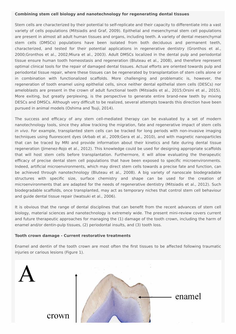
Enamel and dentin of the tooth crown are most often the first tissues to be affected following traumaticinjuries or carious lesions (Figure 1).

Figure 1. Tooth structure in physiological and pathological conditions. A) Histological section of ahuman premolar (blue colour: toluidine blue). B) Histological sections of human carious teeth. Left side:ground unstained section, showing mild carious injuries (red arrows) affecting only enamel. Right side:decalcified section stained with hematoxylin and eosin, showing a severe carious injury (red arrows) withbacterial invasion (asterisks: bacterial front within dentin). Abbreviations: d, dentin; e, enamel; p, pulp; td,tertiary dentin.
Prompt and efficient repair of enamel and/or dentin is fundamental to prevent infection and damageextending towards dental soft tissues (i.e., dental pulp, periodontium) and alveolar bone. The most used

approach for treating enamel and dentin harm is the substitution and restoration of the destroyed or lostdental hard tissues by sophisticated composite materials. However, traditional adhesive systems areunstable and fail over time, thus leading to marginal leakage and poor retention of the restoration in thetooth (Breschi et al., 2018). Therefore, a major task of nanotechnology in dentistry is to develop noveldurable materials and adhesive systems with improved enamel- and/or dentin-bonding performance inorder to increase the longevity of the restorations and prevent repeated treatments. Indeed, theintroduction of novel materials such as phosphine oxide initiators and monomethacrylate diluents has ledto dental composites with satisfactory and adequate properties (Kilambi et al., 2009). The introduction ofnanofillers and nanomaterials led to even more significant advances in terms of optimizing the propertiesand performance of the composites (Ilie et al., 2013;Goracci et al., 2014;Monterubbianesi et al., 2016).These nanotechnology-based strategies using cross-linking agents and Ca- and P-releasing means, whichmimic the process of natural dentin mineralization, have also reduced the degradation of the resin-dentinbonded interface (Mazzoni et al., 2018).
Ceramic-based materials are privileged by dentists for the restoration of damaged tooth crowns, mainlybecause of their superior aesthetic appearance and biocompatibility (Wittneben et al., 2017;Ozcan andJonasch, 2018). To overcome the fact that ceramic materials are brittle and prone to cracks propagation,several transformation/toughening mechanisms have been developed, leading to higher aging resistant-ceramics (Zhang et al., 2017) such as zirconia with exceptional toughness and flexural strength (Guazzatoet al., 2004).
Nanomodified materials could be also designed for controlling oral microbiota and the formation of dentalplaque, and, furthermore, for enhancing the mineralization process in the cases of enamel wear and/ordentin hypersensitivity due to the extensive consumption of acidic drinks (Orsini et al., 2013;Lelli et al.,2014). Indeed, the use of synthetic nanohydroxyapatite particles and other Ca-based nanomodifiedmaterials in dentifrices may offer a protective nanostructured coating on the tooth surface thatsimultaneously restores the lost minerals from enamel (Orsini et al., 2013;Lelli et al., 2014).
The preservation of the dental pulp, which is a living tissue ensuring tooth physiological function, is ofprime importance during the treatment of a damaged tooth crown. In very severe tooth injury, the pulp isalso affected and may lose its vitality. Therefore, the endodontic therapy (i.e., pulp tissue removal) isimposed in order to prevent further bacterial progression and damage of the surrounding alveolar bone.This is followed by disinfection of the dental root canals and the replacement of the pulp tissue withinorganic materials. Devitalized teeth are more fragile than normal teeth and consequently arepredisposed to postoperative fractures (DeRosa, 2006).
Regenerative endodontics aims at reforming the original pulp tissue morphology and physiology based ontissue engineering principles (Murray and Garcia-Godoy, 2006;Diogenes and Hargreaves, 2017).Nanomaterials can be used either alone or implemented with growth factors and stem cells in order tostimulate and enhance the regenerative capacity of the pulp tissue. Adjustment of biomaterials for dentalspecific purposes would require adjustments at a nanoscale level, thus allowing multifunctionality within agiven small surface, increasing the quality of targeting, and better controlling bioactive molecules delivery(Fioretti et al., 2011;Diogenes and Ruparel, 2017). Nanomaterials developed for endodontic purposes candeliver antibacterial and anti-inflammatory molecules, as well as growth factors that will guide thebehaviour (e.g., cell migration, proliferation, and differentiation) of the various dental pulp cell populations(e.g., pulp fibroblasts, endothelial cells, neuronal cells, immune cells). Biomimetic scaffolds composed ofnatural molecules, such as type I collagen, hyaluronic acid and chitosan, combined with nanoassembledmaterials possessing anti-inflammatory capabilities have been generated to stimulate pulp tissueregeneration and to prevent inflammation (Fioretti et al., 2011). Although such nanofibrous andmicroporous membranes have provided promising results, significant improvements are still needed to
Challenges in dentin-pulp regeneration

create scaffolds that promote proper pulp regeneration (Yamauchi et al., 2011;Albuquerque et al., 2014).
Numerous attempts using human DMSCs have been made in a variety of animal models in order to achievecomplete dental pulp regeneration (Figure 2A), a process that also requires neovascularization and re-innervation of this tissue. Experiments have shown that human DMSCs are capable to differentiate intoodontoblasts and to form dentin-like structures when transplanted together with a ceramic powder inimmune-compromised mice ex vivo (Gronthos et al., 2000;Gronthos et al., 2002). Similar studies haverevealed that human DMSCs seeded on poly-D, L-lactide/glycol scaffolds are able to regeneratevascularized pulp tissue when transplanted into an empty mouse tooth root canal (Volponi et al.,2010;Hayashi et al., 2015). Recently, new experimental strategies have been elaborated, where DMSCs-seeded scaffolds combined with bioactive molecules fulfil the empty pulp chamber immediately after pulpremoval (Albuquerque et al., 2014;Piva et al., 2014). Pilot studies in humans have demonstrated the safetyand efficacy of DMSCs for complete dental pulp regeneration and new dentin formation (Nakashima andIohara, 2017;Nakashima et al., 2017). Bone morphogenetic proteins (BMPs) have been commonly used foraccelerating and enhancing the production of dentin during dental pulp regeneration (Luiz de Oliveira daRosa et al., 2017). While these stem cell-based procedures appear to improve pulp tissue regeneration,their effectiveness for achieving accurate, precise and long-lasting therapies is still unclear. As a matter offact, most approaches aiming at dental pulp regeneration led to the formation of fibrotic tissues that canundergo degeneration over time or be replaced with bone. The possibility to decellularise healthy humandental pulps (Song et al., 2017) opens new horizons in regenerative dentistry since these decellularisedtissues could serve as natural scaffolds for supporting transplanted autologous DMSCs. Decellularised pulpsrepresent ideal biomaterials for hosting stem cells and guiding neovascularization and re-innervationwithin the regenerating tissues.
While significant efforts have been produced so far, regenerative procedures have to be furtherinvestigated in order to ultimately provide evidence of functional dental pulp regeneration in vivo (Figure2A) (Torabinejad and Faras, 2012;Diogenes and Ruparel, 2017).

Figure 2. Schematic representation of stem cell-based regenerative approaches in dentistry. A)Use of scaffolds (yellow colour) seeded with dental pulp stem cells (DPSCs) for the repair of the dentin-pulpcomplex. B) Use of scaffolds (yellow colour) seeded with periodontal ligament stem cells (PDLSCs) for theregeneration of the damaged periodontium.
Challenges in enamel regeneration
De novo formation of enamel in humans is one of the greatest challenges in regenerative dentistry, sinceamelogenesis is a very complex process and dental epithelial stem cells (DESCs) that could regenerateenamel are very rare in adult human teeth. Very few dental epithelial cells with stem cell properties havebeen isolated from the periodontal tissue (i.e., epithelial rests of Malassez, ERM). Experiments usingporcine ERM have demonstrated that these cells can differentiate into ameloblasts when co-cultured withdental pulp cells in vitro and can form enamel structures after their transplantation in vivo (Shinmura et al.,2008). Although ERM is a potential stem cell source for enamel regeneration, availability of these cells inhuman teeth is scarce, making thus necessary the identification of other epithelial stem cell populations ofnon-dental origin that could differentiate into enamel-producing ameloblasts.
Another key issue in generating new enamel is time. The accomplishment of proper enamel formationrequires many years, a time frame clearly incompatible with clinical needs. Moreover, mild disturbancesduring this process could lead to the generation of defective enamel (Cantu et al., 2017). Therefore, anyprocedure and technique that will be able to considerably accelerate the process of amelogenesis will be ofbenefit to the patients and dental community.

Periodontal diseases - Current periodontal therapies
Periodontium is a common site of pathologies that severely affect not only the structure of the surroundingtissues (i.e., dental root, alveolar bone) but also tooth functionality. Severe inflammation to theperiodontium leads to significant alterations in both the structure and quantity of the alveolar bone, aprocess that ultimately may cause tooth loss (Lindhe et al., 1983). Contemporary, periodontal therapiesinclude a wide range of surgical procedures along with use of bone grafts as tissue substitutes, barriermembranes for protecting the healing area from undesirable epithelial tissues (Howell et al., 1997;Aghalooand Moy, 2007), and growth factors for enhancing the healing capacity of the harmed tissues (Lynch et al.,1991b). Bone grafting materials, aiming to stimulate bone augmentation and periodontal regeneration,include intraoral or extraoral autografts, freeze-dried and fresh-frozen bone allografts, animal-derived bonedeproteinised xenografts, and hydroxyapatite and beta-tricalcium phosphate alloplasts (Pilipchuk et al.,2015;Sheikh et al., 2017). These grafting materials could be used alone or in association with variousgrowth factors. It has been shown that application of these regenerative methods in clinics allowed theformation of novel osseous tissues with similar to the pre-existent native bone characteristics (Scarano etal., 2006;De Angelis et al., 2011;Danesh-Sani et al., 2016;Clark et al., 2018). Even though, theseapproaches do not always ensure a predictable and desirable outcome of periodontal regeneration andoften result in healing with epithelial lining rather than new periodontal tissue formation (Lynch, 1992).
Challenges in periodontal regeneration
A fundamental goal in regenerative dentistry is to reconstruct a functional periodontium consisting of newcementum, alveolar bone and periodontal ligament around the tooth root damaged area (Figure 2B).DMSCs isolated from the periodontal space (i.e., periodontal ligament stem cells, dental follicle stem cells)of human teeth can differentiate into the various cell types of the periodontium in vitro when combinedwith different scaffolds or dentin matrix (Takahashi and Yamanaka, 2006;Washio et al., 2010;Arakaki et al.,2012;Yang et al., 2012). These stem cell populations have been shown to improve periodontal regenerationwhen transplanted into immunocompromised animals ex vivo, indicating their great potential for futurestem cell-based therapies in dentistry (Seo et al., 2004;Caton et al., 2011). A variety of growth factors havebeen also used for improving the regenerative efficacy of stem cells in the periodontium. Diverseexperiments have demonstrated that platelet-derived growth factors (PDGFs) stimulate periodontal tissueregeneration (Lynch et al., 1991b;Howell et al., 1997;Clark et al., 2018), while BMPs enhance alveolar boneand cementum production (Lynch et al., 1989;Howell et al., 1997;Selvig et al., 2002). However, excessivebone formation that results in tooth ankylosis can be a frequent side effect following the use of BMPs, sincethese molecules favour and direct stem cells differentiation towards the osteogenic fate.
Clinical studies have demonstrated that enamel matrix derivatives also assist and promote periodontaltissue regeneration (Miron et al., 2016b;Miron et al., 2017). Advanced new bone grafting materials withimproved physicochemical properties have been used as carriers of enamel protein derivatives in order tofurther improve their clinical performance (Miron et al., 2016a;Miron et al., 2016b). Nevertheless, despitethe very encouraging clinical outcomes, the mechanism of action of these enamel matrix molecules is notyet clear.
TOOTH LOSS
Current dental implant treatments
The use of dental implants has become a common and successful treatment for replacing missing teeth forpathologic, traumatic and genetic causes (Figure 3) (Esposito et al., 2014).

Figure 3. Tooth replacement and correction of aesthetics in a patient. A) Preoperative intraoralview of a young patient with a congenital missing tooth and compromised aesthetics of the other teeth. B)Upon orthodontic, surgical and implant treatment, the teeth were prepared for aesthetic prostheticrehabilitation. Asterisk indicates dental implant impression coping. C) Postoperative intraoral view of thepatient with the final ceramic restorations.
A typical dental implant is composed of a metal screw part that interfaces and integrates within thealveolar bone, and another part where tooth crown substitutes are placed. The retention of a dentalimplant requires its close contact with the alveolar bone, a process termed osseointegration. Despite theirlarge and regular usage in dental clinics, implants still need significant improvements, particularly in theircapacity to stimulate cellular events at the implantation site that would guarantee their long-termintegration and retention (Variola et al., 2009;Variola et al., 2011). The use of nanotechnology hasimproved the osseointegration of implants by modifying their surfaces, thus allowing the shortening of thehealing period (Barbucci et al., 2003;Mendonca et al., 2008). Indeed, zinc-modified calcium silicatecoatings, nanohydroxyapatite-blasted surfaces, nanotextured blasted titanium surfaces, as well as goldnanoparticles coated surfaces have considerably enhanced the adhesive properties of implants andtherefore their osseointegration (Coelho et al., 2016;Heo et al., 2016;Bezerra et al., 2017;Yu et al., 2017).However, there is a major risk of infection of tissues surrounding the implant, a pathology termed peri-implantitis (Singh, 2011). In vitro and in vivo studies have shown that the incorporation of antibacterialagents to dental implants (e.g., silver nanoparticles) could partly prevent the growth of bacteria andtherefore decrease the percentage of implant treatments failure (Godoy-Gallardo et al., 2016;Pokrowieckiet al., 2017). It has been also demonstrated that gallium-modified chitosan/poly (acrylic acid) bilayercoatings might improve titanium implant performances by limiting bacterial adhesion and proliferation(Bonifacio et al., 2017). Dental implants have also benefited from regenerative technologies usingscaffolds, stem cells and growth factors that contribute to enhanced osseointegration and host tissueresponse (Pilipchuk et al., 2015). Despite a good number of preclinical studies in large animal models forguided bone and periodontal regeneration around implants using growth factors and protein deliverysystems (Lynch et al., 1991a;Selvig et al., 2002;Sauerbier et al., 2011;Alvarez et al., 2012;Larsson et al.,2016), and the evident clinical advantages, well-conducted human randomized clinical studies that willdefinitively validate these approaches are still lacking (Caton et al., 2011) (Seo et al., 2004;Rickert et al.,2011;Sauerbier et al., 2011). To date, only few randomized clinical trials have been performed andtherefore it is absolutely necessary the realization of larger trials (Kaigler et al., 2013;Kaigler et al., 2015).
Challenges in entire tooth regeneration
Regeneration of entire brand-new teeth for the replacement of missing or lost teeth is the most ambitiousgoal in dentistry and requires the use and recombination of dental mesenchymal and epithelial stem cells(Papagerakis, 2013;Otsu et al., 2014). DMSCs can form all mesenchymal components of the tooth organand the surrounding tissues such as dentin, cementum and alveolar bone, while DESCs are essential for thegeneration of enamel. Since most of the dental epithelial cell populations disappear shortly after tootheruption and DESCs are limited in human adult teeth, current knowledge on DESCs has been obtainedmainly from rodents, where they contribute to the renewal of the enamel in the continuously growingincisors (Mitsiadis and Harada, 2015).

Two main strategies have been elaborated for constructing whole new teeth (Mitsiadis and Papagerakis,2011;Otsu et al., 2014;Mitsiadis and Harada, 2015). One approach consists in recombining and culturingDESCs and DMSCs in vitro until they will form a tooth germ that subsequently will be transplanted into thealveolar bone. It is expected that this tooth germ will further develop and grow, erupt and finally become afunctional tooth. Another approach relies to tooth-shaped polymeric biodegradable scaffolds that are filledwith both DESCs and DMSCs and implanted into the alveolar bone, expecting that will finally give rise tofunctional teeth. The three-dimensional structure of the scaffolds should drive the differentiation of thetransplanted stem cells into odontoblasts and ameloblasts (Bluteau et al., 2008). Indeed, severalexperiments in mice using bioengineered approaches have revealed that functional teeth with appropriatecrowns, dental pulp, roots and periodontal ligament can be formed following their implantation inmandibles (Jimenez-Rojo et al., 2012;Oshima and Tsuji, 2014;Otsu et al., 2014;Mitsiadis and Harada, 2015).However, similar results have not yet been obtained with human cells, due mainly to the limited number ofadult DESCs and the significantly elongated time period that is needed for proper human toothdevelopment. Penury of DESCs within human adult teeth might be successfully addressed bydifferentiating patient-specific inducible pluripotent stem cells (iPSCs) into DESCs. Certain studies haveshown that iPSCs technology could be successfully used in regenerative dentistry, since re-aggregation ofhuman iPSC-derived mesenchymal cells and mouse dental epithelium resulted in the formation of entireteeth ex vivo (Takahashi and Yamanaka, 2006;Arakaki et al., 2012;Otsu et al., 2014;Mitsiadis and Harada,2015). Although promising, this approach also needs further investigation, as effective protocols for thedifferentiation of human DESCs from iPSCs are not available yet.
Novel Platforms for Tooth Modelling, Drug Discovery and Diagnostics
Use of organoids and organ-on-chip devices in dentistry
Appropriate systems for modelling human organs and pathologies represent a constant need in allbranches of biomedical research and practice, included dentistry. Animal models and two-dimensional (2D)human cell culture systems have been traditionally used for most pre-clinical studies aiming at thedevelopment of novel cell-based and pharmaceutical therapies. However, translation of preclinical resultsinto effective treatments remains poor (Weeber et al., 2017), highlighting the need for accurate human-emulation systems (Skardal et al., 2016). In this context, great expectations are accompanying the recentdevelopments on spheroids, organoids, microfluidics and organ-on-chip technologies.
Spheroids and organoids are 3D culture systems, obtained by primary stem cells and tissues, which areincreasingly used to model and understand tissue-specific physiology (Figure 4). The 3D structure of bothsystems allows establishment of complex cell-cell interactions and gradients of oxygen, nutrients andsoluble signals that generate tissue-specific heterogeneous cell types. Organoids provide additionalfeatures compared to spheroids, as they are able of self-organization, exhibit similar architecture to thetissue of origin and exert tissue-specific complex functions (Yin et al., 2016).

Figure 4. Overview of new platforms for tooth modelling, drug discovery and diagnostics. Fromtop right clockwise: LabDisk systems for bacteria detection (adapted from Czilwik et al., 2015), graphene-based wearable sensors (adapted from Mannoor et al., 2012), organ-on-chip (Emulate©), organoids(courtesy of Dr. T. Valenta, University of Zurich), spheroids (from (Natsiou et al., 2017).
Dental spheroids or dentospheres have been successfully generated from both mouse and human dentalepithelial and mesenchymal (e.g., pulp and periodontium) tissues (Berahim et al., 2011;Bonnamain et al.,2011;Miquel, 2011;Natsiou et al., 2017). Epithelial dentospheres formed from mouse incisors and molars,upon modulation of their culture conditions, have either demonstrated strong stem cell capabilities orgenerated differentiation gradients (Natsiou et al., 2017). Human mesenchymal spheroids consistentlydisplayed higher expression of odontoblast- and periodontal-specific differentiation markers whencompared to 2D culture systems (Berahim et al., 2011;Bonnamain et al., 2011). These aspects makespheroids valuable tools for studying cytodifferentiation events in human dental tissues in vitro, and mightbe a source of stem cells for personalized dental regenerative approaches. Indeed, genetic diseases areoften associated with dental anomalies (Mitsiadis and Luder, 2011;Klein et al., 2014), which could beproperly modelled and investigated in patient-specific dental spheroids. Similarly, such spheroidsrepresent novel tools for studying the behaviour of definite human dental cell populations to novelmaterials and drugs. However, despite their incontestable advantages, it is not yet clear to what extentspheroids and organoids could faithfully represent the in vivo dental status. Organoids and spheroids infact lack many features that are critical for the function of any organ, such as vasculature, innervation,mechanical cues, and immune responses (Ingber, 2016).
These limitations are the basis for the rise of microfluidic “organ-on-chip” systems. Organ-on-chips aremicrofluidic or nanofluidic devices composed of different chambers, where organ-specific elements such asepithelial, mesenchymal, endothelial and neuronal cells and/or tissues are cultured (Figure 4) (Bhatia andIngber, 2014). Porous membranes allow the passage of molecular cues between the different chambers,while blood circulation is simulated by the regulated flow of enriched and specific media. These devicescan incorporate mechanical forces to recreate physiological movements and stresses (Bhatia and Ingber,2014), as well as electrical stimuli, allowing the modelling and analysis of complex organ-specific

physiological and pathological processes. Importantly, circulating immune cells and even livingmicrobiomes can be integrated in these devices to mimic complex organ-level responses (Bhatia andIngber, 2014;Ingber, 2016). Microfluidic devices involving dental tissues have been used for the first timefor analysing the crosstalk of tooth germs and DMSCs with trigeminal innervation (Pagella et al.,2014;Pagella et al., 2015). These pioneer studies have shown that microfluidics can faithfully imitate andreproduce the in vivo dental situation and thus reinforce the options to study dental tissues in “organ-on-chip” systems. Results obtained from these devices contribute to successfully emulate human- andpatient-specific dental tissues in vitro. The most ambitious goal of these microfluidic devices consists inthe modelling of the functional interconnection between different human organs, by the realization of so-called “bodies-on-chip”. In fact, “organ-on-chip” devices can be interconnected via microfluidic tubes,which emulate systemic blood circulation. Via such emulated vasculature, molecular cues as well asimmune responses can propagate to all organs, allowing the study of body-level responses to organ-specific events (Ingber, 2016). Such approach is already being used and optimized for modelling ofpharmacokinetic and pharmacodynamics of systemic human drug responses (Prantil-Baun et al., 2018).With these platforms, it will be possible to study body-level responses to the various dental pathologies.While it is long known that oral diseases are strongly associated with a plethora of systemic disorders,including atherosclerosis, stoke, and systemic infections (Slavkin and Baum, 2000), the mechanismsunderlying these connections and thus their therapeutic relevance are far from being understood. A human“body-on-chip” system would finally allow understanding how dental and systemic health are correlated,thus testing how the treatment of dental diseases affects general physiology.
Microfluidic devices could be also employed for the detection of both specific metabolites (Wu et al., 2017)and particular bacterial strains (Czilwik et al., 2015) that are involved in chronic diseases. Within the dentalfield, microfluidic devices have been used for the detection of pathogenic bacteria that lead toperiodontitis and carious diseases. Recent technological developments allowed the significant shorteningof this process via optimization of fully automated and integrated DNA extraction, multiplex PCR pre-amplification and species-specific real-time PCR (Figure 4) (Chen et al., 2007;Czilwik et al., 2015). Thesesystems allow a fast processing of samples, without loss of sensitivity and complex laboratoryinstrumentation.
Recent nanotechnology tools permitted the detection of single oral bacteria in situ via graphene-biosensors equipped with electrodes and antennae, which were printed onto enamel as “temporary-tattoos” (Figure 4) (Mannoor et al., 2012). These wearable devices are thus capable of monitoring bacteriapresent in the mouth and more specifically on the tooth surface (Mannoor et al., 2012). The same principlehas been applied very recently for detecting and identifying ingested food and liquids (Tseng et al., 2018).These mounted onto enamel nanodevices could be optimized to sense a wide variety of properties ofdrinks, such as alcohol content, salinity, sugars, pH, and temperature (Tseng et al., 2018). Although still intheir experimental phase, such sensors represent excellent tools for the refined control and understandingof oral environment that will greatly help the field of preventive dentistry.
Concluding remarks
The important advances in stem cells and materials sciences are driving innovative approaches indentistry. These progresses hold a great promise for the development of efficient and personalizedtreatments in the near future. At the same time, optimization of sophisticated systems for the modellingand monitoring of human tissues is leading to unprecedented possibilities for the study of diseases,diagnostics, and drug testing. Although extremely exciting, most of these approaches are not yetapplicable in dental clinics. Stem cell-based dental regenerative approaches still lack reliable techniquesthat allow controlling stem cell behaviour upon transplantation. Similarly, state-of-the-art diagnosticsystems still need to be validated in proper clinical settings. Nevertheless, these innovative approachesoffer exciting perspectives to regenerative dentistry and might prove fundamental for the long-soughtregeneration of fully functional dental tissues.

Acknowledgements
This work was supported by institutional funds from University of Zurich. The authors contributed to thewriting, reading, and editing of the present review article. The authors confirm that there are no conflicts ofinterest associated with this work.
This work has been published in Frontiers in Physiology, 08 August 2018
doi: 10.3389/fphys.2018.01102
Conflict of Interest Statement
The authors declare that the research was conducted in the absence of any commercial or financialrelationship that could be construed as a potential conflict of interest.
References
Aghaloo, T.L., and Moy, P.K. (2007). Which hard tissue augmentation techniques are the most successful infurnishing bony support for implant placement? Int J Oral Maxillofac Implants 22 Suppl, 49-70.
Albuquerque, M.T., Valera, M.C., Nakashima, M., Nor, J.E., and Bottino, M.C. (2014). Tissue-engineering-based strategies for regenerative endodontics. J Dent Res 93, 1222-1231.
Alvarez, P., Hee, C.K., Solchaga, L., Snel, L., Kestler, H.K., Lynch, S.E., and Hollinger, J.O. (2012). Growthfactors and craniofacial surgery. J Craniofac Surg 23, 20-29.
Arakaki, M., Ishikawa, M., Nakamura, T., Iwamoto, T., Yamada, A., Fukumoto, E., Saito, M., Otsu, K., Harada,H., Yamada, Y., and Fukumoto, S. (2012). Role of epithelial-stem cell interactions during dental celldifferentiation. J Biol Chem 287, 10590-10601.
Arbab, A.S., Janic, B., Haller, J., Pawelczyk, E., Liu, W., and Frank, J.A. (2009). In Vivo Cellular Imaging forTranslational Medical Research. Curr Med Imaging Rev 5, 19-38.
Banerjee, A., Thompson, I.D., and Watson, T.F. (2011). Minimally invasive caries removal using bio-activeglass air-abrasion. J Dent 39, 2-7.
Barbucci, R., Pasqui, D., Wirsen, A., Affrossman, S., Curtis, A., and Tetta, C. (2003). Micro and nano-structured surfaces. J. Mater. Sci.-Mater. Med. 14, 721-725.
Berahim, Z., Moharamzadeh, K., Rawlinson, A., and Jowett, A.K. (2011). Biologic interaction of three-dimensional periodontal fibroblast spheroids with collagen-based and synthetic membranes. J Periodontol82, 790-797.
Beuer, F., Schweiger, J., and Edelhoff, D. (2008). Digital dentistry: an overview of recent developments forCAD/CAM generated restorations. Br Dent J 204, 505-511.
Bezerra, F., Ferreira, M.R., Fontes, G.N., Da Costa Fernandes, C.J., Andia, D.C., Cruz, N.C., Da Silva, R.A., andZambuzzi, W.F. (2017). Nano hydroxyapatite-blasted titanium surface affects pre-osteoblast morphology bymodulating critical intracellular pathways. Biotechnol Bioeng 114, 1888-1898.
Bhatia, S.N., and Ingber, D.E. (2014). Microfluidic organs-on-chips. Nat Biotechnol 32, 760-772.
Bluteau, G., Luder, H.U., De Bari, C., and Mitsiadis, T.A. (2008). Stem cells for tooth engineering. Eur CellMater 16, 1-9.

Bonifacio, M.A., Cometa, S., Dicarlo, M., Baruzzi, F., De Candia, S., Gloria, A., Giangregorio, M.M., Mattioli-Belmonte, M., and De Giglio, E. (2017). Gallium-modified chitosan/poly(acrylic acid) bilayer coatings forimproved titanium implant performances. Carbohydr Polym 166, 348-357.
Bonnamain, V., Neveu, I., and Naveilhan, P. (2011). In vitro analyses of the immunosuppressive propertiesof neural stem/progenitor cells using anti-CD3/CD28-activated T cells. Methods Mol Biol 677, 233-243.
Boyde, A. (1997). Microstructure of enamel. Ciba Found Symp 205, 18-27; discussion 27-31.
Breschi, L., Maravic, T., Cunha, S.R., Comba, A., Cadenaro, M., Tjaderhane, L., Pashley, D.H., Tay, F.R., andMazzoni, A. (2018). Dentin bonding systems: From dentin collagen structure to bond preservation andclinical applications. Dent Mater 34, 78-96.
Cantu, C., Pagella, P., Shajiei, T.D., Zimmerli, D., Valenta, T., Hausmann, G., Basler, K., and Mitsiadis, T.A.(2017). A cytoplasmic role of Wnt/beta-catenin transcriptional cofactors Bcl9, Bcl9l, and Pygopus in toothenamel formation. Sci Signal 10.
Caton, J., Bostanci, N., Remboutsika, E., De Bari, C., and Mitsiadis, T.A. (2011). Future dentistry: cell therapymeets tooth and periodontal repair and regeneration. J Cell Mol Med 15, 1054-1065.
Chen, Z., Mauk, M.G., Wang, J., Abrams, W.R., Corstjens, P.L., Niedbala, R.S., Malamud, D., and Bau, H.H.(2007). A microfluidic system for saliva-based detection of infectious diseases. Ann N Y Acad Sci 1098, 429-436.
Clark, D., Rajendran, Y., Paydar, S., Ho, S., Cox, D., Ryder, M., Dollard, J., and Kao, R.T. (2018). Advancedplatelet-rich fibrin and freeze-dried bone allograft for ridge preservation: A randomized controlled clinicaltrial. J Periodontol 89, 379-387.
Coelho, P.G., Zavanelli, R.A., Salles, M.B., Yeniyol, S., Tovar, N., and Jimbo, R. (2016). Enhanced BoneBonding to Nanotextured Implant Surfaces at a Short Healing Period: A Biomechanical Tensile Testing in theRat Femur. Implant Dent 25, 322-327.
Czilwik, G., Messinger, T., Strohmeier, O., Wadle, S., Von Stetten, F., Paust, N., Roth, G., Zengerle, R.,Saarinen, P., Niittymaki, J., Mcallister, K., Sheils, O., O'leary, J., and Mark, D. (2015). Rapid and fullyautomated bacterial pathogen detection on a centrifugal-microfluidic LabDisk using highly sensitive nestedPCR with integrated sample preparation. Lab Chip 15, 3749-3759.
Danesh-Sani, S.A., Loomer, P.M., and Wallace, S.S. (2016). A comprehensive clinical review of maxillarysinus floor elevation: anatomy, techniques, biomaterials and complications. Br J Oral Maxillofac Surg 54,724-730.
De Angelis, N., Felice, P., Pellegrino, G., Camurati, A., Gambino, P., and Esposito, M. (2011). Guided boneregeneration with and without a bone substitute at single post-extractive implants: 1-year post-loadingresults from a pragmatic multicentre randomised controlled trial. Eur J Oral Implantol 4, 313-325.
Del Fabbro, M., Taschieri, S., Lodi, G., Banfi, G., and Weinstein, R.L. (2015). Magnification devices forendodontic therapy. Cochrane Database Syst Rev, CD005969.
Derosa, T.A. (2006). A retrospective evaluation of pulpotomy as an alternative to extraction. Gen Dent 54,37-40.
Diogenes, A., and Hargreaves, K.M. (2017). Microbial Modulation of Stem Cells and Future Directions inRegenerative Endodontics. J Endod 43, S95-S101.
Diogenes, A., and Ruparel, N.B. (2017). Regenerative Endodontic Procedures: Clinical Outcomes. Dent Clin
North Am 61, 111-125.
Esposito, M., Ardebili, Y., and Worthington, H.V. (2014). Interventions for replacing missing teeth: differenttypes of dental implants. Cochrane Database Syst Rev, CD003815.
Fioretti, F., Mendoza-Palomares, C., Avoaka-Boni, M.C., Ramaroson, J., Bahi, S., Richert, L., Granier, F.,Benkirane-Jessel, N., and Haikel, Y. (2011). Nano-odontology: nanostructured assemblies for endodonticregeneration. J Biomed Nanotechnol 7, 471-475.
Gera, A., Steinberg, G.K., and Guzman, R. (2010). In vivo neural stem cell imaging: current modalities andfuture directions. Regen Med 5, 73-86.
Godoy-Gallardo, M., Manzanares-Cespedes, M.C., Sevilla, P., Nart, J., Manzanares, N., Manero, J.M., Gil, F.J.,Boyd, S.K., and Rodriguez, D. (2016). Evaluation of bone loss in antibacterial coated dental implants: Anexperimental study in dogs. Mater Sci Eng C Mater Biol Appl 69, 538-545.
Goracci, C., Cadenaro, M., Fontanive, L., Giangrosso, G., Juloski, J., Vichi, A., and Ferrari, M. (2014).Polymerization efficiency and flexural strength of low-stress restorative composites. Dent Mater 30, 688-694.
Gronthos, S., Brahim, J., Li, W., Fisher, L.W., Cherman, N., Boyde, A., Denbesten, P., Robey, P.G., and Shi, S.(2002). Stem cell properties of human dental pulp stem cells. J Dent Res 81, 531-535.
Gronthos, S., Mankani, M., Brahim, J., Robey, P.G., and Shi, S. (2000). Postnatal human dental pulp stemcells (DPSCs) in vitro and in vivo. Proc Natl Acad Sci U S A 97, 13625-13630.
Guazzato, M., Albakry, M., Ringer, S.P., and Swain, M.V. (2004). Strength, fracture toughness andmicrostructure of a selection of all-ceramic materials. Part II. Zirconia-based dental ceramics. Dent Mater20, 449-456.
Hammerle, C.H., Stone, P., Jung, R.E., Kapos, T., and Brodala, N. (2009). Consensus statements andrecommended clinical procedures regarding computer-assisted implant dentistry. Int J Oral MaxillofacImplants 24 Suppl, 126-131.
Hancocks, S. (2017). What is digital about dentistry? Br Dent J 223, 305.
Hayashi, Y., Murakami, M., Kawamura, R., Ishizaka, R., Fukuta, O., and Nakashima, M. (2015). CXCL14 andMCP1 are potent trophic factors associated with cell migration and angiogenesis leading to higherregenerative potential of dental pulp side population cells. Stem Cell Res Ther 6, 111.
Heo, D.N., Ko, W.K., Lee, H.R., Lee, S.J., Lee, D., Um, S.H., Lee, J.H., Woo, Y.H., Zhang, L.G., Lee, D.W., andKwon, I.K. (2016). Titanium dental implants surface-immobilized with gold nanoparticles as osteoinductiveagents for rapid osseointegration. J Colloid Interface Sci 469, 129-137.
Howell, T.H., Fiorellini, J.P., Paquette, D.W., Offenbacher, S., Giannobile, W.V., and Lynch, S.E. (1997). Aphase I/II clinical trial to evaluate a combination of recombinant human platelet-derived growth factor-BBand recombinant human insulin-like growth factor-I in patients with periodontal disease. J Periodontol 68,1186-1193.
Ilie, N., Kessler, A., and Durner, J. (2013). Influence of various irradiation processes on the mechanicalproperties and polymerisation kinetics of bulk-fill resin based composites. J Dent 41, 695-702.
Ingber, D.E. (2016). Reverse Engineering Human Pathophysiology with Organs-on-Chips. Cell 164, 1105-1109.

Iwatsuki, S., Honda, M.J., Harada, H., and Ueda, M. (2006). Cell proliferation in teeth reconstructed fromdispersed cells of embryonic tooth germs in a three-dimensional scaffold. Eur J Oral Sci 114, 310-317.
Jimenez-Rojo, L., Granchi, Z., Graf, D., and Mitsiadis, T.A. (2012). Stem Cell Fate Determination duringDevelopment and Regeneration of Ectodermal Organs. Front Physiol 3, 107.
Kaigler, D., Avila-Ortiz, G., Travan, S., Taut, A.D., Padial-Molina, M., Rudek, I., Wang, F., Lanis, A., andGiannobile, W.V. (2015). Bone Engineering of Maxillary Sinus Bone Deficiencies Using Enriched CD90+Stem Cell Therapy: A Randomized Clinical Trial. J Bone Miner Res 30, 1206-1216.
Kaigler, D., Pagni, G., Park, C.H., Braun, T.M., Holman, L.A., Yi, E., Tarle, S.A., Bartel, R.L., and Giannobile,W.V. (2013). Stem cell therapy for craniofacial bone regeneration: a randomized, controlled feasibility trial.Cell Transplant 22, 767-777.
Kilambi, H., Cramer, N.B., Schneidewind, L.H., Shah, P., Stansbury, J.W., and Bowman, C.N. (2009).Evaluation of highly reactive mono-methacrylates as reactive diluents for BisGMA-based dentalcomposites. Dent Mater 25, 33-38.
Klein, C., Le Goff, C., Topouchian, V., Odent, S., Violas, P., Glorion, C., and Cormier-Daire, V. (2014).Orthopedics management of acromicric dysplasia: follow up of nine patients. Am J Med Genet A 164A, 331-337.
Larsson, L., Decker, A.M., Nibali, L., Pilipchuk, S.P., Berglundh, T., and Giannobile, W.V. (2016). RegenerativeMedicine for Periodontal and Peri-implant Diseases. J Dent Res 95, 255-266.
Lelli, M., Putignano, A., Marchetti, M., Foltran, I., Mangani, F., Procaccini, M., Roveri, N., and Orsini, G.(2014). Remineralization and repair of enamel surface by biomimetic Zn-carbonate hydroxyapatitecontaining toothpaste: a comparative in vivo study. Front Physiol 5, 333.
Levato, C.M., Farman, A.G., and Miles, D.A. (2015). The "inevitability" of digital radiography in dentistry.Compend Contin Educ Dent 36, 238, 240.
Lindhe, J., Haffajee, A.D., and Socransky, S.S. (1983). Progression of periodontal disease in adult subjects inthe absence of periodontal therapy. J Clin Periodontol 10, 433-442.
Luiz De Oliveira Da Rosa, W., Machado Da Silva, T., Fernando Demarco, F., Piva, E., and Fernandes Da Silva,A. (2017). Could the application of bioactive molecules improve vital pulp therapy success? A systematicreview. J Biomed Mater Res A 105, 941-956.
Lynch, C. (2017). Defining digital dentistry: A survey of recent literature. J Dent 59, 1.
Lynch, S.E. (1992). Methods for evaluation of regenerative procedures. J Periodontol 63, 1085-1092.
Lynch, S.E., Buser, D., Hernandez, R.A., Weber, H.P., Stich, H., Fox, C.H., and Williams, R.C. (1991a). Effectsof the platelet-derived growth factor/insulin-like growth factor-I combination on bone regeneration aroundtitanium dental implants. Results of a pilot study in beagle dogs. J Periodontol 62, 710-716.
Lynch, S.E., De Castilla, G.R., Williams, R.C., Kiritsy, C.P., Howell, T.H., Reddy, M.S., and Antoniades, H.N.(1991b). The effects of short-term application of a combination of platelet-derived and insulin-like growthfactors on periodontal wound healing. J Periodontol 62, 458-467.
Lynch, S.E., Williams, R.C., Polson, A.M., Howell, T.H., Reddy, M.S., Zappa, U.E., and Antoniades, H.N.(1989). A combination of platelet-derived and insulin-like growth factors enhances periodontalregeneration. J Clin Periodontol 16, 545-548.

Mannoor, M.S., Tao, H., Clayton, J.D., Sengupta, A., Kaplan, D.L., Naik, R.R., Verma, N., Omenetto, F.G., andMcalpine, M.C. (2012). Graphene-based wireless bacteria detection on tooth enamel. Nat Commun 3, 763.
Mazzoni, A., Angeloni, V., Comba, A., Maravic, T., Cadenaro, M., Tezvergil-Mutluay, A., Pashley, D.H., Tay,F.R., and Breschi, L. (2018). Cross-linking effect on dentin bond strength and MMPs activity. Dent Mater 34,288-295.
Mendonca, G., Mendonca, D.B., Aragao, F.J., and Cooper, L.F. (2008). Advancing dental implant surfacetechnology--from micron- to nanotopography. Biomaterials 29, 3822-3835.
Miquel, A. (2011). [In the 11- M terrorist tragedy in Madrid]. Rev Clin Esp 211, 158-162.
Miron, R.J., Chandad, F., Buser, D., Sculean, A., Cochran, D.L., and Zhang, Y. (2016a). Effect of EnamelMatrix Derivative Liquid on Osteoblast and Periodontal Ligament Cell Proliferation and Differentiation. JPeriodontol 87, 91-99.
Miron, R.J., Sculean, A., Cochran, D.L., Froum, S., Zucchelli, G., Nemcovsky, C., Donos, N., Lyngstadaas, S.P.,Deschner, J., Dard, M., Stavropoulos, A., Zhang, Y., Trombelli, L., Kasaj, A., Shirakata, Y., Cortellini, P., Tonetti,M., Rasperini, G., Jepsen, S., and Bosshardt, D.D. (2016b). Twenty years of enamel matrix derivative: thepast, the present and the future. J Clin Periodontol 43, 668-683.
Miron, R.J., Zucchelli, G., Pikos, M.A., Salama, M., Lee, S., Guillemette, V., Fujioka-Kobayashi, M., Bishara, M.,Zhang, Y., Wang, H.L., Chandad, F., Nacopoulos, C., Simonpieri, A., Aalam, A.A., Felice, P., Sammartino, G.,Ghanaati, S., Hernandez, M.A., and Choukroun, J. (2017). Use of platelet-rich fibrin in regenerativedentistry: a systematic review. Clin Oral Investig 21, 1913-1927.
Mitsiadis, T.A., and Graf, D. (2009). Cell fate determination during tooth development and regeneration.Birth Defects Res C Embryo Today 87, 199-211.
Mitsiadis, T.A., and Harada, H. (2015). Regenerated teeth: the future of tooth replacement. An update.Regen Med 10, 5-8.
Mitsiadis, T.A., and Luder, H.U. (2011). Genetic basis for tooth malformations: from mice to men and backagain. Clin Genet 80, 319-329.
Mitsiadis, T.A., Orsini, G., and Jimenez-Rojo, L. (2015). Stem cell-based approaches in dentistry. Eur CellMater 30, 248-257.
Mitsiadis, T.A., and Papagerakis, P. (2011). Regenerated teeth: the future of tooth replacement? Regen Med6, 135-139.
Mitsiadis, T.A., Woloszyk, A., and Jimenez-Rojo, L. (2012). Nanodentistry: combining nanostructuredmaterials and stem cells for dental tissue regeneration. Nanomedicine (Lond) 7, 1743-1753.
Miura, M., Gronthos, S., Zhao, M., Lu, B., Fisher, L.W., Robey, P.G., and Shi, S. (2003). SHED: stem cells fromhuman exfoliated deciduous teeth. Proc Natl Acad Sci U S A 100, 5807-5812.
Monterubbianesi, R., Orsini, G., Tosi, G., Conti, C., Librando, V., Procaccini, M., and Putignano, A. (2016).Spectroscopic and Mechanical Properties of a New Generation of Bulk Fill Composites. Front Physiol 7, 652.
Murray, P.E., and Garcia-Godoy, F. (2006). The outlook for implants and endodontics: a review of the tissueengineering strategies to create replacement teeth for patients. Dent Clin North Am 50, 299-315, x.
Nakashima, M., and Iohara, K. (2017). Recent Progress in Translation from Bench to a Pilot Clinical Study onTotal Pulp Regeneration. J Endod 43, S82-S86.

Nakashima, M., Iohara, K., Murakami, M., Nakamura, H., Sato, Y., Ariji, Y., and Matsushita, K. (2017). Pulpregeneration by transplantation of dental pulp stem cells in pulpitis: a pilot clinical study. Stem Cell ResTher 8, 61.
Natsiou, D., Granchi, Z., Mitsiadis, T.A., and Jimenez-Rojo, L. (2017). Generation of Spheres from DentalEpithelial Stem Cells. Front Physiol 8, 7.
Orsini, G., Jimenez-Rojo, L., Natsiou, D., Putignano, A., and Mitsiadis, T.A. (2015). In vivo administration ofdental epithelial stem cells at the apical end of the mouse incisor. Front Physiol 6, 112.
Orsini, G., Procaccini, M., Manzoli, L., Sparabombe, S., Tiriduzzi, P., Bambini, F., and Putignano, A. (2013). A3-day randomized clinical trial to investigate the desensitizing properties of three dentifrices. J Periodontol84, e65-73.
Oshima, M., and Tsuji, T. (2014). Functional tooth regenerative therapy: tooth tissue regeneration andwhole-tooth replacement. Odontology 102, 123-136.
Otsu, K., Kumakami-Sakano, M., Fujiwara, N., Kikuchi, K., Keller, L., Lesot, H., and Harada, H. (2014). Stemcell sources for tooth regeneration: current status and future prospects. Front Physiol 5, 36.
Ozcan, M., and Jonasch, M. (2018). Effect of Cyclic Fatigue Tests on Aging and Their TranslationalImplications for Survival of All-Ceramic Tooth-Borne Single Crowns and Fixed Dental Prostheses. JProsthodont 27, 364-375.
Pagella, P., Miran, S., and Mitsiadis, T. (2015). Analysis of Developing Tooth Germ Innervation UsingMicrofluidic Co-culture Devices. J Vis Exp, e53114.
Pagella, P., Neto, E., Jimenez-Rojo, L., Lamghari, M., and Mitsiadis, T.A. (2014). Microfluidics co-culturesystems for studying tooth innervation. Front Physiol 5, 326.
Papagerakis, P., Mitsiadis, T. (2013). Development and structure of teeth and periodontal tissues.: JohnWiley & Sons, Inc.
Pilipchuk, S.P., Plonka, A.B., Monje, A., Taut, A.D., Lanis, A., Kang, B., and Giannobile, W.V. (2015). Tissueengineering for bone regeneration and osseointegration in the oral cavity. Dent Mater 31, 317-338.
Piva, E., Silva, A.F., and Nor, J.E. (2014). Functionalized scaffolds to control dental pulp stem cell fate. JEndod 40, S33-40.
Pokrowiecki, R., Zareba, T., Szaraniec, B., Palka, K., Mielczarek, A., Menaszek, E., and Tyski, S. (2017). Invitro studies of nanosilver-doped titanium implants for oral and maxillofacial surgery. Int J Nanomedicine12, 4285-4297.
Prantil-Baun, R., Novak, R., Das, D., Somayaji, M.R., Przekwas, A., and Ingber, D.E. (2018). PhysiologicallyBased Pharmacokinetic and Pharmacodynamic Analysis Enabled by Microfluidically Linked Organs-on-Chips. Annu Rev Pharmacol Toxicol 58, 37-64.
Rickert, D., Sauerbier, S., Nagursky, H., Menne, D., Vissink, A., and Raghoebar, G.M. (2011). Maxillary sinusfloor elevation with bovine bone mineral combined with either autogenous bone or autogenous stem cells:a prospective randomized clinical trial. Clin Oral Implants Res 22, 251-258.
Sauerbier, S., Rickert, D., Gutwald, R., Nagursky, H., Oshima, T., Xavier, S.P., Christmann, J., Kurz, P., Menne,D., Vissink, A., Raghoebar, G., Schmelzeisen, R., Wagner, W., and Koch, F.P. (2011). Bone marrowconcentrate and bovine bone mineral for sinus floor augmentation: a controlled, randomized, single-blindedclinical and histological trial--per-protocol analysis. Tissue Eng Part A 17, 2187-2197.
Scarano, A., Degidi, M., Iezzi, G., Pecora, G., Piattelli, M., Orsini, G., Caputi, S., Perrotti, V., Mangano, C., andPiattelli, A. (2006). Maxillary sinus augmentation with different biomaterials: a comparative histologic andhistomorphometric study in man. Implant Dent 15, 197-207.
Selvig, K.A., Sorensen, R.G., Wozney, J.M., and Wikesjo, U.M. (2002). Bone repair following recombinanthuman bone morphogenetic protein-2 stimulated periodontal regeneration. J Periodontol 73, 1020-1029.
Seo, B.M., Miura, M., Gronthos, S., Bartold, P.M., Batouli, S., Brahim, J., Young, M., Robey, P.G., Wang, C.Y.,and Shi, S. (2004). Investigation of multipotent postnatal stem cells from human periodontal ligament.Lancet 364, 149-155.
Sheikh, Z., Hamdan, N., Ikeda, Y., Grynpas, M., Ganss, B., and Glogauer, M. (2017). Natural graft tissues andsynthetic biomaterials for periodontal and alveolar bone reconstructive applications: a review. BiomaterRes 21, 9.
Shinmura, Y., Tsuchiya, S., Hata, K., and Honda, M.J. (2008). Quiescent epithelial cell rests of Malassez candifferentiate into ameloblast-like cells. J Cell Physiol 217, 728-738.
Singh, P. (2011). Understanding peri-implantitis: a strategic review. J Oral Implantol 37, 622-626.
Skardal, A., Shupe, T., and Atala, A. (2016). Organoid-on-a-chip and body-on-a-chip systems for drugscreening and disease modeling. Drug Discov Today 21, 1399-1411.
Slavkin, H.C., and Baum, B.J. (2000). Relationship of dental and oral pathology to systemic illness. JAMA284, 1215-1217.
Song, J.S., Takimoto, K., Jeon, M., Vadakekalam, J., Ruparel, N.B., and Diogenes, A. (2017). DecellularizedHuman Dental Pulp as a Scaffold for Regenerative Endodontics. J Dent Res 96, 640-646.
Takahashi, K., and Yamanaka, S. (2006). Induction of pluripotent stem cells from mouse embryonic andadult fibroblast cultures by defined factors. Cell 126, 663-676.
Torabinejad, M., and Faras, H. (2012). A clinical and histological report of a tooth with an open apex treatedwith regenerative endodontics using platelet-rich plasma. J Endod 38, 864-868.
Tseng, P., Napier, B., Garbarini, L., Kaplan, D.L., and Omenetto, F.G. (2018). Functional, RF-Trilayer Sensorsfor Tooth-Mounted, Wireless Monitoring of the Oral Cavity and Food Consumption. Adv Mater 30, e1703257.
Variola, F., Brunski, J.B., Orsini, G., Tambasco De Oliveira, P., Wazen, R., and Nanci, A. (2011). Nanoscalesurface modifications of medically relevant metals: state-of-the art and perspectives. Nanoscale 3, 335-353.
Variola, F., Vetrone, F., Richert, L., Jedrzejowski, P., Yi, J.-H., Zalzal, S., Clair, S., Sarkissian, A., Perepichka,D.F., Wuest, J.D., Rosei, F., and Nanci, A. (2009). Improving Biocompatibility of Implantable Metals byNanoscale Modification of Surfaces: An Overview of Strategies, Fabrication Methods, and Challenges. Small5, 996-1006.
Volponi, A.A., Pang, Y., and Sharpe, P.T. (2010). Stem cell-based biological tooth repair and regeneration.Trends Cell Biol 20, 715-722.
Washio, K., Iwata, T., Mizutani, M., Ando, T., Yamato, M., Okano, T., and Ishikawa, I. (2010). Assessment ofcell sheets derived from human periodontal ligament cells: a pre-clinical study. Cell Tissue Res 341, 397-404.
Weeber, F., Ooft, S.N., Dijkstra, K.K., and Voest, E.E. (2017). Tumor Organoids as a Pre-clinical Cancer Model
for Drug Discovery. Cell Chem Biol 24, 1092-1100.
Wittneben, J.G., Gavric, J., Belser, U.C., Bornstein, M.M., Joda, T., Chappuis, V., Sailer, I., and Bragger, U.(2017). Esthetic and Clinical Performance of Implant-Supported All-Ceramic Crowns Made withPrefabricated or CAD/CAM Zirconia Abutments: A Randomized, Multicenter Clinical Trial. J Dent Res 96,163-170.
Wu, J., Dong, M., Santos, S., Rigatto, C., Liu, Y., and Lin, F. (2017). Lab-on-a-Chip Platforms for Detection ofCardiovascular Disease and Cancer Biomarkers. Sensors (Basel) 17.
Yamauchi, N., Yamauchi, S., Nagaoka, H., Duggan, D., Zhong, S., Lee, S.M., Teixeira, F.B., and Yamauchi, M.(2011). Tissue engineering strategies for immature teeth with apical periodontitis. J Endod 37, 390-397.
Yang, B., Chen, G., Li, J., Zou, Q., Xie, D., Chen, Y., Wang, H., Zheng, X., Long, J., Tang, W., Guo, W., and Tian,W. (2012). Tooth root regeneration using dental follicle cell sheets in combination with a dentin matrix -based scaffold. Biomaterials 33, 2449-2461.
Yang, W.F., Choi, W.S., Leung, Y.Y., Curtin, J.P., Du, R., Zhang, C.Y., Chen, X.S., and Su, Y.X. (2018). Three-dimensional printing of patient-specific surgical plates in head and neck reconstruction: A prospective pilotstudy. Oral Oncol 78, 31-36.
Yin, X., Mead, B.E., Safaee, H., Langer, R., Karp, J.M., and Levy, O. (2016). Engineering Stem Cell Organoids.Cell Stem Cell 18, 25-38.
Yu, J., Xu, L., Li, K., Xie, N., Xi, Y., Wang, Y., Zheng, X., Chen, X., Wang, M., and Ye, X. (2017). Zinc-modifiedCalcium Silicate Coatings Promote Osteogenic Differentiation through TGF-beta/Smad Pathway andOsseointegration in Osteopenic Rabbits. Sci Rep 7, 3440.
Zhang, F., Chevalier, J., Olagnon, C., Batuk, M., Hadermann, J., Van Meerbeek, B., and Vleugels, J. (2017).Grain-Boundary Engineering for Aging and Slow-Crack-Growth Resistant Zirconia. J Dent Res 96, 774-779.
Zhou, W., Liu, Z., Song, L., Kuo, C.L., and Shafer, D.M. (2018). Clinical Factors Affecting the Accuracy ofGuided Implant Surgery-A Systematic Review and Meta-analysis. J Evid Based Dent Pract 18, 28-40.
Retrieved from https://encyclopedia.pub/617
Keywords
tooth;dental implants;dental treatment;stem cells;dental pulp;organ-on-chip;organoids