novel expression pattern of neuropeptide y immunoreactivity in the
TRANSCRIPT
MOLECULAR PAINMagnussen et al. Molecular Pain (2015) 11:31 DOI 10.1186/s12990-015-0029-y
RESEARCH Open Access
Novel expression pattern of neuropeptide Yimmunoreactivity in the peripheral nervoussystem in a rat model of neuropathic painClaire Magnussen1,2, Shih-Ping Hung1 and Alfredo Ribeiro-da-Silva1,2,3*
Abstract
Background: Neuropeptide Y (NPY) has been implicated in the modulation of pain. Under normal conditions, NPYis found in interneurons in the dorsal horn of the spinal cord and in sympathetic postganglionic neurons but isabsent from the cell bodies of sensory neurons. Following peripheral nerve injury NPY is dramatically upregulatedin the sensory ganglia. How NPY expression is altered in the peripheral nervous system, distal to a site of nervelesion, remains unknown. To address this question, NPY expression was investigated using immunohistochemistryat the level of the trigeminal ganglion, the mental nerve and in the skin of the lower lip in relation to markers ofsensory and sympathetic fibers in a rat model of trigeminal neuropathic pain.
Results: At 2 and 6 weeks after chronic constriction injury (CCI) of the mental nerve, de novo expression of NPYwas seen in the trigeminal ganglia, in axons in the mental nerve, and in fibers in the upper dermis of the skin. Inlesioned animals, NPY immunoreactivity was expressed primarily by large diameter mental nerve sensory neuronsretrogradely labelled with Fluorogold. Many axons transported this de novo NPY to the periphery as NPY-immunoreactive(IR) fibers were seen in the mental nerve both proximal and distal to the CCI. Some of these NPY-IR axons co-expressedNeurofilament 200 (NF200), a marker for myelinated sensory fibers, and occasionally colocalization was seen intheir terminals in the skin. Peptidergic and non-peptidergic C fibers expressing calcitonin gene-related peptide(CGRP) or binding isolectin B4 (IB4), respectively, never expressed NPY. CCI caused a significant de novo sproutingof sympathetic fibers into the upper dermis of the skin, and most, but not all of these fibers, expressed NPY.
Conclusions: This is the first study to provide a comprehensive description of changes in NPY expression in theperiphery after nerve injury. Novel expression of NPY in the skin comes mostly from sprouted sympathetic fibers.This information is fundamental in order to understand where endogenous NPY is expressed, and how it mightbe acting to modulate pain in the periphery.
Keywords: Neuropeptide tyrosine, NPY, Innervation, NF200, CGRP, IB4, Sympathetic nervous system, Skin, Mentalnerve, Trigeminal, Pain
IntroductionIn animal models of neuropathic pain, the landscapes ofthe central (CNS) and peripheral nervous systems (PNS)are vastly modified with some of the most strikingchanges occurring in the skin, where a noxious stimulus
* Correspondence: [email protected] of Pharmacology and Therapeutics, McGill University, McIntyreMedical Building, 3655 Promenade Sir William Osler, Room 1215, Montreal,Quebec H3G 1Y6, Canada2Alan Edwards Centre for Research on Pain, McGill University, Montreal,Quebec H3A 0G1, CanadaFull list of author information is available at the end of the article
© 2015 Magnussen et al.; licensee BioMed CenCommons Attribution License (http://creativecreproduction in any medium, provided the orDedication waiver (http://creativecommons.orunless otherwise stated.
is first detected. Injury to the trigeminal or sciatic nervecauses a loss of sensory fibers in the skin, including neu-rofilament 200 (NF200)-immunoreactive (IR) myelinatedand calcitonin gene-relate peptide (CGRP)-IR fibers, thattransmit touch and nociceptive information, respectively[1–5]. In these models, sympathetic fibers invade theupper dermis of the skin, a region from where they arenormally devoid, and form close associations with sen-sory fibers [2, 3, 6, 7]. These gross anatomical changesare often accompanied by alterations in neurochemistry,including changes in the peptide content of primaryafferents [8, 9].
tral. This is an Open Access article distributed under the terms of the Creativeommons.org/licenses/by/4.0), which permits unrestricted use, distribution, andiginal work is properly credited. The Creative Commons Public Domaing/publicdomain/zero/1.0/) applies to the data made available in this article,
Magnussen et al. Molecular Pain (2015) 11:31 Page 2 of 12
Neuropeptide tyrosine, also named neuropeptide Y(NPY), is a tyrosine-rich peptide that is implicated inmany homeostatic roles including the modulation ofpain. In mammals, it acts by binding to its receptors, afamily of five G-protein coupled receptors (Y1, Y2, Y4,Y5 and Y6), with the Y1 and Y2 receptors being mostheavily connected to pain mechanisms [10]. Pharmaco-logically, NPY delivered intrathecally reduces tactile andthermal allodynia that develop following the sparednerve injury (SNI) model of neuropathic pain [11], andconditional knockdown of NPY either prior to, or afterSNI, significantly increases both mechanical and coldallodynia [12]. To complicate matters, NPY administeredsubcutaneously into the paw after partial sciatic nerveligation (PSNL) exacerbates mechanical and thermalallodynia [13]. While it is clear that NPY modulates pain,its effect might be determined by its site of action.Due to its diverse physiological roles, NPY is distributed
throughout the CNS and PNS [14]. It is colocalized withnorepinephrine in postganglionic sympathetic neurons[15, 16] and is especially abundant in the superficial dorsalhorn (laminae I and II) [17, 18]. Following nerve injury,large dorsal root ganglia (DRG) cell bodies, which nor-mally do not express NPY, upregulate NPY [19–26]. Incontrast, C fibers almost never express NPY after injury[27]. Parallel observations were seen after transectionof the mental nerve, a branch of the trigeminal nervethat innervates the lower lip. [26].NPY also accumulates in the neuroma that develops
proximal to sciatic nerve transection [28–30], butwhether NPY is transported through the site of injuryremains unknown. Furthermore, the innervation patternof NPY in the skin has never been described in a modelof chronic pain. To this end, NPY expression was
A B
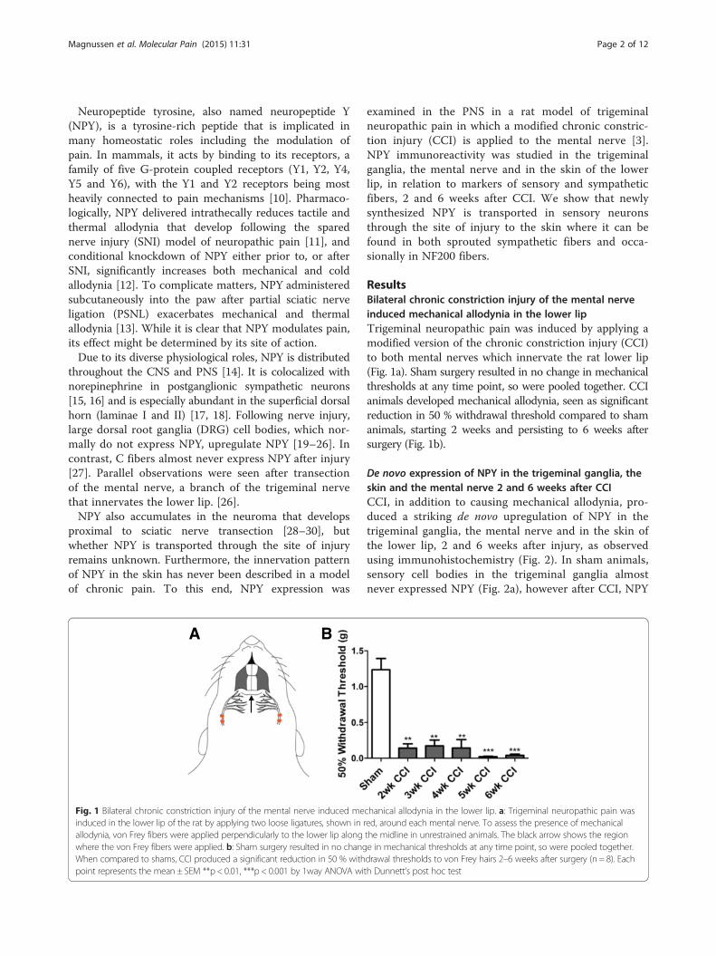
Fig. 1 Bilateral chronic constriction injury of the mental nerve induced meinduced in the lower lip of the rat by applying two loose ligatures, shown in rallodynia, von Frey fibers were applied perpendicularly to the lower lip alongwhere the von Frey fibers were applied. b: Sham surgery resulted in no changWhen compared to shams, CCI produced a significant reduction in 50 % withpoint represents the mean ± SEM **p < 0.01, ***p < 0.001 by 1way ANOVA wi
examined in the PNS in a rat model of trigeminalneuropathic pain in which a modified chronic constric-tion injury (CCI) is applied to the mental nerve [3].NPY immunoreactivity was studied in the trigeminalganglia, the mental nerve and in the skin of the lowerlip, in relation to markers of sensory and sympatheticfibers, 2 and 6 weeks after CCI. We show that newlysynthesized NPY is transported in sensory neuronsthrough the site of injury to the skin where it can befound in both sprouted sympathetic fibers and occa-sionally in NF200 fibers.
ResultsBilateral chronic constriction injury of the mental nerveinduced mechanical allodynia in the lower lipTrigeminal neuropathic pain was induced by applying amodified version of the chronic constriction injury (CCI)to both mental nerves which innervate the rat lower lip(Fig. 1a). Sham surgery resulted in no change in mechanicalthresholds at any time point, so were pooled together. CCIanimals developed mechanical allodynia, seen as significantreduction in 50 % withdrawal threshold compared to shamanimals, starting 2 weeks and persisting to 6 weeks aftersurgery (Fig. 1b).
De novo expression of NPY in the trigeminal ganglia, theskin and the mental nerve 2 and 6 weeks after CCICCI, in addition to causing mechanical allodynia, pro-duced a striking de novo upregulation of NPY in thetrigeminal ganglia, the mental nerve and in the skin ofthe lower lip, 2 and 6 weeks after injury, as observedusing immunohistochemistry (Fig. 2). In sham animals,sensory cell bodies in the trigeminal ganglia almostnever expressed NPY (Fig. 2a), however after CCI, NPY
chanical allodynia in the lower lip. a: Trigeminal neuropathic pain wased, around each mental nerve. To assess the presence of mechanicalthe midline in unrestrained animals. The black arrow shows the regione in mechanical thresholds at any time point, so were pooled together.drawal thresholds to von Frey hairs 2–6 weeks after surgery (n = 8). Eachth Dunnett’s post hoc test
Sham 2 week CCI 6 week CCI
TGS
kin
MN
A B C
E F G
D
H
IJ K
L M
NPY
p p
d
d
d
p
d
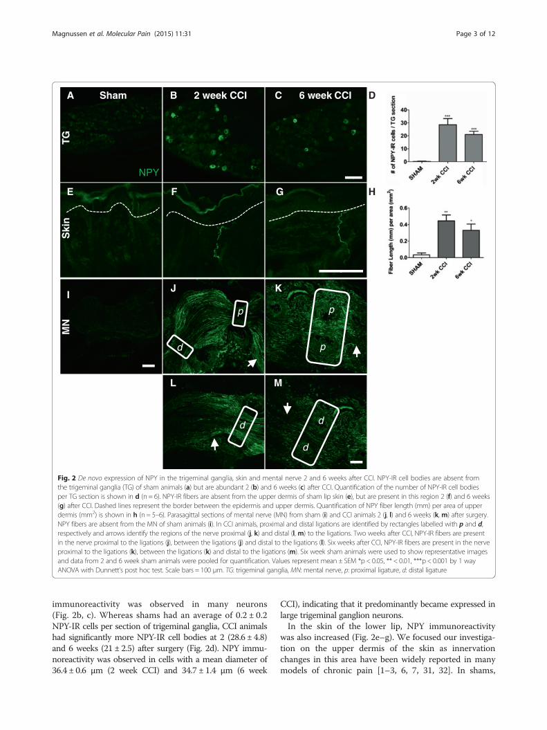
Fig. 2 De novo expression of NPY in the trigeminal ganglia, skin and mental nerve 2 and 6 weeks after CCI. NPY-IR cell bodies are absent fromthe trigeminal ganglia (TG) of sham animals (a) but are abundant 2 (b) and 6 weeks (c) after CCI. Quantification of the number of NPY-IR cell bodiesper TG section is shown in d (n = 6). NPY-IR fibers are absent from the upper dermis of sham lip skin (e), but are present in this region 2 (f) and 6 weeks(g) after CCI. Dashed lines represent the border between the epidermis and upper dermis. Quantification of NPY fiber length (mm) per area of upperdermis (mm2) is shown in h (n = 5–6). Parasagittal sections of mental nerve (MN) from sham (i) and CCI animals 2 (j, l) and 6 weeks (k, m) after surgery.NPY fibers are absent from the MN of sham animals (i). In CCI animals, proximal and distal ligations are identified by rectangles labelled with p and d,respectively and arrows identify the regions of the nerve proximal (j, k) and distal (l, m) to the ligations. Two weeks after CCI, NPY-IR fibers are presentin the nerve proximal to the ligations (j), between the ligations (j) and distal to the ligations (l). Six weeks after CCI, NPY-IR fibers are present in the nerveproximal to the ligations (k), between the ligations (k) and distal to the ligations (m). Six week sham animals were used to show representative imagesand data from 2 and 6 week sham animals were pooled for quantification. Values represent mean ± SEM *p < 0.05, ** < 0.01, ***p < 0.001 by 1 wayANOVA with Dunnett’s post hoc test. Scale bars = 100 μm. TG: trigeminal ganglia, MN: mental nerve, p: proximal ligature, d: distal ligature
Magnussen et al. Molecular Pain (2015) 11:31 Page 3 of 12
immunoreactivity was observed in many neurons(Fig. 2b, c). Whereas shams had an average of 0.2 ± 0.2NPY-IR cells per section of trigeminal ganglia, CCI animalshad significantly more NPY-IR cell bodies at 2 (28.6 ± 4.8)and 6 weeks (21 ± 2.5) after surgery (Fig. 2d). NPY immu-noreactivity was observed in cells with a mean diameter of36.4 ± 0.6 μm (2 week CCI) and 34.7 ± 1.4 μm (6 week
CCI), indicating that it predominantly became expressed inlarge trigeminal ganglion neurons.In the skin of the lower lip, NPY immunoreactivity
was also increased (Fig. 2e–g). We focused our investiga-tion on the upper dermis of the skin as innervationchanges in this area have been widely reported in manymodels of chronic pain [1–3, 6, 7, 31, 32]. In shams,
Magnussen et al. Molecular Pain (2015) 11:31 Page 4 of 12
almost no NPY immunoreactivity was observed (Fig. 2e),however after CCI, NPY-IR fibers were readily seen inthe upper dermis (Fig. 2f, g). NPY fiber density, as mea-sured by fiber length (mm) per area of upper dermis(mm2), was significantly greater than in sham at bothtime points after CCI (Fig. 2h; Sham 0.032 ± 0.02; 2 weekCCI 0.4 ± 0.07, p < 0.01; 6 week CCI 0.3 ± 0.07, p < 0.05).NPY immunoreactivity was not seen in the mental
nerve of sham animals (Fig. 2i), but 2 and 6 weeks afterCCI, many NPY-IR fibers were found proximal to thefirst ligature. These fibers passed through both the firstand second ligatures (Fig. 2j, k), and could be seen run-ning in the nerve distal to injury (Fig. 2l, m).
NPY was expressed in Fluorogold labelled mental nervecell bodies after CCIThe increase in NPY immunoreactivity in the trigeminalganglia after CCI was evident, but was this upregulationoccurring specifically in the cell bodies of mental nervesensory neurons, or was it occurring through the entiretrigeminal ganglia? To address this question, Fluorogoldwas injected into the mental nerve following sham or
NPY
FG NPY/FG
A Sham
C
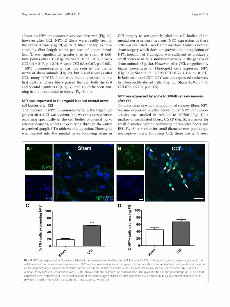
Fig. 3 NPY was expressed in Fluorogold labelled mental nerve cell bodiescell bodies of mental nerve sensory neurons. NPY immunoreactivity is showin the adjacent larger panel. Colocalization of the two signals is shown in tanimals many NPY cells colocalized with FG (b). Arrows indicate examplesexpressed NPY is shown in c. The quantification of the percentage of NPY(n = 6); *p < 0.05, ***p < 0.001 by Student’s t-test. Scale bar = 100 μm
CCI surgery to retrogradely label the cell bodies of themental nerve sensory neurons. NPY expression in thesecells was evaluated 1 week after injection. Unlike a normalsham surgery which does not provoke the upregulation ofNPY, injection of Fluorogold was sufficient to produce asmall increase in NPY immunoreactivity in the ganglia ofsham animals (Fig. 3a). However, after CCI, a significantlyhigher percentage of Fluorogold cells expressed NPY(Fig. 3b, c; Sham 19.2 ± 2.7 %; CCI 58.5 ± 1.3 %, p < 0.001).In both sham and CCI, NPY was not expressed exclusivelyby Fluorogold-labelled cells (Fig. 3d; Sham 45.0 ± 5.7 %;CCI 67.4 ± 5.7 %, p < 0.05).
NPY was expressed by some NF200-IR sensory neuronsafter CCITo determine in which population of sensory fibers NPYbecame expressed in after nerve injury, NPY immunore-activity was studied in relation to NF200 (Fig. 4), amarker of myelinated fibers, CGRP (Fig. 5), a marker forsmall diameter peptide containing nociceptive fibers andIB4 (Fig. 6), a marker for small diameter non-peptidergicnociceptive fibers. Following CCI, there was a de novo
D
CCIB
after CCI. Fluorogold (FG), in blue, was used to retrogradely label then in green. Signals are shown separately in small panels, and togetherurquoise. Few NPY cells were seen in sham animals (a), but in CCIof colocalization. The quantification of the percentage of FG cells thatcells that expressed FG is shown in d. Values represent mean ± SEM
Sham 2 week CCI 6 week CCI
TG
A B C
Dis
tal M
NS
kin
D E F
G H I
Proximal MN
NPY/NF200
Proximal MN
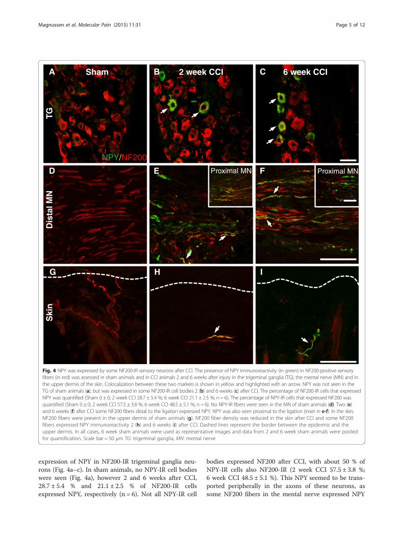
Fig. 4 NPY was expressed by some NF200-IR sensory neurons after CCI. The presence of NPY immunoreactivity (in green) in NF200-positive sensoryfibers (in red) was assessed in sham animals and in CCI animals 2 and 6 weeks after injury in the trigeminal ganglia (TG), the mental nerve (MN) and inthe upper dermis of the skin. Colocalization between these two markers is shown in yellow and highlighted with an arrow. NPY was not seen in theTG of sham animals (a), but was expressed in some NF200-IR cell bodies 2 (b) and 6 weeks (c) after CCI. The percentage of NF200-IR cells that expressedNPY was quantified (Sham 0 ± 0; 2 week CCI 28.7 ± 5.4 %; 6 week CCI 21.1 ± 2.5 %; n = 6). The percentage of NPY-IR cells that expressed NF200 wasquantified (Sham 0 ± 0; 2 week CCI 57.5 ± 3.8 %; 6 week CCI 48.5 ± 5.1 %; n = 6). No NPY-IR fibers were seen in the MN of sham animals (d). Two (e)and 6 weeks (f) after CCI some NF200 fibers distal to the ligation expressed NPY. NPY was also seen proximal to the ligation (inset in e-f). In the skin,NF200 fibers were present in the upper dermis of sham animals (g). NF200 fiber density was reduced in the skin after CCI and some NF200fibers expressed NPY immunoreactivity 2 (h) and 6 weeks (i) after CCI. Dashed lines represent the border between the epidermis and theupper dermis. In all cases, 6 week sham animals were used as representative images and data from 2 and 6 week sham animals were pooledfor quantification. Scale bar = 50 μm TG: trigeminal ganglia, MN: mental nerve
Magnussen et al. Molecular Pain (2015) 11:31 Page 5 of 12
expression of NPY in NF200-IR trigeminal ganglia neu-rons (Fig. 4a–c). In sham animals, no NPY-IR cell bodieswere seen (Fig. 4a), however 2 and 6 weeks after CCI,28.7 ± 5.4 % and 21.1 ± 2.5 % of NF200-IR cellsexpressed NPY, respectively (n = 6). Not all NPY-IR cell
bodies expressed NF200 after CCI, with about 50 % ofNPY-IR cells also NF200-IR (2 week CCI 57.5 ± 3.8 %;6 week CCI 48.5 ± 5.1 %). This NPY seemed to be trans-ported peripherally in the axons of these neurons, assome NF200 fibers in the mental nerve expressed NPY
A B C
TG
Dis
tal M
N
Ski
n
Proximal MNNPY/CGRP
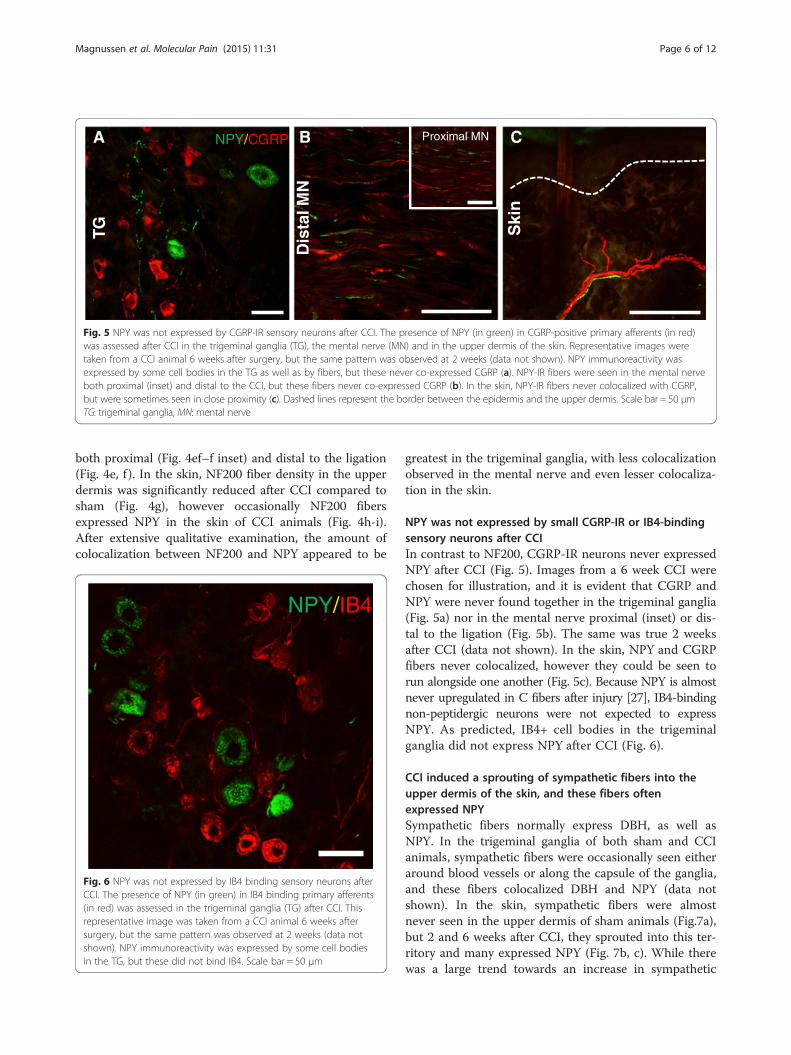
Fig. 5 NPY was not expressed by CGRP-IR sensory neurons after CCI. The presence of NPY (in green) in CGRP-positive primary afferents (in red)was assessed after CCI in the trigeminal ganglia (TG), the mental nerve (MN) and in the upper dermis of the skin. Representative images weretaken from a CCI animal 6 weeks after surgery, but the same pattern was observed at 2 weeks (data not shown). NPY immunoreactivity wasexpressed by some cell bodies in the TG as well as by fibers, but these never co-expressed CGRP (a). NPY-IR fibers were seen in the mental nerveboth proximal (inset) and distal to the CCI, but these fibers never co-expressed CGRP (b). In the skin, NPY-IR fibers never colocalized with CGRP,but were sometimes seen in close proximity (c). Dashed lines represent the border between the epidermis and the upper dermis. Scale bar = 50 μmTG: trigeminal ganglia, MN: mental nerve
Magnussen et al. Molecular Pain (2015) 11:31 Page 6 of 12
both proximal (Fig. 4ef–f inset) and distal to the ligation(Fig. 4e, f ). In the skin, NF200 fiber density in the upperdermis was significantly reduced after CCI compared tosham (Fig. 4g), however occasionally NF200 fibersexpressed NPY in the skin of CCI animals (Fig. 4h-i).After extensive qualitative examination, the amount ofcolocalization between NF200 and NPY appeared to be
NPY/IB4
Fig. 6 NPY was not expressed by IB4 binding sensory neurons afterCCI. The presence of NPY (in green) in IB4 binding primary afferents(in red) was assessed in the trigeminal ganglia (TG) after CCI. Thisrepresentative image was taken from a CCI animal 6 weeks aftersurgery, but the same pattern was observed at 2 weeks (data notshown). NPY immunoreactivity was expressed by some cell bodiesin the TG, but these did not bind IB4. Scale bar = 50 μm
greatest in the trigeminal ganglia, with less colocalizationobserved in the mental nerve and even lesser colocaliza-tion in the skin.
NPY was not expressed by small CGRP-IR or IB4-bindingsensory neurons after CCIIn contrast to NF200, CGRP-IR neurons never expressedNPY after CCI (Fig. 5). Images from a 6 week CCI werechosen for illustration, and it is evident that CGRP andNPY were never found together in the trigeminal ganglia(Fig. 5a) nor in the mental nerve proximal (inset) or dis-tal to the ligation (Fig. 5b). The same was true 2 weeksafter CCI (data not shown). In the skin, NPY and CGRPfibers never colocalized, however they could be seen torun alongside one another (Fig. 5c). Because NPY is almostnever upregulated in C fibers after injury [27], IB4-bindingnon-peptidergic neurons were not expected to expressNPY. As predicted, IB4+ cell bodies in the trigeminalganglia did not express NPY after CCI (Fig. 6).
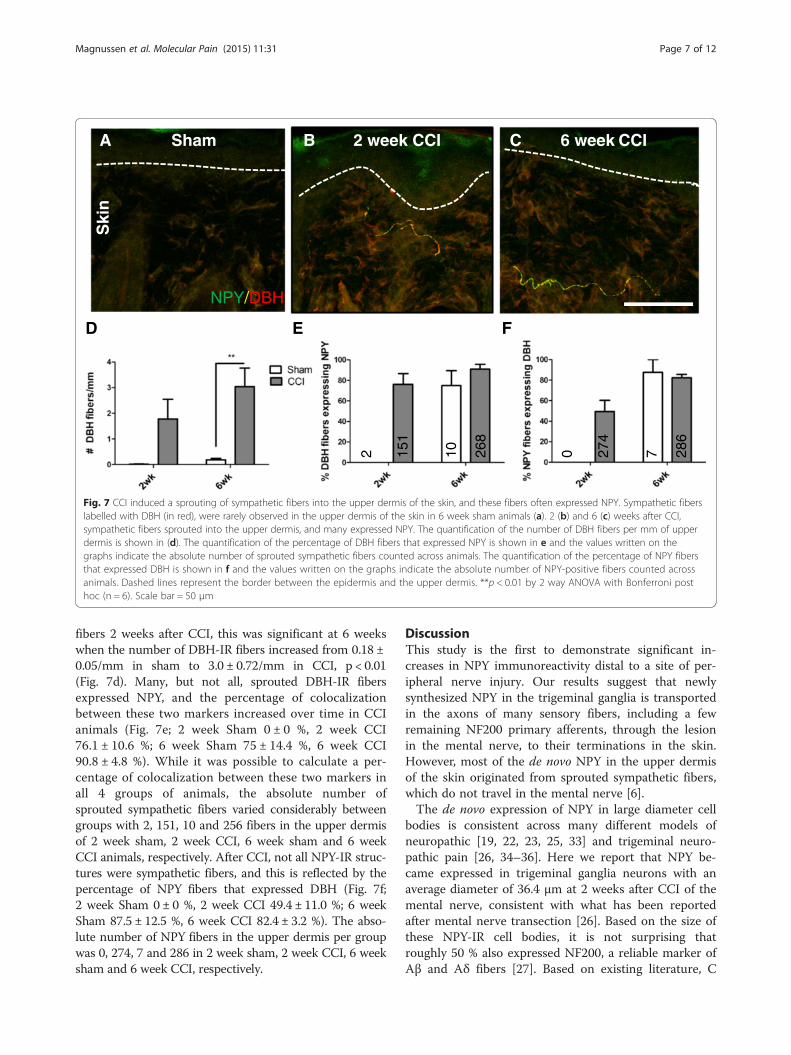
CCI induced a sprouting of sympathetic fibers into theupper dermis of the skin, and these fibers oftenexpressed NPYSympathetic fibers normally express DBH, as well asNPY. In the trigeminal ganglia of both sham and CCIanimals, sympathetic fibers were occasionally seen eitheraround blood vessels or along the capsule of the ganglia,and these fibers colocalized DBH and NPY (data notshown). In the skin, sympathetic fibers were almostnever seen in the upper dermis of sham animals (Fig.7a),but 2 and 6 weeks after CCI, they sprouted into this ter-ritory and many expressed NPY (Fig. 7b, c). While therewas a large trend towards an increase in sympathetic
FED
Sham 2 week CCI 6 week CCICBA
Ski
n
NPY/DBH
2 151
10 268
0 274
7 286
Fig. 7 CCI induced a sprouting of sympathetic fibers into the upper dermis of the skin, and these fibers often expressed NPY. Sympathetic fiberslabelled with DBH (in red), were rarely observed in the upper dermis of the skin in 6 week sham animals (a). 2 (b) and 6 (c) weeks after CCI,sympathetic fibers sprouted into the upper dermis, and many expressed NPY. The quantification of the number of DBH fibers per mm of upperdermis is shown in (d). The quantification of the percentage of DBH fibers that expressed NPY is shown in e and the values written on thegraphs indicate the absolute number of sprouted sympathetic fibers counted across animals. The quantification of the percentage of NPY fibersthat expressed DBH is shown in f and the values written on the graphs indicate the absolute number of NPY-positive fibers counted acrossanimals. Dashed lines represent the border between the epidermis and the upper dermis. **p < 0.01 by 2 way ANOVA with Bonferroni posthoc (n = 6). Scale bar = 50 μm
Magnussen et al. Molecular Pain (2015) 11:31 Page 7 of 12
fibers 2 weeks after CCI, this was significant at 6 weekswhen the number of DBH-IR fibers increased from 0.18 ±0.05/mm in sham to 3.0 ± 0.72/mm in CCI, p < 0.01(Fig. 7d). Many, but not all, sprouted DBH-IR fibersexpressed NPY, and the percentage of colocalizationbetween these two markers increased over time in CCIanimals (Fig. 7e; 2 week Sham 0 ± 0 %, 2 week CCI76.1 ± 10.6 %; 6 week Sham 75 ± 14.4 %, 6 week CCI90.8 ± 4.8 %). While it was possible to calculate a per-centage of colocalization between these two markers inall 4 groups of animals, the absolute number ofsprouted sympathetic fibers varied considerably betweengroups with 2, 151, 10 and 256 fibers in the upper dermisof 2 week sham, 2 week CCI, 6 week sham and 6 weekCCI animals, respectively. After CCI, not all NPY-IR struc-tures were sympathetic fibers, and this is reflected by thepercentage of NPY fibers that expressed DBH (Fig. 7f;2 week Sham 0 ± 0 %, 2 week CCI 49.4 ± 11.0 %; 6 weekSham 87.5 ± 12.5 %, 6 week CCI 82.4 ± 3.2 %). The abso-lute number of NPY fibers in the upper dermis per groupwas 0, 274, 7 and 286 in 2 week sham, 2 week CCI, 6 weeksham and 6 week CCI, respectively.
DiscussionThis study is the first to demonstrate significant in-creases in NPY immunoreactivity distal to a site of per-ipheral nerve injury. Our results suggest that newlysynthesized NPY in the trigeminal ganglia is transportedin the axons of many sensory fibers, including a fewremaining NF200 primary afferents, through the lesionin the mental nerve, to their terminations in the skin.However, most of the de novo NPY in the upper dermisof the skin originated from sprouted sympathetic fibers,which do not travel in the mental nerve [6].The de novo expression of NPY in large diameter cell
bodies is consistent across many different models ofneuropathic [19, 22, 23, 25, 33] and trigeminal neuro-pathic pain [26, 34–36]. Here we report that NPY be-came expressed in trigeminal ganglia neurons with anaverage diameter of 36.4 μm at 2 weeks after CCI of themental nerve, consistent with what has been reportedafter mental nerve transection [26]. Based on the size ofthese NPY-IR cell bodies, it is not surprising thatroughly 50 % also expressed NF200, a reliable marker ofAβ and Aδ fibers [27]. Based on existing literature, C
Magnussen et al. Molecular Pain (2015) 11:31 Page 8 of 12
fibers, with small diameter cell bodies, were not ex-pected to express NPY after CCI [27], and as anticipated,NPY never colocalized with CGRP or IB4 in the ganglia.The exact triggers that result in the do novo expression
of NPY by sensory neurons remain unknown. However,it has been proposed that nerve trauma, and not painper se, might be the precipitating factor since painful in-flammation does not increase DRG NPY immunoreactiv-ity [23, 37]. In models of neuropathic pain, the expressionof NPY seems to correlate with the extent of nerve injury[36, 38, 39] and in our hands, a small trauma, like the onethat occurred when Fluorogold was injected into thenerve, triggered a limited NPY expression in the ganglia ofsham animals. 1 week after CCI, the majority (67 %) ofNPY-IR cells also expressed Fluorogold, indicating a certainspatial specificity to this response. Because Fluorogold isnot taken up by intact axons at nonterminal sites [40], thistechnique was used to retrogradely label injured neurons,and underestimates the total number of mental nerve sen-sory neurons. While we and the literature agree that NPYis expressed by injured sensory neurons [21, 25, 41], our re-sults suggest that a smaller proportion of uninjured neu-rons also begin to express NPY after CCI, in agreementwith Ma and Bisby [41]. This is not the case after mediannerve transection where only 1 % of uninjured neuronsexpressed NPY [25].We suggest that these uninjured neurons are the first
to transport NPY through the site of injury in the mentalnerve. While many NPY-IR fibers are seen in the nerve,only some co-express NF200. In fact, the colocalizationbetween NF200 and NPY appears to be the greatest inthe ganglia, with lower levels observed in the nerve andin the skin, respectively. This is unlikely due to slowshipment of the peptide to the periphery as NPY, synthe-sized in the cell body, is sent by fast axonal transport ata rate of approximately 9 mm/h [42]. Given that itsmRNA and protein are increased as early as 3 days afternerve injury [25, 35, 43], NPY could theoretically reachthe terminals of NF200 fibers only 4 days after CCI. In-stead the reduced colocalization between these twomarkers in distal nerves is most likely explained by thefindings that 90 % of A fibers did not conduct impulsesthrough the site of injury 3 days after CCI [44] and thatNF200 fiber density in the skin is drastically reduced upto 16 weeks after CCI [1]. As NPY immunoreactivity re-mains increased in the ganglia up to 24 weeks afternerve injury [21], it is possible that greater colocalizationbetween NF200 and NPY could be observed distal toCCI at these later time points when there is moreNF200 fiber recovery. The induction of NPY in thesesensory fibers may reflect a mechanism for altered neur-onal activity after injury. Indeed, following spinal nerveligation, Aβ DRGs from NPY overexpressing mice dis-played longer refractory periods and had higher voltage
thresholds compared to wild type animals, indicatingthat NPY in surplus can decrease the responses of thesefibers to nerve injury [45].While NF200 fibers represent a minor source of de
novo NPY in the skin, sprouted sympathetic fibers,which invade the upper dermis at 2 weeks [3, 7] repre-sent the major source. The colocalization between DBHand NPY increased over time, and by 6 weeks after CCIapproximately 90 % of DBH-IR sprouted sympatheticfibers expressed NPY. NPY is co-expressed with nor-epinephrine in many sympathetic fibers [42, 46, 47],however there exists heterogeneity within this popula-tion and not all fibers contain NPY [16]. In our model,previous studies have shown evidence that these ectopicfibers migrate from the lower dermis [3, 6], where sym-pathetic fibers predominantly innervate vasculature andalmost always contain NPY (unpublished observations).We are therefore not surprised that these sprouted sym-pathetic fibers mostly maintain their original phenotype.Normally, NPY is preferentially released following highfrequency stimulation and leads to long lasting vasocon-striction [48], however because these sprouted sympa-thetic fibers are most often not associated with bloodvessels, they may function in a different capacity afternerve injury. Finally, because our data indicate that totalNPY immunoreactivity in the upper dermis cannot beexplained alone by its expression in sympathetic fibers,NPY should not be used as a reliable marker of this fiberpopulation after nerve injury.In this study, we show two novel sources of NPY in
the upper dermis of the skin after CCI, however howthis increased NPY contributes to pain is difficult to dir-ectly examine. Mental nerve CCI resulted in mechanicalallodynia that lasted from 2 to 6 weeks, the last timepoint tested. Because neither the withdrawal thresholdsnor levels of NPY changed significantly during this time,we are unable to correlate alterations in NPY immunoreac-tivity in the periphery with pain behaviour. In the literaturehowever there is considerable evidence in support of theidea that upregulation of NPY by sensory neurons repre-sents an adaptive mechanism that counteracts the evolutionof neuropathic pain [27]—for reviews see [10, 49]. Indeed,conditional knockdown of NPY resulted in a significantincrease in the intensity and duration of mechanicaland thermal hypersensitivity after SNI suggesting thatNPY inhibits pain [12].In the periphery, the situation might be different, with
NPY being pronociceptive through its action at Y2 recep-tors, or having mixed effects through the Y1 receptor.Subcutaneous injections of NPY and an NPY Y2 receptoragonist into the paw after PSNL exacerbated mechanicalallodynia and thermal hyperalgesia [13]. Could NPY re-leased from these sprouted sympathetic fibers worsenpain? A novel physical proximity exists between sprouted
Magnussen et al. Molecular Pain (2015) 11:31 Page 9 of 12
sympathetic fibers and sensory fibers after nerve injury[2, 3, 6, 7], and we have shown here that NPY fibersrun in bundles with CGRP fibers in the skin. It is con-ceivable that NPY released from these fibers could dir-ectly modulate the activity of primary afferents, knownto express Y1 and Y2 [50–53]. Indeed, NPY through itsactions on Y2 increased the excitability of sensory neu-rons [54]. Alternatively, NPY might act directly onthese sprouted sympathetic fibers, through Y2 autore-ceptors [55], to modulate the release of excitatory nor-epinephrine, or even NPY itself [13, 56, 57]. NPY in theskin can also act on Y1 receptors, and it has beenshown that an intraplantar injection of a Y1 agonistworsens mechanical hyperalgesia, while concurrentlyimproving thermal hyperalgesia after PSNL [13]. Inmodels involving neurogenic inflammation, Y1 agonistsinhibit capsaicin evoked mechanical allodynia through areduction in CGRP release from primary afferent termi-nals [58]. Thus the effects of NPY in the skin are ex-tremely complex, and depend on the receptor on which itacts, the location of these receptors, and the model used.Not all of the NPY immunoreactivity has been accounted
for as many NPY-IR cell bodies and axons were not shownto co-express any of the markers used in this study. Futurestudies will be required to identify these primary afferents,however it is expected that they will likely possess mediumto large diameter cell bodies and send central projections tolamina III and IV.In conclusion, these results show novel and increased
sources of NPY in the periphery in a rat model of tri-geminal neuropathic pain. How exactly this increase inNPY contributes to pain should be the subject of futureinvestigations.
MethodsAnimalsAdult male Sprague-Dawley rats (250–350 g; CharlesRiver, Canada) were maintained on a 12-h light/darkcycle and allowed access to food and water ad libitum.All protocols were approved by the McGill UniversityAnimal Care Committee and complied with the policiesand guidelines outlined by the Canadian Council onAnimal Care and the International Association for theStudy of Pain.
Surgical proceduresA modified chronic constriction injury [59], as describedpreviously [3], was applied to the mental nerve of therat, a purely sensory branch of the trigeminal nerve thatinnervates the rat lower lip [6]. Briefly, the animals wereanesthetized with isofluorane and the mental nerveswere bilaterally exposed from their point of exit fromthe mental foramina. Proximal to its branching, thenerve was freed from adhering tissue and 2 loose silk
ligatures (8.0, Covidien), separated by approximately2 mm, were tied around the nerve. The incision wasclosed with absorbable sutures (Vicryl, Ethicon). A shamsurgery, where the mental nerve was visualized but nosutures were applied, was used as a control.
Fluorogold injectionsBecause the trigeminal ganglion contains the sensoryneurons from all three branches of the trigeminal nerve,Fluorogold was used to determine if the upregulation ofNPY was occurring specifically in the cell bodies of men-tal nerve sensory neurons, or if it was occurring globallythroughout the ganglion. Using a calibrated glass micro-pipette, 1 μl of 5 % Fluorogold solution (Fluorochrome,LLC) diluted in distilled water was injected directly intothe mental nerve of sham and CCI animals, proximal tothe first suture. As above, the incision was closed withabsorbable sutures. Animals were sacrificed one weekafter injection.
Behaviour: mechanical allodyniaAll animals (n = 8/group) were tested 2–6 weeks followingCCI surgery by a blinded experimenter. We used von Freyfilaments applied to the lower lip to determine the 50 %withdrawal threshold to punctate mechanical stimulation.Each rat was placed in a transparent Plexiglas cage atop awire mesh grid and was allowed to habituate to theirsurroundings for 30 min before behaviour testing. VonFrey filaments of increasing stiffness were applied perpen-dicularly, in the midline, to the lower lip skin following theup-and-down method described by Dixon [60] until apositive reaction, considered a brisk head withdrawal, wasobserved [7]. Following the first positive reaction, the nextlighter filament was applied. If no reaction, the next stifferfilament was applied; if a reaction was observed, the nextlighter filament was applied. After the first positive fila-ment, four additional filaments were applied and the 50 %withdrawal threshold was calculated using the methodsoutlined by Chaplan et al. [61]. Mechanical allodynia wasconsidered as a significant reduction in withdrawalthreshold when compared to shams, as measured by a1way analysis of variance (ANOVA) with Dunnett’s posthoc test. Statistical significance was set at p < 0.05.
Tissue preparationNPY expression in the trigeminal ganglion, the mentalnerve and in the skin of the lower lip following CCIwas visualized using immunohistochemistry. Rats weredeeply anesthetized with Equithesin (0.3 mL/100 g) andperfused transcardially with 100 mL of perfusion bufferand 500 mL of 3 % paraformaldehyde and 15 % satu-rated picric acid (v/v) in 0.1 M phosphate buffer (PB),pH 7.4, for 30 min. Trigeminal ganglia, mental nervesand hairy lower lip skin were dissected out and postfixed
Magnussen et al. Molecular Pain (2015) 11:31 Page 10 of 12
for 1 h in the above fixative and cryoprotected in 30 % su-crose in PB for 24 h at 4 °C. Tissue was embedded in anoptimum cutting temperature medium (Tissue Tek, OCT)and cut on a cryostat at − 20 °C. Parasagittal sections ofmental nerve and horizontal sections of trigeminal gangliawere cut at a thickness of 14 μm and 20 μm, respectively,and mounted directly on slides. Sections of skin were cutat 50 μm and collected as free-floating in 0.01 Mphosphate-buffered saline (PBS).
Immunohistochemisty and lectin bindingTo determine in which population of fibers NPY wasexpressed in after CCI, the mental nerves, trigeminalganglia and skin were processed for immunohistochemistryusing antibodies against NPY in combination with thoseagainst either neurofilament 200 (NF200), calcitonin gene-related peptide (CGRP) or dopamine β-hydroxylase (DBH)to label myelinated fibers, peptidergic sensory fibers andsympathetic fibers, respectively. The lectin IB4 was used toidentify non-peptidergic sensory fibers. Fluorogold is afluorescent dye and requires no additional staining. All sec-tions were washed for 30 min with PBS containing 0.2 %Triton-X (PBS-T), incubated in 50 % ethanol for 30 minand washed in PBS-T. Depending on the species in whichthe secondary antibody was raised in, the tissue was eitherblocked in 10 % normal donkey or normal goat serum for1 h. Primary antibodies were used at the following concen-trations – anti-NPY (1:4000, Rabbit polyclonal, Abcam,ab10980, lot#GR16697-2), anti-NF200 (in skin and nerve,1:5, mouse monoclonal, Abcam, ab910, lot# GR130394-2;in ganglia, 1:1000, mouse monoclonal, Sigma, N0142, lot#053 M4756), anti-CGRP (1:1000, mouse monoclonal,Sigma, C7113, lot#032 M4862), anti-DBH (1:50, mousemonoclonal, Medimabs, MM-0008-P, lot#83120927-P) andIB4 conjugated to Alexa Fluor 568 (1:200, MolecularProbes, I21412, lot#880292) made in 5 % blocking serum(goat or donkey) in PBS-T and left to incubate overnight onthe shaker at 4 °C. Following 30 min of washes with PBS-T,the tissue was incubated with the appropriate secondaryantibody diluted in PBS-T for 2 h at room temperature–goat anti-rabbit conjugated to Alexa Fluor 488 (1:800,Molecular Probes, A11034, lot#1212189), goat anti-mouse conjugated to Alexa Fluor 568 (1:800, MolecularProbes, A11031, lot# 822389), or donkey anti-mouseconjugated to Rhodamine Red X (1:200, Jackson Immu-noresearch, 715-296-151, lot#72430). Following 30 minof washes, free-floating lip sections were mounted ongelatin-subbed slides and all slides were coverslippedwith Aqua Polymount (Polysciences).Representative images were taken using a Zeiss LSM510
confocal microscope equipped with Ar and He-Ne lasersusing either 10× dry, 40× water-immersion, or 63× oil-immersion objectives. For co-localization studies both zstack and single optical slice images were taken to ensure
bona fide detection of co-localization between markers.In the figures, all images of the mental nerve and trigemi-nal ganglia are single plane images, with z stack images ofthe skin.
QuantificationImages used for quantification were taken on a ZeissAxioplan 2e imaging fluorescence microscope (Carl ZeissMicroscopy LLC), with either a 20× or a 40× objective.Images were acquired with a high-resolution color digitalcamera with Zeiss Axiovision 4.8 software.
NPY fiber density quantification in the skinChanges in NPY-IR innervation in the lower lip skinwere determined by analyzing the density of fiberswithin the upper dermis, defined as the area above theopening of the sebaceous glands [6]. Six randomlychosen fields per lip section from 3 sections were cap-tured, totaling 18 images per animal. Six animals wereused for each time point. Quantification was performedusing an MCID Elite image analysis system (Imaging Re-search Inc., St. Catharines, ON, Canada) to determinethe total fiber length (mm) per unit area of upper dermis(mm2), as described by us previously [7]. Briefly, we useda function of the MCID software that was developed tospecifically and accurately detect fibers. After detection,fibers were skeletonized to 1 pixel in width, and the totalfiber length per unit area was determined by the soft-ware and compared using an ANOVA with Dunnett’spost hoc test, with p < 0.05 considered significant.
Trigeminal ganglia quantificationTo determine the changes in NPY expression in the tri-geminal ganglia, 3 entire sections per ganglion (and peranimal) were captured using the 20× objective. Fromthese images, the numbers of NPY-IR cell bodies withvisible nuclei were counted. In addition the diameter ofNPY-IR cells was measured using the Axiovision software.The extent of colocalization between NPY and eitherFluorogold, CGRP, IB4 or NF200, was determined bycounting both the total number of cell bodies expressingeach individual marker as well as those labelled with bothmarkers. A percentage was calculated from these valuesand when applicable was compared using a Student t-testwith p < 0.05 considered significant.
Quantification of NPY/DBH colocalization in the skinThree random sections per animal were visualized usingthe 40× objective from the fluorescence microscope andall of the DBH-IR and NPY-IR fibers in the upper dermiswere counted, taking note of whether the fiber was alsoNPY-IR or DBH-IR, respectively. Total section lengthwas measured in ImageJ. The number of DBH-IR fibersper section length (mm) as well as the percentage of
Magnussen et al. Molecular Pain (2015) 11:31 Page 11 of 12
DBH fibers expressing NPY or NPY fibers expressingDBH was calculated and in all cases was compared usinga 2 way ANOVA with Bonferroni post-hoc analysis withp < 0.05 considered significant.
Competing interestsThe authors declare that they have no competing interests.
Authors’ contributionsCM planned and performed most of the experiments, analysed the data,prepared the figures and wrote the first version of the manuscript. SPHhelped with the immuhistochemistry and quantification. ARdS wasresponsible for the coordination and overall supervision of the study andedited the manuscript. All authors have read and approved the final draft ofthis manuscript.
AcknowledgementsThis work was supported by grants from the Louise and Alan EdwardsFoundation and Canadian Institutes of Health Research (CIHR). CM is arecipient of the Louise and Alan Edwards Foundation PhD Fellowship. Theauthors would like to thank Noosha Yousefpour for her drawing of themental nerve chronic constriction injury in Fig. 1a and Manon St. Louis forher technical support.
Author details1Department of Pharmacology and Therapeutics, McGill University, McIntyreMedical Building, 3655 Promenade Sir William Osler, Room 1215, Montreal,Quebec H3G 1Y6, Canada. 2Alan Edwards Centre for Research on Pain, McGillUniversity, Montreal, Quebec H3A 0G1, Canada. 3Department of Anatomyand Cell Biology, McGill University, Montreal, Quebec H3A 0C7, Canada.
Received: 13 March 2015 Accepted: 20 May 2015
References1. Peleshok JC, Ribeiro-da-Silva A. Delayed reinnervation by nonpeptidergic
nociceptive afferents of the glabrous skin of the rat hindpaw in a neuropathicpain model. J Comp Neurol. 2011;519:49–63.
2. Yen LD, Bennett GJ, Ribeiro-da-Silva A. Sympathetic sprouting and changesin nociceptive sensory innervation in the glabrous skin of the rat hind pawfollowing partial peripheral nerve injury. J Comp Neurol. 2006;495:679–90.
3. Grelik C, Bennett GJ, Ribeiro-da-Silva A. Autonomic fibre sprouting andchanges in nociceptive sensory innervation in the rat lower lip skin followingchronic constriction injury. Eur J Neurosci. 2005;21:2475–87.
4. Duraku LS, Hossaini M, Hoendervangers S, Falke LL, Kambiz S, Mudera VC,et al. Spatiotemporal dynamics of re-innervation and hyperinnervationpatterns by uninjured CGRP fibers in the rat foot sole epidermis afternerve injury. Mol Pain. 2012;8:61.
5. Duraku LS, Hossaini M, Schuttenhelm BN, Holstege JC, Baas M, Ruigrok TJ,et al. Re-innervation patterns by peptidergic Substance-P, non-peptidergicP2X3, and myelinated NF-200 nerve fibers in epidermis and dermis of ratswith neuropathic pain. Exp Neurol. 2013;241:13–24.
6. Ruocco I, Cuello AC, Ribeiro-Da-Silva A. Peripheral nerve injury leads to theestablishment of a novel pattern of sympathetic fibre innervation in the ratskin. J Comp Neurol. 2000;422:287–96.
7. Taylor AM, Ribeiro-da-Silva A. GDNF levels in the lower lip skin in a ratmodel of trigeminal neuropathic pain: implications for nonpeptidergic fiberreinnervation and parasympathetic sprouting. Pain. 2011;152:1502–10.
8. Wiesenfeld-Hallin Z, Xu XJ. Neuropeptides in neuropathic and inflammatorypain with special emphasis on cholecystokinin and galanin. Eur JPharmacol. 2001;429:49–59.
9. Fried K, Bongenhielm U, Boissonade FM, Robinson PP. Nerve injury-inducedpain in the trigeminal system. Neuroscientist. 2001;7:155–65.
10. Brumovsky P, Shi TS, Landry M, Villar MJ, Hokfelt T. Neuropeptide tyrosineand pain. Trends Pharmacol Sci. 2007;28:93–102.
11. Intondi AB, Dahlgren MN, Eilers MA, Taylor BK. Intrathecal neuropeptide Yreduces behavioral and molecular markers of inflammatory or neuropathicpain. Pain. 2008;137:352–65.
12. Solway B, Bose SC, Corder G, Donahue RR, Taylor BK. Tonic inhibition ofchronic pain by neuropeptide Y. Proc Natl Acad Sci U S A. 2011;108:7224–9.
13. Tracey DJ, Romm MA, Yao NN. Peripheral hyperalgesia in experimentalneuropathy: exacerbation by neuropeptide Y. Brain Res. 1995;669:245–54.
14. Tatemoto K. Neuropeptide Y: history and overview. In: Michel MC, editor.Neuropeptide Y and related peptides. Berlin: Springer; 2004. p. 1–21. MichelMC (Series Editor).
15. Lundberg JM, Franco-Cereceda A, Lacroix JS, Pernow J. Neuropeptide Y andsympathetic neurotransmission. Ann N Y Acad Sci. 1990;611:166–74.
16. Lundberg JM, Hokfelt T. Multiple co-existence of peptides and classicaltransmitters in peripheral autonomic and sensory neurons–functional andpharmacological implications. Prog Brain Res. 1986;68:241–62.
17. Rowan S, Todd AJ, Spike RC. Evidence that neuropeptide Y is present inGABAergic neurons in the superficial dorsal horn of the rat spinal cord.Neuroscience. 1993;53:537–45.
18. Gibson SJ, Polak JM, Allen JM, Adrian TE, Kelly JS, Bloom SR. The distributionand origin of a novel brain peptide, neuropeptide Y, in the spinal cord ofseveral mammals. J Comp Neurol. 1984;227:78–91.
19. Wakisaka S, Kajander KC, Bennett GJ. Increased neuropeptide Y (NPY)-likeimmunoreactivity in rat sensory neurons following peripheral axotomy.Neurosci Lett. 1991;124:200–3.
20. Zhang X, Meister B, Elde R, Verge VM, Hokfelt T. Large calibre primaryafferent neurons projecting to the gracile nucleus express neuropeptide Yafter sciatic nerve lesions: an immunohistochemical and in situ hybridizationstudy in rats. Eur J Neurosci. 1993;5:1510–9.
21. Intondi AB, Zadina JE, Zhang X, Taylor BK. Topography and time course ofchanges in spinal neuropeptide Y immunoreactivity after spared nerveinjury. Neuroscience. 2010;165:914–22.
22. Ma W, Bisby MA. Partial and complete sciatic nerve injuries induce similarincreases of neuropeptide Y and vasoactive intestinal peptideimmunoreactivities in primary sensory neurons and their central projections.Neuroscience. 1998;86:1217–34.
23. Wakisaka S, Kajander KC, Bennett GJ. Effects of peripheral nerve injuries andtissue inflammation on the levels of neuropeptide Y-like immunoreactivityin rat primary afferent neurons. Brain Res. 1992;598:349–52.
24. Kashiba H, Noguchi K, Ueda Y, Senba E. Neuropeptide Y and galanin arecoexpressed in rat large type A sensory neurons after peripheral transection.Peptides. 1994;15:411–6.
25. Tsai YJ, Lin CT, Lue JH. Characterization of the induced neuropeptide Y-likeimmunoreactivity in primary sensory neurons following complete mediannerve transection. J Neurotrauma. 2007;24:1878–88.
26. Wakisaka S, Takikita S, Sasaki Y, Kato J, Tabata MJ, Kurisu K. Cell size-specificappearance of neuropeptide Y in the trigeminal ganglion following peripheralaxotomy of different branches of the mandibular nerve of the rat. Brain Res.1993;620:347–50.
27. Ruscheweyh R, Forsthuber L, Schoffnegger D, Sandkuhler J. Modification ofclassical neurochemical markers in identified primary afferent neurons withAbeta-, Adelta-, and C-fibers after chronic constriction injury in mice. JComp Neurol. 2007;502:325–36.
28. Fried K, Frisen J. End structure and neuropeptide immunoreactivity of axonsin sciatic neuromas following nerve section in neonatal rats. Exp Neurol.1990;109:286–93.
29. Frisen J, Risling M, Theodorsson E, Fried K. NPY-like immunoreactivity in sen-sory nerve fibers in rat sciatic neuroma. Brain Res. 1992;577:142–6.
30. Zochodne DW, Cheng C, Miampamba M, Hargreaves K, Sharkey KA. Peptideaccumulations in proximal endbulbs of transected axons. Brain Res. 2001;902:40–50.
31. Almarestani L, Longo G, Ribeiro-da-Silva A. Autonomic fiber sprouting in theskin in chronic inflammation. Mol Pain. 2008;4:56.
32. Longo G, Osikowicz M, Ribeiro-da-Silva A. Sympathetic fiber sprouting ininflamed joints and adjacent skin contributes to pain-related behavior inarthritis. J Neurosci. 2013;33:10066–74.
33. Ossipov MH, Zhang ET, Carvajal C, Gardell L, Quirion R, Dumont Y, et al.Selective mediation of nerve injury-induced tactile hypersensitivity byneuropeptide Y. J Neurosci. 2002;22:9858–67.
34. Sasaki Y, Wakisaka S, Kurisu K. Effects of peripheral axotomy of the inferioralveolar nerve on the levels of neuropeptide Y in rat trigeminal primaryafferent neurons. Brain Res. 1994;664:108–14.
35. Wakisaka S, Sasaki Y, Kurisu K. Temporal analysis of neuropeptide Yexpression in the rat trigeminal ganglion following peripheral axotomy ofthe inferior alveolar nerve. Neurosci Lett. 1995;188:49–52.
36. Benoliel R, Eliav E, Iadarola MJ. Neuropeptide Y in trigeminal ganglionfollowing chronic constriction injury of the rat infraorbital nerve: is therecorrelation to somatosensory parameters? Pain. 2001;91:111–21.
Magnussen et al. Molecular Pain (2015) 11:31 Page 12 of 12
37. Ji RR, Zhang X, Wiesenfeld-Hallin Z, Hokfelt T. Expression of neuropeptide Yand neuropeptide Y (Y1) receptor mRNA in rat spinal cord and dorsal rootganglia following peripheral tissue inflammation. J Neurosci. 1994;14:6423–34.
38. Brumovsky PR, Bergman E, Liu HX, Hokfelt T, Villar MJ. Effect of a gradedsingle constriction of the rat sciatic nerve on pain behavior and expressionof immunoreactive NPY and NPY Y1 receptor in DRG neurons and spinalcord. Brain Res. 2004;1006:87–99.
39. Ohara S, Roth KA, Beaudet LN, Schmidt RE. Transganglionic neuropeptide Yresponse to sciatic nerve injury in young and aged rats. J Neuropathol ExpNeurol. 1994;53:646–62.
40. Schmued LC, Fallon JH. Fluoro-gold: a new fluorescent retrograde axonaltracer with numerous unique properties. Brain Res. 1986;377:147–54.
41. Ma W, Bisby MA. Partial sciatic nerve ligation induced more dramatic increaseof neuropeptide Y immunoreactive axonal fibers in the gracile nucleus ofmiddle-aged rats than in young adult rats. J Neurosci Res. 2000;60:520–30.
42. Fried G, Lundberg JM, Theodorsson-Norheim E. Subcellular storage andaxonal transport of neuropeptide Y (NPY) in relation to catecholamines inthe cat. Acta Physiol Scand. 1985;125:145–54.
43. Nahin RL, Ren K, De Leon M, Ruda M. Primary sensory neurons exhibit alteredgene expression in a rat model of neuropathic pain. Pain. 1994;58:95–108.
44. Kajander KC, Bennett GJ. Onset of a painful peripheral neuropathy in rat: apartial and differential deafferentation and spontaneous discharge in A betaand A delta primary afferent neurons. J Neurophysiol. 1992;68:734–44.
45. Sapunar D, Modric-Jednacak K, Grkovic I, Michalkiewicz M, Hogan QH. Effectof peripheral axotomy on pain-related behavior and dorsal root ganglionneurons excitability in NPY transgenic rats. Brain Res. 2005;1063:48–58.
46. Lundberg JM, Terenius L, Hokfelt T, Martling CR, Tatemoto K, Mutt V, et al.Neuropeptide Y (NPY)-like immunoreactivity in peripheral noradrenergicneurons and effects of NPY on sympathetic function. Acta Physiol Scand.1982;116:477–80.
47. Morris JL. Cotransmission from sympathetic vasoconstrictor neurons tosmall cutaneous arteries in vivo. Am J Physiol. 1999;277:H58–64.
48. Lundberg JM, Rudehill A, Sollevi A, Theodorsson-Norheim E, Hamberger B.Frequency- and reserpine-dependent chemical coding of sympathetictransmission: differential release of noradrenaline and neuropeptide Yfrom pig spleen. Neurosci Lett. 1986;63:96–100.
49. Smith PA, Moran TD, Abdulla F, Tumber KK, Taylor BK. Spinal mechanisms ofNPY analgesia. Peptides. 2007;28:464–74.
50. Brumovsky P, Stanic D, Shuster S, Herzog H, Villar M, Hokfelt T. NeuropeptideY2 receptor protein is present in peptidergic and nonpeptidergic primarysensory neurons of the mouse. J Comp Neurol. 2005;489:328–48.
51. Brumovsky PR, Shi TJ, Matsuda H, Kopp J, Villar MJ, Hokfelt T. NPY Y1receptors are present in axonal processes of DRG neurons. Exp Neurol.2002;174:1–10.
52. Hokfelt T, Brumovsky P, Shi T, Pedrazzini T, Villar M. NPY and pain as seenfrom the histochemical side. Peptides. 2007;28:365–72.
53. Marchand JE, Cepeda MS, Carr DB, Wurm WH, Kream RM. Alterations inneuropeptide Y, tyrosine hydroxylase, and Y-receptor subtype distributionfollowing spinal nerve injury to rats. Pain. 1999;79:187–200.
54. Abdulla FA, Smith PA. Nerve injury increases an excitatory action of neuropeptideY and Y2-agonists on dorsal root ganglion neurons. Neuroscience. 1999;89:43–60.
55. Zhang X, Shi T, Holmberg K, Landry M, Huang W, Xiao H, et al. Expressionand regulation of the neuropeptide Y Y2 receptor in sensory andautonomic ganglia. Proc Natl Acad Sci U S A. 1997;94:729–34.
56. Lin Q, Zou X, Ren Y, Wang J, Fang L, Willis WD. Involvement of peripheralneuropeptide Y receptors in sympathetic modulation of acute cutaneousflare induced by intradermal capsaicin. Neuroscience. 2004;123:337–47.
57. Lin Q, Zou X, Fang L, Willis WD. Sympathetic modulation of acute cutaneousflare induced by intradermal injection of capsaicin in anesthetized rats.J Neurophysiol. 2003;89:853–61.
58. Gibbs JL, Flores CM, Hargreaves KM. Attenuation of capsaicin-evokedmechanical allodynia by peripheral neuropeptide Y Y1 receptors. Pain.2006;124:167–74.
59. Bennett GJ, Xie YK. A peripheral mononeuropathy in rat that producesdisorders of pain sensation like those seen in man. Pain. 1988;33:87–107.
60. Dixon W. The up-and-down method for small samples. J Am Stat Assoc.1965;60:967–78.
61. Chaplan SR, Bach FW, Pogrel JW, Chung JM, Yaksh TL. Quantitativeassessment of tactile allodynia in the rat paw. J Neurosci Methods.1994;53:55–63.
Submit your next manuscript to BioMed Centraland take full advantage of:
• Convenient online submission
• Thorough peer review
• No space constraints or color figure charges
• Immediate publication on acceptance
• Inclusion in PubMed, CAS, Scopus and Google Scholar
• Research which is freely available for redistribution
Submit your manuscript at www.biomedcentral.com/submit