novel human adenovirus causing nosocomial epidemic ... · nosocomial infection, hokkaido university...
TRANSCRIPT

JOURNAL OF CLINICAL MICROBIOLOGY, June 2008, p. 2002–2008 Vol. 46, No. 60095-1137/08/$08.00�0 doi:10.1128/JCM.01835-07Copyright © 2008, American Society for Microbiology. All Rights Reserved.
Novel Human Adenovirus Causing NosocomialEpidemic Keratoconjunctivitis�
Hiroaki Ishiko,1* Yasushi Shimada,1 Tsunetada Konno,1 Akio Hayashi,1 Takeshi Ohguchi,2Yoshitsugu Tagawa,2 Koki Aoki,2 Shigeaki Ohno,2 and Shudo Yamazaki3
Host Defense Laboratory, Mitsubishi Chemical Medience Corporation, Tokyo 174-8555, Japan1; Department ofOphthalmology and Visual Sciences, Hokkaido University Graduate School of Medicine, Sapporo 060-8638,
Japan2; and AIDS Vaccine Development Association, Tokyo 169-0075, Japan3
Received 14 September 2007/Returned for modification 30 November 2007/Accepted 23 March 2008
In 2000, we encountered cases of nosocomial infections with epidemic keratoconjunctivitis (EKC) at auniversity hospital in Kobe, in the western part of Japan. Two human adenovirus (HAdV) strains, Kobe-H andKobe-S, were isolated from patients with nosocomial EKC infection. They were untypeable by existing neu-tralizing antisera; however, the isolate was neutralized with homologous antisera. We then encountered severalcases of EKC due to nosocomial infections in eye clinics in different parts of Japan. A total of 80 HAdVs wereisolated from patients with EKC at eight different hospitals. The partial hexon gene sequences of the isolateswere determined and compared to those of the prototype strains of 51 serotypes. All isolates had identicalpartial hexon nucleotide sequences. Phylogenetic analysis classified these isolates into species of HAdV-D. Theisolates showed 93.9 to 96.7% nucleotide identity with HAdV-D prototype strains, while all 32 HAdV-Dprototype strains ranged from 93.2 to 99.2% identity. The sequences of the loop 2 and fiber knob regions fromthe representative strain, Kobe-H, were dissimilar in all prototype strains of 51 serotypes. We believe that thisvirus is a novel serotype of HAdV that causes EKC.
Human adenoviruses (HAdVs), which belong to the genusMastadenovirus of the family Adenoviridae, infect billions ofpeople worldwide and cause various diseases, including con-junctivitis, respiratory infectious disease, diarrhea in infantsand young children, and hemorrhagic cystitis (41). HAdVsinitially were grouped into six subgenera (A to F) on the basisof several biochemical and biophysical criteria (8, 41). In 1999,however, a reclassification on the basis of nucleotide and de-duced amino acid sequences was approved by the InternationalCommittee on Taxonomy of Viruses; under this reclassifica-tion, the 51 serotypes of HAdVs were grouped into six species,HAdV-A to HAdV-F (8).
In Japan, roughly 8,500 HAdV isolates were reported in2003 to 2007 (Infectious Agents Surveillance Report [http://idsc.nih.go.jp/iasr/index.html]). They were obtained from pa-tients with epidemic conjunctivitis (979; 11.5%), upper andlower respiratory tract infections (1,582; 18.6%), and gastro-enteritis (348; 4.1%). Virus isolation followed by a neutraliza-tion test (NT) using a type-specific antiserum has been thestandard procedure for serotyping (39). To date, the amplifi-cation of the genome by PCR, the determination of the nucle-otide sequences, and phylogenetic analysis have becomegeneralized techniques for both the classification and identifi-cation of viruses. We recently determined the partial hexonsequences of all 51 prototype strains and have developed arapid and reliable method of molecular diagnosis based onphylogenetic analysis (25, 33). This method has successfully
classified the 51 prototype strains of HAdVs into the six des-ignated species, as approved by the International Committeeon Taxonomy of Viruses. Over the last 30 years, we haveapplied this molecular diagnosis to the identification of hun-dreds of isolates and swabs without virus isolation from pa-tients from different parts of the world with EKC and lowerrespiratory tract infections (5, 6, 21, 25, 33).
In 2000, we encountered cases of nosocomial EKC infectionat a university hospital in Kobe, in the western part of Japan.Two HAdV strains, Kobe-H and Kobe-S, were isolated fromthe patients and showed identical nucleotide sequences of par-tial hexon genes. Adenoviral conjunctivitis is caused primarilyby HAdV-3 (of the HAdV-B species), HAdV-4 (of HAdV-E),and HAdV-8, HAdV-19, and HAdV-37 (all of HAdV-D).Among these, HAdV-8, HAdV-19, and HAdV-37 cause moresevere EKC than the others (3, 4, 7). The more serious out-comes occur in all age groups and can trigger highly contagiousnosocomial infections (3, 7, 20, 26, 38). Kobe-H and Kobe-Swere untypeable by using existing neutralizing antisera, sug-gesting that they represent a novel adenovirus causing EKC.After the nosocomial infections of 2000, we encountered sev-eral cases of nosocomial EKC infection, including cases at eyeclinics, in different parts in Japan. Surprisingly, the 80 isolatesfrom patients with EKC at eight different hospitals sharedpartial nucleotide sequences that were identical.
In this paper, we characterize the isolates from the patientswith EKC. The present phylogenetic analysis classified theseisolates into the HAdV-D species, and the Kobe-H strain rep-resents only 93.9 to 96.7% nucleotide identity between the 32prototype strains of HAdV-D, while all prototype strains ofHAdV-D ranged from 93.2 to 99.2% identity. Quantitative NTand genetic analysis of the main neutralization ε determinant,loops 1 (L1) and 2 (L2) in the hexon gene, clearly indicated
* Corresponding author. Mailing address: Host Defense Labora-tory, Mitsubishi Chemical Medience Corporation, Shimura 3-30-1,Itabashi-ku, Tokyo 174-8555, Japan. Phone: 81-3-5994-2340. Fax: 81-3-5994-2972. E-mail: [email protected].
� Published ahead of print on 2 April 2008.
2002
on February 18, 2020 by guest
http://jcm.asm
.org/D
ownloaded from

that the representative strain Kobe-H is different from previ-ously known HAdVs. We believe that the Kobe-H strain is anovel HAdV that causes nosocomial EKC infection.
MATERIALS AND METHODS
Viruses. The isolates and conjunctival swabs were obtained from nosocomialor sporadic infections from 2000 to 2005 (Table 1). Kobe-H and -S were isolatedfrom patients with nosocomial EKC infections at a university hospital in Kobe,Japan, in 2000. These strains were supplied from the Reference Center ofNosocomial Infection, Hokkaido University Graduate School of Medicine, Sap-poro, Japan. The prototype strains of 51 HAdVs were obtained from the Amer-ican Type Culture Collection and from the National Institute of InfectiousDiseases, Tokyo, Japan. These viruses were used directly for DNA extractionwithout further propagation.
Phylogeny-based classification using a partial hexon sequence. A partialhexon sequence of HAdV was amplified from the isolates as described previously(25, 33). In brief, viral DNA was extracted from 100 �l of virus suspension usinga Sumitest EX-R&D kit (Medical & Biological Laboratories Co., Ltd., Naggoya,Japan) by following the manufacturer’s instructions. The DNA was dissolved in100 �l of TE buffer (10 mM Tris [pH 8.0], 1 mM EDTA). The 1,004-bp fragmentof the hexon gene was amplified with 50 pmol of a pair of primers, AdTU7(nucleotide positions 20,734 to 20,753; 5�-GCCACCTTCTTCCCCATGGC-3�)and AdTU4� (positions 21,718 to 21,737; 5�-GTAGCGTTGCCGGCCGAGAA-3�). The positions of the primers for PCR were numbered according to thecomplete nucleotide sequence of the HAdV-2 strain (GenBank accession no.J01917). Using 10 �l of the PCR product, nested PCR was performed to amplifythe 956-bp DNA fragment with a pair of primers, AdnU-S� (positions 20,743 to20,762; 5�-TTCCCCATGGCNCACAACAC-3�) and AdnU-A (positions 21,679to 21,698; 5�-GCCTCGATGACGCCGCGGTG-3�). PCR was carried out for 36cycles in a Cetus 9600 Thermal Cycler (PE Applied Biosystems, Foster City, CA).Each cycle consisted of denaturation at 94°C for 1 min, annealing at 50°C for 1min, and primer extension at 72°C for 2 min. After the last cycle, extension wascontinued at 72°C for 7 min. The PCR products were separated on 3% agarosegels and purified with a QIAquick gel extraction kit (Qiagen, Valencia, CA).
The nucleotide sequence of the PCR products was determined using the CEQ2000XL DNA analysis system with a Dye Terminator cycle sequencing kit (Beck-man Coulter, Inc., Fullerton, CA). The nucleotide sequences of the partialhexons were analyzed by a comparison to those of prototype strains of all 51HAdV serotypes from GenBank using the SINCA software (Fujitsu, Ltd., Tokyo,Japan). Evolutionary distances were estimated using Kimura’s two-parametermethod (22), and unrooted phylogenetic trees were constructed using the neigh-bor-joining (N-J) method (30). Bootstrap analyses were performed by 1,000resamplings of the data sets (16). Similarity plots depicting the relationshipsamong the aligned hexon nucleotide sequences were generated using SimPlot(version 3.5.1) (23). The similarity of the hexon gene between the novel HAdVand the prototype strains of the 51 serotypes was calculated in each window of200 nucleotides by the Kimura two-parameter method (22) with a transition/
transversion ratio of 2.0. The window was successively advanced along the ge-nome alignment in 20-nucleotide increments. We previously determined thenucleotide sequences of the partial hexon genes (916 bp) of all 32 prototypestrains of HAdV-D and HAdV-E (33). In the previous study, we used theresidual HAdV prototype strains, AdV-6 of HAdV-C, AdV-31 of HAdV-A, andAdV-50 of HAdV-B, to complete the database based on 350 bp of the hexongene of HAdV; these sequences were not available from GenBank (25).
Serological analysis. Antisera against the Kobe-H strain were raised in rabbitsby following the conventional procedure (12). Serological analyses were per-formed by a quantitative NT with type-specific antisera (for HAdV-8, HAdV-9,HAdV-19, and HAdV-37) purchased from Denka Seiken Co., Ltd. (Tokyo,Japan), or from the American Type Culture Collection (for HAdV-8). NTs wereperformed with A549 human lung cancer cells in 96-well microplates. The 50%tissue culture infectious doses of challenge virus that caused a cytopathic effectafter 7 days of incubation at 37°C were used. Duplicates of the twofold serial-diluted antisera were used in the HAdV NTs (32).
Genome typing. Viral DNA was extracted from infected cells and placed in a75-cm2 plastic flask using 3 ml of Hirt lysis solution (10 mM Tris, 1 mM EDTA,0.6% sodium dodecyl sulfate, pH 8.0) (18). Proteinase K was added at a finalconcentration of 50 �g/ml, and the samples were incubated at 37°C for 1 h.Cellular DNA was precipitated with 1 M of NaCl (final concentration) overnightat 4°C. After phenol-chloroform extraction, the supernatant was treated with amixture of ribonucleases A (25 mg/ml) and T1 (80 U/ml) (Sigma, St. Louis, MO),and phenol-chloroform extraction was performed. Viral DNA was precipitatedwith isopropanol and suspended in 50 �l of TE buffer (1 mM Tris-HCl, pH 8.0,0.1 mM EDTA). One microgram of viral genomic DNA was digested with 5 Uof each of the following restriction enzymes: BamHI, HindIII, and SmaI (TakaraShuzo Co., Ltd., Kyoto, Japan). The digested viral DNA was loaded onto 1%agarose gels containing 1 �g/ml ethidium bromide. DNA bands were photo-graphed with a UV transilluminator and a Polaroid camera. The migrationpatterns of the DNA fragments were compared to those of previously reportedgenome types (1, 11, 17, 31, 40).
Nucleotide sequence accession numbers. The GenBank accession numbers ofthe nucleotide sequences presented in this study are AB333801 and AB359056.GenBank sequences AB330082 to AB330132 were used to generate alignmentsof the hexon gene.
RESULTS
Phylogeny-based classification using a partial hexon se-quence. In 2000, we encountered cases of nosocomial EKC ata university hospital in Kobe, in the western part of Japan. TwoHAdVs, the Kobe-H and Kobe-S strains, were isolated fromthe EKC patients. These strains had identical partial hexonsequences. After the Kobe episode, several studies reported anHAdV in different parts of Japan that was similar to the
TABLE 1. Type identification of clinical isolates from patients with EKC by phylogenetic analysis
Representative isolate/origin (in Japan)/year
No. ofisolates or
swabs
Typing byphylogeny
Typing of partial hexon regions (916 and 350 bp) for:
Highest-scoring prototype Next-highest-scoring prototype
916 bp 350 bp 916 bp 350 bp
Type % Identity Type % Identity Type % Identity Type % Identity
Kobe-H/Kobe/2000 1 isolate NTa HAdV-8 96.7 HAdV-8b 95.4 HAdV-22 95.4 HAdV-22c 95.1Kobe-S/Kobe/2000 1 isolate NT HAdV-8 96.7 HAdV-8b 95.4 HAdV-22 95.4 HAdV-22c 95.1Hamamatsu/2000 5 isolates NT HAdV-8 96.7 HAdV-8b 95.4 HAdV-22 95.4 HAdV-22c 95.133371/Sapporo/2001 6 isolates NT HAdV-8 96.7 HAdV-8b 95.4 HAdV-22 95.4 HAdV-22c 95.133537/Kumamoto/2001 3 isolates NT HAdV-8 96.7 HAdV-8b 95.4 HAdV-22 95.4 HAdV-22c 95.1C029/Matsuyama/2003 12 isolates NT HAdV-8 96.7 HAdV-8b 95.4 HAdV-22 95.4 HAdV-22c 95.1085/Itoman/2003 1 isolate NT HAdV-8 96.7 HAdV-8b 95.4 HAdV-22 95.4 HAdV-22c 95.136876/Tokyo/2005 16 isolates NT HAdV-8 96.7 HAdV-8b 95.4 HAdV-22 95.4 HAdV-22c 95.165041/Nagoya/2005d 33 swabs NT HAdV-8 96.7 HAdV-8b 95.4 HAdV-22 95.4 HAdV-22c 95.1
a NT, not typed.b HAdV-8, HAdV-29, HAdV-38, HAdV-43, and HAdV-46.c HAdV-22, HAdV-24, HAdV-25, HAdV-30, HAdV-32, HAdV-33, HAdV-37, HAdV-45, and HAdV-47.d HAdV DNA was detected in conjunctival swabs.
VOL. 46, 2008 NOVEL HAdV CAUSING NOSOCOMIAL INFECTIONS IN JAPAN 2003
on February 18, 2020 by guest
http://jcm.asm
.org/D
ownloaded from

Kobe-H and Kobe-S strains and that caused nosocomial EKC(Table 1). To assess the genetic constellation of the HAdV thatcaused the nosocomial infections, Kobe-H was selected as arepresentative isolate, and the alignment of the partial hexonnucleotide sequences was performed with 51 prototype strainsand three HAdV-8 variants, including HAdV-8A, HAdV-8B,and HAdV-8E, using the SINCA genetic software program(34).
As shown in Fig. 1, the prototype strains were segregatedinto six major clusters, which corresponded well with the sixnewly designated HAdV species A to F (8). Kobe-H was seg-regated into the D cluster with 32 prototype strains and clus-tered with the HAdV-8 prototype strain and its genome typestrains, HAdV-8A, HAdV-8B, and HAdV-8E (Fig. 1). Withinthis cluster, however, Kobe-H had a lineage that was differentfrom that of the prototype HAdV-8 and the three genome typestrains. The genetic relationships between Kobe-H and theHAdV-D prototype strains were further compared one by one.The nucleotide identity among the 32 HAdV-D serotypesranged from 93.2 (between HAdV-8 and HAdV-51) to 99.2%(between HAdV-23 and HAdV-25), with an average of 97.3%,and those between the prototype HAdV-8 strain and the three
genome type strains were 99.3 to 99.9%. Interestingly, Kobe-Hshowed only 96.7% nucleotide identity with HAdV-8 and 96.3to 96.8% identity with the three genome type strains. Thesedata suggest that Kobe-H is a novel serotype in the HAdV-Dstrains.
Serological analysis. The clinical isolates includingKobe-H formed a monophyletic cluster with the HAdV-8prototype strain of HAdV-D. To examine the serologicalreactivity between the Kobe-H and HAdV-D strains, aquantitative neutralization assay was performed with theantiserum against HAdV-8 along with the antisera againstthe HAdV-9, HAdV-19, and HAdV-37 prototype strains,which are the causative agents of conjunctivitis. As shown inTable 2, no prototype-specific antiserum reacted withKobe-H at a titer higher than 1:64 of homologous titer,except for the HAdV-8 antiserum. Conversely, the anti-Kobe-H serum did not react with any of the six prototypestrains, except that a low-NT titer was found in the reactionmixture with HAdV-9 (Table 2). As the serotype has beendefined on the basis of its immunological distinctiveness, thenew serotype should show a homologous/heterologous titerratio of �16 in either direction (39). Therefore, these results
FIG. 1. Phylogenetic analyses of the representative strain Kobe-H and isolates from patients with EKC in different parts of Japan. The 916-bpsequence of a partial hexon gene of the representative samples was analyzed by the N-J method together with the prototype strains of all 51 HAdVserotypes and three genome type strains of HAdV-8. HAdV-8 is the prototype strain (Trim). HAdV-8A, HAdV-8B, and HAdV-E are the genometype strains of HAdV-8. The numbers at the nodes are percentages of 1,000 bootstrap pseudoreplicates containing the cluster distal to the node.
2004 ISHIKO ET AL. J. CLIN. MICROBIOL.
on February 18, 2020 by guest
http://jcm.asm
.org/D
ownloaded from
again suggest that Kobe-H is a novel serotype of the speciesHAdV-D.
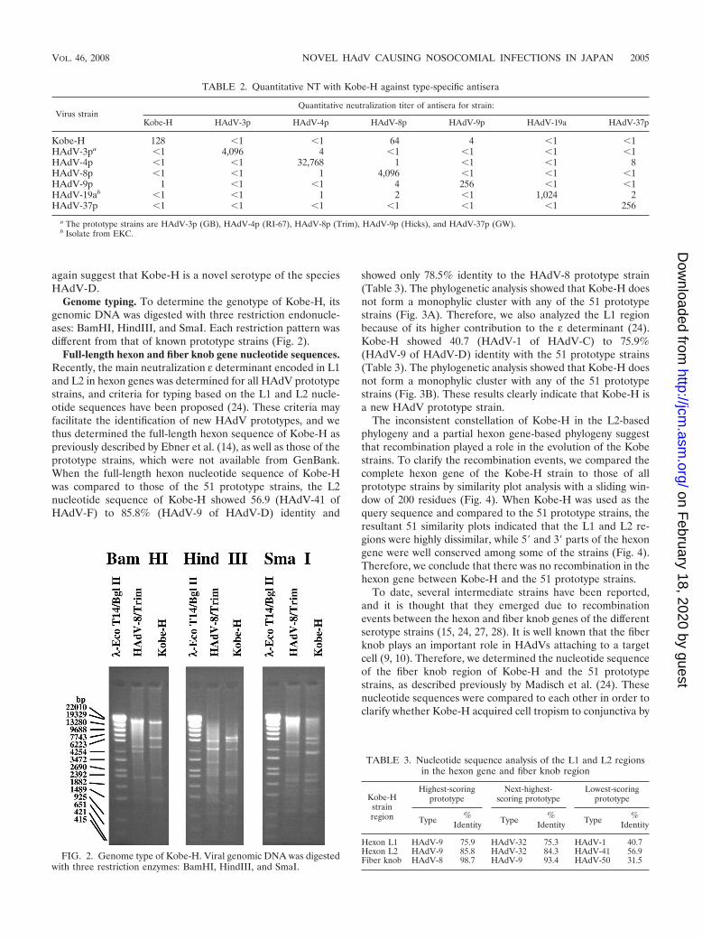
Genome typing. To determine the genotype of Kobe-H, itsgenomic DNA was digested with three restriction endonucle-ases: BamHI, HindIII, and SmaI. Each restriction pattern wasdifferent from that of known prototype strains (Fig. 2).
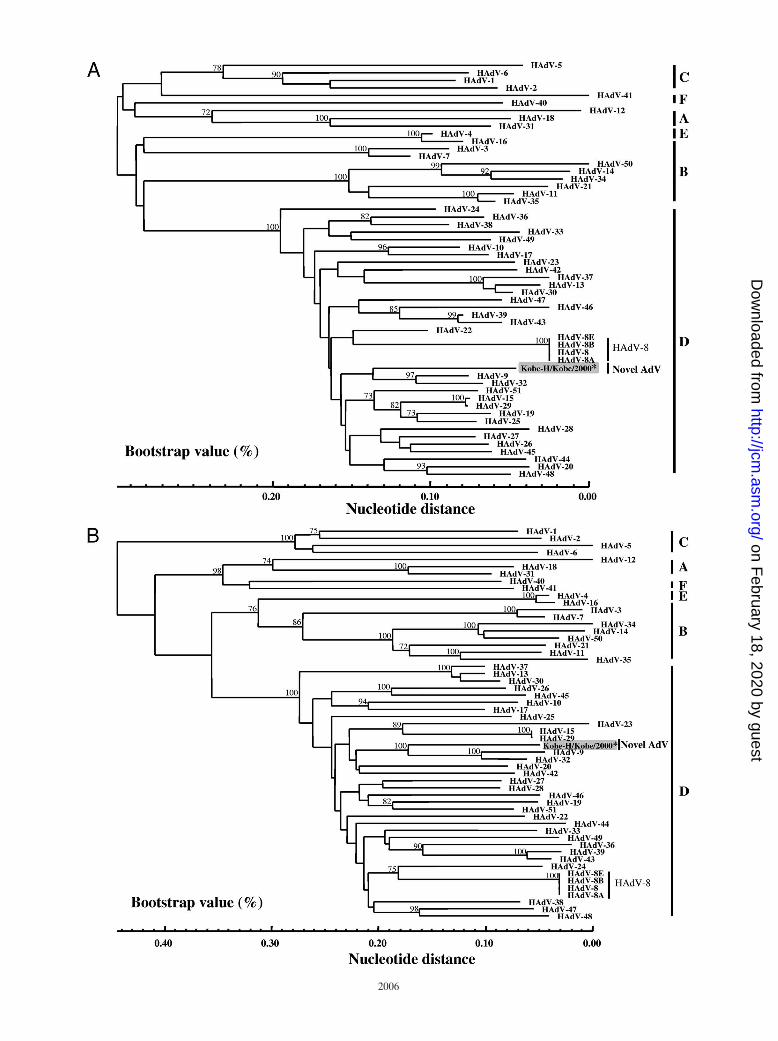
Full-length hexon and fiber knob gene nucleotide sequences.Recently, the main neutralization ε determinant encoded in L1and L2 in hexon genes was determined for all HAdV prototypestrains, and criteria for typing based on the L1 and L2 nucle-otide sequences have been proposed (24). These criteria mayfacilitate the identification of new HAdV prototypes, and wethus determined the full-length hexon sequence of Kobe-H aspreviously described by Ebner et al. (14), as well as those of theprototype strains, which were not available from GenBank.When the full-length hexon nucleotide sequence of Kobe-Hwas compared to those of the 51 prototype strains, the L2nucleotide sequence of Kobe-H showed 56.9 (HAdV-41 ofHAdV-F) to 85.8% (HAdV-9 of HAdV-D) identity and
showed only 78.5% identity to the HAdV-8 prototype strain(Table 3). The phylogenetic analysis showed that Kobe-H doesnot form a monophylic cluster with any of the 51 prototypestrains (Fig. 3A). Therefore, we also analyzed the L1 regionbecause of its higher contribution to the ε determinant (24).Kobe-H showed 40.7 (HAdV-1 of HAdV-C) to 75.9%(HAdV-9 of HAdV-D) identity with the 51 prototype strains(Table 3). The phylogenetic analysis showed that Kobe-H doesnot form a monophylic cluster with any of the 51 prototypestrains (Fig. 3B). These results clearly indicate that Kobe-H isa new HAdV prototype strain.
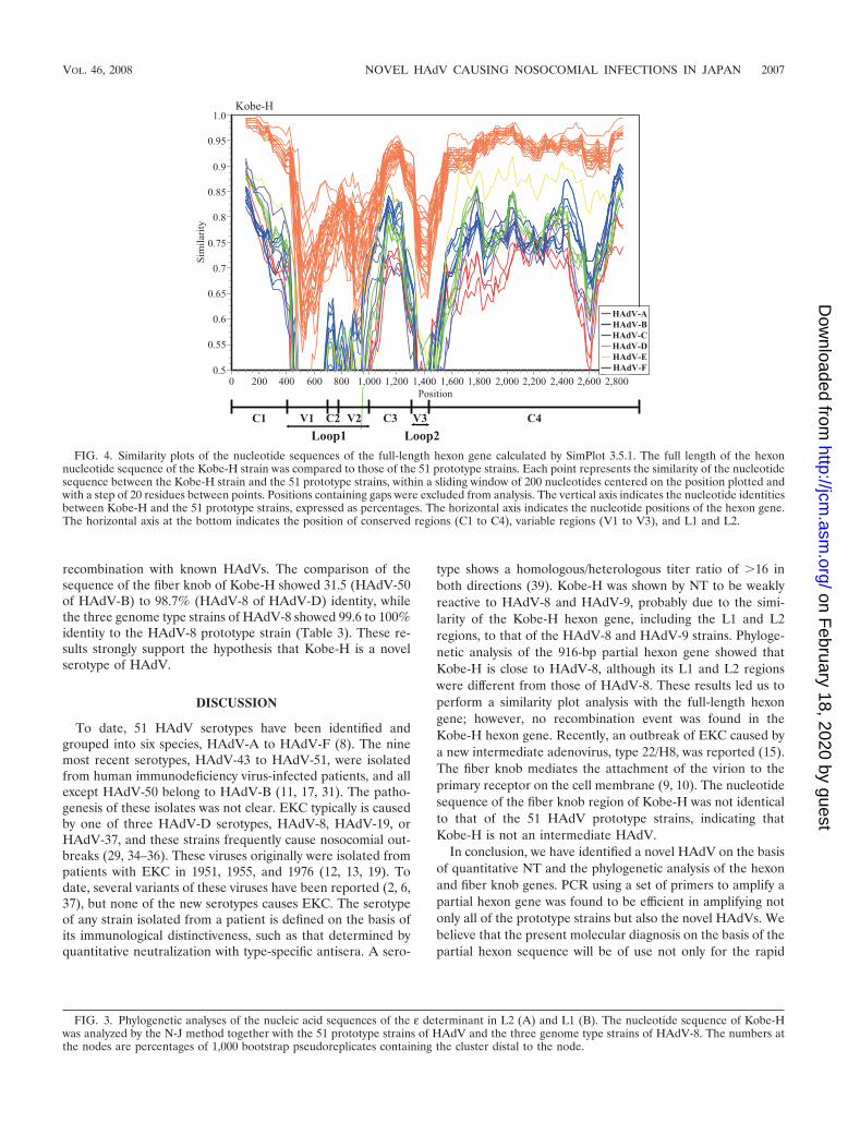
The inconsistent constellation of Kobe-H in the L2-basedphylogeny and a partial hexon gene-based phylogeny suggestthat recombination played a role in the evolution of the Kobestrains. To clarify the recombination events, we compared thecomplete hexon gene of the Kobe-H strain to those of allprototype strains by similarity plot analysis with a sliding win-dow of 200 residues (Fig. 4). When Kobe-H was used as thequery sequence and compared to the 51 prototype strains, theresultant 51 similarity plots indicated that the L1 and L2 re-gions were highly dissimilar, while 5� and 3� parts of the hexongene were well conserved among some of the strains (Fig. 4).Therefore, we conclude that there was no recombination in thehexon gene between Kobe-H and the 51 prototype strains.
To date, several intermediate strains have been reported,and it is thought that they emerged due to recombinationevents between the hexon and fiber knob genes of the differentserotype strains (15, 24, 27, 28). It is well known that the fiberknob plays an important role in HAdVs attaching to a targetcell (9, 10). Therefore, we determined the nucleotide sequenceof the fiber knob region of Kobe-H and the 51 prototypestrains, as described previously by Madisch et al. (24). Thesenucleotide sequences were compared to each other in order toclarify whether Kobe-H acquired cell tropism to conjunctiva by
FIG. 2. Genome type of Kobe-H. Viral genomic DNA was digestedwith three restriction enzymes: BamHI, HindIII, and SmaI.
TABLE 2. Quantitative NT with Kobe-H against type-specific antisera
Virus strainQuantitative neutralization titer of antisera for strain:
Kobe-H HAdV-3p HAdV-4p HAdV-8p HAdV-9p HAdV-19a HAdV-37p
Kobe-H 128 �1 �1 64 4 �1 �1HAdV-3pa �1 4,096 4 �1 �1 �1 �1HAdV-4p �1 �1 32,768 1 �1 �1 8HAdV-8p �1 �1 1 4,096 �1 �1 �1HAdV-9p 1 �1 �1 4 256 �1 �1HAdV-19ab �1 �1 1 2 �1 1,024 2HAdV-37p �1 �1 �1 �1 �1 �1 256
a The prototype strains are HAdV-3p (GB), HAdV-4p (RI-67), HAdV-8p (Trim), HAdV-9p (Hicks), and HAdV-37p (GW).b Isolate from EKC.
TABLE 3. Nucleotide sequence analysis of the L1 and L2 regionsin the hexon gene and fiber knob region
Kobe-Hstrainregion
Highest-scoringprototype
Next-highest-scoring prototype
Lowest-scoringprototype
Type %Identity Type %
Identity Type %Identity
Hexon L1 HAdV-9 75.9 HAdV-32 75.3 HAdV-1 40.7Hexon L2 HAdV-9 85.8 HAdV-32 84.3 HAdV-41 56.9Fiber knob HAdV-8 98.7 HAdV-9 93.4 HAdV-50 31.5
VOL. 46, 2008 NOVEL HAdV CAUSING NOSOCOMIAL INFECTIONS IN JAPAN 2005
on February 18, 2020 by guest
http://jcm.asm
.org/D
ownloaded from
recombination with known HAdVs. The comparison of thesequence of the fiber knob of Kobe-H showed 31.5 (HAdV-50of HAdV-B) to 98.7% (HAdV-8 of HAdV-D) identity, whilethe three genome type strains of HAdV-8 showed 99.6 to 100%identity to the HAdV-8 prototype strain (Table 3). These re-sults strongly support the hypothesis that Kobe-H is a novelserotype of HAdV.
DISCUSSION
To date, 51 HAdV serotypes have been identified andgrouped into six species, HAdV-A to HAdV-F (8). The ninemost recent serotypes, HAdV-43 to HAdV-51, were isolatedfrom human immunodeficiency virus-infected patients, and allexcept HAdV-50 belong to HAdV-B (11, 17, 31). The patho-genesis of these isolates was not clear. EKC typically is causedby one of three HAdV-D serotypes, HAdV-8, HAdV-19, orHAdV-37, and these strains frequently cause nosocomial out-breaks (29, 34–36). These viruses originally were isolated frompatients with EKC in 1951, 1955, and 1976 (12, 13, 19). Todate, several variants of these viruses have been reported (2, 6,37), but none of the new serotypes causes EKC. The serotypeof any strain isolated from a patient is defined on the basis ofits immunological distinctiveness, such as that determined byquantitative neutralization with type-specific antisera. A sero-
type shows a homologous/heterologous titer ratio of �16 inboth directions (39). Kobe-H was shown by NT to be weaklyreactive to HAdV-8 and HAdV-9, probably due to the simi-larity of the Kobe-H hexon gene, including the L1 and L2regions, to that of the HAdV-8 and HAdV-9 strains. Phyloge-netic analysis of the 916-bp partial hexon gene showed thatKobe-H is close to HAdV-8, although its L1 and L2 regionswere different from those of HAdV-8. These results led us toperform a similarity plot analysis with the full-length hexongene; however, no recombination event was found in theKobe-H hexon gene. Recently, an outbreak of EKC caused bya new intermediate adenovirus, type 22/H8, was reported (15).The fiber knob mediates the attachment of the virion to theprimary receptor on the cell membrane (9, 10). The nucleotidesequence of the fiber knob region of Kobe-H was not identicalto that of the 51 HAdV prototype strains, indicating thatKobe-H is not an intermediate HAdV.
In conclusion, we have identified a novel HAdV on the basisof quantitative NT and the phylogenetic analysis of the hexonand fiber knob genes. PCR using a set of primers to amplify apartial hexon gene was found to be efficient in amplifying notonly all of the prototype strains but also the novel HAdVs. Webelieve that the present molecular diagnosis on the basis of thepartial hexon sequence will be of use not only for the rapid
FIG. 3. Phylogenetic analyses of the nucleic acid sequences of the ε determinant in L2 (A) and L1 (B). The nucleotide sequence of Kobe-Hwas analyzed by the N-J method together with the 51 prototype strains of HAdV and the three genome type strains of HAdV-8. The numbers atthe nodes are percentages of 1,000 bootstrap pseudoreplicates containing the cluster distal to the node.
FIG. 4. Similarity plots of the nucleotide sequences of the full-length hexon gene calculated by SimPlot 3.5.1. The full length of the hexonnucleotide sequence of the Kobe-H strain was compared to those of the 51 prototype strains. Each point represents the similarity of the nucleotidesequence between the Kobe-H strain and the 51 prototype strains, within a sliding window of 200 nucleotides centered on the position plotted andwith a step of 20 residues between points. Positions containing gaps were excluded from analysis. The vertical axis indicates the nucleotide identitiesbetween Kobe-H and the 51 prototype strains, expressed as percentages. The horizontal axis indicates the nucleotide positions of the hexon gene.The horizontal axis at the bottom indicates the position of conserved regions (C1 to C4), variable regions (V1 to V3), and L1 and L2.
VOL. 46, 2008 NOVEL HAdV CAUSING NOSOCOMIAL INFECTIONS IN JAPAN 2007
on February 18, 2020 by guest
http://jcm.asm
.org/D
ownloaded from

diagnosis of HAdVs but also in molecular epidemiology. Todate, only three HAdVs, HAdV-8, HAdV-19, and HAdV-37,are known to cause severe EKC (4). The origin and mechanismof transmission of the novel HAdV are not yet clear. Ourphylogenetic analysis based on the partial hexon gene indicatesthat the novel HAdV had already appeared in 1995 as thecausative agent of EKC (data not shown). The novel HAdVhas sometimes been identified as HAdV-8 because of its weakcross-reaction with anti-HAdV-8 serum, possibly delaying itsrecognition as a novel HAdV. It also is possible that this virusis circulating in humans with asymptomatic infection; however,no asymptomatic infection was detectable in the present sur-veillance. The virus might gain pathogenesis by some unknownmechanism, becoming causative of large nosocomial EKC in-fections. The novel HAdV has caused nosocomial EKC infec-tions in at least eight hospitals in Japan during the past 5 years,and it should be monitored as an emerging adenoviral infec-tion.
REFERENCES
1. Adrian, T., G. Wadell, J. C. Hierholzer, and R. Wigand. 1986. DNA restric-tion analysis of adenovirus prototype 1 to 41. Arch. Virol. 91:277–290.
2. Adrian, T., U. Wolf, H. J. Lauer, and R. Wigand. 1990. Restriction sitemapping of adenovirus type 8 genome types. Res. Virol. 141:611–624.
3. Aoki, K., M. Kato, H. Ohtsuka, K. Ishii, N. Nakazono, and H. Sawada. 1982.Clinical and aetiological study of adenoviral conjunctivitis, with special ref-erence to adenovirus type 4 and 19 infections. Br. J. Ophthalmol. 66:776–780.
4. Aoki, K., and Y. Tagawa. 2002. A twenty-one year surveillance of adenoviralconjunctivitis in Sapporo, Japan. Int. Ophthalmol. Clin. 42:49–54.
5. Ariga, T., Y. Shimada, K. Ohgami, Y. Tagawa, H. Ishiko, K. Aoki, and S.Ohno. 2004. New genome type of adenovirus serotype 4 caused nosocomialinfections associated with epidemic conjunctivitis in Japan. J. Clin. Micro-biol. 42:3644–3648.
6. Ariga, T., Y. Shimada, K. Shiratori, K. Ohgami, S. Yamazaki, Y. Tagawa, M.Kikuchi, Y. Miyakita, K. Fujita, H. Ishiko, K. Aoki, and S. Ohno. 2005. Fivenew genome types of adenovirus type 37 caused epidemic keratoconjuncti-vitis in Sapporo, Japan, for more than 10 years. J. Clin. Microbiol. 43:726–732.
7. Becroft, D. M. 1967. Histopathology of fatal adenovirus infection of therespiratory tract in young children. J. Clin. Pathol. 20:561–569.
8. Benko, M., B. Harrach, G. W. Bothe, and W. C. Russel. 2005. FamilyAdenoviridae, p. 213–228. In C. M. Fauquet, M. A. Mayo, J. Maniloff, U.Desselberger, and L. A. Ball (ed.), Virus taxonomy: eighth report of theInternational Committee on Taxonomy of Viruses. Elsevier Academic Press,San Diego, CA.
9. Bergelson, J. M., J. A. Cunningham, G. Droguett, E. A. Kurt-Jones, A.Krithivas, J. S. Hong, M. S. Horwitz, R. L. Crowell, and R. W. Finberg. 1997.Isolation of a common receptor for coxsackie b viruses and adenoviruses 2and 5. Science 275:1320–1323.
10. Burmeister, W. P., D. Guilligay, S. Cusack, G. Wadell, and N. Arnberg. 2004.Crystal structure of species D adenovirus fiber knobs and their sialic acidbinding sites. J. Virol. 78:7727–7736.
11. De Jong, J. C., A. G. Wermenbol, M. W. Verweij-Uijterwaal, K. W. Slaterus,P. Wertheim-Van Dillen, G. J. Van Doornum, S. H. Khoo, and J. C. Hier-holzer. 1999. Adenoviruses from human immunodeficiency virus-infectedindividuals, including two strains that represent new candidate serotypesAd50 and Ad51 of species B1 and D, respectively. J. Clin. Microbiol. 37:3940–3945.
12. De Jong, J. C., R. Wigand, G. Wadell, D. Keller, C. J. Muzerie, A. G.Wermenbol, and G. J. Schaap. 1981. Adenovirus 37: identification and char-acterization of a medically important new adenovirus type of subgroup D.J. Med. Virol. 7:105–118.
13. Desmyter, J., J. C. De Jong, K. W. Slaterus, and H. Verlaeckt. 1974. Kerato-conjunctivitis caused by adenovirus type 19. Br. Med. J. 4:406.
14. Ebner, K., W. Pinsker, and T. Lion. 2005. Comparative sequence analysis ofthe hexon gene in the entire spectrum of human adenovirus serotypes:phylogenetic, taxonomic, and clinical implications. J. Virol. 79:12635–12642.
15. Engelmann, I., I. Madisch, H. Pommer, and A. Heim. 2006. An outbreak ofepidemic keratoconjunctivitis caused by a new intermediate adenovirus22/H8 identified by molecular typing. Clin. Infect. Dis. 43:e64–e66.
16. Felsenstein, J. 1985. Confidence limits on phylogeneies: an approach usingthe bootstrap. Evolution 39:783–791.
17. Hierholzer, J. C., R. Wigand, L. J. Anderson, T. Adrian, and J. W. M. Gold.1988. Adenovirus from patient with AIDS: a plethora of serotypes and adescription of five new serotypes of subgenus D (types 43-47). J. Infect. Dis.154:804–813.
18. Hirt, B. 1967. Selective extraction of polyoma DNA. J. Mol. Biol. 26:365–369.
19. Jawetz, E., S. J. Kimura, A. N. Nicholas, P. Thygeson, and L. Hanna. 1955.New type of APC virus from epidemic keratoconjunctivitis. Science 122:1190–1191.
20. Jernigan, J. A., B. S. Lowry, F. G. Hayden, S. A. Kyger, B. P. Conway, D. H.Groschel, and B. M. Farr. 1993. Adenovirus type 8 epidemic keratoconjunc-tivitis in an eye clinic: risk factors and control. J. Infect. Dis. 167:1307–1313.
21. Jin, X. H., H. Ishiko, T. H. Nguyen, T. Ohguchi, M. Akanuma, K. Aoki, andS. Ohno. 2006. Molecular epidemiology of adenoviral conjunctivitis inHanoi, Vietnam. Am. J. Ophthalmol. 142:1064–1066.
22. Kimura, M. 1980. A simple method for estimating evolutionary rates of basesubstitutions through comparative studies of nucleotide sequence. J. Mol.Evol. 16:111–120.
23. Lole, K. S., R. C. Bollinger, R. S. Paranjape, D. Gadkari, S. S. Kulkarni,N. G. Novak, R. Ingersoll, H. W. Sheppard, and S. C. Ray. 1999. Full-lengthhuman immunodeficiency virus type 1 genomes from subtype C-infectedseroconverters in India, with evidence of intersubtype recombination. J. Vi-rol. 73:152–160.
24. Madisch, I., G. Harste, H. Pommer, and A. Heim. 2005. Phylogenetic anal-ysis of the main neutralization and hemagglutination determinants of allhuman adenovirus prototypes as a basis for molecular classification andtaxonomy. J. Virol. 79:15265–15276.
25. Miura-Ochiai, R., Y. Shimada, T. Konno, S. Yamazaki, K. Aoki, S. Ohno, E.Suzuki, and H. Ishiko. 2007. Quantitative detection and rapid identificationof human adenoviruses. J. Clin. Microbiol. 45:958–967.
26. Nauheim, R. C., E. G. Romanowski, T. Araullo-Cruz, R. P. Kowalski, P. W.Turgeon, S. S. Stopak, and Y. J. Gordon. 1990. Prolonged recoverability ofdesiccated adenovirus type 19 from various surfaces. Ophthalmology 97:1450–1453.
27. Noda, M., Y. Miyamoto, Y. Ikeda, T. Matsuishi, and T. Ogino. 1991. Inter-mediate human adenovirus type 22/H10,19,37 as a new etiological agent ofconjunctivitis. J. Clin. Microbiol. 29:1286–1289.
28. Pring-Akerblom, P., and T. Adrian. 1995. Characterization of adenovirussubgenus D fiber genes. Virology 206:564–571.
29. Richmond, S., R. Burman, E. Crosdale, L. Cropper, D. Longson, B. E.Enoch, and C. L. Dodd. 1984. A large outbreak of keratoconjunctivitis dueto adenovirus type 8. J. Hyg. (London) 93:285–291.
30. Saitou, N., and M. Nei. 1987. The neighbor-joining method: a new methodfor reconstructing phylogenetic trees. Mol. Biol. Evol. 4:406–425.
31. Schnurr, D., and M. E. Dondero. 1993. Tow new candidate adenovirusserotypes. Intervirology 36:79–83.
32. Schrader, E., and R. Wigand. 1981. Neutralization of adenovirus infectivityand cytotoxin in various cell cultures. J. Virol. Methods 2:321–330.
33. Shimada, Y., T. Ariga, Y. Tagawa, K. Aoki, S. Ohno, and H. Ishiko. 2004.Molecular diagnosis of human adenoviruses D and E by a phylogeny-basedclassification method using a partial hexon sequence. J. Clin. Microbiol.42:1577–1584.
34. Tabery, H. M. 1995. Two outbreaks of adenovirus type 8 keratoconjunctivitiswith different outcome. Acta Ophthalmol. Scand. 73:358–360.
35. Takeuchi, R., Y. Nomura, M. Kojima, E. Uchio, N. Kobayashi, and M.Matumoto. 1990. A nosocomial outbreak of epidemic keratoconjunctivitisdue to adenovirus type 37. Microbiol. Immunol. 34:749–754.
36. Tanaka-Yokogui, K., N. Itoh, N. Usui, S. Takeuchi, E. Uchio, K. Aoki, M.Usui, and S. Ohno. 2001. New genome type of adenovirus serotype 19causing nosocomial infections of epidemic keratoconjunctivitis in Japan.J. Med. Virol. 65:530–533.
37. Wadell, G., and J. C. De Jong. 1980. Restriction endonucleases in identifi-cation of a genome type of adenovirus 19 associated with keratoconjuncti-vitis. Infect. Immun. 27:292–296.
38. Warren, D., K. E. Nelson, J. A. Farrar, E. Hurwitz, J. Hierholzer, E. Ford,and L. J. Anderson. 1989. A large outbreak of epidemic keratoconjunctivitis:problems in controlling nosocomial spread. J. Infect. Dis. 160:938–943.
39. Wigand, R., A. Bartha, R. S. Dreizin, H. Esche, H. S. Ginsberg, M. Green,J. C. Hierholzer, S. S. Kalter, J. B. McFerran, U. Pettersson, W. C. Russell,and G. Wadell. 1982. Adenoviridae: second report. Intervirology 18:169–176.
40. Wigand, R., T. H. Adrian, and F. Bricout. 1987. A new human adenovirus ofsubgenus D: candidate adenovirus type 42. Arch. Virol. 94:283–286.
41. World, W. S. M., and M. S. Howrwitz. 2007. Adenoviridae, p. 2395–2436. InD. M. H. Knipe and P. M. Howley (ed.), Fields virology, 5th ed., vol. 2.Lippincott Williams & Wilkins, Philadelphia, PA.
2008 ISHIKO ET AL. J. CLIN. MICROBIOL.
on February 18, 2020 by guest
http://jcm.asm
.org/D
ownloaded from