novel method for high-throughput colony pcr screening in nanoliter-reactors
TRANSCRIPT
Published online 12 March 2009 Nucleic Acids Research, 2009, Vol. 37, No. 8 e57doi:10.1093/nar/gkp160
Novel method for high-throughput colony PCRscreening in nanoliter-reactorsMarcel Walser1, Rene Pellaux1, Andreas Meyer1, Matthias Bechtold1,
Herve Vanderschuren2, Richard Reinhardt3, Joseph Magyar4,
Sven Panke1 and Martin Held1,*
1ETH Zurich, Institute of Process Engineering, BioProcess Laboratory (BPL), 2ETH Zurich, Institute of PlantScience, Plant Biotechnology, Zurich, Switzerland, 3Max Planck Institute for Molecular Genetics, Analytics& Computing, Berlin, Germany and 4Harlan Laboratories Ltd., Environmental Safety and Metabolism,Itingen, Switzerland
Received December 17, 2008; Revised and Accepted February 26, 2009
ABSTRACT
We introduce a technology for the rapid identifica-tion and sequencing of conserved DNA elementsemploying a novel suspension array based on nano-liter (nl)-reactors made from alginate. The reactorshave a volume of 35 nl and serve as reaction com-partments during monoseptic growth of microbiallibrary clones, colony lysis, thermocycling andscreening for sequence motifs via semi-quantitativefluorescence analyses. nl-Reactors were kept insuspension during all high-throughput steps whichallowed performing the protocol in a highly space-effective fashion and at negligible expenses ofconsumables and reagents. As a first application,11 high-quality microsatellites for polymorphismstudies in cassava were isolated and sequencedout of a library of 20 000 clones in 2 days. The tech-nology is widely scalable and we envision thatthroughputs for nl-reactor based screenings canbe increased up to 100 000 and more samplesper day thereby efficiently complementing pro-tocols based on established deep-sequencingtechnologies.
INTRODUCTION
Identification of conserved DNA fragments in poorlycharacterized DNA samples (e.g. metagenomic orgenomic libraries) is a challenge (1,2). We introduce anovel technology based on nl-reactors for screening of
large libraries. The technology was adapted to the identi-fication of conserved sequence motifs in the genome ofcassava and we argue that any microbial library canbe screened in this way. As key steps are based on well-established and robust techniques (e.g. DNA amplificationby microbial growth, PCR and Sanger sequencing) theprotocol can be easily extended to other applications.
Fishing for conserved DNAmotifs
A prerequisite for the successful sequencing of an individ-ual DNA molecule out of a mixture of DNA moleculesis the dilution of the DNA molecules down to a level atwhich single molecules are obtained. Afterwards thesesingle molecules are arrayed in a fashion guaranteeingthat no cross-contamination between samples occurs.One of the most-efficient methods for compartmentaliza-tion of single DNA molecules is to clone them into repli-cating elements (e.g. plasmids, fosmids or BACs) (3–5)and to transform microbial hosts with these. By doingso, single DNA molecules are arrayed in a very smallvolume which in the case of single Escherichia coli cells,for instance, is in the order of a few femtoliters. The DNAis then rapidly amplified upon growth of the clones.However, in order not to mix the clones and thereforealso the genetic information during growth, a secondaryarray for monoseptic (i.e. comprising offspring of oneclone only) cell cultivation has to be employed. Sucharrays are typically agar plates providing convenientmeans for monoseptic growth of single cells on solidsupport (6,7).However, processing and handling of larger quantities
of plates employed for colony expansion and pickingis rather laborious and requires a highly automated
Author’s contributionsM.W. carried out the described experiments and wrote the manuscript. M.B. contributed with thermocycler programming. H.V. reviewed the plantpart of the manuscript. R.P., A.J.M., R.R., J.M. and S.P. supervised and improved critical parts of the work. M.H. was the responsible seniorresearcher on the project.
*To whom correspondence should be addressed. Tel: +41 44 632 4315; Fax: +41 44 632 1325; Email: [email protected]
� 2009 The Author(s)This is an Open Access article distributed under the terms of the Creative Commons Attribution Non-Commercial License (http://creativecommons.org/licenses/by-nc/2.0/uk/) which permits unrestricted non-commercial use, distribution, and reproduction in any medium, provided the original work is properly cited.
by guest on February 16, 2011
nar.oxfordjournals.orgD
ownloaded from
environment (5). Furthermore, agar plates cannot be usedas reaction vessels for in vitro DNA manipulations(e.g. PCR or sequencing). Thus, cells have to be trans-ferred into another array, typically a microtiter plate,thereby slowing down the process and requiring additionalequipment. As a result, processing of 100 000 clones perday is probably already close to the practicable limit formost screening applications (8) employing microbialclones. This is one of the reasons why novel technologies(1,9) from within the field of ultra-high-throughputscreening (HTS) do not rely on the arraying of singlemolecules in cells anymore but employ immobilizationon surfaces [e.g. microbeads (10,11) or microchips (12)]or in microemulsions (13–17) as main compartmentaliza-tion mechanisms.Still, single molecule arraying in cells has a number of
advantages: First, error rates during amplification of theDNA by cell growth are much lower than in vitro (5).Second, the information content per array element, i.e.the length of the screened fragment is much larger thanin the competing in vitro systems (3). Third, by the simpleapplication of antibiotic selection pressure, array elementswith contents (transformed clones) can be very efficientlyseparated from empty ones (i.e. clones that do not containa DNA fragment) (6). Fourth, amplification of certainsequences such as repeating regions requires specific con-ditions in vitro (9) whereas cells generally provide anappropriate environment for polynucleotide amplificationprobably with the exception of a few cases where coinci-dently toxic gene products are synthesized (18). Fifth, cellscan also be used for functional screenings, i.e. for screen-ings where first the phenotype is screened and only clonesfeaturing the desired properties are sequenced (19).We demonstrate a PCR-based method for HTS of
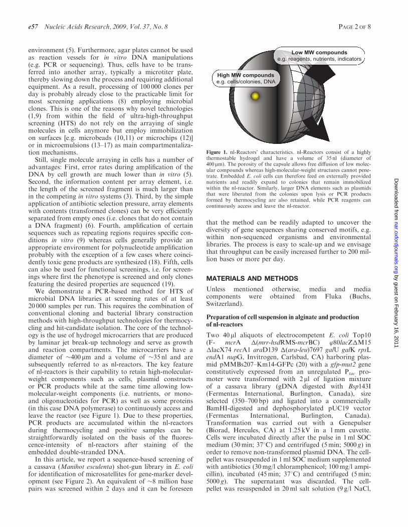
microbial DNA libraries at screening rates of at least20 000 samples per run. This requires the combination ofconventional cloning and bacterial library constructionmethods with high-throughput technologies for thermocy-cling and hit-candidate isolation. The core of the technol-ogy is the use of hydrogel microcarriers that are producedby laminar jet break-up technology and serve as growthand reaction compartments. The microcarriers have adiameter of �400 mm and a volume of �35 nl and aresubsequently referred to as nl-reactors. The key featureof nl-reactors is their capability to retain high-molecular-weight components such as cells, plasmid constructsor PCR products while at the same time allowing low-molecular-weight components (i.e. nutrients, or mono-and oligonucleotides for PCR) as well as some proteins(in this case DNA polymerase) to continuously access andleave the reactor (see Figure 1). Due to these properties,PCR products are accumulated within the nl-reactorsduring thermocycling and positive samples can bestraightforwardly isolated on the basis of the fluores-cence-intensity of nl-reactors after staining of theembedded double-stranded DNA.In this article, we report a sequence-based screening of
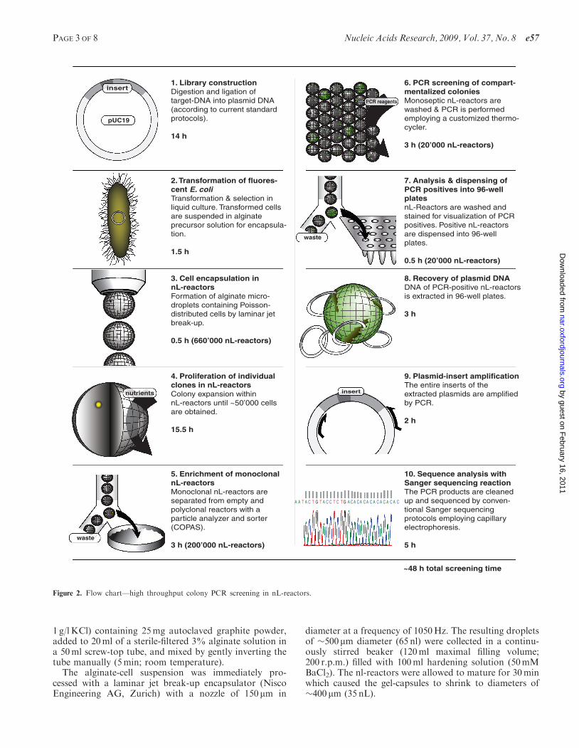
a cassava (Manihot esculenta) shot-gun library in E. colifor identification of microsatellites for gene-marker devel-opment (see Figure 2). An equivalent of �8 million basepairs was screened within 2 days and it can be foreseen
that the method can be readily adapted to uncover thediversity of gene sequences sharing conserved motifs, e.g.within non-sequenced organisms and environmentallibraries. The process is easy to scale-up and we envisagethat throughput can be easily increased further to 200 mil-lion bases or more per day.
MATERIALS AND METHODS
Unless mentioned otherwise, media and mediacomponents were obtained from Fluka (Buchs,Switzerland).
Preparation of cell suspension in alginate and productionof nl-reactors
Two 40 ml aliquots of electrocompetent E. coli Top10(F- mcrA �(mrr-hsdRMS-mcrBC) ’80lacZ�M15�lacX74 recA1 araD139 �(ara-leu)7697 galU galK rpsLendA1 nupG, Invitrogen, Carlsbad, CA) harboring plas-mid pMMBs207–Km14-GFPc (20) with a gfp-mut2 geneconstitutively expressed from an unregulated Ptac pro-moter were transformed with 2 ml of ligation mixtureof a cassava library (gDNA digested with Bsp143I(Fermentas International, Burlington, Canada), sizeselected (350–700 bp) and ligated into a commerciallyBamHI-digested and dephosphorylated pUC19 vector(Fermentas International, Burlington, Canada).Transformation was carried out with a Genepulser(Biorad, Hercules, CA) at 1.25 kV in a 1mm cuvette.Cells were incubated directly after the pulse in 1ml SOCmedium (30min; 378C) and centrifuged (5min; 5000 g) inorder to remove non-transformed plasmid DNA. The cell-pellet was resuspended in 1ml SOC medium supplementedwith antibiotics (30mg/l chloramphenicol; 100mg/l ampi-cillin), incubated (45min; 378C) and centrifuged (5min;5000 g). The supernatant was discarded. The cell-pellet was resuspended in 20ml salt solution (9 g/l NaCl,
High MW compoundse.g. cells/colonies, DNA
Low MW compoundse.g. reagents, nutrients, indicators
Figure 1. nl-Reactors’ characteristics. nl-Reactors consist of a highlythermostable hydrogel and have a volume of 35 nl (diameter of400mm). The porosity of the capsule allows free diffusion of low molec-ular compounds whereas high-molecular-weight structures cannot pene-trate. Embedded E. coli cells can therefore feed on externally providednutrients and readily expand to colonies that remain immobilizedwithin the nl-reactor. Similarly, larger DNA elements such as plasmidsthat were liberated from the colonies upon lysis or PCR productsformed by thermocycling are also retained, while PCR reagents cancontinuously access and leave the nl-reactor.
e57 Nucleic Acids Research, 2009, Vol. 37, No. 8 PAGE 2 OF 8
by guest on February 16, 2011
nar.oxfordjournals.orgD
ownloaded from
1 g/lKCl) containing 25mg autoclaved graphite powder,added to 20ml of a sterile-filtered 3% alginate solution ina 50ml screw-top tube, and mixed by gently inverting thetube manually (5min; room temperature).
The alginate-cell suspension was immediately pro-cessed with a laminar jet break-up encapsulator (NiscoEngineering AG, Zurich) with a nozzle of 150mm in
diameter at a frequency of 1050Hz. The resulting dropletsof �500mm diameter (65 nl) were collected in a continu-ously stirred beaker (120ml maximal filling volume;200 r.p.m.) filled with 100ml hardening solution (50mMBaCl2). The nl-reactors were allowed to mature for 30minwhich caused the gel-capsules to shrink to diameters of�400 mm (35 nL).
insert
A A T A C T G T A C C T C T G A C A C A C A C A C A C A C A C
1. Library constructionDigestion and ligation of target-DNA into plasmid DNA (according to current standard protocols).
14 h
2. Transformation of fluores-cent E. coliTransformation & selection in liquid culture. Transformed cells are suspended in alginate precursor solution for encapsula-tion.
1.5 h
3. Cell encapsulation in nL-reactorsFormation of alginate micro-droplets containing Poisson-distributed cells by laminar jet break-up.
0.5 h (660’000 nL-reactors)
4. Proliferation of individual clones in nL-reactorsColony expansion within nL-reactors until ~50’000 cells are obtained.
15.5 h
5. Enrichment of monoclonal nL-reactorsMonoclonal nL-reactors are separated from empty and polyclonal reactors with a particle analyzer and sorter (COPAS).
3 h (200’000 nL-reactors)
6. PCR screening of compart-mentalized coloniesMonoseptic nL-reactors are washed & PCR is performed employing a customized thermo- cycler.
3 h (20’000 nL-reactors)
7. Analysis & dispensing of PCR positives into 96-well platesnL-Reactors are washed and stained for visualization of PCR positives. Positive nL-reactors are dispensed into 96-well plates.
0.5 h (20’000 nL-reactors)
8. Recovery of plasmid DNA DNA of PCR-positive nL-reactors is extracted in 96-well plates.
3 h
9. Plasmid-insert amplificationThe entire inserts of the extracted plasmids are amplified by PCR.
2 h
10. Sequence analysis with Sanger sequencing reactionThe PCR products are cleaned up and sequenced by conven-tional Sanger sequencing protocols employing capillary electrophoresis.
5 h
PCR reagents
nutrients
insert
pUC19
waste
waste
~48 h total screening time
Figure 2. Flow chart—high throughput colony PCR screening in nL-reactors.
PAGE 3 OF 8 Nucleic Acids Research, 2009, Vol. 37, No. 8 e57
by guest on February 16, 2011
nar.oxfordjournals.orgD
ownloaded from
Proliferation ofE. coli in nl-reactors and determinationof colony diameter
The nl-reactors were recovered from the hardening solu-tion by sieving (100mm Falcon sieve; BD, Franklin Lakes,NJ) and washed three times in 150ml of growth-medium(4 g/l Bacto yeast extract; 1 g/l Bacto tryptone; 1 g/l gly-cerol, 1mM BaCl2; 10mM Tris–HCl pH 7.0; 30mg/lchloramphenicol; 100mg/l ampicillin). Aliquots of �3 gwet nl-reactors were added to Petri dishes containing30ml of growth medium. The plates were covered withtheir lids and incubated for 14 h at 308C. Afterwardsampicillin was added to the medium to 100mg/l and theplates were incubated for an additional 1.5 h (378C).nl-Reactors were recovered from the dishes, sieved,washed three times with 50ml washing solution (10mMTris pH 8.0, 0.1mM BaCl2; pH 8.0) and kept on ice untilprocessing by Complex Object Parametric Analyzer andSorter (COPAS; Union Biometrica, Holliston, MA). Laterthe average colony diameter and cell number was deter-mined by analyzing 30 colonies that had been grownwithin the nl-reactor by fluorescence microscopy underthe assumption that a typical E. coli cell volume is 2 fl.
COPAS analysis for enrichment of monoclonalmicrocarriers
Monoseptic nl-reactors harboring one colony only wereenriched by COPAS sorting employing the ‘Profiler’ soft-ware (21). Pulse shape diagram recording was triggered bythe opacity signal [threshold >25 AU (arbitrary units);signal gain factor 1.5; measuring range 0–65500 AU]. nl-Reactor size is expressed as time-of-flight [(ToF); gatedrange 400–750 ToF, arbitrary units]. Fluorescence signalsfor colony detection were generally recorded at 510 nm,which is the emission maximum of the employed GFP[ex 488 nm, photon multiplier settings (PMT) 800V; gainfactor 1.0; measuring range 0–65 500 AU; peak-profiling was initiated at >20 000 AU; gated range60 000–65 500]. The COPAS-device was operated at anaverage frequency of 30Hz and coincidence settings fordispensing into microtiter plates were adjusted to ‘pure’-mode which guarantees that nl-reactors are only sorted-out if no other reactor is coincidentally within the samedroplet. A detailed description of the cell encapsulation,proliferation and isolation procedure employed for enrich-ment of monoseptic nl-reactors by COPAS, was publishedelsewhere (21).
In-bead PCR screening of embedded colonies
A total number of 20 000 enriched monoseptical nl-reac-tors were suspended in a 50ml Falcon tube in 10ml ofddH2O. The cells were lyzed by heat (10min in a waterbath at 968C). The microcarriers were washed once in50ml washing solution and three times in 50mL ddH2Oand directly added to a poly(fluoro acrylate) PCR-bag(Welch Fluorocarbon, Dover, NH; 100� 100mm; thick-ness: 50 mm) containing 10ml PCR reagent [ddH2O con-taining: 1M betaine; 5% DMSO; 1 � PCR buffer; 0.1mMBaCl2; 0.2mM of each dNTPs; 0.2mM primer M13F(–21); 0.2 mM primer GA9; 700U Taq polymerase;
Genscript, Piscataway, NJ]. All gas bubbles wereremoved, the PCR-bag was air-tightly sealed, and cycledon a custom-built flat-screen thermocycler that consistedof a chamber flanked by aluminum plates which containedchannels for water circulation for temperature control (32cycles; 90 s at 968C; 180 s at 558C) exerted by two water-baths (Huber Polystat CC3; set to 968C and 568C, respec-tively). Numerically, controlled thermo-switching betweenthe two water cycles was realized with four valves (m&minternational, Bedford, UK) controlling the in- and outletof the water baths and a customized LabView (NationalInstruments; Austin; TX) program controlling the relay-switch station USB-Erb24 (Measurement ComputingCorp., Norton, MA). After cycling, the beads were recov-ered from the bag by sieving, washed with 50ml washingsolution and 250ml ddH2O and suspended in a Petri dishwith 20ml washing solution containing 1� SYBR Green Idye (Invitrogen; Carlsbad; CA) in order to stain PCR-products prior to another COPAS analysis. Microscopicpictures were taken by a Zeiss Axio Star Plus fluorescencemicroscope (Carl Zeiss AG; Gottingen; Germany) usingan excitation filter at 488 nm and an emission long-passfilter >520 nm in combination with phase contrastmicroscopy.
Screening for PCR positive nl-reactors
All nl-reactors recovered after thermocycling were addedto the COPAS sample cup and analyzed. Both, objectdiameter (based on extinction) as well as fluorescenceintensity (510 nm) of each nl-reactor, were measured con-comitantly by pulse shape analysis (COPAS profiler soft-ware). First, the system was calibrated by analyzing 300nl-reactors in order to set an adaptive fluorescence inten-sity threshold just above the main, non-fluorescent, popu-lation, which was evaluated to be at 780 AU (at 510 nm).Next, gates for a ToF value of 600–1000 AU were appliedfor dispensing of nL-reactors into a 96-well microtiterplate. For sampling of the control group, the same ToFgates were applied and 48 putatively PCR-negativenl-reactors featuring signal intensities below the adaptivethreshold (600–780 AU) were sorted out. All samples wereimmediately processed further in order to recover the plas-mids (see next section).
Plasmid extraction with cetyl trimethylammoniumbromide (CTAB)
For plasmid extraction, we adapted a method developedby Allen et al. (22). An aliquot of 90 ml of freshly preparedand pre-warmed (508C) CTAB DNA extraction buffer[100mM Tris–HCl (pH 8.0); 1.4M NaCl; 2.5% w/vCTAB; 0.5% w/v N-lauryl sarcosine] was added to awell containing a single isolated nl-reactor (one reactorper MT-plate well), plates were air-tightly sealed by8-cap strips, incubated in a PCR cycler (Mastercycler,Eppendorf, 658C; 45min) and every 5min vigorously agi-tated by hand for 10 s in order to dissolve the nl-reactor.After centrifugation at 3200 g for 30 s, 90 ml of chloroform/isoamylalcohol (24/1) were added and plates were agitatedby hand until turbid emulsions were formed in the wells(�15 s). Plates were centrifuged (30min at 3200 g) and an
e57 Nucleic Acids Research, 2009, Vol. 37, No. 8 PAGE 4 OF 8
by guest on February 16, 2011
nar.oxfordjournals.orgD
ownloaded from
aliquot of 33 ml was withdrawn from the aqueous phase,transferred to another microtiter plate, and DNA wasprecipitated by ethanol (77 ml per well). After overnightincubation at –808C, samples were centrifuged (45min;3220 g; 08C) and supernatants were discarded prior towashing (100 ml of 70% EtOH, chilled to –208C and cen-trifuged for 30min; 3220 g; 08C). Once more, supernatantswere discarded and the remaining liquid was allowed toevaporate at room temperature.
Insert amplification by PCR
PCR reagents [25ml comprising: 1� PCR Buffer, 200 mMof each dNTP, 200 nM of primer M13F(–43) andM13R(+86), 1.75U Taq DNA polymerase] were addedto the dried pellet and the plasmid inserts were amplifiedby PCR (denaturation at 958C for 1min, then 40 cycles of:958C for 30 s, 558C for 30 s, 728C for 1min, and finalelongation: 728C for 2min). After cycling, 5 ml of eachsample were loaded on a 1.5% agarose gel in order todetermine the number of PCR-amplified fragments persample, the insert size as well as the approximate PCRproduct concentration. All samples yielding a singleband of at least 100 ng of DNA on the gel (equivalent to20 ng/ml concentration in the well) were sent out forSanger sequencing with a M13 reverse primer (GATCBiotech, Konstanz, Germany).
Polymorphism test
Clone-redundancy was tested with the DNA Star Software(DNAstar inc., Madison, WI). Default assembly param-eters were used: match size: 12 bp; minimum matchpercentage: 80%; minimum sequence length 100 bp; max-imum added gaps per kb in contig: 70 bp; maximum addedgaps per kb in sequence: 70 bp; last group considered: 2;gap penalty: 0.00; gap length penalty: 0.70.
Only sequences with a match to the screening primer of10 bases or more were used to subsequently design micro-satellite markers. Next, PCR primers flanking the CT-richregion were designed by the primer-3 software; http://fokker.wi.mit.edu/primer3/input.htm). Amplicon length poly-morphism of microsatellite markers was tested by PCR onseven different cassava cultivars (91/02322; TMS60444;95/0306; 98/0002; TAI-8; PER-183; COL-1505) usingFAM-labeled primers. PCRs were performed in a finalvolume of 20 ml: 1� PCR Buffer, 200 mM each dNTP,200 nM of each primer, 5 ng template; 1.75U Taq DNApolymerase; cycling conditions: initial denaturation: 958Cfor 1min; 35 cycles: 958C for 30 s, 568C for 30 s, 728C for40 s; final elongation: 728C for 2min. Fragments wereseparated by capillary electrophoresis on an ABI 3700(Applied Biosystems; Foster City; CA) and analyzedby the GeneMarker (Softgenetics; State College; PA) soft-ware. Polymorphic information content (PIC) was calcu-lated according to Anderson et al. (23).
RESULTS
The objective of this study was the detection andsequence determination of specific DNA-motifs from agenomic cassava library using an essentially freely scalable
approach employing gel-like, suspended nl-reactors asreaction compartments.
Cell encapsulation and proliferation in nl-reactors
The feasibility of the approach was investigated fora genomic cassava library (average insert length350–700 bp) in E. coli. The sample throughput wasmonitored over all process steps (see Table 1). Freshlytransformed cells synthesizing a green fluorescent protein(GFP) from Aequorea victoria were embedded intonl-reactors (660 000 reactors total; average degree of occu-pation with colonies 10.8%) and propagated from singlecell status to colonies of �50 000 cells. This was confirmedmanually by fluorescence microscopy. Next, 20 000 mono-septic nl-reactors were isolated by COPAS-sorting (21).During this step, the number of reactors containing noor more than one colony, and reactor multiplets (24), inev-itably formed during nl-reactor production, was reducedto a minimum (Table 1).
Highly parallelized nl-PCR and PCR-product detection
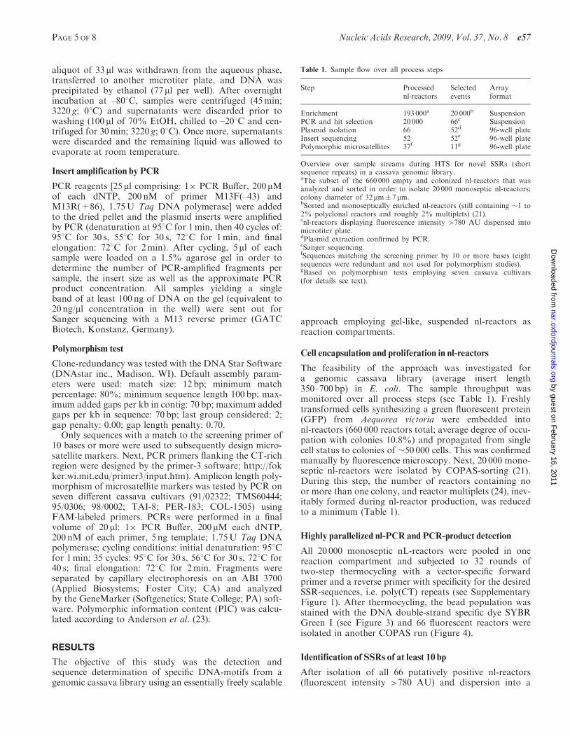
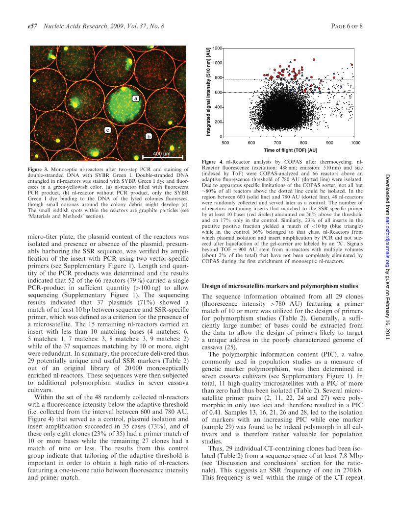
All 20 000 monoseptic nL-reactors were pooled in onereaction compartment and subjected to 32 rounds oftwo-step thermocycling with a vector-specific forwardprimer and a reverse primer with specificity for the desiredSSR-sequences, i.e. poly(CT) repeats (see SupplementaryFigure 1). After thermocycling, the bead population wasstained with the DNA double-strand specific dye SYBRGreen I (see Figure 3) and 66 fluorescent reactors wereisolated in another COPAS run (Figure 4).
Identification of SSRs of at least 10 bp
After isolation of all 66 putatively positive nl-reactors(fluorescent intensity >780 AU) and dispersion into a
Table 1. Sample flow over all process steps
Step Processednl-reactors
Selectedevents
Arrayformat
Enrichment 193 000a 20 000b SuspensionPCR and hit selection 20 000 66c SuspensionPlasmid isolation 66 52d 96-well plateInsert sequencing 52 52e 96-well platePolymorphic microsatellites 37f 11g 96-well plate
Overview over sample streams during HTS for novel SSRs (shortsequence repeats) in a cassava genomic library.aThe subset of the 660 000 empty and colonized nl-reactors that wasanalyzed and sorted in order to isolate 20 000 monoseptic nl-reactors;colony diameter of 32 mm� 7 mm.bSorted and monoseptically enriched nl-reactors (still containing �1 to2% polyclonal reactors and roughly 2% multiplets) (21).cnl-reactors displaying fluorescence intensity >780 AU dispensed intomicrotiter plate.dPlasmid extraction confirmed by PCR.eSanger sequencing.fSequences matching the screening primer by 10 or more bases (eightsequences were redundant and not used for polymorphism studies).gBased on polymorphism tests employing seven cassava cultivars(for details see text).
PAGE 5 OF 8 Nucleic Acids Research, 2009, Vol. 37, No. 8 e57
by guest on February 16, 2011
nar.oxfordjournals.orgD
ownloaded from
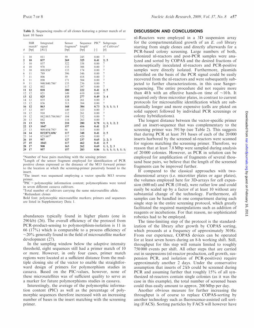
micro-titer plate, the plasmid content of the reactors wasisolated and presence or absence of the plasmid, presum-ably harboring the SSR sequence, was verified by ampli-fication of the insert with PCR using two vector-specificprimers (see Supplementary Figure 1). Length and quan-tity of the PCR products was determined and the resultsindicated that 52 of the 66 reactors (79%) carried a singlePCR-product in sufficient quantity (>100 ng) to allowsequencing (Supplementary Figure 1). The sequencingresults indicated that 37 plasmids (71%) showed amatch of at least 10 bp between sequence and SSR-specificprimer, which was defined as a criterion for the presence ofa microsatellite. The 15 remaining nl-reactors carried aninsert with less than 10 matching bases (4 matches: 6,5 matches: 1, 7 matches: 3, 8 matches: 3, 9 matches: 2)while of the 37 sequences matching by 10 or more, eightwere redundant. In summary, the procedure delivered thus29 potentially unique and useful SSR markers (Table 2)out of an original library of 20 000 monosepticallyenriched nl-reactors. These sequences were then subjectedto additional polymorphism studies in seven cassavacultivars.Within the set of the 48 randomly collected nl-reactors
with a fluorescence intensity below the adaptive threshold(i.e. collected from the interval between 600 and 780 AU,Figure 4) that served as a control, plasmid isolation andinsert amplification succeeded in 35 cases (73%), and ofthese only eight clones (23% of 35) had a primer match of10 or more bases while the remaining 27 clones had amatch of nine or less. The results from this controlgroup indicate that tailoring of the adaptive threshold isimportant in order to obtain a high ratio of nl-reactorsfeaturing a one-to-one ratio between fluorescence intensityand primer match.
Design of microsatellite markers and polymorphism studies
The sequence information obtained from all 29 clones(fluorescence intensity >780 AU) featuring a primermatch of 10 or more was utilized for the design of primersfor polymorphism studies (Table 2). Generally, a suffi-ciently large number of bases could be extracted fromthe data to allow the design of primers likely to targeta unique address in the poorly characterized genome ofcassava (25).
The polymorphic information content (PIC), a valuecommonly used in population studies as a measure ofgenetic marker polymorphism, was then determined inseven cassava cultivars (see Supplementary Figure 1). Intotal, 11 high-quality microsatellites with a PIC of morethan zero had thus been isolated (Table 2). Several micro-satellite primer pairs (2, 11, 22, 24 and 27) were poly-morphic in only two loci and therefore resulted in a PICof 0.41. Samples 13, 16, 21, 26 and 28, led to the isolationof markers with an increasing PIC while one marker(sample 29) was found to be indeed polymorph in all cul-tivars and is therefore rather valuable for populationstudies.
Thus, 29 individual CT-containing clones had been iso-lated (Table 2) from a sequence space of at least 7.8 Mbp(see ‘Discussion and conclusions’ section for the ratio-nale). This suggests an SSR frequency of one in 270 kb.This frequency is well within the range of the CT-repeat
0
200
400
600
800
1000
1200
500 600 700 800 900 1000
Time of flight (TOF) [AU]
Inte
gra
ted
sig
nal
inte
nsi
ty (
510
nm
) [A
U]
Figure 4. nl-Reactor analysis by COPAS after thermocycling. nl-Reactor fluorescence (excitation: 488 nm; emission: 510 nm) and size(indexed by ToF) were COPAS-analyzed and 66 reactors above anadaptive fluorescence threshold of 780 AU (dotted line) were isolated.Due to apparatus specific limitations of the COPAS sorter, not all but�80% of all reactors above the dotted line could be isolated. In theregion between 600 (solid line) and 780 AU (dotted line), 48 nl-reactorswere randomly collected and served later as a control. The number ofnl-reactors containing inserts that matched to the SSR-specific primerby at least 10 bases (red circles) amounted on 56% above the thresholdand on 17% only in the control. Similarly, 23% of all inserts in theputative positive fraction yielded a match of <10 bp (blue triangle)while in the control 56% belonged to that class. nl-Reactors fromwhich plasmid isolation and insert amplification by PCR did not suc-ceed after liquefaction of the gel-carrier are labeled by an ‘X’. Signalsbeyond TOF=900 AU stem from nl-reactors with multiple volumes(about 2% of the total) that have not been completely eliminated byCOPAS during the first enrichment of monoseptic nl-reactors.
a
cb
400 µm
Figure 3. Monoseptic nl-reactors after two-step PCR and staining ofdouble-stranded DNA with SYBR Green I. Double-stranded DNAentangled in nl-reactors was stained with SYBR Green I dye and fluor-esces in a green-yellowish color. (a) nl-reactor filled with fluorescentPCR product. (b) nl-reactor without PCR product, only the SYBRGreen I dye binding to the DNA of the lysed colonies fluoresces,though small coronas around the colony debris might develop (c).The small reddish spots within the reactors are graphite particles (see‘Materials and Methods’ section).
e57 Nucleic Acids Research, 2009, Vol. 37, No. 8 PAGE 6 OF 8
by guest on February 16, 2011
nar.oxfordjournals.orgD
ownloaded from
abundances typically found in higher plants (one in290 kb) (26). The overall efficiency of the protocol fromPCR-product-sensing to polymorphism-isolation is 11 in66 (17%) which is comparable to a process efficiency of�20% generally found in the field of microsatellite markerdevelopment (27).
In the sampling window below the adaptive intensitythreshold, eight sequences still had a primer match of 10or more. However, in only four cases, primer matchregions were located at a sufficient distance from the mul-tiple cloning site of the vector to enable the straightfor-ward design of primers for polymorphism studies incassava. Based on the PIC-values, however, none ofthese microsatellites was of sufficient quality to serve asa marker for future polymorphisms studies in cassava.
Interestingly, the average of the polymorphic informa-tion content (PIC) as well as the percentage of poly-morphic sequences therefore increased with an increasingnumber of bases in the insert matching with the screeningprimer.
DISCUSSION AND CONCLUSIONS
nl-Reactors were employed in a 3D suspension arrayfor the compartmentalized growth of an E. coli librarystarting from single clones and directly afterwards for aPCR-based colony screening. Large numbers of both,colonized nl-reactors and post-PCR samples were ana-lyzed and sorted by COPAS and the desired fractions ofmonoseptically inoculated nl-reactors and PCR-positivesamples were directly isolated. Furthermore, plasmidsidentified on the basis of the PCR signal could be easilyrecovered from the nl-reactors and were subsequently sub-jected to further characterizations, in this case Sanger-sequencing. The entire procedure did not require morethan 48 h with an effective hands-on time of �10 h. Itrequired only three microtiter plates, in contrast to currentprotocols for microsatellite identification which are sub-stantially longer and more expensive (cells are plated onsolid support followed by individual PCR screenings orcolony hybridizations).The longest distance between the vector-specific primer
and an insert-sequence that was complementary to thescreening primer was 391 bp (see Table 2). This suggeststhat during PCR at least 391 bases of each of the 20 000inserts harbored by the screened nl-reactors were scannedfor regions matching the screening primer. Therefore, wereason that at least 7.8Mbp were sampled during analysisof 20 000 colonies. However, as PCR in solution can beemployed for amplification of fragments of several thou-sand base pairs, we believe that the length of the screenedfragments can be improved further.If compared to the classical approaches with two-
dimensional arrays (i.e. microtiter plates or agar plates),the volumes employed here for 3D-arrays for cell expan-sion (600ml) and PCR (10ml), were rather low and couldeasily be scaled up by a factor of at least 10 without anysubstantial change of the technology. Furthermore, allsamples can be handled in one compartment during eachsingle step in the entire screening protocol, which greatlyfacilitated the required manipulations such as addition ofreagents or incubations. For that reason, no sophisticatedrobotics had to be employed.The time-limiting step of the protocol is the standard-
ization of the library after growth by COPAS sorting,which proceeds at a frequency of approximately 30Hz.From our experience, COPAS devices can be operatedfor at least seven hours during an 8-h working shift. Still,throughput for this step will remain limited to roughly750 000 events per shift. All other steps that are carriedout in suspensions (nl-reactor production, cell growth, sus-pension PCR, and isolation of PCR-positives) requireapproximately another 2 days. Under the conservativeassumption that inserts of 2 kb could be screened duringPCR and assuming further that roughly 15% of all syn-thesized nl-reactors contain single colonies (as it was thecase in this example), the total number of screened basescould thus easily amount to approx. 200Mbp per day.Another obvious measure for further increasing the
throughput is of course to replace COPAS-sorting byanother technology such as fluorescence-assisted cell sort-ing (FACS). Sorting particles by FACS will however have
Table 2. Sequencing results of all clones featuring a primer match of at
least 10 bases
SSRmatcha
Integratedsignal
Sensorfragmentb
Sequencelengthc
PICd Subgroupsof Cultivarse
# [bp] [AU] [bp] [bp] [–] [#]
1 10 811 59 245 0.00 72 10 837 269 325 0.41 2; 5
3 10 837 322 338 0.00 74 10 978 133 308 0.00 75 10 808/856� 133 174 0.00 76 11 789 396 546 0.00 77 11 806 59 418 0.00 78 11 890 171 504 0.00 79 11 940/840/786� 133 236 0.00 710 12 802 117 397 0.00 711 12 818 200 222 0.41 2; 5
12 12 821 149 610 0.00 713 12 825 133 277 0.49 3; 4
14 12 835 124 261 0.00 715 12 836 313 384 0.00 716 12 863 168 384 0.73 3; 1; 1; 1; 1
17 12 897 149 323 0.00 718 12 910 112 261 0.00 719 12 882/885/784/861� 104 332 0.00 720 13 842 118 263 0.00 721 13 919 210 280 0.78 1; 1; 1; 2; 2
22 13 1047 157 416 0.41 2; 5
23 13 909/654/793� 86 315 0.00 724 14 815/871/696� 117 348 0.41 2; 5
25 14 823/607� 117 420 0.00 726 15 957 127 237 0.65 2; 2; 3
27 15 1043 117 462 0.41 2; 5
28 17 908 163 262 0.69 1; 1, 2; 3
29 17 957 157 283 0.86 1; 1; 1; 1; 1; 1; 1;
aNumber of base pairs matching with the sensing primer.bLength of the sensor fragment employed for identification of PCRpositive clones expressed as the distance of the M13 forward primerto the location at which the screening-primer putatively bound to theinsert.cThe insert was sequenced employing a vector specific M13 reverseprimer.dPIC=polymorphic information content; polymorphisms were testedin seven different cassava cultivars.eTotal number of cultivars carrying the same microsatellite allele.�Redundant clonesBold font: polymorphic microsatellite markers; primers and sequencesare listed in Supplementary Data 1.
PAGE 7 OF 8 Nucleic Acids Research, 2009, Vol. 37, No. 8 e57
by guest on February 16, 2011
nar.oxfordjournals.orgD
ownloaded from
to provide a volume in the lower picoliter (pl)-range andsuch particles can not be reliably produced by the laminarjet technology that was employed for capsule synthesishere. However, we are currently working on the develop-ment and standardization of the technologies required forthe synthesis of hydrogel pl-reactors.We argue that the sequence-specific amplification com-
bined with high throughput screening makes this technol-ogy suitable for other protocols and generally for all caseswhere ‘rare’ events are sought. Especially screeningof gene homologues or miRNA precursors (28,29) innon-sequenced species and particularly metagenomicapproaches, e.g. ‘fishing’ for new enzymes in metagenomiclibraries, or localization of insertion sequences in muta-genized genomes (e. g. transposons for random mutagen-esis) will be greatly facilitated.
SUPPLEMENTARY DATA
Supplementary Data are available at NAR Online.
ACKNOWLEDGEMENTS
We are indebted to the R’equipe program of the SwissNational Science Foundation for generous support inacquiring the COPAS Plus Biosorter. We also areindebted to H. Hilbi for the gift of plasmid pMMB207–Km14-GFPc, to Bernhard Koller for the support withSSR libraries in preliminary tests, and to MariaDomenica Moccia for the gift of plasmid pCT16 used inoptimization experiments. Additionally, we would like tothank the four anonymous reviewers for the valuableinput.
FUNDING
The Swiss Commission for Technology and Innovationgrants (to M.W., M.H. and R.P.). Funding for openaccess charge: The Swiss Commission for Technologyand Innovation (KTI/CTI).
Conflict of interest statement. None declared.
REFERENCES
1. Hutchison,C.A. III. (2007) DNA sequencing: bench to bedside andbeyond. Nucleic Acids Res., 35, 6227–6237.
2. Lorenz,P. and Eck,J. (2005) Metagenomics and industrialapplications. Nat. Rev., 3, 510–516.
3. Farrar,K. and Donnison,I. (2007) Construction and screening ofBAC libraries made from brachypodium genomic DNA. Nat.Protoc., 2, 1661–1674.
4. Beja,O. (2004) To BAC or not to BAC: marine ecogenomics. Curr.Opin. Biotechnol., 15, 187–190.
5. Venter,J.C., Remington,K., Heidelberg,J.F., Halpern,A.L.,Rusch,D., Eisen,J.A., Wu,D., Paulsen,I., Nelson,K.E., Nelson,W.et al. (2004) Environmental genome shotgun sequencing of theSargasso Sea. Science, 304, 66–74.
6. Dahlroth,S.L., Nordlund,P. and Cornvik,T. (2006) Colony filtrationblotting for screening soluble expression in Escherichia coli. Nat.Protoc., 1, 253–258.
7. Jones,P., Watson,A., Davies,M. and Stubbings,S. (1992) Integrationof image analysis and robotics into a fully automated colonypicking and plate handling system. Nucleic Acids Res., 20,4599–4606.
8. Kim,C.G., Fujiyama,A. and Saitou,N. (2003) Construction of agorilla fosmid library and its PCR screening system. Genomics, 82,571–574.
9. Shendure,J. and Ji,H. (2008) Next-generation DNA sequencing.Nat. Biotechnol., 26, 1135–1145.
10. Freeman,A., Cohen-Hadar,N., Abramow,S., Modai-Hod,R.,Dror,Y. and Georgiou,G. (2004) Screening of large protein librariesby the ‘cell immobilized on adsorbed bead’ approach. Biotechnol.Bioeng., 86, 196–200.
11. Dressman,D., Yan,H., Traverso,G., Kinzler,K.W. and Vogelstein,B.(2003) Transforming single DNA molecules into fluorescent mag-netic particles for detection and enumeration of genetic variations.Proc. Natl Acad. Sci. USA, 100, 8817–8822.
12. Fan,J.B. and Chee,M.S. (2006) Highly parallel genomic assays.Nature Rev., 7, 632–644.
13. Lee,Y.-F., Tawfik,D.S. and Griffiths,A.D. (2002) Investigating thetarget recognition of DNA cytosine-5 methyltransferase HhaI bylibrary selection using in vitro compartmentalisation. Nucleic AcidsRes., 30, 4937–4944.
14. Miller,O.J., Bernath,K., Agresti,J., Amitai,G., Kelly,B.T.,Mastrobattista,E., Taly,V., Magdassi,S., Tawfik,D.S. andGriffiths,A.D. (2006) Directed evolution by in vitro compartmen-talization. Nat. Methods, 3, 561–570.
15. Leamon,J.H., Link,D.R., Egholm,M. and Rothberg,J.M. (2006)Overview: methods and applications for droplet compartmentaliza-tion of biology. Nat. Methods, 3, 541–543.
16. Diehl,F., Li,M., He,Y., Kinzler,K.W., Vogelstein,B. andDressman,D. (2006) BEAMing: single-molecule PCR on micropar-ticles in water-in-oil emulsions. Nat. Methods, 3, 551–559.
17. Margulies,M., Egholm,M., Altman,W.E., Attiya,S., Bader,J.S.,Bemben,L.A., Berka,J., Braverman,M.S., Chen,Z., Dewell,S.B.et al. (2005) Genome sequencing in microfabricated high-densitypicolitre reactors. Nature, 437, 376–380.
18. Cowan,D., Meyer,Q., Stafford,W., Muyanga,S., Cameron,R. andWittwer,P. (2005) Metagenomic gene discovery: past, present,future. Trends Biotechnol., 23, 321–329.
19. Inglese,J., Johnson,R.L., Simeonov,A., Xia,M., Zheng,W.,Austin,C.P. and Auld,D.S. (2007) High-throughput screening assaysfor the identification of chemical probes. Nat. Chem. Biol., 3,466–479.
20. Mampel,J., Spirig,T., Weber,S.S., Haagensen,J.A.J., Molin,S. andHilbi,H. (2006) Planktonic replication is essential for biofilmformation by legionella pneumophila in a complex medium understatic and dynamic flow conditions. Appl. Environ. Microbiol., 72,2885–2895.
21. Walser,M., Leibundgut,R., Pellaux,R., Panke,S. and Held,M. (2008)Isolation of monoclonal microcarriers colonized by fluorescentE. coli. Cytometry Part A, 73A, 788–798.
22. Allen,G.C., Flores-Vergara,M.A., Krasnyanski,S., Kumar,S. andThompson,W.F. (2006) A modified protocol for rapid DNA isola-tion from plant tissues using cetyltrimethylammonium bromide.Nat. Protoc., 1, 2320–2325.
23. Anderson,J.A., Churchill,G.A., Autrique,J.E., Tanksley,S.D. andSorrells,M.E. (1993) Optimizing parental selection for geneticlinkage maps. Genome, 36, 181–186.
24. Brandenberger,H. and Widmer,F. (1999) Immobilization of highlyconcentrated cell suspensions using the laminar jet breakup techni-que. Biotechnol Prog., 15, 366–372.
25. Okogbenin,E., Marin,J. and Fregene,M. (2006) An SSR-basedmolecular genetic map of cassava. Euphytica, 147, 433–440.
26. Gupta,P.K. and Varshney,R.K. (2000) The development and use ofmicrosatellite markers for genetic analysis and plant breeding withemphasis on bread wheat. Euphytica, 113, 163–185.
27. Squirrell,J., Hollingsworth,P.M., Woodhead,M., Russell,J.,Lowe,A.J., Gibby,M. and Powell,W. (2003) How much effort isrequired to isolate nuclear microsatellites from plants? Mol. Ecol.,12, 1339–1348.
28. Terai,G., Komori,T., Asai,K. and Kin,T. (2007) miRRim: a novelsystem to find conserved miRNAs with high sensitivity and speci-ficity. RNA, 13, 2081–2090.
29. Ohler,U.W.E., Yekta,S., Lim,L.P., Bartel,D.P. and Burge,C.B.(2004) Patterns of flanking sequence conservation and a character-istic upstream motif for microRNA gene identification. RNA, 10,1309–1322.
e57 Nucleic Acids Research, 2009, Vol. 37, No. 8 PAGE 8 OF 8
by guest on February 16, 2011
nar.oxfordjournals.orgD
ownloaded from