novel stenotic microchannels to study thrombus formation
TRANSCRIPT
International Journal of
Molecular Sciences
Article
Novel Stenotic Microchannels to Study ThrombusFormation in Shear Gradients: Influence of ShearForces and Human Platelet-Related Factors
Mathew Lui 1,2, Elizabeth E. Gardiner 3 , Jane F. Arthur 4, Isaac Pinar 1,2, Woei Ming Lee 5,Kris Ryan 1,2, Josie Carberry 1,2,* and Robert K. Andrews 4,*
1 Department of Mechanical and Aerospace Engineering, Monash University, 3800 Clayton, Australia;[email protected] (M.L.); [email protected] (I.P.); [email protected] (K.R.)
2 Monash Institute of Medical Engineering, Monash University, 3800 Clayton, Australia3 Department of Cancer Biology and Therapeutics, John Curtin School of Medical Research,
Australian National University, 2600 Canberra, Australia; [email protected] Australian Centre for Blood Diseases, Monash University, 3004 Melbourne, Australia; [email protected] Research School of Electrical, Energy and Materials Engineering, The Australian National University,
2600 Canberra, Australia; [email protected]* Correspondence: [email protected] (J.C.); [email protected] (R.K.A.);
Tel.: +61-3-9905-9646 (J.C.); +61-3-9902-0136 (R.K.A.)
Received: 16 May 2019; Accepted: 15 June 2019; Published: 18 June 2019�����������������
Abstract: Thrombus formation in hemostasis or thrombotic disease is initiated by the rapid adhesion,activation, and aggregation of circulating platelets in flowing blood. At arterial or pathologicalshear rates, for example due to vascular stenosis or circulatory support devices, platelets maybe exposed to highly pulsatile blood flow, while even under constant flow platelets are exposedto pulsation due to thrombus growth or changes in vessel geometry. The aim of this study isto investigate platelet thrombus formation dynamics within flow conditions consisting of eitherconstant or variable shear. Human platelets in anticoagulated whole blood were exposed ex vivo tocollagen type I-coated microchannels subjected to constant shear in straight channels or variable sheargradients using different stenosis geometries (50%, 70%, and 90% by area). Base wall shears between1800 and 6600 s−1, and peak wall shears of 3700 to 29,000 s−1 within stenoses were investigated,representing arterial-pathological shear conditions. Computational flow-field simulations and stenosisplatelet thrombi total volume, average volume, and surface coverage were analysed. Interestingly,shear gradients dramatically changed platelet thrombi formation compared to constant base shearalone. Such shear gradients extended the range of shear at which thrombi were formed, that is, plateletsbecame hyperthrombotic within shear gradients. Furthermore, individual healthy donors displayedquantifiable differences in extent/formation of thrombi within shear gradients, with implications forfuture development and testing of antiplatelet agents. In conclusion, here, we demonstrate a specificcontribution of blood flow shear gradients to thrombus formation, and provide a novel platform forplatelet functional testing under shear conditions.
Keywords: platelets; shear gradients; stenosis; thrombosis
1. Introduction
The rapid adhesion, activation, and aggregation of circulating platelets in flowing blood is crucialfor initiating thrombus formation in hemostasis or thrombotic diseases. The capacity to form a thrombusdepends on multiple parameters, including platelet-related factors (receptor expression and function,and reactivity towards various prothrombotic stimuli), vascular factors (exposure of prothrombotic
Int. J. Mol. Sci. 2019, 20, 2967; doi:10.3390/ijms20122967 www.mdpi.com/journal/ijms
Int. J. Mol. Sci. 2019, 20, 2967 2 of 10
surfaces by activation or disruption of endothelium), disease state comorbidities (atherosclerosis,inflammation, diabetes, or other prothrombotic conditions), and blood flow [1–6]. Currently availableplatelet functional testing based on platelet aggregometry is of limited value clinically, and evenlarge-scale clinical trials have failed to show benefits in terms of antiplatelet therapy [7]. The potentialvalue of testing pharmacological agents in microfluidic systems, in particular for screening drugs orcombinations of drugs and determining effective doses and individual patient responses before orduring treatment, has recently been evaluated using collagen-coated devices [8].
Platelet adhesion and thrombus formation in vivo is affected by the complex flow in the humancirculatory system where arterial flows may be highly pulsatile and subject to temporal shear changesdue to altered vessel geometries or occlusive thrombus. Platelets respond to changes in flow ratethrough mechanotransduction of forces that the flow generates [9–11]: a dominant aspect of theseforces is the shear stress (force per unit area) due to velocity gradients that are inherently generatedby the no-slip boundary condition at a vessel wall. Mechanotransduction relates directly to thereference frame (Lagrangian) of the platelet and is therefore independent of how the local shearenvironment is generated [12]. For example, equivalent shear gradients can be generated either by atemporally unsteady pulsatile flow in a straight vessel or by a temporally steady flow passing throughan appropriately shaped stenosis. Determining the consequences of varying shear flows and sheargradients on platelet function and thrombus formation is critical for understanding targets/mechanismsof antiplatelet agents and development of new antithrombotic therapies.
To investigate platelet function within complex flow conditions experimentally, there are potentiallytwo approaches to generating shear gradients. One method involves generating shear gradients bymimicking physiological pulsatile flows, for example, by using a variable pumping device. However,since platelets forming a thrombus in a temporally unsteady flow experience the full range of shearsand shear gradients in the flow pulse, it is extremely difficult to identify how various components of thepulse are regulating platelet function. To overcome this problem, we utilized shear gradients generatedby steady flow in spatially varying stenosis geometries, so that platelets adhering at a particular pointhave experienced the same known pulse of shear. Platelets adhering at different adhesion pointsexperience different pulses of shear. As a flowing platelet moves through a stenosis, it experiences aperiod of increasing shear (positive shear gradient) followed by a period of decreasing shear (negativeshear gradient). At the point where initial adhesion occurs, both the local shear magnitude and gradientas well as the shear histories are known and approximately temporally constant. As more plateletsadhere and the wall surface shear is altered by growing thrombus geometries, values in the free-streamimmediately prior to adhesion remain relatively unchanged.
In this study, human platelet function under flow conditions consisting of constant shear orvariable shear gradients was investigated using collagen-coated microchannels, with variable wallshear rates and either constant shear in straight channels or shear gradients generated using varyingstenosis geometries. Analysis of mature platelet thrombus formation by confocal imaging combinedwith computational flow-field simulations demonstrated specific outcomes of shear gradients onplatelet function and thrombi formation, which improve understanding of pathophysiological factorsregulating thrombus formation and have relevance for future development of antithrombotic agents.
2. Results
Flow experiments were conducted in polydimethylsiloxane (PDMS) microchannels that wereeither straight channels with uniform rectangular cross-section (600 µm wide, 200 µm high) or stenosedchannels with straight regions (600 µm wide, 200 µm high) separated by regions of stenosis witha diminished area (Figure 1). Each stenosis channel contained 4 consecutive stenoses of the samepercentage area reduction (50%, 70%, or 90%), connected by regions of straight channel.
Int. J. Mol. Sci. 2019, 20, 2967 3 of 10
Int. J. Mol. Sci. 2019, 20, x FOR PEER REVIEW 3 of 10
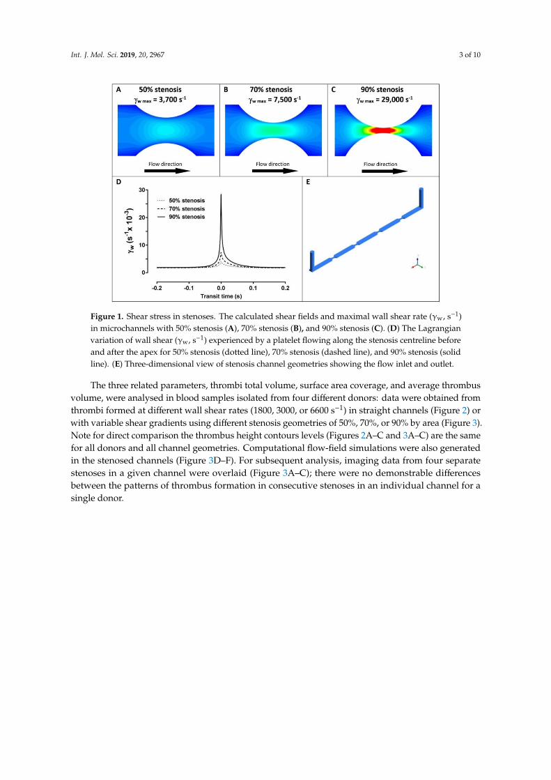
Figure 1. Shear stress in stenoses. The calculated shear fields and maximal wall shear rate (γw, s–1) in microchannels with 50% stenosis (A), 70% stenosis (B), and 90% stenosis (C). (D) The Lagrangian variation of wall shear (γw, s–1) experienced by a platelet flowing along the stenosis centreline before and after the apex for 50% stenosis (dotted line), 70% stenosis (dashed line), and 90% stenosis (solid line). (E) Three-dimensional view of stenosis channel geometries showing the flow inlet and outlet.
The three related parameters, thrombi total volume, surface area coverage, and average thrombus volume, were analysed in blood samples isolated from four different donors: data were obtained from thrombi formed at different wall shear rates (1800, 3000, or 6600 s–1) in straight channels (Figure 2) or with variable shear gradients using different stenosis geometries of 50%, 70%, or 90% by area (Figure 3). Note for direct comparison the thrombus height contours levels (Figures 2A–C and 3A–C) are the same for all donors and all channel geometries. Computational flow-field simulations were also generated in the stenosed channels (Figure 3D–F). For subsequent analysis, imaging data from four separate stenoses in a given channel were overlaid (Figure 3A–C); there were no demonstrable differences between the patterns of thrombus formation in consecutive stenoses in an individual channel for a single donor.
Figure 1. Shear stress in stenoses. The calculated shear fields and maximal wall shear rate (γw, s−1)in microchannels with 50% stenosis (A), 70% stenosis (B), and 90% stenosis (C). (D) The Lagrangianvariation of wall shear (γw, s−1) experienced by a platelet flowing along the stenosis centreline beforeand after the apex for 50% stenosis (dotted line), 70% stenosis (dashed line), and 90% stenosis (solidline). (E) Three-dimensional view of stenosis channel geometries showing the flow inlet and outlet.
The three related parameters, thrombi total volume, surface area coverage, and average thrombusvolume, were analysed in blood samples isolated from four different donors: data were obtained fromthrombi formed at different wall shear rates (1800, 3000, or 6600 s−1) in straight channels (Figure 2) orwith variable shear gradients using different stenosis geometries of 50%, 70%, or 90% by area (Figure 3).Note for direct comparison the thrombus height contours levels (Figures 2A–C and 3A–C) are the samefor all donors and all channel geometries. Computational flow-field simulations were also generatedin the stenosed channels (Figure 3D–F). For subsequent analysis, imaging data from four separatestenoses in a given channel were overlaid (Figure 3A–C); there were no demonstrable differencesbetween the patterns of thrombus formation in consecutive stenoses in an individual channel for asingle donor.
Int. J. Mol. Sci. 2019, 20, 2967 4 of 10
Int. J. Mol. Sci. 2019, 20, x FOR PEER REVIEW 4 of 10
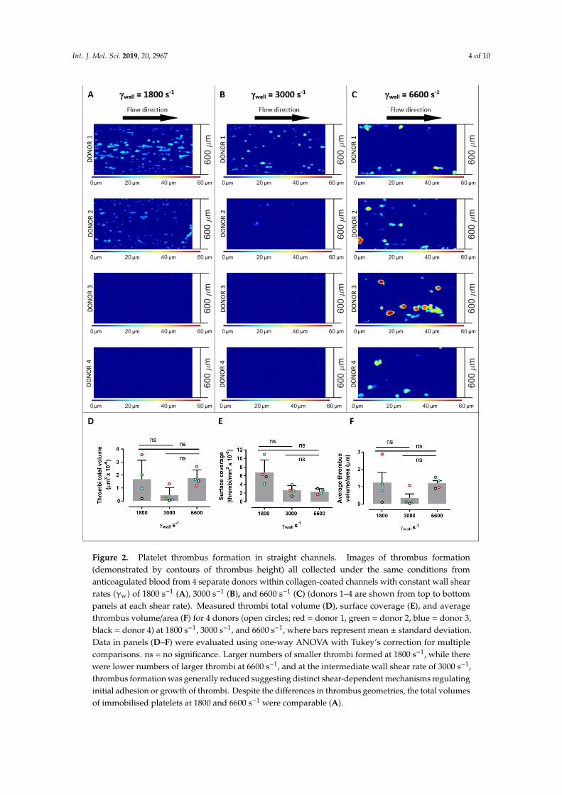
Figure 2. Platelet thrombus formation in straight channels. Images of thrombus formation (demonstrated by contours of thrombus height) all collected under the same conditions from anticoagulated blood from 4 separate donors within collagen-coated channels with constant wall shear rates (γw) of 1800 s–1 (A), 3000 s–1 (B), and 6600 s–1 (C) (donors 1–4 are shown from top to bottom panels at each shear rate). Measured thrombi total volume (D), surface coverage (E), and average thrombus volume/area (F) for 4 donors (open circles; red = donor 1, green = donor 2, blue = donor 3, black = donor 4) at 1800 s–1, 3000 s–1, and 6600 s–1, where bars represent mean ± standard deviation. Data in panels (D–F) were evaluated using one-way ANOVA with Tukey’s correction for multiple comparisons. ns = no significance. Larger numbers of smaller thrombi formed at 1800 s–1, while there were lower numbers of larger thrombi at 6600 s–1, and at the intermediate wall shear rate of 3000 s–1, thrombus formation was generally reduced suggesting distinct shear-dependent mechanisms regulating initial adhesion or growth of thrombi. Despite the differences in thrombus geometries, the total volumes of immobilised platelets at 1800 and 6600 s–1 were comparable (A).
Figure 2. Platelet thrombus formation in straight channels. Images of thrombus formation(demonstrated by contours of thrombus height) all collected under the same conditions fromanticoagulated blood from 4 separate donors within collagen-coated channels with constant wall shearrates (γw) of 1800 s−1 (A), 3000 s−1 (B), and 6600 s−1 (C) (donors 1–4 are shown from top to bottompanels at each shear rate). Measured thrombi total volume (D), surface coverage (E), and averagethrombus volume/area (F) for 4 donors (open circles; red = donor 1, green = donor 2, blue = donor 3,black = donor 4) at 1800 s−1, 3000 s−1, and 6600 s−1, where bars represent mean ± standard deviation.Data in panels (D–F) were evaluated using one-way ANOVA with Tukey’s correction for multiplecomparisons. ns = no significance. Larger numbers of smaller thrombi formed at 1800 s−1, while therewere lower numbers of larger thrombi at 6600 s−1, and at the intermediate wall shear rate of 3000 s−1,thrombus formation was generally reduced suggesting distinct shear-dependent mechanisms regulatinginitial adhesion or growth of thrombi. Despite the differences in thrombus geometries, the total volumesof immobilised platelets at 1800 and 6600 s−1 were comparable (A).
Int. J. Mol. Sci. 2019, 20, 2967 5 of 10
Int. J. Mol. Sci. 2019, 20, x FOR PEER REVIEW 5 of 10
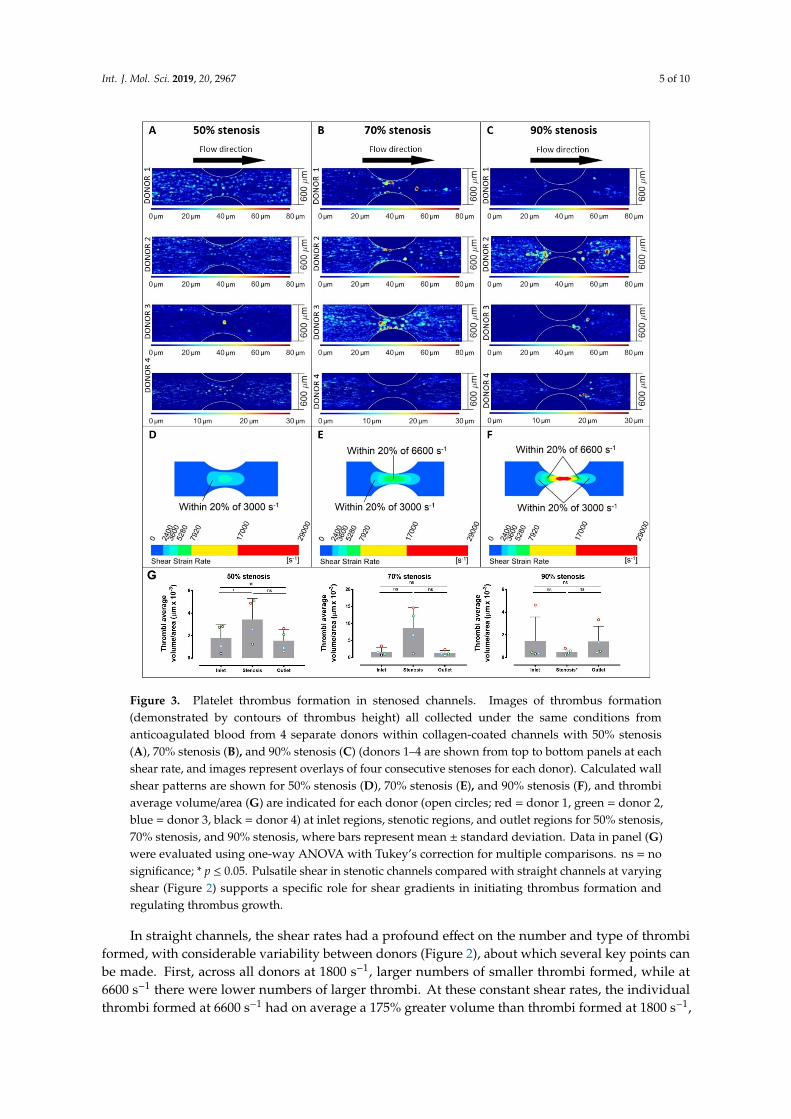
Figure 3. Platelet thrombus formation in stenosed channels. Images of thrombus formation (demonstrated by contours of thrombus height) all collected under the same conditions from anticoagulated blood from 4 separate donors within collagen-coated channels with 50% stenosis (A), 70% stenosis (B), and 90% stenosis (C) (donors 1–4 are shown from top to bottom panels at each shear rate, and images represent overlays of four consecutive stenoses for each donor). Calculated wall shear patterns are shown for 50% stenosis (D), 70% stenosis (E), and 90% stenosis (F), and thrombi average volume/area (G) are indicated for each donor (open circles; red = donor 1, green = donor 2, blue = donor 3, black = donor 4) at inlet regions, stenotic regions, and outlet regions for 50% stenosis, 70% stenosis, and 90% stenosis, where bars represent mean ± standard deviation. Data in panel (G) were evaluated using one-way ANOVA with Tukey’s correction for multiple comparisons. ns = no significance; * p ≤ 0.05. Pulsatile shear in stenotic channels compared with straight channels at varying shear (Figure 2) supports a specific role for shear gradients in initiating thrombus formation and regulating thrombus growth.
In straight channels, the shear rates had a profound effect on the number and type of thrombi formed, with considerable variability between donors (Figure 2), about which several key points can be made. First, across all donors at 1800 s–1, larger numbers of smaller thrombi formed, while at 6600
Figure 3. Platelet thrombus formation in stenosed channels. Images of thrombus formation(demonstrated by contours of thrombus height) all collected under the same conditions fromanticoagulated blood from 4 separate donors within collagen-coated channels with 50% stenosis(A), 70% stenosis (B), and 90% stenosis (C) (donors 1–4 are shown from top to bottom panels at eachshear rate, and images represent overlays of four consecutive stenoses for each donor). Calculated wallshear patterns are shown for 50% stenosis (D), 70% stenosis (E), and 90% stenosis (F), and thrombiaverage volume/area (G) are indicated for each donor (open circles; red = donor 1, green = donor 2,blue = donor 3, black = donor 4) at inlet regions, stenotic regions, and outlet regions for 50% stenosis,70% stenosis, and 90% stenosis, where bars represent mean ± standard deviation. Data in panel (G)were evaluated using one-way ANOVA with Tukey’s correction for multiple comparisons. ns = nosignificance; * p ≤ 0.05. Pulsatile shear in stenotic channels compared with straight channels at varyingshear (Figure 2) supports a specific role for shear gradients in initiating thrombus formation andregulating thrombus growth.
In straight channels, the shear rates had a profound effect on the number and type of thrombiformed, with considerable variability between donors (Figure 2), about which several key points canbe made. First, across all donors at 1800 s−1, larger numbers of smaller thrombi formed, while at6600 s−1 there were lower numbers of larger thrombi. At these constant shear rates, the individualthrombi formed at 6600 s−1 had on average a 175% greater volume than thrombi formed at 1800 s−1,
Int. J. Mol. Sci. 2019, 20, 2967 6 of 10
while there were 189% more individual thrombi at 1800 than at 6600 s−1 (Figure 2D). There wereno significant differences in total volume, surface coverage, or thrombus volume/area when usinginput shear rate as the only variable in a 1-way ANOVA analysis. However, using 2-way ANOVAto simultaneously evaluate donor and shear rate variables, the input shear rate influenced thrombussurface area coverage (p = 0.0198) and accounted for 62% of the variance observed in this parameter.Donor-related factors did not significantly affect any of the parameters in Figure 2. Interestingly, at anintermediate wall shear rate of 3000 s−1, thrombus formation was generally reduced suggesting distinctshear-dependent mechanisms at elevated shear and greater dependence on growth of existing thrombicompared to initiation of new thrombi. That is, thrombus growth was more amenable at 6600 s−1 thanplatelet adhesion to the collagen surface, whereas at a lower shear rate (1800 s−1) platelet adhesionwas relatively more achievable with greater limitations on thrombus growth. Therefore, this resultsin qualitative differences in types of thrombus formed at lower (1800 s−1) versus higher (6600 s−1)shear rates, and while the transition between these conditions appears to be donor-specific, a flex pointcommonly occurs at a shear of ~3000 s−1 where there appears clear to be reduced levels of thrombusformation. Despite the differences in the thrombus geometries, the total volumes of immobilisedplatelets at 1800 and 6600 s−1 were comparable.
In stenosed channels, increasing shear rates within 50%, 70%, and 90% stenoses also had aprofound effect on the number and type of thrombi formed, and as for straight channels there werealso marked differences between individual donors (Figure 3). In the stenosed channels, the variabilityin thrombus number and volume was related to the location within the stenosis, that is, either the inletregion, stenotic region, or outlet region (Figure 3G). In this case, the different percentages of stenosispresent not only different values of wall shears but different gradients of shear (Figure 1). Despitedifferences between individual donors, the 50% stenosis showed an increased number of lower volumethrombi throughout the stenosis, the 70% stenosis showed a smaller number of larger thrombi within300 µm of the stenosis, and the 90% stenosis also showed a smaller number of larger thrombi butpredominantly 150 µm or more from the centre of the stenosis and with no stable adhesion within150 µm (Figure 3). Using 2-way ANOVA analysis, the sampling position (inlet, stenosis, or outlet)and donor variables significantly affected the thrombus volume recorded (p = 0.0035 for position andp = 0.014 for donor) through the 50% stenosis. However at the 70% stenosis, only the sampling positionsignificantly affected thrombus volume (p = 0.026). At the 90% stenosis, neither sampling positionnor donor had any significant effect on thrombi volume. Notably, in the 50% stenosis, the differencesin shear rates between the straight sections of the channels and the stenoses were relatively minor,with the peak shear in the stenosis at ~3000 s−1. In the 70% stenosis, larger stable thrombi tended toform towards the centre of the stenosis where the peak shear reached ~6600 s−1. In the 90% stenosis,larger thrombi formed mainly in inlet/outlet regions where the shear was 6600 s−1 or below, but notwithin the very high shear central stenosis region (peak shear up to 29,000 s−1) (Figure 3). However,although there was little or no stable adhesion within the centre of the 90% stenosis, video analysisshowed a cycle of unstable contact adhesion and embolism.
While the three constant shear rates examined in the straight channels (Figure 2) are present inregions of the stenosed channels, a platelet moving through the stenosed channels experiences theselevels in the context of shear gradients. The regions within the stenoses where shear amplitudeswere within 20% of levels in the 3000 and 6600 s−1 straight channels are highlighted in Figure 3D–F.The shear gradients generated by the stenoses extended the range of shear rates at which thrombiwere formed, that is, platelets became hyperthrombotic within shear gradients. In the 50% stenoses,where the surface area with shear rates in the order of 3000 s−1 was largest, pulsed flow passingthrough 3000 s−1 did not result in reduced thrombus formation (Figure 3A,E,G) as observed in steadyflow at 3000 s−1 (Figure 2). In the 70% and 90% stenoses, the larger thrombi formed at lower shearlocations (well below 6600 s−1) further indicated a role for shear gradients. The shear gradient effectappeared to be more pronounced in some donors and the threshold gradient at which it occurs may bedonor-specific; donor 2 responded strongly to the 90% stenosis while donor 3 responded more to the
Int. J. Mol. Sci. 2019, 20, 2967 7 of 10
70% stenosis. Together, the differences in thrombus formation in both straight channels at varyingshear and pulsatile shear in stenotic channels highlights a specific functional role for shear gradientsin mediating thrombus formation, and suggest distinct shear-dependent pathways promoting eitherincreased adhesion resulting in larger numbers of smaller thrombi or enhanced thrombus growthresulting in smaller numbers of larger thrombi.
3. Discussion
Improved understanding of how changes in shear stress and shear gradients regulate thrombusformation is critical for evaluation of hemostatic function and thrombotic risk in stenosed coronaryvessels, as well as for identification of improved molecular targets and application of existing antiplateletagents [1–7]. At arterial shear rates of 1800 s−1 or higher, key platelet-specific receptors glycoprotein(GP)Ibα of the GPIb-IX-V complex, and GPVI form a complex on the platelet surface and regulatebinding of von Willebrand factor (VWF), collagen, and other prothrombotic/procoagulant ligands criticalfor thrombus formation under shear conditions [13–16]. Receptor expression and density specificallycontrols cell adhesion dynamics [17], and is critical for shear-induced thrombus formation [18].
Recent studies investigated how receptor expression and GPIbα/GPVI shedding contributes todonor-related differences and/or shear-dependent thrombus formation [13,19–21]. Exposure of humanplatelets to uniform shear stress ex vivo rapidly induces shedding of GPIbα and GPVI, while patientswith cardiovascular disease, heart failure, and/or circulatory support devices (with acutely elevatedfluid shear stress within these devices) show decreased platelet surface GPIbα/GPVI [1,20–22] andelevated shed soluble GPVI (sGPVI) in plasma, consistent with increased shedding [22,23]. There arenow identified inter-subject variations that can exceed intra-subject variations up to 4-fold, in ex vivothrombus formation that are due to multiple factors [24]. The latter could also include GPVI-linkedgenetic variations.
In summary, our new studies used computational flow-field simulations and measurement ofthrombi total volume, average volume, and surface coverage in sequential stenotic micro-channelswith geometries of 50%, 70%, and 90% by area to generate pulsatile shear and defined shear gradients.These methods can detect discrete differences in the initial adhesion and thrombus formation by humanplatelets that are not only varied between different donors, but are dramatically distinct from thethrombi formed with the same donor’s platelets in straight channels at constant wall shear rates of 1800,3000, or 6600 s−1. Together, these findings provide a more patho-physiologically relevant analyticalapproach for future testing of antiplatelet targets or agents under pulsatile shear.
4. Materials and Methods
4.1. Blood Collection and Analysis
Whole blood was collected in accordance with the Declaration of Helsinki after informed writtenconsent from 4 healthy volunteers, free of use of antiplatelet medication in the prior 10 days, using a19-gauge winged infusion kit as approved by the Monash University Standing Committee for Researchin Humans (CF07/0141 – 2007000025, issued 14.01.16). The blood was collected into hirudin (800 U/mL)anticoagulant and platelet membranes stained with a fluorescent dye (DiOC6) to enable confocalimaging [12].
4.2. Fabrication of the Stenosed Microchannels
The PDMS microchannels were fabricated using a standard PDMS soft lithography methodwhere PDMS was casted onto a master containing the negatives of the channels. The master wascreated using standard photolithography techniques where two layers of 100 µm-thick SU-2075(MicroChem, Westborough, MA, USA) photoresist were initially spin-coated onto a clean silicon wafer.After the spin-coating first and second photoresist layer, the layers were baked for 40 and 90 min,respectively, at 95 ◦C. The patterns of the channels were exposed onto the photoresist using 400 mJ/cm2
Int. J. Mol. Sci. 2019, 20, 2967 8 of 10
of ultraviolet light. After the exposure, the photoresist was further baked for 90 min at 95 ◦C followedby development in SU-8 developer (MicroChem) to remove unexposed photoresist. The silicon waferwas then silanized (20 µL of Trichloro (1H,1H,2H,2H-perfluorooctyl) silane (Sigma-Aldrich, Castle Hill,NSW, Australia)) before casting with Sylgard-184 PDMS (Dow Corning, Midland, MI, USA) with acuring agent-to-base weight ratio of 1:10. The cast was cured at 65 ◦C for 5 h, and inlets and outlets ofthe channels were cut using a 0.75 mm hole puncher. The channels were sealed using #1.5H coverslips(Menzel-Gläser, Braunscheig, Germany) bonded using air plasma at 300 mTorr for 45 s.
4.3. Calculations of the Stenosis Percentages
Periodic stenoses in the channel cross-sectional area were created by two parallel channel wallswith circular segment profiles where each had a radius of 0.5 mm. The stenosis percentage (%sten) wasdefined by
%sten =Anon−sten −Asten
Anon−sten× 100%,
where Asten is the minimum channel cross-sectional area within the stenosis and Anon−sten is the channelcross-sectional area of the non-stenosed regions of the channel.
4.4. Platelet Thrombus Formation under Flow
Microchannels were cleaned with 80% (v/v) ethanol followed by Tris-saline (TS) buffer (0.01 MTris-HCl, 0.15 M NaCl, pH 7.4) and then coated with bovine type 1 collagen (100 µg/mL) as previouslydescribed [11]. Blood was perfused through the channels for 3 min using a syringe pump (HarvardApparatus PHD2000, Holliston, MA, USA) in refill mode, followed by buffer flow to flush red bloodcells. The thrombus geometries were then fixed using 1% (w/v) paraformaldehyde solution, and themicrochannel was sealed prior to scanning. The pulsed studies were conducted with a base wall shearrate of 1800 s−1 from which platelets were subjected to elevated shear pulses using three differentstenosis geometries, 50%, 70%, and 90% by area. The flow fields within channels were simulated usingthe ANSYS CFX solver, revealing the spatial variation of shear within each stenotic region (Figure 1).The temporal variation of the shear experienced by a platelet flowing along the centreline of the channeljust above the wall was also calculated (Figure 1D), with positive shear gradients upstream of the apexand negative shear gradients downstream. Platelet adhesion in the stenosis geometries with pulsatileshear was also compared to flow adhesion assays in straight channels at different shear rates (γwall) of1800, 3000 and 6600 s−1, where these values were selected to correspond to key shear rates within thestenosis channels.
4.5. Analysis of Thrombus Formation
Sections of the channels were imaged using confocal fluorescence microscopy (Nikon A1r,512 × 512 pixel image resolution; Melville, NY, USA) to acquire overlapping z-stacks with ∆z of 0.7 µm.The z-stacks captured all thrombi adhered to the bottom surface of the microchannels within the x-yscan area. The thrombus geometries were isolated using a density-based segmentation algorithm [25]and the floor of the channel identified, allowing for correction for channel tilt during scanning.
4.6. Shear Force Equations and Units
The force generated by velocity gradients was given (in two-dimensions for simplicity) by:
F = µdudy
A
where µ is the dynamic viscosity (~3.2 × 10⁻³ Pa·s for whole blood), dudy is a velocity gradient (the
rate at which the velocity changes in the direction normal to which it is travelling), and A is the areaover which the force is acting. The velocity gradient is also known as the shear rate (units of inverse

Int. J. Mol. Sci. 2019, 20, 2967 9 of 10
seconds) and is directly proportional to the shear-induced force exerted by the flow on a platelet.The shear rate varies across a blood vessel but is often characterised by the shear rate at the vessel
wall or wall shear γwall = dudy
]y=0
, where the shear at the wall is typically maximised and also most
relevant to platelet adhesion. The shear rates calculated within the stenosed microchannels werethree-dimensional and derived from the three-dimensional strain rate tensor. For completion, the shearrate in the three-dimensional space was given by:
.γ =
√2
(∂UX
∂X
)2
+
(∂UY∂Y
)2
+
(∂UZ
∂Z
)2+ (∂UX
∂Y+∂UY∂X
)2
+
(∂UX
∂Z+∂UZ
∂X
)2
+
(∂UY∂Z
+∂UZ
∂Y
)2,where
.γ is the shear rate, X, Y, and Z are the orthogonal directions in the 3-dimensional space, and U is
the velocity; the subscripts denote the directional components of the velocity.
5. Conclusions
These studies demonstrate key differences in thrombus formation in straight channels at varyingshear and at pulsatile shear in stenotic channels, and support a specific functional role for sheargradients in mediating initiation and growth of thrombi involving human platelets, with contributionsalso from platelet-specific parameters. These data have implications for understanding mechanismsfor thrombus formation under altered flow conditions in disease and clear implications for targetingand monitoring antiplatelet therapies in future.
Author Contributions: Conceptualization, M.L., E.E.G., J.F.A., K.R., J.C., and R.K.A.; methodology, M.L., E.E.G.,J.F.A., and J.C.; writing of review and editing, I.P. and W.M.L.; writing of original draft preparation, R.K.A.
Funding: We acknowledge financial support from the National Health and Medical Research Council, Australia(APP1147214, APP1042865, APP1126536) and The Australian Research Council (DP190100039). This work wasperformed in part at the Melbourne Centre for Nanofabrication (MCN) in the Victorian Node of the AustralianNational Fabrication Facility (ANFF).
Acknowledgments: We acknowledge current and past colleagues for many helpful discussions.
Conflicts of Interest: The authors declare no conflicts of interest. Funders played no role in the design of thestudy; in the collection, analyses, or interpretation of data; in the writing of the manuscript, or in the decision topublish the results.
References
1. Al-Tamimi, M.; Tan, C.W.; Qiao, J.; Pennings, G.J.; Javadzadegan, A.; Yong, A.S.C.; Arthur, J.F.; Davis, A.K.;Jing, J.; Mu, F.-T.; et al. Pathological shear triggers shedding of vascular receptors: A novel mechanism fordownregulation of platelet glycoprotein (GP)VI in stenosed coronary vessels. Blood 2012, 119, 4311–4320.[CrossRef]
2. Gardiner, E.E.; Andrews, R.K. Platelet adhesion. In Platelets in Thrombotic and Non-Thrombotic Disorders;Gresele, P., Lopez, J.A., Kleiman, N.S., Page, C.P., Eds.; Springer: Berlin/Heidelberg, Germany, 2017;pp. 309–319.
3. Maxwell, M.J.; Westein, E.; Nesbitt, W.S.; Giuliano, S.; Dopheide, S.M.; Jackson, S.P. Identification of a 2-stageplatelet aggregation process mediating shear-dependent thrombus formation. Blood 2007, 109, 566–576.[CrossRef]
4. Nesbitt, W.S.; Westein, E.; Tovar-Lopez, F.J.; Tolouei, E.; Mitchell, A.; Fu, J.; Carberry, J.; Fouras, A.; Jackson, S.P.A shear gradient-dependent platelet aggregation mechanism drives thrombus formation. Nat. Med. 2009, 15,665–673. [CrossRef]
5. Shi, X.; Yang, J.; Huang, J.; Long, Z.; Ruan, Z.; Xiao, B.; Xi, X. Effects of different shear rates on the attachmentand detachment of platelet thrombi. Mol. Med. Rep. 2016, 13, 2447–2456. [CrossRef]
6. Chopard, B.; de Sousa, D.R.; Lätt, J.; Mountrakis, L.; Dubois, F.; Yourassowsky, C.; Van Antwerpen, P.;Eker, O.; Vanhamme, L.; Perez-Morga, D.; et al. A physical description of the adhesion and aggregation ofplatelets. R. Soc. Open Sci. 2017, 4, 170219. [CrossRef]

Int. J. Mol. Sci. 2019, 20, 2967 10 of 10
7. Koltai, K.; Kesmarky, G.; Feher, G.; Tibold, A.; Toth, K. Platelet aggregometry testing: Molecular mechanisms,techniques and clinical implications. Int. J. Mol. Sci. 2017, 18, 1803. [CrossRef]
8. Li, R.; Grosser, T.; Diamond, S.L. Microfluidic whole blood testing of platelet response to pharmacologicalagents. Platelets 2017, 28, 457–462. [CrossRef]
9. Qiu, Y.; Ciciliano, J.; Myers, D.R.; Tran, R.; Lam, W.A. Platelets and physics: How platelets “feel” and respondto their mechanical microenvironment. Blood Rev. 2015, 29, 377–386. [CrossRef]
10. Zhang, P.; Zhang, L.; Slepian, M.J.; Deng, Y.; Bluestein, D. A multiscale biomechanical model of platelets:Correlating with in-vitro results. J. Biomech. 2017, 50, 26–33. [CrossRef]
11. Slepian, M.J.; Sheriff, J.; Hutchinson, M.; Tran, P.; Bajaj, N.; Garcia, J.G.; Scott Saavedra, S.; Bluestein, D.Shear-mediated platelet activation in the free flow: Perspectives on the emerging spectrum of cellmechanobiological mechanisms mediating cardiovascular implant thrombosis. J. Biomech. 2017, 50,20–25. [CrossRef]
12. Pinar, I.P.; Arthur, J.F.; Andrews, R.K.; Gardiner, E.E.; Ryan, K.; Carberry, J. Methods to determine theLagrangian shear experienced by platelets during thrombus growth. PlosONE 2015, 10, e0144860. [CrossRef]
13. Gardiner, E.E.; Andrews, R.K. Platelet receptor expression and shedding: GPIb-IX-V and GPVI. TransfusionMed. Rev. 2014, 28, 56–60. [CrossRef]
14. Andrews, R.K.; Berndt, M.C. Platelet adhesion: A game of catch and release. J. Clin. Invest. 2008, 118,3009–3011. [CrossRef]
15. Yago, T.; Lou, J.; Wu, T.; Yang, J.; Miner, J.J.; Coburn, L.; López, J.A.; Cruz, M.A.; Dong, J.F.; McIntire, L.V.; et al.Platelet glycoprotein Ibα forms catch bonds with human WT vWF but not with type 2B von Willebranddisease vWF. J. Clin. Invest. 2008, 118, 3195–3207. [CrossRef]
16. Arthur, J.F.; Gardiner, E.E.; Matzaris, M.; Taylor, S.G.; Wijeyewickrema, L.; Ozaki, Y.; Kahn, M.L.;Andrews, R.K.; Berndt, M.C. Glycoprotein VI is associated with GPIb-IX-V on the membrane of resting andactivated platelets. Thromb. Haemost. 2005, 93, 716–723.
17. Kruss, S.; Erpenbeck, L.; Amschler, K.; Mundinger, T.A.; Boehm, H.; Helms, H.J.; Friede, T.; Andrews, R.K.;Schön, M.P.; Spatz, J.P. Adhesion maturation of neutrophils on nanoscopically presented platelet glycoproteinIbα. ACS Nano 2013, 7, 9984–9996. [CrossRef]
18. Kamada, H.; Imai, Y.; Nakamura, M.; Ishikawa, T.; Yamaguchi, T. Computational study on thrombusformation regulated by platelet glycoprotein and blood flow shear. Microvasc. Res. 2013, 89, 95–106.[CrossRef]
19. Andrews, R.K.; Gardiner, E.E. Basic mechanisms of platelet receptor shedding. Platelets 2017, 28, 319–324.[CrossRef]
20. Chen, Z.; Mondal, N.K.; Ding, J.; Gao, J.; Griffith, B.P.; Wu, Z.J. Shear-induced platelet receptor sheddingby non-physiological high shear stress with short exposure time: Glycoprotein Ibα and glycoprotein VI.Thromb. Res. 2015, 135, 692–698. [CrossRef]
21. Montague, S.J.; Andrews, R.K.; Gardiner, E.E. Mechanisms of receptor shedding in platelets. Blood 2018, 132,2535–2545. [CrossRef]
22. Lukito, P.; Wong, A.; Jing, J.; Mado, B.; Arthur, J.F.; Marasco, S.F.; Murphy, D.A.; Pellegrino, V.A.; Bergin, P.J.;Shaw, J.; et al. Mechanical circulatory support is associated with loss of platelet receptors glycoprotein (GP)Ibα and GPVI. J. Thromb. Haemost. 2016, 14, 2253–2260. [CrossRef]
23. Muthiah, K.; Connor, D.; Ly, K.; Gardiner, E.E.; Andrews, R.K.; Qiao, J.; Rutgers, D.; Robson, D.; Low, J.;Jarvis, S.; et al. Longitudinal changes in haemostatic parameters and reduced pulsatility contribute tonon-surgical bleeding in patients with centrifugal continuous flow left ventricular assist devices. J. HeartLung Transpl. 2016, 35, 743–751. [CrossRef]
24. Geffen, J.P.V.; Brouns, S.L.N.; Batista, J.; McKinney, H.; Kempster, C.; Nagy, M.; Sivapalaratnam, S.;Baaten, C.C.F.M.J.; Bourry, N.; Frontini, M.; et al. High-throughput elucidation of thrombus formationreveals sources of platelet function variability. Haematologica. 2018. [CrossRef]
25. Chan, P.; Cheng, S.H.; Poon, T.C. Automated segmentation in confocal images using a density clusteringmethod. J. Electron Imaging 2007, 16, 043003. [CrossRef]
© 2019 by the authors. Licensee MDPI, Basel, Switzerland. This article is an open accessarticle distributed under the terms and conditions of the Creative Commons Attribution(CC BY) license (http://creativecommons.org/licenses/by/4.0/).