nrf2 is a direct perk substrate and effector of perk ...zhangd/donna's paper/8.pdf · nrf2 is...
TRANSCRIPT
MOLECULAR AND CELLULAR BIOLOGY, Oct. 2003, p. 7198–7209 Vol. 23, No. 200270-7306/03/$08.00�0 DOI: 10.1128/MCB.23.20.7198–7209.2003Copyright © 2003, American Society for Microbiology. All Rights Reserved.
Nrf2 Is a Direct PERK Substrate and Effector of PERK-DependentCell Survival
Sara B. Cullinan,1 Donna Zhang,2 Mark Hannink,2 Edward Arvisais,3Randal J. Kaufman,4 and J. Alan Diehl1*
The Leonard and Madlyn Abramson Family Cancer Research Institute and Cancer Center, Department of CancerBiology, University of Pennsylvania Cancer Center, Philadelphia, Pennsylvania 191041; Department of
Biochemistry, University of Missouri—Columbia, Columbia Missouri2; University of NebraskaMedical Center, Omaha, Nebraska 681983; and Howard Hughes Medical Institute
and Department of Biological Chemistry, University of MichiganMedical Center, Ann Arbor, Michigan 481094
Received 22 April 2003/Returned for modification 28 May 2003/Accepted 15 July 2003
Activation of PERK following the accumulation of unfolded proteins in the endoplasmic reticulum (ER)promotes translation inhibition and cell cycle arrest. PERK function is essential for cell survival followingexposure of cells to ER stress, but the mechanisms whereby PERK signaling promotes cell survival are notthoroughly understood. We have identified the Nrf2 transcription factor as a novel PERK substrate. Inunstressed cells, Nrf2 is maintained in the cytoplasm via association with Keap1. PERK-dependent phosphor-ylation triggers dissociation of Nrf2/Keap1 complexes and inhibits reassociation of Nrf2/Keap1 complexes invitro. Activation of PERK via agents that trigger the unfolded protein response is both necessary and sufficientfor dissociation of cytoplasmic Nrf2/Keap1 and subsequent Nrf2 nuclear import. Finally, we demonstrate thatcells harboring a targeted deletion of Nrf2 exhibit increased cell death relative to wild-type counterpartsfollowing exposure to ER stress. Our data demonstrate that Nrf2 is a critical effector of PERK-mediated cellsurvival.
Mammalian cells possess a signaling network that sensesendoplasmic reticulum (ER) stress and determines cell fatefollowing exposure to stress. The ER signaling network con-sists of three related transmembrane protein kinases, Ire1�,Ire1�, and PERK (31). Ire1� and Ire1� are composed of aluminal domain that senses stress, a single membrane-spanningdomain, and a cytosolic tail that contains both a serine/threo-nine kinase domain and an RNase domain (40). Ire1-depen-dent signals induce expression of ER chaperones (40) andCHOP (42), a transcription factor that may participate in theapoptotic program. Activation of PERK, which lacks an RNasedomain, following ER stress promotes phosphorylation of thetranslation initiation factor eukaryotic initiation factor 2�(eIF2�), thereby attenuating translation initiation (15, 22).PERK is one of at least four eIF2� protein kinases that includethe heme-regulated kinase (HRI), the interferon-inducible,RNA-dependent protein kinase (PKR), and GCN2 (3, 39).Among these, only PERK function is required for the cellularresponse to ER stress (22).
Activation of PERK via ER stress initiates cell cycle arrestvia inhibition of cyclin D1 translation (5). PERK-dependentsignaling is also critical for cell survival following the initiationof an ER stress response. Targeted deletion of PERK dramat-ically reduces survival of embryonic stem cells following expo-sure to ER stress-inducing agents (21). Mice that contain in-activated PERK exhibit severe defects in the exocrine pancreas
due to a high rate of secretory cell apoptosis (20, 44). While themechanism that underlies PERK-dependent cell cycle arrest isestablished, the nature of PERK-dependent cell survival isunresolved.
A majority of PERK’s biological activities have been attrib-uted to its function as an eIF2� kinase. However, the possibil-ity that PERK targets additional downstream substrates thatfunction as cellular effectors has not been addressed. In orderto identify novel PERK substrates, we performed a yeast two-hybrid screen with the PERK catalytic domain. We describethe identification of the Cap ’n’ Collar transcription factor Nrf2as a PERK substrate. In unstressed cells, Nrf2 is maintained inlatent cytoplasmic complexes via association with the cytoskel-etal anchor, Keap1. Previous work revealed that oxidativestress triggers dissociation of this complex via an uncharacter-ized mechanism, thereby allowing Nrf2 nuclear import, whereit promotes expression of its downstream target genes (30). Wedemonstrate that PERK-dependent phosphorylation is bothnecessary and sufficient to trigger dissociation of Nrf2/Keap1complexes and thereby promote Nrf2 nuclear import. Further-more, Nrf2 nuclear translocation is independent of eIF2�phosphorylation. Finally, targeted deletion of Nrf2 reduces cellsurvival following ER stress. Collectively, these data support amodel wherein Nrf2 functions as an effector of PERK cellsurvival signaling.
MATERIALS AND METHODS
Tissue culture conditions. Cells were maintained in Dulbecco’s modified Ea-gle’s medium (DMEM) supplemented with 10% fetal calf serum (FCS), antibi-otics, nonessential amino acids, and glutamine (Mediatech, Inc.). Wild-type andNrf2�/� mouse embryonic fibroblasts (MEFs) were immortalized via a standard
* Corresponding author. Mailing address: AFCRI, University ofPennsylvania Cancer Center 454 BRB II/III, 421 Curie Blvd., Phila-delphia, PA 19104. Phone: (215) 746-6389. Fax: (215) 746-5511. E-mail: [email protected].
7198
3T9 protocol (41). Transfections were performed with Lipofectamine Plus re-agent (Life Technologies, Inc.).
Plasmids. �NPERK was generated from murine PERK cDNA using PCR(primers 5�-CCCGGGTCTAGAGAGCGCGCCACCCCGGCCCGG-3� and 5�-GGGCCCGAATTCTCAGGAGAAGGAGCTTGACTT-3�). �NPERKK618Awas generated through site-directed PCR mutagenesis (primers 5�-CAATTACGCTATCGCGAGGATCCGGCT-3� and 5�-AGCCGGATCCTCGCGATAGCGTAATTG-3� [mutated residues are underlined]). �NPERK and �NPERKK618A were subcloned into pGEX4T1 and pAS2 (Amersham) vectors usingEcoRI sites. Murine Nrf1 and MafG were cloned from a mouse cDNA library(Nrf1, 5�-TCTAGAATGGAAATGCAGGCTATG-3� and 5�-CAGCTGGTATTTGGACAGCAG-3�; MafG, 5�-GTCCCCCGGGTTATGACGAC-3� and 5�-GAATTCGTGTCCCTATGACCGAGCATC-3�). Cloned cDNAs were ligated intothe pcDNA3 vector (Invitrogen) using EcoRI sites. Myc-Keap1 and His-Keap1were generated from Keap1 cDNA and cloned into pcDNA3 or pET15b vectors.HA-Nrf2 and GST-Nrf2 were generated from Nrf2 cDNA and cloned into thepCI and pGEX5T2 vectors. The antioxidant response element (ARE) reporterplasmid was generated by cloning four ARE motifs (36) upstream of the TATAsequence in pGL2 (Promega). The mutant ARE reporter contains a minimalinitiator sequence upstream of the TATA sequence in pGL2. PERK�C andmyc-PERK plasmids and viruses were described previously (5).
Yeast two-hybrid screen. �NPERK(K618A) was cloned into the pAS2(TRP)vector using EcoRI sites, resulting in GAL4-�NPERK(K618A). Saccharomycescerevisiae strain AH109 was transformed with GAL4-�NPERK(K618A) andpACT(LEU) plasmids containing cDNAs from MEFs. Double transformantswere selected for their ability to grow on medium lacking tryptophan, leucine,and histidine and supplemented with 50 mM 3-amino-1,2,4-triazole. Transfor-mants were further selected for their ability to drive lacZ expression in a GAL4-�NPERKK618A-dependent manner. cDNAs from positive transformants wererecovered and sequenced.
EMSA. A consensus ARE oligomer (primers 5�-CTACGATTTCTGCTTAGTCATTGTCTTCC-3� and 5�-GGAAGACAATGACTAAGCACAATCGTAG-3�) was end labeled with [�-32P]ATP and incubated with 10 �g of cell extract(lysed in 50 mM Tris, 250 mM NaCl, 5 mM EDTA, 0.1% Triton, and 4 mMdithiothreitol [DTT]) for 30 min at room temperature in a reaction mixturecontaining 20 mM HEPES (pH 8.0), 1 mM EDTA, 20 mM KCl, 4 mM MgCl2,4% glycerol, 2 �g of poly(dI-dC), and 5 mM DTT. Where indicated, proteinswere first preincubated for 1 h at 4°C in the presence of antibodies. To achievemaximum resolution of in vivo-derived protein-DNA adducts, native gels wererun at low voltage for 4 h with the free probe routinely running off the gel. Forin vitro reactions, recombinant Nrf2 (0.5 �g) and in vitro-transcribed and -trans-lated MafG (Promega) were combined in electrophoretic mobility shift assay(EMSA) reaction mixtures as described above.
Immunofluorescence. Cells proliferating on glass coverslips were transfectedand treated as indicated. Cells were fixed in 3% paraformaldehyde and perme-abilized in 0.1% Triton in phosphate-buffered saline (PBS). The hemagglutinin(HA) epitope was detected with either the 12CA5 or the 3F10 monoclonalantibody. The myc epitope was detected with either the 9E10 or the Jac6 mono-clonal antibody. Calreticulin was detected using a polyclonal anticalreticulinantibody (Stressgen). Cells were stained with either fluorescein isothiocyanate(FITC)-conjugated or biotinylated immunoglobulin G and Texas Red-streptavi-din (Vector). DNA was detected with Hoechst 33258 dye (Sigma). Cells werevisualized using a Nikon microscope fitted with appropriate filters or a Bio-Radconfocal imaging system.
Reporter assays. NIH 3T3 cells transfected with the indicated plasmids wereleft untreated or treated with 5 �g of tunicamycin/ml or 100 �M tert-butylhyd-roquinone (tBHQ) for the indicated intervals. Luciferase assays were carried outaccording to the manufacturer’s instructions (Dual Luciferase Reporter assaysystem; Promega) with a luminometer (PE Applied Biosystems). Firefly lucif-erase activity was normalized against Renilla luciferase activity from the samelysates.
Cellular fractionation, immunoblotting, and Northern blot analysis. Wild-type, PERK�/�, and eIF2�(S51A) MEFs were treated with 5 �g of tunicamy-cin/ml for the indicated time intervals. Nuclei were collected in buffer A (10 mMHEPES [pH 8.0], 10 mM KCl, 1.5 mM MgCl2, 0.1 mM EDTA, 0.1 mM EGTA,0.1% IGEPAL) and buffer C (420 mM NaCl, 1.5 mM MgCl2, 1.0 mM EDTA, 1.0mM EGTA, 20% glycerol) and stored in buffer D (20 mM HEPES [pH 8.0], 50mM KCl, 0.2 mM EDTA, 0.2 mM EGTA, 20% glycerol). All buffers weresupplemented with 0.1 mM phenylmethylsulfonyl fluoride, 1 �g of aprotinin/ml,1 �g of leupeptin/ml, 1 �g of pepstatin/ml, and 25 mM �-glycerol phosphate.Proteins were resolved by sodium dodecyl sulfate-polyacrylamide gel electro-phoresis (SDS-PAGE), transferred to nitrocellulose membranes (Osmonics),and blotted with an anti-Nrf2 polyclonal antibody (Santa Cruz). For detection of
eIF2�, cells were lysed in EBC buffer, proteins were resolved by SDS-PAGE andtransferred to a nitrocellulose membrane (Osmonics), and total and phosphor-ylated eIF2� was detected by immunoblotting (Cell Signaling). RNA was ex-tracted using TRIzol reagent (Life Technologies, Inc.). RNA immobilized onnylon membranes (Osmonics) was hybridized with 32P-labeled probes specific foreither luciferase, �-glutamylcysteine synthetase ligase (GCLC), or �-actin (5).Signals were detected by phosphorimaging.
Protein kinase assays, metabolic labeling, and immunoprecipitation. Affinity-purified recombinant GST-�NPERK was used as the kinase, and equivalentamounts of affinity-purified GST-Nrf2, glutathione S-transferase (GST), andHis-eIF2� were used as substrates in the in vitro kinase assays. �NPERK waspreincubated with kinase buffer (50 mM HEPES [pH 8.0], 10 mM MgCl2, 2.5mM EGTA, 20 �M ATP) at 30°C for 1 h prior to addition of substrates in orderto reduce incorporation of radioactive phosphate into PERK itself due to itspropensity to autophosphorylate (22). Kinase reactions were initiated followingaddition of substrates by the addition of 10 �Ci of [�-32P]ATP and placement at30°C for 30 min. Reactions were terminated by the addition of sample buffercontaining SDS and incubation at 100°C for 3 min. Proteins were resolved bySDS-PAGE and visualized by autoradiography. To obtain PERK produced inmammalian cells, NIH 3T3 cells infected with retrovirus encoding PERK werelysed in radioimmunoprecipitation assay buffer (150 mM NaCl, 10 mM sodiumphosphate, 2 mM EDTA, 1% NP-40, 0.1% SDS, 1% deoxycholic acid,) andimmunoprecipitated with anti-PERK antiserum. Proteins were mixed with theindicated recombinant substrates in kinase reactions as described above. Formetabolic labeling, NIH 3T3 cells were cultured for 1 h in phosphate-freeDMEM supplemented with 0.1% dialyzed FCS and then labeled with 1 mCi of[32P]orthophosphate (ICN) and in the presence of 5 �g of tunicamycin/ml. Cellswere lysed in NP-40 lysis buffer (50 mM Tris [pH 7.5], 150 mM NaCl, 1% NP-40,0.5% deoxycholic acid). HA-Nrf2 was precipitated from cell lysates with the12CA5 antibody and visualized by autoradiography.
In vitro binding assays. Nrf2, in vitro transcribed and translated (Promega) inthe presence of [35S]methionine (ICN), was mixed with purified His-Keap1 orHis-eIF2� immobilized on Talon beads in binding buffer (20 mM Tris, 150 mMNaCl, 0.5% IGEPAL) for 2 h at 4°C. After washes with buffer, Nrf2-containingcomplexes were incubated with �NPERK or �NPERKK618A in the absence orpresence of 500 �M ATP at 30°C for the indicated intervals in kinase bufferlacking EGTA.
Annexin V staining and clonogenic survival assays. Cells proliferating on glasscoverslips were left untreated or were treated with 5 �g of tunicamycin/ml, 4 �Mstaurosporine, or tumor necrosis factor alpha (TNF-�; 5 ng/ml) plus cyclohexi-mide (10 �g/ml) for the indicated intervals. Cells were then washed with PBS andstained with a FITC-conjugated annexin V antibody and propidium iodide(Pharmingen) and examined by fluorescence and light microscopy. AnnexinV-positive cells are expressed as the percentage of total cells. For colony out-growth, cells were plated at 2 104/60-mm dish, and 24 h later cells were leftuntreated or treated with 1 �g of tunicamycin/ml for 8 h. Following treatment,cells were washed with PBS, refed with complete medium, and allowed to growfor 7 days.
RESULTS
Activation of the Nrf2 transcription factor and Nrf2 targetgenes by the UPR. To identify novel PERK substrates, weperformed a yeast two-hybrid screen with the catalytic domainof murine PERK. Sequence analysis of these clones revealedthree overlapping cDNAs predicted to encode the C-terminal219 amino acids of the Nrf1 transcription factor. Nrf1 belongsto the Cap ’n’ Collar family of transcription factors (9), whichincludes NF-E2 (1) and Nrf2 (34). Nrf1 and -2 are homologouswithin their C-terminal DNA-binding domains, but they aredivergent at their N-terminal transactivation domains (25). Weconfirmed that PERK could bind directly to both Nrf1 andNrf2 in an in vitro binding assay (data not shown). To deter-mine the likely PERK target, we evaluated the capacity of theunfolded protein response (UPR)-inducing agent, tunicamycin(Fig. 1A), an inhibitor of N-linked glycosylation, or phorbolmyristate acetate (PMA) (100 nM), a phorbol ester, to induceNrf1 or Nrf2 DNA-binding activity. Extracts prepared from
VOL. 23, 2003 ACTIVATION OF Nrf2 BY THE UPR 7199
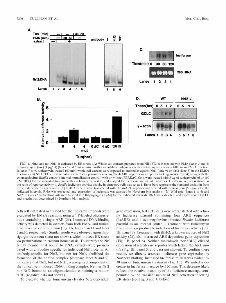
cells left untreated or treated for the indicated intervals wereevaluated by EMSA reactions using a 32P-labeled oligonucle-otide containing a single ARE (36). Increased DNA-bindingactivity was detected in extracts from both PMA- and tunica-mycin-treated cells by 30 min (Fig. 1A, lanes 3 and 4 and lanes5 and 6, respectively). Similar results were observed upon thap-sigargin treatment (data not shown), which induces ER stressvia perturbations in calcium homeostasis. To identify the Nrffamily member that bound to DNA, extracts were preincu-bated with antibodies specific for individual Nrf proteins. Anantibody specific for Nrf2, but not for Nrf1, abolished theformation of the shifted complex (compare lanes 8 and 9),indicating that Nrf2, but not Nrf1, is a required component ofthe tunicamycin-induced DNA-binding complex. Neither Nrf1nor Nrf2 bound to an oligonucleotide containing a mutantARE (negative data not shown).
To evaluate whether tunicamycin elevates Nrf2-dependent
gene expression, NIH 3T3 cells were cotransfected with a fire-fly luciferase plasmid containing four ARE sequences(4xARE) and a cytomegalovirus-directed Renilla luciferaseplasmid as an internal control. Treatment with tunicamycinresulted in a reproducible induction of luciferase activity (Fig.1B, panel 2). Treatment with tBHQ, a known inducer of Nrf2activity (28), also increased ARE-dependent gene expression(Fig. 1B, panel 6). Neither tunicamycin nor tBHQ elicitedexpression of a luciferase reporter which lacked the ARE mo-tifs (Fig. 1B, panel 3, and data not shown). To confirm theseresults, we directly assessed luciferase gene expression byNorthern blotting. Increased luciferase mRNA was evident by30 min of tunicamycin treatment (Fig. 1C). We noted a de-crease in luciferase message by 120 min. This decrease likelyreflects the relative instability of the luciferase message com-pounded by the transient nature of Nrf2 activation followingER stress (see Fig. 3 and 4, below).
FIG. 1. Nrf2, and not Nrf1, is activated by ER stress. (A) Whole-cell extracts prepared from NIH 3T3 cells treated with PMA (lanes 3 and 4)or tunicamycin (tun) (1 �g/ml) (lanes 5 and 6) were mixed with a radiolabeled oligonucleotide containing a consensus ARE in an EMSA reaction.In lanes 7 to 9, tunicamycin-treated (60 min) whole-cell extracts were exposed to antibodies against Nrf1 (lane 9) or Nrf2 (lane 8) in the EMSAreactions. (B) NIH 3T3 cells were cotransfected with plasmids encoding the 4xARE reporter or a reporter lacking an ARE (mut) along with thecytomegalovirus-Renilla control (internal normalization control) with or without PERK�C. Cells were treated with 5 �g of tunicamycin/ml or 100�M tBHQ for the indicated time intervals (in hours), harvested, and assayed for luciferase and Renilla activities. Luciferase activity is shown asthe ratio of reporter activity to Renilla luciferase activity; activity in untreated cells was set as 1. Error bars represent the standard deviation fromthree independent experiments. (C) NIH 3T3 cells were transfected with the 4xARE reporter and treated with tunicamycin (5 �g/ml) for theindicated intervals. RNA was extracted, and expression of luciferase was assessed by Northern blot analysis. (D) Wild-type (lanes 1 to 4) andNrf2�/� (lanes 5 to 8) fibroblasts were treated with thapsigargin (1 �M) for the indicated intervals. RNA was extracted, and expression of GCLCand �-actin was determined by Northern blot analysis.
7200 CULLINAN ET AL. MOL. CELL. BIOL.
To determine if UPR-dependent induction of the ARE re-porter required PERK activity, we measured expression of theARE reporter in NIH 3T3 cells cotransfected with a dominant-negative PERK (PERK�C) (5). While the cotransfection ofPERK�C did not influence basal luciferase gene expression(Fig. 1B, bar 4), cotransfection of PERK�C eliminated tuni-camycin-dependent induction of 4xARE-luciferase (bar 5).
We next assessed the capacity of ER stress-inducing agentsto induce expression of an endogenous Nrf2 target gene. Forthis experiment, we utilized fibroblasts prepared from eitherwild-type or Nrf2-null mouse embryos. Wild-type and Nrf2�/�
MEFs were challenged with thapsigargin, total RNA was col-lected, and expression of the Nrf2 target gene, GCLC (7), wasassessed. Thapsigargin treatment resulted in a rapid increasein GCLC mRNA in wild-type (Fig. 1D, lanes 1 to 4) but not inNrf2�/� (lanes 5 to 8) MEFs. Induction of GCLC was alsonoted in MEFs containing a homozygous knock-in of a non-phosphorylatable allele of eIF2�, eIF2�(S51A) (data notshown) (37). These data demonstrate that induction of theUPR triggers the activation of Nrf2 and Nrf2-dependent geneexpression and suggest that PERK function is essential forNrf2 activation.
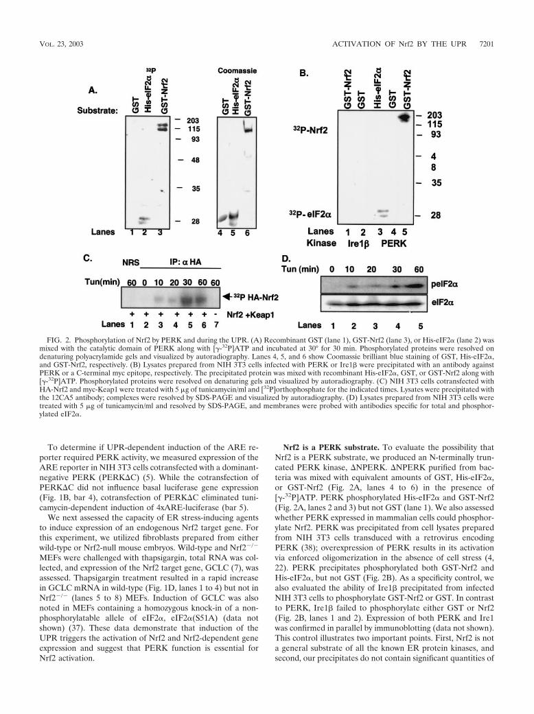
Nrf2 is a PERK substrate. To evaluate the possibility thatNrf2 is a PERK substrate, we produced an N-terminally trun-cated PERK kinase, �NPERK. �NPERK purified from bac-teria was mixed with equivalent amounts of GST, His-eIF2�,or GST-Nrf2 (Fig. 2A, lanes 4 to 6) in the presence of[�-32P]ATP. PERK phosphorylated His-eIF2� and GST-Nrf2(Fig. 2A, lanes 2 and 3) but not GST (lane 1). We also assessedwhether PERK expressed in mammalian cells could phosphor-ylate Nrf2. PERK was precipitated from cell lysates preparedfrom NIH 3T3 cells transduced with a retrovirus encodingPERK (38); overexpression of PERK results in its activationvia enforced oligomerization in the absence of cell stress (4,22). PERK precipitates phosphorylated both GST-Nrf2 andHis-eIF2�, but not GST (Fig. 2B). As a specificity control, wealso evaluated the ability of Ire1� precipitated from infectedNIH 3T3 cells to phosphorylate GST-Nrf2 or GST. In contrastto PERK, Ire1� failed to phosphorylate either GST or Nrf2(Fig. 2B, lanes 1 and 2). Expression of both PERK and Ire1was confirmed in parallel by immunoblotting (data not shown).This control illustrates two important points. First, Nrf2 is nota general substrate of all the known ER protein kinases, andsecond, our precipitates do not contain significant quantities of
FIG. 2. Phosphorylation of Nrf2 by PERK and during the UPR. (A) Recombinant GST (lane 1), GST-Nrf2 (lane 3), or His-eIF2� (lane 2) wasmixed with the catalytic domain of PERK along with [�-32P]ATP and incubated at 30° for 30 min. Phosphorylated proteins were resolved ondenaturing polyacrylamide gels and visualized by autoradiography. Lanes 4, 5, and 6 show Coomassie brilliant blue staining of GST, His-eIF2�,and GST-Nrf2, respectively. (B) Lysates prepared from NIH 3T3 cells infected with PERK or Ire1� were precipitated with an antibody againstPERK or a C-terminal myc epitope, respectively. The precipitated protein was mixed with recombinant His-eIF2�, GST, or GST-Nrf2 along with[�-32P]ATP. Phosphorylated proteins were resolved on denaturing gels and visualized by autoradiography. (C) NIH 3T3 cells cotransfected withHA-Nrf2 and myc-Keap1 were treated with 5 �g of tunicamycin/ml and [32P]orthophosphate for the indicated times. Lysates were precipitated withthe 12CA5 antibody; complexes were resolved by SDS-PAGE and visualized by autoradiography. (D) Lysates prepared from NIH 3T3 cells weretreated with 5 �g of tunicamycin/ml and resolved by SDS-PAGE, and membranes were probed with antibodies specific for total and phosphor-ylated eIF2�.
VOL. 23, 2003 ACTIVATION OF Nrf2 BY THE UPR 7201

contaminating protein kinases that nonspecifically phosphory-late Nrf2. These data demonstrate that Nrf2 is a direct PERKsubstrate.
We next determined if Nrf2 is phosphorylated in vivo fol-lowing induction of an ER stress response. NIH 3T3 cellscotransfected with plasmids encoding Nrf2 engineered to ex-press an amino-terminal HA epitope tag (HA-Nrf2) andKeap1 were cultured in medium containing [32P]orthophos-phate and subsequently challenged with tunicamycin for theindicated intervals (Fig. 2C). Lysates were prepared and sub-jected to precipitation with the 12CA5 monoclonal antibody.Nrf2 phosphorylation was evident by 10 min of tunicamycintreatment (lane 3) and reached peak levels by 30 min (lane 5),indicating that Nrf2 is rapidly phosphorylated in response toER stress conditions. Similar results were observed in cellschallenged with tBHQ (data not shown) (28). No phosphory-lation was detected in mock-transfected cells (lane 7) or incontrol precipitates (lane 1). Additionally, Nrf2 phosphoryla-tion was eliminated in cells that had been transfected with aplasmid encoding PERK�C (negative data not shown), impli-cating PERK as the Nrf2 kinase in intact cells. The rapidphosphorylation of Nrf2 led us to compare the kinetics of Nrf2phosphorylation with that of eIF2�, a known in vivo substrateof PERK. NIH 3T3 cells were treated with tunicamycin for theindicated intervals, and eIF2� phosphorylation was assessed byimmunoblotting using an eIF2� phospho-specific antibody. In-creased eIF2� phosphorylation was detectable by 10 to 20 min(Fig. 2D, lanes 2 and 3) and was maximal at 60 min (lane 4).These data demonstrate that Nrf2 is phosphorylated during anER stress response in a PERK-dependent fashion, with kinet-ics that are temporally similar to that of the other knownPERK substrate, eIF2�.
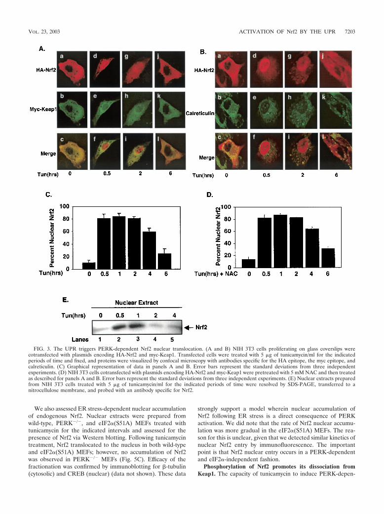
ER stress triggers PERK-dependent Nrf2 nuclear import.Under homeostatic conditions, Nrf2 is maintained in inactivecytoplasmic complexes by Keap1 (30). Dissociation of Nrf2/Keap1 complexes and subsequent Nrf2 nuclear import can beachieved by treatment of cells with electrophiles such as tBHQ(28). While the mechanism underlying activation of Nrf2 re-mains unclear, models invoking oxidation of redox-sensitivecysteines within Keap1 (13, 14, 24) or phosphorylation of Nrf2(28, 43, 45) have been proposed. Given the capacity of ERstress to induce both Nrf2 DNA-binding activity and Nrf2-dependent gene expression, we reasoned that ER stress mustalso induce Nrf2 nuclear translocation. To assess regulatednuclear entry of Nrf2, NIH 3T3 cells were cotransfected withplasmids encoding HA-Nrf2 and Keap1 engineered with anN-terminal myc epitope (myc-Keap1). Cells expressing HA-Nrf2 and myc-Keap1 were left untreated or treated with tuni-camycin for various intervals, and Nrf2 localization was visu-alized using epitope-specific antibodies followed by confocalmicroscopy. In the absence of stimulus, Nrf2 was cytoplasmic(Fig. 3A, panel a, B, panel a, and C); by 30 min of tunicamycintreatment, HA-Nrf2 was primarily nuclear (Fig. 3A, panel d, B,panel d, and C). In contrast, myc-Keap1 remained cytoplasmicat all intervals (Fig. 3A, panels b, e, h, and k). While nuclearNrf2 staining remained evident at 2 h (Fig. 3A, panel g, B,panel g, and C), by 6 h Nrf2 increasingly relocalized to thecytoplasm (Fig. 3A, panel j, B, panel j, and C). Similar resultswere observed following treatment with other known inducersof ER stress, including thapsigargin (data not shown). Tran-
sient nuclear accumulation of endogenous Nrf2 was also ob-served following ER stress as determined by subcellular frac-tionation (Fig. 3E). Loss of endogenous Nrf2 nuclearlocalization was evident by 2 h, suggesting that overexpressionmay result in some attenuation of Nrf2 downregulation.
Because the UPR is known to elicit oxidative stress (18, 31),a known inducer of Nrf2 nuclear translocation, we assessedwhether tunicamycin-dependent activation of Nrf2 was di-rected by a burst of reactive oxygen species (ROS) generatedas a consequence of ER stress. NIH 3T3 cells transfected withplasmids encoding both HA-Nrf2 and myc-Keap1 were pre-treated with the ROS scavenger N-acetylcysteine (5 mM) priorto treatment with tunicamycin. While this dose was sufficient toreduce levels of tunicamycin-induced oxidative stress by over80%, as measured by dichlorodihydrofluorescein diacetate flu-orescence (data not shown), tunicamycin still induced the nu-clear accumulation of Nrf2 (Fig. 3D). These results suggestthat Nrf2 nuclear transport is unlikely to result from the gen-eration of ROS following ER stress.
The capacity of ER stress to induce Nrf2 nuclear import isconsistent with the notion that a subset of cytoplasmic Nrf2complexes must be in the proximity of the ER. We thus deter-mined whether HA-Nrf2 colocalized with calreticulin, an ERresident protein, in either untreated or tunicamycin-treatedcells. As anticipated, in unstimulated cells HA-Nrf2 colocal-ized with endogenous, ER-localized calreticulin (Fig. 3B, panelc); however, treatment of cells with tunicamycin resulted inHA-Nrf2 nuclear import and a loss of HA-Nrf2–calreticulincolocalization (Fig. 3B, panels f and i). In addition, myc-Keap1colocalized with calreticulin in the absence or presence of ERstress (data not shown).
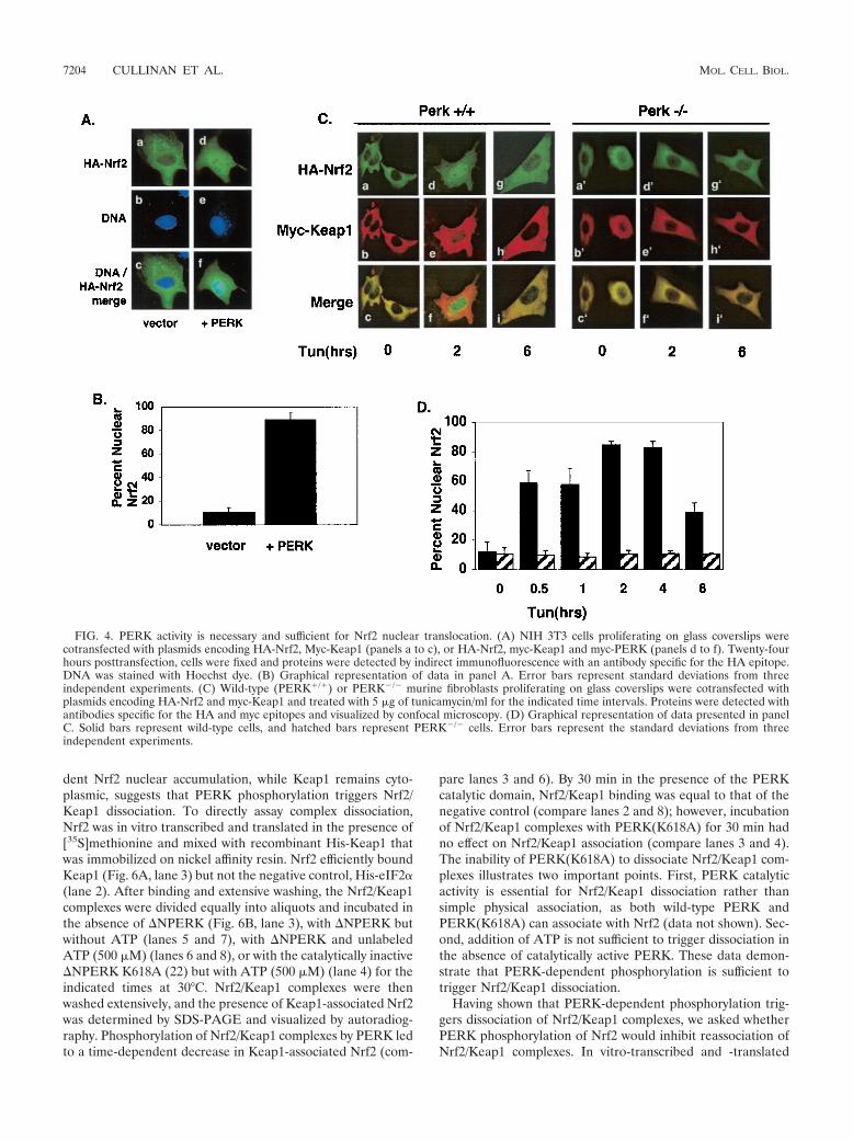
The above data suggest that ER stress triggers Nrf2-Keap1dissociation and Nrf2 nuclear import, but they do not addresswhether PERK activity is required for this action. To assess therole of PERK in Nrf2 nuclear import, we cotransfected NIH3T3 cells with plasmids encoding HA-Nrf2 and Keap1 with orwithout PERK. Cexpression of wild-type PERK efficiently pro-moted Nrf2 nuclear localization in the absence of a stress-inducing agent (Fig. 4A, panel d, and B), demonstrating thatPERK activity is sufficient for Nrf2 nuclear import. To assesswhether PERK is required for Nrf2 nuclear import followingER stress, we examined Nrf2 localization in fibroblasts derivedfrom PERK null embryos (PERK�/�). PERK�/� fibroblasts orfibroblasts derived from wild-type littermates (PERK�/�) werecotransfected with plasmids encoding HA-Nrf2 and myc-Keap1. In PERK�/� cells, Nrf2 remained cytoplasmic in theabsence or presence of tunicamycin (Fig. 4C, panels a�, d�, andg�, and D), where it colocalized with Keap1 (Fig. 4C, panels c�,f�, and i�). In contrast, in wild-type cells that express endoge-nous PERK, tunicamycin triggered Nrf2 nuclear localization(Fig. 4C, compare panels a and d).
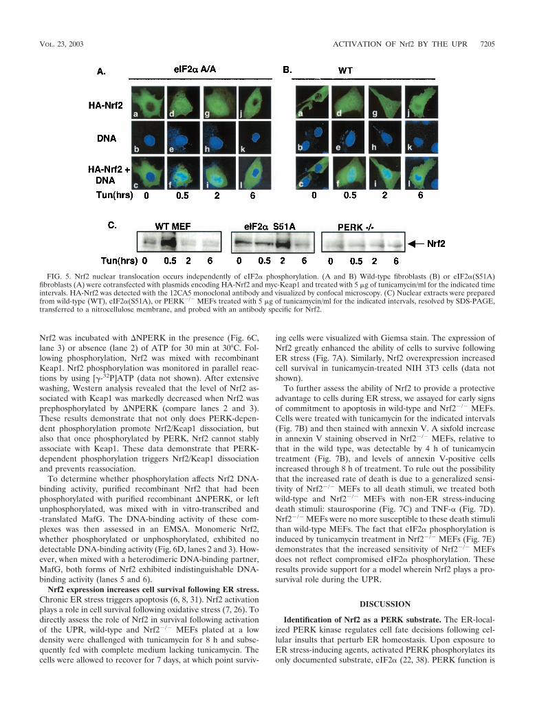
To determine whether Nrf2 nuclear translocation was anindirect consequence of PERK-dependent eIF2� phosphory-lation, we assessed Nrf2 localization in MEFs containing ahomozygous knock-in of a nonphosphorylatable eIF2� allele,eIF2�(S51A) (37). Fibroblasts derived from either wild-type oreIF2�(S51A) embryos were transfected with vectors encodingHA-Nrf2 or myc-Keap1. Treatment of both wild-type andeIF2�(S51A) cells resulted in Nrf2 nuclear accumulation (Fig.5A and B).
7202 CULLINAN ET AL. MOL. CELL. BIOL.
We also assessed ER stress-dependent nuclear accumulationof endogenous Nrf2. Nuclear extracts were prepared fromwild-type, PERK�/�, and eIF2�(S51A) MEFs treated withtunicamycin for the indicated intervals and assessed for thepresence of Nrf2 via Western blotting. Following tunicamycintreatment, Nrf2 translocated to the nucleus in both wild-typeand eIF2�(S51A) MEFs; however, no accumulation of Nrf2was observed in PERK�/� MEFs (Fig. 5C). Efficacy of thefractionation was confirmed by immunoblotting for �-tubulin(cytosolic) and CREB (nuclear) (data not shown). These data
strongly support a model wherein nuclear accumulation ofNrf2 following ER stress is a direct consequence of PERKactivation. We did note that the rate of Nrf2 nuclear accumu-lation was more gradual in the eIF2�(S51A) MEFs. The rea-son for this is unclear, given that we detected similar kinetics ofnuclear Nrf2 entry by immunofluorescence. The importantpoint is that Nrf2 nuclear entry occurs in a PERK-dependentand eIF2�-independent fashion.
Phosphorylation of Nrf2 promotes its dissociation fromKeap1. The capacity of tunicamycin to induce PERK-depen-
FIG. 3. The UPR triggers PERK-dependent Nrf2 nuclear translocation. (A and B) NIH 3T3 cells proliferating on glass coverslips werecotransfected with plasmids encoding HA-Nrf2 and myc-Keap1. Transfected cells were treated with 5 �g of tunicamycin/ml for the indicatedperiods of time and fixed, and proteins were visualized by confocal microscopy with antibodies specific for the HA epitope, the myc epitope, andcalreticulin. (C) Graphical representation of data in panels A and B. Error bars represent the standard deviations from three independentexperiments. (D) NIH 3T3 cells cotransfected with plasmids encoding HA-Nrf2 and myc-Keap1 were pretreated with 5 mM NAC and then treatedas described for panels A and B. Error bars represent the standard deviations from three independent experiments. (E) Nuclear extracts preparedfrom NIH 3T3 cells treated with 5 �g of tunicamycin/ml for the indicated periods of time were resolved by SDS-PAGE, transferred to anitrocellulose membrane, and probed with an antibody specific for Nrf2.
VOL. 23, 2003 ACTIVATION OF Nrf2 BY THE UPR 7203
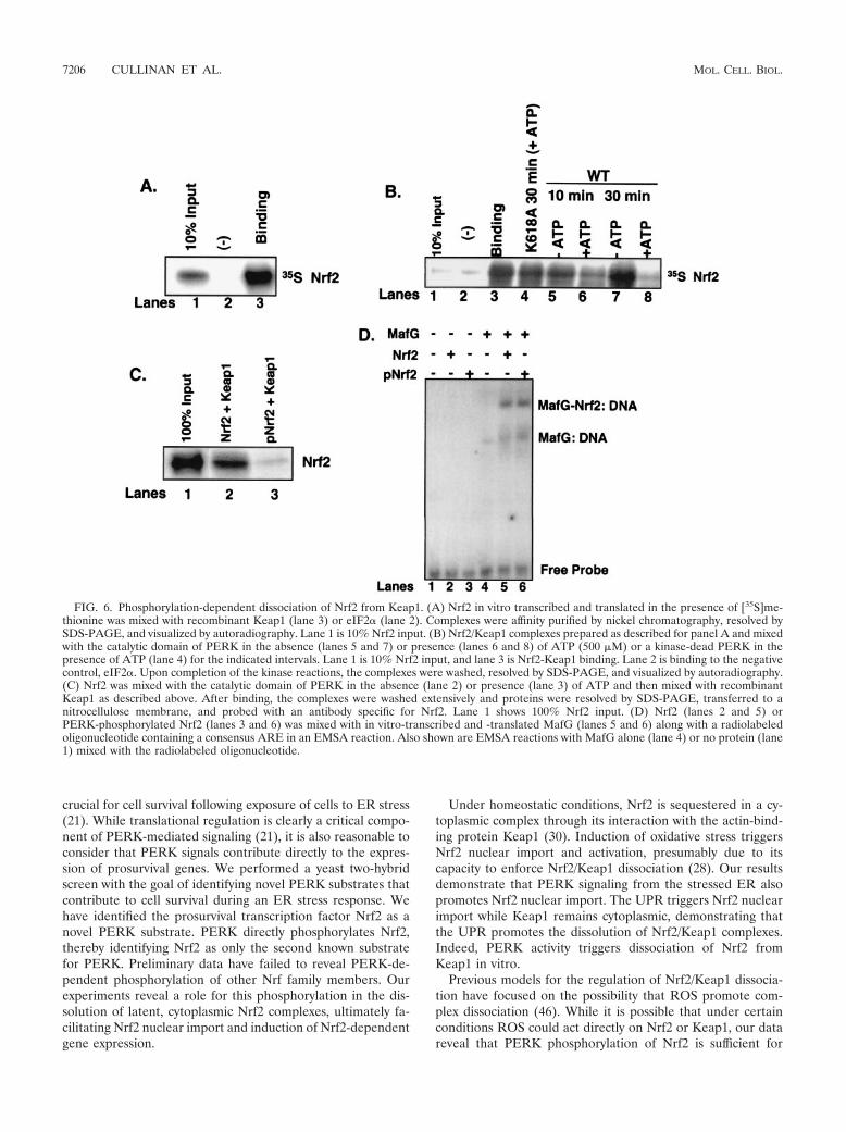
dent Nrf2 nuclear accumulation, while Keap1 remains cyto-plasmic, suggests that PERK phosphorylation triggers Nrf2/Keap1 dissociation. To directly assay complex dissociation,Nrf2 was in vitro transcribed and translated in the presence of[35S]methionine and mixed with recombinant His-Keap1 thatwas immobilized on nickel affinity resin. Nrf2 efficiently boundKeap1 (Fig. 6A, lane 3) but not the negative control, His-eIF2�(lane 2). After binding and extensive washing, the Nrf2/Keap1complexes were divided equally into aliquots and incubated inthe absence of �NPERK (Fig. 6B, lane 3), with �NPERK butwithout ATP (lanes 5 and 7), with �NPERK and unlabeledATP (500 �M) (lanes 6 and 8), or with the catalytically inactive�NPERK K618A (22) but with ATP (500 �M) (lane 4) for theindicated times at 30°C. Nrf2/Keap1 complexes were thenwashed extensively, and the presence of Keap1-associated Nrf2was determined by SDS-PAGE and visualized by autoradiog-raphy. Phosphorylation of Nrf2/Keap1 complexes by PERK ledto a time-dependent decrease in Keap1-associated Nrf2 (com-
pare lanes 3 and 6). By 30 min in the presence of the PERKcatalytic domain, Nrf2/Keap1 binding was equal to that of thenegative control (compare lanes 2 and 8); however, incubationof Nrf2/Keap1 complexes with PERK(K618A) for 30 min hadno effect on Nrf2/Keap1 association (compare lanes 3 and 4).The inability of PERK(K618A) to dissociate Nrf2/Keap1 com-plexes illustrates two important points. First, PERK catalyticactivity is essential for Nrf2/Keap1 dissociation rather thansimple physical association, as both wild-type PERK andPERK(K618A) can associate with Nrf2 (data not shown). Sec-ond, addition of ATP is not sufficient to trigger dissociation inthe absence of catalytically active PERK. These data demon-strate that PERK-dependent phosphorylation is sufficient totrigger Nrf2/Keap1 dissociation.
Having shown that PERK-dependent phosphorylation trig-gers dissociation of Nrf2/Keap1 complexes, we asked whetherPERK phosphorylation of Nrf2 would inhibit reassociation ofNrf2/Keap1 complexes. In vitro-transcribed and -translated
FIG. 4. PERK activity is necessary and sufficient for Nrf2 nuclear translocation. (A) NIH 3T3 cells proliferating on glass coverslips werecotransfected with plasmids encoding HA-Nrf2, Myc-Keap1 (panels a to c), or HA-Nrf2, myc-Keap1 and myc-PERK (panels d to f). Twenty-fourhours posttransfection, cells were fixed and proteins were detected by indirect immunofluorescence with an antibody specific for the HA epitope.DNA was stained with Hoechst dye. (B) Graphical representation of data in panel A. Error bars represent standard deviations from threeindependent experiments. (C) Wild-type (PERK�/�) or PERK�/� murine fibroblasts proliferating on glass coverslips were cotransfected withplasmids encoding HA-Nrf2 and myc-Keap1 and treated with 5 �g of tunicamycin/ml for the indicated time intervals. Proteins were detected withantibodies specific for the HA and myc epitopes and visualized by confocal microscopy. (D) Graphical representation of data presented in panelC. Solid bars represent wild-type cells, and hatched bars represent PERK�/� cells. Error bars represent the standard deviations from threeindependent experiments.
7204 CULLINAN ET AL. MOL. CELL. BIOL.
Nrf2 was incubated with �NPERK in the presence (Fig. 6C,lane 3) or absence (lane 2) of ATP for 30 min at 30°C. Fol-lowing phosphorylation, Nrf2 was mixed with recombinantKeap1. Nrf2 phosphorylation was monitored in parallel reac-tions by using [�-32P]ATP (data not shown). After extensivewashing, Western analysis revealed that the level of Nrf2 as-sociated with Keap1 was markedly decreased when Nrf2 wasprephosphorylated by �NPERK (compare lanes 2 and 3).These results demonstrate that not only does PERK-depen-dent phosphorylation promote Nrf2/Keap1 dissociation, butalso that once phosphorylated by PERK, Nrf2 cannot stablyassociate with Keap1. These data demonstrate that PERK-dependent phosphorylation triggers Nrf2/Keap1 dissociationand prevents reassociation.
To determine whether phosphorylation affects Nrf2 DNA-binding activity, purified recombinant Nrf2 that had beenphosphorylated with purified recombinant �NPERK, or leftunphosphorylated, was mixed with in vitro-transcribed and-translated MafG. The DNA-binding activity of these com-plexes was then assessed in an EMSA. Monomeric Nrf2,whether phosphorylated or unphosphorylated, exhibited nodetectable DNA-binding activity (Fig. 6D, lanes 2 and 3). How-ever, when mixed with a heterodimeric DNA-binding partner,MafG, both forms of Nrf2 exhibited indistinguishable DNA-binding activity (lanes 5 and 6).
Nrf2 expression increases cell survival following ER stress.Chronic ER stress triggers apoptosis (6, 8, 31). Nrf2 activationplays a role in cell survival following oxidative stress (7, 26). Todirectly assess the role of Nrf2 in survival following activationof the UPR, wild-type and Nrf2�/� MEFs plated at a lowdensity were challenged with tunicamycin for 8 h and subse-quently fed with complete medium lacking tunicamycin. Thecells were allowed to recover for 7 days, at which point surviv-
ing cells were visualized with Giemsa stain. The expression ofNrf2 greatly enhanced the ability of cells to survive followingER stress (Fig. 7A). Similarly, Nrf2 overexpression increasedcell survival in tunicamycin-treated NIH 3T3 cells (data notshown).
To further assess the ability of Nrf2 to provide a protectiveadvantage to cells during ER stress, we assayed for early signsof commitment to apoptosis in wild-type and Nrf2�/� MEFs.Cells were treated with tunicamycin for the indicated intervals(Fig. 7B) and then stained with annexin V. A sixfold increasein annexin V staining observed in Nrf2�/� MEFs, relative tothat in the wild type, was detectable by 4 h of tunicamycintreatment (Fig. 7B), and levels of annexin V-positive cellsincreased through 8 h of treatment. To rule out the possibilitythat the increased rate of death is due to a generalized sensi-tivity of Nrf2�/� MEFs to all death stimuli, we treated bothwild-type and Nrf2�/� MEFs with non-ER stress-inducingdeath stimuli: staurosporine (Fig. 7C) and TNF-� (Fig. 7D).Nrf2�/� MEFs were no more susceptible to these death stimulithan wild-type MEFs. The fact that eIF2� phosphorylation isinduced by tunicamycin treatment in Nrf2�/� MEFs (Fig. 7E)demonstrates that the increased sensitivity of Nrf2�/� MEFsdoes not reflect compromised eIF2� phosphorylation. Theseresults provide support for a model wherein Nrf2 plays a pro-survival role during the UPR.
DISCUSSION
Identification of Nrf2 as a PERK substrate. The ER-local-ized PERK kinase regulates cell fate decisions following cel-lular insults that perturb ER homeostasis. Upon exposure toER stress-inducing agents, activated PERK phosphorylates itsonly documented substrate, eIF2� (22, 38). PERK function is
FIG. 5. Nrf2 nuclear translocation occurs independently of eIF2� phosphorylation. (A and B) Wild-type fibroblasts (B) or eIF2�(S51A)fibroblasts (A) were cotransfected with plasmids encoding HA-Nrf2 and myc-Keap1 and treated with 5 �g of tunicamycin/ml for the indicated timeintervals. HA-Nrf2 was detected with the 12CA5 monoclonal antibody and visualized by confocal microscopy. (C) Nuclear extracts were preparedfrom wild-type (WT), eIF2�(S51A), or PERK�/� MEFs treated with 5 �g of tunicamycin/ml for the indicated intervals, resolved by SDS-PAGE,transferred to a nitrocellulose membrane, and probed with an antibody specific for Nrf2.
VOL. 23, 2003 ACTIVATION OF Nrf2 BY THE UPR 7205
crucial for cell survival following exposure of cells to ER stress(21). While translational regulation is clearly a critical compo-nent of PERK-mediated signaling (21), it is also reasonable toconsider that PERK signals contribute directly to the expres-sion of prosurvival genes. We performed a yeast two-hybridscreen with the goal of identifying novel PERK substrates thatcontribute to cell survival during an ER stress response. Wehave identified the prosurvival transcription factor Nrf2 as anovel PERK substrate. PERK directly phosphorylates Nrf2,thereby identifying Nrf2 as only the second known substratefor PERK. Preliminary data have failed to reveal PERK-de-pendent phosphorylation of other Nrf family members. Ourexperiments reveal a role for this phosphorylation in the dis-solution of latent, cytoplasmic Nrf2 complexes, ultimately fa-cilitating Nrf2 nuclear import and induction of Nrf2-dependentgene expression.
Under homeostatic conditions, Nrf2 is sequestered in a cy-toplasmic complex through its interaction with the actin-bind-ing protein Keap1 (30). Induction of oxidative stress triggersNrf2 nuclear import and activation, presumably due to itscapacity to enforce Nrf2/Keap1 dissociation (28). Our resultsdemonstrate that PERK signaling from the stressed ER alsopromotes Nrf2 nuclear import. The UPR triggers Nrf2 nuclearimport while Keap1 remains cytoplasmic, demonstrating thatthe UPR promotes the dissolution of Nrf2/Keap1 complexes.Indeed, PERK activity triggers dissociation of Nrf2 fromKeap1 in vitro.
Previous models for the regulation of Nrf2/Keap1 dissocia-tion have focused on the possibility that ROS promote com-plex dissociation (46). While it is possible that under certainconditions ROS could act directly on Nrf2 or Keap1, our datareveal that PERK phosphorylation of Nrf2 is sufficient for
FIG. 6. Phosphorylation-dependent dissociation of Nrf2 from Keap1. (A) Nrf2 in vitro transcribed and translated in the presence of [35S]me-thionine was mixed with recombinant Keap1 (lane 3) or eIF2� (lane 2). Complexes were affinity purified by nickel chromatography, resolved bySDS-PAGE, and visualized by autoradiography. Lane 1 is 10% Nrf2 input. (B) Nrf2/Keap1 complexes prepared as described for panel A and mixedwith the catalytic domain of PERK in the absence (lanes 5 and 7) or presence (lanes 6 and 8) of ATP (500 �M) or a kinase-dead PERK in thepresence of ATP (lane 4) for the indicated intervals. Lane 1 is 10% Nrf2 input, and lane 3 is Nrf2-Keap1 binding. Lane 2 is binding to the negativecontrol, eIF2�. Upon completion of the kinase reactions, the complexes were washed, resolved by SDS-PAGE, and visualized by autoradiography.(C) Nrf2 was mixed with the catalytic domain of PERK in the absence (lane 2) or presence (lane 3) of ATP and then mixed with recombinantKeap1 as described above. After binding, the complexes were washed extensively and proteins were resolved by SDS-PAGE, transferred to anitrocellulose membrane, and probed with an antibody specific for Nrf2. Lane 1 shows 100% Nrf2 input. (D) Nrf2 (lanes 2 and 5) orPERK-phosphorylated Nrf2 (lanes 3 and 6) was mixed with in vitro-transcribed and -translated MafG (lanes 5 and 6) along with a radiolabeledoligonucleotide containing a consensus ARE in an EMSA reaction. Also shown are EMSA reactions with MafG alone (lane 4) or no protein (lane1) mixed with the radiolabeled oligonucleotide.
7206 CULLINAN ET AL. MOL. CELL. BIOL.

Nrf2/Keap1 dissociation and that ROS are not required. Whilewe have not yet identified the residues within Nrf2 targeted byPERK, our preliminary data indicate that the phosphorylationsite lies within the amino terminus of Nrf2. This portion ofNrf2 contains the Neh2 domain, which is critical for interactionwith Keap1 (30). It seems likely that Nrf2 phosphorylationtriggers a conformational change that disrupts Nrf2/Keap1complexes.
Distinct mechanisms of PERK-dependent regulation of geneexpression. While cellular protein translation is repressed dueto eIF2� phosphorylation following ER stress, translation ofATF4 is selectively increased (19). ATF4 translation is notaugmented in PERK�/� cells (19), indicating that during theUPR, ATF4 synthesis is PERK dependent. While PERK sig-naling is necessary for ATF4 translation, an intermediate sig-naling step, eIF2� phosphorylation, is also required (21, 37). Incontrast, our data demonstrate that Nrf2 is an immediatePERK substrate and its activation is independent of eIF2�phosphorylation. Consistent with this idea, we found thatstress-dependent Nrf2 nuclear import was lost in PERK�/�
cells; furthermore, dominant-negative PERK moleculesblocked the nuclear import (data not shown) and transcrip-tional activation of Nrf2, indicating that PERK activity is re-quired for Nrf2 activation. Unlike ATF4, Nrf2 is synthesized in
unstressed cells, but is maintained in latent cytoplasmic com-plexes, by virtue of its interaction with Keap1 (30). PERKsignals Nrf2 directly, resulting in a rapid response to ER stress.As illustrated in Fig. 8, our data demonstrate that followingPERK activation signals bifurcate, resulting in the regulationof cell survival via two distinct pathways. The first involves thetranslation-dependent accumulation of ATF4, and the secondis mediated by PERK-dependent phosphorylation of inactiveNrf2.
PERK, Nrf2, and cell survival signaling. While most exper-iments have utilized cell culture systems to evaluate the con-sequences of increased ER stress, cells in the context of thewhole organism are also vulnerable to perturbations in ERhomeostasis. Indeed, PERK�/� mice exhibit massive levels ofapoptosis of pancreatic beta cells, leading to the developmentof diabetes (20, 44). Mice homozygous for a mutant, nonphos-phorylatable eIF2� (S51A) also display a beta cell deficiencyand die soon after birth (37), indicating the importance oftranslational control during the UPR. These results suggest animportant role for PERK signaling in cell survival followingER stress.
Our results demonstrate that Nrf2 activation regulates cellsurvival following UPR activation. The absence of Nrf2 in-creased apoptosis following ER stress, but not in response to
FIG. 7. Nrf2 promotes increased cell survival after chronic ER stress. (A) Wild-type and Nrf2�/� MEFs were left untreated or treated with 1�g of tunicamycin/ml for 8 h and then allowed to recover for 7 days in normal medium. Colonies were visualized by using Giemsa stain. (B to D)Wild-type (Œ) and Nrf2�/� (■ ) MEFs proliferating on glass coverslips were left untreated or treated with 5 �g of tunicamycin/ml (B), 4 �Mstaurosporine (C), or 5 ng of TNF-�/ml plus 10 �g of cycloheximide/ml (D) for the indicated intervals (in hours). Apoptotic cells were visualizedwith a FITC-conjugated annexin V antibody. Error bars represent the standard deviations from three independent experiments. (E) Wild-type andNrf2�/� MEFs were treated with 5 �g of tunicamycin/ml for the indicated intervals. Proteins were resolved by SDS-PAGE, and membranes wereprobed with antibodies specific for total and phosphorylated eIF2�.
VOL. 23, 2003 ACTIVATION OF Nrf2 BY THE UPR 7207
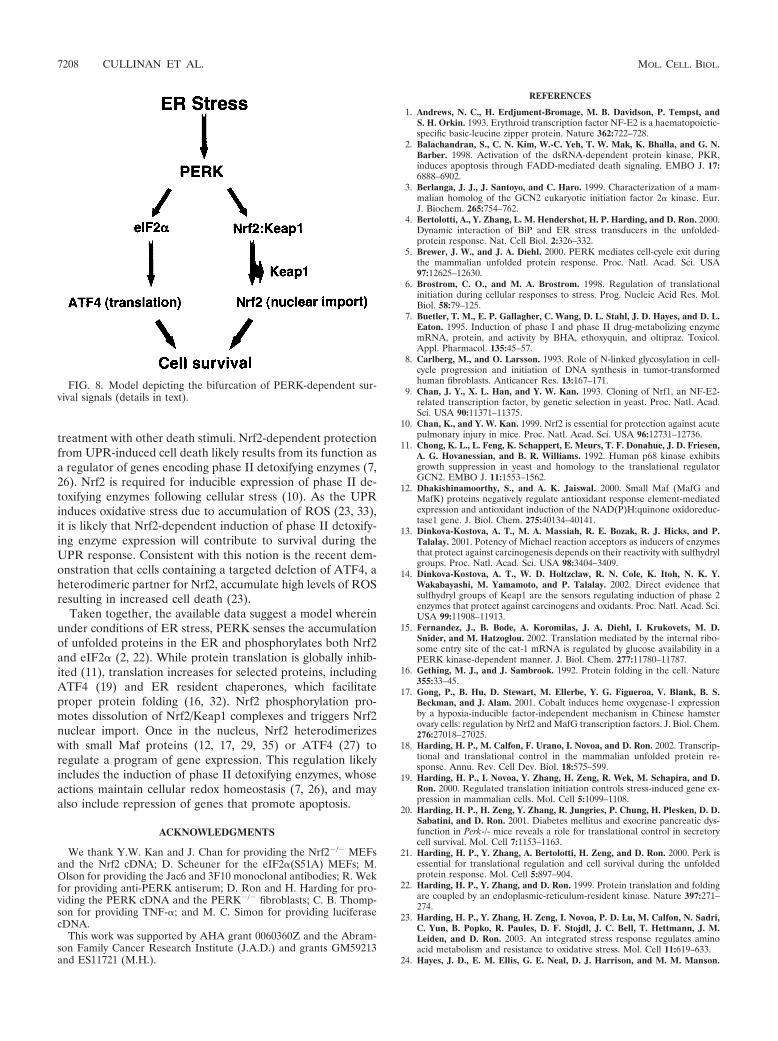
treatment with other death stimuli. Nrf2-dependent protectionfrom UPR-induced cell death likely results from its function asa regulator of genes encoding phase II detoxifying enzymes (7,26). Nrf2 is required for inducible expression of phase II de-toxifying enzymes following cellular stress (10). As the UPRinduces oxidative stress due to accumulation of ROS (23, 33),it is likely that Nrf2-dependent induction of phase II detoxify-ing enzyme expression will contribute to survival during theUPR response. Consistent with this notion is the recent dem-onstration that cells containing a targeted deletion of ATF4, aheterodimeric partner for Nrf2, accumulate high levels of ROSresulting in increased cell death (23).
Taken together, the available data suggest a model whereinunder conditions of ER stress, PERK senses the accumulationof unfolded proteins in the ER and phosphorylates both Nrf2and eIF2� (2, 22). While protein translation is globally inhib-ited (11), translation increases for selected proteins, includingATF4 (19) and ER resident chaperones, which facilitateproper protein folding (16, 32). Nrf2 phosphorylation pro-motes dissolution of Nrf2/Keap1 complexes and triggers Nrf2nuclear import. Once in the nucleus, Nrf2 heterodimerizeswith small Maf proteins (12, 17, 29, 35) or ATF4 (27) toregulate a program of gene expression. This regulation likelyincludes the induction of phase II detoxifying enzymes, whoseactions maintain cellular redox homeostasis (7, 26), and mayalso include repression of genes that promote apoptosis.
ACKNOWLEDGMENTS
We thank Y.W. Kan and J. Chan for providing the Nrf2�/� MEFsand the Nrf2 cDNA; D. Scheuner for the eIF2�(S51A) MEFs; M.Olson for providing the Jac6 and 3F10 monoclonal antibodies; R. Wekfor providing anti-PERK antiserum; D. Ron and H. Harding for pro-viding the PERK cDNA and the PERK�/� fibroblasts; C. B. Thomp-son for providing TNF-�; and M. C. Simon for providing luciferasecDNA.
This work was supported by AHA grant 0060360Z and the Abram-son Family Cancer Research Institute (J.A.D.) and grants GM59213and ES11721 (M.H.).
REFERENCES
1. Andrews, N. C., H. Erdjument-Bromage, M. B. Davidson, P. Tempst, andS. H. Orkin. 1993. Erythroid transcription factor NF-E2 is a haematopoietic-specific basic-leucine zipper protein. Nature 362:722–728.
2. Balachandran, S., C. N. Kim, W.-C. Yeh, T. W. Mak, K. Bhalla, and G. N.Barber. 1998. Activation of the dsRNA-dependent protein kinase, PKR,induces apoptosis through FADD-mediated death signaling. EMBO J. 17:6888–6902.
3. Berlanga, J. J., J. Santoyo, and C. Haro. 1999. Characterization of a mam-malian homolog of the GCN2 eukaryotic initiation factor 2� kinase. Eur.J. Biochem. 265:754–762.
4. Bertolotti, A., Y. Zhang, L. M. Hendershot, H. P. Harding, and D. Ron. 2000.Dynamic interaction of BiP and ER stress transducers in the unfolded-protein response. Nat. Cell Biol. 2:326–332.
5. Brewer, J. W., and J. A. Diehl. 2000. PERK mediates cell-cycle exit duringthe mammalian unfolded protein response. Proc. Natl. Acad. Sci. USA97:12625–12630.
6. Brostrom, C. O., and M. A. Brostrom. 1998. Regulation of translationalinitiation during cellular responses to stress. Prog. Nucleic Acid Res. Mol.Biol. 58:79–125.
7. Buetler, T. M., E. P. Gallagher, C. Wang, D. L. Stahl, J. D. Hayes, and D. L.Eaton. 1995. Induction of phase I and phase II drug-metabolizing enzymemRNA, protein, and activity by BHA, ethoxyquin, and oltipraz. Toxicol.Appl. Pharmacol. 135:45–57.
8. Carlberg, M., and O. Larsson. 1993. Role of N-linked glycosylation in cell-cycle progression and initiation of DNA synthesis in tumor-transformedhuman fibroblasts. Anticancer Res. 13:167–171.
9. Chan, J. Y., X. L. Han, and Y. W. Kan. 1993. Cloning of Nrf1, an NF-E2-related transcription factor, by genetic selection in yeast. Proc. Natl. Acad.Sci. USA 90:11371–11375.
10. Chan, K., and Y. W. Kan. 1999. Nrf2 is essential for protection against acutepulmonary injury in mice. Proc. Natl. Acad. Sci. USA 96:12731–12736.
11. Chong, K. L., L. Feng, K. Schappert, E. Meurs, T. F. Donahue, J. D. Friesen,A. G. Hovanessian, and B. R. Williams. 1992. Human p68 kinase exhibitsgrowth suppression in yeast and homology to the translational regulatorGCN2. EMBO J. 11:1553–1562.
12. Dhakishinamoorthy, S., and A. K. Jaiswal. 2000. Small Maf (MafG andMafK) proteins negatively regulate antioxidant response element-mediatedexpression and antioxidant induction of the NAD(P)H:quinone oxidoreduc-tase1 gene. J. Biol. Chem. 275:40134–40141.
13. Dinkova-Kostova, A. T., M. A. Massiah, R. E. Bozak, R. J. Hicks, and P.Talalay. 2001. Potency of Michael reaction acceptors as inducers of enzymesthat protect against carcinogenesis depends on their reactivity with sulfhydrylgroups. Proc. Natl. Acad. Sci. USA 98:3404–3409.
14. Dinkova-Kostova, A. T., W. D. Holtzclaw, R. N. Cole, K. Itoh, N. K. Y.Wakabayashi, M. Yamamoto, and P. Talalay. 2002. Direct evidence thatsulfhydryl groups of Keap1 are the sensors regulating induction of phase 2enzymes that protect against carcinogens and oxidants. Proc. Natl. Acad. Sci.USA 99:11908–11913.
15. Fernandez, J., B. Bode, A. Koromilas, J. A. Diehl, I. Krukovets, M. D.Snider, and M. Hatzoglou. 2002. Translation mediated by the internal ribo-some entry site of the cat-1 mRNA is regulated by glucose availability in aPERK kinase-dependent manner. J. Biol. Chem. 277:11780–11787.
16. Gething, M. J., and J. Sambrook. 1992. Protein folding in the cell. Nature355:33–45.
17. Gong, P., B. Hu, D. Stewart, M. Ellerbe, Y. G. Figueroa, V. Blank, B. S.Beckman, and J. Alam. 2001. Cobalt induces heme oxygenase-1 expressionby a hypoxia-inducible factor-independent mechanism in Chinese hamsterovary cells: regulation by Nrf2 and MafG transcription factors. J. Biol. Chem.276:27018–27025.
18. Harding, H. P., M. Calfon, F. Urano, I. Novoa, and D. Ron. 2002. Transcrip-tional and translational control in the mammalian unfolded protein re-sponse. Annu. Rev. Cell Dev. Biol. 18:575–599.
19. Harding, H. P., I. Novoa, Y. Zhang, H. Zeng, R. Wek, M. Schapira, and D.Ron. 2000. Regulated translation initiation controls stress-induced gene ex-pression in mammalian cells. Mol. Cell 5:1099–1108.
20. Harding, H. P., H. Zeng, Y. Zhang, R. Jungries, P. Chung, H. Plesken, D. D.Sabatini, and D. Ron. 2001. Diabetes mellitus and exocrine pancreatic dys-function in Perk-/- mice reveals a role for translational control in secretorycell survival. Mol. Cell 7:1153–1163.
21. Harding, H. P., Y. Zhang, A. Bertolotti, H. Zeng, and D. Ron. 2000. Perk isessential for translational regulation and cell survival during the unfoldedprotein response. Mol. Cell 5:897–904.
22. Harding, H. P., Y. Zhang, and D. Ron. 1999. Protein translation and foldingare coupled by an endoplasmic-reticulum-resident kinase. Nature 397:271–274.
23. Harding, H. P., Y. Zhang, H. Zeng, I. Novoa, P. D. Lu, M. Calfon, N. Sadri,C. Yun, B. Popko, R. Paules, D. F. Stojdl, J. C. Bell, T. Hettmann, J. M.Leiden, and D. Ron. 2003. An integrated stress response regulates aminoacid metabolism and resistance to oxidative stress. Mol. Cell 11:619–633.
24. Hayes, J. D., E. M. Ellis, G. E. Neal, D. J. Harrison, and M. M. Manson.
FIG. 8. Model depicting the bifurcation of PERK-dependent sur-vival signals (details in text).
7208 CULLINAN ET AL. MOL. CELL. BIOL.
1999. Cellular response to cancer chemopreventive agents: contribution ofthe antioxidant responsive element to the adaptive response to oxidative andchemical stress. Biochem. Soc. Symp. 64:141–168.
25. Hayes, J. D., and M. McMahon. 2001. Molecular basis for the contributionof the antioxidant responsive element to cancer chemoprevention. CancerLett. 174:103–113.
26. Hayes, J. D., and D. J. Pulford. 1995. The glutathione S-transferase super-gene family: regulation of GST and the contribution of the isoenzymes tocancer chemoprotection and drug resistance. Crit. Rev. Biochem. Mol. Biol.30:446–600.
27. He, C. H., P. Gong, B. Hu, D. Stewart, M. E. Choi, A. M. K. Choi, and J.Alam. 2001. Identification of the activating transcription factor (ATF4) as anNrf2-interacting protein. J. Biol. Chem. 276:20858–20865.
28. Huang, H.-C., T. Nguyen, and C. B. Pickett. 2000. Regulation of the anti-oxidant response element by protein kinase C-mediated phosphorylation ofNF-E2-related factor 2. Proc. Natl. Acad. Sci. USA 97:12475–12480.
29. Itoh, K., T. Chiba, S. Takahashi, T. Ishii, K. Igarashi, Y. Katoh, T. Oyake,N. Hayahsi, K. Satoh, I. Hatayama, M. Yamamoto, and Y. Nabeshima. 1997.An Nrf2/small Maf heterodimer mediates the induction of phase II detoxi-fying enzyme genes through antioxidant response elements. Biochem. Bio-phys. Res. Commun. 2236:313–322.
30. Itoh, K., N. Wakabayashi, Y. Kotoh, T. Ishii, K. Igarashi, J. D. Engel, and M.Yamamoto. 1999. Keap1 represses nuclear activation of antioxidant respon-sive elements by Nrf2 through binding to the amino-terminal Neh2 domain.Genes Dev. 13:76–86.
31. Kaufman, R. J. 1999. Stress signaling from the lumen of the endoplasmicreticulum: coordination of gene transcriptional and translational controls.Genes Dev. 13:1211–1233.
32. Kozutsumi, Y., M. Segal, K. Normington, M. J. Gething, and J. Sambrook.1988. The presence of malfolded proteins in the endoplasmic reticulumsignals the induction of glucose-regulated proteins. Nature 332:462–464.
33. McCullough, K. D., J. L. Martindale, L.-O. Klotz, T.-Y. Aw, and N. J.Holbrook. 2001. Gadd153 sensitizes cells to endoplasmic reticulum stress bydown-regulating BclII and perturbing the cellular redox state. Mol. Cell.Biol. 21:1249–1259.
34. Moi, P., K. Chan, I. Asunis, A. Cao, and Y. W. Kan. 1994. Isolation ofNF-E2-related factor 2 (Nrf2), a NF-E2-like basic leucine zipper transcrip-tional activator that binds to the tandem NF-E2/AP1 repeat of the beta-globin locus control region. Proc. Natl. Acad. Sci. USA 91:9926–9930.
35. Nguyen, T., H. C. Huang, and C. B. Pickett. 2000. Transcriptional regulationof the antioxidant response element. Activation by Nrf2 and repression byMafK. J. Biol. Chem. 275:15466–15473.
36. Rushmore, T. H., M. R. Morton, and C. B. Pickett. 1991. The antioxidantresponsive element. Activation by oxidative stress and identification of theDNA consensus sequence required for functional activity. J. Biol. Chem.266:11632–11639.
37. Scheuner, D., B. Song, E. McEwen, C. Liu, R. Laybutt, P. Gillespie, T.Saunders, S. Bonner-Weir, and R. J. Kaufman. 2001. Translational controlis required for the unfolded protein response and in vivo glucose homeosta-sis. Mol. Cell 7:1165–1176.
38. Shi, Y., K. M. Vattem, R. Sood, J. An, J. Liang, L. Stramm, and R. C. Wek.1998. Identification and characterization of pancreatic eukaryotic initiationfactor 2-subunit kinase, PEK, involved in translational control. Mol. Cell.Biol. 18:7499–7509.
39. Sood, R., A. C. Porter, D. Olsen, D. R. Cavener, and R. C. Wek. 2000. Amammalian homologue of GCN2 protein kinase important for translationalcontrol by phosphorylation of eukaryotic initiation factor-2�. Genetics 154:787–801.
40. Tirasophon, W., A. A. Welihinda, and R. J. Kaufman. 1998. A stress responsepathway from the endoplasmic reticulum to the nucleus requires a novelbifunctional protein kinase/endoribonuclease (Ire1p) in mammalian cells.Genes Dev. 12:2416–2423.
41. Todaro, G. J., and H. Green. 1963. Quantitative studies of the growth ofmouse embryo cells in culture and their development into established lines.J. Cell Biol. 17:299–313.
42. Wang, X. Z., B. Lawson, J. W. Brewer, H. Zinszner, A. Sanjay, L. J. Mi, R.Boorstein, G. Kreibich, L. M. Hendershot, and D. Ron. 1996. Signals fromthe stressed endoplasmic reticulum induce C/EBP-homologous protein(CHOP/GADD153). Mol. Cell. Biol. 16:4273–4280.
43. Yu, R., C. Chen, Y. Y. Mo, V. Hebbar, E. D. Owuor, T. H. Tan, and A. N.Kong. 2000. Activation of mitogen-activated protein kinase pathways inducesantioxidant response element-mediated gene expression via a Nrf2-depen-dent mechanism. J. Biol. Chem. 275:39907–39913.
44. Zhang, P., B. McGrath, S. Li, A. Frank, F. Zambito, J. Reinert, M. Gannon,K. Ma, K. McNaughton, and D. R. Caverner. 2002. The PERK eukaryoticinitiation factor 2� kinase is required for the development of the skeletalsystem, postnatal growth, and the function and viability of the pancreas. Mol.Cell. Biol. 22:3864–3874.
45. Zipper, L. M., and R. T. Mulcahy. 2000. Inhibition of Erk and p38 MAPkinases inhibits binding of Nrf2 and induction of GCS genes. Biochem.Biophys. Res. Commun. 278:484–492.
46. Zipper, L. M., and R. T. Mulcahy. 2002. The Keap1 BTB/POZ dimerizationfunction is required to sequester Nrf2 in cytoplasm. J. Biol. Chem. 277:36544–36552.
VOL. 23, 2003 ACTIVATION OF Nrf2 BY THE UPR 7209