nutritional intervention in animals: benchmarking of ... · 6 interaction between gastro-intestinal...
TRANSCRIPT
Together with our clients, we integrate scientific know-how and practical experience
to develop livestock concepts for the 21st century. With our expertise on innovative
livestock systems, nutrition, welfare, genetics and environmental impact of livestock
farming and our state-of-the art research facilities, such as Dairy Campus and Swine
Innovation Centre Sterksel, we support our customers to find solutions for current
and future challenges.
The mission of Wageningen UR (University & Research centre) is ‘To explore
the potential of nature to improve the quality of life’. Within Wageningen UR,
nine specialised research institutes of the DLO Foundation have joined forces
with Wageningen University to help answer the most important questions in the
domain of healthy food and living environment. With approximately 30 locations,
6,000 members of staff and 9,000 students, Wageningen UR is one of the leading
organisations in its domain worldwide. The integral approach to problems and
the cooperation between the various disciplines are at the heart of the unique
Wageningen Approach.
Wageningen UR Livestock Research
P.O. Box 338
6700 AH Wageningen
The Netherlands
T +31 (0)317 480 10 77
www.wageningenUR.nl/livestockresearch
Livestock Research Report 800
M.M. van Krimpen, M.M. Hulst, J. van der Meulen, D. Schokker
H.F.J. Savelkoul, E.J. Tijhaar
V.P.M.G. Rutten
Nutritional intervention in animals: benchmarking of strategies, monitoring biomarkers and immune competence
LSR_Rapport_omslag_800.indd 1 15-10-14 13:40

Nutritional intervention in animals:benchmarking of strategies, monitoringbiomarkers and immune competence
M.M. van Krimpen, M.M. Hulst, J. van der Meulen, D. Schokker1
H.F.J. Savelkoul, E.J. Tijhaar2
V.P.M.G. Rutten3
1 Wageningen UR Livestock Research
2 Wageningen UR
3 Utrecht University
Dit onderzoek is uitgevoerd door Wageningen UR Livestock Research, in opdracht van en gefinancierd door hetFeed4Foodure consortium (http://www.wageningenur.nl/nl/Onderzoek-Resultaten/Projecten/Feed4Foodure.htm)
Wageningen UR Livestock ResearchWageningen, Oktober 2014
Livestock Research Report 800
M.M. van Krimpen, M.M. Hulst, J. van der Meulen, D. Schokker, H.F.J. Savelkoul, E.J. Tijhaar andV.P.M.G. Rutten 2014. Nutritional intervention in animals: benchmarking of strategies, monitoring
biomarkers and immune competence. Lelystad, Wageningen UR (University & Research centre)Livestock Research, Livestock Research Report 110 blz.
Samenvatting NLHet huidige rapport omvat een literatuurstudie naar diverse aspecten die gerelateerd zijn aan deimmuun competentie van landbouwhuisdieren. Deze aspecten, die in hoofdstuk 1 geïntroduceerdworden, zijn: Het aantonen van verbanden tussen functionele componenten in diervoedergrondstoffen en de
expressie van genen/biologische processen, die van invloed zijn op de darmgezondheid vanlandbouwhuisdieren;
Het beschrijven van modellen die gebruikt kunnen worden bij onderzoek naar effecten vanvoedingsinterventies op immuun gerelateerde kenmerken in landbouwhuisdieren;
Het samenvatten van effecten van nutritionele interventies in de maternale, neonatale en post-neonatale fase op de ontwikkeling van het aangeboren en verworven immuunsysteem;
Een literatuurstudie naar de relatie tussen het immuunsysteem in de darm en in de bovensteluchtwegen, waarbij ingegaan wordt op de vraag hoe voedingsinterventies het immuunsysteem inde bovenste luchtwegen kunnen ondersteunen.
Summary UKThe current study covers a review of literature regarding a number of topics related to immunecompetence in farm animals, which are introduced in chapter 1. These topics are: A demonstration of the relationship between functional feed components and the expression of
genes/biological processes that are involved in gut health of farm animals; A description of available models, that can be used to investigate the effects of nutritional
interventions on immune related parameters in animals; A review of the effects of nutritional interventions in the maternal, neonatal and post-neonatal
phase on the development of the innate and acquired immune system; A review of the relationship between the immune system in the gut and in the upper airways,
whereas the question will be addressed how the immune system in the upper airways can beaffected by nutritional interventions.
© 2014 Wageningen UR Livestock Research, P.O. Box 338, 6700 AH Wageningen, The Netherlands,T +31 (0)317 48 39 53, E [email protected], www.wageningenUR.nl/en/livestockresearch.Livestock Research is part of Wageningen UR (University & Research centre).
All rights reserved. No part of this publication may be reproduced and/or made public, whether byprint, photocopy, microfilm or any other means, without the prior permission of the publisher orauthor.
The ISO 9001 certification by DNV underscores our quality level. All our researchcommissions are in line with the Terms and Conditions of the Animal Sciences Group. Theseare filed with the District Court of Zwolle.
Livestock Research Report 800

Livestock Research Report 800 | 3
Table of contents
Foreword 9
Samenvatting 11
Summary 16
1 Rationale of the report 20
2 Introduction 23
2.1 Functional feed components 232.2 Animal models available for studying immune competence 232.3 Nutritional immune modulation 232.4 Relation between immune system of the gut and airways 24
3 Feed ingredients and functional components 25
3.1 Gene expression datasets of nutritional interventions in farm animals 263.2 Functional analysis and data-mining using a gene expression dataset 27
4 Human and animal models for testing immune competence 30
4.1 In vitro models 304.1.1 Primary cells 304.1.2 Epithelial mini-guts 314.1.3 Cell cultures 314.1.4 2D Cell cultures 314.1.5 3D Cell cultures 324.1.6 Intestine-on-a-chip 32
4.2 Ex vivo models 344.2.1 Perfusion system 344.2.2 Everted sac technique 344.2.3 Ussing chamber 344.2.4 In vitro organ culture (IVOC) 344.2.5 Non-polarized (or traditional) IVOC 354.2.6 Polarized in vitro organ culture (pIVOC) 354.2.7 Novel polarized ex vivo organ culture 354.2.8 IVOC in the Ussing chamber 364.2.9 Precision-cut tissue slices (PCTS) 374.2.10Porcine intestinal tissue model 37
4.3 In situ models 384.3.1 Intestinal loop model 384.3.2 Small intestinal segment perfusion (SISP) 38
4.4 In vivo models 384.5 Correlations between models and real practice in humans and monogastrics 38
5 Nutritional intervention strategies for monogastrics 40
5.1 Effects of interventions during the maternal stage on the immune competence ofthe progeny 405.1.1 Trans-generational effects 405.1.2 Trans-generational transfer of immune competence 405.1.3 Impact of nutritional interventions in the maternal diets on trans-
generational epigenetic effects 41
Livestock Research Report 800 | 5
5.1.4 Conclusions 455.2 Impact of nutritional interventions in the neonatal phase on immune related
parameters 455.2.1 Results from literature 455.2.2 Human 515.2.3 Conclusions 53
5.3 Impact of nutritional interventions in the post-neonatal phase on immune relatedparameters 53
5.4 Mode of action of dietary interventions on immunity 59
6 Interaction between gastro-intestinal and airway mucosal immune systems 63
6.1 Immune system of the respiratory tract 636.2 The “common” mucosal immune system 646.3 Gastrointestinal Immunity. 646.4 The respiratory tract in pigs and its immune system 696.5 Secretory IgA: Designed for Anti-Microbial Defence and minimal mucosal
inflammation 70
References 73
Appendix 1 Gene expression datasets of nutritional interventions in cattle 81
Appendix 2a Gene expression datasets of nutritional interventions in pigs 82
Appendix 2b Gene expression datasets of nutritional interventions in pigs 83
Appendix 3a Gene expression datasets of nutritional interventions in chickens
84
Appendix 3b Gene expression datasets of nutritional interventions in chickens
85
Appendix 4 Tutorial for data mining 86
Appendix 5 David subset > Immune system phenotype / cell adhesion /
cytoskelet / transcription (IAC) 87
Appendix 6 GENEDECKS subset > small molecule metabolic process (SMMP) 88
Appendix 7 Pathways / Compounds / Tissue expression / Transcription binding
sequences 89
Appendix 8 Pathways / Compounds / Tissue expression / Transcription binding
sequences 90
Appendix 9 STITCH interactors (type of association and confidential scores) 91
Appendix 10 Predicted chemical (Comparative Toxicogenomics Database
hyperlinks) 92
Appendix 11 Detailed study information of the experiments that tested trans-
generational epigenetic effects (maternal phase) 93
Appendix 12 Detailed study information regarding the effect of nutritional
interventions on immune related parameters in pigs (neonatal
phase) 97
Appendix 13 Detailed study information regarding the effect of nutritional
interventions on immune related parameters in poultry (neonatal
phase) 102
Appendix 14 Detailed study information of interventions that were tested in
the post-neonatal phase 107
Appendix 15 Detailed study information of interventions that were tested in
neonatal humans 111
Appendix 15 A brief introduction to the immune system 113


Livestock Research Report 800 | 7

Livestock Research Report 800 | 9
Foreword
Feed4Foodure is a public-private partnership between the Dutch Ministry of Economic Affairs, aconsortium of various organizations within the animal production chain and Wageningen UR LivestockResearch. Feed4Foodure aims to contribute to sustainable and healthy livestock farming in theNetherlands, simultaneously strengthening our competitive position on the global market. TheFeed4Foodure program line “Nutrition, Intestinal Health, and Immunity”, aims to contribute to areduction in the use of antibiotics in livestock farming by increasing general health and diseaseresistance. The main goals are to develop innovative measurement techniques and to test new health-promoting nutritional additives in the field of gut health and immunity.The current study covers a review of literature regarding a number of topics related to the focus areaof the Feed4Foodure program line “Nutrition, Intestinal Health, and Immunity”. These topics are:The impact of functional feed components on the expression of genes/biological processes that arerelated to gut health of farm animals;Available models, that can be used to investigate the effects of nutritional interventions on immunerelated parameters in animals;The effects of nutritional interventions in the maternal, neonatal and post-neonatal phase on thedevelopment of the innate and acquired immune system;The relationship between the immune system in the gut and in the upper airways, and the possibilitiesto affects the immune system in the upper airways by nutritional interventions.For the current study, scientist of Wageningen UR Livestock Research, Wageningen University andUtrecht University worked together with representatives from the various private partners, includingAgrifirm, ForFarmers, Nutreco, De Heus, Denkavit, and Darling Ingredients International. The authorsthank the industry partners of the project team for their worthwhile input.
Dr. Mari Smits, leader Feed4Foodure program line “Nutrition, Intestinal Health, and Immunity”.

10 | Livestock Research Report 800
Livestock Research Report 800 | 11
Samenvatting
Besmettelijke dierziekten hebben een sterk remmend effect op het welzijn van dieren, het efficiëntgebruik van nutriënten en de ecologische footprint van de dierlijke productie. Voeding kan bijdragenaan het vergroten van de immuun competentie en daarmee aan het beperken van de incidentie vanziekten. Immuun competentie wordt gedefinieerd als het vermogen van het immuunsysteem om opadequate wijze te reageren op een antigene stimulus door middel van een evenwichtigeimmuunrespons met een goede balans tussen tolerantie en ontsteking. Het belang van immuniteitversterkende diervoeding is toegenomen, omdat bacterieremmers, anti-parasitaire toevoegmiddelenen sommige andere additieven die de diergezondheid bevorderen omwille van de volksgezondheid nietmeer toegevoegd worden aan het voer.Het huidige rapport omvat een literatuurstudie naar diverse aspecten die gerelateerd zijn aan deimmuun competentie van landbouwhuisdieren. Deze aspecten, die in hoofdstuk 1 geïntroduceerdworden, zijn: Het aantonen van verbanden tussen functionele componenten in diervoedergrondstoffen en de
expressie van genen/biologische processen, die van invloed zijn op de darmgezondheid vanlandbouwhuisdieren;
Het beschrijven van modellen die gebruikt kunnen worden bij onderzoek naar effecten vanvoedingsinterventies op immuun gerelateerde kenmerken in landbouwhuisdieren;
Het samenvatten van effecten van nutritionele interventies in de maternale, neonatale en post-neonatale fase op de ontwikkeling van het aangeboren en verworven immuunsysteem;
Een literatuurstudie naar de relatie tussen het immuunsysteem in de darm en in de bovensteluchtwegen, waarbij ingegaan wordt op de vraag hoe voedingsinterventies het immuunsysteem inde bovenste luchtwegen kunnen ondersteunen.
Het combineren van informatie over enerzijds de chemische kenmerken van natuurlijke bioactievestoffen en anderzijds hun mogelijke effecten op expressie van genen die betrokken zijn bijdarmgezondheid kan bijdragen aan het voorspellen van hun biologische activiteiten in het dier zelf.Dergelijke informatie is van veel natuurlijk voorkomende bioactieve stoffen vastgelegd in onlinebeschikbare databases en op basis hiervan kunnen bioactieve componenten geselecteerd worden dieheel gericht onderzocht kunnen worden in dierexperimenten. Hoofdstuk 2 geeft een overzicht vangenexpressie datasets, die gekoppeld zijn aan twee datasets waarin informatie overdiervoedingsstudies is opgeslagen. Ook bevat dit hoofdstuk een handleiding die beschrijft hoe deinformatie in deze databases te gebruiken is om - op basis van de chemische samenstelling van debetreffende bioactieve stof, die de potentie heeft om de expressie en functie van specifieke genen enprocessen te beïnvloeden – te voorspellen welk mogelijk effecten er kunnen zijn op dedarmgezondheid. Hoofdstuk 3 beschrijft de kenmerken, mogelijke toepassingen en voordelen vanverschillende typen in situ, ex vivo, in vitro en in vivo modellen, die gebruikt kunnen worden voor hetonderzoeken van immuno-modulerende onderzoeksvragen. In dit hoofdstuk worden de voordelen enbeperkingen van de meeste modellen besproken. De nutritionele interventies die rechtstreeks ofindirect (via de microbiota) van invloed zijn op de ontwikkeling van het aangeboren en verworvenimmuunsysteem, zowel in de maternale, neonatale als post-neonatale fase, worden besproken inhoofdstuk 4.Het is de vraag of voedingsinterventies, naast een effect op het immuunsysteem in de darm,gelijktijdig ook een effect kunnen hebben op het immuunsysteem van de bovenste luchtwegen. Dezevraag komt aan de orde in hoofdstuk 5.
In hoofdstuk 2 wordt geconcludeerd dat de hoeveelheid databases met genexpressie data en andere‘omics’ data in openbare bio informatica databases snel toeneemt. Het koppelen van dergelijke dataafkomstig van diervoedingsexperimenten aan die van ‘omics’ onderzoek gericht op darmgezondheid,zoals in deze studie is uitgewerkt voor het probioticum L. plantarum, kan de zoektocht naar nieuwe en
12 | Livestock Research Report 800
alternatieve diervoedingsadditieven in de juiste richting sturen. Daarnaast kan een dergelijke integralebenadering leiden tot de ontdekking of verbetering van bioactieve componenten. Tevens kan hierdoorhet gebruik van dure diermodellen bij het verwerven van de eerste inzichten m.b.t. de potentie vanspecifieke componenten als nieuw diervoederadditief beperkt worden.
In hoofdstuk 3 zijn de diverse soorten in vitro, ex vivo, in situ en in vivo modellen beschreven. Bij dein vitro modellen wordt onderscheid gemaakt tussen primaire cellen, epitheliale “mini-darmen”, celculturen (een, twee- en driedimensionaal) en darmen-op-een chip. De ex vivo modellen wordenonderverdeeld in perfusiesystemen, de omgekeerde zaktechniek, de Ussing kamer, de in vitro orgaancultuur (IVOC, zowel gepolariseerd als niet-gepolariseerd), de nieuwe gepolariseerde ex vivo orgaancultuur, de IVOC in de Ussing kamer, de precieze weefselsnede plakjes (PCTS) en het varkensdarmmodel. Voorbeelden van in situ modellen zijn het darmlus model en de techniek, waarbij segmenten inde dunne darm doorgespoeld worden (SISP). Tot slot worden sommige in vivo modellen kortbesproken. Het hangt van de (wetenschappelijk) vraagstelling af welk model het beste gebruikt kanworden. Hoewel tot nu toe nog niet alle modellen gebruikt zijn voor onderzoek naar immuniteit vanmucosaweefsel, zijn alle modellen in principe wel in staat om met behulp van mucosaal weefsel en/ofdarmvloeistof het effect van interventies op immunologische processen te meten.
Hoofdstuk 4 beschrijft de directe en indirecte (via de microbiota) effecten van voedingsinterventies opde ontwikkeling van het aangeboren en verworven immuunsysteem, zowel in de maternale, neonataleen post-neonatale fase. Met betrekking tot voedingsinterventies in de maternale fase kangeconcludeerd worden dat er positieve transgenerationele effecten op de darmgezondheid en immuuncompetentie van het nageslacht zijn aangetoond bij zowel zoogdieren (muizen, ratten, varkens,mensen) als bij pluimvee. In deze fase zijn interventies uitgevoerd met vetzuren, zeewierextracten,pre- en probiotica, vitaminen en mineralen. De interventies verschillen in werkingsmechanisme.Afhankelijk van het type interventie zijn er effecten gemeten op de samenstelling van de microbiota,aantal en activiteit van specifieke immuun cellen, morfologie van de darmwand en de expressie vangenen die betrokken zijn bij een immuunrespons. Sommige toevoegingen aan het voer van deouderdieren resulteerden in verbeterde technische resultaten of een lagere incidentie van diarree bijde nakomelingen. Dergelijke transgenerationele effecten zijn aangetoond bij toevoeging vanzeewierextract, Saccharomyces cerevisiae, zink en β-caroteen. Slechts een heel beperkt aantal studiesrapporteerden transgenerationele effecten van de interventies op de expressie van genen diebetrokken zijn bij immuun competentie. Alleen in studies, waarin omega-3 vetzuren ofvitaminen/mineralen werden toegevoegd zijn deze effecten gemeten. Toevoeging van specifiekevetzuren aan maternale voeders, met name vetzuren afkomstig uit visolie, hadden een sterk positiefeffect op diverse aspecten van de immuun competentie van de nakomelingen. Op basis van dezeresultaten kan daarom gesteld worden dat beïnvloeding van het vetzuurprofiel van het voer vanouderdieren veel perspectief biedt voor verbetering van de immuun competentie van denakomelingen.Op basis van de studies die gericht waren op het effect van voedingsinterventies op de immuuncompetentie in de neonatale fase van biggen kan vastgesteld worden dat met name prebiotica enbloedplasma voor deze diercategorie potentiële additieven zijn. Aanbevolen wordt om dezeinterventies in vervolgonderzoek mee te nemen. Uit onderzoek met jonge kuikens bleek dat hetverstrekken van tarwe/sojaschroot voeders (in vergelijking met maïs/sojaschroot oftarwe/rogge/sojaschroot voeders), het toevoegen van de zwavelhoudende aminozuren methionine +cysteine boven het niveau dat nodig is voor het bereiken van maximale dierperformance, en hetverstrekken van vitamine E (100 IU/kg) samen met selenium (0,2 ppm) in vergelijking met een voerdat geen vitamine E en selenium bevatte, positieve effecten had om immuun gerelateerde parameters(o.a. cytokines, T cellen en productie van antilichamen). In deze literatuurstudie zijn ook humanestudies verwerkt. In deze studies met baby’s is met name gekeken naar het effect van prebiotica (o.a.oligosachariden), probiotica (o.a. lactobacilli en bifidobacteriën) en langketenige onverzadigdevetzuren.Uit onderzoek naar voedingsinterventies in de post-neonatale fase bleek dat het toevoegen vanglutamine de integriteit van de darmwand bevorderde en het aantal CD4+ cellen in het bloedverhoogde. Toevoeging van kruiden en bioactieve plantenstoffen, o.a. zwarte komijn, chitosan enquercitine, verbeterden de darmintegriteit, de lokale en systemische immuunrespons, desamenstelling van de microflora in de darm en de dierprestaties. Bestendig zetmeel bleek t.o.v.
Livestock Research Report 800 | 13
verteerbaar zetmeel een aanzienlijk gunstig effect te hebben op immunologische processen in hetcolon van varkens en op de samenstelling van de microflora. Bepaalde probiotica (o.a. Bacillus subtilli
en Lactobacillus Bulgaricus) verbeterden de immuun competentie, zoals bleek uit een hoger serumIgG en IgA gehalte, een verlaagd gehalte van de pro-inflammatoire cytokine IL-8, verlaagde aantallenclostridia en coliformen in de blinde darm en langere darmvilli. Toevoeging van antimicrobiële eiwitten,o.a. buforin II en lysozym, verbeterden de dierprestaties en de darmintegriteit en verminderden defecale uitscheiding van clostridia, E. coli en coliformen.
De waargenomen positieve effecten van verhoogde aminozuurgehalten in het voer op de immuuncompetentie kunnen toegeschreven worden aan het effect dat ze hebben op DNA methylatie, activatieen proliferatie van lymfocyten, NK cellen en macrofagen. Kruiden en bioactieve plantenstoffen hebbeneen effect op de lokale immuunrespons (minder macrofagen en neutrofielen in de darminhoud) en opde systemische immuunrespons (minder TNF-alfa, haptoglobine, witte bloedcellen en lymfocyten inhet serum) na een bacteriële challenge. De positieve effecten van bloedplasma op de immuuncompetentie van jonge biggen hangen in sterke mate samen met het IgG gehalte van bloedplasma.Het gunstige effect van meervoudig onverzadigde vetzuren hangt samen met een vermindering vanontstekingsreacties (minder macrofagen en T cellen) en met een verminderde acute fase respons.Bovendien stimuleren ze de productie van antilichamen. Antimicrobiële eiwitten hebben een directeffect op de samenstelling van de microbiota door het doden van schadelijke bacteriën (o.a. E. coli) ofdoor te voorkomen dat ze zich aan mucosale epitheelcellen hechten. Daarnaast zijn antimicrobiëleeiwitten in staat om het aantal gobletcellen te verhogen, evenals de expressie van tight junctioneiwitten in de darmwand van het jejunum en ileum. Deze tight junction eiwitten beschermen demucosa tegen beschadigingen, stabiliseren de mucus laag en bevorderen herstel van het epitheel.Op basis van deze studie kan geconcludeerd worden dat het voorkómen van deficiënties aanaminozuren, mineralen en vitaminen in het voer een belangrijke voorwaarde is voor een goedewerking van het immuunsysteem. Uit de literatuur blijkt dat voedingsinterventies met pre- enprobiotica, bioactieve plantenstoffen, antimicrobiële eiwitten, bloedplasma en omega-3 vetzuren hetmeeste perspectief bieden om de immuun competentie van landbouwhuisdieren te verbeteren.In de huidige literatuur is nog nauwelijks gekeken naar het effect van voedingsinterventies op deexpressie van genen die betrokken zijn bij immunologische processen. Ook is de genoom sequentietechniek nog nauwelijks toegepast in dit type onderzoek, zodat er nog zeer weinig bekend over heteffect van deze interventies op verschuivingen in de totale microflora. Aanbevolen wordt om dezetechnieken in toekomstige studies toe te passen.
In hoofdstuk 5 komt de interactie tussen de darm- en luchtwegimmuniteit aan de orde. De immuuncompetentie van de diepere luchtwegen (trachea, primaire bronchiën en longen) blijkt niet wezenlijkbeïnvloed te kunnen worden door voedingsinterventies. Het mucosale immuunsysteem van debovenste luchtwegen (neusholte, farynx en larynx) zijn echter gedeeltelijk verbonden met hetmucosale immuunsysteem van de darm. Een orale vaccinatie (met een cholera toxine) bleek nietalleen in het maagdarmkanaal de IgA productie te vergroten, maar ook o.a. in de bovensteluchtwegen. Immunologische weefsel in het hoofd/nek gebied bevindt zich vooral in tonsillen,lymfknopen en lymffollikels. Het grootste contactoppervlak tussen het voedsel, neusinhoud en hetimmuunsysteem bevindt zich in het gebied waar neus en mond samenkomen. Inductie van eenmucosale IgA reactie kan ook al in het immunologische weefsel van de mond en keel (denk aan deamandelen) plaatsvinden. Het is daarmee waarschijnlijk dat voedingscomponenten/additieven tijdenshet passeren van deze weefsels in de mond- en keelholte, invloed op het mucosale immuunsysteemvan de bovenste luchtwegen kunnen uitoefenen. Luchtweg pathogenen en luchtallergenen, die inneusafscheiding aanwezig zijn, kunnen terecht komen in de darm en daar een effect hebben op hetimmuunsysteem. Dit kan resulteren in immunologische reacties in zowel de luchtweg- als darmmucosaals gevolg van circulerende specifieke B-cellen. Een challenge met pathogenen vanuit keel/mond kanresulteren in de productie van IgA door het mucosale weefsel van de bovenste luchtwegen. SecretoirIgA (sIgA) is een niet-inflammatoire immunoglobuline, dat in staat is te voorkomen dat pathogenenmucosale weefsels binnen te dringen (neutralisatie), zonder dat hierbij een ontstekingsreactie op gangkomt, met mogelijk nadelige gevolgen voor de gastheer. Voedingsinterventies die de productie vanIgA bevorderen, kunnen bijdragen aan het ondersteunen van het immuunsysteem, zowel in debovenste luchtwegen, als in de darm en illustreren daarbij hun immuun competentie bevorderendeactiviteit. Uit de experimenten die in deze studie zijn beschreven blijkt dat toevoeging van Bacillus
14 | Livestock Research Report 800
subtillus, Saccharomyces cerevisiae, chitosan, rijstevoermeel, glutamine en bloedplasma aan het voerresulteerde in een verhoogde IgA productie. In ons instituut is een groot aantal voeradditieven getestals potentiele vervanger van antimicrobiële groeibevorderaars (AMGB). Het betrof o.a. levende gisten,mengsels van organische zuren, lecithinen, boterzuur, humuszuur, bioactieve plantenstoffen enprebiotica. In al deze experimenten werd naast dierprestaties ook het aantal veterinairebehandelingen en uitgevallen biggen als gevolg van luchtweginfecties geregistreerd. In de meesteexperimenten was het niveau van veterinaire behandelingen en uitval vanwege luchtweginfecties telaag om verschillen tussen behandelingen statistisch te kunnen toetsen, of was er geen aantoonbaareffect van de interventie. De enige uitzondering vormde een interventie met een mengsel vanmiddellange vetzuren (Aromabiotic). Geen enkele big die het voer met Aromabiotic kreeg ontving eenveterinaire behandeling tegen luchtwegaandoeningen, terwijl 7 van de 220 biggen in zowel denegatieve als positieve (behandeling met AMGB) controle groep veterinair behandeld werden. Er isbehoefte aan meer experimenten, die specifiek het effect onderzoeken van voedingsinterventies op deimmuun competentie in de bovenste luchtwegen.

Livestock Research Report 800 | 15
16 | Livestock Research Report 800
Summary
Infectious diseases greatly impair animal welfare and efficiency of nutrient use and thus theenvironmental footprint of animal production. Nutrition may aid in minimising the incidence ofdiseases by enhancing immune competence. Immune competence is defined as the ability of theimmune system to respond adequately on an antigenic stimulus by an appropriate immune responsewith a balance between tolerance and inflammation. The importance of immune competenceenhancing nutrition becomes even more critical as anti-bacterials, anti-parasitics and other additivesthat promote animal health are eliminated due to consumer demands.The current study covers a review of literature regarding a number of topics related to immunecompetence in farm animals, which are introduced in chapter 1. These topics are: A demonstration of the relationship between functional feed components and the expression of
genes/biological processes that are involved in gut health of farm animals; A description of available models, that can be used to investigate the effects of nutritional
interventions on immune related parameters in animals; A review of the effects of nutritional interventions in the maternal, neonatal and post-neonatal
phase on the development of the innate and acquired immune system; A review of the relationship between the immune system in the gut and in the upper airways,
whereas the question will be addressed how the immune system in the upper airways can beaffected by nutritional interventions.
Combined information about gene-chemical associations retrieved from on-line available databasescan be used to predict the in-vivo biological activity of natural occurring chemical compounds. Theseknowledge can be used to select bioactive components for further assessment in animal experiments.In chapter 2, an overview of gene-expression datasets of feed-related studies in farm animals postedin two databases were discussed. Secondly, a tutorial is provided how to explore all information inthese databases in order to (in-silico) predict chemical compounds and additives that have potential tosteer the expression and function of specific genes/processes in the GI tract of farm animals.Chapter 3 describes the characteristics, applications and benefits of different types of in situ, ex vivo,in vitro as well as in vivo models that can be used to investigate immuno modulating researchquestions. Advantages and limitations of most models have been indicated.The nutritional interventions that directly or indirectly (via the microbiota) engage the optimaldevelopment of the innate and acquired immune system, both in the maternal, neonatal and post-neonatal phase are reviewed. The results of this literature search are described in chapter 4.It can be questioned whether nutritional interventions may not only affect the immune system in thegut, but simultaneously the immune system of the upper airways. This question is discussed inchapter 5.
In chapter 2, it is concluded that the amount of gene expression databases and other “omics” data inpublic bioinformatics databases still increases rapidly. Integration of such data from feed-relatedstudies, as well as from other “omics” research studying the function of the gut, as was done in thisstudy for the probiotic bacterium L. plantarum , can push the search for new and/or alternative feedadditives in the right direction. Besides, such an integrative approach may lead to the discovery orimprovement of new functional compounds, it may also reduce the use of expensive animal models togain the primary data needed to prove the potential of specific compounds as functional feed additivesin an early stage of their development traject.
In chapter 3, the principles of in vitro, ex vivo, in situ and in vivo models are described. The followingin vitro models are distinguished: primary cells, epithelial “mini guts”, cell cultures (one-, two- andthree-dimensional), and intestines-on-a-chip. The ex vivo models are subdivided in the perfusionsystem, the
Livestock Research Report 800 | 17
everted sac technique, the Ussing chamber, in vitro organ culture (IVOC, both polarized or non-polarized), the novel polarized ex vivo organ culture, the IVOC in the Ussing chamber, the precisioncut tissue slices (PCTS), and the porcine intestinal model. The intestinal loop model and the smallintestinal segment perfusion (SISP) are representatives of in situ models. Finally, some in vivo modelsare briefly described. It depends on the (scientific) question to be answered, which model should beused. Assessing mucosal immunity had not been applied in all models described, but in principal in allmodels mucosal tissues and/or gut fluid samples can be used for measuring immunological responses.
Chapter 4 presents the direct or indirect (via the microbiota) effects of nutritional interventions on thedevelopment of the innate and acquired immune system, both in the maternal, neonatal and post-neonatal phase. The conclusions regarding the maternal phase are summarized as follows. Trans-generational effects of maternal dietary interventions on improvement of gut health and immunecompetence has been demonstrated in mammals (mice, rats, pigs, humans) as well as in poultry. Thereviewed dietary interventions can be categorized to fatty acids, seaweed extract, pre- and probiotics,and vitamins and minerals. The dietary interventions showed a variety in modes of action. They couldaffect the composition of the microbiota, the numbers or activity of specific immune cells, the gutmorphology, and the expression of genes involved in immune response. In some studies, it was shownthat the maternal dietary interventions resulted in improved animal performance or reduced incidenceof diarrhea of the offspring. These effects were demonstrated for seaweed extract, Saccharomyces
cerevisiae, zinc and β-carotene. Only a few studies reported trans-generational effects on geneexpression. These studies investigated the effects of supplemented omega-3 fatty acids or vitaminsand minerals. Supplementation of maternal diets with specific fatty acids, in particular with fish oil,showed to have a wide impact on the immune competence of the progeny. Therefore, modulating thedietary fatty acid profile of maternal diets seems to be a promising intervention for improving immunecompetence of the progeny.Based on the studies that were focused on nutritional interventions in the neonatal phase in pigs, itcan be concluded that in terms of immune parameters (e.g. cytokines) prebiotics and spray driedplasma could be potential candidates to further investigation. For chickens, evidence of nutritionaleffects on improved immune competence was found in supplementation of ‘wheat/soy’ diets(compared to maize/soy or wheat/rye/soy diets), addition of ‘Methionine and Cysteine’ above therequirements for maximal growth, as well as ‘vitamin E (100 IU/kg) together with selenium (0.2 ppm)’vs. an unsupplemented vitamin E/selenium diet (e.g. cytokines, T cells and antibodies). Human infantswere also studied extensively within the context of early life variation and (impact on) immunity. Fromthese studies we concluded that one must avoid a depletion of vitamins and/or minerals in the diet.The most abundant investigated nutritional interventions were prebiotics (e.g. oligosaccharides),probiotics (e.g. lactobacilli and bifidobacteria) and long-chain poly unsaturated fatty acids.In the post-neonatal phase, it was shown that dietary addition of glutamine improved gut integrity andthe number of CD4+ cells in blood. Herbs or functional components in plants, e.g. black cumin,chitosan or quercitine, showed to improve gut integrity, local or systemic immune responses,composition of gut microbiota and performance parameters. Compared to digestive starch, resistantstarch showed to have significant improving effects on immunological pathways in the colon of pigs aswell as on the composition of the microflora. Some probiotics (Bacillus subtillis and Lactobacillus
Bulgaricus) also positively affected the immune competence, as shown by increased serum IgG andIgA concentrations, less pro-inflammatory cytokine IL-8, decreased numbers of clostridia andcoliforms in the ceaca, and greater villus height. Antimicrobial proteins, e.g. buforin II and lysozyme,improved performance levels and gut integrity, and reduced faecal excretion of clostridia, E. coli, andcoliforms.
The positive observed effects of amino acids on immune competence can be attributed to their role inDNA methylation, activation and proliferation of lymphocytes, NK cells, and macrophages. Herbs andfunctional plant components have their effects though reduced local (les macrophages and neutrophilsin the ileum) and systemic inflammation (less serum TNF-alpha, haptoglobin, white blood cells andlymphocytes) after bacterial challenges. The immuno competence improving effects of spray-driedanimal plasma in young pigs is most likely mediated by the IgG component in the plasma. Polyunsaturated fatty acids exert their effects via reducing inflammatory responses in macrophages and T
18 | Livestock Research Report 800
cells, and by reducing the acute phase response. Moreover, it was shown that they stimulate theantibody response. Antimicrobial proteins are able to affect the microbial population by directly killingharmful bacteria (e.g. E. coli) or by preventing binding of bacteria to the mucosal epithelial cells.Moreover, antimicrobial proteins might stimulate the number of goblet cells and the expression of tightjunction proteins in the jejunum and ileum. These tight junction proteins may protect the mucosa frominsults, stabilize the mucus layer and affect healing of epithelium.It can be concluded that an important condition for a well-functioning immune system is theprevention of nutritional deficiencies in terms of amino acids, minerals and vitamins. Based on theresults of this review, pre- and probiotics, functional plant components, antimicrobial proteins, spray-dried animal plasma, and omega-3 fatty acids seem the most perspective categories of dietaryinterventions for improving immune competence of farm animals. Transcriptomics data and microbiotadata based on genome sequencing techniques, however, are hardly available in the reviewedliterature. For a better understanding of the impact of nutritional interventions on immunecompetence, the use of these techniques are highly recommended.
Chapter 5 shows the interaction between the gastro-intestinal and airway mucosal immune system. Itis concluded that the lower respiratory tract (trachea, primary bronchi and lungs) is not significantlymodulated by nutritional modulation. The mucosal immune system of the upper respiratory tract(nasal cavity, pharynx and larynx), however, is partly connected to the mucosal immune system ofthe gut. Oral vaccination (with cholera toxin) did not only increase IgA secretion in the upper airways,but also in the digestive tract and in breast tissue. Immunological tissues in the head/neck area aremainly structured in tonsils, lymph nodes and lymph follicles. The largest site of contact between foodand nasal contents and the immune system occurs in the area where nose and mouse come together.The mouth and throat are important inductive sites for initiating specific IgA secretion at the mucosalsurfaces of the upper respiratory tract. Airway pathogens and aeroallergens, present in nasalsecretions, can become available in to the intestinal immune system, which may result in immuneeffects in both mucosal compartments due to recirculating specific B-cells. A challenge with suchpathogens from mouth and throat is an important trigger for the secretion of IgA at the mucosalsurfaces of the upper respiratory tract. IgA, which is a non-inflammatory immunoglobulin, which canexclude pathogens from entering the tissues, without promoting inflammation.It is expected that nutritional interventions that stimulate IgA production might be helpful insupporting the mucosal immune system, both in the upper airways as well as in the gut, therebydemonstrating their immune competence enhancing activities. In the current study, it was shown fromliterature that dietary addition of Bacillus subtillis, Saccharomyces cerevisiae, chitosan, rice bran,glutamine, and spray-dried animal plasma resulted in increased secretion of IgA.In our institute, a large number of feed additives, e.g. live yeast, mixtures of organic acids, lecithin’s,butyric acid, humic acid, bioactive plant components and prebiotics, were tested as potential replacersof antimicrobial growth promoters. In all these animal performance experiments, veterinarytreatments and mortality due to airway disorders were registered. In most of the experiments,however, veterinary treatment and mortality levels were too low for statistical analysis, or were notaffected by the dietary interventions. The only exception was an intervention with a mixture ofmedium chain fatty acids (Aromabiotic), with no veterinary treatments in the piglets that received theAromabiotic diet, whereas 7 out of 220 piglets were veterinary treated in both the negative andpositive (with antimicrobial growth promoter) control groups. More studies, and probably with a moretailored design, should be performed to determine the possible contribution of dietary interventions onthe immune competence of the upper airways.

Livestock Research Report 800 | 19
20 | Livestock Research Report 800
1 Rationale of the report
The fields of immunology, microbiology, nutrition and metabolism are rapidly converging to induce andmaintain gastro-intestinal health of agricultural animals. Diet has a considerable effect on thecomposition of the gut microbiota, and composition and products of the gut microbiota have profoundeffects on immune and inflammatory responses. The intestinal microbiota is derived at least in partfrom the mother during the birthing process and is modified thereafter by factors such as diet,antibiotic use, host genetics and other environmental factors.
This suggests that balanced microbial composition results in symbiosis; this provides regulation ofimmune and inflammatory responses through anti-inflammatory and/or immunomodulatory dietaryproducts, which helps maintain homeostasis. Dysbiosis would lead to dysregulation of the immunesystem through lack of beneficial microbial products and an increase in virulence factors, which couldleave the host susceptible to inflammation. Dysbiosis could occur through the consumption of a poordiet, as well as through changes induced by factors such as host genetics, maternal transfer andantibiotic use. It has been recognized only recently that the gut microbiota can influence immunefunction beyond the gut, thereby affecting also immune protection in the upper airways.
Postnatal, bacteria colonize the neonatal intestine immediately, initiating multiple events that affectthe development or functional maturation of the mucosa and gut-associated lymphoid tissues.Microbe-associated molecular patterns (MAMPs) sensed by pattern-recognition receptors on intestinalepithelial cells and dendritic cells adjacent to cryptopatches stimulate the further recruitment of B cellsand T cells, causing the cryptopatches to develop into mature isolated lymphoid follicles. The isolatedlymphoid follicles release IgA-producing plasma cells, which are formed through T-cell-dependent andindependent interactions, into the lamina propria. Microbes also cross the epithelium and enter Peyer’spatches through M cells, from which they are endocytosed by dendritic cells in the sub epithelial dome.Antigen-loaded dendritic cells in the Peyer’s patch interact with local lymphocytes to induce T-celldifferentiation and T-cell-dependent B-cell maturation in the germinal centre to induce thedevelopment of IgA- producing plasma cells that home to the lamina propria, where they releasedimeric IgA for transport into the intestinal lumen. Dendritic-cell-mediated luminal sampling ofmicrobial products or transcytosis of bacteria across the epithelium results in antigen loading of laminapropria dendritic cells, which then migrate through the afferent lymphatics vessels to a drainingmesenteric lymph node to induce differentiation of effector T cells that traffic to the lamina propria.Sensing of MAMPs stimulates the proliferation of intestinal epithelial cells in crypts, resulting in theirincreased depth and, in the small intestine, increased density of Paneth cells. This sensing also armsthe intestinal epithelial cells for release of antimicrobial peptides.
Appropriate activation of an immunological activity of production animals is thus considered importantto become resistant to infectious diseases, in the respiratory and gastro-intestinal tract. Stressinduced by husbandry conditions and management activities can lead to changes in the immuneresponse resulting in both increased and decreased resistance to opportunistic pathogens. Growth-promoting antibiotics have been a major tool in modulating host-pathogen interactions and limitingclinical and subclinical bacterial infections. Regulatory pressures to limit antibiotic use in poultry andpig production have limited the disease-fighting tools available to poultry and livestock producers.There is thus a need to evaluate potential antibiotic alternatives to improve both production anddisease resistance in high-intensity food animal production.
The nexus between nutrient metabolism and the immune system occurs at many levels, ranging fromendocrine signalling to direct sensing of nutrients by immune cells. Diet shapes gut microbialcommunity structure and function, and the microbiota adapts in ways that promote nutrientprocessing; the ability of the microbiota to process a given diet affects the nutrient and energeticvalue of that diet. The microbiota and immune systems co-evolve: malnutrition affects the innate andadaptive immune systems as well as the microbiota. The microbiota acts as a barrier toenteropathogen infection; this barrier function may be disrupted by malnutrition, as well as byperturbations in immune system function. The microbiota affects nutrient processing by the host,including the expression of host genes involved in nutrient transport and metabolism. Immune-cell-associated receptors use information about the local nutrient or metabolite milieu to organize localimmune responses. The dietary intake of nutrients shapes microbial community structure, which, inturn, changes the nutritional value of the consumed food. Unmodified dietary components are
Livestock Research Report 800 | 21
absorbed in the intestine, where they can interact with immune cells. Microbial signals in the form ofmicrobe-associated molecular patterns (MAMPs) modify local mucosal immune responses throughinnate signalling pathways such as the inflammasome or TLRs. Microbe-modified dietary components(such as acetate produced by the fermentation of polysaccharides) provide signals by which theimmune system can monitor the metabolic activities of the microbiota. In turn, feed factors candirectly modifying intestinal microbial ecology.
There is thus a clear need to create more and improved databases for monitoring changing patterns offood consumption and allowing the identification of new host and microbial biomarkers and mediatorsof nutritional status, determining the nutritional value of various feeds, and characterizing the functionof the immune system, including mucosal barrier integrity and mucosal immunity, and the dynamicoperations of the microbiota. These biomarkers should be reliable, robust, relevant and reflectcompromised health, and need to be determined in several body fluids like peripheral blood (serum orplasma), saliva, urine and stool in sufficient sensitivity and specificity, in small volumes and with thenecessary throughput. Such biomarkers are as yet hardly available. Basic research is required inrelevant model and target species to define such biomarkers based on -omics studies (usinggenomics, proteomics and metabolomics tools) combined with bioinformatics (network analyses). Forproper utilization, economically relevant diseases (including respiratory infections in pigs and chicken,and feed-induced inflammatory bowel diseases in husbandry species) should be analysed for suitableapplication of techniques and biomarkers.
Nutritional approaches to counteract the debilitating effects of stress and infection may provideproducers with useful alternatives to antibiotics and increased food safety. Since diet is a majorcontributing factor to the health status of agricultural animals, the feed industry should consciouslyadapt their available products. This report provides a solid background based on benchmarking theavailable literature to accommodate a cost-effective effort to new feed approaches for the industry.

22 | Livestock Research Report 800
Livestock Research Report 800 | 23
2 Introduction
Infectious diseases greatly impair animal welfare and efficiency of nutrient use and thus theenvironmental footprint of poultry production [1]. Appropriate nutrition becomes even more critical asanti-bacterials, anti-parasitics and other additives that promote animal health are eliminated due toconsumer demands. Nutrition may aid in minimising the incidence of diseases by enhancing immunecompetence. Immune competence is defined as the ability of the immune system to respondadequately on an antigenic stimulus by an appropriate immune response with a balance betweentolerance and inflammation.
2.1 Functional feed components
The immuno modulatory effects of specific feed ingredients is assumed to be related to somefunctional components in these feed matrices. The mode of action of these components, however, isoften unknown.Integration of “omics” techniques like gene-expression profiles (micro-arrays), proteomics, andmetabolomics, could fill in this gap of knowledge. The data gained with these techniques wouldsignificantly contribute to the elucidation of the mechanisms how specific components and/oringredients in feed positively influence functional processes in the GI tract, and with this, the overallperformance of farm animals.Combined information about gene-chemical associations retrieved from on-line available databasescan be used to predict the in-vivo biological activity of natural occurring chemical compounds, i.e.predict their “defined mode of action”. Or contrariwise, this information can be used to findcompounds that have potential to influence the function of specific genes/proteins or biologicalprocesses. Such a data-mining approach has already accelerates the development of alternative ornew dedicated supplements/ additives for human foods. In chapter 2, an overview of gene-expressiondatasets of feed-related studies in farm animals posted in two databases will be discussed. Secondly,a tutorial is provided how to explore all information in these databases in order to (in-silico) predictchemical compounds and additives that have potential to steer the expression and function of specificgenes/processes in the GI tract of farm animals.
2.2 Animal models available for studying immunecompetence
In vivo experiments are widely used to investigate the effects of nutritional interventions on immunerelated parameters in animals. In these studies animals are used as goal animals, or as a model forhumans. In vivo experiments, however, might increase the level of discomfort of the animals, are timeand labour intensive, and costly. For some research questions, other types of experiments can be usedas well, thereby lowering the level of disadvantages compared to in vivo experiments. Chapter 3describes the characteristics, applications and benefits of different types of in situ, ex vivo, in vitro aswell as in vivo models that can be used to investigate immuno modulating research questions.
2.3 Nutritional immune modulation
Infectious diseases greatly impair animal welfare and efficiency of nutrient use and thus theenvironmental footprint of animal production [1]. Appropriate nutrition becomes even more critical asanti-bacterials, anti-parasitics and other additives that promote animal health are eliminated due toconsumer demands. Nutrition may aid in minimising the incidence of diseases by enhancing immunity.It can be questioned, however, whether the diets in use today for realizing maximal productionefficiency are sufficient for optimal immunity. Klasing [1] distinguished six different mechanisms bywhich diet might affect immunity: 1) feeding the cells of the immune system, 2) feeding pathogens, 3)
24 | Livestock Research Report 800
modifying the responses of leukocytes, 4) protecting against immunopathology, 5) influencing themicrobial ecology of the gut, and 6) stimulating the immune system.Growing evidence highlights the importance of a mother's nutrition from preconception throughlactation in programming the developing immune system of her offspring [2]. Examples of nutrition-mediated immune programming can be found in the literature on intra-uterine growth retardation,maternal micronutrient deficiencies, and infant feeding. One programming mechanism involvesactivation of the maternal hypothalamic-pituitary-adrenal axis in response to nutritional stress.Maternal malnutrition may also have a direct influence, as evidenced by nutrient-driven epigeneticchanges to developing T regulatory cells and subsequent risk of allergy or asthma. A thirdprogramming pathway involves placental or breast milk transfer of maternal immune factors withimmuno modulatory functions (e.g. cytokines). Early immune system programming can give rise tochanges in the foetal immune system that can persist over the life course [3]. An inappropriatematernal immune activation may contribute to an increased risk in the offspring ofneurodevelopmental disorders, auto immune diseases and allergies later in life, but well-controlledmaternal immune responses might play a positive physiological role in foetal immune and nervoussystem development [3]. The foetal immune system is particularly vulnerable to environmental insults(e.g., malnutrition, toxins, infections, and stress). Therefore, adequate maternal/foetal nutrition isnecessary to stimulate the appropriate development of foetal and neonatal immune responses andimmune cell proliferation, placentation, and the development of oral tolerance. According to Korver [4]nutritional immunomodulation holds great promise as a means to increase poultry productivity andhealth. The induced changes in immune function, however, may predispose the animals to otherdiseases, or decrease production characteristics.This study aims, among others, to review the nutritional interventions that directly or indirectly (viathe microbiota) engage the optimal development of the innate and acquired immune system of farmanimals and humans, both in the maternal, neonatal and post-neonatal phase. This The results of thisliterature search are described in chapter 4.
2.4 Relation between immune system of the gut andairways
Farm animals may be vulnerable to respiratory diseases. The upper airways are protected by amucosal immune system that is partly connected to other mucosa of the body. Together this mucosalimmune system is referred to as the “common mucosal immune system”. The primary specific defenceat this mucosa is formed by IgA antibodies. A hypothesis on the common mucosal immune systemsuggested that antigenic stimulation at any inductive site of the mucosal immune system results in aspecific immune response characterized by IgA antibody production in other mucosa included in thissystem. Therefore, nutritional interventions may not only affect the immune system in the gut, butsimultaneously the immune system of the upper airways. This question is discussed in chapter 5.
Livestock Research Report 800 | 25
3 Feed ingredients and functionalcomponents
The need of efficacious, safe and low-cost feed additives displaying a defined mode of action urges theanimal production industry to invest more money in more specialized research to discover such newadditives. Still most in-vivo feed-intervention studies performed in farm animals today measureconventional “performance” parameters" (e.g. feed and water intake, growth, resistance to infection,general health status). Although these parameters are essential data for the feed industry, they do notprovide detailed insight in how functional components in feed-formulations interact with the animal.Integration of “omics” data like gene-expression profiles (micro-arrays), proteomics, andmetabolomics, could fill in this gap of knowledge. The data gained with these techniques wouldsignificantly contribute to the elucidation of the mechanisms how specific components and/oringredients in feed positively influence functional processes in the GI tract, and with this, the overallperformance of farm animals.The last two decades a large number of gene-expression datasets describing nutritional interventionstudies in the intestines of rats and mice, and in cultured intestinal cells (human and animal) wereposted in the Gene Expression databases (GEO-NCBI and ArrayExpress-EMBL). Together with datafrom conventional biological and immunological in-vitro and ex-vivo experiments, these expressiondata of genes are linked to interacting chemical compounds in databases like the ComparativeToxicogenomics Database (CTD) and PubChem (NCBI), and interactive web-based resources likeSTITCH. The power of these databases lies in their comprehensive interconnectivity to other biologicaland chemical data-resources. Public and commercial resources that store information about genefunction (e.g. NCBI-Entrez Gene and Genecards: including Gene Ontology terms), transcriptionalregulation (e.g. GNCpro), expression in specific cell-types/tissues (BioGPS), (genetic) diseases (e.g.NCBI OMIN), specific biological processes and pathways (e.g. KEGG, DAVID), enzyme activity andmetabolism (e.g. Brenda), and relevant literature (PubMed). Combined information about gene-chemical associations retrieved from all these databases can be used to predict the in-vivo biologicalactivity of natural occurring chemical compounds, i.e. predict their “defined mode of action”. Orcontrariwise, to find compounds that have potential to influence the function of specific genes/proteinsor biological processes. Such a data-mining approach has already accelerates the development ofalternative or new dedicated supplements/additives for human foods(http://www.ncbi.nlm.nih.gov/pubmed/20233651/ http://www.ncbi.nlm.nih.gov/pubmed/17506913).
For farm animals the majority of gene expression profiles recorded in the GI track and posted in GEOand ArrayExpress, were obtained after a challenge with enteric pathogens, mainly bacteria. So far onlya dozen gene-expression datasets were posted in GEO and ArrayExpress in which nutritionalinterventions in the GI tract of farm animals was studied without, or in combination with a challengewith enteric pathogens. Unfortunately, not all these datasets are linked to a scientific paper thatsummarizes and discusses the outcome of these data. This makes these data less accessible forresearches not skilled in analysing gene-expression data. This backlog in available “omics” datacompared to human, mouse and rat, does not necessary has to be a limitation to apply data-miningstrategies also for farm animal feed-research. A clear advantage compared to a decade ago is thecompletion of the sequence of the cattle, pig, chicken and sheep genomes. Comparison of thesegenomes to the more in detail characterized human and mouse genomes has accelerated gene-annotation and identification of regulatory elements in farm animal genomes, making extrapolation ofhuman, mouse and rat gene expression data to farm animals more reliable.
In the first part of this paragraph an overview of gene-expression datasets of feed-related studies infarm animals posted in GEO and/or ArrayExpress will be discussed. In the second part of thisparagraph a tutorial is provided how to explore all information in the above mentioned databases inorder to (in-silico) predict chemical compounds and additives that have potential to steer theexpression and function of specific genes/processes in the GI tract of farm animals.
26 | Livestock Research Report 800
3.1 Gene expression datasets of nutritional interventionsin farm animals
A total of 2, 11, and 12 gene expression datasets of feed-intervention studies performed in cattle,pigs, and chicken, respectively, were posted in GEO and ArrayExpress. In all these studies changes ingene-expression were measured in different parts of the GI track as well as in cultured gut-derivedex-vivo cells or cell-lines derived from gut tissues. In Appendix 1, 2, and 3 an overview of theseintervention studies is listed (marked with p1, b1, or c1 etc.), including a brief description of theexperimental design and the most important genes and/or processes found regulated. Interventionstudies with bacterial endotoxins, LPS and probiotic bacteria were also included in this overview. Ifavailable a PubMed web link of the peer-reviewed scientific publications describing the results of theseintervention studies is provided in Appendix 1,2, and 3..
Compared to the dozens of gene-expression studies conducted with tissues collected from uddersand/or mammary glands of dairy cows, so far only two gene expression datasets were posted in whichgene expression was measured in tissue collected from the GI tract of cattle (b1, b2). Both datasetsdescribe changes in gene expression recorded after imposing sub-acute ruminal acidosis by feedingdairy cows with dietary grain or high and low concentrate grain diets (HC). In both these studiesgenes involved in maintenance of the integrity of the ruminal epithelial layer were found to beregulated.
Most intervention studies, in which gene expression profiles were recorded in the gut of pigs andchickens, were aimed to find alternatives for antibiotic treatment against enteric pathogens. Four geneexpression datasets were recorded from intestinal lymphocytes (IEL’s) isolated from chickens fed witha diet supplemented with chemicals and/or phytogenic compounds/extracts (anethol [c4], secondarymetabolites of garlic [c7], and plant extracts of C. annuum fruits [c12] and Curcuma longa [c6]), allfour intended to reduce the negative impact of coccidiosis (Eimeria infection) on overall performanceof chickens. Gene expression was measured in the jejunum of chicken fed with mannose-richoligosaccharides (isolated from Saccharomyces) in order to compare this additive to treatment withthe antibiotic virginiamycin (c8). In addition, gene expression experiments were conducted in whichthe effect of toxins produced by enteric pathogens on gene expression in intestinal cells was studied.In-vitro cultured chicken intestinal macrophages (HD11 cell-line; c9) and pig jejunal segments in (in-situ SISP model; see above) were challenged with endotoxins from Salmonella and ETEC (p3),respectively. The genes responding in these studies provided valuable information about the innateimmune mechanisms activated by bacterial endotoxins. Challenge of cultured “Intestinal PorcineEpithelial Cells” (IPEC‑J2) with the fungal toxin deoxynivalenol (DON), a contaminant present in farmanimal diets produced of grains, influenced expression of genes involved in maintenance of theintegrity of the epithelial layer (p5). With respect to the integrity and the barrier function of theintestinal epithelial layer, several gene expression studies have been conducted in a pig modeldeveloped to study induction of necrotizing enterocolitis (NEC) in human preterm neonates (p3, p8,p9). NEC is often induced in preterm neonates when formula milk is provided using enteral tubes.Feeding of formula milk to preterm piglets was compared to colostrum, and gene expression wasmeasured in the distal small intestine of preterm pigs fed by an enteral tube or orally. Geneexpression profiles in these studies showed that enteral tube feeding with formula milk induces a pro-inflammatory insult to the immature intestine of the preterm pigs, resulting in development of NEC.Amniotic fluid (AF) added as additive to formula milk suppressed this inflammatory insult, and it wassuggested that components in AF reduced the pathogenesis of NEC (p9). The studies reported abovewere primarily performed in pigs and poultry, and extrapolation to other species therefore should beadministered with caution.
In experiments conducted to relate feed efficiency and daily gain of chickens to their diet, 2 studiesused gene expression profiles to investigate the metabolic mechanisms of feed conversion and uptakein the gut. Expression profiles recorded in the jejunum after feeding with organic or conventionallyproduced starter and grower diets were compared in chicken lines with different genetic backgrounds(c11). In a second study, duodenal expression profiles were compared between groups of chickensshowing different efficiencies in food-to-energy conversion (Apparent Metabolizable Energy [c1]).Direct-fed microbials (DFM) have been shown to increase overall performance of daily farm animals.
Livestock Research Report 800 | 27
In one pig (p6), and two chicken studies (c1, c10) gene expression profiles were recorded in theintestine after repeated oral administration with cultures of probiotic bacteria. The expression profilesrecorded in the pig study (p6) clearly indicated that repeated oral administration with Lactobacillus
plantarum 299v had a beneficial effect on the mucosal innate and adaptive immune system. Briefly,genes involved in the intestinal immune network for IgA production were regulated by Lactobacillus
plantarum 299v, especially in the ileum.
3.2 Functional analysis and data-mining using a geneexpression dataset
As depicted in Figure 13 (Chapter 5), chemokine’s CCL25 and CCL28 initiate the attraction, migrationand homing of (pre)-IgA Ab-secreting cells (ASCs) from the GALT to effector sites in the lamina propiaof the small intestine and colon, and to other mucosal effector sites in the body [5]. Gene expressionof CCL25, and of the pre-B cell surface markers CR2 (complement component receptor 2) and MME(membrane metallo-endopeptidase) was up-regulated more than 10 times in the colon of pigs afterrepeated oral administration with Lactobacillus plantarum 299v preparations (dataset p6 in Appendix2a). This led to the hypothesis that infiltration of the colon with activated pIgA+ precursor cells or IgAAb-secreting cells (ASCs), likely, originating from ileum GALT (e.g. Peyers patches) may haveoccurred. We used this set of 122 regulated genes as input list in web-based bioinformatics programsand retrieved relevant biological and chemical information from databases in order to predict a set of“candidate” feed-additives which could induce similar processes in the colon of pigs as Lactobacillus
plantarum 299v did. The bioinformatics analysis of the gene expression dataset recorded in the ileumof this pig study will be presented elsewhere (Hulst et. al., submitted 2014).
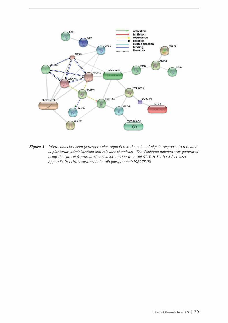
In Appendix 4 a point to point tutorial is given to perform such a data-mining strategy. Two sub-listsof genes were extracted from bioinformatics programs DAVID and GeneDecks; 1 representing genesinvolved in “small molecule metabolic processes” (SMMP list; Appendix 5) and 1 list of genes involvedin “immune processes” (“immune phenotype”), cell-adhesion, and cytoskeletal rearrangements (IAClist; Appendix 6). These lists were separately analysed in DAVID and GeneDecks to i) retrieve relevantpathways/processes, ii) identify the most frequently present transcription factor binding sequence(TFBS) within the loci of the regulated genes, iii) predict which type of cell or tissue supportsexpression of the majority of the regulated genes, and iiii) to find significant associations of theregulated with relevant chemicals. In supplementary Appendix 7 and 8 the results of thesebioinformatics analysis are presented for the SMMP list and IAC list, respectively. In addition, toidentify functional associations and crosstalk between proteins encoded by differential expressedgenes and relevant chemicals, a combined list of SMMP and IAC genes was uploaded in the (protein)-protein-chemical interaction web tool STITCH 3.1 beta. An interactive network was built from 18 genesand 4 chemicals (Figure 1) which all scored associations with a high level of confidence (≥0.7; seeAppendix 9). By clicking on the lines which connects two nodes (genes or chemicals), relevantliterature and information about the type of association (e.g. a physical interaction, or an enzyme-substrate relation, etc.) can be accessed directly in other biological and chemical data-resources(note; only interactive on-line). Four key-genes, displaying a central role in this network (APOC3,NR1H4, CYP3A4, and CYP4F3), were selected together with 3 genes mapped to the “B Cell ReceptorSignaling Pathway” (CR2, APOC3, and SPP1; highlighted in red in Appendix 8). The latter 3 geneswere selected based on the “pIgA+ precursor cell” hypothesis (see above), the prediction that amajority of genes from the IAC list can be expressed by “lymphomaburkitts” cells (B lymphoma celline), and their gene loci contains a TFBS of the transcription factor T (BRACH; Brachyury HomologMouse), a transcription factor highly active in B-lymphocytes (highlighted in red in Appendix 8).CYP4F3A catalyses the omega-hydroxylation of leukotrine B4, a potent chemoattractant forpolymorphonuclear leukocytes. NR1H4 (alias Farnesoid-X-receptor or ileal bile acid transporter) is abile acid-activated transcription factor which regulates the expression of genes involved in bile acidsynthesis and transport. CYP3A4 is a monooxygenase involved in linoleic acid, steroid, and retinolmetabolism. With respect to retinol metabolism, 4 remarkable genes were up-regulated. APOA1,APOA4, APOB, and RBP2, all mapped to the KEGG “Vitamin digestion and absorption” pathway(hsa04977).The proteins encoded by these genes are components of the chylomicrons-mediatedexocytosis mechanism, a mechanism by which enterocytes secrete luminal absorbed fat-soluble
28 | Livestock Research Report 800
vitamins, retinol, and cholesterol esters into the lymphatic system (highlighted in blue in Appendix 7and 8). APOC3 inhibits the uptake of lymph chylomicrons by cells. According to the literature retrievedfrom the STITCH network NR1H4 influences gene expression and function of APOC3(http://www.ncbi.nlm.nih.gov/pubmed/22187655).
The Comparative Toxicogenomics Database (CTD) was explored to find chemicals having potential toinfluence the expression and/or the function of these seven selected key-genes. In Appendix 10, foreach key-gene 3 chemicals with potential were listed (web links provide direct access to literaturedescribing the association between a gene and a chemical). The NR1H4 appeared as a centralgene/protein in the STITCH network. In addition to the predicted role of NR1H4 in regulation of thetransport of fat-soluble vitamins (see above), this nuclear receptor also plays a role in regulating geneexpression in intestinal immune cells (see literature links in Appendix 10). Chemicals like alpha-Linolenic Acid and Epigallocatechingallate (EGCG; a natural phenol with antioxidant properties found inhigh concentrations in green tea), may therefore be promising “candidate additives” to steer geneexpression of NR1H4, and with this, immune processes in the intestine. In addition, EGCG alsomodulates gene expression of NCK1, a key coordinator of cytoskeletal changes in various types ofimmune cells (including B-cells), enabling polarization and directional migration of these cells. At first,such “candidate” additives have to be tested in an in-vitro test. Preferably, a test capable ofmeasuring parameters reflecting the mechanisms predicted by the here performed data-miningstrategy. In case of the Lactobacillus plantarum 299v dataset analyzed here, for example, mixedcultures of enterocyte cell lines and ex-vivo IEL’s and/or B cell fractions may be challenged with thesechemicals, and tested for expression level of the 3 key-genes CR2, APOC3, and SPP1, or other genesfrom the IAC list by quantitative real-time PCR. If the outcomes of such experiments are in agreementwith the processes predicted by data-mining, an in-vivo intervention study, specifically designed forone chemical or a group of related chemicals, may prove whether the “candidate” additives or theiranalogs can replace DFM like Lactobacillus plantarum 299v as an feed-additive that initiates theattraction, migration and homing of (pre)-IgA Ab-secreting cells.
Livestock Research Report 800 | 29
Figure 1 Interactions between genes/proteins regulated in the colon of pigs in response to repeated
L. plantarum administration and relevant chemicals. The displayed network was generated
using the (protein)-protein-chemical interaction web tool STITCH 3.1 beta (see also
Appendix 9; http://www.ncbi.nlm.nih.gov/pubmed/19897548).
30 | Livestock Research Report 800
4 Human and animal models for testingimmune competence
The intestine is at the same time the place for nutrient uptake and the barrier against pathogenicantigens. A direct consequence of these physiological functions is a very complex interplay of differentcell types leading to a tightly controlled mucosal immune balance in the healthy gut [6].Using several animal models, intestinal mucosal immunology has been studied in vivo (Latin for"within the living")[7] mostly in relationship with pathological conditions, such as inflammatory boweldiseases (Crohn's disease and ulcerative colitis)[8]. As a consequence of, among others, theprotection of animals used for scientific purposes and the 3R (reduction, refinement and replacement)approach in animal experimentation, intestinal ex vivo models have been developed and employed. Ex
vivo (Latin for "out of the living") refers to experiments done in or on tissue in an artificialenvironment outside the organism with the minimum alteration of natural conditions, allowingexperimentation under more controlled conditions than possible in in vivo experiments, at the expenseof altering the "natural" environment [7]. Driven by the ask for faster and cheaper systems, besidespathology related models, intestinal in situ, ex vivo and in vitro models have also been used inresearch on absorption [9], bacteria-host interactions [10-13] and food science and nutrition [14-17].In vitro (Latin: in glass) models are those that are conducted using components of an organism thathave been isolated from their usual biological surroundings in order to permit a more detailed or moreconvenient analysis than can be done with whole organisms, and are also known as “test tubeexperiments” or “petri dish experiments” [7]. In situ (Latin: in position) models are intermediatebetween in vivo or ex vivo and in vitro models. For example, examining cells within a whole organintact and under perfusion may be an in situ investigation. This is not in vivo as the donor is sacrificedby the experimentation, but it is not the same as working with the cells alone, a common scenario forin vitro experiments [7].Although not all of the latter models have been used to study intestinal immunology, they may beemployed for such an immunological driven approach. In this chapter these models are described,disregarding in silico modelling (modelling performed on a computer or via computer simulation)[18].
4.1 In vitro models
The intestinal wall is composed of several cell types forming the epithelial barrier (mainly enterocytes)and immune cells on the basolateral side [11, 15]. A perfect in vitro cell model has to be a directsubstitute of the in vivo environment, reflecting both the natural responses as well as the complexphysiology of the intestine. Therefore, cell lines involved in single or co-culture models need to besufficiently characterized. Such a characterization includes epithelial markers (cytokeratins), brushborder (digestive) enzymes, expression of tight junction proteins for the formation of epithelial barrier,integrity and polarity (ZO-1, occludins and claudins, transepithelial resistance (TEER)/transepithelialelectrical potential (TEEP) when grown on a microporous membrane, molecular transporters, internalmetabolism (cytochromes P450-CYPs) and responsiveness to environmental factors like cytokines andinflammatory molecules (lipopolysaccharides (LPS), cytokines). Depending on the model, cells indifferent activation/differentiation state can be used. For example, immature non-activated immunecells are useful in host-bacterial intestinal interaction models, while activated cells can be used tostudy inflammatory intestinal disorders.
4.1.1 Primary cells
Primary cells, isolated from (human or animal) tissue, retain the majority of in vivo functionality [11,15]. They are however rarely used as their survivability is limited and usually need to be derived fromdifferent individuals in subsequent tests affecting the reproducibility of the results.On the other hand, recently commercial sources of primary human intestinal epithelial cells (hlnEpCs)have become available [19].
Livestock Research Report 800 | 31
4.1.2 Epithelial mini-guts
However, recently an in vitro culture system to grow three-dimensional (3D) intestinal epithelialorganoids (‘epithelial mini-guts’) for periods greater than 1.5 year from Lgr5+-CBC cells has beenestablished [20]. Slender crypt base columnar (CBC) cells are present at the crypt bottom,intercalated between post-mitotic Paneth cells. Lgr5+ marked CBC cells persist for the life-time of amouse, whereas their progeny include all differential cell lineages of the epithelium. Lgr5+-CBC cellsrepresent cycling, long-lived, multipotent stem cells. Isolated crypts require Matrigel, a 3D laminin andcollagen-rich matrix that mimics the basal lamina and a cocktail of R-spondin (a protein interactingwith Wnt that constitutes the key pathway to maintain stem cell fate and drive proliferation),epidermal growth factor (EGF) and the polypeptide Noggin is the minimal essential stem cellmaintenance cocktail. In vitro-generated organoids occur as cysts with a central lumen flanked by asimple, highly polarized villus epithelium. The basal side of the cells is orientated toward the outside,touching the matrigel, whereas enterocyte brush borders form the luminal surface. Secretion byPaneth and goblet cells occurs toward the lumen. The organoids can be passaged weekly for at least1.5 years with a phenotype and karyotype that reamin unchanged. To test how normal the epithelialmini-guts are, they were introduced per anum into the colons of mice with chemical-induced mucosallesions. The engrafted epithelial mini-guts regenerated epithelial patches that were indiscernible fromsurrounding recipient epithelium and persisted for at least 6 months without changing theirhistological appearance.
4.1.3 Cell cultures
Most currently used intestinal cell line models are transformed cell lines isolated from cancer affectedindividuals [21, 22]. Among them, Caco-2 is the most widely used and established cell line. Caco-2(colon adenocarcinoma) were isolated from a 72-year old Caucasian man and upon reachingconfluence, cells were found to express characteristics of enterocyte differentiation and functionality.Another human colon adenocarcinoma cell line, HT-29, was isolated from the colon tumour of a 44-year old Caucasian female, while the Intestine-407 cell line has been established via HeLa cellcontamination and CaSki cells were originally isolated from a metastasis in the small intestine. TheHIEC-6 cell line was established from normal human crypt small intestinal cells. The H4 cell line wasestablished from human foetal small intestinal cells and subcloned to obtain a small intestinalepithelial cell line, capable of TEER and TEEP formation.The most widely used rodent lines are IEC-6 and IEC-18 cell lines derived from the rat small intestine[21, 22]. The IPEC-J2 cell line is a non-transformed intestinal cell line derived from jejunal epitheliaisolated from a neonatal piglet, maintained as a continuous culture and subcloned to obtain cultures ofenterocyte-like and mucin producing cell types.
4.1.4 2D Cell cultures
Cells are mostly cultivated as one-dimensional monolayers on plastic surfaces, where theestablishment of a functional, epithelial character is not achieved, possibly resulting in misleadingresults, especially in adhesion assays [11, 15].Growing cells on microporous membranes (e.g. insert filters, Transwell models) results in spontaneouscell differentiation and polarization, enabling basolateral feeding of epithelia similar to the in vivo
setting and facilitating transport studies through the polarised epithelial monolayer [11, 15].Semi-wet interface culture in combination with mechanical stimulation and DAPT causes HT29, MTX-P8, MTX-E12 and LS513 cells to polarize, form functional tight junctions, form a three-dimensionalarchitecture resembling colonic crypts and produce an adherent mucus layer [23]. Caco-2 and T84cells also polarize, form functional tight junctions and produce a thin adherent mucus layer after sucha treatment, but this is less consistent.Newly commercially available primary human intestinal epithelial cells (hlnEpCs) and inducedpluripotent stem cell (iPSC)-derived intestinal cells in Transwell inserts may become an alternativesource of human intestinal cells [19].Epithelial cells growing on microporous membranes can be co-cultured with monocyte/macrophages(derived from the same species) in the lower compartment or on the microporous membrane using aninverted setting [15, 24]. In this way the in vivo environment of the intestine is better stimulated and
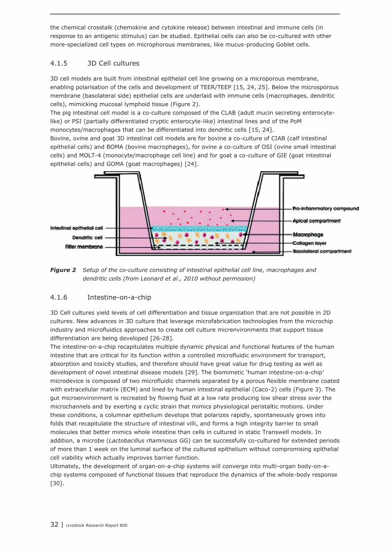
32 | Livestock Research Report 800
the chemical crosstalk (chemokine and cytokine release) between intestinal and immune cells (inresponse to an antigenic stimulus) can be studied. Epithelial cells can also be co-cultured with othermore-specialized cell types on microphorous membranes, like mucus-producing Goblet cells.
4.1.5 3D Cell cultures
3D cell models are built from intestinal epithelail cell line growing on a microporous membrane,enabling polarisation of the cells and development of TEER/TEEP [15, 24, 25]. Below the microsporousmembrane (basolateral side) epithelial cells are underlaid with immune cells (macrophages, dendriticcells), mimicking mucosal lymphoid tissue (Figure 2).The pig intestinal cell model is a co-culture composed of the CLAB (adult mucin secreting enterocyte-like) or PSI (partially differentiated cryptic enterocyte-like) intestinal lines and of the PoMmonocytes/macrophages that can be differentiated into dendritic cells [15, 24].Bovine, ovine and goat 3D intestinal cell models are for bovine a co-culture of CIAB (calf intestinalepithelial cells) and BOMA (bovine macrophages), for ovine a co-culture of OSI (ovine small intestinalcells) and MOLT-4 (monocyte/macrophage cell line) and for goat a co-culture of GIE (goat intestinalepithelial cells) and GOMA (goat macrophages) [24].
Figure 2 Setup of the co-culture consisting of intestinal epithelial cell line, macrophages and
dendritic cells (from Leonard et al., 2010 without permission)
4.1.6 Intestine-on-a-chip
3D Cell cultures yield levels of cell differentiation and tissue organization that are not possible in 2Dcultures. New advances in 3D culture that leverage microfabrication technologies from the microchipindustry and microfluidics approaches to create cell culture micrenvironments that support tissuedifferentiation are being developed [26-28].The intestine-on-a-chip recapitulates multiple dynamic physical and functional features of the humanintestine that are critical for its function within a controlled microfluidic environment for transport,absorption and toxicity studies, and therefore should have great value for drug testing as well asdevelopment of novel intestinal disease models [29]. The biomimetic ‘human intestine-on-a-chip’microdevice is composed of two microfluidic channels separated by a porous flexible membrane coatedwith extracellular matrix (ECM) and lined by human intestinal epithelial (Caco-2) cells (Figure 3). Thegut microenvironment is recreated by flowing fluid at a low rate producing low shear stress over themicrochannels and by exerting a cyclic strain that mimics physiological peristaltic motions. Underthese conditions, a columnar epithelium develops that polarizes rapidly, spontaneously grows intofolds that recapitulate the structure of intestinal villi, and forms a high integrity barrier to smallmolecules that better mimics whole intestine than cells in cultured in static Transwell models. Inaddition, a microbe (Lactobacillus rhamnosus GG) can be successfully co-cultured for extended periodsof more than 1 week on the luminal surface of the cultured epithelium without compromising epithelialcell viability which actually improves barrier function.Ultimately, the development of organ-on-a-chip systems will converge into multi-organ body-on-a-chip systems composed of functional tissues that reproduce the dynamics of the whole-body response[30].
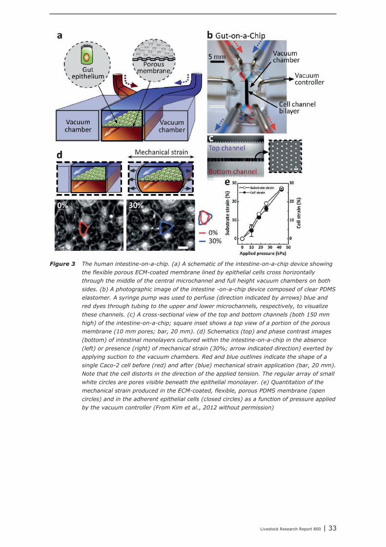
Livestock Research Report 800 | 33
Figure 3 The human intestine-on-a-chip. (a) A schematic of the intestine-on-a-chip device showing
the flexible porous ECM-coated membrane lined by epithelial cells cross horizontally
through the middle of the central microchannel and full height vacuum chambers on both
sides. (b) A photographic image of the intestine -on-a-chip device composed of clear PDMS
elastomer. A syringe pump was used to perfuse (direction indicated by arrows) blue and
red dyes through tubing to the upper and lower microchannels, respectively, to visualize
these channels. (c) A cross-sectional view of the top and bottom channels (both 150 mm
high) of the intestine-on-a-chip; square inset shows a top view of a portion of the porous
membrane (10 mm pores; bar, 20 mm). (d) Schematics (top) and phase contrast images
(bottom) of intestinal monolayers cultured within the intestine-on-a-chip in the absence
(left) or presence (right) of mechanical strain (30%; arrow indicated direction) exerted by
applying suction to the vacuum chambers. Red and blue outlines indicate the shape of a
single Caco-2 cell before (red) and after (blue) mechanical strain application (bar, 20 mm).
Note that the cell distorts in the direction of the applied tension. The regular array of small
white circles are pores visible beneath the epithelial monolayer. (e) Quantitation of the
mechanical strain produced in the ECM-coated, flexible, porous PDMS membrane (open
circles) and in the adherent epithelial cells (closed circles) as a function of pressure applied
by the vacuum controller (From Kim et al., 2012 without permission)
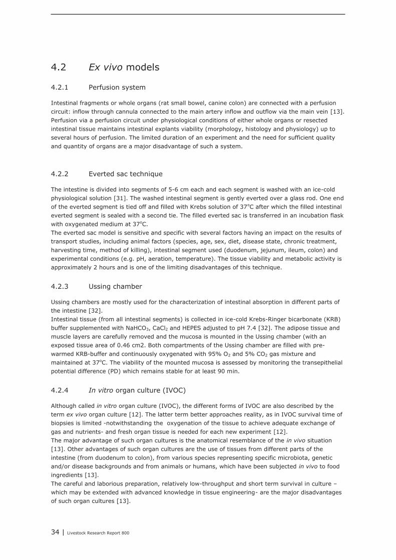
34 | Livestock Research Report 800
4.2 Ex vivo models
4.2.1 Perfusion system
Intestinal fragments or whole organs (rat small bowel, canine colon) are connected with a perfusioncircuit: inflow through cannula connected to the main artery inflow and outflow via the main vein [13].Perfusion via a perfusion circuit under physiological conditions of either whole organs or resectedintestinal tissue maintains intestinal explants viability (morphology, histology and physiology) up toseveral hours of perfusion. The limited duration of an experiment and the need for sufficient qualityand quantity of organs are a major disadvantage of such a system.
4.2.2 Everted sac technique
The intestine is divided into segments of 5-6 cm each and each segment is washed with an ice-coldphysiological solution [31]. The washed intestinal segment is gently everted over a glass rod. One endof the everted segment is tied off and filled with Krebs solution of 37oC after which the filled intestinaleverted segment is sealed with a second tie. The filled everted sac is transferred in an incubation flaskwith oxygenated medium at 37oC.The everted sac model is sensitive and specific with several factors having an impact on the results oftransport studies, including animal factors (species, age, sex, diet, disease state, chronic treatment,harvesting time, method of killing), intestinal segment used (duodenum, jejunum, ileum, colon) andexperimental conditions (e.g. pH, aeration, temperature). The tissue viability and metabolic activity isapproximately 2 hours and is one of the limiting disadvantages of this technique.
4.2.3 Ussing chamber
Ussing chambers are mostly used for the characterization of intestinal absorption in different parts ofthe intestine [32].Intestinal tissue (from all intestinal segments) is collected in ice-cold Krebs-Ringer bicarbonate (KRB)buffer supplemented with NaHCO3, CaCl2 and HEPES adjusted to pH 7.4 [32]. The adipose tissue andmuscle layers are carefully removed and the mucosa is mounted in the Ussing chamber (with anexposed tissue area of 0.46 cm2. Both compartments of the Ussing chamber are filled with pre-warmed KRB-buffer and continuously oxygenated with 95% O2 and 5% CO2 gas mixture andmaintained at 37oC. The viability of the mounted mucosa is assessed by monitoring the transepithelialpotential difference (PD) which remains stable for at least 90 min.
4.2.4 In vitro organ culture (IVOC)
Although called in vitro organ culture (IVOC), the different forms of IVOC are also described by theterm ex vivo organ culture [12]. The latter term better approaches reality, as in IVOC survival time ofbiopsies is limited -notwithstanding the oxygenation of the tissue to achieve adequate exchange ofgas and nutrients- and fresh organ tissue is needed for each new experiment [12].The major advantage of such organ cultures is the anatomical resemblance of the in vivo situation[13]. Other advantages of such organ cultures are the use of tissues from different parts of theintestine (from duodenum to colon), from various species representing specific microbiota, geneticand/or disease backgrounds and from animals or humans, which have been subjected in vivo to foodingredients [13].The careful and laborious preparation, relatively low-throughput and short term survival in culture –which may be extended with advanced knowledge in tissue engineering- are the major disadvantagesof such organ cultures [13].
Livestock Research Report 800 | 35
4.2.5 Non-polarized (or traditional) IVOC
Intestinal biopsies (obtained from patients receiving upper endoscopy, ileo-colonoscopy or surgicalintestinal resection) are placed on a thick supporting sponge or agarose gel base (with the apical sideupward and the serosal side downward in direct contact with the underlying sponge) in IVOC mediumto create a thin film of medium on the apical sides of biopsies [12](Figure 4A). Bacterial overnightcultures are inoculated on the mucosal side of the biopsies and incubated at 37oC on a rocker in anatmosphere of 95% O2 and 5% CO2 with regular replacement of medium to keep the biopsies viable.At harvest, the biopsies were removed and shaken in medium to remove mucus and bacteria.Disadvantages of non/polarized IVOC comprise the inability to apply different conditions to either sideof the biopsies [12]. Furthermore, no interactions with deep tissue, different severity of traumacaused by biopsy sampling procedure, diverse underlying diseases of the patients and individualvariations in the gut flora associating with the biopsy tissues, are inevitable weaknesses of this model.
Figure 4 Mounting of intestinal biopsies for traditional in vitro organ culture (IVOC) and polarized
IVOC. (A) A biopsy specimen mounted on a sponge or an agarose plug immersed in the
medium with its mucosal surface upward. (B) After orientation of the tissue on a
nitrocellulose filter, the apical disc was fixed to the mucosal side of the biopsy via tissue
glue. The sandwich was then inserted between a Snapwell support (from Fang et al., 2013
without permission)
4.2.6 Polarized in vitro organ culture (pIVOC)
In the pIVOC each biopsy is positioned as a sandwich between two acrylic glass discs with a centralaperture slightly smaller than the size of the biopsy [12](Figure 4B). The upper disc is sealed to theperipheral area of the mucosal side of the biopsy to minimize leakage from the apical to the basalcompartment. The sandwich holding the biopsy is then mounted in a Snapwell insert. After transferinto a six/well plate, the explant tissue is submerged in medium and incubated at 37oC on a rocker inan atmosphere of 95% O2 and 5% CO2 with regular replacement of medium to keep the biopsiesviable. At harvest, the biopsies were removed and shaken in medium to remove mucus and bacteria.
4.2.7 Novel polarized ex vivo organ culture
Samples are excised from the intestines of patients at the time of surgery and directly transferred inHBSS buffer supplemented with bacteriostatic antibiotics at 4oC to the laboratory [33, 34]. The cleanmucosal layer is washed in Hank's buffer and cut with sterile scalpels into 1 cm2 pieces. The pieces areplaced on sterile metal grids with the basolateral side facing downwards. and the cylinder (cloning
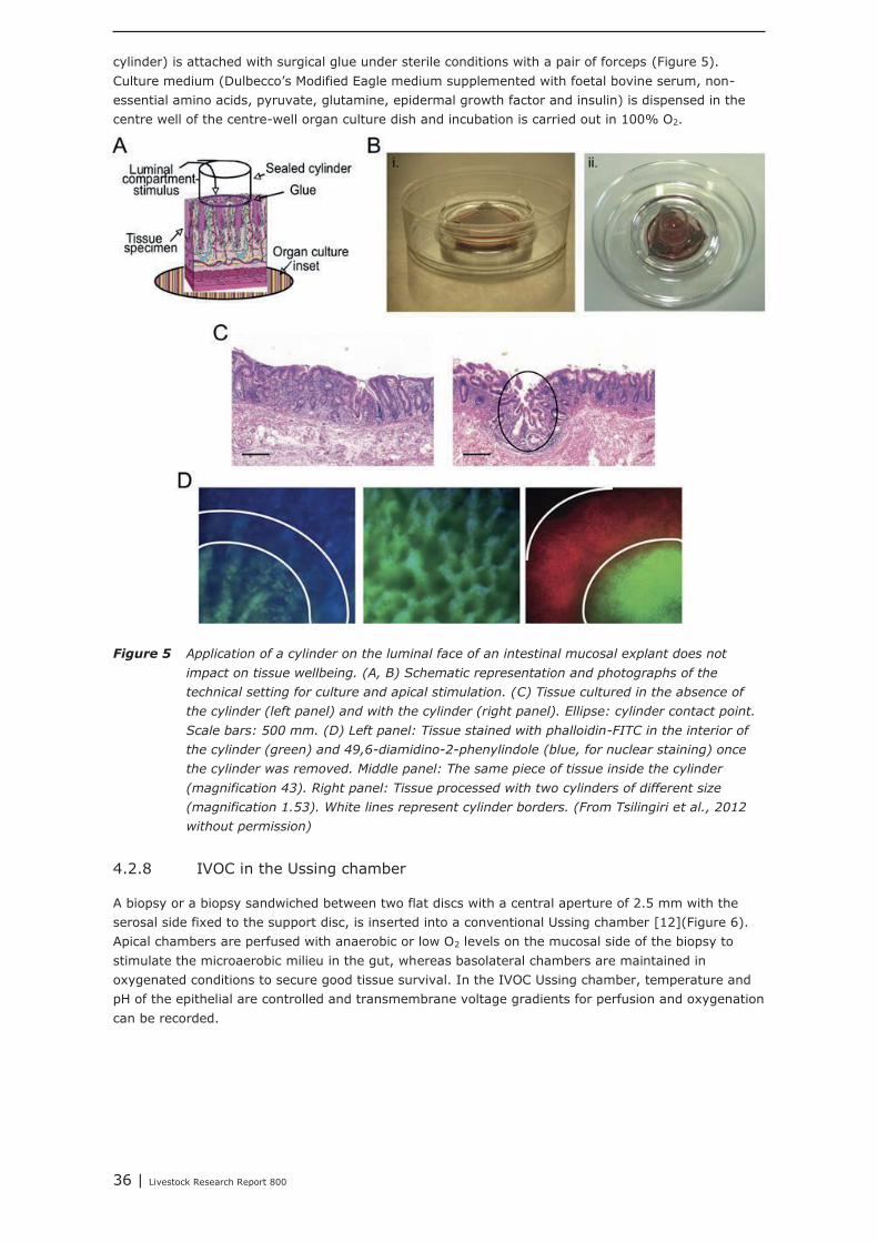
36 | Livestock Research Report 800
cylinder) is attached with surgical glue under sterile conditions with a pair of forceps (Figure 5).Culture medium (Dulbecco's Modified Eagle medium supplemented with foetal bovine serum, non-essential amino acids, pyruvate, glutamine, epidermal growth factor and insulin) is dispensed in thecentre well of the centre-well organ culture dish and incubation is carried out in 100% O2.
Figure 5 Application of a cylinder on the luminal face of an intestinal mucosal explant does not
impact on tissue wellbeing. (A, B) Schematic representation and photographs of the
technical setting for culture and apical stimulation. (C) Tissue cultured in the absence of
the cylinder (left panel) and with the cylinder (right panel). Ellipse: cylinder contact point.
Scale bars: 500 mm. (D) Left panel: Tissue stained with phalloidin-FITC in the interior of
the cylinder (green) and 49,6-diamidino-2-phenylindole (blue, for nuclear staining) once
the cylinder was removed. Middle panel: The same piece of tissue inside the cylinder
(magnification 43). Right panel: Tissue processed with two cylinders of different size
(magnification 1.53). White lines represent cylinder borders. (From Tsilingiri et al., 2012
without permission)
4.2.8 IVOC in the Ussing chamber
A biopsy or a biopsy sandwiched between two flat discs with a central aperture of 2.5 mm with theserosal side fixed to the support disc, is inserted into a conventional Ussing chamber [12](Figure 6).Apical chambers are perfused with anaerobic or low O2 levels on the mucosal side of the biopsy tostimulate the microaerobic milieu in the gut, whereas basolateral chambers are maintained inoxygenated conditions to secure good tissue survival. In the IVOC Ussing chamber, temperature andpH of the epithelial are controlled and transmembrane voltage gradients for perfusion and oxygenationcan be recorded.
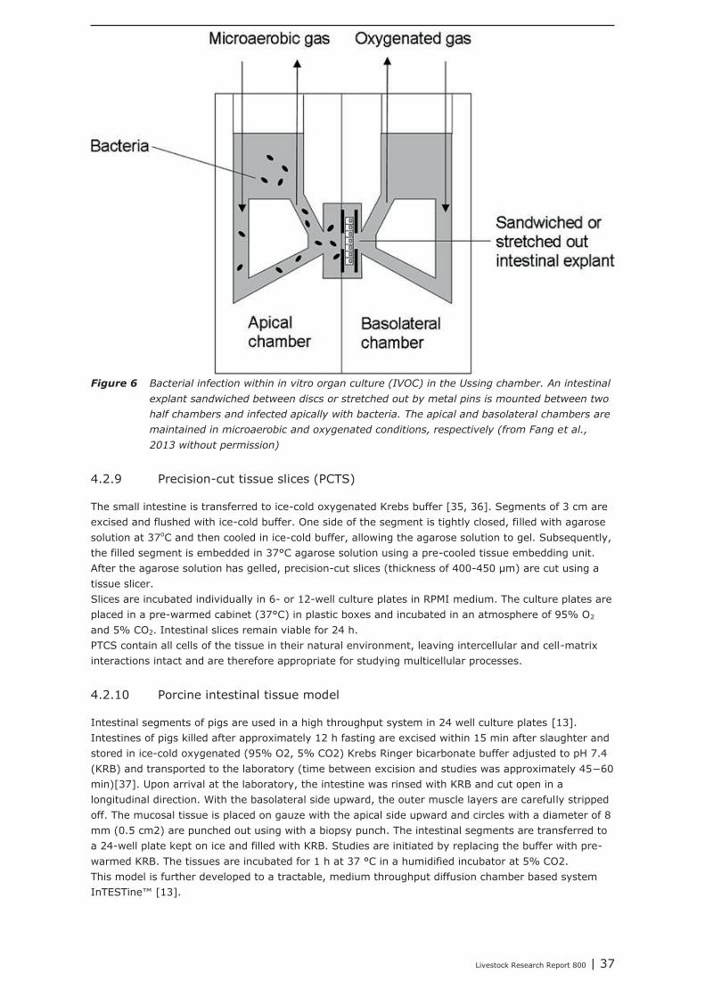
Livestock Research Report 800 | 37
Figure 6 Bacterial infection within in vitro organ culture (IVOC) in the Ussing chamber. An intestinal
explant sandwiched between discs or stretched out by metal pins is mounted between two
half chambers and infected apically with bacteria. The apical and basolateral chambers are
maintained in microaerobic and oxygenated conditions, respectively (from Fang et al.,
2013 without permission)
4.2.9 Precision-cut tissue slices (PCTS)
The small intestine is transferred to ice-cold oxygenated Krebs buffer [35, 36]. Segments of 3 cm areexcised and flushed with ice-cold buffer. One side of the segment is tightly closed, filled with agarosesolution at 37oC and then cooled in ice-cold buffer, allowing the agarose solution to gel. Subsequently,the filled segment is embedded in 37°C agarose solution using a pre-cooled tissue embedding unit.After the agarose solution has gelled, precision-cut slices (thickness of 400-450 μm) are cut using atissue slicer.Slices are incubated individually in 6- or 12-well culture plates in RPMI medium. The culture plates areplaced in a pre-warmed cabinet (37°C) in plastic boxes and incubated in an atmosphere of 95% O2
and 5% CO2. Intestinal slices remain viable for 24 h.PTCS contain all cells of the tissue in their natural environment, leaving intercellular and cell-matrixinteractions intact and are therefore appropriate for studying multicellular processes.
4.2.10 Porcine intestinal tissue model
Intestinal segments of pigs are used in a high throughput system in 24 well culture plates [13].Intestines of pigs killed after approximately 12 h fasting are excised within 15 min after slaughter andstored in ice-cold oxygenated (95% O2, 5% CO2) Krebs Ringer bicarbonate buffer adjusted to pH 7.4(KRB) and transported to the laboratory (time between excision and studies was approximately 45−60min)[37]. Upon arrival at the laboratory, the intestine was rinsed with KRB and cut open in alongitudinal direction. With the basolateral side upward, the outer muscle layers are carefully strippedoff. The mucosal tissue is placed on gauze with the apical side upward and circles with a diameter of 8mm (0.5 cm2) are punched out using with a biopsy punch. The intestinal segments are transferred toa 24-well plate kept on ice and filled with KRB. Studies are initiated by replacing the buffer with pre-warmed KRB. The tissues are incubated for 1 h at 37 °C in a humidified incubator at 5% CO2.This model is further developed to a tractable, medium throughput diffusion chamber based systemInTESTine™ [13].
38 | Livestock Research Report 800
4.3 In situ models
4.3.1 Intestinal loop model
In the intestinal loop model, the animal (typically rat, rabbit or mouse) is fasted for approximately 3days and anaesthetised for the entirety of the surgical procedure [9]. An incision is made in themidline of the abdomen to expose the small intestine. The desired intestinal segments are sliced, heldwith a clamp, and then washed to remove excess food particles while keeping the intestine attachedand in close proximity to the body cavity. The segment is then ligated using suture to form a loop,assuring normal blood flow and muscle movement to the looped segment. An alternative way toperform the initial preparation is to make an incision at the desired location and then ligating that end.To determine particle transport across the intestine, the particle suspension is injected directly into theloop, and the intestinal loop is returned to the body cavity of the animal for up to 2 h while theanimals are maintained under anaesthesia. After a chosen time period, the animal is sacrificed and theintestinal loop is removed from the body cavity of the animal. A majority of the time, the closed loopintestine is returned to the body cavity for 2 h and then the entire intestinal segment is removed.Another way to use the closed loop method is to inject a solution into the closed loop, remove thesuturing after 2 h, and return the intact intestine to the body, allowing the animal to resume normaldigestive processes and movement. After no more than 24 h, the animal is killed and the intestinesremoved for analysis.
4.3.2 Small intestinal segment perfusion (SISP)
After overnight fasting, a piglet is anaesthetized and in the small intestine maximal 10 segments ofapproximately 20 cm each are prepared, each with a cranial inflow tube and a caudal outflow tube[38]. The segments are either paired (in one infected and one non-infected) or not, and perfused forup to 8 hours with, in the case of paired segments, up to 5 different and, in the case of non-pairedsegments, up to 10 different perfusion fluids. For each segment over 8 h net fluid absorption isdetermined and from each segment mucosal samples (after 8 hours or at different time points afterthe start of perfusion) can be collected to analyse genome responses of early pathogen-inducedinflammation [39].
4.4 In vivo models
For pathological human conditions, such as Crohn's disease and ulcerative colitis, different animalmodels using less complex organisms (roundworm, fruit fly, fish) or rodents (mouse, rat, guinea-pig)have been developed including spontaneous models, chemically-induced models, geneticallyengineered models (transgenic animals), genetically engineered (knockout mice) and T cell transfermodels [6, 8, 40, 41]. In such models blood samples can be taken and by endoscopy or at dissectionintestinal tissue samples. For studying the in vivo functions of human cells and tissues and the humanimmune system, ‘humanized' mouse strains have been developed based on immunodeficient micewith mutations in the interleukin-2 receptor common γ-chain locus [42].Also in the pig a model for chemically-induced colitis has been described [43-46]. Pigs can be fittedwith catheters allowing frequent blood sampling and intestinal cannulas for taking intestinal biopsiesmore frequently than just at dissection [47].
4.5 Correlations between models and real practice inhumans and monogastrics
Advantages and limitations of most models have been indicated. Assessing mucosal immunity [48]has not been implied in all models described, but in principal from all models tissue and/or fluidsamples can be taken which may be used for immunological analysis.The correlation between a model and the ‘real practice' of humans or monogastrics, depends on the(scientific) question to be answered and the model used. Different in vitro, in situ or ex vivo models,for instance, not always indicate the same:
Livestock Research Report 800 | 39
- In vitro, Lactobacillus gasseri was found to be effective in reducing Escherichia coli adhesion to Caco-2 cells, but in ex vivo conditions no exclusion or displacement of Escherichia coli by Lactobacillus
gasseri was observed on jejunal piglet tissue [49].Furthermore, both studies showing a good correlation and studies showing a bad or no correlationbetween a model and the ‘real practice’ of monogastrics have been published. Three examples of bothkind of results, in which the in vitro, in situ or ex vivo experiments are reported in the same source asthe in vivo results, are:- In vitro, in the human small intestinal epithelial Caco-2 cell model, the supernatants of dietscontaining either pre-fermented cereals or their fermentation end-products (short chain fatty acids)clearly modulated cellular growth, metabolism, differentiation and mucosal integrity [50]. Such effectswere not observed in the same study for in vivo intestinal characteristics (morphology, mitoticactivity) and performance in weanling pigs [50], although in other in vivo experiments positive effectsof fermented wheat have been reported [51].- In vitro, tempeh, a fermented soya bean, interfered with adhesion of enterotoxigenic Escherichia coli
(ETEC), in situ tempeh reduced ETEC-induced fluid loss in piglet small intestinal segments and in vivo
severity of diarrhoea was less, feed intake increased and daily weight gain increased in ETECchallenged piglets [52].- In vitro, a porcine Lactobacillus amylovorus-like strain (pLAI) protected the intestinal permeability ofporcine IPEC-1 cells from the perturbation induced by ETEC and in vivo, in piglets challenged withETEC, pLAI increased daily weight gain and tended to increase ETEC shedding and totalimmunoglobulin A (IgA) in saliva and serum [53].Also advancing insight can affect the way models are looked at. In modern biomedical research,mouse models are often used to explore basic pathophysiological mechanisms, evaluate newtherapeutic approaches and make go or no-go decisions to carry new drug candidates forward intoclinical trials [54]. But while in humans acute inflammatory stresses from different aetiologies result inhighly similar genomic responses, the responses in corresponding mouse models correlate poorly withthe human conditions and also, one another. Among genes changed significantly in humans, themurine orthologs are close to random in matching their human counterparts, questioning the use ofmouse models to study human inflammatory diseases [54].
A systematic review, focusing on a single question which tries to identify, appraise, select andsynthesize all high quality research evidence relevant to that question, as recently introduced foranimal models [55], may be employed (and may be technically supported by ZonMw).
40 | Livestock Research Report 800
5 Nutritional intervention strategies formonogastrics
This chapter aims to review the nutritional interventions that directly or indirectly (via the microbiota)engage the optimal development of the innate and acquired immune system, both in the maternal(par. 4.1), neonatal (par. 4.2) and post-neonatal phase (par. 4.3).
The strategy to search the relevant literature was as follows; the input to the query were 1) general orspecific nutritional factors; 2) immune competence related words, i.e. intestinal/gut health, immunesystem, or immunity; 3) species (e.g. pig, Sus scrofa or chicken, broiler, Gallus gallus); 4) ‘maternal’was used as a key word for searching to studies related to interventions during the maternal stage. Itshould be noted that the data presented here are incomplete, however it gives an overview ofliterature describing nutritional interventions in neonatal livestock that may affect performance and/orimmune related processes.
5.1 Effects of interventions during the maternal stage onthe immune competence of the progeny
5.1.1 Trans-generational effects
Environmental conditions that are experienced in early life can profoundly influence biologyand long-term health. Early-life nutrition is a well-documented example of suchconditions because they influence the adult risk of developing metabolic diseases. A human Swedishstudy showed that diabetes mortality increased in men if the paternal grandfather was exposed toabundant nutrition during his pre-pubertal growth period, an effect later extended to paternalgrandmother/granddaughter pairs and transmitted in a gender-specific fashion. Poor maternalnutrition has also been associated with increased risk of type 2 diabetes mellitus over severalgenerations in North American Indians, and individuals whose grandparents were in utero during theDutch Hunger Winter had lower birth weight [56]. The above mentioned examples show thatmalnutrition during pregnancy exerts negative effects in the offspring. In this study, however, we areinterested in dietary interventions that particularly improve the immune competence of the progeny.
5.1.2 Trans-generational transfer of immune competence
Below, the mode of action of dietary trans-generational effects on the immune competence of pigletsand broilers is described.
In mammals
According to Salmon et al. [57], immunoglobulins cannot cross the placenta in pregnant sows.Neonatal pigs are therefore agammaglobulinemic at birth. Therefore, they cannot provide rapidimmune responses at systemic and mucosal sites. Their survival depends directly on the acquisition ofmaternal immunity via colostrum and milk. Protection by maternal immunity is mediated by a numberof factors, including specific systemic humoral immunity, involving mostly maternal IgG transferredfrom blood to colostrum and typically absorbed within the first 36 h of life. Passive mucosal immunityinvolves local humoral immunity, including the production of secretary IgA (slgA) by the mammarygland (MG), which is transferred principally via milk until weaning. These antibodies are produced inresponse to intestinal and respiratory antigens, like pathogens and commensal organisms. Protectionis also mediated by cellular immunity, which is transferred via maternal cells present in mammarysecretions. The hormones addressins and chemokines are involved in the regulation of the cellularimmunity. The enhancement of colostrogenic immunity depends on the stimulation of systemicimmunity, whereas the enhancement of lactogenic immunity depends on appropriate stimulation atinduction sites, an increase in cell trafficking from the gut and upper respiratory tract to the MG and,possibly, enhanced immunoglobulin production at the effector site and secretion in milk. In addition,
Livestock Research Report 800 | 41
mammary secretions provide factors other than immunoglobulins that protect the neonate andregulate the development of mucosal immunity, which is a key element of postnatal adaptation toenvironmental antigens [57].
In birds
The egg is the main source of maternal antibodies of broilers. Although maternal antibodies are onlypresent in broilers during the first 2 wk of life, they may exert a lifelong determinative influence on theneonatal immune system [58]. Maternal antibodies have strong immuno-regulatory properties whichaffect the specific neonatal B and T cell binding repertoire. Maternal antibodies, therefore, can beconsidered as non-genetic, information-carrying molecules that transfer knowledge about theimmunologically relevant environment gathered by the mother to her offspring. One of theseantibodies, avidin, was expected to guide the gut microbiota composition [1]. It is bacteriostatic andinhibits pathogenic bacterial growth. Moreover, parental natural antibodies might be transferred viathe egg shell to the young chicken, thereby preventing the vertical transmission of pathogens. Finally,evidence for heritability of natural and specific antibody responses was found [59].
5.1.3 Impact of nutritional interventions in the maternal diets on trans-generational epigenetic effects
It has been demonstrated that maternal dietary interventions could interfere with different parametersin the offspring, as shown in Table 1. The mode of action, however, differs between interventionstrategies. Addition of the probiotic Saccharomyces cerevisiae to sow diets was found to increase theIgA concentration in serum of their piglets [60], indicating that this intervention affected the amountof antibodies. Fish oil supplementation in mice reduced the levels of total OVA-specific antibodies andOVA-IgG1 titers in the pup blood [61].Maternal consumption of seaweed extract, fructo oligosaccharides or zinc resulted in increasedcytokine levels in the ileal piglet digesta, in human breast milk, and in 6-month old babies respectively[62-64], whereas maternal consumption of β-carotene reduced cytokine levels in 6-month old babies.Dietary supplementation with probiotic (Bacillus cereus var. Toyoi) or with additional vitamins andtrace minerals increased the number of T cells and Leukocytes in piglets and broilers, respectively[65, 66]. From a study conducted in rats, it could be concluded that lymphocyte proliferation, whichwas impaired at birth after prenatal exposure to a low protein diet, could be overcome in postnatalrats if 50% or 90% of corn oil in the prenatal diet was replaced by coconut oil [67]. In another ratstudy it was shown that a reduced maternal selenium intake of rats reduced the neonatal seleniumstatus which in turn hampers the neonatal immune system development [68]. Replacing sunflower oilby fish oil in broiler breeder diets resulted in the offspring in an increased production of leukotriene B5by thrombocytes, indicating that modulating maternal dietary n-6 and n-3 fatty acid could lead to lessinflammatory-related disorders in poultry [69-71].Some dietary interventions in the maternal diet (e.g. addition of seaweed extract or vitamins/traceminerals) resulted in an improved gut integrity of the progeny [63, 65]. Addition of probiotics (e.g.Bacillus cereus var. Toyoi, Saccharomyces cerevisiae) to sow diets reduced the diarrhea scores of theirpiglets [60, 66], whereas maternal zinc supplementation would be an effective way to reducediarrhoeal disease in babies [64].Supplementation of Lactobacillus rhamnosus and Bifidobacterium lactis to pregnant women increasedthe adiponectin content of their colostrum [72].Until now, transcriptomics data is scarce, but addition of addition vitamins/trace minerals to broilerbreeder diets was found to increase the expression of genes that are related to intestinal turnover,proliferation and development, metabolism and feed absorption in the offspring [73].The improved immune response of the offspring was not always related to improved performance ofthe progeny. Maternal supplementation with seaweed extract, however, resulted in improved bodyweight gain of the piglets, and this effect maintained even till the growing finishing phase [63].Growth depression in broilers that passed through a malabsorption syndrome infection was less if theirmothers received additional vitamins/trace minerals [65]. More details regarding the investigatedstudies are provided in appendix 14.
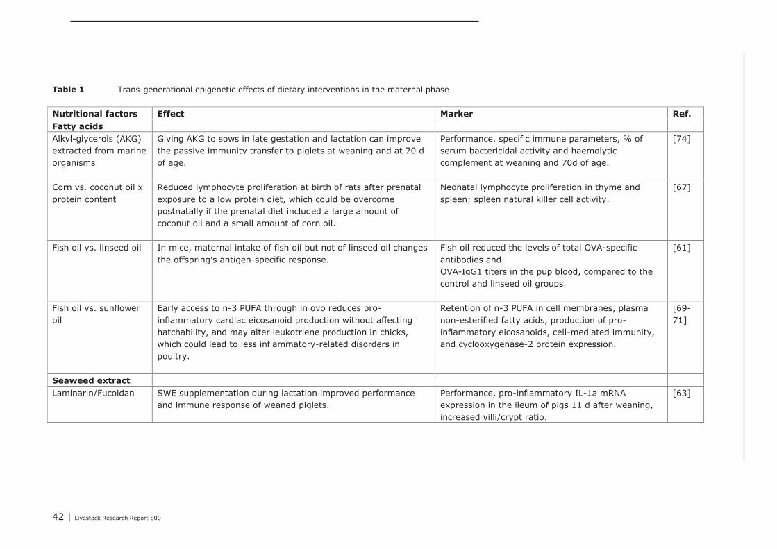
42 | Livestock Research Report 800
Table 1 Trans-generational epigenetic effects of dietary interventions in the maternal phase
Nutritional factors Effect Marker Ref.Fatty acidsAlkyl-glycerols (AKG)extracted from marineorganisms
Giving AKG to sows in late gestation and lactation can improvethe passive immunity transfer to piglets at weaning and at 70 dof age.
Performance, specific immune parameters, % ofserum bactericidal activity and haemolyticcomplement at weaning and 70d of age.
[74]
Corn vs. coconut oil xprotein content
Reduced lymphocyte proliferation at birth of rats after prenatalexposure to a low protein diet, which could be overcomepostnatally if the prenatal diet included a large amount ofcoconut oil and a small amount of corn oil.
Neonatal lymphocyte proliferation in thyme andspleen; spleen natural killer cell activity.
[67]
Fish oil vs. linseed oil In mice, maternal intake of fish oil but not of linseed oil changesthe offspring’s antigen-specific response.
Fish oil reduced the levels of total OVA-specificantibodies andOVA-IgG1 titers in the pup blood, compared to thecontrol and linseed oil groups.
[61]
Fish oil vs. sunfloweroil
Early access to n-3 PUFA through in ovo reduces pro-inflammatory cardiac eicosanoid production without affectinghatchability, and may alter leukotriene production in chicks,which could lead to less inflammatory-related disorders inpoultry.
Retention of n-3 PUFA in cell membranes, plasmanon-esterified fatty acids, production of pro-inflammatory eicosanoids, cell-mediated immunity,and cyclooxygenase-2 protein expression.
[69-71]
Seaweed extractLaminarin/Fucoidan SWE supplementation during lactation improved performance
and immune response of weaned piglets.Performance, pro-inflammatory IL-1a mRNAexpression in the ileum of pigs 11 d after weaning,increased villi/crypt ratio.
[63]
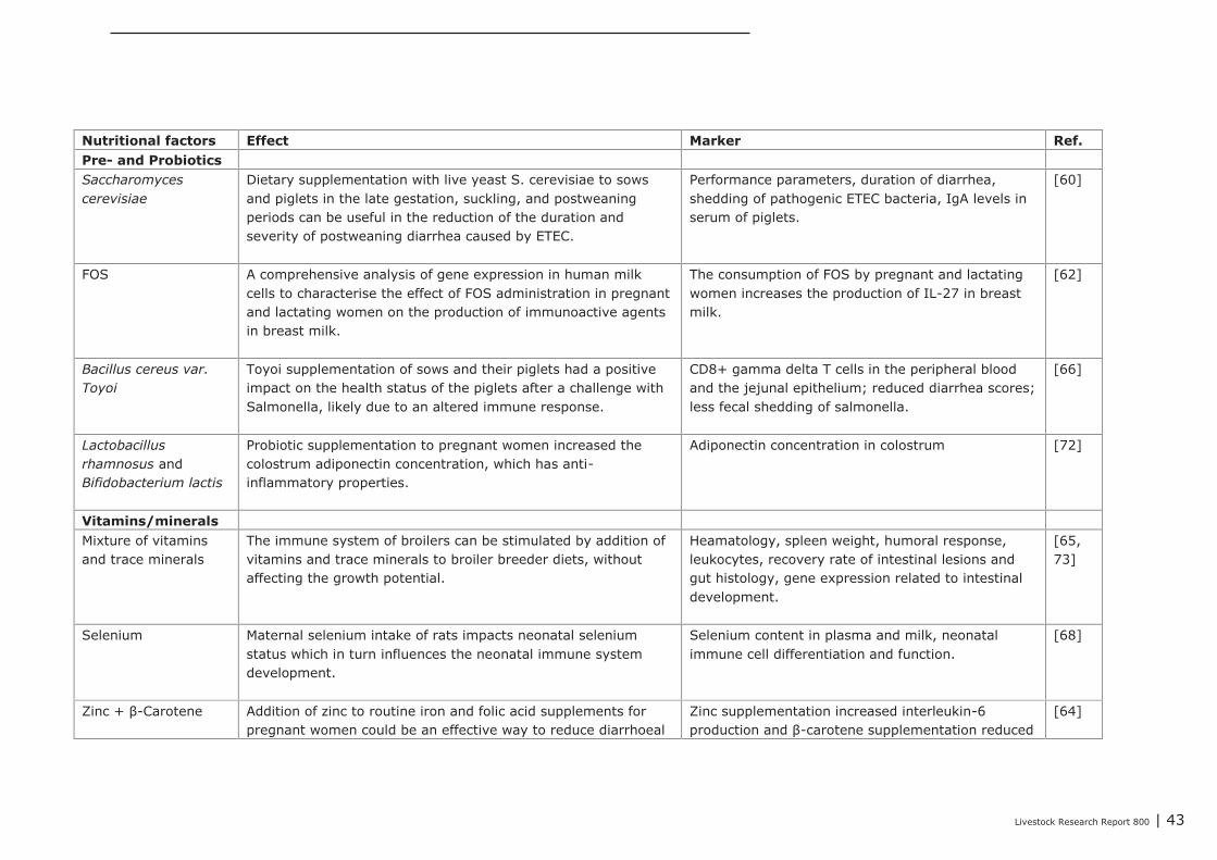
Livestock Research Report 800 | 43
Nutritional factors Effect Marker Ref.Pre- and ProbioticsSaccharomycescerevisiae
Dietary supplementation with live yeast S. cerevisiae to sowsand piglets in the late gestation, suckling, and postweaningperiods can be useful in the reduction of the duration andseverity of postweaning diarrhea caused by ETEC.
Performance parameters, duration of diarrhea,shedding of pathogenic ETEC bacteria, IgA levels inserum of piglets.
[60]
FOS A comprehensive analysis of gene expression in human milkcells to characterise the effect of FOS administration in pregnantand lactating women on the production of immunoactive agentsin breast milk.
The consumption of FOS by pregnant and lactatingwomen increases the production of IL-27 in breastmilk.
[62]
Bacillus cereus var.Toyoi
Toyoi supplementation of sows and their piglets had a positiveimpact on the health status of the piglets after a challenge withSalmonella, likely due to an altered immune response.
CD8+ gamma delta T cells in the peripheral bloodand the jejunal epithelium; reduced diarrhea scores;less fecal shedding of salmonella.
[66]
Lactobacillusrhamnosus andBifidobacterium lactis
Probiotic supplementation to pregnant women increased thecolostrum adiponectin concentration, which has anti-inflammatory properties.
Adiponectin concentration in colostrum [72]
Vitamins/mineralsMixture of vitaminsand trace minerals
The immune system of broilers can be stimulated by addition ofvitamins and trace minerals to broiler breeder diets, withoutaffecting the growth potential.
Heamatology, spleen weight, humoral response,leukocytes, recovery rate of intestinal lesions andgut histology, gene expression related to intestinaldevelopment.
[65,73]
Selenium Maternal selenium intake of rats impacts neonatal seleniumstatus which in turn influences the neonatal immune systemdevelopment.
Selenium content in plasma and milk, neonatalimmune cell differentiation and function.
[68]
Zinc + β-Carotene Addition of zinc to routine iron and folic acid supplements forpregnant women could be an effective way to reduce diarrhoeal
Zinc supplementation increased interleukin-6production and β-carotene supplementation reduced
[64]

44 | Livestock Research Report 800
disease interferon-g production.
45 | Livestock Research Report 800
5.1.4 Conclusions
The most important conclusions were: Trans-generational effects of maternal dietary interventions on improvement of gut health and
immune competence has been demonstrated in mammals (mice, rats, pigs, humans) as well as inpoultry.
The reviewed dietary interventions can be categorized to fatty acids, seaweed extract, pre- andprobiotics, and vitamins and minerals.
The dietary interventions showed a variety in modes of action. They could affect the compositionof the microbiota, the numbers or activity of specific immune cells, the gut morphology, and theexpression of genes involved in immune response.
In some studies, it was shown that the maternal dietary interventions resulted in improved animalperformance or reduced incidence of diarrhea of the offspring.
Only a few studies reported trans-generational effects on gene expression. These studiesinvestigated the effects of supplemented omega-3 fatty acids or vitamins and minerals.
Supplementation of maternal diets with specific fatty acids, in particular with fish oil, showed tohave a wide impact on the immune competence of the progeny. Therefore, modulating the dietaryfatty acid profile of maternal diets seems to be the most promising intervention for improvingimmune competence of the progeny.
5.2 Impact of nutritional interventions in the neonatalphase on immune related parameters
5.2.1 Results from literature
Neonatal development is defined as; day 0 to weaning for pigs and day 0 to 14 for broilers, thus theearly development after birth/hatch. Which is hallmarked by the development and differentiation ofthe intestine. Morphological, functional and immunological development occur simultaneously, and canbe influenced by the host (genetics), microbiota, environment (feed, antibiotics, etc.). For both pigs(Table 2) and poultry (Table 3) we have identified a number of macronutrients and micronutrients, aswell as microbiota that influence the performance and/or intestinal immune status of the host.Macronutrients can be categorized into carbohydrates (sugars / saccharides), fats (vegetable, animal,etc.), and proteins (polymeric chains of amino acids). Micronutrients are categorized into vitamins(e.g. A, B, C, and E) and minerals (e.g. zinc, copper, selenium, and iodine). The microbiota is moredifficult to categorize because of the multitude of processes in which they are involved, howeverlactobacilli and bifido bacteria are often defined as health promoters. The strategy to search therelevant literature was as follows; the input to the query were 1) general or specific nutritional factor;2) immune competence related words, i.e. intestinal/gut health, immune system, or immunity; 3)species (e.g. pig, Sus scrofa OR chicken, broiler, Gallus gallus OR infants, babies). It should be notedthat the data presented here are incomplete, however it gives an overview of literature describingnutritional interventions in neonatal livestock that may affect performance and/or immune relatedprocesses.More details regarding the investigated studies are provided in appendix 12 (pigs) and 13 (poultry).

46 | Livestock Research Report 800
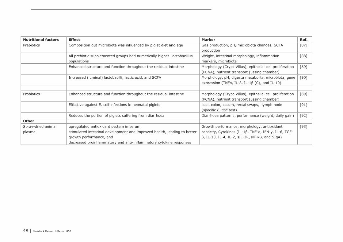
48 | Livestock Research Report 800
Nutritional factors Effect Marker Ref.
Prebiotics Composition gut microbiota was influenced by piglet diet and age Gas production, pH, microbiota changes, SCFAproduction
[87]
All prebiotic supplemented groups had numerically higher Lactobacilluspopulations
Weight, intestinal morphology, inflammationmarkers, microbiota
[88]
Enhanced structure and function throughout the residual intestine Morphology (Crypt-Villus), epithelial cell proliferation(PCNA), nutrient transport (ussing chamber)
[89]
Increased (luminal) lactobacilli, lactic acid, and SCFA Morphology, pH, digesta metabolits, microbiota, geneexpression (TNFα, IL-8, IL-1β (C), and IL-10)
[90]
Probiotics Enhanced structure and function throughout the residual intestine Morphology (Crypt-Villus), epithelial cell proliferation(PCNA), nutrient transport (ussing chamber)
[89]
Effective against E. coli infections in neonatal piglets ileal, colon, cecum, rectal swaps, lymph node(specific E. coli test)
[91]
Reduces the portion of piglets suffering from diarrhoea Diarrhoea patterns, performance (weight, daily gain) [92]Other
Spray-dried animalplasma
upregulated antioxidant system in serum,stimulated intestinal development and improved health, leading to bettergrowth performance, anddecreased proinflammatory and anti-inflammatory cytokine responses
Growth performance, morphology, antioxidantcapacity, Cytokines (IL-1β, TNF-α, IFN-γ, IL-6, TGF-β, IL-10, IL-4, IL-2, sIL-2R, NF-κB, and SIgA)
[93]
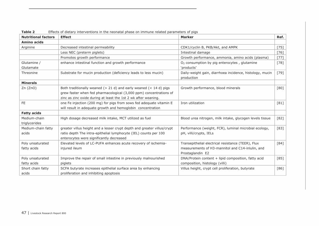
47 | Livestock Research Report 800
Table 2 Effects of dietary interventions in the neonatal phase on immune related parameters of pigsNutritional factors Effect Marker Ref.
Amino acids
Arginine Decreased intestinal permeability CDK1/cyclin B, PKB/Akt, and AMPK [75]Less NEC (preterm piglets) Intestinal damage [76]Promotes growth performance Growth performance, ammonia, amino acids (plasma) [77]
Glutamine /Glutamate
enhance intestinal function and growth performance O2 consumption by pig enterocytes , glutamine‘products’
[78]
Threonine Substrate for mucin production (deficiency leads to less mucin) Daily-weight gain, diarrhoea incidence, histology, mucinproduction
[79]
Minerals
Zn (ZnO) Both traditionally weaned (> 21 d) and early weaned (< 14 d) pigsgrew faster when fed pharmacological (3,000 ppm) concentrations ofzinc as zinc oxide during at least the 1st 2 wk after weaning.
Growth performance, blood minerals [80]
FE one Fe injection (200 mg) for pigs from sows fed adequate vitamin Ewill result in adequate growth and hemoglobin concentration
Iron utilization [81]
Fatty acids
Medium-chaintriglycerides
High dosage decreased milk intake, MCT utilized as fuel Blood urea nitrogen, milk intake, glycogen levels tissue [82]
Medium-chain fattyacids
greater villus height and a lesser crypt depth and greater villus/cryptratio depth The intra-epithelial lymphocyte (IEL) counts per 100enterocytes were significantly decreased
Performance (weight, FCR), luminal microbial ecology,pH, villi/crypts, IELs
[83]
Poly unsaturatedfatty acids
Elevated levels of LC-PUFA enhances acute recovery of ischemia-injured ileum
Transepithelial electrical resistance (TEER), Fluxmeasurements of H3-mannitol and C14-inlulin, andProstaglandin E2
[84]
Poly unsaturatedfatty acids
Improve the repair of small intestine in previously malnourishedpiglets
DNA/Protein content + lipid composition, fatty acidcomposition, histology (villi)
[85]
Short chain fattyacids
SCFA butyrate increases epithelial surface area by enhancingproliferation and inhibiting apoptosis
Villus height, crypt cell proliferation, butyrate [86]

48 | Livestock Research Report 800
Nutritional factors Effect Marker Ref.
Prebiotics Composition gut microbiota was influenced by piglet diet and age Gas production, pH, microbiota changes, SCFAproduction
[87]
All prebiotic supplemented groups had numerically higher Lactobacilluspopulations
Weight, intestinal morphology, inflammationmarkers, microbiota
[88]
Enhanced structure and function throughout the residual intestine Morphology (Crypt-Villus), epithelial cell proliferation(PCNA), nutrient transport (ussing chamber)
[89]
Increased (luminal) lactobacilli, lactic acid, and SCFA Morphology, pH, digesta metabolits, microbiota, geneexpression (TNFα, IL-8, IL-1β (C), and IL-10)
[90]
Probiotics Enhanced structure and function throughout the residual intestine Morphology (Crypt-Villus), epithelial cell proliferation(PCNA), nutrient transport (ussing chamber)
[89]
Effective against E. coli infections in neonatal piglets ileal, colon, cecum, rectal swaps, lymph node(specific E. coli test)
[91]
Reduces the portion of piglets suffering from diarrhoea Diarrhoea patterns, performance (weight, daily gain) [92]Other
Spray-dried animalplasma
upregulated antioxidant system in serum,stimulated intestinal development and improved health, leading to bettergrowth performance, anddecreased proinflammatory and anti-inflammatory cytokine responses
Growth performance, morphology, antioxidantcapacity, Cytokines (IL-1β, TNF-α, IFN-γ, IL-6, TGF-β, IL-10, IL-4, IL-2, sIL-2R, NF-κB, and SIgA)
[93]
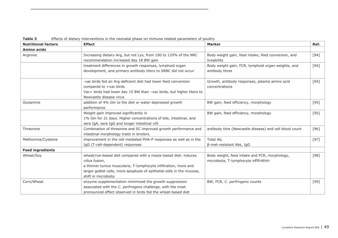
Livestock Research Report 800 | 49
Table 3 Effects of dietary interventions in the neonatal phase on immune related parameters of poultryNutritional factors Effect Marker Ref.
Amino acids
Arginine Increasing dietary Arg, but not Lys, from 100 to 120% of the NRCrecommendation increased day 18 BW gain
Body weight gain, feed intake, feed conversion, andliveability
[94]
treatment differences in growth responses, lymphoid organdevelopment, and primary antibody titers to SRBC did not occur
Body weight gain, FCR, lymphoid organ weights, andantibody titres
[94]
-vac birds fed an Arg-deficient diet had lower feed conversioncompared to +vac birdsVac+ birds had lower day 15 BW than –vac birds, but higher titers toNewcastle disease virus
Growth, antibody responses, plasma amino acidconcentrations
[94]
Glutamine addition of 4% Gln to the diet or water depressed growthperformance
BW gain, feed efficiency, morphology [95]
Weight gain improved significantly in1% Gln for 21 days. Higher concentrations of bile, intestinal, andsera IgA, sera IgG and longer intestinal villi
BW gain, feed efficiency, morphology [95]
Threonine Combination of threonine and SC improved growth performance andintestinal morphology traits in broilers.
antibody titre (Newcastle disease) and cell blood count [96]
Methionine/Cysteine improvement in the cell mediated PHA-P responses as well as in theIgG (T-cell-dependent) responses
Total Ab,β-met-resistant Abs, IgG
[97]
Feed ingredients
Wheat/Soy wheat/rye-based diet compared with a maize-based diet: inducesvillus fusion,a thinner tunica muscularis, T-lymphocyte infiltration, more andlarger goblet cells, more apoptosis of epithelial cells in the mucosa,shift in microbiota
Body weight, feed intake and FCR, morphology,microbiota, T-lymphocyte infiltration
[98]
Corn/Wheat enzyme supplementation minimized the growth suppressionassociated with the C. perfringens challenge, with the mostpronounced effect observed in birds fed the wheat-based diet
BW, FCR, C. perfringens counts [99]
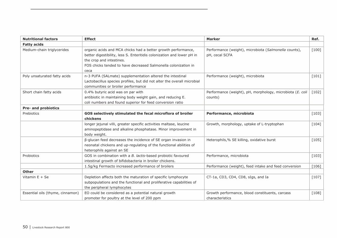
50 | Livestock Research Report 800
Nutritional factors Effect Marker Ref.
Fatty acids
Medium-chain triglycerides organic acids and MCA chicks had a better growth performance,better digestibility, less S. Enteritidis colonization and lower pH inthe crop and intestines.FOS chicks tended to have decreased Salmonella colonization inceca
Performance (weight), microbiota (Salmonella counts),pH, cecal SCFA
[100]
Poly unsaturated fatty acids n-3 PUFA (SALmate) supplementation altered the intestinalLactobacillus species profiles, but did not alter the overall microbialcommunities or broiler performance
Performance (weight), microbiota [101]
Short chain fatty acids 0.4% butyric acid was on par withantibiotic in maintaining body weight gain, and reducing E.coli numbers and found superior for feed conversion ratio
Performance (weight), pH, morphology, microbiota (E. coli
counts)[102]
Pre- and probiotics
Prebiotics GOS selectively stimulated the fecal microflora of broiler
chickens
Performance, microbiota [103]
longer jejunal villi, greater specific activities maltase, leucineaminopeptidase and alkaline phosphatase. Minor improvement inbody weight.
Growth, morphology, uptake of L-tryptophan [104]
β-glucan feed decreases the incidence of SE organ invasion inneonatal chickens and up-regulating of the functional abilities ofheterophils against an SE
Heterophils,% SE killing, oxidative burst [105]
Probiotics GOS in combination with a B. lactis-based probiotic favouredintestinal growth of bifidobacteria in broiler chickens.
Performance, microbiota [103]
1.5g/kg Fermacto increased performance of broilers Performance (weight), feed intake and feed conversion [106]Other
Vitamin E + Se Depletion affects both the maturation of specific lymphocytesubpopulations and the functional and proliferative capabilities ofthe peripheral lymphocytes
CT-1a, CD3, CD4, CD8, sIgs, and Ia [107]
Essential oils (thyme, cinnamon) EO could be considered as a potential natural growthpromoter for poultry at the level of 200 ppm
Growth performance, blood constituents, carcasscharacteristics
[108]
51 | Livestock Research Report 800
5.2.2 Human
Minerals are essential for a proper development of the neonate and the immune system, examples arezinc, selenium, copper, manganese, calcium and iron (Table 4). Suppletion of minerals can have apositive effect on the host, but bear in mind that the (resident) microbiota also make use theseminerals or trace elements for their maintenance. In literature many studies conclude that a deficiencyof minerals lead to impaired immunity, however an excessive dose can be harmful as well. This meansthat one must carefully asses what the ‘normal’ range of these minerals is for the host. Addition ofvitamins can have a positive effect on the host’s immune system, as with minerals a depletion isdetrimental for the immune system. Another category are the fatty acids, in literature mainly the long-chain poly unsaturated fatty acids are described for (young) infants, here two case studies are given.Pre- and probiotics are often used when infants are given formula, instead of mother’s milk, both pre-and probiotics try to mimic the mechanism(s) of mother’s milk. Prebiotics are often oligosaccharides,whereas probiotics are mainly (specific) Lactobacilli and Bifidobacteria. An overview of case studiesdescribing different nutritional interventions is given in Table 4.
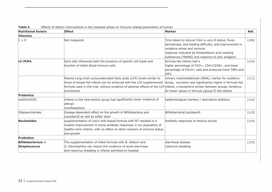
52 | Livestock Research Report 800
Table 4 Effects of dietary interventions in the neonatal phase on immune related parameters of humanNutritional factors Effect Marker Ref.
Vitamins
C + E Not measured Time taken to recover from a very ill status, fever,tachypnoea, and feeding difficulty; and improvement inoxidative stress and immuneresponse indicated by thiobarbituric acid reactingsubstances (TBARS) and response to skin antigens.
[109]
LC-PUFA Early diet influences both the presence of specific cell types andfunction of infant blood immune cells
formula fed infants had ahigher percentage of CD3+, CD4+CD28+, and lowerpercentage of CD14+ cells and produced more TNFα andINFγ
[110]
Plasma Long-chain polyunsaturated fatty acids (LCP) levels similar tothose of breast fed infants can be achieved with the LCP supplementedformula used in this trial, without evidence of adverse effects of the LCPenrichment
Urinary malondialdehyde (MDA), marker for oxidativestress, excretion was significantly higher in formula fedinfants. α-tocopherol similar between groups, tendencyfor lower values in formula (group F) fed infants
[111]
Prebiotics
scGOS/lcFOS Infants in the intervention group had significantly lower incidence ofallergicmanifestations
Epidemiological markers / descriptive statistics [112]
Oligosaccharides Dosage-dependent effect on the growth of Bifidobacteria andLactobacilli as well as softer stool
Bifidobacteria/Lactobacilli [113]
Nucleotides supplementation of cow's milk-based formula with NT resulted in amodest improvement in some antibody responses in our population ofhealthy term infants, with no effect on other markers of immune statusand growth
Antibody responses to tetanus toxoid [114]
Probiotics
Bifidobacterium +
Streptococcus
The supplementation of infant formula with B. bifidum andS. thermophilus can reduce the incidence of acute diarrhoeaand rotavirus shedding in infants admitted to hospital
diarrhoeal diseaserotavirus shedding
[115]
53 | Livestock Research Report 800
5.2.3 Conclusions
From these different studies it can be concluded that it is possible to modulate the host by a multitudeof nutritional factors. However one must bear in mind that most of the (published) literature is biasedtowards publishing (significant) measurable effects on the host. Therefor by definition this overview isincomplete, nevertheless these studies do show that different nutritional factors affect the host indifferent ways. Taken together, no clear biomarker/endomarker can be extracted from this long list ofavailable studies to investigate the effect on the immune competence of the host. Due to the fact thatin most studies performance (i.e. body weight (gain) and feed conversion rate) is an important markerto test the impact of the intervention, this is often investigated in conjunction with the morphology ofthe small intestine. Nevertheless, in a few studies immunological or microbial parameters were used toinvestigate the impact of the intervention, in this way more information is gained of the possible modeof action associated to immune competence. In other words which genes, proteins and/or microbialgroups that contribute to intestinal immune development and health. From Table 2 it can be extractedthat in terms of immune parameters (e.g. cytokines) prebiotics (and spray dried plasma) could bepotential candidates to further investigate in pigs. For chickens (Table 3), evidence was found in‘wheat/soy’ diets, ‘Methionine and Cysteine’, as well as ‘vitamin E together with Selenium’, thatshowed effects on the immune related parameters (e.g. cytokines, T cells and antibodies). Humaninfants were also studied extensively within the context of early life variation and (impact on)immunity. From these studies we can conclude that one must avoid a depletion of vitamins and/orminerals in the diet. Furthermore the most abundant nutritional factors were; prebiotics (e.g.oligosaccharides), probiotics (e.g. lactobacilli and bifidobacteria) and long-chain poly unsaturated fattyacids.
5.3 Impact of nutritional interventions in the post-neonatal phase on immune related parameters
In this paragraph, the impact of nutritional interventions on the immune competence in the adultphase is discussed. It is based on research in calves, weaned pigs, adult mice, laying hens, ducks,broilers (> 10 d), and humans. The results are shown in Table 5, whereas more details are provided inAppendix 14.
Microbiota parameters
Dietary supplementation of rice bran [116], quercitine [117], alfalfa [118], bacillus subtillis [119],antimicrobial peptide A3 and P5 [120], Buforin II [121] and lysozyme [122] reduced the amount ofpathogenic bacteria (e.g. Clostridia, E. Coli), whereas in some studies the number of Lactobacilli andBifidobacteria was increased
Gut integrity
The gut integrity was improved by dietary addition of glutamine [123], black cumin [124], bacillus
subtillis [119], antimicrobial peptide A3 and P5 [120], Buforin II [121] and lysozyme [122]. Theimproved gut integrity was shown by greater villus height and reduced crypt depth, and in somestudies an increased production of tight junction proteins, reduced lesion scores and less bacterialtranslocation from the gut to the spleen.
Immune cells
Several dietary interventions positively affected immune cells. Such effects were found aftersupplementation of glutamine [123], chitosan [125], garlic botanical [126], rice bran [116], alfalfa[118], black cumin [124], bacillus subtillis [119], and Lactobacillus delbrueckii subsp. Bulgaricus
[127]. These interventions affected a wide range of immune cells: Increased IgG, IgA and secretory IgA levels Increased numbers of TNF-α, IL-1β cells Increased numbers of myeloid dendritic cells (CD11c+CD11b+ DCs) in the Lamina propria Increased proliferation of T and B lymphocytes Increased number of goblet cells Increased or decreased number of CD4+ cells and decreased CD8+ cells Increased ratio between CD4+/CD8+
54 | Livestock Research Report 800
Increased number of monocytes Ratio between CD8+/CD3+ in human peripheral blood. Ratio between CD16+56 (NK cells)/CD45+ in human peripheral blood.
Gene expression
Gene expression data were only provided in limited number of studies. In these studies, Lactobacillus
delbrueckii subsp. Bulgaricus [127], lysozyme [122], Buforin II [121], and rice bran [116] were usedas dietary interventions. These gene expression data showed: a reduced cytokine quantification in human serum an increased Lysozyme activity and lysozyme g2 mRNA content an increased expression of surface IgA on B220+ B cells in Peyers Patches an enhanced expression of protective factors (HGF, hepatocyte growth factor, reg-3 gamma,
regeneration protein gamma, TFF-3, and trefoil factor-3) an enhanced expression of TGF-β1, transforming growth factor β1; VEGF, and vessel endothelium
growth factor.
Performance parameters
Dietary addition of quercetin [117], black cumin [124], bacillus subtillis [119], antimicrobial peptideA3 and P5 [120], Buforin II [121] and lysozyme [122] resulted in improved animal performance.Improved performance was shown by a decreased FCR and an increased egg mass production, dailygain, nutrient digestibility and retention. In some experiments [121, 122], animals were challengedwith pathogenic bacteria, and although animal performance was negatively affected by the challenge,the dietary interventions could partly prevent this reduced performance.

Livestock Research Report 800 | 55
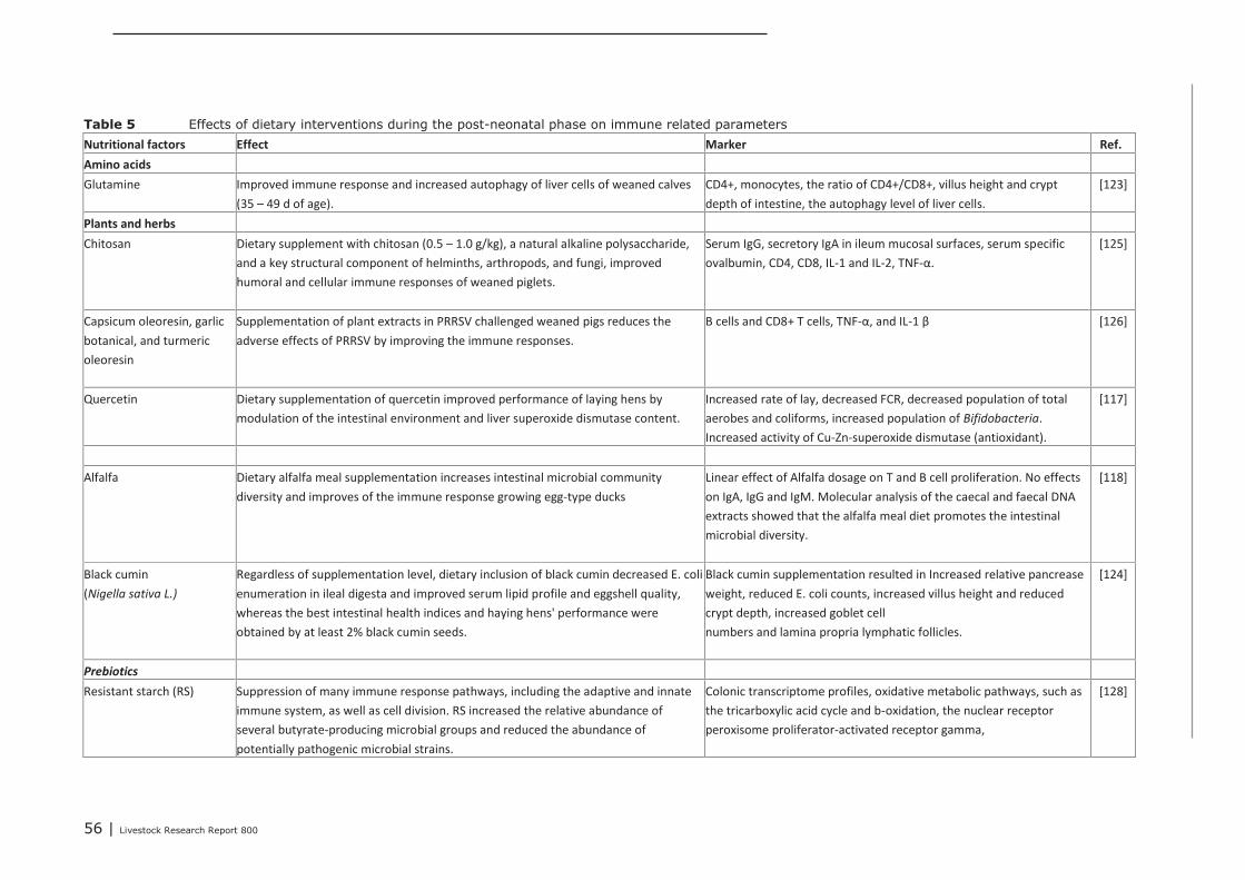
56 | Livestock Research Report 800
Table 5 Effects of dietary interventions during the post-neonatal phase on immune related parametersNutritional factors Effect Marker Ref.
Amino acids
Glutamine Improved immune response and increased autophagy of liver cells of weaned calves
(35 – 49 d of age).
CD4+, monocytes, the ratio of CD4+/CD8+, villus height and crypt
depth of intestine, the autophagy level of liver cells.
[123]
Plants and herbs
Chitosan Dietary supplement with chitosan (0.5 – 1.0 g/kg), a natural alkaline polysaccharide,
and a key structural component of helminths, arthropods, and fungi, improved
humoral and cellular immune responses of weaned piglets.
Serum IgG, secretory IgA in ileum mucosal surfaces, serum specific
ovalbumin, CD4, CD8, IL-1 and IL-2, TNF-α.
[125]
Capsicum oleoresin, garlic
botanical, and turmeric
oleoresin
Supplementation of plant extracts in PRRSV challenged weaned pigs reduces the
adverse effects of PRRSV by improving the immune responses.
B cells and CD8+ T cells, TNF-α, and IL-1 β [126]
Quercetin Dietary supplementation of quercetin improved performance of laying hens by
modulation of the intestinal environment and liver superoxide dismutase content.
Increased rate of lay, decreased FCR, decreased population of total
aerobes and coliforms, increased population of Bifidobacteria.
Increased activity of Cu-Zn-superoxide dismutase (antioxidant).
[117]
Alfalfa Dietary alfalfa meal supplementation increases intestinal microbial community
diversity and improves of the immune response growing egg-type ducks
Linear effect of Alfalfa dosage on T and B cell proliferation. No effects
on IgA, IgG and IgM. Molecular analysis of the caecal and faecal DNA
extracts showed that the alfalfa meal diet promotes the intestinal
microbial diversity.
[118]
Black cumin
(Nigella sativa L.)
Regardless of supplementation level, dietary inclusion of black cumin decreased E. coli
enumeration in ileal digesta and improved serum lipid profile and eggshell quality,
whereas the best intestinal health indices and haying hens' performance were
obtained by at least 2% black cumin seeds.
Black cumin supplementation resulted in Increased relative pancrease
weight, reduced E. coli counts, increased villus height and reduced
crypt depth, increased goblet cell
numbers and lamina propria lymphatic follicles.
[124]
Prebiotics
Resistant starch (RS) Suppression of many immune response pathways, including the adaptive and innate
immune system, as well as cell division. RS increased the relative abundance of
several butyrate-producing microbial groups and reduced the abundance of
potentially pathogenic microbial strains.
Colonic transcriptome profiles, oxidative metabolic pathways, such as
the tricarboxylic acid cycle and b-oxidation, the nuclear receptor
peroxisome proliferator-activated receptor gamma,
[128]

Livestock Research Report 800 | 57
Probiotics
Bacillus subtilis
fermentation biomass
4.5 g/kg BS 1–2 fermentation biomass had beneficial effects on weanling pigs
performance, CTTAD of nutrients, cecal microbiota, serum immunoglobulins and
intestinal morphology.
ADG, G:F, digestibility of DM, GE and CP, serum IgA and IgG, cecal Cl.
spp. and coliforms, villus height and VH:CD (duodenum, jejunum and
ileum).
[129]
Nutritional factors Effect Marker
Lactobacillus delbrueckii
subsp. bulgaricus
Immunomodulation induced by L. delbrueckii subsp. bulgaricus could favour the
maintenance of an adequate immune response.
Slowing the aging of the T cell subpopulations and increasing the
number of immature T cells which are potential responders to new
antigens.
[127]
Antimicrobial proteins
Antimicrobial peptide-A3
and P5
AMP-A3 and P5 have potential to improve performance, nutrient digestibility,
intestinal morphology and to reduce pathogenic bacteria in weanling pigs
Performance, nutrient digestibility, fecal Clostridium spp. and
coliforms, anaerobic bacteria, gut morphology
[130]
Buforin II Buforin II protects small intestinal mucosal membrane integrity of weaned pigs. Increasing tight junction proteins, enhancing the expression of
protective factors, reducing hemolytic E. coli concentrations in the
Intestines.
[121]
Lysozyme Exogenous lysozyme could decrease C. perfringens colonization and improve intestinal
barrier function and growth performance of chickens.
A reduction of C. perfringens in the ileum and of the intestinal lesion
scores, less overgrowth of E. coli and Lactobacillus in the ileum and
less intestinal bacteria translocation to the spleen, and improved
intestinal lysozyme activity in the duodenum, and improved feed
conversion ratio of chickens.
[122]

58 | Livestock Research Report 800
59 | Livestock Research Report 800
5.4 Mode of action of dietary interventions on immunity
In this paragraph, the mode of action of the mentioned dietary interventions is briefly described. Thisparagraph is partly based on two review reports regarding this topic [131, 132]
Fungal and bacterial cell wall (β-glucans)
β-Glucans bind Dectin-1-like receptor on animal cells (e.g. neutrophils, monocytes). This supports theinnate immunity by increased neutrophil functioning (i.e. production of reactive oxygen species,cytokine production), resulting in reduced susceptibility for pathogen invasion (e.g. Salmonella
enteritidis).
Mannan-oligosaccharide (MOS)
MOS acts as a competition molecule for the adhesion of certain bacterial species in the gut, therebyreducing the risk of diseases. Next to this, MOS also has an anti-inflammatory working, but exactmode of action is not known. However, MOS was also reported to have an increased inflammatoryresponse (i.e. phagocytosis and complement pathways).
Alfalfa
Alfalfa was shown to increase heterophil functioning and was also shown to prevent Salmonella
enteritidis infection in molting chicken.
Amino acids
Several amino acids were found to alter histone and DNA methylation/acetylation and thereby toregulate gene expressions. A similar working mechanism might be possible in other organs/cells. Nodirect link of gene expression and immune functioning under influence of amino acids is reported yet.However, amino acids were reported to activate and proliferate lymphocytes, NK cells andmacrophages and to alter the acute phase protein response.
Poly unsaturated fatty acids (PUFA)
PUFA react via the PPAR and RXR on cells and thereby affect eicosanoid releases. Additionally, PUFAsare also incorporated in cell membranes and influence cell functioning depending on PUFA type andconcentration. Finally, PUFA are known for their regulation of gene expression, possibly via epigeneticmechanisms (in humans). Consequently, PUFAs reduce inflammatory response in macrophages and inT cells and also reduce the acute phase response. They are reported to stimulate antibody response.Chicken become less vulnerable to humoral (B cell) associated diseases, but more vulnerable tocellular (T cell) associated diseases. Also other cells are influenced. PUFA are reported to improveepithelial integrity in the gut. The (n-3) LCPUFA metabolites induce eicosanoid production, alter geneexpression, and modify lipid raft composition, altering T-cell signalling; all contribute to immunologicalfunctional changes. However, the roles of these mechanisms and the types of T or otherimmunological cells involved remain unclear at present. Moreover, the effect of (n-3) LCPUFA on theimmune system of infants may vary according to dose, time of exposure, and profile of the immunesystem (T-helper, Th1/Th2). Most of the interventional studies in infancy have been performed for theprevention of allergy. They all confirmed influence on T-cell function and cytokine profiles, butclinically beneficial effects are more conflicting. Supplementation of the maternal diet in pregnancy orearly childhood with (n-3) LCPUFA is potentially a non-invasive intervention strategy to prevent thedevelopment of allergy, infection, and possibly other immune-mediated diseases. However, any long-term in vivo effects on (n-3) LCPUFA early in life for immuno-modulatory defence in infants and lateron immune status and health remain to be assessed [133].
Spray-dried animal plasma
The immune competence improving effects of spray-dried animal plasma in young pigs is most likelymediated by the IgG component in the plasma. The IgG prevents viruses and bacteria from damagingthe gut wall, thereby resulting in a more functional intestinal wall, improves intestinal morphology andenzyme activity as evidenced by increased villus surface area, and increased mucosal maltase andlactase activities [134].
60 | Livestock Research Report 800
Minerals
Mineral deficiencies are well described in literature with often detrimental immuno modulatoryinfluences. Minerals are also reported to affect macrophage functioning and to reduce inflammation(as antioxidants) by protection of neutrophils from oxygen-derived radicals. Also (pro-inflammatory)gene and receptor expression is influenced by minerals. Also lymphocytes require selenium for betterfunctioning. Next to this, the essence of minerals for gut microbiota should not be forgotten.
Vitamins
Vitamin serve as antioxidants in the body and thereby protect cells from oxygen damage.Inflammation and macrophage activity were reported to be reduced, although macrophage activitywas also reported to be increased. Vitamins were also reported to induce DNA methylation and alterthe immune response, although no direct link between these observations was proven.
Seaweed extract
The reduction in colonic E. coli and Enterobacteriaceae populations may be the result of theagglutination properties of seaweed extract, thereby preventing the attachment to epithelial cellsurfaces and subsequent colonisation of mucosal surfaces [63]. Seaweed inclusion post-weaningtended to up-regulate colonic MUC2 mRNA expression. Mucin, the primary component of mucus,functions as a biophysical barrier against enzymatic, mechanical insult and pathogenic invasion withinthe GIT. The fibrous components of Laminarin may stimulate the colonic mucosa or may have a directeffect at the cellular level because of becoming internalized by intestinal epithelial cells and gut-associated lymphoid tissue cells. Alterations in colonic microflora composition may also directlyinfluence mucin synthesis and secretion from goblet cells. The usual up-regulation of intestinalinflammatory cytokine gene expression in ileal and colonic tissues after weaning was not observed ortended to be lower in pigs weaned from seaweed-supplemented sows. Seaweed, therefore, seems tomodulate the production of pro- and anti-inflammatory cytokines, which might be beneficial to animalhealth and performance.
Probiotics
In vitro studies have shown that Sachoromyces Cerevisiae secretes anti-inflammatory factors andinhibits the phosphorylation of both p38 and ERK1/2 kinases after co-culture of porcine intestinalepithelial cells with ETEC [135, 136]. Supplementation of Lactobacillus rhamnosus and Bifidobacterium
lactis to pregnant women increased the adiponectin content of their colostrum [72]. The assumedhealth effects of adiponectin are mediated through its anti-inflammatory properties via regulation ofthe expression of proinflammatory cytokines and through its insulin-sensitizing properties viainhibition of liver gluconeogenesis and promotion of fatty acid oxidation.
Lysozyme
Toxins produced by C. perfringens may destruct the mucosal tissue, causing macroscopic lesions inthe duodenum, jejunum and ileum. This results in a disturbance of the interface between enterocytesand the lamina propria, which might induce a strong inflammatory response in birds, therebynegatively affecting digestive functions and impairing nutrient utilization, finally suppressing bird’sperformance. Lysozyme is suggested to have a direct inhibitive effect on C. perfringens proliferationand its immune modulating activity. Based on in vitro experiments [137] is was concluded thatlysozyme could not only kill C. perfringens, but also reduce the production of α-toxin, the causativeagent for intestinal lesions. Other in vitro experiments showed that lysozyme stimulatesimmunoglobulin production of specific blood lymphocytes [138]. After absorption of orallyadministered lysozyme in the gut, plasma levels of lysozyme will increase, which could producesystemic effects like an increased activity of monocytes and macrophages [139]. Lysozyme has beenreported to interact with intestinal bacteria to liberate immune-modulating peptidoglycans and Peyer’spatches, and intraepithelial lymphocytes to activate the host’s immune system [140].
Rice bran
It is suggested that various bioactive and prebiotic components in rice bran, e.g. γ-oryzanol,polyphenols, fatty acids, essential amino acids and micronutrients, are involved in the enhancement ofimmunity [141].
Livestock Research Report 800 | 61
Lactobacilli
Lactobacilli are able to stimulate the number and activity of NK cells. NK cells play a critical role inimmune surveillance against the development of tumors and viral infections, and intestinal microfloracan modulate NK activity [142]. It is suggested that Lactobacilli stimulate the activity of the thymusgland [127]. Lactobacilli could reduce the IL-8 content in the serum, confirming that some probioticsare able to inhibit the production of pro-inflammatory cytokines [143]. Moreover, Lactobacillisupplementation was associated with a very marked increase in hBD-2 antimicrobial molecule inserum, suggesting an enhancement in the innate immunity through defensins induction [144].Lactobacilli, therefore, do not only have local effects in the intestinal mucosa, but also in theperipheral blood.
Plant extracts
Plant extracts are able to reduce serum TNF-α and haptoglobin, white blood cells and lymphocytes,and to mitigate villus atrophy after an E. coli challenge, thereby modulating the systemic inflammationcaused by E. coli infection [126]. TNF-α is one of the most important cytokines released especiallyform macrophages and monocytes, in response to bacterial cell wall products and bacterial toxins. Theeffects of plant extracts on white blood cells and TNF-α showed that they may alleviate theoverstimulation of the systemic immunity and early immune response in E. coli infected pigs. Plantextracts could reduce the recruitment of macrophages and neutrophils in the ileum of E. coli infectedpigs, indicating that these reduce both systemic and local inflammation.
Buforin II
Buforin II kills E. coli by penetrating the cell membrane, subsequently inhibiting cellular functions[145]. Buforin supplementation could decrease the jejunal and ileal mucosal lymphocyte amount aswell as the calprotectin level. Calprotectin exerts antimicrobial effects through competition for zinc andby preventing bacteria from binding to mucosal epithelial cells. Buforin II significantly increased gobletcell amounts and the expression level of tight junction proteins (VEGF, HGH, Reg-3γ, TGF-β1, andTFF-3) in the jejunum and ileum. HGF acts on a wide variety of epithelial cells, as a mitogen(stimulation of cell growth), a mitogen (stimulation of cell motility), and a morphogen (induction ofmulticellular architecture), and is considered to be a key molecule for construction of structure duringorgan regeneration. TFF is a secretory protein expressed in gastrointestinal mucosa and may protectthe mucosa from insults, stabilize the mucus layer and affect healing of epithelium [146].

62 | Livestock Research Report 800
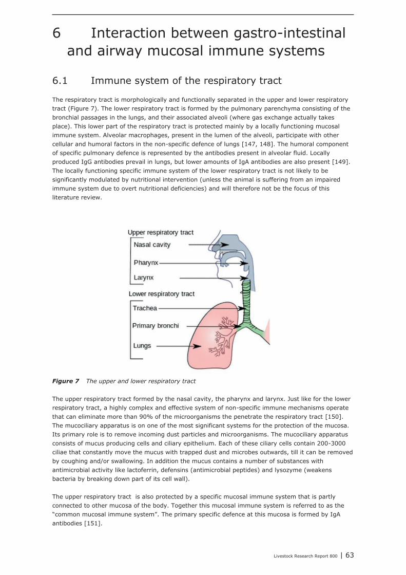
Livestock Research Report 800 | 63
6 Interaction between gastro-intestinaland airway mucosal immune systems
6.1 Immune system of the respiratory tract
The respiratory tract is morphologically and functionally separated in the upper and lower respiratorytract (Figure 7). The lower respiratory tract is formed by the pulmonary parenchyma consisting of thebronchial passages in the lungs, and their associated alveoli (where gas exchange actually takesplace). This lower part of the respiratory tract is protected mainly by a locally functioning mucosalimmune system. Alveolar macrophages, present in the lumen of the alveoli, participate with othercellular and humoral factors in the non-specific defence of lungs [147, 148]. The humoral componentof specific pulmonary defence is represented by the antibodies present in alveolar fluid. Locallyproduced IgG antibodies prevail in lungs, but lower amounts of IgA antibodies are also present [149].The locally functioning specific immune system of the lower respiratory tract is not likely to besignificantly modulated by nutritional intervention (unless the animal is suffering from an impairedimmune system due to overt nutritional deficiencies) and will therefore not be the focus of thisliterature review.
Figure 7 The upper and lower respiratory tract
The upper respiratory tract formed by the nasal cavity, the pharynx and larynx. Just like for the lowerrespiratory tract, a highly complex and effective system of non-specific immune mechanisms operatethat can eliminate more than 90% of the microorganisms the penetrate the respiratory tract [150].The mucociliary apparatus is on one of the most significant systems for the protection of the mucosa.Its primary role is to remove incoming dust particles and microorganisms. The mucociliary apparatusconsists of mucus producing cells and ciliary epithelium. Each of these ciliary cells contain 200-3000ciliae that constantly move the mucus with trapped dust and microbes outwards, till it can be removedby coughing and/or swallowing. In addition the mucus contains a number of substances withantimicrobial activity like lactoferrin, defensins (antimicrobial peptides) and lysozyme (weakensbacteria by breaking down part of its cell wall).
The upper respiratory tract is also protected by a specific mucosal immune system that is partlyconnected to other mucosa of the body. Together this mucosal immune system is referred to as the“common mucosal immune system”. The primary specific defence at this mucosa is formed by IgAantibodies [151].
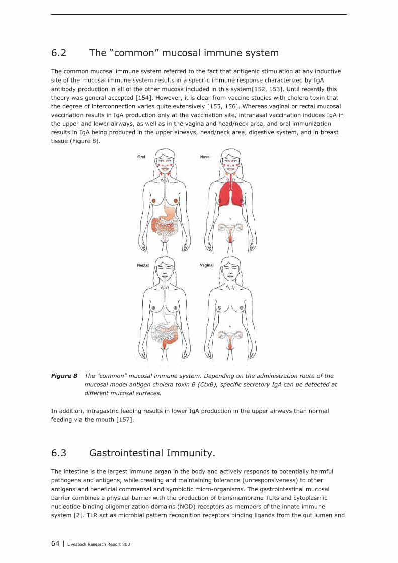
64 | Livestock Research Report 800
6.2 The “common” mucosal immune system
The common mucosal immune system referred to the fact that antigenic stimulation at any inductivesite of the mucosal immune system results in a specific immune response characterized by IgAantibody production in all of the other mucosa included in this system[152, 153]. Until recently thistheory was general accepted [154]. However, it is clear from vaccine studies with cholera toxin thatthe degree of interconnection varies quite extensively [155, 156]. Whereas vaginal or rectal mucosalvaccination results in IgA production only at the vaccination site, intranasal vaccination induces IgA inthe upper and lower airways, as well as in the vagina and head/neck area, and oral immunizationresults in IgA being produced in the upper airways, head/neck area, digestive system, and in breasttissue (Figure 8).
Figure 8 The “common” mucosal immune system. Depending on the administration route of the
mucosal model antigen cholera toxin B (CtxB), specific secretory IgA can be detected at
different mucosal surfaces.
In addition, intragastric feeding results in lower IgA production in the upper airways than normalfeeding via the mouth [157].
6.3 Gastrointestinal Immunity.
The intestine is the largest immune organ in the body and actively responds to potentially harmfulpathogens and antigens, while creating and maintaining tolerance (unresponsiveness) to otherantigens and beneficial commensal and symbiotic micro-organisms. The gastrointestinal mucosalbarrier combines a physical barrier with the production of transmembrane TLRs and cytoplasmicnucleotide binding oligomerization domains (NOD) receptors as members of the innate immunesystem [2]. TLR act as microbial pattern recognition receptors binding ligands from the gut lumen and
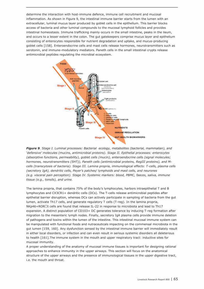
Livestock Research Report 800 | 65
determine the interaction with host-immune defence, immune cell recruitment and mucosalinflammation. As shown in Figure 9, the intestinal immune barrier starts from the lumen with anextracellular, luminal mucus layer produced by goblet cells in the epithelium. This barrier blocksaccess of bacteria and other luminal compounds to the mucosal lymphoid follicles and providesintestinal homeostasis. Immune trafficking mainly occurs in the small intestine, peaks in the ileum,and occurs to a lesser extent in the colon. The gut gatekeepers comprise mucus layer and epitheliumconsisting of enterocytes responsible for nutrient degradation and uptake, and mucus-producinggoblet cells [158]. Enteroendocrine cells and mast cells release hormones, neurotransmitters such asserotonin, and immune-modulatory mediators. Paneth cells in the small intestinal crypts releaseantimicrobial peptides regulating the microbial ecosystem.
Figure 9. Stage I. Luminal processes: Bacterial ecology, metabolites (bacterial, mammalian), and
‘defensive’ molecules (mucins, antimicrobial proteins). Stage II. Epithelial processes: enterocytes
(absorptive functions, permeability), goblet cells (mucin), enteroendocrine cells (signal molecules;
hormones, neurotransmitters (5HT)), Paneth cells (antimicrobial proteins, RegIII proteins), and M-
cells (transcytosis of bacteria). Stage III. Lamina propria, immunological effects: T-cells, plasma cells
(secretory IgA), dendritic cells, Peyer’s patches/ lymphoids and mast cells, and neurones
(e.g. visceral pain perception). Stage IV. Systemic markers: blood, PBMC, faeces, saliva, immune
tissue (e.g., tonsils), and urine.
The lamina propria, that contains 75% of the body’s lymphocytes, harbors intraepithelial T and Blymphocytes and CX3CR1+ dendritic cells (DCs). The T-cells release antimicrobial peptides afterepithelial barrier disruption, whereas DCs can actively participate in sampling of bacteria from the gutlumen, activate Th17 cells, and generate regulatory T cells (T-reg). In the lamina propria,NKp46+RORt cells are found that release IL-22 in response to microbiota and lead to Th17expansion. A distinct population of CD103+ DC generates tolerance by inducing T-reg formation aftermigration to the mesenteric lymph nodes. Finally, secretory IgA plasma cells provide immune deletionof pathogens and toxins within the lumen of the intestine. This intestinal mucosal immune system canbe manipulated with functional foods and nutraceuticals impacting on the commensal microbiota in thegut lumen [159, 160]. Any dysfunction sensed by the intestinal immune barrier will immediately resultin either local disorders, or infection and can even result in serious systemic disorders all deleteriousto health [161].The immune system in the mouth and upper respiratory tract: inductive sites formucosal immunity.A proper understanding of the anatomy of mucosal immune tissues is important for designing rationalapproaches to enhance immunity in the upper airways. This section will focus on the anatomicalstructure of the upper airways and the presence of immunological tissues in the upper digestive tract,i.e. the mouth and throat.
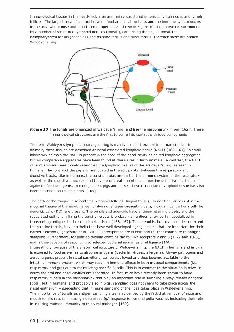
66 | Livestock Research Report 800
Immunological tissues in the head/neck area are mainly structured in tonsils, lymph nodes and lymphfollicles. The largest area of contact between food and nasal contents and the immune system occursin the area where nose and mouth come together. As shown in Figure 10, the pharynx is surroundedby a number of structured lymphoid nodules (tonsils), comprising the lingual tonsil, thenasopharyngeal tonsils (adenoids), the palatine tonsils and tubal tonsils. Together these are namedWaldeyer’s ring.
Figure 10 The tonsils are organized in Waldeyer’s ring, and line the nasopharynx (from [162]). Theseimmunological structures are the first to come into contact with food components
The term Waldeyer’s lymphoid pharyngeal ring is mainly used in literature in human studies. Inanimals, these tissues are described as nasal associated lymphoid tissue (NALT) [163, 164]. In smalllaboratory animals the NALT is present in the floor of the nasal cavity as paired lymphoid aggregates,but no comparable aggregates have been found at these sites in farm animals. In contrast, the NALTof farm animals more closely resembles the lymphoid tissues of the Waldeyer’s ring, as seen inhumans. The tonsils of the pig e.g. are located in the soft palate, between the respiratory anddigestive tracts. Like in humans, the tonsils in pigs are part of the immune system of the respiratoryas well as the digestive mucosae and they are of great importance in porcine defensive mechanismsagainst infectious agents. In cattle, sheep, pigs and horses, larynx-associated lymphoid tissue has alsobeen described on the epiglottis [165].
The back of the tongue also contains lymphoid follicles (lingual tonsil). In addition, dispersed in themucosal tissues of the mouth large numbers of antigen-presenting cells, including Langerhans cell-likedendritic cells (DC), are present. The tonsils and adenoids have antigen-retaining crypts, and thereticulated epithelium lining the tonsillar crypts is probably an antigen entry portal, specialized intransporting antigens to the subepithelial tissue [166, 167]. The adenoids, but to a much lesser extentthe palatine tonsils, have epithelia that have well developed tight junctions that are important for theirbarrier function (Ogasawara et al., 2011). Interspersed are M cells and DC that contribute to antigensampling. Furthermore, tonsillar epithelium contains the toll-like receptors 2 and 3 (TLR2 and TLR3),and is thus capable of responding to selected bacterial as well as viral ligands [166].Interestingly, because of the anatomical structure of Waldeyer’s ring, the NALT in humans and in pigsis exposed to food as well as to airborne antigens (bacteria, viruses, allergens). Airway pathogens andaeroallergens, present in nasal secretions, can be swallowed and thus become available to theintestinal immune system, which may result in immune effects in both mucosal compartments (i.e.respiratory and gut) due to recirculating specific B-cells. This is in contrast to the situation in mice, inwhich the oral and nasal cavities are separated. In fact, mice have recently been shown to haverespiratory M cells in the nasopharynx that play an important role in sampling airway-related antigens[168], but in humans, and probably also in pigs, sampling does not seem to take place across thenasal epithelium – suggesting that immune sampling of the nose takes place in Waldeyer’s ring.The importance of tonsils as antigen sampling sites is evidenced by the fact that removal of nose andmouth tonsils results in strongly decreased IgA response to live oral polio vaccine, indicating their rolein inducing mucosal immunity to this viral pathogen [169].
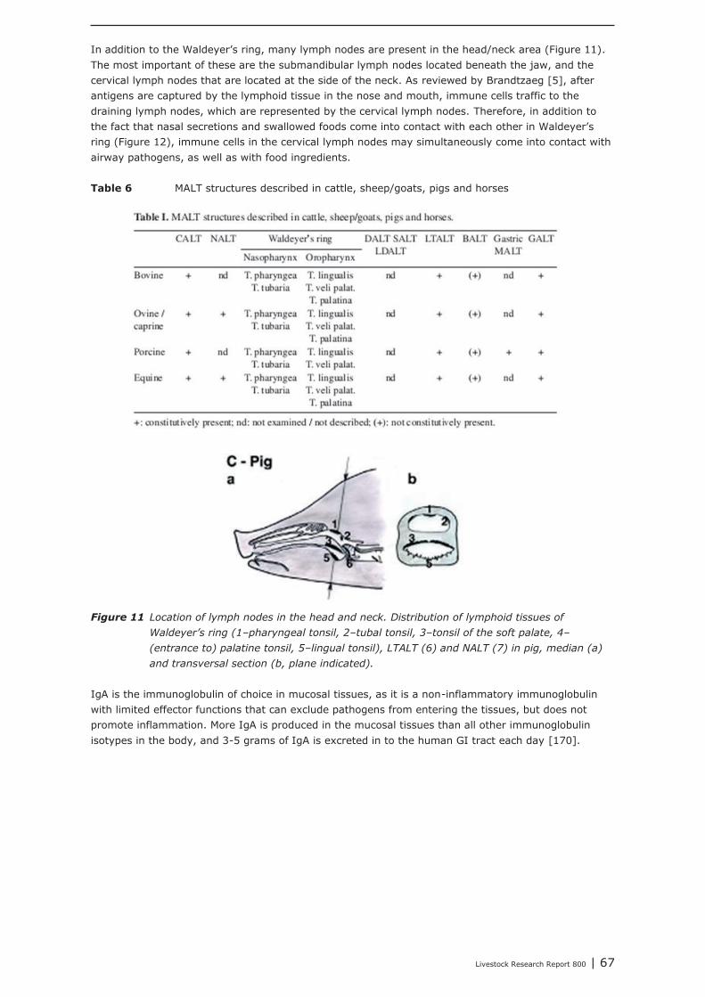
Livestock Research Report 800 | 67
In addition to the Waldeyer’s ring, many lymph nodes are present in the head/neck area (Figure 11).The most important of these are the submandibular lymph nodes located beneath the jaw, and thecervical lymph nodes that are located at the side of the neck. As reviewed by Brandtzaeg [5], afterantigens are captured by the lymphoid tissue in the nose and mouth, immune cells traffic to thedraining lymph nodes, which are represented by the cervical lymph nodes. Therefore, in addition tothe fact that nasal secretions and swallowed foods come into contact with each other in Waldeyer’sring (Figure 12), immune cells in the cervical lymph nodes may simultaneously come into contact withairway pathogens, as well as with food ingredients.
Table 6 MALT structures described in cattle, sheep/goats, pigs and horses
Figure 11 Location of lymph nodes in the head and neck. Distribution of lymphoid tissues of
Waldeyer’s ring (1–pharyngeal tonsil, 2–tubal tonsil, 3–tonsil of the soft palate, 4–
(entrance to) palatine tonsil, 5–lingual tonsil), LTALT (6) and NALT (7) in pig, median (a)
and transversal section (b, plane indicated).
IgA is the immunoglobulin of choice in mucosal tissues, as it is a non-inflammatory immunoglobulinwith limited effector functions that can exclude pathogens from entering the tissues, but does notpromote inflammation. More IgA is produced in the mucosal tissues than all other immunoglobulinisotypes in the body, and 3-5 grams of IgA is excreted in to the human GI tract each day [170].
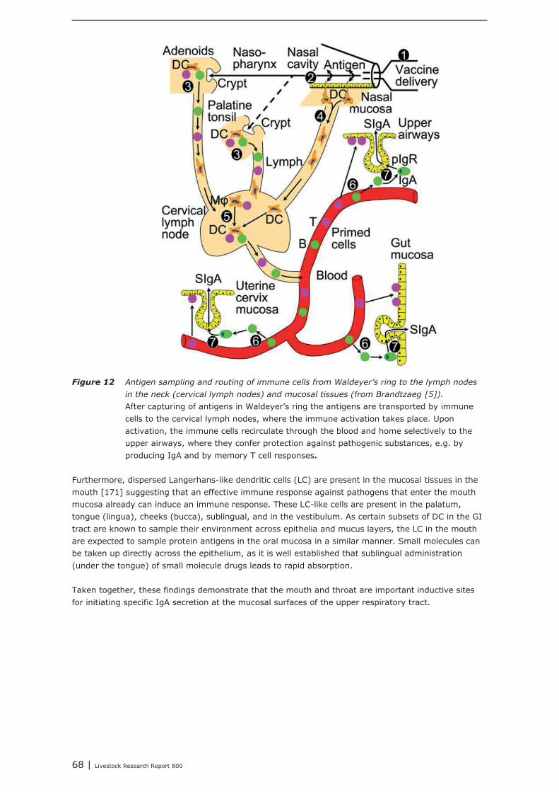
68 | Livestock Research Report 800
Figure 12 Antigen sampling and routing of immune cells from Waldeyer’s ring to the lymph nodes
in the neck (cervical lymph nodes) and mucosal tissues (from Brandtzaeg [5]).
After capturing of antigens in Waldeyer’s ring the antigens are transported by immunecells to the cervical lymph nodes, where the immune activation takes place. Uponactivation, the immune cells recirculate through the blood and home selectively to theupper airways, where they confer protection against pathogenic substances, e.g. byproducing IgA and by memory T cell responses.
Furthermore, dispersed Langerhans-like dendritic cells (LC) are present in the mucosal tissues in themouth [171] suggesting that an effective immune response against pathogens that enter the mouthmucosa already can induce an immune response. These LC-like cells are present in the palatum,tongue (lingua), cheeks (bucca), sublingual, and in the vestibulum. As certain subsets of DC in the GItract are known to sample their environment across epithelia and mucus layers, the LC in the mouthare expected to sample protein antigens in the oral mucosa in a similar manner. Small molecules canbe taken up directly across the epithelium, as it is well established that sublingual administration(under the tongue) of small molecule drugs leads to rapid absorption.
Taken together, these findings demonstrate that the mouth and throat are important inductive sitesfor initiating specific IgA secretion at the mucosal surfaces of the upper respiratory tract.
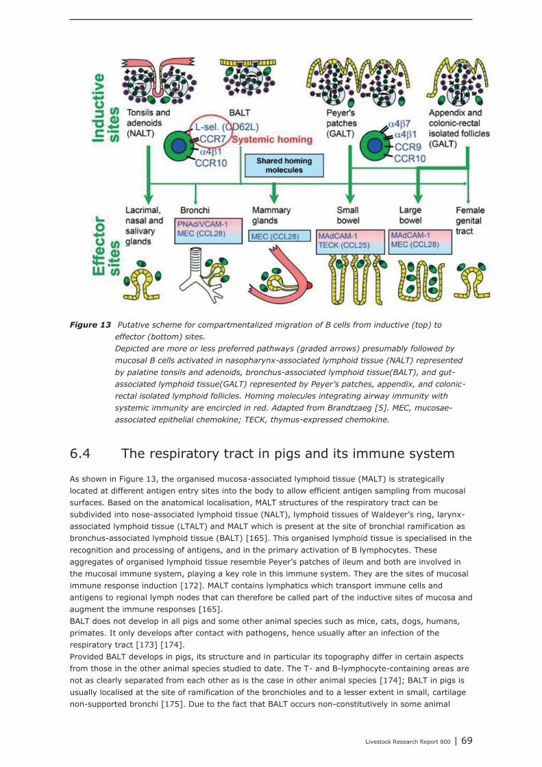
Livestock Research Report 800 | 69
Figure 13 Putative scheme for compartmentalized migration of B cells from inductive (top) to
effector (bottom) sites.
Depicted are more or less preferred pathways (graded arrows) presumably followed by
mucosal B cells activated in nasopharynx-associated lymphoid tissue (NALT) represented
by palatine tonsils and adenoids, bronchus-associated lymphoid tissue(BALT), and gut-
associated lymphoid tissue(GALT) represented by Peyer’s patches, appendix, and colonic-
rectal isolated lymphoid follicles. Homing molecules integrating airway immunity with
systemic immunity are encircled in red. Adapted from Brandtzaeg [5]. MEC, mucosae-
associated epithelial chemokine; TECK, thymus-expressed chemokine.
6.4 The respiratory tract in pigs and its immune system
As shown in Figure 13, the organised mucosa-associated lymphoid tissue (MALT) is strategicallylocated at different antigen entry sites into the body to allow efficient antigen sampling from mucosalsurfaces. Based on the anatomical localisation, MALT structures of the respiratory tract can besubdivided into nose-associated lymphoid tissue (NALT), lymphoid tissues of Waldeyer’s ring, larynx-associated lymphoid tissue (LTALT) and MALT which is present at the site of bronchial ramification asbronchus-associated lymphoid tissue (BALT) [165]. This organised lymphoid tissue is specialised in therecognition and processing of antigens, and in the primary activation of B lymphocytes. Theseaggregates of organised lymphoid tissue resemble Peyer’s patches of ileum and both are involved inthe mucosal immune system, playing a key role in this immune system. They are the sites of mucosalimmune response induction [172]. MALT contains lymphatics which transport immune cells andantigens to regional lymph nodes that can therefore be called part of the inductive sites of mucosa andaugment the immune responses [165].BALT does not develop in all pigs and some other animal species such as mice, cats, dogs, humans,primates. It only develops after contact with pathogens, hence usually after an infection of therespiratory tract [173] [174].Provided BALT develops in pigs, its structure and in particular its topography differ in certain aspectsfrom those in the other animal species studied to date. The T- and B-lymphocyte-containing areas arenot as clearly separated from each other as is the case in other animal species [174]; BALT in pigs isusually localised at the site of ramification of the bronchioles and to a lesser extent in small, cartilagenon-supported bronchi [175]. Due to the fact that BALT occurs non-constitutively in some animal
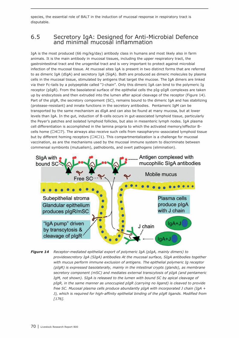
70 | Livestock Research Report 800
species, the essential role of BALT in the induction of mucosal response in respiratory tract isdisputable.
6.5 Secretory IgA: Designed for Anti-Microbial Defenceand minimal mucosal inflammation
IgA is the most produced (66 mg/kg/day) antibody class in humans and most likely also in farmanimals. It is the main antibody in mucosal tissues, including the upper respiratory tract, thegastrointestinal tract and the urogenital tract and is very important to protect against microbialinfection of the mucosal tissue. At mucosal sites IgA is present in two distinct forms that are referredto as dimeric IgA (dIgA) and secretory IgA (SigA). Both are produced as dimeric molecules by plasmacells in the mucosal tissue, stimulated by antigens that target the mucose. The IgA dimers are linkedvia their Fc-tails by a polypeptide called “J-chain”. Only this dimeric IgA can bind to the polymeric Igreceptor (pIgR). From the basolateral surface of the epithelial cells the pIg-pIgR complexes are takenup by endocytosis and then extruded into the lumen after apical cleavage of the receptor (Figure 14).Part of the pIgR, the secretory compoment (SC), remains bound to the dimeric IgA and has stabilizing(protease-resistant) and innate functions in the secretory antibodies. Pentameric IgM can betransported by the same mechanism as dIgA and can also be found at many mucosa, but at lowerlevels than IgA. In the gut, induction of B-cells occurs in gut-associated lymphoid tissue, particularlythe Peyer's patches and isolated lymphoid follicles, but also in mesenteric lymph nodes. IgA plasmacell differentiation is accomplished in the lamina propria to which the activated memory/effector B-cells home (47). The airways also receive such cells from nasopharynx-associated lymphoid tissuebut by different homing receptors (41). This compartmentalization is a challenge for mucosalvaccination, as are the mechanisms used by the mucosal immune system to discriminate betweencommensal symbionts (mutualism), pathobionts, and overt pathogens (elimination).
Figure 14 Receptor-mediated epithelial export of polymeric IgA (pIgA, mainly dimers) to
providesecretory IgA (SIgA) antibodies At the mucosal surface, SIgA antibodies together
with mucus perform immune exclusion of antigens. The epithelial polymeric Ig receptor
(pIgR) is expressed basolaterally, mainly in the intestinal crypts (glands), as membrane
secretory component (mSC) and mediates external transcytosis of pIgA (and pentameric
IgM, not shown). SIgA is released to the lumen with bound SC by apical cleavage of
pIgR, in the same manner as unoccupied pIgR (carrying no ligand) is cleaved to provide
free SC. Mucosal plasma cells produce abundantly pIgA with incorporated J chain (IgA +
J), which is required for high-affinity epithelial binding of the pIgR ligands. Modified from
[176].

Livestock Research Report 800 | 71
It is expected that nutritional interventions that stimulate IgA production might be helpful insupporting the mucosal immune system, both in the upper airways as well as in the gut. In this study,dietary addition of Bacillus subtillis, Saccharomyces cerevisiae, chitosan, rice bran, glutamine, andspray-dried animal plasma resulted in increased secretion of IgA.In our institute, a large number of feed additives, e.g. live yeast, mixtures of organic acids, lecithins,butyric acid, humic acid, bioactive plant components and prebiotics, were tested as potential replacersof antimicrobial growth promoters. In all these animal performance experiments, veterinarytreatments and mortality due to airway disorders were registered. In most of the experiments,however, veterinary treatment and mortality levels were too low for statistical analysis, or were notaffected by the dietary interventions. The only exception was an intervention with a mixture ofmedium chain fatty acids (Aromabiotic), with no veterinary treatments in the Aromabiotic group and 7out of 220 veterinary treated piglets in both the negative and positive (with antimicrobial growthpromoter) control groups [177]. More studies, and probably with a more tailored design, should beperformed to determine the possible contribution of dietary interventions on the immune competenceof the upper airways.Modulation of the capacity and activity of the immune system through nutrition and the use ofimmuno-modulating compounds, results in innovative animal nutrition concepts and feedinterventions. The programming, development and maintenance of a properly functioninggastrointestinal tract in pigs, with an improved resistance to pathogens and other stressors willimprove the health and welfare of animals and leads to a further reduction in the use of antibiotics inlivestock farming and saving on the cost for animal health. The identified bio-indicators of resilienceand robustness with predictive value for good animal health will result in improved feed efficiency andecological footprint. Nowhere did we suggest to use the immuno-modulatory compounds of relevanceto this project to treat sick animals, but we focus on strengthening the immune system to betterprepare animals for infections and, thereby, improve their resilience.

72 | Livestock Research Report 800
Livestock Research Report 800 | 73
References
1. Klasing, K.C., Nutrition and the immune system. British Poultry Science, 2007. 48(5): p. 525-537.2. Palmer, A.C., Nutritionally Mediated Programming of the Developing Immune System. Advances in
Nutrition, 2011. 2(5): p. 377-395.3. Marques, A.H., et al., The influence of maternal prenatal and early childhood nutrition and maternal
prenatal stress on offspring immune system development and neurodevelopmental disorders. FrontNeurosci, 2013. 7: p. 120.
4. Korver, D.R., Implications of changing immune function through nutrition in poultry. Animal FeedScience and Technology, 2012. 173(1-2): p. 54-64.
5. Brandtzaeg, P., Potential of Nasopharynx-associated Lymphoid Tissue for Vaccine Responses in theAirways. American Journal of Respiratory and Critical Care Medicine, 2011. 183(12): p. 1595-1604.
6. Wirtz, S. and M.F. Neurath, Animal models of intestinal inflammation: New insights into themolecular pathogenesis and immunotherapy of inflammatory bowel disease. International Journal ofColorectal Disease, 2000. 15(3): p. 144-160.
7. Wikipedia, The Free Encyclopedia.8. Dothel, G., et al., Animal models of chemically induced intestinal inflammation: Predictivity and
ethical issues. Pharmacology and Therapeutics, 2013. 139(1): p. 71-86.9. Gamboa, J.M. and K.W. Leong, In vitro and in vivo models for the study of oral delivery of
nanoparticles. Advanced Drug Delivery Reviews, 2013. 65(6): p. 800-810.10. Bermudez-Brito, M., et al., In vitro cell and tissue models for studying host-microbe interactions: A
review. British Journal of Nutrition, 2013. 109(SUPPL. 2): p. S27-S34.11. Cencic, A. and T. Langerholc, Functional cell models of the gut and their applications in food
microbiology - A review. International Journal of Food Microbiology, 2010. 141(Suppl..): p. S4-S14.12. Fang, S.B., S. Schüller, and A.D. Phillips, Human intestinal in vitro organ culture as a model for
investigation of bacteria-host interactions. Journal of Experimental and Clinical Medicine, 2013.5(2): p. 43-50.
13. Roeselers, G., et al., Ex vivo systems to study host-microbiota interactions in the gastrointestinaltract. Best Practice and Research: Clinical Gastroenterology, 2013. 27(1): p. 101-113.
14. Cheli, F., et al., State of the art in feedstuff analysis: A technique-oriented perspective. Journal ofAgricultural and Food Chemistry, 2012. 60(38): p. 9529-9542.
15. Langerholc, T., et al., Novel and established intestinal cell line models - An indispensable tool infood science and nutrition. Trends in Food Science and Technology, 2011. 22(SUPPL. 1): p. S11-S20.
16. Purup, S. and T.S. Nielsen, Cell-based models to test the effects of milk-derived bioactives. Animal,2012. 6(3): p. 423-432.
17. Trapecar, M., et al., The use of a porcine intestinal cell model system for evaluating the food safetyrisk of Bacillus cereus probiotics and the implications for assessing enterotoxigenicity. APMIS, 2011.119(12): p. 877-884.
18. Smallwood, R., Computational modeling of epithelial tissues. Wiley Interdisciplinary Reviews:Systems Biology and Medicine, 2009. 1(2): p. 191-201.
19. Kauffman, A.L., et al., Alternative functional in vitro models of human intestinal epithelia. Frontiersin Pharmacology, 2013. 4 JUL.
20. Sato, T. and H. Clevers, Growing self-organizing mini-guts from a single intestinal stem cell:Mechanism and applications. Science, 2013. 340(6137): p. 1190-1194.
21. Cencic, A. and T. Langerholc, Functional cell models of the gut and their applications in foodmicrobiology A review. International Journal of Food Microbiology, 2010. 141: p. S4-S14.
22. Langerholc, T., et al., Novel and established intestinal cell line models - An indispensable tool infood science and nutrition. Trends in Food Science & Technology, 2011. 22: p. S11-S20.
23. Navabi, N., M.A. McGuckin, and S.K. Lindén, Gastrointestinal cell lines form polarized epithelia withan adherent mucus layer when cultured in semi-wet interfaces with mechanical stimulation. PLoSONE, 2013. 8(7).
24. Cencic, A., Can functional cell models replace laboratory animals in biomedical research? Journal ofBioanalysis and Biomedicine, 2012. 4(3): p. 2-3.
25. Leonard, F., E.M. Collnot, and C.M. Lehr, A three-dimensional coculture of enterocytes, monocytesand dendritic cells to model inflamed intestinal mucosa in vitro. Molecular Pharmaceutics, 2010.7(6): p. 2103-2119.
74 | Livestock Research Report 800
26. Huh, D., G.A. Hamilton, and D.E. Ingber, From 3D cell culture to organs-on-chips. Trends in CellBiology, 2011. 21(12): p. 745-754.
27. van der Meer, A.D. and A. van den Berg, Organs-on-chips: Breaking the in vitro impasse.Integrative Biology, 2012. 4(5): p. 461-470.
28. Ghaemmaghami, A.M., et al., Biomimetic tissues on a chip for drug discovery. Drug DiscoveryToday, 2012. 17(3-4): p. 173-181.
29. Kim, H.J., et al., Human gut-on-a-chip inhabited by microbial flora that experiences intestinalperistalsis-like motions and flow. Lab on a Chip - Miniaturisation for Chemistry and Biology, 2012.12(12): p. 2165-2174.
30. Sung, J.H., et al., Microfabricated mammalian organ systems and their integration into models ofwhole animals and humans. Lab on a Chip - Miniaturisation for Chemistry and Biology, 2013. 13(7):p. 1201-1212.
31. Alam, M.A., F.I. Al-Jenoobi, and A.M. Al-Mohizea, Everted gut sac model as a tool in pharmaceuticalresearch: Limitations and applications. Journal of Pharmacy and Pharmacology, 2012. 64(3): p.326-336.
32. Rozehnal, V., et al., Human small intestinal and colonic tissue mounted in the Ussing chamber as atool for characterizing the intestinal absorption of drugs. European Journal of PharmaceuticalSciences, 2012. 46(5): p. 367-373.
33. Tsilingiri, K., et al., Probiotic and postbiotic activity in health and disease: Comparison on a novelpolarised ex-vivo organ culture model. Gut, 2012. 61(7): p. 1007-1015.
34. Tsilingiri, K. and M. Rescigno, Should probiotics be tested on ex vivo organ culture models. GutMicrobes, 2012. 3(5).
35. De Graaf, I.A.M., et al., Preparation and incubation of precision-cut liver and intestinal slices forapplication in drug metabolism and toxicity studies. Nature Protocols, 2010. 5(9): p. 1540-1551.
36. Tzankova, V., et al., Pressision-cut slices as a tool for studying phase II drug metabolism in ratsmall intestine in vitro. Biotechnology and Biotechnological Equipment, 2011. 25(1): p. 2279-2283.
37. Voortman, T., et al., Effects of long- and short-chain fatty acids on the release of gastrointestinalhormones using an ex vivo porcine intestinal tissue model. Journal of Agricultural and FoodChemistry, 2012. 60(36): p. 9035-9042.
38. van der Meulen, J., et al., Small intestinal segment perfusion test in piglets: future applications instudying probiotics-gut crosstalk in infectious diarrhoea? Beneficial Microbes, 2010. 1(4): p. 439-445.
39. Hulst, M., et al., Transcription networks responsible for early regulation of Salmonella-inducedinflammation in the jejunum of pigs. Journal of Inflammation, 2013. 10(1).
40. Nanau, R.M. and M.G. Neuman, Nutritional and probiotic supplementation in colitis models.Digestive Diseases and Sciences, 2012. 57(11): p. 2786-2810.
41. Neurath, M.F., Animal models of inflammatory bowel diseases: Illuminating the pathogenesis ofcolitis, ileitis and cancer. Digestive Diseases, 2012. 30(SUPPL. 1): p. 91-94.
42. Shultz, L.D., et al., Humanized mice for immune system investigation: Progress, promise andchallenges. Nature Reviews Immunology, 2012. 12(11): p. 786-798.
43. Kim, C.J., et al., L-Tryptophan exhibits therapeutic function in a porcine model of dextran sodiumsulfate (DSS)-induced colitis. Journal of Nutritional Biochemistry, 2010. 21(6): p. 468-475.
44. Lee, M., et al., Hen egg lysozyme attenuates inflammation and modulates local gene expression in aporcine model of dextran sodium sulfate (DSS)-induced colitis. Journal of Agricultural and FoodChemistry, 2009. 57(6): p. 2233-2240.
45. Negaard, A., et al., Feasibility of MRI in experimentally induced inflammatory small bowel disease:A pilot study in a porcine model. Digestive Diseases and Sciences, 2010. 55(1): p. 14-20.
46. Pouillart, P.R., et al., Nutriose, a prebiotic low-digestible carbohydrate, stimulates gut mucosalimmunity and prevents TNBS-induced colitis in piglets. Inflammatory Bowel Diseases, 2010. 16(5):p. 783-794.
47. Haenen, D., et al., Dietary reistant starch improves mucosal gene expression profile and luminalmicrobiota composition in porcine colon. Journal of Nutrition, submitted.
48. Erickson, K.L. and N.E. Hubbard, Assessing mucosal immunity with new concepts and innovative,time-honored strategies. Nutrition Reviews, 2009. 67(SUPPL. 2): p. S172-S182.
49. Bogovič Matijasic, B., et al., Ability of Lactobacillus gasseri K7 to inhibit Escherichia coli adhesion invitro on Caco-2 cells and ex vivo on pigs' jejunal tissue. International Journal of Food Microbiology,2006. 107(1): p. 92-96.
50. Bruininx, E.M.A.M., et al., Effects of prefermented cereals or the end products of fermentation ongrowth and metabolism of enterocyte-like Caco-2 cells and on intestinal health of restrictedly fedweanling pigs. Animal, 2010. 4(1): p. 40-51.
51. Scholten, R.H.J., et al., Fermented wheat in liquid diets: Effects on gastrointestinal characteristics inweanling piglets. Journal of Animal Science, 2002. 80(5): p. 1179-1186.
Livestock Research Report 800 | 75
52. Kiers, J.L., Effects of fermented soya bean on digestion, absorption and diarrhoea. 2001, Thesis,Wageningen University, The Netherlands.
53. Roselli, M., et al., Alternatives to in-feed antibiotics in pigs: Evaluation of probiotics, zinc or organicacids as protective agents for the intestinal mucosa. A comparison of in vitro and in vivo results.Animal Research, 2005. 54(3): p. 203-218.
54. Seok, J., et al., Genomic responses in mouse models poorly mimic human inflammatory diseases.Proceedings of the National Academy of Sciences of the United States of America, 2013. 110(9): p.3507-3512.
55. Leenaars, M., et al., A step-by-step guide to systematically identify all relevant animal studies.Laboratory Animals, 2012. 46(1): p. 24-31.
56. Portha, B., et al., Early environmental factors, alteration of epigenetic marks and metabolic diseasesusceptibility. Biochimie, 2014. 97: p. 1-15.
57. Salmon, H., et al., Humoral and cellular factors of maternal immunity in swine. Developmental andComparative Immunology, 2009. 33(3): p. 384-393.
58. Berghof, T.V.L., H.K. Parmentier, and A. Lammers, Transgenerational epigenetic effects on innateimmunity in broilers: An underestimated field to be explored? Poultry Science, 2013. 92(11): p.2904-2913.
59. Wijga, S., et al., Genetic parameters for levels of natural antibodies in chicken lines divergentlyselected for specific antibody response. Poultry Science, 2009. 88(9): p. 1805-1810.
60. Trckova, M., et al., The effects of live yeast Saccharomyces cerevisiae on postweaning diarrhea,immune response, and growth performance in weaned piglets. Journal of Animal Science, 2014. 92:p. 767-774.
61. Lauritzen, L., et al., Maternal Intake of Fish Oil but not of Linseed Oil Reduces the AntibodyResponse in Neonatal Mice. Lipids, 2011. 46(2): p. 171-178.
62. Kubot, T., et al., Prebiotic consumption in pregnant and lactating women increases IL-27 expressionin human milk. British journal of Nutrition, 2014. 111: p. 625-632.
63. Leonard, S.G., et al., Effects of dietary seaweed extract supplementation in sows and post-weanedpigs on performance, intestinal morphology, intestinal microflora and immune status. British Journalof Nutrition, 2011. 106(5): p. 688-699.
64. Wieringa, F.T., et al., Maternal micronutrient supplementation with zinc and beta-carotene affectsmorbidity and immune function of infants during the first 6 months of life. European Journal ofClinical Nutrition, 2010. 64(10): p. 1072-1079.
65. Rebel, J.M.J., et al., Vitamin and trace mineral content in feed of breeders and their progeny:effects of growth, feed conversion and severity of malabsorption syndrome of broilers. BritishPoultry Science, 2004. 45(2): p. 201-209.
66. Scharek-Tedin, L., et al., Bacillus cereus var. Toyoi modulates the immune reaction and reduces theoccurrence of diarrhea in piglets challenged with Salmonella Typhimurium DT104. Journal of AnimalScience, 2013. 91(12): p. 5696-5704.
67. Calder, P.C. and P. Yaqoob, The level of protein and type of fat in the diet of pregnant rats bothaffect lymphocyte function in the offspring. Nutrition Research, 2000. 20(7): p. 995-1005.
68. Dylewski, M.L., A.M. Mastro, and M.F. Picciano, Maternal selenium nutrition and neonatal immunesystem development. Biology of the Neonate, 2002. 82(2): p. 122-127.
69. Bautista-Ortega, J., D.E. Goeger, and G. Cherian, Egg yolk omega-6 and omega-3 fatty acidsmodify tissue lipid components, antioxidant status, and ex vivo eicosanoid production in chickcardiac tissue. Poultry Science, 2009. 88(6): p. 1167-1175.
70. Cherian, G., Essential fatty acids and early life programming in meat-type birds. Worlds PoultryScience Journal, 2011. 67(4): p. 599-614.
71. Hall, J.A., et al., Maternal dietary n-3 fatty acids alter immune cell fatty acid composition andleukotriene production in growing chicks. Prostaglandins Leukotrienes and Essential Fatty Acids,2007. 76(1): p. 19-28.
72. Luoto, R., et al., Impact of maternal probiotic-supplemented dietary counseling during pregnancyon colostrum adiponectin concentration: A prospective, randomized, placebo-controlled study. EarlyHuman Development, 2012. 88(6): p. 339-344.
73. Rebel, J.M.J., et al., Maternal diet influences gene expression in intestine of offspring in chicken(Gallus gallus). Comparative Biochemistry and Physiology a-Molecular & Integrative Physiology,2006. 145(4): p. 502-508.
74. Benzoni, G., et al., Specific and non-specific immunity of piglets from sows fed diets containingspecific fatty acids in field conditions. Journal of Animal Physiology and Animal Nutrition, 2013.97(5): p. 996-1005.
75. Corl, B.A., et al., Arginine activates intestinal p70(S6k) and protein synthesis in piglet rotavirusenteritis. Journal of Nutrition, 2008. 138(1): p. 24-9.
76 | Livestock Research Report 800
76. Di Lorenzo, M., J. Bass, and A. Krantis, Use of L-arginine in the treatment of experimentalnecrotizing enterocolitis. J Pediatr Surg, 1995. 30(2): p. 235-40; discussion 240-1.
77. Kim, S.W. and G. Wu, Dietary arginine supplementation enhances the growth of milk-fed youngpigs. Journal of Nutrition, 2004. 134(3): p. 625-30.
78. Wu, G., et al., Glutamine and glucose metabolism in enterocytes of the neonatal pig. Am J Physiol,1995. 268(2 Pt 2): p. R334-42.
79. Law, G.K., et al., Adequate oral threonine is critical for mucin production and gut function inneonatal piglets. Am J Physiol Gastrointest Liver Physiol, 2007. 292(5): p. G1293-301.
80. Carlson, M.S., G.M. Hill, and J.E. Link, Early- and traditionally weaned nursery pigs benefit fromphase-feeding pharmacological concentrations of zinc oxide: Effect on metallothionein and mineralconcentrations. Journal of Animal Science, 1999. 77(5): p. 1199-1207.
81. Hill, G.M., et al., Effect of vitamin E and selenium on iron utilization in neonatal pigs. J Anim Sci,1999. 77(7): p. 1762-8.
82. Benevenga, N.J., et al., Utilization of medium-chain triglycerides by neonatal piglets: I. Effects onmilk consumption and body fuel utilization. J Anim Sci, 1989. 67(12): p. 3331-9.
83. Dierick, N.A., J.A. Decuypere, and I. Degeyter, The combined use of whole Cuphea seeds containingmedium chain fatty acids and an exogenous lipase in piglet nutrition. Archives of Animal Nutrition-Archiv Fur Tierernahrung, 2003. 57(1): p. 49-63.
84. Jacobi, S.K., et al., Dietary long-chain PUFA enhance acute repair of ischemia-injured intestine ofsuckling pigs. Journal of Nutrition, 2012. 142(7): p. 1266-71.
85. Lopez-Pedrosa, J.M., et al., Dietary phospholipids rich in long-chain polyunsaturated fatty acidsimprove the repair of small intestine in previously malnourished piglets. Journal of Nutrition, 1999.129(6): p. 1149-55.
86. Bartholome, A.L., D.M. Albin, and K.A. Tappenden, Intestinal epithelial proliferation and villus heightincreased by short-chain fatty acid supplemented total parenteral nutrition following 80% jejunoilealresection in piglets. Faseb Journal, 2003. 17(5): p. A1204-A1204.
87. Li, M., et al., Microbial composition and in vitro fermentation patterns of human milkoligosaccharides and prebiotics differ between formula-fed and sow-reared piglets. Journal ofNutrition, 2012. 142(4): p. 681-9.
88. Monaco, M.H., et al., Addition of polydextrose and galactooligosaccharide to formula does not affectbacterial translocation in the neonatal piglet. J Pediatr Gastroenterol Nutr, 2011. 52(2): p. 210-6.
89. Barnes, J.L., et al., Intestinal adaptation is stimulated by partial enteral nutrition supplemented withthe prebiotic short-chain fructooligosaccharide in a neonatal intestinal failure piglet model. JPEN JParenter Enteral Nutr, 2012. 36(5): p. 524-37.
90. Herfel, T.M., et al., Polydextrose enrichment of infant formula demonstrates prebiotic characteristicsby altering intestinal microbiota, organic acid concentrations, and cytokine expression in sucklingpiglets. Journal of Nutrition, 2011. 141(12): p. 2139-45.
91. Genovese, K.J., et al., Competitive exclusion treatment reduces the mortality and fecal sheddingassociated with enterotoxigenic Escherichia coli infection in nursery-raised neonatal pigs. Can J VetRes, 2000. 64(4): p. 204-7.
92. Zeyner, A. and E. Boldt, Effects of a probiotic Enterococcus faecium strain supplemented from birthto weaning on diarrhoea patterns and performance of piglets. J Anim Physiol Anim Nutr (Berl),2006. 90(1-2): p. 25-31.
93. Gao, Y.Y., et al., Effects of spray-dried animal plasma on serous and intestinal redox status andcytokines of neonatal piglets. Journal of Animal Science, 2011. 89(1): p. 150-157.
94. Kidd, M.T., et al., Growth and immunity of broiler chicks as affected by dietary arginine. PoultryScience, 2001. 80(11): p. 1535-1542.
95. Bartell, S.M. and A.B. Batal, The effect of supplemental glutamine on growth performance,development of the gastrointestinal tract, and humoral immune response of broilers. PoultryScience, 2007. 86(9): p. 1940-1947.
96. Rezaeipour, V., H. Fononi, and M. Irani, Effects of dietary L-threonine and Saccharomycescerevisiae on performance, intestinal morphology and immune response of broiler chickens. SouthAfrican Journal of Animal Science, 2012. 42(3): p. 266-273.
97. Tsiagbe, V.K., et al., Efficacy of Cysteine in Replacing Methionine in the Immune-Responses ofBroiler Chicks. Poultry Science, 1987. 66(7): p. 1138-1146.
98. Teirlynck, E., et al., The cereal type in feed influences gut wall morphology and intestinal immunecell infiltration in broiler chickens. British Journal of Nutrition, 2009. 102(10): p. 1453-1461.
99. Jia, W., et al., Effects of diet type and enzyme addition on growth performance and gut health ofbroiler chickens during subclinical Clostridium perfringens challenge. Poult Sci, 2009. 88(1): p. 132-40.
Livestock Research Report 800 | 77
100. Chotikatum, S., I. Kramomthong, and K. Angkanaporn, Effects of Medium Chain Fatty Acids,Organic Acids and Fructo-oligosaccharide on Cecal Salmonella Enteritidis Colonization and IntestinalParameters of Broilers. Thai Journal of Veterinary Medicine, 2009. 39(3): p. 245-258.
101. Geier, M.S., et al., Dietary omega-3 polyunsaturated fatty acid does not influence the intestinalmicrobial communities of broiler chickens. Poultry Science, 2009. 88(11): p. 2399-2405.
102. Panda, A.K., et al., Effect of Butyric Acid on Performance, Gastrointestinal Tract Health and CarcassCharacteristics in Broiler Chickens. Asian-Australasian Journal of Animal Sciences, 2009. 22(7): p.1026-1031.
103. Jung, S.J., et al., Effects of galacto-oligosaccharides and a Bifidobacteria lactis-based probioticstrain on the growth performance and fecal microflora of broiler chickens. Poult Sci, 2008. 87(9): p.1694-9.
104. Iji, P.A., A.A. Saki, and D.R. Tivey, Intestinal structure and function of broiler chickens on dietssupplemented with a mannan oligosaccharide. Journal of the Science of Food and Agriculture, 2001.81(12): p. 1186-1192.
105. Lowry, V.K., et al., Purified beta-glucan as an abiotic feed additive up-regulates the innate immuneresponse in immature chickens against Salmonella enterica serovar Enteritidis. International Journalof Food Microbiology, 2005. 98(3): p. 309-318.
106. Piray, A.H., et al., Effects of Cecal Cultures and Aspergillus Meal Prebiotic (Fermacto) on growthPerformance and Organ Weights of Broiler Chickens. International Journal of Poultry Science, 2007.6(5): p. 340-344.
107. Chang, W.P., et al., Effect of dietary vitamin-E and selenium deficiency on chicken splenocyteproliferation and cell-surface marker expression. Immunopharmacology and Immunotoxicology,1994. 16(2): p. 203-223.
108. Al-Kassie, G.A.M., Influence of Two Plant Extracts Derived from Thyme and Cinnamon on BroilerPerformance. Pakistan Veterinary Journal, 2009. 29(4): p. 169-173.
109. Mahalanabis, D., et al., Antioxidant vitamins E and C as adjunct therapy of severe acute lower-respiratory infection in infants and young children: a randomized controlled trial. European Journalof Clinical Nutrition, 2006. 60(5): p. 673-80.
110. Field, C.J., et al., Effect of providing a formula supplemented with long-chain polyunsaturated fattyacids on immunity in full-term neonates. Br J Nutr, 2008. 99(1): p. 91-9.
111. Koletzko, B., et al., Fatty acid profiles, antioxidant status, and growth of preterm infants fed dietswithout or with long-chain polyunsaturated fatty acids. A randomized clinical trial. Eur J Nutr, 2003.42(5): p. 243-53.
112. Arslanoglu, S., et al., Early dietary intervention with a mixture of prebiotic oligosaccharides reducesthe incidence of allergic manifestations and infections during the first two years of life. Journal ofNutrition, 2008. 138(6): p. 1091-5.
113. Moro, G., et al., Dosage-related bifidogenic effects of galacto- and fructooligosaccharides informula-fed term infants. J Pediatr Gastroenterol Nutr, 2002. 34(3): p. 291-5.
114. Hawkes, J.S., et al., Effect of dietary nucleotide supplementation on growth and immune function interm infants: a randomized controlled trial. European Journal of Clinical Nutrition, 2006. 60(2): p.254-64.
115. Saavedra, J.M., et al., Feeding of Bifidobacterium bifidum and Streptococcus thermophilus to infantsin hospital for prevention of diarrhoea and shedding of rotavirus. Lancet, 1994. 344(8929): p.1046-9.
116. Henderson, A.J., et al., Consumption of Rice Bran Increases Mucosal Immunoglobulin AConcentrations and Numbers of Intestinal Lactobacillus spp. Journal of Medicinal Food, 2012. 15(5):p. 469-475.
117. Liu, H.N., et al., effects of dietary supplementation of quercetin on performance, egg quality, cecalmicroflora populations, and antioxidant status in laying hens. Poultry Science, 2014. 93: p. 347-353.
118. Jiang, J.F., et al., Effects of alfalfa meal on the intestinal microbial diversity and immunity ofgrowing ducks. Journal of Animal Physiology and Animal Nutrition, 2014.
119. Lee, K.W., et al., Effect of Bacillus Subtilis-based Direct-fed Microbials on Immune Status in BroilerChickens Raised on Fresh or Used Litter. Asian-Australasian Journal of Animal Sciences, 2013.26(11): p. 1592-1597.
120. Choi, S.C., et al., An antimicrobial peptide-A3: effects on growth performance, nutrient retention,intestinal and faecal microflora and intestinal morphology of broilers. British Poultry Science, 2013.54(6): p. 738-746.
121. Tang, Z.R., et al., Effects of orally administering the antimicrobial peptide buforin II on smallintestinal mucosal membrane integrity, expression of tight junction proteins and protective factorsin weaned piglets challenged by enterotoxigenic Escherichia coli. Animal Feed Science andTechnology, 2013. in press.
78 | Livestock Research Report 800
122. Liu, D., et al., Exogenous lysozyme influences Clostridium perfringens colonization and intestinalbarrier function in broiler chickens. Avian Pathology, 2010. 39(1): p. 17-24.
123. Hu, Z.Y., et al., Effect of parenteral administration of glutamine on autophagy of liver cell andimmune responses in weaned calves. Journal of Animal Physiology and Animal Nutrition, 2013.97(6): p. 1007-1014.
124. Boka, J., et al., Effect of different levels of black cumin (Nigella sativa L.) on performance, intestinalEscherichia coli colonization and jejunal morphology in laying hens. Journal of Animal Physiologyand Animal Nutrition, 2014. 98: p. 373-383.
125. Li, J.L., et al., Effects of dietary supplementation of chitosan on humoral and cellular immunefunction in weaned piglets. Animal Feed Science and Technology, 2013. 186(3-4): p. 204-208.
126. Liu, Y., et al., Dietary plant extracts improve immune responses and growth efficiency of pigsexperimentally infected with porcine reproductive and respiratory syndrome virus. Journal of AnimalScience, 2013. 91(12): p. 5668-5679.
127. Moro-Garcia, M.A., et al., Oral supplementation with Lactobacillus delbrueckii subsp bulgaricus 8481enhances systemic immunity in elderly subjects. Age, 2013. 35(4): p. 1311-1326.
128. Haenen, D., et al., Resistant Starch Induces Catabolic but Suppresses Immune and Cell DivisionPathways and Changes the Microbiome in the Proximal Colon of Male Pigs. Journal of Nutrition,2013. 143(12): p. 1889-1898.
129. Lee, S.H., et al., Effects of dietary supplementation with Bacillus subtilis LS 1-2 fermentationbiomass on growth performance, nutrient digestibility, cecal microbiota and intestinal morphology ofweanling pig. Animal Feed Science and Technology, In press.
130. Yoon, J.H., et al., Effects of dietary supplementation of synthetic antimicrobial peptide-A3 and P5 ongrowth performance, apparent total tract digestibility of nutrients,fecal and intestinal microflora andintestinal morphology in weanling pigs. Livestock Science, 2014. 159: p. 53-60.
131. Van Knegsel, A., J. Van der Meulen, and A. Lammers, Nutritional effects on development andfunction of the mucosal immune system; with a focus on pigs and poultry. Report of the AnimalSciences Group, Lelystad, 2008: p. 1-120.
132. Berghof, T.V.L., H.K. Parmentier, and A. Lammers, Improvement of innate immunity and microbiotacomposition in broilers by transgenerational epigenetic effects. Report of the Adaptation PhysiologyGroup - Immunology section; Wageningen University, The Netherlands, 2013: p. 1-49.
133. Gottrand, F., Long-chain polyunsaturated fatty acids influence the immune system of infants.Journal of Nutrition, 2008. 138(9): p. 1807S-1812S.
134. Pierce, J.L., et al., Effects of spray-dried animal plasma and immunoglobulins on performance ofearly weaned pigs. Journal of Animal Science, 2005. 83(12): p. 2876-2885.
135. Zanello, G., et al., Saccharomyces cerevisiae Modulates Immune Gene Expressions and InhibitsETEC-Mediated ERK1/2 and p38 Signaling Pathways in Intestinal Epithelial Cells. Plos One, 2011.6(4).
136. Zanello, G., et al., Saccharomyces cerevisiae decreases inflammatory responses induced by F4(+)enterotoxigenic Escherichia coli in porcine intestinal epithelial cells. Veterinary Immunology andImmunopathology, 2011. 141(1-2): p. 133-138.
137. Zhang, G., et al., In vitro inhibitory effect of hen egg white lysozyme on Clostridium perfringenstype A associated with broiler necrotic enteritis and its alpha-toxin production. Letters in AppliedMicrobiology, 2006. 42(2): p. 138-143.
138. Sugahara, T., et al., The mode of actions of lysozyme as an immunoglobulin production stimulatingfactor. Biochimica Et Biophysica Acta-General Subjects, 2000. 1475(1): p. 27-34.
139. Seno, S., et al., Intestinal absorption of lysozyme molecules and their destination, animmunohistochemical study on rat. Acta Histochemica Et Cytochemica, 1998. 31(4): p. 329-334.
140. Namba, Y., et al., Effect of oral-administration of lysozyme or digested bacterial-cell walls onimmunostimulation in guinea-pigs. Infection and Immunity, 1981. 31(2): p. 580-583.
141. Ryan, E.P., Bioactive food components and health properties of rice bran. Javma-Journal of theAmerican Veterinary Medical Association, 2011. 238(5): p. 593-600.
142. Takeda, K. and K. Okumura, Effects of a fermented milk drink containing Lactobacillus casei strainshirota on the human NK-cell activity. Journal of Nutrition, 2007. 137(3): p. 791S-793S.
143. Lopez, M., et al., Live and Ultraviolet-inactivated Lactobacillus Rhamnosus GG Decrease Flagellin-induced Interleukin-8 Production in Caco-2 Cells. Journal of Nutrition, 2008. 138(11): p. 2264-2268.
144. Hellgren, O. and B.C. Sheldon, Locus-specific protocol for nine different innate immune genes(antimicrobial peptides: beta-defensins) across passerine bird species reveals within-species codingvariation and a case of trans-species polymorphisms. Molecular Ecology Resources, 2011. 11(4): p.686-692.
Livestock Research Report 800 | 79
145. Park, C.B., H.S. Kim, and S.C. Kim, Mechanism of action of the antimicrobial peptide buforin II:Buforin II kills microorganisms by penetrating the cell membrane and inhibiting cellular functions.Biochemical and Biophysical Research Communications, 1998. 244(1): p. 253-257.
146. Yamachika, T., et al., Intestinal trefoil factor: A marker of poor prognosis in gastric carcinoma.Clinical Cancer Research, 2002. 8(5): p. 1092-1099.
147. Cruijsen, T.L.M., et al., Phagocytosis and killing of Actinobacillus-Pleuropneumoniae by alveolarmacrophages and polymorphonuclear leukocytes isolated from pigs. Infection and Immunity, 1992.60(11): p. 4867-4871.
148. Pabst, R., The respiratory immune system of pigs. Veterinary Immunology and Immunopathology,1996. 54(1-4): p. 191-195.
149. Bradley, P.A., F.J. Bourne, and P.J. Brown, The respiratory tract immune system in the pig. I.Distribution of immunoglobulin-containing cells in the respiratory tract mucosa. Vet. Pathol., 1976.13(2): p. 81-89.
150. Krejci, J., et al., The respiratory tract in pigs and its immune system: a review. VeterinarniMedicina, 2013. 58(4): p. 206-220.
151. Morgan, K.L., et al., Quantification and origin of the immunoglobulins in porcine respiratory tractsecretions. Immunology Today, 1980. 41(3): p. 729-736.
152. Bienenstock, J., et al., Rabbit IgA - Reconstitution experiments with Bronchus Associated Lymphoid-Tissue (BALT) and Peyers patches (GALT). Federation Proceedings, 1974. 33(3): p. 594-594.
153. McDermott, M.R. and J. Bienenstock, Evidence for a common mucosal immunological system .1.Migration of B immunoblasts into intestinal, respiratory, and genital tissues. Journal of Immunology,1979. 122(5): p. 1892-1898.
154. Kiyono, H. and S. Fukuyama, Nalt- versus Peyer's-patch-mediated mucosal immunity. NatureReviews Immunology, 2004. 4(9): p. 699-710.
155. Holmgren, J. and C. Czerkinsky, Mucosal immunity and vaccines. Nature Medicine, 2005. 11(4): p.S45-S53.
156. Kozlowski, P.A., et al., Differential induction of mucosal and systemic antibody responses in womenafter nasal, rectal, or vaginal immunization: Influence of the menstrual cycle. Journal ofImmunology, 2002. 169(1): p. 566-574.
157. Johnson, C.D., et al., Route of nutrition influences generation of antibody-forming cells and initialdefense to an active viral infection in the upper respiratory tract. Ann. Surg., 2003. 237(4): p. 565-573.
158. Oswald, I.P., Role of intestinal epithelial cells in the innate immune defence of the pig intestine.Veterinary Research, 2006. 37(3): p. 359-368.
159. Hansen, C.H.F., et al., Patterns of Early Gut Colonization Shape Future Immune Responses of theHost. Plos One, 2012. 7(3): p. 7.
160. Kelly, D. and I.E. Mulder, Microbiome and immunological interactions. Nutrition Reviews, 2012. 70:p. S18-S30.
161. Mulder, I.E., et al., Restricting Microbial Exposure in Early Life Negates the Immune BenefitsAssociated with Gut Colonization in Environments of High Microbial Diversity. Plos One, 2011.6(12): p. 10.
162. Perry, M. and A. Whyte, Immunology of the tonsils. Immunol. Today, 1998. 19(9): p. 414-421.163. Kuper, C.F., J.H.E. Arts, and V.J. Feron, Toxicity to nasal associated lymphoid tissue. Toxicology
Letters, 2003. 140: p. 281-285.164. Kuper, C.F., et al., The role of Nasopharyngeal Lymphoid Tissue. Immunology Today, 1992. 13(6):
p. 219-224.165. Liebler-Tenorio, E.M. and R. Pabst, MALT structure and function in farm animals. Veterinary
Research, 2006. 37(3): p. 257-280.166. Lange, M.J., J.C. Lasiter, and M.L. Misfeldt, Toll-like receptors in tonsillar epithelial cells.
International Journal of Pediatric Otorhinolaryngology, 2009. 73(4): p. 613-621.167. Perry, M.E., The specialized structure of crypt epithelium in the human palatine tonsil and its
functional-significance. Journal of Anatomy, 1994. 185: p. 111-127.168. Kim, D.Y., et al., The Airway Antigen Sampling System: Respiratory M Cells as an Alternative
Gateway for Inhaled Antigens. Journal of Immunology, 2011. 186(7): p. 4253-4262.169. Ogra, P.L., Effect of tonsilectomy and adenoidectomy on nasopharyngeal antibody response to
poliovirus. New England Journal of Medicine, 1971. 284(2): p. 59-&.170. Fagarasan, S. and T. Honjo, Intestinal IgA synthesis: Regulation of front-line body defences. Nature
Reviews Immunology, 2003. 3(1): p. 63-72.171. Allam, J.P., et al., Distribution of Langerhans cells and mast cells within the human oral mucosa:
new application sites of allergens in sublingual immunotherapy? Allergy, 2008. 63(6): p. 720-727.172. Bienenstock, J. and M.R. McDermott, Bronchus- and nasal-associated lymphoid tissues.
Immunological Reviews, 2005. 206: p. 22-31.
80 | Livestock Research Report 800
173. Jericho, K.W., Intrapulmonary lymphoid tissue of healthy pigs. Res. Vet. Sci., 1970. 11(6): p. 548-552.
174. Delventhal, S., et al., Effects of microbial stimulation on the number, size and activity of Bronchus-Associated Lymphoid-Tissue (BALT) structures in the pig. International Journal of ExperimentalPathology, 1992. 73(3): p. 351-357.
175. Huang, Y.T., et al., Morphologic studies of intrapulmonary airway mucosa-associated lymphoidtissues in swine. Vet. Immunol. Immunopathol., 1990. 25(1): p. 13-22.
176. Brandtzaeg, P., Secretory IgA: Designed for Anti-Microbial Defense. Front Immunol., 2013. 6(4): p.222.
177. Van krimpen, M.M., A.H.A.A.M. Van Lierop, and G.P. Binnendijk, Aromabiotic als alternatief voorAMGB's bij gespeende biggen. Praktijkboek 25, Praktijkonderzoek Veehouderij, Lelystad, TheNetherlands, pp 1-15, 2003.
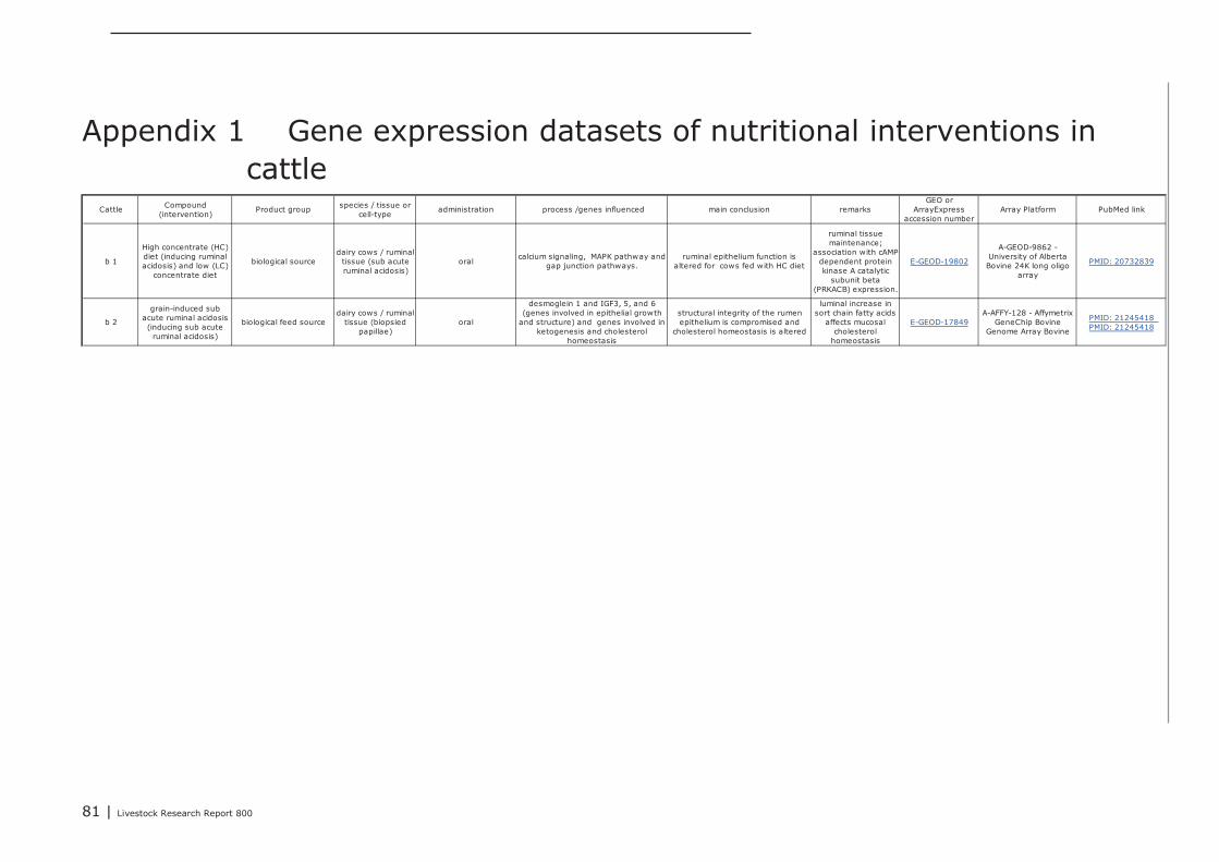
81 | Livestock Research Report 800
Cattle Compound(intervention)
Product group species / tissue orcell-type
administration process /genes influenced main conclusion remarksGEO or
ArrayExpressaccession number
Array Platform PubMed link
b 1
High concentrate (HC)diet (inducing ruminalacidosis) and low (LC)
concentrate diet
biological sourcedairy cows / ruminal
tissue (sub acuteruminal acidosis)
oralcalcium signaling, MAPK pathway and
gap junction pathways.ruminal epithelium function is
altered for cows fed with HC diet
ruminal tissuemaintenance;
association with cAMP-dependent proteinkinase A catalytic
subunit beta(PRKACB) expression.
E-GEOD-19802
A-GEOD-9862 -University of AlbertaBovine 24K long oligo
array
PMID: 20732839
b 2
grain-induced subacute ruminal acidosis(inducing sub acute
ruminal acidosis)
biological feed sourcedairy cows / ruminal
tissue (biopsiedpapillae)
oral
desmoglein 1 and IGF3, 5, and 6(genes involved in epithelial growth
and structure) and genes involved inketogenesis and cholesterol
homeostasis
structural integrity of the rumenepithelium is compromised and
cholesterol homeostasis is altered
luminal increase insort chain fatty acids
affects mucosalcholesterol
homeostasis
E-GEOD-17849A-AFFY-128 - Affymetrix
GeneChip BovineGenome Array Bovine
PMID: 21245418PMID: 21245418
Appendix 1 Gene expression datasets of nutritional interventions incattle
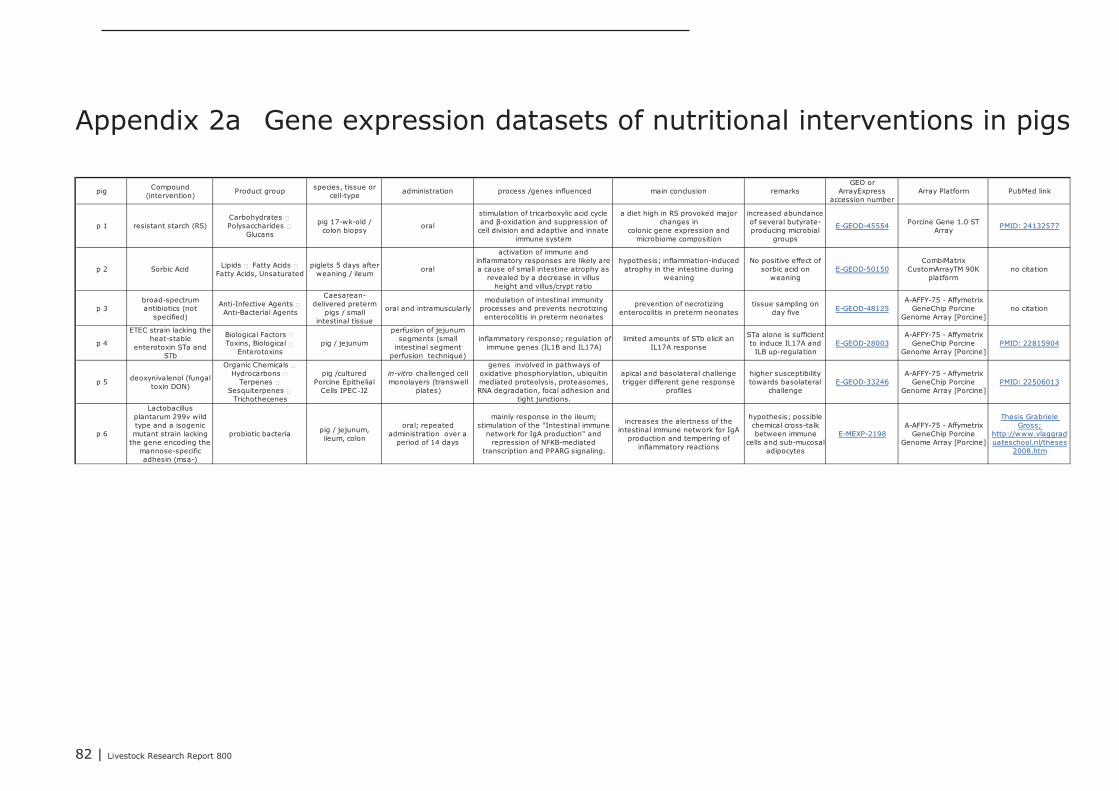
82 | Livestock Research Report 800
pig Compound(intervention)
Product group species, tissue orcell-type
administration process /genes influenced main conclusion remarksGEO or
ArrayExpressaccession number
Array Platform PubMed link
p 1 resistant starch (RS)Carbohydrates ←Polysaccharides ←
Glucans
pig 17-wk-old /colon biopsy
oral
stimulation of tricarboxylic acid cycleand β-oxidation and suppression ofcell division and adaptive and innate
immune system
a diet high in RS provoked majorchanges in
colonic gene expression andmicrobiome composition
increased abundanceof several butyrate-producing microbial
groups
E-GEOD-45554 Porcine Gene 1.0 STArray
PMID: 24132577
p 2 Sorbic Acid Lipids ← Fatty Acids ←Fatty Acids, Unsaturated
piglets 5 days afterweaning / ileum
oral
activation of immune andinflammatory responses are likely area cause of small intestine atrophy as
revealed by a decrease in villusheight and villus/crypt ratio
hypothesis; inflammation-inducedatrophy in the intestine during
weaning
No positive effect ofsorbic acid on
weaningE-GEOD-50150
CombiMatrixCustomArrayTM 90K
platformno citation
p 3broad-spectrumantibiotics (not
specified)
Anti-Infective Agents ←Anti-Bacterial Agents
Caesarean-delivered preterm
pigs / smallintestinal tissue
oral and intramuscularlymodulation of intestinal immunity
processes and prevents necrotizingenterocolitis in preterm neonates
prevention of necrotizingenterocolitis in preterm neonates
tissue sampling onday five E-GEOD-48125
A-AFFY-75 - AffymetrixGeneChip Porcine
Genome Array [Porcine]no citation
p 4
ETEC strain lacking theheat-stable
enterotoxin STa andSTb
Biological Factors ←Toxins, Biological ←
Enterotoxinspig / jejunum
perfusion of jejunumsegments (small
intestinal segmentperfusion technique)
inflammatory response; regulation ofimmune genes (IL1B and IL17A)
limited amounts of STb elicit anIL17A response
STa alone is sufficientto induce IL17A and
ILB up-regulationE-GEOD-28003
A-AFFY-75 - AffymetrixGeneChip Porcine
Genome Array [Porcine]PMID: 22815904
p 5 deoxynivalenol (fungaltoxin DON)
Organic Chemicals ←Hydrocarbons ←
Terpenes ←Sesquiterpenes ←
Trichothecenes
pig /culturedPorcine Epithelial
Cells IPEC‑J2
in-vitro challenged cellmonolayers (transwell
plates)
genes involved in pathways ofoxidative phosphorylation, ubiquitinmediated proteolysis, proteasomes,RNA degradation, focal adhesion and
tight junctions.
apical and basolateral challengetrigger different gene response
profiles
higher susceptibilitytowards basolateral
challengeE-GEOD-33246
A-AFFY-75 - AffymetrixGeneChip Porcine
Genome Array [Porcine]PMID: 22506013
p 6
Lactobacillusplantarum 299v wildtype and a isogenicmutant strain lacking
the gene encoding themannose-specificadhesin (msa-)
probiotic bacteria pig / jejunum,ileum, colon
oral; repeatedadministration over a
period of 14 days
mainly response in the ileum;stimulation of the "Intestinal immune
network for IgA production" andrepression of NFKB-mediated
transcription and PPARG signaling.
increases the alertness of theintestinal immune network for IgA
production and tempering ofinflammatory reactions
hypothesis; possiblechemical cross-talkbetween immune
cells and sub-mucosaladipocytes
E-MEXP-2198A-AFFY-75 - Affymetrix
GeneChip PorcineGenome Array [Porcine]
Thesis GrabrieleGross;
http://www.vlaggraduateschool.nl/theses
2008.htm
Appendix 2a Gene expression datasets of nutritional interventions in pigs
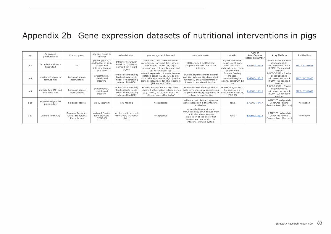
Livestock Research Report 800 | 83
pig Compound(intervention)
Product group species, tissue orcell-type
administration process /genes influenced main conclusion remarksGEO or
ArrayExpressaccession number
Array Platform PubMed link
p 7 Intrauterine GrowthRestricted
NA
piglets (age 0, 2and 5 days of life) /
distal smallintestine (ileum)
and colon
Intrauterine GrowthRestricted (IUGR) vs.normal birth weight
piglets
ileum and colon: macromoleculemetabolism, transport, biosynthesis,
physiological processes, signaltransduction, cell development, and
cell death processes.
IUGR affected proliferation-apoptosis homeostasis in the
intestine
Piglets with IUGRpossess a thinner
intestine and areduced surface area
of exchange
E-GEOD-15384
A-GEOD-7576 - Porcineoligonucleotide
microarray version 4(POM4) (Condensed
version)
PMID: 20335628
p 8 porcine colostrum orformula milk
biological source(formulated)
preterm pigs /distal smallintestine
oral or enteral (tube)feeding/preterm pigmodel for necrotizingenterocolitis (NEC)
altered expression of innate immunedefense genes (IL-1α, IL-6, IL-18),
nitric oxide synthetase, tight junctionproteins (claudins), Toll-like receptors
(TLR-4), and TNF-α
Switchs of parenteral to enteralnutrition induces diet-dependentfunctional, and proinflammatoryinsults to immature intestine.
Formula feedinginduced
histopathologicallesions, colostrum did
not
E-GEOD-13516
A-GEOD-7576 - Porcineoligonucleotide
microarray version 4(POM4) (Condensed
version)
PMID: 21700903
p 9 amniotic fluid (AF) andor formula milk
biological source(formulated)
preterm pigs /distal smallintestine
oral or enteral (tube)feeding/preterm pigmodel for necrotizingenterocolitis (NEC)
Formula-enteral feeded pigs down-regulated inflammation-related genes
(e.g., TNF-α, IL-1α, IL-6, NOS). Noeffect of enteral feeded AF
AF reduces NEC development inpreterm neonates by suppressingthe proinflammatory responses to
enteral formula feeding
AF down-regulated IL-6 expression in
intestinal cells (IEC-6,IPEC-J2)
E-GEOD-13515
A-GEOD-7576 - Porcineoligonucleotide
microarray version 4(POM4) (Condensed
version)
PMID: 23518680
p 10animal or vegetable
protein diet biological source pigs / jejunum oral feeding not specifiedevidence that diet can regulatesgene expression in the intestinal
epitheliumnone E-GEOD-13457
A-AFFY-75 - AffymetrixGeneChip Porcine
Genome Array [Porcine]no citation
p 11 Cholera toxin (CT)Biological Factors ←Toxins, Biological ←
Enterotoxins
cultured PorcineEpithelial Cells
(IPEC‑J2)
in-vitro challenged cellmonolayers (transwell
plates)not specified
mucosal adjuvanticity andimmunogenicity of CT derives from
rapid alterations in geneexpression at the site of firstantigen encounter with theintestinal immune system
none E-GEOD-10314A-AFFY-75 - Affymetrix
GeneChip PorcineGenome Array [Porcine]
no citation
Appendix 2b Gene expression datasets of nutritional interventions in pigs
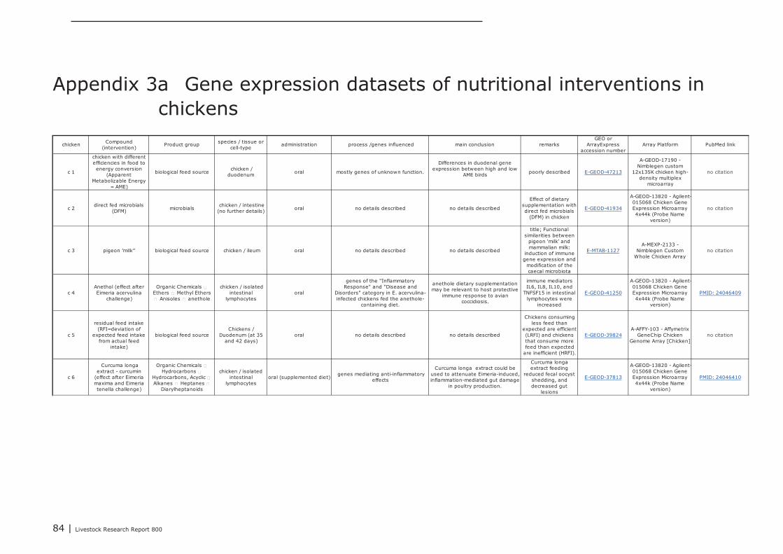
84 | Livestock Research Report 800
chicken Compound(intervention)
Product group species / tissue orcell-type
administration process /genes influenced main conclusion remarksGEO or
ArrayExpressaccession number
Array Platform PubMed link
c 1
chicken with differentefficiencies in food toenergy conversion
(ApparentMetabolizable Energy
= AME)
biological feed source chicken /duodenum oral mostly genes of unknown function.
Differences in duodenal geneexpression between high and low
AME birds poorly described E-GEOD-47213
A-GEOD-17190 -Nimblegen custom
12x135K chicken high-density multiplex
microarray
no citation
c 2direct fed microbials
(DFM) microbials chicken / intestine(no further details) oral no details described no details described
Effect of dietarysupplementation withdirect fed microbials
(DFM) in chicken
E-GEOD-41934
A-GEOD-13820 - Agilent-015068 Chicken GeneExpression Microarray4x44k (Probe Name
version)
no citation
c 3 pigeon 'milk'' biological feed source chicken / ileum oral no details described no details described
title; Functionalsimilarities between
pigeon ‘milk' andmammalian milk:
induction of immunegene expression and
modification of thecaecal microbiota
E-MTAB-1127A-MEXP-2133 -
Nimblegen CustomWhole Chicken Array
no citation
c 4Anethol (effect afterEimeria acervulina
challenge)
Organic Chemicals ←Ethers ← Methyl Ethers← Anisoles ← anethole
chicken / isolatedintestinal
lymphocytesoral
genes of the "InflammatoryResponse" and "Disease and
Disorders" category in E. acervulina-infected chickens fed the anethole-
containing diet.
anethole dietary supplementationmay be relevant to host protective
immune response to aviancoccidiosis.
immune mediatorsIL6, IL8, IL10, and
TNFSF15 in intestinallymphocytes were
increased
E-GEOD-41250
A-GEOD-13820 - Agilent-015068 Chicken GeneExpression Microarray4x44k (Probe Name
version)
PMID: 24046409
c 5
residual feed intake(RFI=deviation of
expected feed intakefrom actual feed
intake)
biological feed sourceChickens /
Duodenum (at 35and 42 days)
oral no details described no details described
Chickens consumingless feed than
expected are efficient(LRFI) and chickensthat consume morefeed than expected
are inefficient (HRFI).
E-GEOD-39824A-AFFY-103 - Affymetrix
GeneChip ChickenGenome Array [Chicken]
no citation
c 6
Curcuma longaextract - curcumin
(effect after Eimeriamaxima and Eimeriatenella challenge)
Organic Chemicals ←Hydrocarbons ←
Hydrocarbons, Acyclic ←Alkanes ← Heptanes ←
Diarylheptanoids
chicken / isolatedintestinal
lymphocytesoral (supplemented diet)
genes mediating anti-inflammatoryeffects
Curcuma longa extract could beused to attenuate Eimeria-induced,inflammation-mediated gut damage
in poultry production.
Curcuma longaextract feeding
reduced fecal oocystshedding, anddecreased gut
lesions
E-GEOD-37813
A-GEOD-13820 - Agilent-015068 Chicken GeneExpression Microarray4x44k (Probe Name
version)
PMID: 24046410
Appendix 3a Gene expression datasets of nutritional interventions inchickens
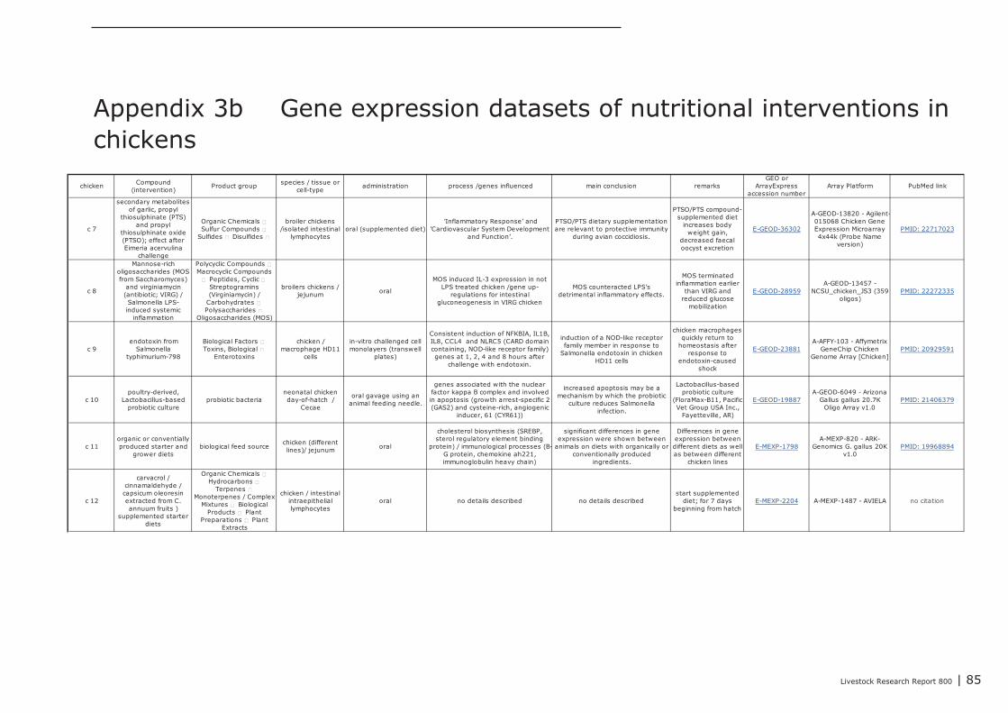
Livestock Research Report 800 | 85
chicken Compound(intervention)
Product group species / tissue orcell-type
administration process /genes influenced main conclusion remarksGEO or
ArrayExpressaccession number
Array Platform PubMed link
c 7
secondary metabolitesof garlic, propyl
thiosulphinate (PTS)and propyl
thiosulphinate oxide(PTSO); effect afterEimeria acervulina
challenge
Organic Chemicals ←Sulfur Compounds ←
Sulfides ← Disulfides ←
broiler chickens/isolated intestinal
lymphocytesoral (supplemented diet)
'Inflammatory Response' and'Cardiovascular System Development
and Function'.
PTSO/PTS dietary supplementationare relevant to protective immunity
during avian coccidiosis.
PTSO/PTS compound-supplemented diet
increases bodyweight gain,
decreased faecaloocyst excretion
E-GEOD-36302
A-GEOD-13820 - Agilent-015068 Chicken GeneExpression Microarray4x44k (Probe Name
version)
PMID: 22717023
c 8
Mannose-richoligosaccharides (MOSfrom Saccharomyces)
and virginiamycin(antibiotic; VIRG) /Salmonella LPS-induced systemic
inflammation
Polycyclic Compounds ←Macrocyclic Compounds← Peptides, Cyclic ←
Streptogramins(Virginiamycin) /
Carbohydrates ←Polysaccharides ←
Oligosaccharides (MOS)
broilers chickens /jejunum oral
MOS induced IL-3 expression in notLPS treated chicken /gene up-
regulations for intestinalgluconeogenesis in VIRG chicken
MOS counteracted LPS'sdetrimental inflammatory effects.
MOS terminatedinflammation earlier
than VIRG andreduced glucose
mobilization
E-GEOD-28959A-GEOD-13457 -
NCSU_chicken_JS3 (359oligos)
PMID: 22272335
c 9endotoxin from
Salmonellatyphimurium-798
Biological Factors ←Toxins, Biological ←
Enterotoxins
chicken /macrophage HD11
cells
in-vitro challenged cellmonolayers (transwell
plates)
Consistent induction of NFKBIA, IL1B,IL8, CCL4 and NLRC5 (CARD domaincontaining, NOD-like receptor family)genes at 1, 2, 4 and 8 hours after
challenge with endotoxin.
induction of a NOD-like receptorfamily member in response to
Salmonella endotoxin in chickenHD11 cells
chicken macrophagesquickly return to
homeostasis afterresponse to
endotoxin-causedshock
E-GEOD-23881A-AFFY-103 - Affymetrix
GeneChip ChickenGenome Array [Chicken]
PMID: 20929591
c 10poultry-derived,
Lactobacillus-basedprobiotic culture
probiotic bacterianeonatal chickenday-of-hatch /
Cecae
oral gavage using ananimal feeding needle.
genes associated with the nuclearfactor kappa B complex and involvedin apoptosis (growth arrest-specific 2(GAS2) and cysteine-rich, angiogenic
inducer, 61 (CYR61))
increased apoptosis may be amechanism by which the probiotic
culture reduces Salmonellainfection.
Lactobacillus-basedprobiotic culture
(FloraMax-B11, PacificVet Group USA Inc.,
Fayetteville, AR)
E-GEOD-19887A-GEOD-6049 - Arizona
Gallus gallus 20.7KOligo Array v1.0
PMID: 21406379
c 11organic or conventiallyproduced starter and
grower dietsbiological feed source
chicken (differentlines)/ jejunum oral
cholesterol biosynthesis (SREBP,sterol regulatory element binding
protein) / immunological processes (B-G protein, chemokine ah221,immunoglobulin heavy chain)
significant differences in geneexpression were shown between
animals on diets with organically orconventionally produced
ingredients.
Differences in geneexpression betweendifferent diets as wellas between different
chicken lines
E-MEXP-1798A-MEXP-820 - ARK-
Genomics G. gallus 20Kv1.0
PMID: 19968894
c 12
carvacrol /cinnamaldehyde /capsicum oleoresinextracted from C.annuum fruits )
supplemented starterdiets
Organic Chemicals ←Hydrocarbons ←
Terpenes ←Monoterpenes / Complex
Mixtures ← BiologicalProducts ← Plant
Preparations ← PlantExtracts
chicken / intestinalintraepitheliallymphocytes
oral no details described no details describedstart supplemented
diet; for 7 daysbeginning from hatch
E-MEXP-2204 A-MEXP-1487 - AVIELA no citation
Appendix 3b Gene expression datasets of nutritional interventions inchickens
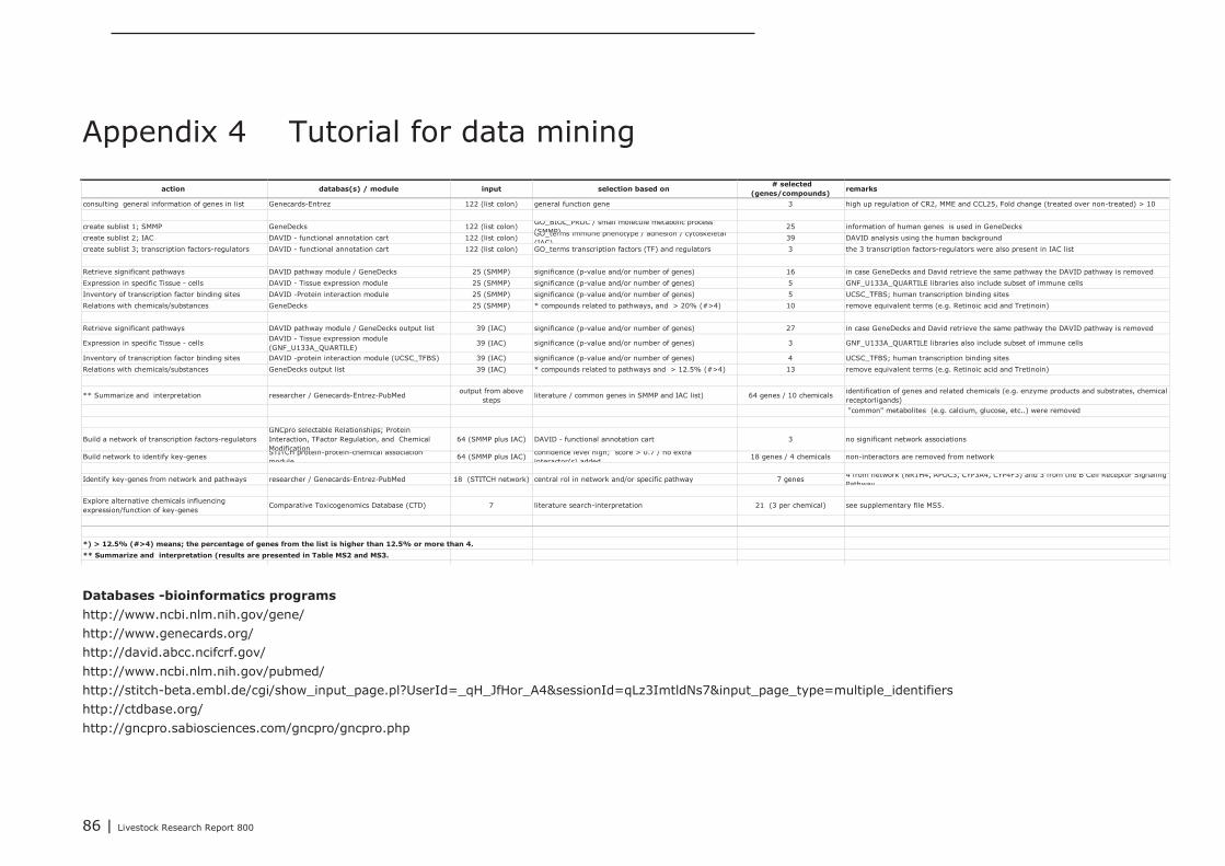
86 | Livestock Research Report 800
action databas(s) / module input selection based on # selected
(genes/compounds)remarks
consulting general information of genes in list Genecards-Entrez 122 (list colon) general function gene 3 high up regulation of CR2, MME and CCL25, Fold change (treated over non-treated) > 10
create sublist 1; SMMP GeneDecks 122 (list colon) GO_BIOL_PROC / small molecule metabolic process(SMMP)
25 information of human genes is used in GeneDecks
create sublist 2; IAC DAVID - functional annotation cart 122 (list colon) GO_terms immune phenotype / adhesion / cytoskeletal(IAC)
39 DAVID analysis using the human background
create sublist 3; transcription factors-regulators DAVID - functional annotation cart 122 (list colon) GO_terms transcription factors (TF) and regulators 3 the 3 transcription factors-regulators were also present in IAC list
Retrieve significant pathways DAVID pathway module / GeneDecks 25 (SMMP) significance (p-value and/or number of genes) 16 in case GeneDecks and David retrieve the same pathway the DAVID pathway is removed
Expression in specific Tissue - cells DAVID - Tissue expression module 25 (SMMP) significance (p-value and/or number of genes) 5 GNF_U133A_QUARTILE libraries also include subset of immune cells
Inventory of transcription factor binding sites DAVID -Protein interaction module 25 (SMMP) significance (p-value and/or number of genes) 5 UCSC_TFBS; human transcription binding sites
Relations with chemicals/substances GeneDecks 25 (SMMP) * compounds related to pathways, and > 20% (#>4) 10 remove equivalent terms (e.g. Retinoic acid and Tretinoin)
Retrieve significant pathways DAVID pathway module / GeneDecks output list 39 (IAC) significance (p-value and/or number of genes) 27 in case GeneDecks and David retrieve the same pathway the DAVID pathway is removed
Expression in specific Tissue - cells DAVID - Tissue expression module(GNF_U133A_QUARTILE)
39 (IAC) significance (p-value and/or number of genes) 3 GNF_U133A_QUARTILE libraries also include subset of immune cells
Inventory of transcription factor binding sites DAVID -protein interaction module (UCSC_TFBS) 39 (IAC) significance (p-value and/or number of genes) 4 UCSC_TFBS; human transcription binding sites
Relations with chemicals/substances GeneDecks output list 39 (IAC) * compounds related to pathways and > 12.5% (#>4) 13 remove equivalent terms (e.g. Retinoic acid and Tretinoin)
** Summarize and interpretation researcher / Genecards-Entrez-PubMed output from abovesteps
literature / common genes in SMMP and IAC list) 64 genes / 10 chemicals identification of genes and related chemicals (e.g. enzyme products and substrates, chemicalreceptorligands) "common" metabolites (e.g. calcium, glucose, etc..) were removed
Build a network of transcription factors-regulatorsGNCpro selectable Relationships; ProteinInteraction, TFactor Regulation, and ChemicalModification
64 (SMMP plus IAC) DAVID - functional annotation cart 3 no significant network associations
Build network to identify key-genes STITCH protein-protein-chemical associationmodule
64 (SMMP plus IAC) confidence level high; score > 0.7 / no extrainteractor(s) added
18 genes / 4 chemicals non-interactors are removed from network
Identify key-genes from network and pathways researcher / Genecards-Entrez-PubMed 18 (STITCH network) central rol in network and/or specific pathway 7 genes 4 from network (NR1H4, APOC3, CYP3A4, CYP4F3) and 3 from the B Cell Receptor SignalingPathway
Explore alternative chemicals influencingexpression/function of key-genes
Comparative Toxicogenomics Database (CTD) 7 literature search-interpretation 21 (3 per chemical) see supplementary file MS5.
*) > 12.5% (#>4) means; the percentage of genes from the list is higher than 12.5% or more than 4.
** Summarize and interpretation (results are presented in Table MS2 and MS3.
Databaes -bioinformatics programs
http://www.ncbi.nlm.nih.gov/gene/
http://www.genecards.org/
http://www.genecards.org/index.php?path=/GeneDecks
http://david.abcc.ncifcrf.gov/
http://www.ncbi.nlm.nih.gov/pubmed/
http://stitch-beta.embl.de/cgi/show_input_page.pl?UserId=_qH_JfHor_A4&sessionId=qLz3ImtldNs7&input_page_type=multiple_identifiers
http://ctdbase.org/
http://gncpro.sabiosciences.com/gncpro/gncpro.php
Appendix 4 Tutorial for data mining
Databases -bioinformatics programs
http://www.ncbi.nlm.nih.gov/gene/http://www.genecards.org/http://david.abcc.ncifcrf.gov/http://www.ncbi.nlm.nih.gov/pubmed/http://stitch-beta.embl.de/cgi/show_input_page.pl?UserId=_qH_JfHor_A4&sessionId=qLz3ImtldNs7&input_page_type=multiple_identifiershttp://ctdbase.org/http://gncpro.sabiosciences.com/gncpro/gncpro.php
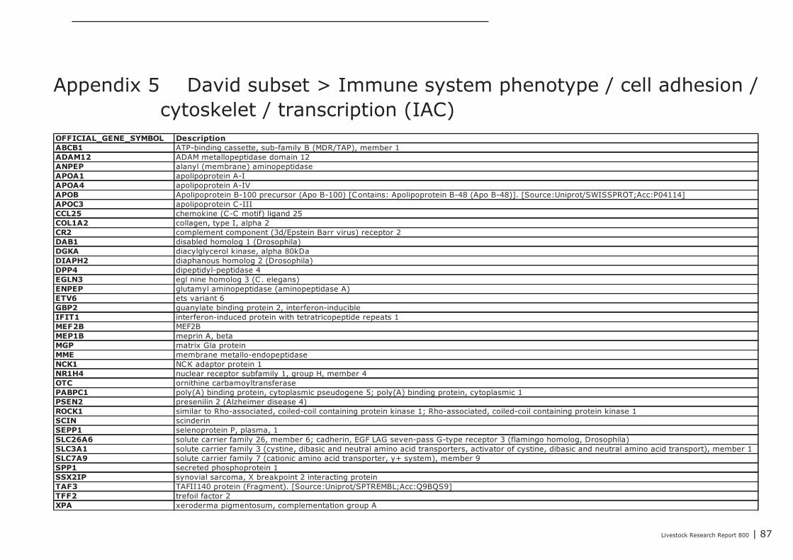
Livestock Research Report 800 | 87
OFFICIAL_GENE_SYMBOL DescriptionABCB1 ATP-binding cassette, sub-family B (MDR/TAP), member 1ADAM12 ADAM metallopeptidase domain 12ANPEP alanyl (membrane) aminopeptidaseAPOA1 apolipoprotein A-IAPOA4 apolipoprotein A-IVAPOB Apolipoprotein B-100 precursor (Apo B-100) [Contains: Apolipoprotein B-48 (Apo B-48)]. [Source:Uniprot/SWISSPROT;Acc:P04114]APOC3 apolipoprotein C-IIICCL25 chemokine (C-C motif) ligand 25COL1A2 collagen, type I, alpha 2CR2 complement component (3d/Epstein Barr virus) receptor 2DAB1 disabled homolog 1 (Drosophila)DGKA diacylglycerol kinase, alpha 80kDaDIAPH2 diaphanous homolog 2 (Drosophila)DPP4 dipeptidyl-peptidase 4EGLN3 egl nine homolog 3 (C. elegans)ENPEP glutamyl aminopeptidase (aminopeptidase A)ETV6 ets variant 6GBP2 guanylate binding protein 2, interferon-inducibleIFIT1 interferon-induced protein with tetratricopeptide repeats 1MEF2B MEF2BMEP1B meprin A, betaMGP matrix Gla proteinMME membrane metallo-endopeptidaseNCK1 NCK adaptor protein 1NR1H4 nuclear receptor subfamily 1, group H, member 4OTC ornithine carbamoyltransferasePABPC1 poly(A) binding protein, cytoplasmic pseudogene 5; poly(A) binding protein, cytoplasmic 1PSEN2 presenilin 2 (Alzheimer disease 4)ROCK1 similar to Rho-associated, coiled-coil containing protein kinase 1; Rho-associated, coiled-coil containing protein kinase 1SCIN scinderinSEPP1 selenoprotein P, plasma, 1SLC26A6 solute carrier family 26, member 6; cadherin, EGF LAG seven-pass G-type receptor 3 (flamingo homolog, Drosophila)SLC3A1 solute carrier family 3 (cystine, dibasic and neutral amino acid transporters, activator of cystine, dibasic and neutral amino acid transport), member 1SLC7A9 solute carrier family 7 (cationic amino acid transporter, y+ system), member 9SPP1 secreted phosphoprotein 1SSX2IP synovial sarcoma, X breakpoint 2 interacting proteinTAF3 TAFII140 protein (Fragment). [Source:Uniprot/SPTREMBL;Acc:Q9BQS9]TFF2 trefoil factor 2XPA xeroderma pigmentosum, complementation group A
Appendix 5 David subset > Immune system phenotype / cell adhesion /cytoskelet / transcription (IAC)
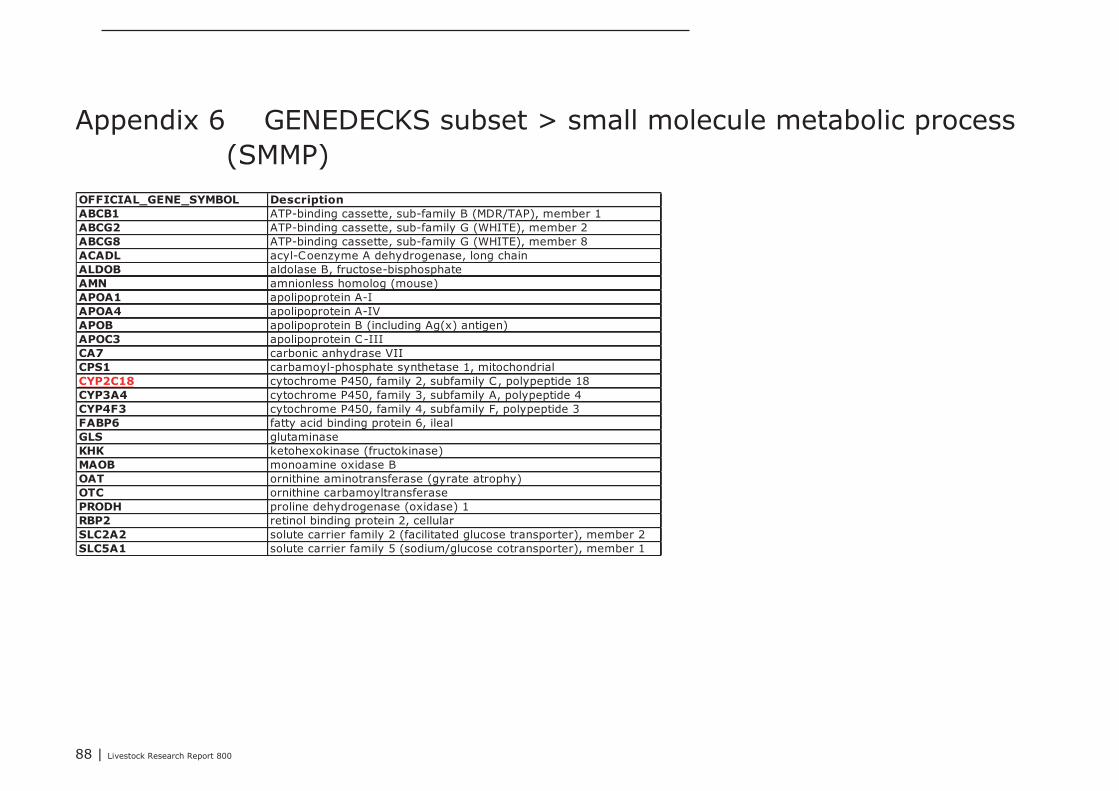
88 | Livestock Research Report 800
OFFICIAL_GENE_SYMBOL DescriptionABCB1 ATP-binding cassette, sub-family B (MDR/TAP), member 1ABCG2 ATP-binding cassette, sub-family G (WHITE), member 2ABCG8 ATP-binding cassette, sub-family G (WHITE), member 8ACADL acyl-Coenzyme A dehydrogenase, long chainALDOB aldolase B, fructose-bisphosphateAMN amnionless homolog (mouse)APOA1 apolipoprotein A-IAPOA4 apolipoprotein A-IVAPOB apolipoprotein B (including Ag(x) antigen)APOC3 apolipoprotein C-IIICA7 carbonic anhydrase VIICPS1 carbamoyl-phosphate synthetase 1, mitochondrialCYP2C18 cytochrome P450, family 2, subfamily C, polypeptide 18CYP3A4 cytochrome P450, family 3, subfamily A, polypeptide 4CYP4F3 cytochrome P450, family 4, subfamily F, polypeptide 3FABP6 fatty acid binding protein 6, ilealGLS glutaminaseKHK ketohexokinase (fructokinase)MAOB monoamine oxidase BOAT ornithine aminotransferase (gyrate atrophy)OTC ornithine carbamoyltransferasePRODH proline dehydrogenase (oxidase) 1RBP2 retinol binding protein 2, cellularSLC2A2 solute carrier family 2 (facilitated glucose transporter), member 2SLC5A1 solute carrier family 5 (sodium/glucose cotransporter), member 1
Appendix 6 GENEDECKS subset > small molecule metabolic process(SMMP)
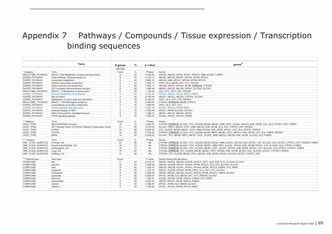
Livestock Research Report 800 | 89
Term # genes % p -value genesa
(N=25)* Category Term Count % PValue GenesREACTOME_PATHWAY REACT_602:Metabolism of lipids and lipoproteins 8 32 9.16E-06 APOA4, ABCG8, APOB, APOA1, APOC3, AMN, ACADL, FABP6SUPER_PATHWAY Statin Pathway, Pharmacodynamics 6 24 4.41E-13 ABCB1, ABCG8, APOA1, APOA4, APOB, APOC3SUPER_PATHWAY Lipoprotein metabolism 6 24 2.86E-12 ABCG8, AMN, APOA1, APOA4, APOB, APOC3SUPER_PATHWAY Arginine and proline metabolism 6 24 1.65E-11 CPS1, GLS, MAOB, OAT, OTC, PRODHSUPER_PATHWAY alpha-Linolenic acid metabolism 6 24 7.56E-11 ABCG8, APOA1, APOA4, APOB, CYP2C18, CYP3A4SUPER_PATHWAY SLC-mediated transmembrane transport 6 24 7.09E-06 ABCB1, ABCG2, ABCG8, APOA1, SLC2A2, SLC5A1REACTOME_PATHWAY REACT_13:Metabolism of amino acids 5 20 1.12E-02 GLS, OTC, CPS1, OAT, PRODHSUPER_PATHWAY Retinoid metabolism and transport 5 20 2.31E-09 APOA1, APOA4, APOB, APOC3, RBP2SUPER_PATHWAY Bile secretion 5 20 8.19E-09 ABCB1, ABCG2, ABCG8, CYP3A4, SLC5A1SUPER_PATHWAY Metabolism of amino acids and derivatives 5 20 9.14E-07 CPS1, GLS, OAT, OTC, PRODHREACTOME_PATHWAY REACT_13433:Biological oxidations 4 16 2.52E-02 CYP3A4, CYP2C18, MAOB, CYP4F3SUPER_PATHWAY superpathway of citrulline metabolism 4 16 1.46E-09 CPS1, GLS, OAT, OTCSUPER_PATHWAY Vitamin digestion and absorption 4 16 9.50E-09 APOA1, APOA4, APOB, RBP2SUPER_PATHWAY Selected targets of HNF1 4 16 7.25E-08 APOA1, APOB, SLC2A2, SLC5A1SUPER_PATHWAY ABC-family proteins mediated transport 4 16 1.38E-07 ABCB1, ABCG2, ABCG8, APOA1SUPER_PATHWAY PPAR signaling pathway 4 16 7.05E-07 ACADL, APOA1, APOC3, FABP6
** Category Term Count % PValue GenesUCSC_TFBS MEIS1B-HOXA9 complex 18 72 4.40E-03 CYP3A4, CYP2C18, SLC5A1, OTC, ALDOB, MAOB, ABCB1, AMN, CPS1, ACADL, ABCG8, KHK, APOB, CA7, GLS, CYP4F3, OAT, FABP6UCSC_TFBS NF1=Nuclear Factor I/X (CCAAT-Binding Transcription Factor) 13 52 3.97E-02 SLC5A1, RBP2, ABCB1, CPS1, AMN, ABCG2, KHK, APOB, GLS, CA7, CYP4F3, OAT, PRODHUCSC_TFBS RORA1 14 56 6.92E-02 OTC, ALDOB, MAOB, ABCB1, CPS1, AMN, APOA4, KHK, APOB, APOA1, CA7, GLS, APOC3, PRODHUCSC_TFBS FOXO4 16 64 7.37E-02 CYP3A4, CYP2C18, SLC5A1, OTC, ALDOB, MAOB, RBP2, ABCB1, CPS1, ABCG2, KHK, APOB, CA7, GLS, FABP6, PRODHUCSC_TFBS BRN2 15 60 8.04E-02 SLC5A1, OTC, MAOB, RBP2, ABCB1, CPS1, ACADL, AMN, ABCG2, ABCG8, KHK, APOB, SLC2A2, GLS, FABP6
*** Category Term Count % PValue GenesGNF_U133A_QUARTILE Prostate_3rd 20 80 NA CYP3A4, CYP2C18, SLC5A1, OTC, ALDOB, MAOB, ABCB1, AMN, ACADL, ABCG2, KHK, APOA1, CA7, SLC2A2, GLS, APOC3, CYP4F3, OAT, PRODH, FABP6GNF_U133A_QUARTILE lymphomaburkittsRaji_3rd 18 72 NA CYP3A4, CYP2C18, SLC5A1, OTC, ALDOB, ABCB1, AMN, CPS1, ACADL, APOA4, KHK, APOB, APOA1, CA7, SLC2A2, GLS, CYP4F3, FABP6GNF_U133A_QUARTILE salivarygland_3rd 18 72 NA CYP3A4, CYP2C18, SLC5A1, OTC, ALDOB, ABCB1, CPS1, ACADL, APOA4, KHK, APOB, APOA1, CA7, SLC2A2, GLS, APOC3, CYP4F3, FABP6GNF_U133A_QUARTILE Lung_3rd 16 64 NA CYP3A4, CYP2C18, OTC, ALDOB, MAOB, ABCB1, CPS1, APOA4, KHK, APOB, APOA1, CA7, SLC2A2, APOC3, CYP4F3, PRODHGNF_U133A_QUARTILE fetallung_3rd 13 52 NA CYP3A4, OTC, ALDOB, MAOB, CPS1, ABCG2, KHK, APOB, APOA1, SLC2A2, APOC3, CYP4F3, OAT
****Attribute type Descriptor Score P Value Genes sharing this descriptorCOMPOUND atp 11 44 6.21E-10 ABCB1, ABCG2, ABCG8, ALDOB, APOC3, CPS1, GLS, KHK, OTC, SLC2A2, SLC5A1COMPOUND glucose 10 40 1.65E-08 ABCG8, ALDOB, APOA1, APOA4, APOB, APOC3, GLS, OTC, SLC2A2, SLC5A1COMPOUND lipid 10 40 1.32E-07 ABCG2, ABCG8, ACADL, APOA1, APOA4, APOB, APOC3, FABP6, OTC, RBP2COMPOUND glutamine 9 36 1.12E-10 ABCG2, ALDOB, APOA4, APOB, CPS1, GLS, OAT, OTC, SLC5A1COMPOUND cholesterol 9 36 5.20E-09 ABCB1, ABCG2, ABCG8, APOA1, APOA4, APOB, APOC3, FABP6, SLC5A1COMPOUND glutamate 8 32 6.85E-07 APOA1, APOB, GLS, MAOB, OAT, OTC, PRODH, SLC5A1COMPOUND fatty acid 7 28 1.20E-07 ACADL, APOA4, APOB, APOC3, FABP6, OTC, RBP2COMPOUND taurocholate 5 20 6.05E-10 ABCB1, APOA1, APOB, APOC3, FABP6COMPOUND choline 5 20 3.38E-07 APOA1, APOB, GLS, MAOB, SLC5A1COMPOUND vitamin a 5 20 1.34E-06 APOA1, APOA4, APOB, APOC3, RBP2
Appendix 7 Pathways / Compounds / Tissue expression / Transcriptionbinding sequences
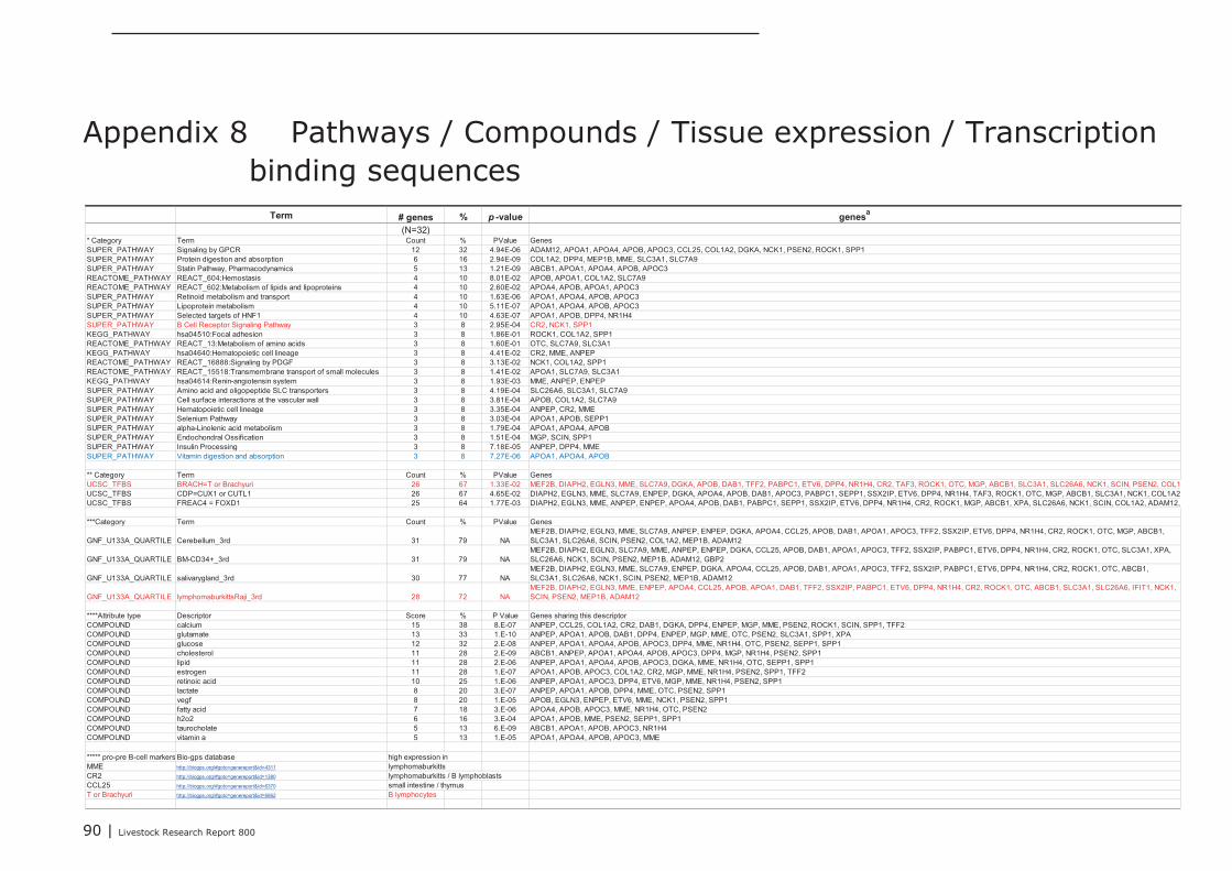
90 | Livestock Research Report 800
Term # genes % p -value genesa
(N=32)* Category Term Count % PValue GenesSUPER_PATHWAY Signaling by GPCR 12 32 4.94E-06 ADAM12, APOA1, APOA4, APOB, APOC3, CCL25, COL1A2, DGKA, NCK1, PSEN2, ROCK1, SPP1SUPER_PATHWAY Protein digestion and absorption 6 16 2.94E-09 COL1A2, DPP4, MEP1B, MME, SLC3A1, SLC7A9SUPER_PATHWAY Statin Pathway, Pharmacodynamics 5 13 1.21E-09 ABCB1, APOA1, APOA4, APOB, APOC3REACTOME_PATHWAY REACT_604:Hemostasis 4 10 8.01E-02 APOB, APOA1, COL1A2, SLC7A9REACTOME_PATHWAY REACT_602:Metabolism of lipids and lipoproteins 4 10 2.60E-02 APOA4, APOB, APOA1, APOC3SUPER_PATHWAY Retinoid metabolism and transport 4 10 1.63E-06 APOA1, APOA4, APOB, APOC3SUPER_PATHWAY Lipoprotein metabolism 4 10 5.11E-07 APOA1, APOA4, APOB, APOC3SUPER_PATHWAY Selected targets of HNF1 4 10 4.63E-07 APOA1, APOB, DPP4, NR1H4SUPER_PATHWAY B Cell Receptor Signaling Pathway 3 8 2.95E-04 CR2, NCK1, SPP1KEGG_PATHWAY hsa04510:Focal adhesion 3 8 1.86E-01 ROCK1, COL1A2, SPP1REACTOME_PATHWAY REACT_13:Metabolism of amino acids 3 8 1.60E-01 OTC, SLC7A9, SLC3A1KEGG_PATHWAY hsa04640:Hematopoietic cell lineage 3 8 4.41E-02 CR2, MME, ANPEPREACTOME_PATHWAY REACT_16888:Signaling by PDGF 3 8 3.13E-02 NCK1, COL1A2, SPP1REACTOME_PATHWAY REACT_15518:Transmembrane transport of small molecules 3 8 1.41E-02 APOA1, SLC7A9, SLC3A1KEGG_PATHWAY hsa04614:Renin-angiotensin system 3 8 1.93E-03 MME, ANPEP, ENPEPSUPER_PATHWAY Amino acid and oligopeptide SLC transporters 3 8 4.19E-04 SLC26A6, SLC3A1, SLC7A9SUPER_PATHWAY Cell surface interactions at the vascular wall 3 8 3.81E-04 APOB, COL1A2, SLC7A9SUPER_PATHWAY Hematopoietic cell lineage 3 8 3.35E-04 ANPEP, CR2, MMESUPER_PATHWAY Selenium Pathway 3 8 3.03E-04 APOA1, APOB, SEPP1SUPER_PATHWAY alpha-Linolenic acid metabolism 3 8 1.79E-04 APOA1, APOA4, APOBSUPER_PATHWAY Endochondral Ossification 3 8 1.51E-04 MGP, SCIN, SPP1SUPER_PATHWAY Insulin Processing 3 8 7.18E-05 ANPEP, DPP4, MMESUPER_PATHWAY Vitamin digestion and absorption 3 8 7.27E-06 APOA1, APOA4, APOB
** Category Term Count % PValue GenesUCSC_TFBS BRACH=T or Brachyuri 26 67 1.33E-02 MEF2B, DIAPH2, EGLN3, MME, SLC7A9, DGKA, APOB, DAB1, TFF2, PABPC1, ETV6, DPP4, NR1H4, CR2, TAF3, ROCK1, OTC, MGP, ABCB1, SLC3A1, SLC26A6, NCK1, SCIN, PSEN2, COL1A2, ADAM12UCSC_TFBS CDP=CUX1 or CUTL1 26 67 4.65E-02 DIAPH2, EGLN3, MME, SLC7A9, ENPEP, DGKA, APOA4, APOB, DAB1, APOC3, PABPC1, SEPP1, SSX2IP, ETV6, DPP4, NR1H4, TAF3, ROCK1, OTC, MGP, ABCB1, SLC3A1, NCK1, COL1A2, ADAM12, GBP2UCSC_TFBS FREAC4 = FOXD1 25 64 1.77E-03 DIAPH2, EGLN3, MME, ANPEP, ENPEP, APOA4, APOB, DAB1, PABPC1, SEPP1, SSX2IP, ETV6, DPP4, NR1H4, CR2, ROCK1, MGP, ABCB1, XPA, SLC26A6, NCK1, SCIN, COL1A2, ADAM12, GBP2
***Category Term Count % PValue Genes
GNF_U133A_QUARTILE Cerebellum_3rd 31 79 NAMEF2B, DIAPH2, EGLN3, MME, SLC7A9, ANPEP, ENPEP, DGKA, APOA4, CCL25, APOB, DAB1, APOA1, APOC3, TFF2, SSX2IP, ETV6, DPP4, NR1H4, CR2, ROCK1, OTC, MGP, ABCB1,SLC3A1, SLC26A6, SCIN, PSEN2, COL1A2, MEP1B, ADAM12
GNF_U133A_QUARTILE BM-CD34+_3rd 31 79 NAMEF2B, DIAPH2, EGLN3, SLC7A9, MME, ANPEP, ENPEP, DGKA, CCL25, APOB, DAB1, APOA1, APOC3, TFF2, SSX2IP, PABPC1, ETV6, DPP4, NR1H4, CR2, ROCK1, OTC, SLC3A1, XPA,SLC26A6, NCK1, SCIN, PSEN2, MEP1B, ADAM12, GBP2
GNF_U133A_QUARTILE salivarygland_3rd 30 77 NAMEF2B, DIAPH2, EGLN3, MME, SLC7A9, ENPEP, DGKA, APOA4, CCL25, APOB, DAB1, APOA1, APOC3, TFF2, SSX2IP, PABPC1, ETV6, DPP4, NR1H4, CR2, ROCK1, OTC, ABCB1,SLC3A1, SLC26A6, NCK1, SCIN, PSEN2, MEP1B, ADAM12
GNF_U133A_QUARTILE lymphomaburkittsRaji_3rd 28 72 NAMEF2B, DIAPH2, EGLN3, MME, ENPEP, APOA4, CCL25, APOB, APOA1, DAB1, TFF2, SSX2IP, PABPC1, ETV6, DPP4, NR1H4, CR2, ROCK1, OTC, ABCB1, SLC3A1, SLC26A6, IFIT1, NCK1,SCIN, PSEN2, MEP1B, ADAM12
****Attribute type Descriptor Score % P Value Genes sharing this descriptorCOMPOUND calcium 15 38 8.E-07 ANPEP, CCL25, COL1A2, CR2, DAB1, DGKA, DPP4, ENPEP, MGP, MME, PSEN2, ROCK1, SCIN, SPP1, TFF2COMPOUND glutamate 13 33 1.E-10 ANPEP, APOA1, APOB, DAB1, DPP4, ENPEP, MGP, MME, OTC, PSEN2, SLC3A1, SPP1, XPACOMPOUND glucose 12 32 2.E-08 ANPEP, APOA1, APOA4, APOB, APOC3, DPP4, MME, NR1H4, OTC, PSEN2, SEPP1, SPP1COMPOUND cholesterol 11 28 2.E-09 ABCB1, ANPEP, APOA1, APOA4, APOB, APOC3, DPP4, MGP, NR1H4, PSEN2, SPP1COMPOUND lipid 11 28 2.E-06 ANPEP, APOA1, APOA4, APOB, APOC3, DGKA, MME, NR1H4, OTC, SEPP1, SPP1COMPOUND estrogen 11 28 1.E-07 APOA1, APOB, APOC3, COL1A2, CR2, MGP, MME, NR1H4, PSEN2, SPP1, TFF2COMPOUND retinoic acid 10 25 1.E-06 ANPEP, APOA1, APOC3, DPP4, ETV6, MGP, MME, NR1H4, PSEN2, SPP1COMPOUND lactate 8 20 3.E-07 ANPEP, APOA1, APOB, DPP4, MME, OTC, PSEN2, SPP1COMPOUND vegf 8 20 1.E-05 APOB, EGLN3, ENPEP, ETV6, MME, NCK1, PSEN2, SPP1COMPOUND fatty acid 7 18 3.E-06 APOA4, APOB, APOC3, MME, NR1H4, OTC, PSEN2COMPOUND h2o2 6 16 3.E-04 APOA1, APOB, MME, PSEN2, SEPP1, SPP1COMPOUND taurocholate 5 13 6.E-09 ABCB1, APOA1, APOB, APOC3, NR1H4COMPOUND vitamin a 5 13 1.E-05 APOA1, APOA4, APOB, APOC3, MME
***** pro-pre B-cell markers Bio-gps database high expression inMME http://biogps.org/#goto=genereport&id=4311 lymphomaburkittsCR2 http://biogps.org/#goto=genereport&id=1380 lymphomaburkitts / B lymphoblastsCCL25 http://biogps.org/#goto=genereport&id=6370 small intestine / thymusT or Brachyuri http://biogps.org/#goto=genereport&id=6862 B lymphocytes
Appendix 8 Pathways / Compounds / Tissue expression / Transcriptionbinding sequences
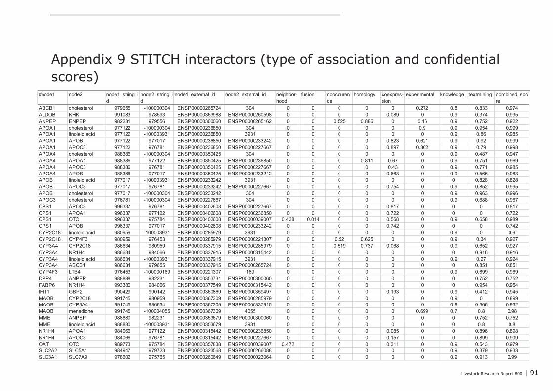
Livestock Research Report 800 | 91
#node1 node2 node1_string_id
node2_string_id
node1_external_id node2_external_id neighbor-hood
fusion cooccurence
homology coexpres-sion
experimental knowledge textmining combined_score
ABCB1 cholesterol 979655 -100000304 ENSP00000265724 304 0 0 0 0 0 0.272 0.8 0.833 0.974ALDOB KHK 991083 978593 ENSP00000363988 ENSP00000260598 0 0 0 0 0.089 0 0.9 0.374 0.935ANPEP ENPEP 982231 979556 ENSP00000300060 ENSP00000265162 0 0 0.525 0.886 0 0.16 0.9 0.752 0.922APOA1 cholesterol 977122 -100000304 ENSP00000236850 304 0 0 0 0 0 0.9 0.9 0.954 0.999APOA1 linoleic acid 977122 -100003931 ENSP00000236850 3931 0 0 0 0 0 0 0.9 0.86 0.985APOA1 APOB 977122 977017 ENSP00000236850 ENSP00000233242 0 0 0 0 0.823 0.621 0.9 0.92 0.999APOA1 APOC3 977122 976781 ENSP00000236850 ENSP00000227667 0 0 0 0 0.897 0.302 0.9 0.79 0.998APOA4 cholesterol 988386 -100000304 ENSP00000350425 304 0 0 0 0 0 0 0.9 0.487 0.947APOA4 APOA1 988386 977122 ENSP00000350425 ENSP00000236850 0 0 0 0.811 0.67 0 0.9 0.751 0.969APOA4 APOC3 988386 976781 ENSP00000350425 ENSP00000227667 0 0 0 0 0.43 0 0.9 0.771 0.985APOA4 APOB 988386 977017 ENSP00000350425 ENSP00000233242 0 0 0 0 0.668 0 0.9 0.565 0.983APOB linoleic acid 977017 -100003931 ENSP00000233242 3931 0 0 0 0 0 0 0 0.828 0.828APOB APOC3 977017 976781 ENSP00000233242 ENSP00000227667 0 0 0 0 0.754 0 0.9 0.852 0.995APOB cholesterol 977017 -100000304 ENSP00000233242 304 0 0 0 0 0 0 0.9 0.963 0.996APOC3 cholesterol 976781 -100000304 ENSP00000227667 304 0 0 0 0 0 0 0.9 0.688 0.967CPS1 APOC3 996337 976781 ENSP00000402608 ENSP00000227667 0 0 0 0 0.817 0 0 0 0.817CPS1 APOA1 996337 977122 ENSP00000402608 ENSP00000236850 0 0 0 0 0.722 0 0 0 0.722CPS1 OTC 996337 975784 ENSP00000402608 ENSP00000039007 0.438 0.014 0 0 0.568 0 0.9 0.658 0.989CPS1 APOB 996337 977017 ENSP00000402608 ENSP00000233242 0 0 0 0 0.742 0 0 0 0.742CYP2C18 linoleic acid 980959 -100003931 ENSP00000285979 3931 0 0 0 0 0 0 0.9 0 0.9CYP2C18 CYP4F3 980959 976453 ENSP00000285979 ENSP00000221307 0 0 0.52 0.625 0 0 0.9 0.34 0.927CYP3A4 CYP2C18 986634 980959 ENSP00000337915 ENSP00000285979 0 0 0.519 0.737 0.068 0 0.9 0.652 0.927CYP3A4 NR1H4 986634 984066 ENSP00000337915 ENSP00000315442 0 0 0 0 0 0 0 0.916 0.916CYP3A4 linoleic acid 986634 -100003931 ENSP00000337915 3931 0 0 0 0 0 0 0.9 0.27 0.924CYP3A4 ABCB1 986634 979655 ENSP00000337915 ENSP00000265724 0 0 0 0 0 0 0 0.851 0.851CYP4F3 LTB4 976453 -100000169 ENSP00000221307 169 0 0 0 0 0 0 0.9 0.699 0.969DPP4 ANPEP 988888 982231 ENSP00000353731 ENSP00000300060 0 0 0 0 0 0 0 0.752 0.752FABP6 NR1H4 993380 984066 ENSP00000377549 ENSP00000315442 0 0 0 0 0 0 0 0.954 0.954IFIT1 GBP2 990429 990142 ENSP00000360869 ENSP00000359497 0 0 0 0 0.193 0 0.9 0.412 0.945MAOB CYP2C18 991745 980959 ENSP00000367309 ENSP00000285979 0 0 0 0 0 0 0.9 0 0.899MAOB CYP3A4 991745 986634 ENSP00000367309 ENSP00000337915 0 0 0 0 0 0 0.9 0.366 0.932MAOB menadione 991745 -100004055 ENSP00000367309 4055 0 0 0 0 0 0.699 0.7 0.8 0.98MME ANPEP 988880 982231 ENSP00000353679 ENSP00000300060 0 0 0 0 0 0 0 0.752 0.752MME linoleic acid 988880 -100003931 ENSP00000353679 3931 0 0 0 0 0 0 0 0.8 0.8NR1H4 APOA1 984066 977122 ENSP00000315442 ENSP00000236850 0 0 0 0 0.085 0 0 0.896 0.898NR1H4 APOC3 984066 976781 ENSP00000315442 ENSP00000227667 0 0 0 0 0.157 0 0 0.899 0.909OAT OTC 989773 975784 ENSP00000357838 ENSP00000039007 0.472 0 0 0 0.311 0 0.9 0.543 0.979SLC2A2 SLC5A1 984947 979723 ENSP00000323568 ENSP00000266088 0 0 0 0 0 0 0.9 0.379 0.933SLC3A1 SLC7A9 978602 975765 ENSP00000260649 ENSP00000023064 0 0 0 0 0 0 0.9 0.913 0.99
Appendix 9 STITCH interactors (type of association and confidentialscores)
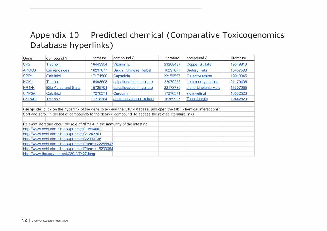
92 | Livestock Research Report 800
Gene compound 1 literature compound 2 literature compound 3 literature
CR2 Tretinoin 16443354 Vitamin E 23208437 Copper Sulfate 19549813
APOC3 Ginsenosides 16297877 Drugs, Chinese Herbal 16297877 Dietary Fats 18457598
SPP1 Calcitriol 17171500 Capsaicin 22150557 Galactosamine 19913045
NCK1 Tretinoin 15498508 epigallocatechin gallate 22079256 beta-methylcholine 21179406
NR1H4 Bile Acids and Salts 15725701 epigallocatechin gallate 22178739 alpha-Linolenic Acid 15307955
CYP3A4 Calcitriol 17270371 Curcumin 17270371 9-cis-retinal 16632523
CYP4F3 Tretinoin 17218384 apple polyphenol extract 16369997 Thapsigargin 19442820
userguide; click on the hyperlink of the gene to access the CTD database, and open the tab " chemical interactions".Sort and scroll in the list of compounds to the desired compound to access the related literature links.
Relevant literature about the role of NR1H4 in the immunity of the intestinehttp://www.ncbi.nlm.nih.gov/pubmed/19864602http://www.ncbi.nlm.nih.gov/pubmed/21242261http://www.ncbi.nlm.nih.gov/pubmed/22993736http://www.ncbi.nlm.nih.gov/pubmed/?term=22285937http://www.ncbi.nlm.nih.gov/pubmed/?term=16230354http://www.jbc.org/content/280/9/7427.long
Appendix 10 Predicted chemical (Comparative ToxicogenomicsDatabase hyperlinks)

93 | Livestock Research Report 800
Appendix 11 Detailed study information of the experiments that testedtrans-generational epigenetic effects (maternal phase)
Nutritional factors n Age (start
experiment)
Duration of
intervention
Dosage Effect Marker Note Ref.
Faty acids
Alkyl-glycerols (AKG)
extracted from
marine organisms
240 sows and Sows: d90 of
gestation
Sows: until
weaning
D90 to 5d before
farrowing: 0.34 g
AKG/sow/d
D5 before to d5 after
farrowing: 1.5 g
AKG/sow/d. From d5
until weaning: 0.75 g
AKG/sow/d.
Giving AKG to sows in
late gestation and
lactation can improve
the passive immunity
transfer to piglets
No effect on
performance and specific
immune parameters.
Increased % of serum
bactericidal activity and
haemolytic complement
(weaning and 70d of age)
No data regarding
microbiota, gut
morphology, and
immuno histochemie.
[74]
Corn vs. coconut oil x
low (9%) or standard
(18%) crude protein
Femal wistar
rats and
progeny
During gestation from
4 d after mating
onwards
Until birth 90 vs. 180 g/kg crude
protein; 100 g corn oil
or 50 g corn oil + 50 g
coconut oil or 10 g
corn oil + 90 g
coconut oil/kg
Reduced lymphocyte
proliferation at birth
after prenatal
exposure to a low
protein diet, which
could be overcome in
postnatal rats if the
prenatal diet included
a large amount of
coconut oil and a
small amount of corn
oil.
Lymphocyte proliferation
in thyme and spleen;
Spleen natural killer cell
activity.
No data regarding
microbiota, gut
morphology, and
immuno histochemie.
[67]
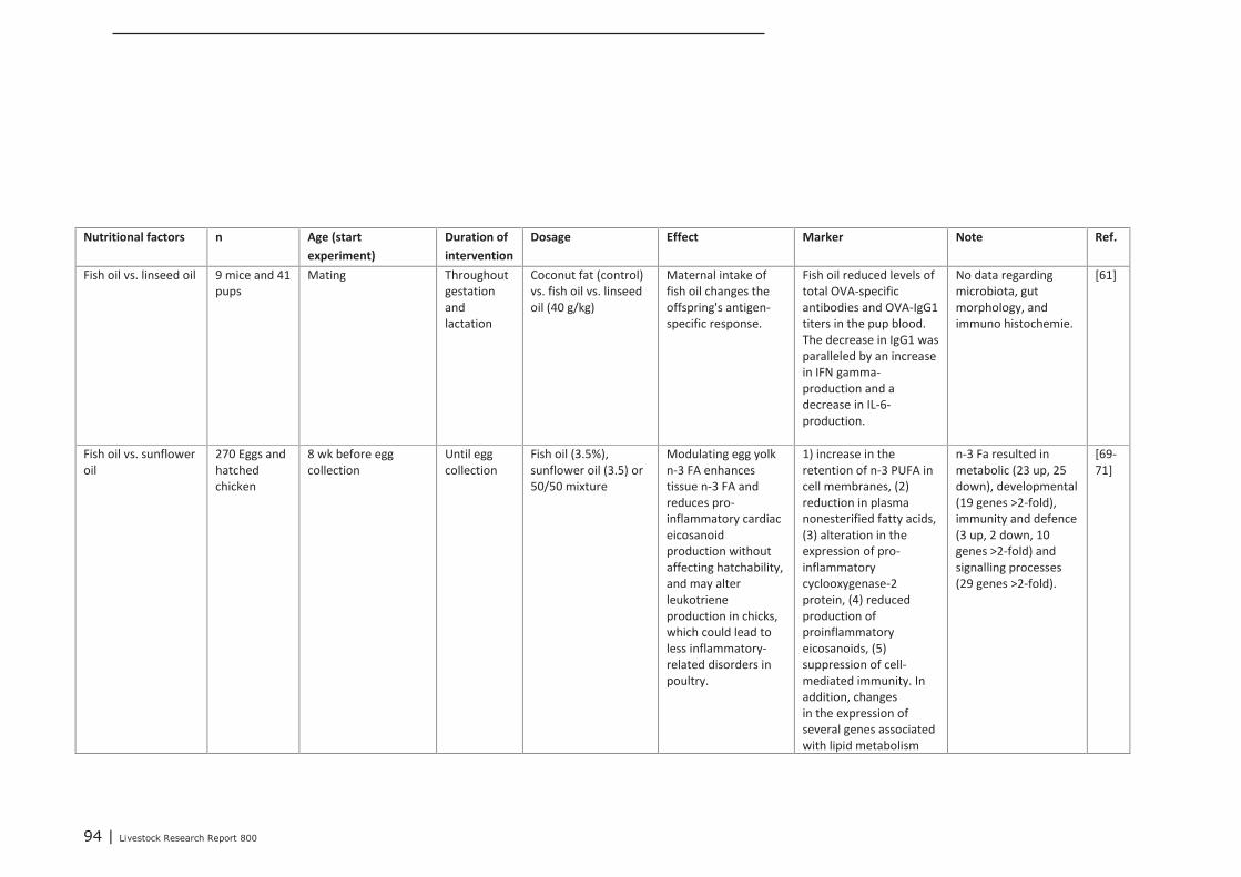
94 | Livestock Research Report 800
Nutritional factors n Age (start
experiment)
Duration of
intervention
Dosage Effect Marker Note Ref.
Fish oil vs. linseed oil 9 mice and 41
pups
Mating Throughout
gestation
and
lactation
Coconut fat (control)
vs. fish oil vs. linseed
oil (40 g/kg)
Maternal intake of
fish oil changes the
offspring's antigen-
specific response.
Fish oil reduced levels of
total OVA-specific
antibodies and OVA-IgG1
titers in the pup blood.
The decrease in IgG1 was
paralleled by an increase
in IFN gamma-
production and a
decrease in IL-6-
production.
No data regarding
microbiota, gut
morphology, and
immuno histochemie.
[61]
Fish oil vs. sunflower
oil
270 Eggs and
hatched
chicken
8 wk before egg
collection
Until egg
collection
Fish oil (3.5%),
sunflower oil (3.5) or
50/50 mixture
Modulating egg yolk
n-3 FA enhances
tissue n-3 FA and
reduces pro-
inflammatory cardiac
eicosanoid
production without
affecting hatchability,
and may alter
leukotriene
production in chicks,
which could lead to
less inflammatory-
related disorders in
poultry.
1) increase in the
retention of n-3 PUFA in
cell membranes, (2)
reduction in plasma
nonesterified fatty acids,
(3) alteration in the
expression of pro-
inflammatory
cyclooxygenase-2
protein, (4) reduced
production of
proinflammatory
eicosanoids, (5)
suppression of cell-
mediated immunity. In
addition, changes
in the expression of
several genes associated
with lipid metabolism
n-3 Fa resulted in
metabolic (23 up, 25
down), developmental
(19 genes >2-fold),
immunity and defence
(3 up, 2 down, 10
genes >2-fold) and
signalling processes
(29 genes >2-fold).
[69-
71]
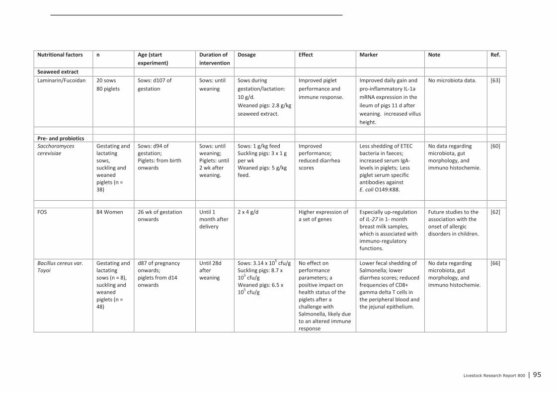
Livestock Research Report 800 | 95
Nutritional factors n Age (start
experiment)
Duration of
intervention
Dosage Effect Marker Note Ref.
Seaweed extract
Laminarin/Fucoidan 20 sows
80 piglets
Sows: d107 of
gestation
Sows: until
weaning
Sows during
gestation/lactation:
10 g/d.
Weaned pigs: 2.8 g/kg
seaweed extract.
Improved piglet
performance and
immune response.
Improved daily gain and
pro-inflammatory IL-1a
mRNA expression in the
ileum of pigs 11 d after
weaning. increased villus
height.
No microbiota data. [63]
Pre- and probiotics
Sacchoromyces
cerevisiae
Gestating and
lactating
sows,
suckling and
weaned
piglets (n =
38)
Sows: d94 of
gestation;
Piglets: from birth
onwards
Sows: until
weaning;
Piglets: until
2 wk after
weaning.
Sows: 1 g/kg feed
Suckling pigs: 3 x 1 g
per wk
Weaned pigs: 5 g/kg
feed.
Improved
performance;
reduced diarrhea
scores
Less shedding of ETEC
bacteria in faeces;
increased serum IgA-
levels in piglets; Less
piglet serum specific
antibodies against
E. coli O149:K88.
No data regarding
microbiota, gut
morphology, and
immuno histochemie.
[60]
FOS 84 Women 26 wk of gestation
onwards
Until 1
month after
delivery
2 x 4 g/d Higher expression of
a set of genes
Especially up-regulation
of IL-27 in 1- month
breast milk samples,
which is associated with
immuno-regulatory
functions.
Future studies to the
association with the
onset of allergic
disorders in children.
[62]
Bacillus cereus var.
Toyoi
Gestating and
lactating
sows (n = 8),
suckling and
weaned
piglets (n =
48)
d87 of pregnancy
onwards;
piglets from d14
onwards
Until 28d
after
weaning
Sows: 3.14 x 10⁵
cfu/g
Suckling pigs: 8.7 x
105
cfu/g
Weaned pigs: 6.5 x
105
cfu/g
No effect on
performance
parameters; a
positive impact on
health status of the
piglets after a
challenge with
Salmonella, likely due
to an altered immune
response
Lower fecal shedding of
Salmonella; lower
diarrhea scores; reduced
frequencies of CD8+
gamma delta T cells in
the peripheral blood and
the jejunal epithelium.
No data regarding
microbiota, gut
morphology, and
immuno histochemie.
[66]
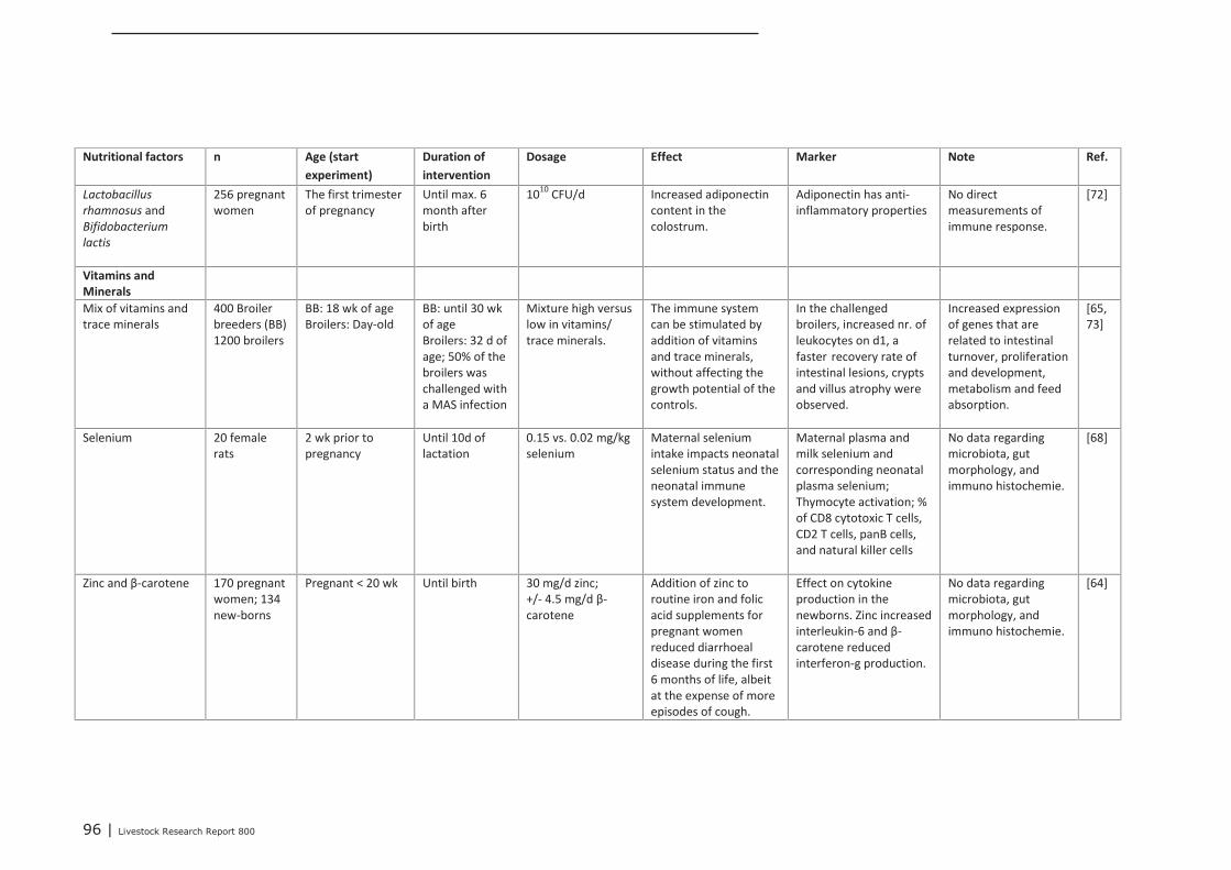
96 | Livestock Research Report 800
Nutritional factors n Age (start
experiment)
Duration of
intervention
Dosage Effect Marker Note Ref.
Lactobacillus
rhamnosus and
Bifidobacterium
lactis
256 pregnant
women
The first trimester
of pregnancy
Until max. 6
month after
birth
1010
CFU/d Increased adiponectin
content in the
colostrum.
Adiponectin has anti-
inflammatory properties
No direct
measurements of
immune response.
[72]
Vitamins and
Minerals
Mix of vitamins and
trace minerals
400 Broiler
breeders (BB)
1200 broilers
BB: 18 wk of age
Broilers: Day-old
BB: until 30 wk
of age
Broilers: 32 d of
age; 50% of the
broilers was
challenged with
a MAS infection
Mixture high versus
low in vitamins/
trace minerals.
The immune system
can be stimulated by
addition of vitamins
and trace minerals,
without affecting the
growth potential of the
controls.
In the challenged
broilers, increased nr. of
leukocytes on d1, a
faster recovery rate of
intestinal lesions, crypts
and villus atrophy were
observed.
Increased expression
of genes that are
related to intestinal
turnover, proliferation
and development,
metabolism and feed
absorption.
[65,
73]
Selenium 20 female
rats
2 wk prior to
pregnancy
Until 10d of
lactation
0.15 vs. 0.02 mg/kg
selenium
Maternal selenium
intake impacts neonatal
selenium status and the
neonatal immune
system development.
Maternal plasma and
milk selenium and
corresponding neonatal
plasma selenium;
Thymocyte activation; %
of CD8 cytotoxic T cells,
CD2 T cells, panB cells,
and natural killer cells
No data regarding
microbiota, gut
morphology, and
immuno histochemie.
[68]
Zinc and β-carotene 170 pregnant
women; 134
new-borns
Pregnant < 20 wk Until birth 30 mg/d zinc;
+/- 4.5 mg/d β-
carotene
Addition of zinc to
routine iron and folic
acid supplements for
pregnant women
reduced diarrhoeal
disease during the first
6 months of life, albeit
at the expense of more
episodes of cough.
Effect on cytokine
production in the
newborns. Zinc increased
interleukin-6 and β-
carotene reduced
interferon-g production.
No data regarding
microbiota, gut
morphology, and
immuno histochemie.
[64]
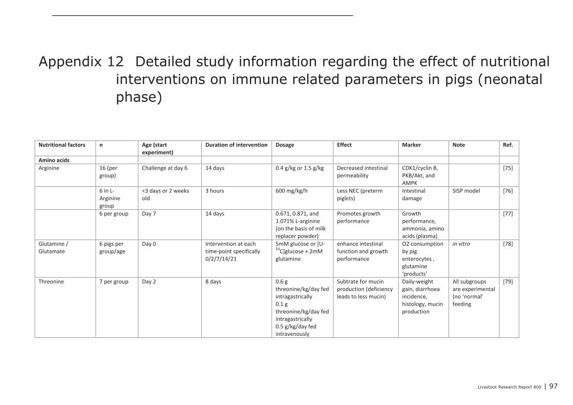
Livestock Research Report 800 | 97
Appendix 12 Detailed study information regarding the effect of nutritionalinterventions on immune related parameters in pigs (neonatalphase)
Nutritional factors n Age (start
experiment)
Duration of intervention Dosage Effect Marker Note Ref.
Amino acids
Arginine 16 (per
group)
Challenge at day 6 14 days 0.4 g/kg or 1.5 g/kg Decreased intestinal
permeability
CDK1/cyclin B,
PKB/Akt, and
AMPK
[75]
6 in L-
Arginine
group
<3 days or 2 weeks
old
3 hours 600 mg/kg/h Less NEC (preterm
piglets)
Intestinal
damage
SISP model [76]
6 per group Day 7 14 days 0.671, 0.871, and
1.071% L-arginine
(on the basis of milk
replacer powder)
Promotes growth
performance
Growth
performance,
ammonia, amino
acids (plasma)
[77]
Glutamine /
Glutamate
6 pigs per
group/age
Day 0 Intervention at each
time-point specifically
0/2/7/14/21
5mM glucose or [U-14
C]glucose + 2mM
glutamine
enhance intestinal
function and growth
performance
O2 consumption
by pig
enterocytes ,
glutamine
‘products’
in vitro [78]
Threonine 7 per group Day 2 8 days 0.6 g
threonine/kg/day fed
intragastrically
0.1 g
threonine/kg/day fed
intragastrically
0.5 g/kg/day fed
intravenously
Subtrate for mucin
production (deficiency
leads to less mucin)
Daily-weight
gain, diarrhoea
incidence,
histology, mucin
production
All subgroups
are experimental
(no ‘normal’
feeding
[79]
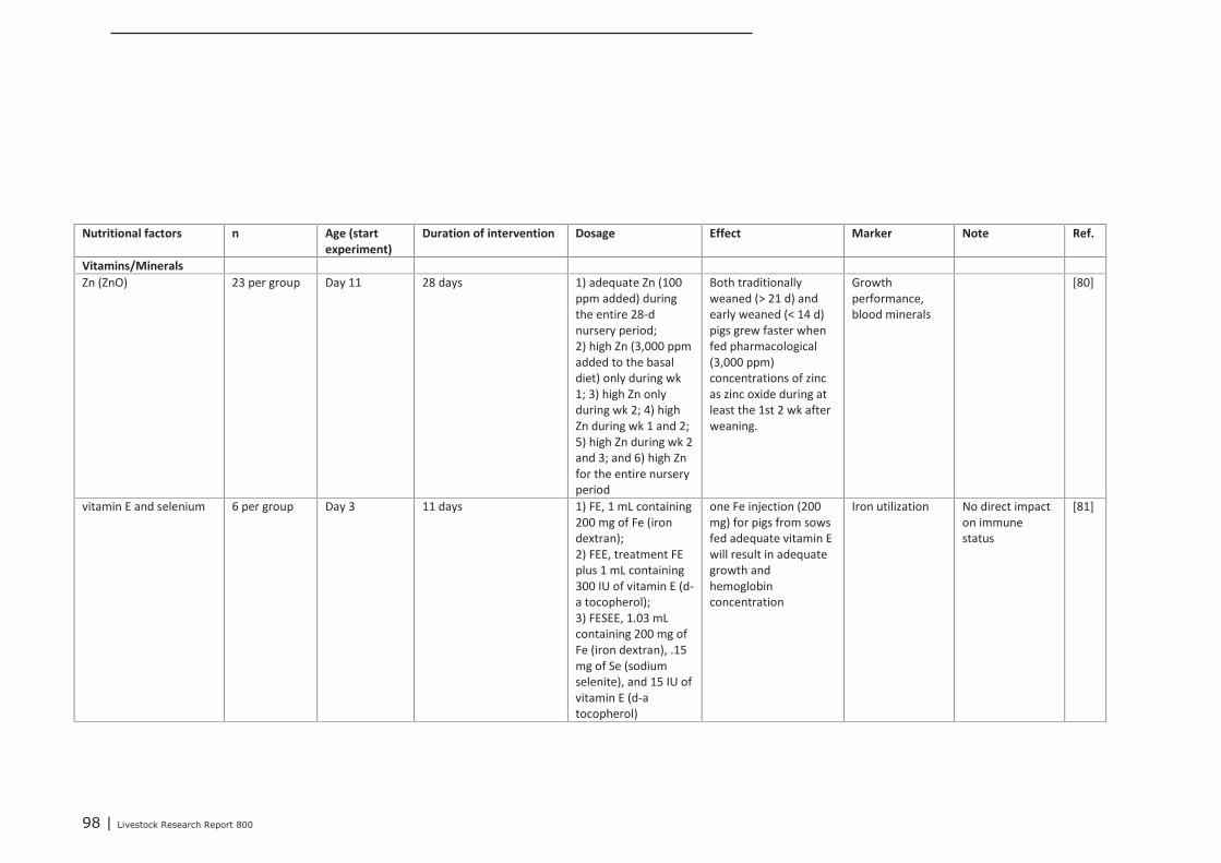
98 | Livestock Research Report 800
Nutritional factors n Age (start
experiment)
Duration of intervention Dosage Effect Marker Note Ref.
Vitamins/Minerals
Zn (ZnO) 23 per group Day 11 28 days 1) adequate Zn (100
ppm added) during
the entire 28-d
nursery period;
2) high Zn (3,000 ppm
added to the basal
diet) only during wk
1; 3) high Zn only
during wk 2; 4) high
Zn during wk 1 and 2;
5) high Zn during wk 2
and 3; and 6) high Zn
for the entire nursery
period
Both traditionally
weaned (> 21 d) and
early weaned (< 14 d)
pigs grew faster when
fed pharmacological
(3,000 ppm)
concentrations of zinc
as zinc oxide during at
least the 1st 2 wk after
weaning.
Growth
performance,
blood minerals
[80]
vitamin E and selenium 6 per group Day 3 11 days 1) FE, 1 mL containing
200 mg of Fe (iron
dextran);
2) FEE, treatment FE
plus 1 mL containing
300 IU of vitamin E (d-
a tocopherol);
3) FESEE, 1.03 mL
containing 200 mg of
Fe (iron dextran), .15
mg of Se (sodium
selenite), and 15 IU of
vitamin E (d-a
tocopherol)
one Fe injection (200
mg) for pigs from sows
fed adequate vitamin E
will result in adequate
growth and
hemoglobin
concentration
Iron utilization No direct impact
on immune
status
[81]
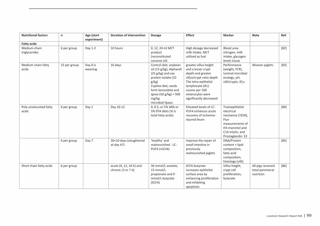
Livestock Research Report 800 | 99
Nutritional factors n Age (start
experiment)
Duration of intervention Dosage Effect Marker Note Ref.
Fatty acids
Medium-chain
triglycerides
6 per group Day 1-2 10 hours 0, 12, 24 ml MCT
product
(reconstituted
coconut oil)
High dosage decreased
milk intake, MCT
utilized as fuel
Blood urea
nitrogen, milk
intake, glycogen
levels tissue
[82]
Medium-chain fatty
acids
15 per group Day 0 is
weaning
16 days Control diet; soybean
oil (15 g/kg), Alphacell
(25 g/kg) and soy
protein isolate (10
g/kg)
Cuphea diet; seeds
form lanceolata and
ignea (50 g/kg) + 500
mg/kg
microbial lipase.
greater villus height
and a lesser crypt
depth and greater
villus/crypt ratio depth
The intra-epithelial
lymphocyte (IEL)
counts per 100
enterocytes were
significantly decreased
Performance
(weight, FCR),
luminal microbial
ecology, pH,
villi/crypts, IELs
Weaner piglets [83]
Poly unsaturated fatty
acids
6 per group Day 1 Day 10-12 0, 0.5, or 5% ARA or
5% EPA diets (% is
total fatty acids)
Elevated levels of LC-
PUFA enhances acute
recovery of ischemia-
injured ileum
Transepithelial
electrical
resistance (TEER),
Flux
measurements of
H3-mannitol and
C14-inlulin, and
Prostaglandin E2
[84]
6 per group Day 7 30+10 days (slaughtered
at day 47)
‘healthy’ and
malnourished - LC-
PUFA (n3/n6)
Improve the repair of
small intestine in
previously
malnourished piglets
DNA/Protein
content + lipid
composition,
fatty acid
composition,
histology (villi)
[85]
Short chain fatty acids 6 per group acute (4, 12, 24 h) and
chronic (3 or 7 d)
36 mmol/L acetate,
15 mmol/L
propionate and 9
mmol/L butyrate
(SCFA)
SCFA butyrate
increases epithelial
surface area by
enhancing proliferation
and inhibiting
apoptosis
Villus height,
crypt cell
proliferation,
butyrate
All pigs received
total parenteral
nutrition
[86]
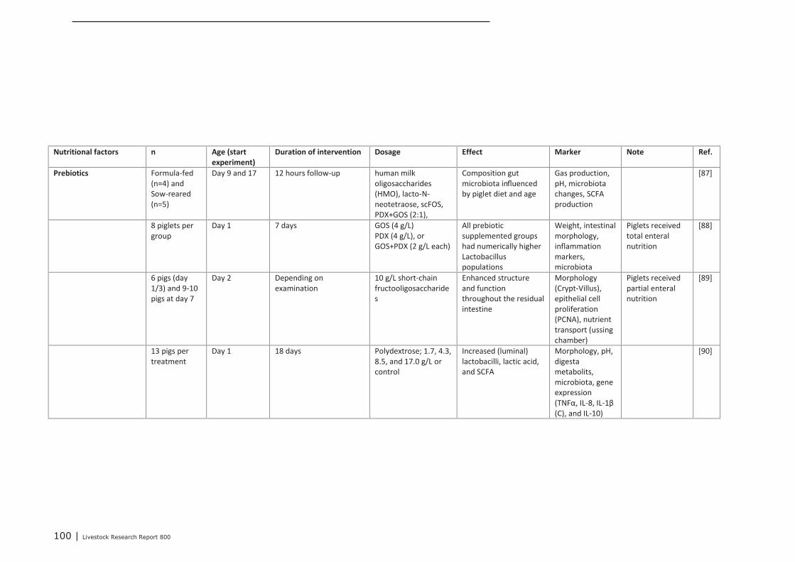
100 | Livestock Research Report 800
Nutritional factors n Age (start
experiment)
Duration of intervention Dosage Effect Marker Note Ref.
Prebiotics Formula-fed
(n=4) and
Sow-reared
(n=5)
Day 9 and 17 12 hours follow-up human milk
oligosaccharides
(HMO), lacto-N-
neotetraose, scFOS,
PDX+GOS (2:1),
Composition gut
microbiota influenced
by piglet diet and age
Gas production,
pH, microbiota
changes, SCFA
production
[87]
8 piglets per
group
Day 1 7 days GOS (4 g/L)
PDX (4 g/L), or
GOS+PDX (2 g/L each)
All prebiotic
supplemented groups
had numerically higher
Lactobacillus
populations
Weight, intestinal
morphology,
inflammation
markers,
microbiota
Piglets received
total enteral
nutrition
[88]
6 pigs (day
1/3) and 9-10
pigs at day 7
Day 2 Depending on
examination
10 g/L short-chain
fructooligosaccharide
s
Enhanced structure
and function
throughout the residual
intestine
Morphology
(Crypt-Villus),
epithelial cell
proliferation
(PCNA), nutrient
transport (ussing
chamber)
Piglets received
partial enteral
nutrition
[89]
13 pigs per
treatment
Day 1 18 days Polydextrose; 1.7, 4.3,
8.5, and 17.0 g/L or
control
Increased (luminal)
lactobacilli, lactic acid,
and SCFA
Morphology, pH,
digesta
metabolits,
microbiota, gene
expression
(TNFα, IL-8, IL-1β
(C), and IL-10)
[90]
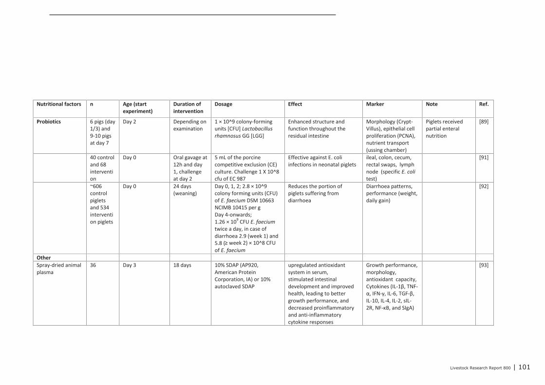
Livestock Research Report 800 | 101
Nutritional factors n Age (start
experiment)
Duration of
intervention
Dosage Effect Marker Note Ref.
Probiotics 6 pigs (day
1/3) and
9-10 pigs
at day 7
Day 2 Depending on
examination
1 × 10^9 colony-forming
units [CFU] Lactobacillus
rhamnosus GG [LGG]
Enhanced structure and
function throughout the
residual intestine
Morphology (Crypt-
Villus), epithelial cell
proliferation (PCNA),
nutrient transport
(ussing chamber)
Piglets received
partial enteral
nutrition
[89]
40 control
and 68
interventi
on
Day 0 Oral gavage at
12h and day
1, challenge
at day 2
5 mL of the porcine
competitive exclusion (CE)
culture. Challenge 1 X 10^8
cfu of EC 987
Effective against E. coli
infections in neonatal piglets
ileal, colon, cecum,
rectal swaps, lymph
node (specific E. coli
test)
[91]
~606
control
piglets
and 534
interventi
on piglets
Day 0 24 days
(weaning)
Day 0, 1, 2; 2.8 × 10^9
colony forming units (CFU)
of E. faecium DSM 10663
NCIMB 10415 per g
Day 4-onwards;
1.26 × 10⁹
CFU E. faecium
twice a day, in case of
diarrhoea 2.9 (week 1) and
5.8 (≥ week 2) × 10^8 CFU
of E. faecium
Reduces the portion of
piglets suffering from
diarrhoea
Diarrhoea patterns,
performance (weight,
daily gain)
[92]
Other
Spray-dried animal
plasma
36 Day 3 18 days 10% SDAP (AP920,
American Protein
Corporation, IA) or 10%
autoclaved SDAP
upregulated antioxidant
system in serum,
stimulated intestinal
development and improved
health, leading to better
growth performance, and
decreased proinflammatory
and anti-inflammatory
cytokine responses
Growth performance,
morphology,
antioxidant capacity,
Cytokines (IL-1β, TNF-
α, IFN-γ, IL-6, TGF-β,
IL-10, IL-4, IL-2, sIL-
2R, NF-κB, and SIgA)
[93]
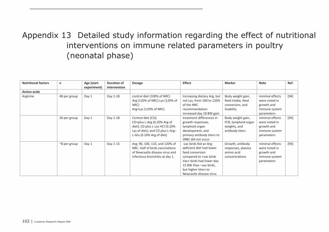
102 | Livestock Research Report 800
Appendix 13 Detailed study information regarding the effect of nutritionalinterventions on immune related parameters in poultry(neonatal phase)
Nutritional factors n Age (start
experiment)
Duration of
intervention
Dosage Effect Marker Note Ref.
Amino acids
Arginine 48 per group Day 1 Day 1-18 control diet (100% of NRC)
Arg (120% of NRC) Lys (120% of
NRC)
Arg+Lys (120% of NRC)
Increasing dietary Arg, but
not Lys, from 100 to 120%
of the NRC
recommendation
increased day 18 BW gain
Body weight gain,
feed intake, feed
conversion, and
livability
minimal effects
were noted in
growth and
immune system
parameters
[94]
30 per group Day 1 Day 1-18 Control diet (CD);
CD+plus L-Arg (0.20% Arg of
diet); CD plus L-Lys HCl (0.20%
Lys of diet); and CD plus L-Arg−
L-Glu (0.10% Arg of diet)
treatment differences in
growth responses,
lymphoid organ
development, and
primary antibody titers to
SRBC did not occur
Body weight gain,
FCR, lymphoid organ
weights, and
antibody titers
minimal effects
were noted in
growth and
immune system
parameters
[94]
~8 per group Day 1 Day 1-15 Arg; 90, 100, 110, and 120% of
NRC. Half of birds vaccinations
of Newcastle disease virus and
infectious bronchitis at day 1.
-vac birds fed an Arg-
deficient diet had lower
feed conversion
compared to +vac birds
Vac+ birds had lower day
15 BW than –vac birds,
but higher titers to
Newcastle disease virus
Growth, antibody
responses, plasma
amino acid
concentrations
minimal effects
were noted in
growth and
immune system
parameters
[94]
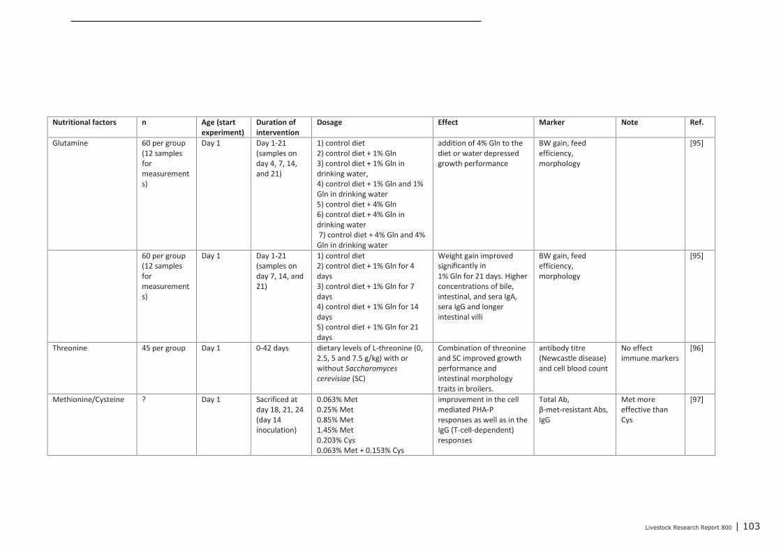
Livestock Research Report 800 | 103
Nutritional factors n Age (start
experiment)
Duration of
intervention
Dosage Effect Marker Note Ref.
Glutamine 60 per group
(12 samples
for
measurement
s)
Day 1 Day 1-21
(samples on
day 4, 7, 14,
and 21)
1) control diet
2) control diet + 1% Gln
3) control diet + 1% Gln in
drinking water,
4) control diet + 1% Gln and 1%
Gln in drinking water
5) control diet + 4% Gln
6) control diet + 4% Gln in
drinking water
7) control diet + 4% Gln and 4%
Gln in drinking water
addition of 4% Gln to the
diet or water depressed
growth performance
BW gain, feed
efficiency,
morphology
[95]
60 per group
(12 samples
for
measurement
s)
Day 1 Day 1-21
(samples on
day 7, 14, and
21)
1) control diet
2) control diet + 1% Gln for 4
days
3) control diet + 1% Gln for 7
days
4) control diet + 1% Gln for 14
days
5) control diet + 1% Gln for 21
days
Weight gain improved
significantly in
1% Gln for 21 days. Higher
concentrations of bile,
intestinal, and sera IgA,
sera IgG and longer
intestinal villi
BW gain, feed
efficiency,
morphology
[95]
Threonine 45 per group Day 1 0-42 days dietary levels of L-threonine (0,
2.5, 5 and 7.5 g/kg) with or
without Saccharomyces
cerevisiae (SC)
Combination of threonine
and SC improved growth
performance and
intestinal morphology
traits in broilers.
antibody titre
(Newcastle disease)
and cell blood count
No effect
immune markers
[96]
Methionine/Cysteine ? Day 1 Sacrificed at
day 18, 21, 24
(day 14
inoculation)
0.063% Met
0.25% Met
0.85% Met
1.45% Met
0.203% Cys
0.063% Met + 0.153% Cys
improvement in the cell
mediated PHA-P
responses as well as in the
IgG (T-cell-dependent)
responses
Total Ab,
β-met-resistant Abs,
IgG
Met more
effective than
Cys
[97]
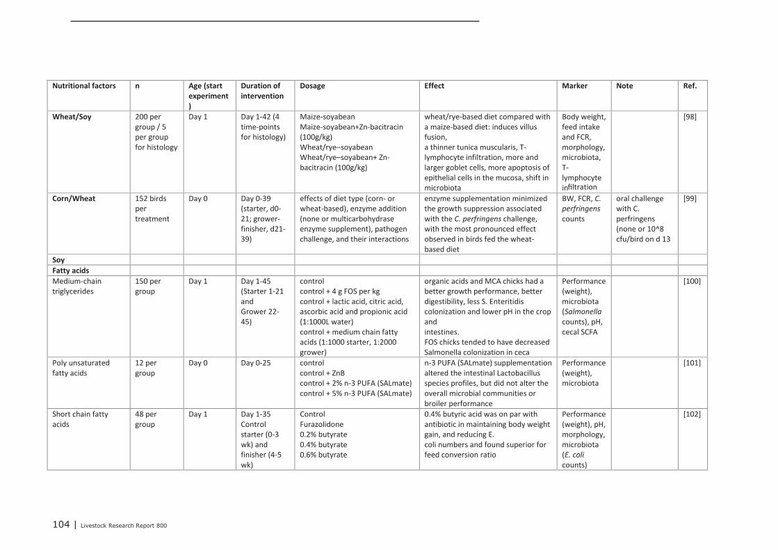
104 | Livestock Research Report 800
Nutritional factors n Age (start
experiment
)
Duration of
intervention
Dosage Effect Marker Note Ref.
Wheat/Soy 200 per
group / 5
per group
for histology
Day 1 Day 1-42 (4
time-points
for histology)
Maize-soyabean
Maize-soyabean+Zn-bacitracin
(100g/kg)
Wheat/rye–soyabean
Wheat/rye–soyabean+ Zn-
bacitracin (100g/kg)
wheat/rye-based diet compared with
a maize-based diet: induces villus
fusion,
a thinner tunica muscularis, T-
lymphocyte infiltration, more and
larger goblet cells, more apoptosis of
epithelial cells in the mucosa, shift in
microbiota
Body weight,
feed intake
and FCR,
morphology,
microbiota,
T-
lymphocyte
infiltration
[98]
Corn/Wheat 152 birds
per
treatment
Day 0 Day 0-39
(starter, d0-
21; grower-
finisher, d21-
39)
effects of diet type (corn- or
wheat-based), enzyme addition
(none or multicarbohydrase
enzyme supplement), pathogen
challenge, and their interactions
enzyme supplementation minimized
the growth suppression associated
with the C. perfringens challenge,
with the most pronounced effect
observed in birds fed the wheat-
based diet
BW, FCR, C.
perfringens
counts
oral challenge
with C.
perfringens
(none or 10^8
cfu/bird on d 13
[99]
Soy
Fatty acids
Medium-chain
triglycerides
150 per
group
Day 1 Day 1-45
(Starter 1-21
and
Grower 22-
45)
control
control + 4 g FOS per kg
control + lactic acid, citric acid,
ascorbic acid and propionic acid
(1:1000L water)
control + medium chain fatty
acids (1:1000 starter, 1:2000
grower)
organic acids and MCA chicks had a
better growth performance, better
digestibility, less S. Enteritidis
colonization and lower pH in the crop
and
intestines.
FOS chicks tended to have decreased
Salmonella colonization in ceca
Performance
(weight),
microbiota
(Salmonella
counts), pH,
cecal SCFA
[100]
Poly unsaturated
fatty acids
12 per
group
Day 0 Day 0-25 control
control + ZnB
control + 2% n-3 PUFA (SALmate)
control + 5% n-3 PUFA (SALmate)
n-3 PUFA (SALmate) supplementation
altered the intestinal Lactobacillus
species profiles, but did not alter the
overall microbial communities or
broiler performance
Performance
(weight),
microbiota
[101]
Short chain fatty
acids
48 per
group
Day 1 Day 1-35
Control
starter (0-3
wk) and
finisher (4-5
wk)
Control
Furazolidone
0.2% butyrate
0.4% butyrate
0.6% butyrate
0.4% butyric acid was on par with
antibiotic in maintaining body weight
gain, and reducing E.
coli numbers and found superior for
feed conversion ratio
Performance
(weight), pH,
morphology,
microbiota
(E. coli
counts)
[102]
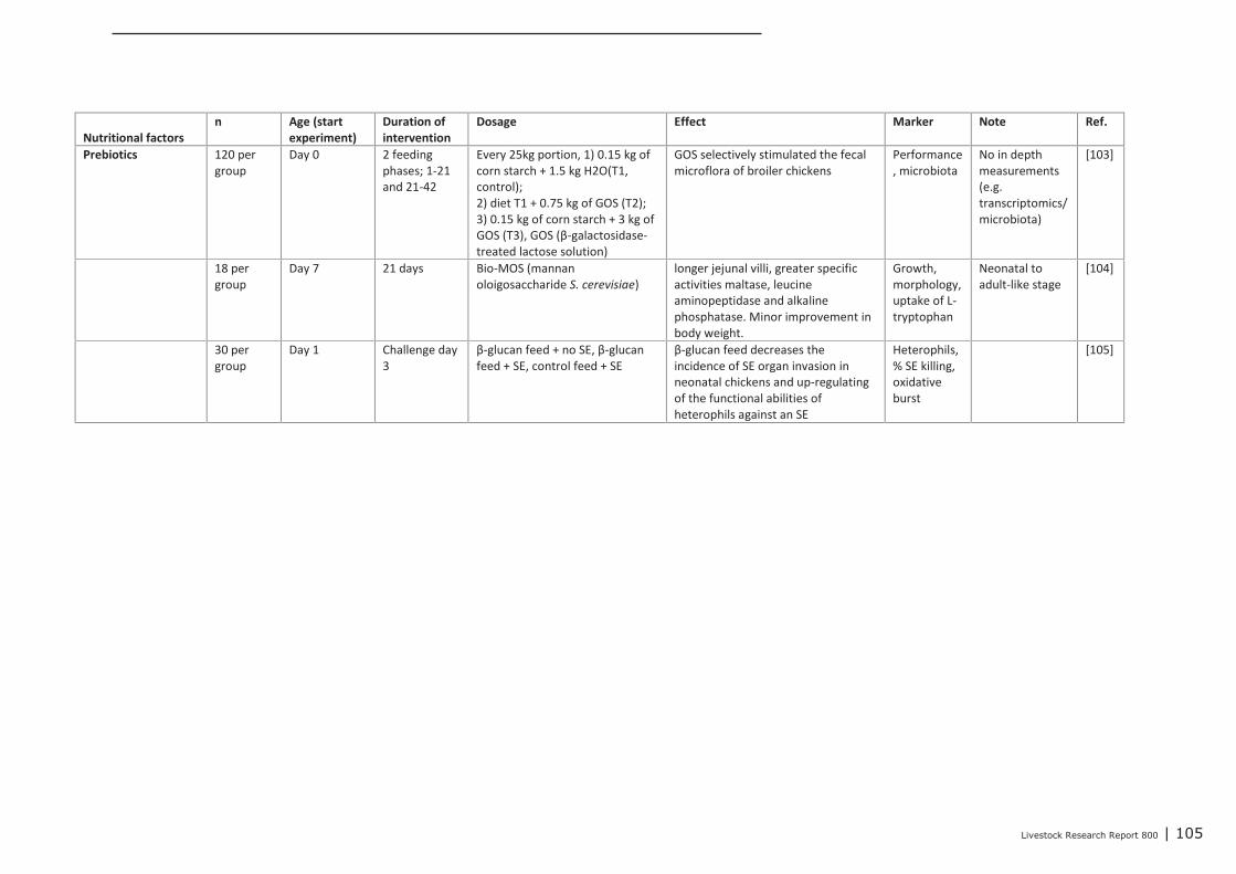
Livestock Research Report 800 | 105
Nutritional factors
n Age (start
experiment)
Duration of
intervention
Dosage Effect Marker Note Ref.
Prebiotics 120 per
group
Day 0 2 feeding
phases; 1-21
and 21-42
Every 25kg portion, 1) 0.15 kg of
corn starch + 1.5 kg H2O(T1,
control);
2) diet T1 + 0.75 kg of GOS (T2);
3) 0.15 kg of corn starch + 3 kg of
GOS (T3), GOS (β-galactosidase-
treated lactose solution)
GOS selectively stimulated the fecal
microflora of broiler chickens
Performance
, microbiota
No in depth
measurements
(e.g.
transcriptomics/
microbiota)
[103]
18 per
group
Day 7 21 days Bio-MOS (mannan
oloigosaccharide S. cerevisiae)
longer jejunal villi, greater specific
activities maltase, leucine
aminopeptidase and alkaline
phosphatase. Minor improvement in
body weight.
Growth,
morphology,
uptake of L-
tryptophan
Neonatal to
adult-like stage
[104]
30 per
group
Day 1 Challenge day
3
β-glucan feed + no SE, β-glucan
feed + SE, control feed + SE
β-glucan feed decreases the
incidence of SE organ invasion in
neonatal chickens and up-regulating
of the functional abilities of
heterophils against an SE
Heterophils,
% SE killing,
oxidative
burst
[105]
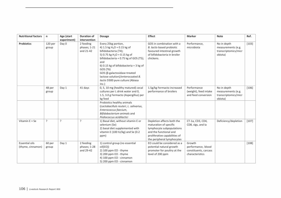
106 | Livestock Research Report 800
Nutritional factors n Age (start
experiment)
Duration of
intervention
Dosage Effect Marker Note Ref.
Probiotics 120 per
group
Day 0 2 feeding
phases; 1-21
and 21-42
Every 25kg portion,
4) 1.5 kg H2O + 0.15 kg of
bifidobacteria (T4);
5) 0.75 kg H2O + 0.15 kg of
bifidobacteria + 0.75 kg of GOS (T5);
and
6) 0.15 kg of bifidobacteria + 3 kg of
GOS (T6)
GOS (β-galactosidase-treated
lactose solution)/enterocoated B.
lactis D300 pure culture (Abiasa
Inc.)
GOS in combination with a
B. lactis-based probiotic
favoured intestinal growth
of bifidobacteria in broiler
chickens.
Performance,
microbiota
No in depth
measurements (e.g.
transcriptomics/micr
obiota)
[103]
48 per
group
Day 1 41 days 0, 5, 10 mg (healthy matured) cecal
cultures per L drink water and 0,
1.5, 3.0 g Fermacto (Aspergillus) per
kg feed
1.5g/kg Fermacto increased
performance of broilers
Performance
(weight), feed intake
and feed conversion
No in depth
measurements (e.g.
transcriptomics/micr
obiota)
[106]
Probiotics healthy animals
(Lactobacilluls reuteri, L. salivarius,
Enterococcus faecium,
Bifidobacterium animals and
Pediococcus acidilactici
Vitamin E + Se ? ? ? 1) Basal diet, without vitamin E or
selenium (Se)
2) basal diet supplemented with
vitamin E (100 IU/kg) and Se (0.2
ppm)
Depletion affects both the
maturation of specific
lymphocyte subpopulations
and the functional and
proliferative capabilities of
the peripheral lymphocytes
CT-1a, CD3, CD4,
CD8, sIgs, and Ia
Deficiency/depletion [107]
Essential oils
(thyme, cinnamon)
60 per
group
Day 1 2 feeding
phases; 1-28
and 29-42
1) control group (no essential
oil[EO])
2) 100 ppm EO - thyme
3) 200 ppm EO - thyme
4) 100 ppm EO - cinnamon
5) 200 ppm EO - cinnamon
EO could be considered as a
potential natural growth
promoter for poultry at the
level of 200 ppm
Growth
performance, blood
constituents, carcass
characteristics
[108]
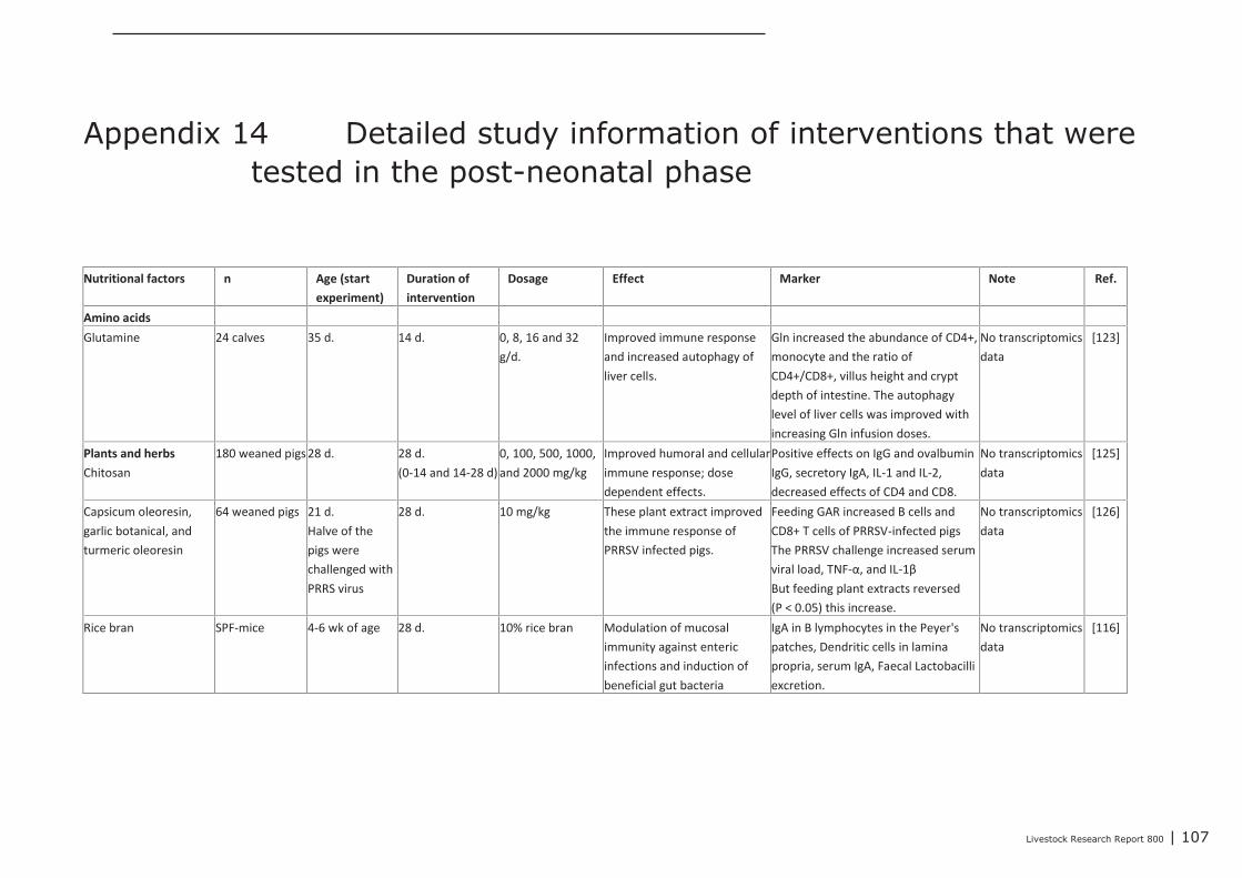
Livestock Research Report 800 | 107
Appendix 14 Detailed study information of interventions that weretested in the post-neonatal phase
Nutritional factors n Age (start
experiment)
Duration of
intervention
Dosage Effect Marker Note Ref.
Amino acids
Glutamine 24 calves 35 d. 14 d. 0, 8, 16 and 32
g/d.
Improved immune response
and increased autophagy of
liver cells.
Gln increased the abundance of CD4+,
monocyte and the ratio of
CD4+/CD8+, villus height and crypt
depth of intestine. The autophagy
level of liver cells was improved with
increasing Gln infusion doses.
No transcriptomics
data
[123]
Plants and herbs
Chitosan
180 weaned pigs 28 d. 28 d.
(0-14 and 14-28 d)
0, 100, 500, 1000,
and 2000 mg/kg
Improved humoral and cellular
immune response; dose
dependent effects.
Positive effects on IgG and ovalbumin
IgG, secretory IgA, IL-1 and IL-2,
decreased effects of CD4 and CD8.
No transcriptomics
data
[125]
Capsicum oleoresin,
garlic botanical, and
turmeric oleoresin
64 weaned pigs 21 d.
Halve of the
pigs were
challenged with
PRRS virus
28 d. 10 mg/kg These plant extract improved
the immune response of
PRRSV infected pigs.
Feeding GAR increased B cells and
CD8+ T cells of PRRSV-infected pigs
The PRRSV challenge increased serum
viral load, TNF-α, and IL-1β
But feeding plant extracts reversed
(P < 0.05) this increase.
No transcriptomics
data
[126]
Rice bran SPF-mice 4-6 wk of age 28 d. 10% rice bran Modulation of mucosal
immunity against enteric
infections and induction of
beneficial gut bacteria
IgA in B lymphocytes in the Peyer's
patches, Dendritic cells in lamina
propria, serum IgA, Faecal Lactobacilli
excretion.
No transcriptomics
data
[116]
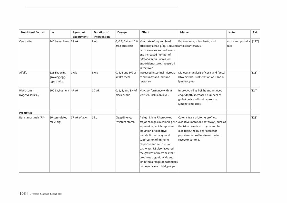
108 | Livestock Research Report 800
Nutritional factors n Age (start
experiment)
Duration of
intervention
Dosage Effect Marker Note Ref.
Quercetin 240 laying hens 28 wk 8 wk 0, 0.2, 0.4 and 0.6
g/kg quercetin
Max. rate of lay and feed
efficiency at 0.4 g/kg. Reduced
nr. of aerobes and coliforms
and increased number of
Bifidobacteria. Increased
antioxidant states measured
in the liver.
Performance, microbiota, and
antioxidant status.
No transcriptomics
data
[117]
Alfalfa 128 Shaoxing
growing egg-
type ducks
7 wk 8 wk 0, 3, 6 and 9% of
alfalfa meal
Increased intestinal microbial
community and immune
response.
Molecular analysis of cecal and faecal
DNA extract. Proliferation of T and B
lymphocytes
[118]
Black cumin
(Nigella sativ L.)
100 Laying hens 49 wk 10 wk 0, 1, 2, and 3% of
black cumin
Max. performance with at
least 2% inclusion level.
Improved villus height and reduced
crypt depth, increased numbers of
globet cells and lamina propria
lymphatic follicles.
[124]
Prebiotics
Resistant starch (RS) 10 cannulated
male pigs
17 wk of age 14 d. Digestible vs.
resistant starch
A diet high in RS provoked
major changes in colonic gene
expression, which represent
induction of oxidative
metabolic pathways and
suppression of immune
response and cell division
pathways. RS also favoured
the growth of microbes that
produces organic acids and
inhibited a range of potentially
pathogenic microbial groups.
Colonic transcriptome profiles,
oxidative metabolic pathways, such as
the tricarboxylic acid cycle and b-
oxidation, the nuclear receptor
peroxisome proliferator-activated
receptor gamma,
[128]
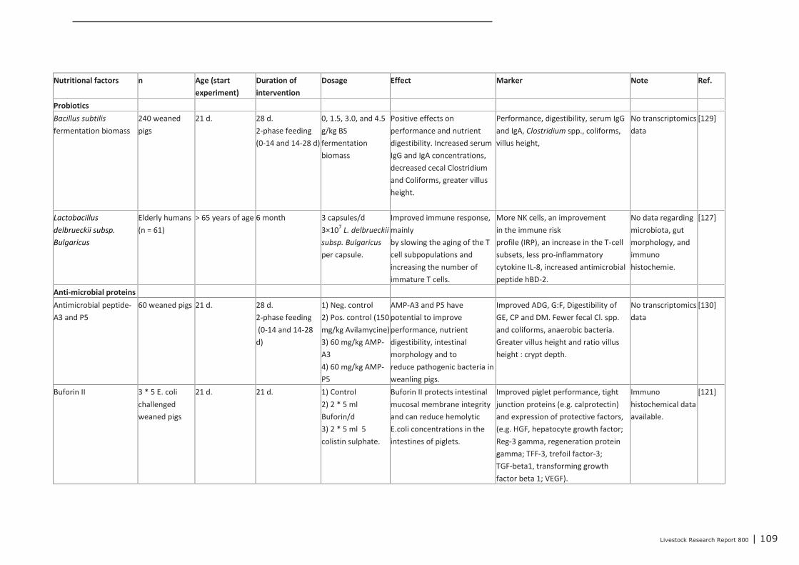
Livestock Research Report 800 | 109
Nutritional factors n Age (start
experiment)
Duration of
intervention
Dosage Effect Marker Note Ref.
Probiotics
Bacillus subtilis
fermentation biomass
240 weaned
pigs
21 d. 28 d.
2-phase feeding
(0-14 and 14-28 d)
0, 1.5, 3.0, and 4.5
g/kg BS
fermentation
biomass
Positive effects on
performance and nutrient
digestibility. Increased serum
IgG and IgA concentrations,
decreased cecal Clostridium
and Coliforms, greater villus
height.
Performance, digestibility, serum IgG
and IgA, Clostridium spp., coliforms,
villus height,
No transcriptomics
data
[129]
Lactobacillus
delbrueckii subsp.
Bulgaricus
Elderly humans
(n = 61)
> 65 years of age 6 month 3 capsules/d
3×10⁷
L. delbrueckii
subsp. Bulgaricus
per capsule.
Improved immune response,
mainly
by slowing the aging of the T
cell subpopulations and
increasing the number of
immature T cells.
More NK cells, an improvement
in the immune risk
profile (IRP), an increase in the T-cell
subsets, less pro-inflammatory
cytokine IL-8, increased antimicrobial
peptide hBD-2.
No data regarding
microbiota, gut
morphology, and
immuno
histochemie.
[127]
Anti-microbial proteins
Antimicrobial peptide-
A3 and P5
60 weaned pigs 21 d. 28 d.
2-phase feeding
(0-14 and 14-28
d)
1) Neg. control
2) Pos. control (150
mg/kg Avilamycine)
3) 60 mg/kg AMP-
A3
4) 60 mg/kg AMP-
P5
AMP-A3 and P5 have
potential to improve
performance, nutrient
digestibility, intestinal
morphology and to
reduce pathogenic bacteria in
weanling pigs.
Improved ADG, G:F, Digestibility of
GE, CP and DM. Fewer fecal Cl. spp.
and coliforms, anaerobic bacteria.
Greater villus height and ratio villus
height : crypt depth.
No transcriptomics
data
[130]
Buforin II 3 * 5 E. coli
challenged
weaned pigs
21 d. 21 d. 1) Control
2) 2 * 5 ml
Buforin/d
3) 2 * 5 ml 5
colistin sulphate.
Buforin II protects intestinal
mucosal membrane integrity
and can reduce hemolytic
E.coli concentrations in the
intestines of piglets.
Improved piglet performance, tight
junction proteins (e.g. calprotectin)
and expression of protective factors,
(e.g. HGF, hepatocyte growth factor;
Reg-3 gamma, regeneration protein
gamma; TFF-3, trefoil factor-3;
TGF-beta1, transforming growth
factor beta 1; VEGF).
Immuno
histochemical data
available.
[121]
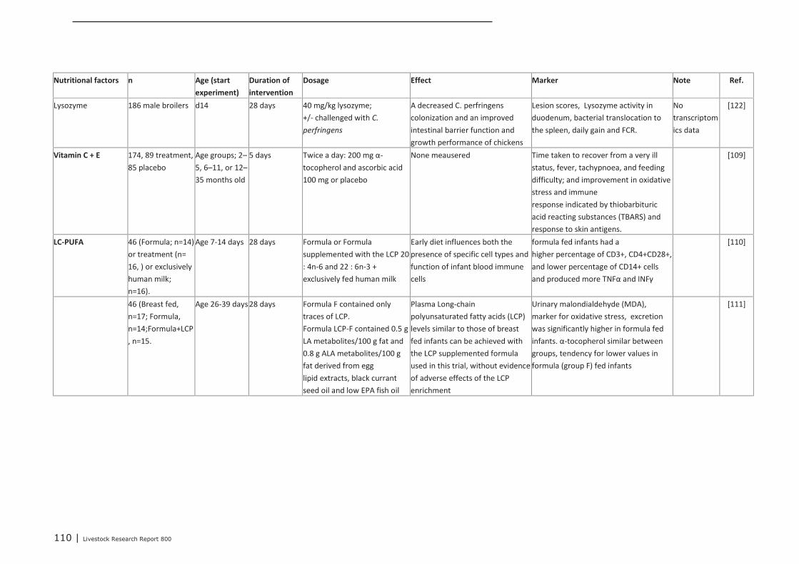
110 | Livestock Research Report 800
Nutritional factors n Age (start
experiment)
Duration of
intervention
Dosage Effect Marker Note Ref.
Lysozyme 186 male broilers d14 28 days 40 mg/kg lysozyme;
+/- challenged with C.
perfringens
A decreased C. perfringens
colonization and an improved
intestinal barrier function and
growth performance of chickens
Lesion scores, Lysozyme activity in
duodenum, bacterial translocation to
the spleen, daily gain and FCR.
No
transcriptom
ics data
[122]
Vitamin C + E 174, 89 treatment,
85 placebo
Age groups; 2–
5, 6–11, or 12–
35 months old
5 days Twice a day: 200 mg α-
tocopherol and ascorbic acid
100 mg or placebo
None meausered Time taken to recover from a very ill
status, fever, tachypnoea, and feeding
difficulty; and improvement in oxidative
stress and immune
response indicated by thiobarbituric
acid reacting substances (TBARS) and
response to skin antigens.
[109]
LC-PUFA 46 (Formula; n=14)
or treatment (n=
16, ) or exclusively
human milk;
n=16).
Age 7-14 days 28 days Formula or Formula
supplemented with the LCP 20
: 4n-6 and 22 : 6n-3 +
exclusively fed human milk
Early diet influences both the
presence of specific cell types and
function of infant blood immune
cells
formula fed infants had a
higher percentage of CD3+, CD4+CD28+,
and lower percentage of CD14+ cells
and produced more TNFα and INFγ
[110]
46 (Breast fed,
n=17; Formula,
n=14;Formula+LCP
, n=15.
Age 26-39 days 28 days Formula F contained only
traces of LCP.
Formula LCP-F contained 0.5 g
LA metabolites/100 g fat and
0.8 g ALA metabolites/100 g
fat derived from egg
lipid extracts, black currant
seed oil and low EPA fish oil
Plasma Long-chain
polyunsaturated fatty acids (LCP)
levels similar to those of breast
fed infants can be achieved with
the LCP supplemented formula
used in this trial, without evidence
of adverse effects of the LCP
enrichment
Urinary malondialdehyde (MDA),
marker for oxidative stress, excretion
was significantly higher in formula fed
infants. α-tocopherol similar between
groups, tendency for lower values in
formula (group F) fed infants
[111]
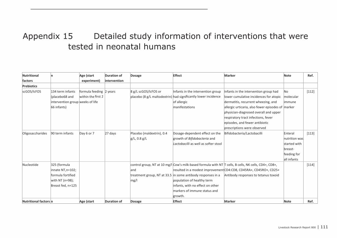
Livestock Research Report 800 | 111
Appendix 15 Detailed study information of interventions that weretested in neonatal humans
Nutritional
factors
n Age (start
experiment)
Duration of
intervention
Dosage Effect Marker Note Ref.
Prebiotics
scGOS/lcFOS 134 term infants
(placebo68 and
intervention group
66 infants)
formula feeding
within the first 2
weeks of life
2 years 8 g/L scGOS/lcFOS or
placebo (8 g/L maltodextrin)
Infants in the intervention group
had significantly lower incidence
of allergic
manifestations
Infants in the intervention group had
lower cumulative incidences for atopic
dermatitis, recurrent wheezing, and
allergic urticaria, also fewer episodes of
physician-diagnosed overall and upper
respiratory tract infections, fever
episodes, and fewer antibiotic
prescriptions were observed
No
molecular
immune
marker
[112]
Oligosaccharides 90 term infants Day 6 or 7 27 days Placebo (maldextrin), 0.4
g/L, 0.8 g/L
Dosage-dependent effect on the
growth of Bifidobacteria and
Lactobacilli as well as softer stool
Bifidobacteria/Lactobacilli Enteral
nutrition was
started with
breast-
feeding for
all infants
[113]
Nucleotide 325 (formula
innate NT,n=102;
formula fortified
with NT (n=98);
Breast fed, n=125
control group, NT at 10 mg/l
and
treatment group, NT at 33.5
mg/l
Cow's milk-based formula with NT
resulted in a modest improvement
in some antibody responses in a
population of healthy term
infants, with no effect on other
markers of immune status and
growth.
T cells, B cells, NK cells, CD4+, CD8+,
CD4:CD8, CD45RA+, CD45RO+, CD25+
Antibody responses to tetanus toxoid
[114]
Nutritional factors n Age (start Duration of Dosage Effect Marker Note Ref.
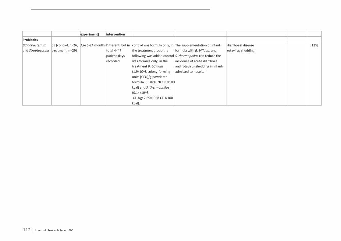
112 | Livestock Research Report 800
experiment) intervention
Probiotics
Bifidobacterium
and Streptococcus
55 (control, n=26;
treatment, n=29)
Age 5-24 months Different, but in
total 4447
patient-days
recorded
control was formula only, in
the treatment group the
following was added control
was formula only, in the
treatment B. bifidum
(1.9x10^8 colony-forming
units [CFU]/g powdered
formula: 35.8x10^8 CFU/100
kcal) and S. thermophilus
(0.14x10^8
CFU/g: 2.69x10^8 CFU/100
kcal).
The supplementation of infant
formula with B. bifidum and
S. thermophilus can reduce the
incidence of acute diarrhoea
and rotavirus shedding in infants
admitted to hospital
diarrhoeal disease
rotavirus shedding
[115]
113 | Livestock Research Report 800
Appendix 15 A brief introduction to theimmune system
1.The innate immune system
The immune system protects against invading pathogens. It is divided the “innate” and the “adaptive”or “acquired” immune system. The innate (also called “natural” or “native” immune system) forms thefirst line of defence that is already present before its first encounter with a pathogen. It consists ofanatomical and (bio)chemical barriers, humoral (soluble) components and cellular components.
1.1 Anatomical and (bio)chemical barriers
The skin forms a strong physical barrier that is hard to break for microbes. Microbes that do attach areusually removed by “flaking” of old skin epithelium. The internal body epithelia, like that of therespiratory and gastrointestinal tract are covered by a mucus layer that is also difficult to penetrate.Microbes trapped in the mucus will be removed with the mucus from the body by the action of the ciliaand due to peristalsis.
The anatomical barriers are covered with antimicrobial (bio)chemical factors, lysozyme on theskin and in tears, gastric acid in the stomach and anti-bacterial proteins (defensins) in the mucosa.Commensal bacteria in the gut and on the skin compete with potential pathogens for space andnutrients.
1.2 Humoral components of the innate immune system
The anatomical en (bio)chemical barriers are a passive part of the innate immune system, butthe innate immune system is also able to recognize Microbial Associated Molecular Patterns (PAMPs)and will respond to these. These PAMPs consist of microbial structures like lipopolysaccharide (LPS),peptidoglycan, bacterial DNA (CpG motifs) and double stranded RNA, that are conserved betweenmany different microbes. Humoral (=soluble) components of the innate immune system that canrecognize PAMPs are the complement system and acute phase proteins (like C-reactive protein).Recognition of bacteria by these systems results in an amplification reaction that can either kill themicrobe directly by forming holes in the bacterial membrane or promotes it destruction by recruitmentof immune cells (inflammation) and “tagging” the microbe for killing by the recruited phagocyticimmune cells.
1.3 Cells of the innate immune system
The cellular part of the innate immune system consists of the granulocytes (neutrophils, mastcells, basophils and eosinophils), the phagocytic antigen presenting cells (macrophages and dendriticcells) and the natural killer (NK) cells.
Mast cells are a type of innate immune cell that reside in connective tissue and in the mucousmembranes. When activated directly by certain microbial products or products of the activatedcomplement system, mast cells rapidly release histamine and tumor necrosis factor alpha (TNFa), thatwill cause recruitment of immune cells (like granulocytes and macrophages) into the tissue a processknown as “inflammation”. Basophils have a very similar function as mast cells, but are normallypresent in the blood. They can be recruited from the blood to sites of infection to contribute to therecruitment of other immune cell, e.g. neutrophils. Neutrophils are the most abundant type ofphagocyte, normally representing 50 to 60% of the total circulating leukocytes (= white blood cells)whose primary function is to phagocytose and destroy bacteria and single cell fungi at the site ofinfection.
Natural killer cells, or NK cells, are a component of the innate immune system that destroysvirus-infected cells, thereby limiting the viral replication.
Macrophages are large phagocytic cells present in the tissues that are differentiated frommonocytes in the blood. They can phagocytose and kill substantial numbers of bacteria or other cellsor microbes. Upon recognition of microbes (PAMPs) they can produce cytokines like TNFa, IL-1, -6,and -8 that promote inflammation, to clear the infection.
114 | Livestock Research Report 800
Dendritic cells (DC) are phagocytic cells present in tissues that are in contact with the externalenvironment, mainly the skin (where they are often called Langerhans cells), and the inner mucosallining of the nose, lungs, stomach, and intestines. Dendritic cells are very important in the process ofantigen presentation, and serve as a link between the innate and adaptive immune systems. Withtheir many Pattern Recognition receptors (PRR), like Toll like receptors, that can bind to PAMPs theycan sense microbial danger. They are the most important cell in the immune system for orchestratingthe right kind of immune response for a particular microbe. Depending on the microbial danger that issensed, they will secrete a cocktail of cytokines that will activate the appropriate immune cells. Theycan activate antigen specific T cells of the adaptive immune system.
Gamma delta T cells (γδ T cells), represent a small subset of T cells in the blood of humansbut are present in relatively larger numbers in the blood of cows and pigs. They consists of severalsubtypes and can perform many of the functions that conventional αβ (alpha beta) T cells can alsoperform, including killing of infected or stressed cells and secretion of many immune modulatingcytokines. Gamma delta T cells exhibit characteristics that place them at the border between innateand adaptive immunity. On one hand, γδ T cells may be considered a component of adaptive immunityin that they rearrange T cell receptor (TCR) genes to produce diversity and because they can developto a certain extend a memory phenotype. However, the various subsets may also be considered partof the innate immune system where a restricted set of TCR receptors may be used as a patternrecognition receptor (PRR) able to recognize conserved microbial structures or other signs of danger.For example, according to this paradigm, large numbers of Vγ9/Vδ2 T cells respond within hours tocommon molecules produced by microbes, and highly restricted intraepithelial Vδ1 T cells will respondto stressed epithelial cells. Gamma delta T cells are more abundantly present in the mucosa wherethey will be one of the first cells to encounter invading pathogens. Still many questions about thesecells remain to be resolved.
2. The adaptive immune system
In contrast to the innate immune system the adaptive immune system is able to very specificallyrecognize pathogens. A second important difference with the innate immune system is that it has“memory” so that in future encounters with the same pathogen it can mount a much faster andstronger immune response compared with the first encounter. The cells of the adaptive immunesystem are T cells and B cells. The both contain antigen specific receptors on their surface that aregenerated by a process called “gene rearrangement”. This process ensures that many different B andT cells are generated of which each cell has one kind of B or T cell receptor with its own uniqueantigen binding site. During an infection, only B or T cells that can bind with their receptor to theinvading pathogen will be selected and will start to proliferate. The process of selection andproliferation takes approximately a week till enough antigen specific B and T cells are generated tomount an effective adaptive immune response. The T cells consist of cytotoxic T cells (CTLs) that cankill virus infected cells and T-helper (Th) cells that are involved in “helping” the appropriate effectorcells. T-helper-1 (Th1) cells will e.g. help macrophages to become more effective in killingphagocytosed microbes and will promote the development of CTLs, while Th17 cells promote therecruitment of neutrophils to the site of infection. B cells often also require the help of Th2 or Th1 cellsto become fully activated and finally develop into antibody secreting plasma cells. The antibodies formthe humoral part of the adaptive immune system.
2.1 Antibodies
In humans 5 antibody isotypes are produced: IgA, IgD, IgE, IgG and IgM. They all consist of 2 lightand 2 heavy chains that together form a Y-shaped protein (T-shaped in the case of IgA). The variablepart of the heavy and light chains form the antigen binding sites (2 identical ones for each monomericantibody), while the constant part of the heavy chains form the Fc part of the antibody thatdetermines what will happen with the antigen/microbe bound by the antibody. Depending on theantibody isotype this could e.g. be activation of complement or binding to Fc receptors on phagocyticcells to enhance uptake of the antigen/microbe by these cells. IgG is the principle isotype in serumwhile IgA is the principle antibody in the gastro intestinal tract and of other mucosal surfaces includingthe upper respiratory tract.
The mucosal immune system
Of all antibody producing cells in the body 70 to 80% are physically located within the tissues of themucosal immune system. The majority of these are IgA producing B cells.
Mechanistically, the induction process can be divided into the following steps:
Livestock Research Report 800 | 115
• Antigens entering the digestive tract are taken up by specialized mucosal cells called M cells. M cellsinternalize the antigen and transport it across the epithelium where antigen can be taken up by APCssuch as dendritic cells (DC). “M” cells are formed in mucosal epithelium in response to signals fromlymphocytes.
• Antigen can be taken up by DC that have dendrites extending through the epithelial tight junctioninto the lumen (drawing on right). Antigens are then presented to lymphocytes (in the intestine, theseare located in Peyer’s patches).
• Lymphocytes (both B and T cells) leave the mucosal site and travel to the mesenteric lymph nodes,then into the lymph.
• Via the thoracic duct, the lymphocytes exit the lymph and enter the circulation.
• Circulating lymphocytes “home” to positions within the mucosal lamina propria throughout the body,including sites distant from the original antigenic encounter. The homing of lymphocytes to mucosalsites involves specific interactions of both adhesion molecules and chemokines.
• B Lymphocytes within the peripheral tissues proliferate and differentiate into IgA secreting plasmacells at effector sites.
C. Features of Mucosal Immunity
1. The administration of antigen at one mucosal site results in specific antibody production at distantmucosal sites (the common mucosal immune system). Some regional preference seems to occur,however. For example, induction via NALT (nasal associated lymphoid tissue) leads to a more robustresponse in the respiratory sites than in induction via mucosal associated lymphoid tissue (MALT) inthe intestine.
2. B cells in the mucosa are selectively induced to produce dimeric IgA rather than other isotypes.The selective switch of B cells to IgA is believed to be mediated by specific cytokines produced by Tcells in the inductive sites.
3. A distinct population of lymphocytes, mostly CD8+ T cells are found in the gut epithelium. Many ofthese intraepithelial lymphocytes are γδ T cells that have an innate character. They can recognizemicrobial structures with their γδ T cell receptor and respond to invading microbes.
4. Conventional T cells, particularly CTLs, are also an important component of the mucosal immuneresponse. The induction and homing requirements for these cells are not as well described as thosefor mucosal B cells.
5. Induction of a response via a mucosal site generally elicits a systemic immune response as well,such that serum antibodies can be detected. This indicates that a mucosal encounter with antigengenerates subsets of T and B cells that home to mucosal sites and also to spleen and regional nodes.
6. Regulatory T cells are a prominent feature at mucosal sites, and may synergize with suppressivedendritic cells. Regulatory populations have been isolated from draining lymph nodes of mucosal sites.

116 | Livestock Research Report 800

Wageningen UR Livestock ResearchP.O. Box 3386700 AH WageningenThe NetherlandsT +31 (0)317 48 39 [email protected]/en/livestockresearch
Livestock Research Report 800
Wageningen UR Livestock Research develops knowledge for meticulous andprofitable livestock farming. This is then translated into practical solutionsand innovations, all the while, ensuring the dissemination of this knowledge.Together with our clients, we combine our scientific knowledge in the field oflivestock farming systems and nutrition, genetics, health and environmentalimpact of livestock into workable livestock concepts for the 21st century.
The mission statement of Wageningen UR (University & Research centre) is‘To explore the potential of nature to improve the quality of life’. WithinWageningen UR, nine specialised research institutes of the DLO Foundationand Wageningen UR have joined forces in order to contribute to the solutionof pressing questions in the field of healthy food and living environment. Witharound 30 branches, 6,000 employees and 9,000 students, Wageningen URis one of the most prestigious knowledge institutes in its field worldwide. Theintegrated approach to problems and the collaboration between differentdisciplines, form the core of Wageningen’s unique approach.


Together with our clients, we integrate scientific know-how and practical experience
to develop livestock concepts for the 21st century. With our expertise on innovative
livestock systems, nutrition, welfare, genetics and environmental impact of livestock
farming and our state-of-the art research facilities, such as Dairy Campus and Swine
Innovation Centre Sterksel, we support our customers to find solutions for current
and future challenges.
The mission of Wageningen UR (University & Research centre) is ‘To explore
the potential of nature to improve the quality of life’. Within Wageningen UR,
nine specialised research institutes of the DLO Foundation have joined forces
with Wageningen University to help answer the most important questions in the
domain of healthy food and living environment. With approximately 30 locations,
6,000 members of staff and 9,000 students, Wageningen UR is one of the leading
organisations in its domain worldwide. The integral approach to problems and
the cooperation between the various disciplines are at the heart of the unique
Wageningen Approach.
Wageningen UR Livestock Research
P.O. Box 338
6700 AH Wageningen
The Netherlands
T +31 (0)317 480 10 77
www.wageningenUR.nl/livestockresearch
Livestock Research Report 800
M.M. van Krimpen, M.M. Hulst, J. van der Meulen, D. Schokker
H.F.J. Savelkoul, E.J. Tijhaar
V.P.M.G. Rutten
Nutritional intervention in animals: benchmarking of strategies, monitoring biomarkers and immune competence
LSR_Rapport_omslag_800.indd 1 15-10-14 13:40