obada abu-rashed or at least part of it physical
TRANSCRIPT

28 3
Obada Abu-Rashed or at least part of it Physical examination-Cranial nerves

Sheet #5 Clinical Skills Lab Record: 15/3/2017 Physical examination-Cranial nerves
------------------------------------------------------------------------------------------------------------------------------------------------------
Page 1
Csl_5 sheet
It’s very long, why? Because I tried to include most of what’s in Macleod in shorter words,
and I didn’t know what to mention and what not. The doctor wasn’t cooperative in this
manner…forgive me
Due its long ,I couldn’t check the sheet more than once, therefore I apologize for any
grammar or spelling mistakes.
Note: this sheet covers all Macleod for the cranial nerves and the doctors lecture,(no need to
go back to it)
References:
- Macleod’s Clinical Examination, 13th Edition
- Doctors lecture (record)
- Different sources on the internet
All the boxes are for illustration and information from Macleod, which I thought to be
necessary to include here, so if you are short on time skip them.
Layout of the sheet is; for each cranial nerve(1N-12N) there’s it:
- Anatomy
- Abnormalities/disorders
- The examination
- Beside some illustration that are important from the doctor or the book.
Good luck for all

Sheet #5 Clinical Skills Lab Record: 15/3/2017 Physical examination-Cranial nerves
------------------------------------------------------------------------------------------------------------------------------------------------------
Page 2
General CNS (cranial nerves) anatomy
The nervous system is divided to two parts: peripheral (spinal nerves and cranial nerves) and
central (brain, brain stem and spinal cord)
Spinal nerves are 31 pairs all of which are mixed (both motor and sensory)
Cranial nerves are paired except the first (olfactory)
Cranial nerves are called so because they rise from the brainstem (midbrain, pons and
medulla oblongata) and pass through different foramina inside the brain skull to reach the
periphery (ex the ear or the eye).
Foramina names can be symbolized in OS ROS
O: Optic foramen, the optic nerve passes through it
S: superior orbit fissure, 3N, 4N, 5aN(ophthalmic branch) and 6N
R: rotundum, 5bN(maxillary branch)
O: ovale, 5cN (mandibular branch)
S: Spinosum, middle meningeal artery and vein
1N (olfactrory) and 2N (optic) emerge from cerebrum of the brain tissue itself (not from the
brain stem)
C.N from 3-12 all arise from brain stem
Two C.N ; 3rd (oculomotor) and fourth(trochlear) from the midbrain
Four C.N ; 5, 6, 7, 8 from the pons
Four C.N ; 9, 10, 11, 12 from medulla oblongata
* the doctor only explained C.N from 2 to 8 but said they are included with us. In this
sheet, I (self acted) and included them as a Macleod reference

Sheet #5 Clinical Skills Lab Record: 15/3/2017 Physical examination-Cranial nerves
------------------------------------------------------------------------------------------------------------------------------------------------------
Page 3
Examination (not done)
Testing of smell is of limited clinical value, and rarely Performed; the pt will come complaining with problems of smelling
Optic nerve (CN 2) – and the 3N 4N 6N
Physioanatomy
The optic nerve examination is accomplished with other related nerves; the third ,fourth and
the sixth C.N all of which are concerned with the orbit and eye movement and vision
In the eye; Rod photoreceptors are responsible for night vision and detection of peripheral
movement. Cone photoreceptors are responsible for color and central vision.
The optic nerve is purely sensory; leaving the eye through the optic disc and entering the
cranium through the optic canal. The pair joins at the optic chiasm. Leaving the chiasm, the
Olfactory nerve (CN1)
Anatomy Bipolar cells in the olfactory bulb form olfactory filaments with small receptors
Abnormal findings
Hyposmia or anosmia (reduction or loss of the sense of smell respectively) may result
from
ear, nose and throat disease,
damage to the olfactory filaments after head injury or
local compression or invasion by basal skull tumors.
Disturbance of smell may also occur in the pre-symptomatic stages of Parkinson’s
and Alzheimer’s diseases.
Hypogeusia/ageusia (altered taste) associated with anosmia. Parosmia : when pleasant smell that is good for others is annoying for you ( from trauma, tumors, or upper respiratory tract infection)
Sheet #5 Clinical Skills Lab Record: 15/3/2017 Physical examination-Cranial nerves
------------------------------------------------------------------------------------------------------------------------------------------------------
Page 4
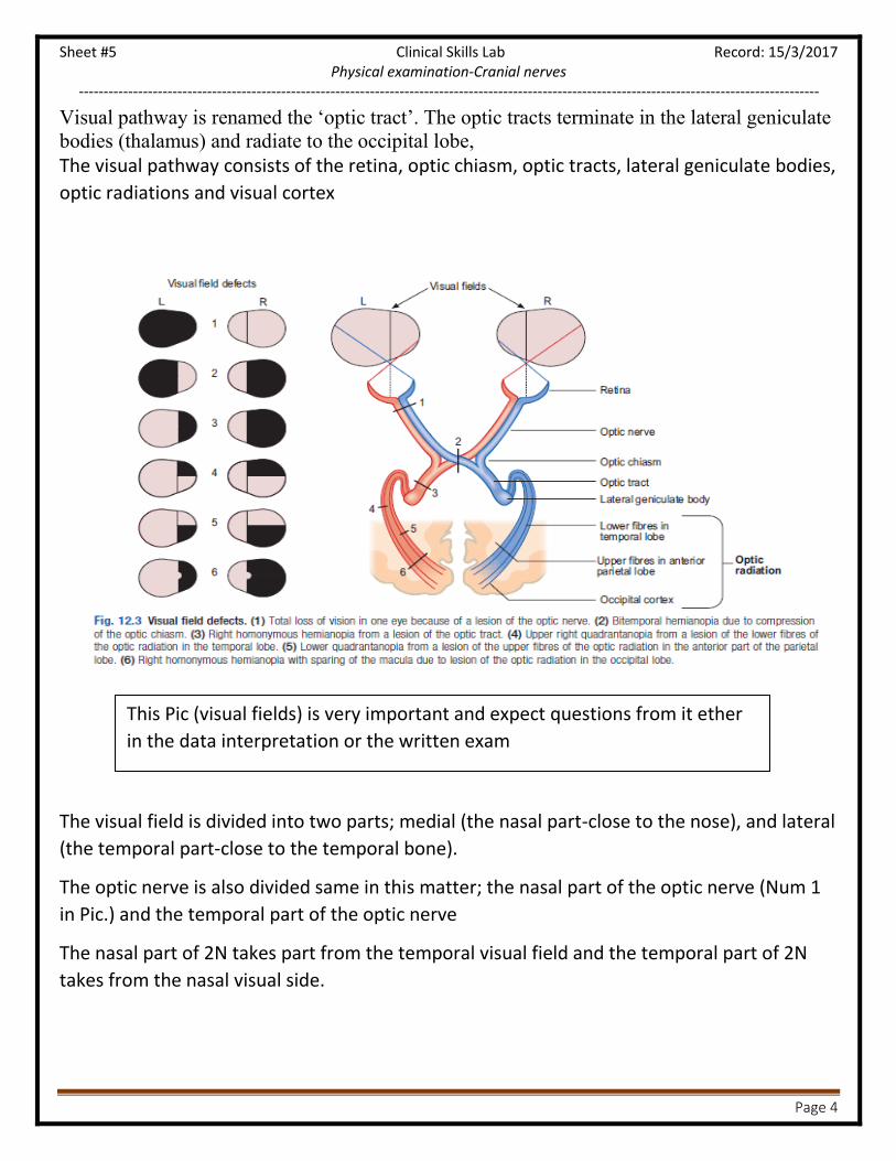
This Pic (visual fields) is very important and expect questions from it ether
in the data interpretation or the written exam
Visual pathway is renamed the ‘optic tract’. The optic tracts terminate in the lateral geniculate
bodies (thalamus) and radiate to the occipital lobe, The visual pathway consists of the retina, optic chiasm, optic tracts, lateral geniculate bodies,
optic radiations and visual cortex
The visual field is divided into two parts; medial (the nasal part-close to the nose), and lateral
(the temporal part-close to the temporal bone).
The optic nerve is also divided same in this matter; the nasal part of the optic nerve (Num 1
in Pic.) and the temporal part of the optic nerve
The nasal part of 2N takes part from the temporal visual field and the temporal part of 2N
takes from the nasal visual side.
Sheet #5 Clinical Skills Lab Record: 15/3/2017 Physical examination-Cranial nerves
------------------------------------------------------------------------------------------------------------------------------------------------------
Page 5
The optic chiasm , in which the nasal parts of both optic nerves cross, is susceptible to
compression from the pituitary gland (ex pituitary adenoma or prolactinoma)lead to inability
to see on the sides for both eyes; called “bitemporal hemianopia”
How does this happen?
The pituitary gland is situated in the sella turcica where the optic chiasm of the optic
nerve(Num 2 in Pic.) is placed. The fibers of the nasal part of optic nerve (from the temporal
visual field), pass though it and they cross to the other side “decussation”
So let’s say if there is a pituitary adenoma; the mass effect of it compresses the optic chiasm
(compressing the nasal part of the left and the nasal part of the right carrying the temporal
vision field of the right and the temporal vision of the left respectively)
Ex of clinical presentation of pituitary adenoma;
A patient comes because multiple car accidents complaining that he can see cars in front of
him but all of a sudden cars hit him from his side, we scan him to see the pituitary (MRI)
The optic tract which carries both nasal and temporal parts continues the visual pathway
until it reaches the cortex area (vision area 17) in the occipital lobe to get analyzed
The Optic tract (num 3 in Pic.) if injured at any place will give us special vision problem
but we (csl5 ) are concerned on the optic caism
Note: Both of the nasal part passes through the optic chaism but those of
the temporal part do not do decussate( meaning they stay at the same
original side )
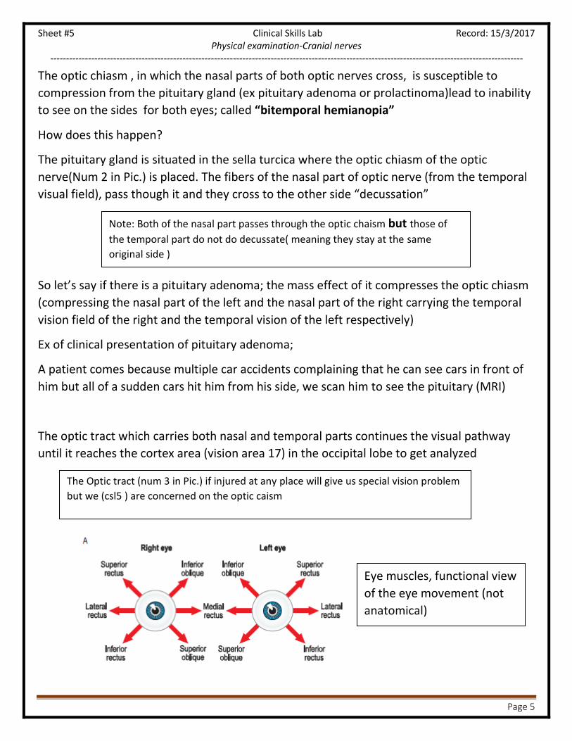
Eye muscles, functional view
of the eye movement (not
anatomical)

Sheet #5 Clinical Skills Lab Record: 15/3/2017 Physical examination-Cranial nerves
------------------------------------------------------------------------------------------------------------------------------------------------------
Page 6
Eye muscles
Inward eye moment (medially) ; adduction
Outward eye moment (laterally); abduction
They are six eye movement muscles grouped in: rectus group and oblique group
Rectus muscle group
1. Lateral movement: abduction
2. Superior rectus :upward and lateral movement
3. Inferior rectus : downward and lateral movement
4. Medial rectus : adduction
Oblique muscle group;
1. Superior oblique :downward and medial movement ( though it’s in a superior
anatomical position)
2. Inferior oblique :upward and medial movement (though it’s in an inferior anatomical
position)
Eye motor muscles are innervated by the 3N 4N 6N
Nerve(num) Emerge Pass through (foramen)
Function
oculomotor (III) tentorium superior
oblique fissure Muscles; open the upper lid (levator palpebrae superioris)
Move the globe upwards (superior rectus, inferior oblique),
downwards (inferior rectus)
Medially (medial rectus). Parasympathetic; supplies
(1)sphincter muscles of the iris, (pupil constriction)
(2) ciliary muscle (focuses the lens for near vision (accommodation))
trochlear (IV) Midbrain (the
left nucleus
innervates the
right trochlear
nerve and vice
versa).
superior orbital
fissure Muscles; superior oblique muscle, which causes downward movement of
the globe when the eye is adducted.
abducens (VI) brainstem superior orbital
fissure Muscles; lateral rectus muscle. Lateral rectus
abducts the eye
The oculomotor (III), trochlear (IV) and abducens (VI) cranial nerves innervate the six external ocular muscles
controlling eye movement and, also affect pupillary size through parasympathetic nerves.

Sheet #5 Clinical Skills Lab Record: 15/3/2017 Physical examination-Cranial nerves
------------------------------------------------------------------------------------------------------------------------------------------------------
Page 7
All muscles of the eye except the superior oblique and lateral rectus are supplied by the 3rd
cranial nerve (oculomotor N.)
superior oblique from 4N trochlear
lateral rectus from the 6N abducens
As like all muscles of the muscular system the eye muscles have an opposite muscle
(agonist and antagonist) functionally to keep the balance. So if one side is damaged the
balance is disrupted and pulls the eye to the opposite of the weak muscle(towards the
normal one)
Ex when the medial rectus muscle is damaged the eye will be sided to the lateral side (to
the strong muscle)
Superior rectus damaged: the eye is towards the ground (downward)
Abnormal findings (disorders/diseases)
eye position displacement:
Can be either caused by damage to the muscle or nerves of the eye
In a neurological born abnormal eye position, it is surgically dealt by cutting both sides of
the muscles repositioning the eye correctly and then its re-stitched
Acronym from doctor; all eye muscles take from 3N except
So4(كبريت : Superior Oblique from the 4N ( and Lr6
Sheet #5 Clinical Skills Lab Record: 15/3/2017 Physical examination-Cranial nerves
------------------------------------------------------------------------------------------------------------------------------------------------------
Page 8
the pupillary reflex
involves the optic nerve,
chiasm (where some
fibers decussate) and the
optic tract, bypassing the
lateral geniculate nucleus
to terminate in
the III nerve (Edinger–
Westphal) nucleus
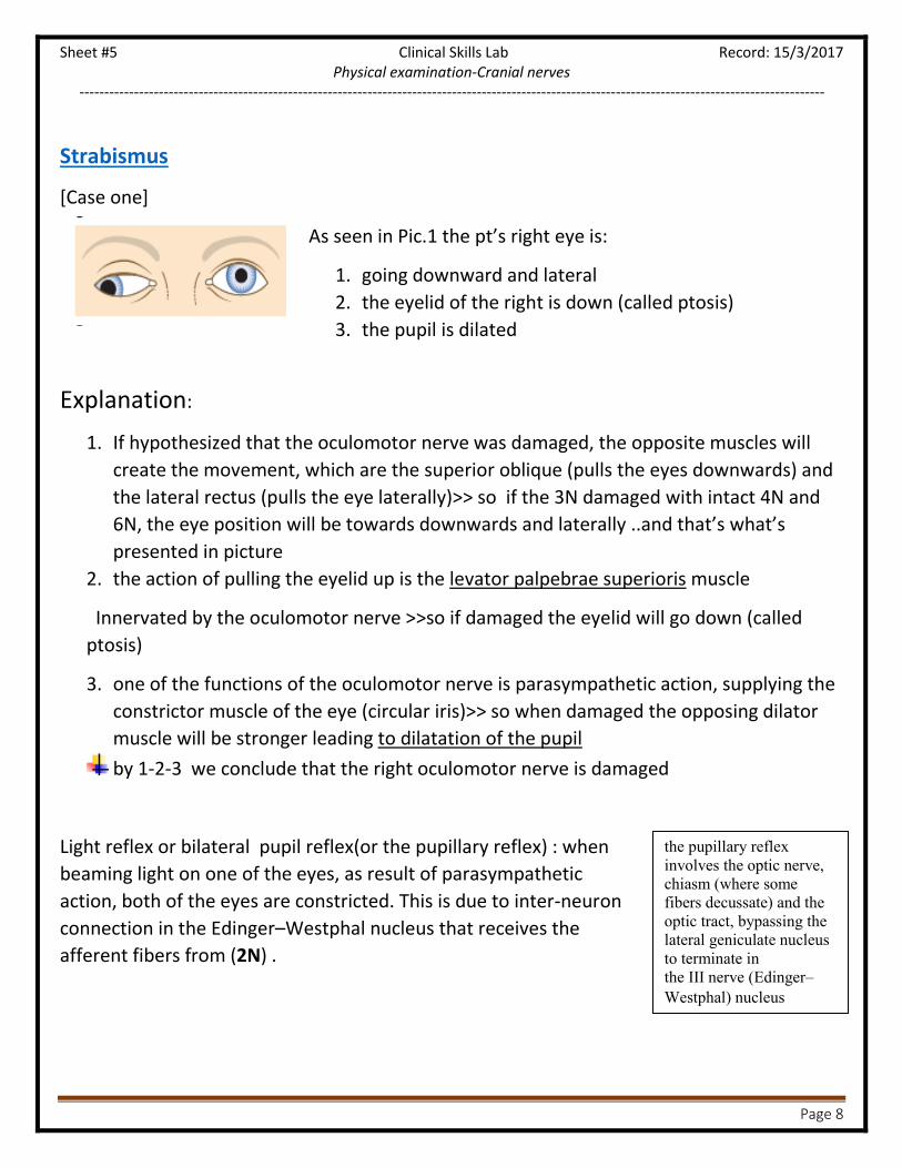
Strabismus
[Case one]
As seen in Pic.1 the pt’s right eye is:
1. going downward and lateral
2. the eyelid of the right is down (called ptosis)
3. the pupil is dilated
Explanation:
1. If hypothesized that the oculomotor nerve was damaged, the opposite muscles will
create the movement, which are the superior oblique (pulls the eyes downwards) and
the lateral rectus (pulls the eye laterally)>> so if the 3N damaged with intact 4N and
6N, the eye position will be towards downwards and laterally ..and that’s what’s
presented in picture
2. the action of pulling the eyelid up is the levator palpebrae superioris muscle
Innervated by the oculomotor nerve >>so if damaged the eyelid will go down (called
ptosis)
3. one of the functions of the oculomotor nerve is parasympathetic action, supplying the
constrictor muscle of the eye (circular iris)>> so when damaged the opposing dilator
muscle will be stronger leading to dilatation of the pupil
by 1-2-3 we conclude that the right oculomotor nerve is damaged
Light reflex or bilateral pupil reflex(or the pupillary reflex) : when
beaming light on one of the eyes, as result of parasympathetic
action, both of the eyes are constricted. This is due to inter-neuron
connection in the Edinger–Westphal nucleus that receives the
afferent fibers from (2N) .

Sheet #5 Clinical Skills Lab Record: 15/3/2017 Physical examination-Cranial nerves
------------------------------------------------------------------------------------------------------------------------------------------------------
Page 9
i.e. : so if we shine a torch on the right eye, the right optic tract gives signal to both the right
and left Edinger–Westphal nucleus. This nucleus by efferent fibers(by the inferior division of
3N) cause a parasypmathetic reflex action causing both of the pupils to constrict.
o The right pupil constriction is called the direct reflex (light pointed on it )
o Left pupil constriction is called indirect reflex (as a result of nucleus connection)
[Case two]
As seen in Pic.2 the pt’s right eye is
1. right eye is going inward (medially )
2. both of the eyelid are normal
3. the pupil is normal
Explanation:
The right eye is going inward (medially) meaning the opposing muscle, the lateral rectus
muscle isn’t functioning..This is called right (convergent squint or esotropia)
Cause : damage to the right 6N abducens nerve supplying the lateral rectus (with
intact 3N and 4N)
[Case three]
In Pic. The eye is going upwards (and only) the medial and
lateral are intact, called (Hypertropia)
Explanation:
Because the of eye is going upwards (and only) meaning the other muscles are functioning
normally
Cause: this abnormality is a result of a dysfunctional Superior oblique muscle
therefore the damage is to the 4N

Sheet #5 Clinical Skills Lab Record: 15/3/2017 Physical examination-Cranial nerves
------------------------------------------------------------------------------------------------------------------------------------------------------
Page 10
Visual disturbance
Visual acuity:
Hypermetropia (long-sightedness): [بعد النظر]; can see the far away things but not the close
ones
Why? Because the cornea (the lens is behind it) abnormally leveled, leading to collecting the
light and focusing it behind the retina than on its normal level
It is common in infants
Myopia (short-sightedness): [قصر النظر]; can see the close things but not far away ones.
Why because the lens is collecting the light and focusing it in front of the retina
Presbyopia happens in old people. They can’t focus on small things in close range, why?
Because the muscles responsible for accommodation are weakened (impaired power) (by
age… >45 ) >> so it’s hard for old people to focus their eye on close range objects but they
still can see in far range.
Pts are given reading glasses to use for close range activities
Note: they do not necessary have abnormalities in their lens or retina therefore not
necessary presented with hypermetropea, anopea or … Astigmatism
Astigmatism: the cornea, which is responsible of collecting the light on the lens, is abnormal
(cornea is irregularly curved), leading to scattering the light and preventing its collection on
the lens therefore the light is weakened and not focused probably on the retina.
The curvature of the optic lens alters to adapt
the focal length to suit the varying distance
and produce a clear, focused image

Sheet #5 Clinical Skills Lab Record: 15/3/2017 Physical examination-Cranial nerves
------------------------------------------------------------------------------------------------------------------------------------------------------
Page 11
Addition eye abnormalities see last page in
this sheet P.32
A student asked about “كسل العين “
It’s a result of a genetic defect in the optic nerve, weakening its functionality. But not
weakness in the muscles of the eye
Treated by closing the strong eye and keeping the weak eye open, this will lead to
hyperactivity (hypertrophy), stimulating the eye to be stronger

Sheet #5 Clinical Skills Lab Record: 15/3/2017 Physical examination-Cranial nerves
------------------------------------------------------------------------------------------------------------------------------------------------------
Page 12
Examination (for the 2n..3N..4N..6N as a group)
1- Introduce you self and take patient name . 2- Take permission and cooperation of patient . 3- Hand hygiene. 4- Ensure privacy(Ensure the pt that all data will be confidential between you and him/her). 5- Make sure there’s a chaperone ( someone with the patient ). 6- Good position of patient and exposure for examining ( in here mostly the pt will be sitting down ) 7- explain the procedure to the pt prior to each act …always do bilateral(or explain to examiner that you have to do bilaterally) 8-gain consent
Ques.: A patient comes with a complaint of decrease visual acuity; please do a cranial
nerve examination for the second third, fourth and sixth cranial nerves
1. inspection
Head position (an abnormal head posture with the head turned or tilted to minimize the
diplopia)
Look at the upper eye lid if for ptosis(drooping eyelid) ,proptosis(forward bulging of the
eyeball.. sclera is visible)or blepharospasm (tonic spasm in the orbicularis oris muscle).
Look for periorbital edema (the surrounding of the eye is enlarged)
- Trauma to the eye
-in nephrotic syndrome
-heart failure patients
Look at the eye ball itself if there’s exopthalmos
we look from the lateral or posterior aspect to check the eye protrusion)
Look for eye discharge
Look at the sclera for redness , discharge and color
Look at the cornea for corneal ulceration
Sheet #5 Clinical Skills Lab Record: 15/3/2017 Physical examination-Cranial nerves
------------------------------------------------------------------------------------------------------------------------------------------------------
Page 13
Pic 2
Pic 1
Pic 3
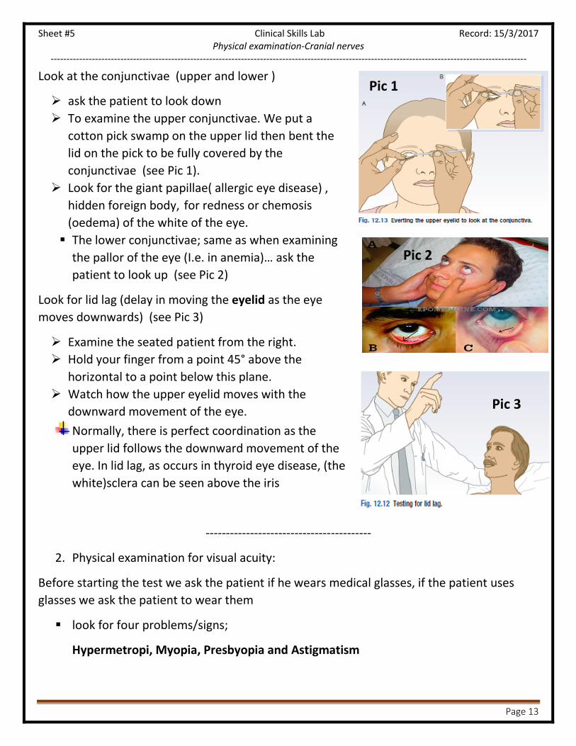
Look at the conjunctivae (upper and lower )
ask the patient to look down
To examine the upper conjunctivae. We put a
cotton pick swamp on the upper lid then bent the
lid on the pick to be fully covered by the
conjunctivae (see Pic 1).
Look for the giant papillae( allergic eye disease) ,
hidden foreign body, for redness or chemosis
(oedema) of the white of the eye.
The lower conjunctivae; same as when examining
the pallor of the eye (I.e. in anemia)… ask the
patient to look up (see Pic 2)
Look for lid lag (delay in moving the eyelid as the eye
moves downwards) (see Pic 3)
Examine the seated patient from the right.
Hold your finger from a point 45° above the
horizontal to a point below this plane.
Watch how the upper eyelid moves with the
downward movement of the eye.
Normally, there is perfect coordination as the
upper lid follows the downward movement of the
eye. In lid lag, as occurs in thyroid eye disease, (the
white)sclera can be seen above the iris
-----------------------------------------
2. Physical examination for visual acuity:
Before starting the test we ask the patient if he wears medical glasses, if the patient uses
glasses we ask the patient to wear them
look for four problems/signs;
Hypermetropi, Myopia, Presbyopia and Astigmatism

Sheet #5 Clinical Skills Lab Record: 15/3/2017 Physical examination-Cranial nerves
------------------------------------------------------------------------------------------------------------------------------------------------------
Page 14
ask pt if wears glasses if so to wear them
We set the pt six (6) meters away from the chart (called
Snellen chart)
Cover one of the patients eyes with a card
ask him to read from the top down until he can no longer
distinguish the letters, while describing what he sees
(character or number or shape)
note down the level (distance) that he can’t describe the
characters
i.e., if a patient couldn’t see at level 12 that means the visual
acuity of the eye is 6/12, meaning what’s supposed to be seen
at a distance of 12 meters can only be seen 6 m from the object
(normal person can see the object clearly at 12 m but the
patient sees it clearly at 6
Scenario:
If the patient didn’t see the first level (60/6), we ask the patient to set one (1) meter away
from the chart, if still he couldn’t see from one meter we wave our hand (big objective like
the palm of the hand )to look for any response, if still couldn’t see we flash a light on his eyes
to observe his visual activity …if the pt reaches this stage then the paten has a serious visual
acuity problem
-----------------------------------------
3.Visual field
Remember: the visual field is divided to nasal and temporal part
Test by four steps:

Sheet #5 Clinical Skills Lab Record: 15/3/2017 Physical examination-Cranial nerves
------------------------------------------------------------------------------------------------------------------------------------------------------
Page 15
1. Homonymous defect
Set the patient so that the distance between you and him is 1 meter
Tell the patient to keep his head fixed during the examination, looking towards you and
to keep his eyes fixed with your eyes نظرك معاي( (
Hold your hands out to their full extent. Wiggle a fingertip and ask the patient to point
to it when he first sees your finger movement (or to tell you)…you move your finger in
x radius way(10 and 2 o’clock, and then 8 and 4 o’clock) (from each angle of a
quadrate start from far and close in to the center)
By doing this the doctor is comparing the visual fields of the patient with his
(normal) because both eyes are lined at same level (opposite to each other)
2. Sensory inattention
Set the patient so that the distance between you and him is 1 meter
Tell the patient to keep his head fixed during the examination looking towards you and
to keep his eyes fixed with your eyes نظرك معاي( (,we Test both eyes together
Both you and the patient should keep your eyes open. (Test both left and right fields at
the same time.)
Raise your hands and move one of them, ask the patient to tell you which had you
moved, do this to both sides to examine for bilateral normal visual sensation or
whether the patient reports seeing only one side.
3. Peripheral visual field
Besides describing the visual field as nasal and temporal it can be called peripheral or central
Set the patient so that the distance between you and him is 1 meter
Tell the patient to keep his head fixed during the examination looking towards you and
to keep his eyes fixed with your eyes (نظرك معاي ), we test each eye separately.
Tell the patient to covers his right eye...the doctor closes the contra to it (here the left)
so that both of the patents eye and the doctors are at the same level
move your finger from far(Peripheral) to center ask the patient to tell you when he
first sees it and compare it to your own observation
Ex if the patient had bitemporal hemeanopea the patient won’t see the fingers coming from
the side to center but the doctor will

Sheet #5 Clinical Skills Lab Record: 15/3/2017 Physical examination-Cranial nerves
------------------------------------------------------------------------------------------------------------------------------------------------------
Page 16
4. Central vision (examine for scotoma)
Central felid of the vision can be observed by the ophthalmoscope looking directly to the
retina of the eye to check the fovea (the blind spot)
It’s a black spot on the retina that doesn’t collect any light, present were the optic disk
originates (start of the optic nerve), normally is to have one black spot…the abnormal is
to have more than one black spot on the retina (called scotoma) meaning more than
one blind spot on the visual field
Set the patient so that the distance between you and him is 1 meter
Tell the patient to keep his head fixed during the examination looking towards you and
to keep his eyes fixed with your eyes (نظرك معاي ), we test each eye separately.
Tell the patient to covers his right eye...the doctor closes the contra to it (here the left)
so that both of the patents eye and the doctors are at the same level
First, Hold the hatpin (pointed pin) at the fixation point; you and the patient focus on
each other’s eye.
Ask the pt to describe the color of the pin.
A ‘pale’ or ‘pink response implies color desaturation, usually because of a
lesion affecting the optic nerve.
Compare the four quadrants of the visual field centrally; each time ask about color
desaturation.
Second, Move the hatpin temporally and horizontally until it disappears from your
visual field. Maintaining the same temporal horizontal position, move it anteriorly or
posteriorly until it also disappears from the patient’s visual field.
Compare the size of the patient’s blind spot to yours.
Third, move the pin in the quadrant movement and ask the pt if the color(redness)
changes at any point, if changed (called color desaturation)
-----------------------------------------

Sheet #5 Clinical Skills Lab Record: 15/3/2017 Physical examination-Cranial nerves
------------------------------------------------------------------------------------------------------------------------------------------------------
Page 17
4. Ocular movements (examination to the muscles of the eye, examine diplopia and
Nystagmus)
Set the patient so that the distance between you and him is 1 meter
Tell the patient to keep his head fixed during the examination) Hold your finger
vertically at least 50 cm away from the patient, (arms length)
Ask the patient to follow your fingers as you move it in a H movement and if he sees your finger doubled (double image) to tell you. Called “diplopia”
If present, cover one of the patient’s eyes to see if diplopia is monocularor binocular.
We do the test twice e, why? The first time to examine for diplopia (if there’s double
imaging) and the second time to examine for Nystagmus (abnormal eye movement the eye
doesn’t move smoothly across a wide movement, (slow drift in one direction: التقطيعزي )
Note we don’t do it twice if the patient in the first time didn’t present with diplopia, just
continue to examine the eye movement for Nystagmus, but if complained about having a
double vision we do another separate one to check the eye movement (Nystagmus)
-----------------------------------------
5. The pupils
Set the patient so that the distance between you and him is 1 meter
Ask the patient to fix his eyes on a distant point straight ahead.
Do two tests:
First, inspection: (we dim the lights in the room)
Bring a bright torchlight from the side to shine on the pupil and look at the symmetry
and the shape of the pupils(there’s no abnormalities in the right and left pupils)
Ex. ..Pt with constricted pupils might be due morphine doses
Pt with fixed dilated pupils might be from internal bleeding, brain stem lesions or death
(it’s a sign of death)

Sheet #5 Clinical Skills Lab Record: 15/3/2017 Physical examination-Cranial nerves
------------------------------------------------------------------------------------------------------------------------------------------------------
Page 18
Second, light reflex (direct and indirect)
Put the torchlight to shine on pupils, coming from the lateral aspect of the pt
And look for pupil construction on the both eyes
The other eye that didn’t receive light, but had a construction reflex is called indirect reflex
or “consensual light reflex”).
-----------------------------------------
6. Convergence and accommodation (to examine the ciliary muscles that are responsible for
the changes in the lens diameter )
Approach the pt from his lateral aspect
ask the pt to look forward at the wall and to fix his vision
approach a pencil(object) to him from the side and ask the patient to focus on it
In normal response, two things occur:
convergence (meaning both the eyes go medially inwards)
accommodation(Look for pupil constriction); the image (pencil) gets clear for the pt …we
ask the pt if he refocused on the pencil once it came in his field of vision
-----------------------------------------
7. Color vision examination, Squint (cover test), Oculocephalic (doll’s-eye) reflex and
Ophthalmoscopy test: not acquired from us

Sheet #5 Clinical Skills Lab Record: 15/3/2017 Physical examination-Cranial nerves
------------------------------------------------------------------------------------------------------------------------------------------------------
Page 19
Trigeminal nerve (5N)
Anatomy :
Is the biggest of the cranial nerves and is divided to three divisions: ophthalmic, maxillary
and mandibular. Those carry both sensory and motor information (mixed)
The sensory part receives from the face and the mouth
Two types of sensation ; general sensation ex touch
And special sensation ex taste (sweet, sour, salty)
For the face its general sensation
The motor information gives its information to muscles of mastication (three of four muscles
of mastication). The masseter, the medial and lateral pterygoid and temporalis
Pic of each muscle position and nerve innervation
for the tongue
The tongue anatomically is divided to anterior 2/3 and posterior 1/3
Anterior 2/3 take its general sensation from the trigeminal nerve 5N and the special
sensation from facial 7N nerve
Posterior 1/3 gives it sensation (both general and special) to glossopharyngeal nerve(9N)
Sensation; ( face, mouth and part of the Dura,) ,sensory ganglion; trigeminal (Gasserian) ganglion.
Motor; temporalis, masseter, medial and lateral pterygoid (muscles of mastication).
Connection; pain and temperature pathways descend to the C2 segment of the spinal cord, so ipsilateral
facial numbness may occur with cervical cord lesions.
Nerve (division)
type Pass through Function
ophthalmic (V1) Sensory superior orbital fissure skin of the upper nose, upper eyelid, forehead and scalp, V1 supplies
sensation to the eye (cornea and conjunctiva) and the mucous
membranes of the sphenoidal and ethmoid sinuses and upper nasal
cavity maxillary (V2) Sensory Foramen rotundum. Upper mouth, roof of pharynx, gums, teeth and palate of the upper jaw
and the maxillary, sphenoidal and ethmoid sinuses. mandibular (V3) Sensory and motor. foramen ovale
floor of the mouth, common sensation, i.e. not taste, to the anterior
two thirds of the tongue, the gums and teeth of the lower jaw, mucosa
of the cheek and the temporomandibular joint in addition to the skin
of the lower lips and jaw area, but not the angle of the jaw

Sheet #5 Clinical Skills Lab Record: 15/3/2017 Physical examination-Cranial nerves
------------------------------------------------------------------------------------------------------------------------------------------------------
Page 20
Abnormal findings
Cause; result
(trigeminal neuralgia); facial numbness and pain.
injury in association with facial fractures ;Unilateral loss of sensation (particularly V2) or by local
invasion by cancer.
Lesions in the cavernous; loss of the corneal reflex , V1 or V2 cutaneous sensory loss.
neurovascular compression(Trigeminal neuralgia); severe, lacerating pain typically in the
distribution of V2 or V3
VZV virus (chickenpox); any sensory nerve,( particularly, thoracic dermatome or V1)
Myasthenia gravis; weakness of the muscles of mastication
pseudobulbar palsy; brisk jaw jerk.

Sheet #5 Clinical Skills Lab Record: 15/3/2017 Physical examination-Cranial nerves
------------------------------------------------------------------------------------------------------------------------------------------------------
Page 21
Examination
Introduce yourself……same as before
1. sensory
**first; general sensation of the face
First examine the sensory part on three places
on the face (skin) -- (that represent the
trigeminal divisions)
When examining the sensation of the
trigeminal we examine the light touch and the
pain
light touch
we ask the patient to close his eyes
tell the him that I am going to swab
on your face and when you feel
anything just say yes
we swab (by a cotton wool tip) on
each area of the trigeminal divisions
as shown in Pic
Compare both sides
pain (superficial pain)
same as before; use a small sharp object (pin) and the same as light touch
Compare both sides
**Second, sensory of that to the anterior 2/3 of tongue (general sensation);
Use like an ear pick (for our exam we use wooden swab sticks)
Ask the pt to close his eyes
Ask the pt to protrude his tongue as much as possible, and touch it by the pick
let him tell you when he feels the pick

Sheet #5 Clinical Skills Lab Record: 15/3/2017 Physical examination-Cranial nerves
------------------------------------------------------------------------------------------------------------------------------------------------------
Page 22
2. Motor part (muscles of mastication) (signs rare)
We inspect (inspection) the muscles looking for at their bulking (atrophy, muscle
wasting or normal).. (Most apparent in temporalis).
Then ask the pt to shut on his teeth (to clench his teeth);to palpate the bulk of the
masseter muscle (feel the cheeks)
Now to examine the medial and lateral pterygoids;
We put our hand underneath the chin and ask the pt to open his mouth against the
resistance of our hand …to asses the power of the pterygoids muscles (if the pt has
weakness in the pterygoids he wouldn’t be able to open his mouth)
3. Reflexes
There are two reflexes to examine; the corneal reflex and the
jaw reflex
The corneal reflexes (only for knowledge, not to be
done...say “I’m going test… “)
Is related to the ophthalmic division as the afferent fiber. When
touching the cornea the pt closes both his eyes (blinks
bilaterally) as action of the facial efferent fibers.
Explain to the patient what you are going to do, and ask him to remove contact lenses, if relevant.
We ask the pt to look upwards we pull his upper eye lid up (pulling the conjunctiva).
We use a swab to touch the lateral edge of the cornea of the pt eye ..normally the pt’s
eyes blink (both; direct and consensual ) as a result of the reflex
For knowledge (not to be done)
Nasal tickle test: use a wisp of cotton wool to tickle the inside of each nostril and ask the patient
to compare: it is an unpleasant sensation easily appreciated by the patient

Sheet #5 Clinical Skills Lab Record: 15/3/2017 Physical examination-Cranial nerves
------------------------------------------------------------------------------------------------------------------------------------------------------
Page 23
the jaw reflex
Is related to the mandibular division for the both afferent and
efferent fibers
we ask the pt to open a slight gap between his teeth (mouth
hangs loosely open)
Place your forefinger in the midline between lower lip and chin.
Percuss your finger gently with the tendon hammer in a
downwards direction
Normally; An absent, or just a slightly present reflex
Why this reflex is imp? Because in upper motor neuron lesions (like in stroke) when
provoking these reflexes they will give us hyperreflexia (shut the teeth powerfully..Most
likely to hear a snap )

Sheet #5 Clinical Skills Lab Record: 15/3/2017 Physical examination-Cranial nerves
------------------------------------------------------------------------------------------------------------------------------------------------------
Page 24
Facial nerve (7N)
Anatomy
has a motor part that is related to facial expressions
parasympathetic part related to the glands (sublingual and submandibular -that make
saliva- and lacrimal gland ) via nervus intermedius
And sensation: special sensation (taste) to the anterior2/3 of the tongue (via the
chorda tympani branch)
provides the efferent supply to several reflexes
Nerve
(num)
emerge Passes through
(foramen) function
Facial nerve
(7N)
emerging from the lateral
pontomedullary junction
Enter the internal acoustic meatus
bone, exiting the skull
Via the stylomastoid foramen.
branches to the stapedius muscle
and its parasympathetic fibers, as
well as being joined by the taste
fibers of the chordae tympani
Abnormal findings
lower motor neuron lesion(weakness of both upper and lower facial muscles). - Bell’s palsy; common in lower motor neuron lesion, preceded by mastoid pain with impairment of taste and
hyperacusis
Bell’s phenomenon occurs when the patient is unable to close his eye. As he tries, the eyeball rolls
upwards, exposing the conjunctiva below the cornea
- Ramsay Hunt syndrome; in herpes zoster infection to (facial) ganglion
severe lower motor neuron facial palsy, ipsilateral loss
of taste and buccal ulceration, and a painful vesicular eruption in the external auditory meatus
- tumours; lower motor neuron lesion
- Synkinesis; (most commonly twitching of the corner of the mouth on ipsilateral blinking) is a sign of
aberrant reinnervation
upper motor neuron lesion: (weakness is marked in the lower facial muscles) this is because there is bilateral cortical innervation of the upper facial muscles
- The nasolabial fold; flattened and the corner of the mouth drooped.
- Involuntary emotional movements,(spontaneous smiling is preserved)
Bilateral facial palsies are less common - Guillain–Barré syndrome
- sarcoidosis
- Lyme disease
- HIV infection
Distinct from VII nerve palsies, Parkinson’s disease can cause loss of spontaneous and
involuntary facial movements
Sheet #5 Clinical Skills Lab Record: 15/3/2017 Physical examination-Cranial nerves
------------------------------------------------------------------------------------------------------------------------------------------------------
Page 25
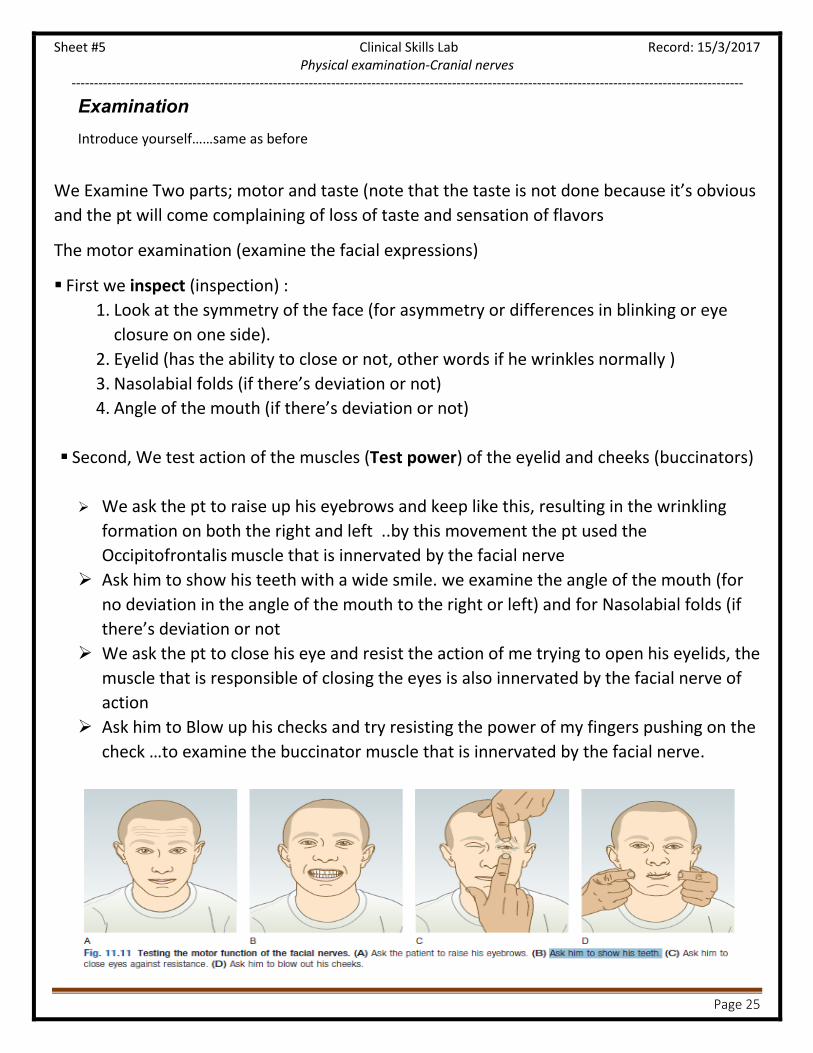
Examination
Introduce yourself……same as before
We Examine Two parts; motor and taste (note that the taste is not done because it’s obvious
and the pt will come complaining of loss of taste and sensation of flavors
The motor examination (examine the facial expressions)
First we inspect (inspection) :
1. Look at the symmetry of the face (for asymmetry or differences in blinking or eye
closure on one side).
2. Eyelid (has the ability to close or not, other words if he wrinkles normally )
3. Nasolabial folds (if there’s deviation or not)
4. Angle of the mouth (if there’s deviation or not)
Second, We test action of the muscles (Test power) of the eyelid and cheeks (buccinators)
We ask the pt to raise up his eyebrows and keep like this, resulting in the wrinkling
formation on both the right and left ..by this movement the pt used the
Occipitofrontalis muscle that is innervated by the facial nerve
Ask him to show his teeth with a wide smile. we examine the angle of the mouth (for
no deviation in the angle of the mouth to the right or left) and for Nasolabial folds (if
there’s deviation or not
We ask the pt to close his eye and resist the action of me trying to open his eyelids, the
muscle that is responsible of closing the eyes is also innervated by the facial nerve of
action
Ask him to Blow up his checks and try resisting the power of my fingers pushing on the
check …to examine the buccinator muscle that is innervated by the facial nerve.

Sheet #5 Clinical Skills Lab Record: 15/3/2017 Physical examination-Cranial nerves
------------------------------------------------------------------------------------------------------------------------------------------------------
Page 26
Examples: (lesions affecting the facial nerve (motor))
From the Pic A , We see
- There’s no winkling
- the eye lid isn’t able to close (bell’s phenomenon)
- The nasolabial fold is absent
- The angle of the moth has deviation to the normal side
This means all upper part and lower parts are effected>> it’s a lower motor neuron lesion
of the right facial nerve
From the Pic B , We see
- There’s wrinkling
- Eye can be closed
- No nasolabial fold
- Mouth deviation to the other side
Meaning the lower part of the face is effected (upper motor neuron lesion of the right 7N)
Remember: The motor part of the facial nerve is divided to upper and lower neurons;
The upper motor neurons control the lower half of the facial expression muscles
Meanwhile the lower motor neurons control all (both upper and lower part) of the
face

Sheet #5 Clinical Skills Lab Record: 15/3/2017 Physical examination-Cranial nerves
------------------------------------------------------------------------------------------------------------------------------------------------------
Page 27
Hearing loss Hearing loss may be due to failure;
- of the VIIIth nerve or its endings( sensorineural )
- the conduction mechanisms of the middle ear by
fluid, fixation or drum perforations or wax
obstructing the external auditory meatus.
Nystagmus: an involuntary rhythmical oscillation of the eyes,
The commonest form;
- Horizontal jerk nystagmus, a slow
(pathological) drift of both eyes in one direction,
then a fast correction in the opposite
direction,(due to a central lesion).
- Pendular nystagmus (oscillations equal in rate
and amplitude about a central point) occurs
with central vision defects.
Vestibulocochlear nerve (8N)
Anatomy
Nerve
(division)
emerge Passes through
(foramen) function
the cochlear
nerve
Lateral floor and wall of
the fourth ventricle in the
pons and medulla.
internal acoustic meatus carries auditory sensory information
from the cochlea of the inner
ear directly to the brain The vestibular
nerve.
Lateral floor and wall of
the fourth ventricle in the
pons and medulla.
internal acoustic meatus travels the vestibular system of the
vestibule of inner ear , reflects
gravity and linear accelerations of the
head
Abnormal findings

Sheet #5 Clinical Skills Lab Record: 15/3/2017 Physical examination-Cranial nerves
------------------------------------------------------------------------------------------------------------------------------------------------------
Page 28
Examination
Introduce yourself……same as before
To testing hearing ..Three methods are included
Check for the conduction of the ear, Conduction problems (ex. Wax, membrane rupture,
inflammation in the bones (osteitis )
1. Whispering test:
Stand behind the patient
close one of the pt’s ear (ex by a cotton)
And at a distance of 15 cm of the pt’s ear, whisper and tell the pt to repeat what
you’ve said
repeat the action but each time get away from the pt
(normally the person can hear your whisper till you’re 60 cm away from his ear
2. Weber’s test:
using the tuning fork (either type doesn’t matter ) hit it then put it on the middle of
the forehead
ask the pt if he can locate were he hears the vibration more (on the left or right ear)
normally the pt hears in both equally, if there’s a problem the pt will hear more
in the normal ear not the obstructed one
3. Rinne’s test :
Note that air conduction is better that bone conduction(AC > BC)
We Place the vibrating fork at the patient’s external auditory meatus; ask if he can
hear it.
Place the still-vibrating base on the mastoid process.
Ask: ‘Is it louder in front, or behind your ear?’
If air pathway is obstructed, the bone conduction is heard better than air
conduction
The function of hearing consist of two parts; the conduction and the nerve part, the conduction pathway is:
air- tube - membrane - bones(3)- receptors

Sheet #5 Clinical Skills Lab Record: 15/3/2017 Physical examination-Cranial nerves
------------------------------------------------------------------------------------------------------------------------------------------------------
Page 29
Check for nerve problems:
1. Weber’s test:
The vibrating sound is more localized to the normal better ear (not the one with
the problem) -sound is heard better in the better-hearing ear.
In symmetrical hearing loss it is heard in the middle.
2. Rinne’s test :
is the same (normally, air conduction is better than bone conduction)

Sheet #5 Clinical Skills Lab Record: 15/3/2017 Physical examination-Cranial nerves
------------------------------------------------------------------------------------------------------------------------------------------------------
Page 30
The glossopharyngeal (9N) and Vagus (10N) nerves
Anatomy Both contain sensory, motor and autonomic components. Main functions of IX and X are swallowing,
phonation/articulation and sensation from the pharynx/larynx.
Nerve
(num)
Emerge Passes through
(foramen) Function
The
glossopharyngeal
(IX)
from the lateral medulla via the jugular foramen Carries sensation from the pharynx and
tonsils, and sensation and special taste from
the posterior one-third of the tongue.
Supply the stylopharyngeus muscle,
Parasympathetic fibers to the parotid gland.
The vagus (X)
from the lateral medulla via the jugular foramen Carries important sensory information
but also innervates upper pharyngeal,
soft palate and laryngeal muscles.
Abnormal findings
Examination Introduce yourself……same as before
Assess the patient’s speech for dysarthria or dysphonia.
Ask him to say ‘Ah ; look at the movements of the palate and uvula using a torch.
Normally, both sides of the palate elevate symmetrically and the uvula remains in the midline.
Ask the patient to puff out his cheeks with the lips tightly closed. Listen for air escaping from the nose.
For the cheeks to puff out, the palate must elevate and occlude the nasopharynx. If palatal movement is
weak, air will escape audibly through the nose.
Ask the patient to cough; assess the strength of the cough.
Testing pharyngeal sensation and the gag reflex is unpleasant and has poor predictive value for
aspiration. Instead, and in fully conscious patients only, use the swallow test. Administer 3
teaspoons of water and observe for absent swallow, cough or delayed cough, or change in voice
quality after each teaspoon. If there are no problems, watch for the same reactions while the
patient swallows a glass of water.
Unilateral X nerve damage (rare) leads to;
- ipsilateral reduced elevation of the soft palate when the patient says‘Ah’, (with deviation of
the uvula)
- dysphonia and a ‘bovine’ cough, from Damage to the recurrent laryngeal branch (due to lung
cancer, thyroid surgery, mediastinal tumours and aortic arch aneurysm)
Bilateral X nerve lesions
- cause dysphagia and dysarthria.
- nasal regurgitation of fluids and nasal air escape when the cheeks are puffed out

Sheet #5 Clinical Skills Lab Record: 15/3/2017 Physical examination-Cranial nerves
------------------------------------------------------------------------------------------------------------------------------------------------------
Page 31
The accessory (11N) nerve
Anatomy
The accessory nerve has two components:
A cranial part closely related to the vagus nerve
A spinal part which provides fibers to the upper trapezius and the
sternocleidomastoid muscles, responsible for elevating (shrugging) the shoulders, and
head turning/flexing. Nerve
(num)
Emerge Passes through
(foramen) function
The accessory (XI) spinal nuclei arise from
the anterior horn cells of
C1–5.( spinal cord)
Ascend through the
foramen magnum, and exit
via the jugular foramen
Responsible for elevating (shrugging) the shoulders, and head turning/flexing.
Abnormal findings
Examination Introduce yourself……same as before
Face the patient and inspect the sternocleidomastoid
muscles for wasting or hypertrophy; palpate them to assess
their bulk.
Stand behind the patient to inspect the trapezius muscle for
wasting or asymmetry.
Ask the patient to shrug the shoulders, then apply downward
pressure with your hands to assess the power (Fig. 11.14A).
Test power in the left sternocleidomastoid by asking the
patient to turn the head to the right(opposite) while you
provide resistance with your hand placed on the right side of
the patients chin.
XI nerve lesions are uncommon,
o Wasting of the upper fibers of trapezius; displacement of the upper vertebral border of the scapula
away from the spine, while the lower border is displaced towards it
o Wasting and weakness of the sternocleidomastoids; myotonic dystrophy.
o head drop; myasthenia, motor neurone disease and some myopathies.
Causes:
- during surgery in the posterior triangle
of the neck,
- penetrating injuries
- local invasion by tumour.

Sheet #5 Clinical Skills Lab Record: 15/3/2017 Physical examination-Cranial nerves
------------------------------------------------------------------------------------------------------------------------------------------------------
Page 32
upper motor nerve lesions (pseudobulbar
palsy) result from;
- vascular disease,
- motor neurone disease( multiple
sclerosis, Parkinson’s disease)
- dyskinesias
The hypoglossal (12N) nerve
Anatomy
Nerve
(num)
Emerge Passes through
(foramen) function
The hypoglossal
(XII)
emerges anteriorly from
the brainstem
exits the skull in
the hypoglossal canal
passing to the root of the tongue innervates all the extrinsic and intrinsic muscles of the tongue, except for the palatoglossus
Abnormal findings
Examination Introduce yoursel……same as before
Ask the patient to open his mouth. Look (inspect) at the tongue at rest for wasting,
fasciculation or involuntary movement.
Ask the patient to put out his tongue. Look for deviation or involuntary movement.
Ask the patient to move the tongue quickly from side to side.
Test power by asking the patient to press the tongue against the inside of each
cheek in turn while you press from the outside with your finger.
Assess speech by asking the patient to say ‘yellow lorry’.
Assess swallowing with a water swallow test.

Sheet #5 Clinical Skills Lab Record: 15/3/2017 Physical examination-Cranial nerves
------------------------------------------------------------------------------------------------------------------------------------------------------
Page 33
Cortex damage:
A lesion, affecting only the posterior tip of the occipital lobe, may produce
central homonymous hemianopia with sparing of peripheral
vision. A lesion, affecting only the anterior occipital lobe, may cause
homonymous hemianopia involving peripheral vision but sparing central
macular vision. Damage to secondary visual areas causes visual agnosia
(inability to recognise visual stimuli) and distorted as macropsia (seeing
things larger) or micropsia (smaller than reality)
pain:
1.with ‘white eye’ Blepharitis
Severe dry eyes are a
feature of Sjögren’s syndrome.
Migraine visual disturbance associated with headache or eye pain
Cluster headaches may present as ocular pain.
raised intraocular pressure), patients describe seeing haloes around lights. Optic neuritis or scleritis:.Pain on eye movement usually indicates
2.with red eye uveitis).
scleritis or conjunctivitis. Diffuse redness .
Entropion (an inverted eyelid) leads to painful corneal
Erosion
ectropion (everted eyelid) causes
dryness in the exposed eye.
glaucoma with high intraocular pressure (with cloudy
Corneal oedema clouds the underlying iris,
Acute iritis(The inflamed iris becomes stuck to the underlying lens). The pain
is not
as severe
Corneal ulceration may be due to Virus: herpes simplex(mostly), or bacterial
infection
scleritis : Pain on moving a red eye
Diplopia (double vision): caused by imbalance of eye movements(Binocular) or intraocular disease.( uniocular)
Blurred vision: an ocular problem,
Sudden-onset visual loss: Transient due to the aura of migraine or unilateral amaurosis fugax
Permanent: due to vascular occlusion. Or indicates occipital
lobe infarction.( in the absence of hemiparesis or dysphasia)
demyelinating optic neuritis (Uthoff’s
phenomenon). (Visual impairment)
Gradual-onset visual loss:cataract, age-related, optic atrophy,
tumours
Distortion of vision: disruption of the photoreceptors
Flashes and floaters: caused by vitreous degeneration,( >65 years and in
myopia), lead to retinal tears.
Haloes: due to corneal oedema: coloured lights seen around bright lights, occur
with glaucoma.
Oscillopsia:, objects appear to oscillate, causes mild blurring to rapid and
periodic jumping (common in acquired nystagmus)
Nystagmus: is an involuntary oscillation of the eyes
that is often rhythmical, with both eyes moving synchronously.
Anisocoria:Anisocoria is inequality of the pupil sizes.