o'brien, j. s., troyer, j. l., roelke, m., maker, l., and
TRANSCRIPT

O'Brien, J. S., Troyer, J. L., Roelke, M., Maker, L., and Pecon-Slattery, J. (2006). Plagues and adaptation: lessons from the Felidae models for SARS and AIDS. Biol. Conserv. 131: 255-267. Keywords: Acinonyx jubatus/AIDS/CDV/cheetah/disease/felidae/FIV/SARS Abstract: Research studies of infectious disease outbreaks in wild species of the cat family Felidae have revealed unusual details regarding forces that shape population survival and genetic resistance in these species. A highly virulent feline coronavirus epidemic in African cheetahs, a disease model for human SARS, illustrates the critical role of ancestral population genetic variation. Widespread prevalence of species specific feline immunodeficiency virus (FIV), a relative of HIV-AIDS, occurs with little pathogenesis in felid species, except in domestic cats, suggesting immunological adaptation in species where FIV is endemic. Resolving the interaction of host and pathogen genomes can shed new light on the process of disease outbreak in wildlife and in humankind. The role of disease in endangered populations and species is difficult to access as opportunities to monitor outbreaks in natural populations are limited. Conservation management may benefit greatly from advances in molecular genetic tools developed for human biomedical research to assay the biodiversity of both host species and emerging pathogen. As these examples illustrate, strong parallels exist between disease in human and endangered wildlife and argue for an integration of the research fields of comparative genomics, infectious disease, epidemiology, molecular genetics and population biology for an effective proactive conservation approach.

B I O L O G I C A L C O N S E R V A T I O N 1 3 1 ( 2 0 0 6 ) 2 5 5 – 2 6 7
. sc iencedi rec t . com
ava i lab le a t wwwjournal homepage: www.elsevier .com/ locate /b iocon
Plagues and adaptation: Lessons from the Felidae models forSARS and AIDS
Stephen J. O’Briena,*, Jennifer L. Troyerb, Melody Roelkeb, Laurie Markerc, Jill Pecon-Slatterya
aLaboratory of Genomic Diversity, National Cancer Institute, Building 560, Room 21-105, Frederick, MD 21702, USAbLaboratory of Genomic Diversity, SAIC-Frederick, NCI-Frederick, Frederick MD USAcCheetah Conservation Fund, Namibia, Southwest Africa
A R T I C L E I N F O
Available online 6 June 2006
Keywords:
FIV
SARS
AIDS
CDV
0006-3207/$ - see front matter � 2006 Elsevidoi:10.1016/j.biocon.2006.05.001
* Corresponding author: Tel.: +1 301 846 1296E-mail address: [email protected] (S.J. O
A B S T R A C T
Research studies of infectious disease outbreaks in wild species of the cat family Felidae
have revealed unusual details regarding forces that shape population survival and genetic
resistance in these species. A highly virulent feline coronavirus epidemic in African chee-
tahs, a disease model for human SARS, illustrates the critical role of ancestral population
genetic variation. Widespread prevalence of species specific feline immunodeficiency virus
(FIV), a relative of HIV–AIDS, occurs with little pathogenesis in felid species, except in
domestic cats, suggesting immunological adaptation in species where FIV is endemic.
Resolving the interaction of host and pathogen genomes can shed new light on the process
of disease outbreak in wildlife and in humankind. The role of disease in endangered pop-
ulations and species is difficult to access as opportunities to monitor outbreaks in natural
populations are limited. Conservation management may benefit greatly from advances in
molecular genetic tools developed for human biomedical research to assay the biodiversity
of both host species and emerging pathogen. As these examples illustrate, strong parallels
exist between disease in human and endangered wildlife and argue for an integration of
the research fields of comparative genomics, infectious disease, epidemiology, molecular
genetics and population biology for an effective proactive conservation approach.
� 2006 Elsevier Ltd. All rights reserved.
1. Introduction
It is becoming increasingly clear that understanding the com-
ponents of disease processes, particularly infectious disease,
will be critical for understanding the survival or demise of
free ranging mammalian species. Genomic variation of both
host species and infectious agent is of critical importance in
the outcome of a disease outbreak, and even more so in
endangered species. Species with reduced genetic variation,
particularly in genes involved with disease resistance, may
be less able to mount an effective immune response against
an emerging pathogen. Representing carnivores, the cat fam-
ily Felidae offers numerous examples of reduced genetic var-
er Ltd. All rights reserved
; fax: +1 301 846 1686.’Brien).
iation in natural populations common to endangered species
including Asian lion (Panthera leo persica) (Gilbert et al., 1991),
cheetah (Acinonyx jubatus) (Menotti-Raymond and O’Brien,
1993), tiger (P. tigris) (Luo et al., 2004), leopard (P. pardus)
(Uphyrkina et al., 2001; Uphyrkina et al., 2002) and the North
American populations of puma (Puma concolor) (Culver et al.,
2000; Roelke et al., 1993) that may signify increased suscepti-
bility to opportunistic infectious disease. In addition, the
pathogens themselves evolve, constantly developing new ge-
netic based strategies to overcome or abrogate the immune
defenses of the host species. The consequence of the co-evo-
lution of pathogen and host are a dynamic ratchet of con-
stantly improving virulence of the agent and resistance of
.

256 B I O L O G I C A L C O N S E R V A T I O N 1 3 1 ( 2 0 0 6 ) 2 5 5 – 2 6 7
the host. Both the pathogens and the host are survivors of an-
cient deadly struggles and the exquisite strategies that have
been retained are only now being deciphered.
Conservation management of endangered carnivores has
limited opportunities to detect, monitor and contain emer-
gent pathogens due to logistic concerns involved with the
continued monitoring of the health status of natural popu-
lations. As a result, we know most about disease processes
in humans, Homo sapiens (Garrett, 1994), so understanding
human–pathogen interactions can help us correctly inter-
pret examples from wildlife. There are far more people
and more human medical researchers than there are for
most large charismatic mammals, so more human disease
outbreaks have been tracked. Tools of molecular biology,
genetics, immunology and epidemiology were developed
with human disease in mind, but they work very well in
other mammals. Thus, efforts in characterizing the genet-
ics, evolution, and pattern of transmission of pathogens,
and the resultant conservation implications for the host
species, benefit directly from advances in human genomic
research.
etis/snoitutitsbus 500.0 YM 2.01
YM 9.8
YM 6.7
YM 0.8
YM 7.6
YM 3.6
YM 8.5
Fig. 1 – Cat family phylogeny from Johnson et al., 2006. Shown
sequence evolution from 18,853 bp of data from autosome, X an
three-letter codes, scientific name, common name and grouped
One of the most significant advances in the genomics era
integral to conservation genetics is the whole genome se-
quence of a score of mammals. Starting with human, mouse,
rat, and chimp, these projects now capture the phylogenetic
divergence across all the orders of placental mammals by
inclusion of dog, cat, elephant, cow and others (NHGRI web-
site: www.genome.gov). Further, a subset of orthologous
genes sequenced in representatives of 5400 species deter-
mined conclusively the evolutionary pattern of divergence
of placental mammals during the last 105 MY (Murphy
et al., 2001a; Murphy et al., 2004; Murphy et al., 2001b; O’Brien
et al., 1999a). The utility of the mammalian phylogeny in con-
servation ranges from defining taxonomic units to using the
evolutionary tree as a reference guide to detect patterns of
mutation, adaptation, and selection in endangered species.
For example, whole genome sequences of human, chimpan-
zee (Pan troglodytes), mouse (Mus musculus) and dog (Canis
familiaris) indicate that not only do genomes evolve at differ-
ent rates (Cooper et al., 2004), but also categories of genes
experience different regimes of selection to either diversify
or maintain function among primates, rodents and carni-
:acF sutac sileF:isF sirtsevlis .F
:ilF acybil.F:ibF iteib .F
:amF atiragram .F:inF sepirgin .F:hcF suahc .F:amO lunam subolocotO
:urP susonigibur surulianoirP:ebP sisnelagneb .P
:ivP sunirreviv .P:lpP specinalp .P:ocP rolocnoc amuP :ayP idnuorauogay .P :ujA sutabuj xynonicA:pyL sunidrap xnyL
:ylL xnyl .L:acL sisnedanac .L:urL sufur .L:apL siladrap sudrapoeL:iwL iideiw .L:ajL sutibocaj .L:ocL olocoloc .L:egL iyorffoeg .L:ugL angiug .L
:itL sunirgit .L :acP lacarac sileforP
:uaP atarua .P :esP lavres .P
:abP aidab silefodraP:etP iikcnimmet .P
:amP ataromram .P :elP oel arehtnaP:noP acno .P:apP sudrap .P
:itP sirgit .P:nuP aicnu .P:enN asoluben silefoeN
gnasnil nodonoirP gnasnil dednaBatucorc atucorC aneayhdettopS
aluvrap elagoleH esoognom frawDS attacirus ataciru etaciruS
xoref atcorpotpyrC assoFsutidorhpamreh suruxodaraP teviC mlaP
atteneg attenneG teneG
tac citsemoDtac dliw naeporuE
tac dliw nacirfAtac tresed esenihC
tac dnaStac detoof-kcalB
tac elgnuJtac sallaP
tac dettops-ytsuRtac drapoel naisA
tac gnihsiFtac dedaeh-talF
amuPidnuoraugaJ
hateehCxnyl nairebI
xnyl naisaruExnyl naidanaC
tacboBtolecOyagraM
tac niatnuom naednAtac sapmaP
tac s’yorffoeGdokdoK
anirgiTlacaraC
tac nedlog nacirfAlavreStac yaB
tac nedlog naisAtac delbraM
noiLraugaJdrapoeL
regiTdrapoel wonS
drapoel deduolC
Do
mes
tic
cat
Asi
anle
op
ard
cat
Pu
ma
Lyn
xO
celo
tC
arac
alP
anth
era
Bay ca
t
nommoCseicepS egaeniL
is the maximum likelihood tree using the GTR +I model of
d Y linked genes. Terminal nodes are labelled with
in to eight major lineages within Felidae.

B I O L O G I C A L C O N S E R V A T I O N 1 3 1 ( 2 0 0 6 ) 2 5 5 – 2 6 7 257
vores. In addition, human and chimpanzee, separated by only
5–7 MY of evolution (Enard and Paabo, 2004) exhibit profound
differences in chromosome recombination (Winckler et al.,
2005), gene expression (Hill and Walsh, 2005) and differential
selection for biological function (Clark et al., 2003). Further,
adaptive evolution of genes involved with the immune re-
sponse to infectious disease can be estimated only by com-
parison across species. For example, the major
histocompatibility complex (MHC) is a large multi-gene com-
plex responsible for adaptive immune response in mammals
and is integral to host resistance to emerging pathogens
(Kumanovics et al., 2003). Whole genome sequence of MHC
reveals differences in gene composition, gene order and puta-
tive gene function between primates, rodents and carnivores
(Belov et al., 2004; Kumanovics et al., 2003; Yuhki et al., 2003).
Thus, conservation genetic strategies for targeting informa-
tive genes benefit from a wealth of sequence data defining
unique differences for these gene families among taxonomic
groups.
Our ongoing research into host–pathogen interactions in
the cat family Felidae offers additional insights on how the
application of molecular genomic technologies to non-human
animal species not traditionally studied in research laborato-
ries holds real promise in conservation. There are 38 species
of felids nearly all of which are listed as threatened or near
threatened with extinction (www.iucnredlist.org). Recently,
we have defined the pattern of divergence of the eight major
lineages of cats from a common origin in Asia and a series of
global migration events over the past 10 MY (Johnson et al.,
2006) (Fig. 1) which provides an important evolutionary con-
text for the analysis of host–pathogen adaptation. In the fol-
lowing, we illustrate how our investigations into the
genomic, evolutionary and population studies of endangered
cats species reviewed in O’Brien and Johnson (2005) uncov-
ered feline disease that resembled those in human and vice
versa. The first involves the SARS epidemic that devastated
human populations in east Asia in 2003 and a parallel out-
break in the cheetah (A. jubatus) years before (Pearks Wilker-
son et al., 2004). The second focuses on the lentivirus genus,
which a generation ago (Korber et al., 2000) leapt from chim-
panzee to humans-likely through the bush meat trade in wes-
tern Africa (Hahn et al., 2000; Sharp et al., 1999; Sharp et al.,
2005), and precipitated perhaps the most deadly scourge in
recorded history, HIV–AIDS . The cat family Felidae is afflicted
with a close relative of HIV, feline immunodeficiency virus
(FIV). In domestic cat Felis catus, FIV infection results in dis-
ease progression and outcome similarly to that of HIV in hu-
mans, and offers a natural model to AIDS (Bendinelli et al.,
1995). Here, we compare and contrast FIV genetics among
additional cat species to yield new perspectives on host–path-
ogen adaptation.
The impact of emerging pathogens in felids in these cases
provides a cautionary tale of the importance of disease out-
breaks in free-ranging species. The unpredictable nature of
these outbreaks argues for conservation management strate-
gies to guard against the introduction of disease by domestic
animals or introduction of infected individuals into naı̈ve
populations. However, these cases show how in humans, ra-
pid genetic and genomic characterization of both pathogen
and host has been effective in development of treatment,
intervention, and therapy, and offers a paradigm for disease
research in conservation of endangered species.
2. SARS
SARS (severe acute respiratory syndrome) first appeared as a
flu-like disease caused by a new human coronavirus in
Guangdong Province in Southern China (Drazen, 2003; Dros-
ten et al., 2003; Holmes, 2003). In the space of nine months
the virus traveled to 29 countries, infected over 8000 people,
and caused nearly 800 deaths (CDC, 2004). The virus spread
with alarming speed among health care workers, through ca-
sual contacts, and across the globe, causing human suffering
and huge economic costs. Published reports indicate a virus
phylogenetically close to the SARS virus was discovered in
samples collected in Chinese food markets from Himalayan
palm civets Paguma lavarta (Guan et al., 2003). Further screen-
ing of wildlife has now identified one or more species of bat as
the presumptive host reservoir for SARS-like coronaviruses
(SL-CoV); (Lau et al., 2005; Li et al., 2005; Poon et al., 2005).
The epidemic subsided by May 2003, presumably consequent
of Draconian quarantine measures. There is still little clear
understanding of the precise mode of transmission, and
while there are promising advances in research of SARS pro-
tease inhibitors (Savarino, 2005; Wei et al., 2006) there is still
no vaccine or efficacious treatment for infected patients.
The SARS outbreak caught many by surprise, since human
coronaviruses are well known as the cause of one third of
common colds and are rarely deadly. Further, veterinary virol-
ogists have commonly studied coronaviruses in livestock,
dogs, cats and poultry and find these viruses seldom cause
fatal diseases (Holmes, 2003). However, exceptions have oc-
curred, for example in pigs, where a single nucleotide variant
of porcine coronavirus leads to virulent pathogenic enteric
coronavirus (Ballesteros et al., 1997; Sanchez et al., 1999).
The second exception involved a devastating feline coronavi-
rus outbreak in cheetahs A. jubatus documented in a wild ani-
mal park, Wildlife Safari, in rural Winston, Oregon (Heeney
et al., 1990; O’Brien et al., 1985).
Wildlife Safari was then the most prolific A. jubatus breed-
ing facility in the world, holding some 60 cheetahs. In May
1982, two young cheetahs arrived from the Sacramento Zoo
in California and rapidly developed symptoms of fever, severe
diarrhea, jaundice, and neurological spasms. Both died and
were diagnosed at necropsy with the wet form of feline infec-
tious peritonitis (FIP) a disease caused by feline coronavirus in
domestic cats (Heeney et al., 1990). Within six months, every
cheetah in the park developed antibodies to the FIP virus
(FIPV), and exhibited diarrhea, jaundice, weight loss, gingivi-
tis, and renal and hepatic pathology (Fig. 2). Within two years
60% of the cheetahs had died of FIP (Evermann et al., 1988;
Heeney et al., 1990). To our knowledge, this was the worst out-
break of FIP in any cat species. In reported domestic cat out-
breaks, mortality seldom exceeds 5% (Foley et al., 1997).
The Winston FIP outbreak preceded PCR and advanced
molecular phylogenetic methods, but when SARS appeared
in 2003, archival specimens were revisited to characterize
the nature of the cheetah coronavirus (Pearks Wilkerson
et al., 2004). Sequences of three viral genes from five cheetahs
were PCR amplified. Phylogenetic analyses of the cheetah

Fig. 2 – Time course of the antibody titers against FCoV in 35 cheetahs living in a wildlife park in Winston, Oregon in 1982–
1986 (modified Fig. 1 from Heeney et al., 1990). Titers are based on immunofluorescent assay of cultured cells. Numbers are
from the North American cheetah studbook numbers for individual cheetahs (Marker and O’Brien, 1989). Arrows represent
date of death by Aju-CoV for each animal.
258 B I O L O G I C A L C O N S E R V A T I O N 1 3 1 ( 2 0 0 6 ) 2 5 5 – 2 6 7
strains and other known coronaviruses placed the cheetah
FIPV within the monophyletic Group I lineage as close rela-
tives of domestic cat FIPV, porcine transmissible gastroenter-
itis virus (TGEV) and canine coronavirus (CcoV) (Fig. 3a).
Furthermore, the polyphyletic arrangement of the cheetah
strains interspersed with domestic cat FIPV suggested few
differences between the deadly cheetah virus and the more
innocuous domestic cat virus (Fig. 3b). Given the high genetic
similarity between domestic cat FIPV and cheetah FIPV, and
the fact that several lions (P. leo) at Winston Park became in-
fected with the virus simultaneously but did not succumb to
FIP, we suggested the reason for the extremely high morbidity
and mortality was due to host genetics.
The cheetah is one of the most genetically homogeneous
species within the cat family. The cheetah has as its closest
relatives two North and South American species of puma (P.
concolor) and jaguarondi (P. yagouaroundi) and together form
the puma lineage (Fig. 1). Fossil records indicate that cheetah
once had a nearly global distribution, compared with its cur-
rent distribution of sub-Saharan Africa and a relict population
in Iran (Nowell and Jackson, 1996). A series of genetic studies
of cheetah populations describe a species depauperate in
genomic variation and this loss in genetic heterogeneity
was most likely due to a severe population bottleneck that
felled scores of large mammal species in North America, Eur-
ope, Asia and Australia 10,000–12,000 years ago (Driscoll et al.,
2002; Menotti-Raymond and O’Brien, 1993; O’Brien et al.,
1987). The near extirpation led to generations of close
inbreeding during the cheetah’s ancestry reducing overall
genomic diversity 10–100-fold (Menotti-Raymond and
O’Brien, 1993). This reduction included variation in the im-
mune response genes within the major histocompatibility
complex (O’Brien et al., 1985; O’Brien and Yuhki, 1999b; Yuhki
and O’Brien, 1990). The genetic basis for disease resistance is
that inherent population genetic diversity provides a broad
moving target for evolving pathogens. Thus, when a microbe
evolves a strategy to abrogate immune defenses of an individ-
ual, the genetically diverse population may still be protected.
However, once virulence was achieved in the first cheetah, the
conditions were set for transmission, pathogenesis and mor-
bidity in the other individuals within the population made
vulnerable by shared genetic and immunologic homogeneity.
The parallels and lessons for SARS have relevance to con-
servation of biodiversity in wildlife. First, the deadly coronav-
iruses in both human and cheetah represent profound
consequences that may accompany viral emergence into a
new host. Phylogenetic analyses of genetic data clearly define
the domestic cat origin of cheetah FIPV (Fig. 3a, b) and that the
virus appeared to be highly pathogenic upon entering the new
host. The similarity of the pattern of high pathogenicity with
SARS infection of humans suggests that the virus is poten-
tially more benign in the reservoir species of bats, albeit this
remains unconfirmed (Lau et al., 2005; Li et al., 2005). Even so,
the pattern of genetic changes within the pathogen among

Fig. 3 – (a) Phylogenetic analysis of cheetah coronavirus Aju-CoV pol 1b (439 bp) sequences from archived liver and kidney
tissues from cheetahs living at Wildlife Safari during the coronavirus outbreak unrooted maximum likelihood tree under the
GTR+I model of sequence evolution (�ln likelihood = 3347.49). Numbers plotted along the branches indicate bootstrap values
and Bayesian posterior probabilities shown as percentages depicted in the following order, maximum likehood/maximum
parsimony/minimum evolution/Bayesian. The three major coronavirus antigenic groups are supported by the genomic
data and are indicated by hatched circles and roman numerals. Abbreviations are as follows, human coronavirus 229E
(HCoV-229E), canine coronavirus (CCoV), feline coronavirus (FCoV), porcine transmissible gastroenteritis virus (TGEV),
porcine epidemic diarrhea virus (PEDV), human coronavirus OC43 (HCoV-OC43), bovine coronavirus (BCoV), porcine
hemagglutinating encephalomyelitis virus (HEV), rat sialodacryoadenitis (SDAV), mouse hepatitis virus (MHV), turkey
coronavirus (TCoV), avian infectious bronchitis virus (avian IBV), SARS coronavirus from human (SARSCoV) and from palm
civet (SZ16). (b) Phylogenetic analyses based on pol 1a (405 bp). Inset depicts portions of the unrooted maximum likelihood
tree for pol 1a (405 bp) under the GTR + I model of sequence evolution (�ln likelihood = 3368.65). CL, FIP1146, FIPTN406,
FIP1683 and UCD1 are FCoV strains from domestic cats. Cheetah isolates from Winston, Oregon outbreak are indicated as
Aju. Details of the sequences and experimental procedures are provided in Pearks Wilkerson et al. (2004).
B I O L O G I C A L C O N S E R V A T I O N 1 3 1 ( 2 0 0 6 ) 2 5 5 – 2 6 7 259
the intermediate host of palm civet P. lavarta was very similar
to those observed in human strains, and is consistent with
the expectation of rapid evolution in the pathogen to adapt
to the new host immune response (Song et al., 2005). Second,
while mortality in humans with SARS symptoms (CDC, 2004)
and in house cats with FIPV (Foley et al., 1997) was low (10% in
humans, 5% in cats), infected cheetahs exhibited the opposite
extreme with 90% morbidity and over 60% mortality (Fig. 2).
As for cheetahs, the combined evidence suggests that genetic
uniformity makes epidemics much worse. It is likely that
260 B I O L O G I C A L C O N S E R V A T I O N 1 3 1 ( 2 0 0 6 ) 2 5 5 – 2 6 7
variation in immune defense genes of humans and domestic
cats protected them from a deadly disease, to which most ex-
posed and genetically impoverished cheetahs succumbed
(Fig. 2). Thus, genomic characterization of both pathogen
and host are key for conservation of biodiversity and preven-
tion of disease outbreak. Lastly, FIPV in cheetahs and SARS in
humans are highly contagious, spreading rapidly in close
quarters in weeks, if not days (Fig. 2; (Shaw, 2006)). This inher-
ent ability of emergent pathogens to rapidly infect endan-
gered populations should be an integral consideration for
management strategies of re-introduction.
3. AIDS and FIV
AIDS had first appeared in humans in the early 1980s as a
clustering of patients from homosexual communities in
Los Angeles and New York City with a rare cancer, Kaposi’s
sarcoma, and pneumonia. AIDS is caused by a lentivirus, a
genus within the family Retroviridae, termed human immu-
nodeficiency virus or HIV that infects and destroys CD4-
bearing T-cells. There are two forms of HIV (types 1 and 2)
and since its recognition in the early 1980s, HIV-1 has spread
across the globe infecting 65 million people and killing over
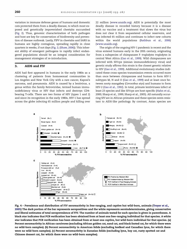
Fig. 4 – Prevalence and distribution of FIV seroreactivity in free-
2005) The dark portion of the bar represents seropositives and th
and liberal estimates of total seroprevalence of FIV. The number
black star indicates that PCR verification has been obtained from
star indicates that PCR verification has been obtained from at le
Percent seroreactivity in African carnivores (excluding African go
no wild-born samples). (b) Percent seroreactivity in American fe
were no wild-born samples). (c) Percent seroreactivity in Eurasi
Chinese dessert cat, for which there were no wild-born sample
22 million (www.unaids.org). AIDS is potentially the most
deadly disease in recorded history because it is a disease
with no vaccine and a treatment that slows the virus but
does not clear it from sequestered cellular reservoirs, and
has infected 65 million and continues to infect new cohorts
within the world populations (Rathbun et al., 2006)
(www.unaids.org).
The origin of the ongoing HIV-1 pandemic is recent and the
virus entered humans early in the 20th century, originating
from a subspecies of chimpanzee P. troglodytes troglodytes in
central West Africa (Gao et al., 1999). Wild chimpanzees are
infected with SIVcpz (simian immunodeficiency virus) and
genetic study affirms this strain is the closest genetic relative
to HIV (Gao et al., 1999). Additional evolutionary studies indi-
cated these cross-species transmission events occurred more
than once between chimpanzee and human to form HIV-1
subtypes M, N and O (Gao et al., 1999) and at least once be-
tween sooty mangabey (Cercocebus atys) and humans to form
HIV-2 (Gao et al., 1992). In total, primate lentiviruses infect at
least 23 species and like SIVcpz are host-specific (Hahn et al.,
2000; Sharp et al., 1999; Sharp et al., 2005). All naturally occur-
ring SIV are in African primates and these species seem resis-
tant to AIDS-like pathology. By contrast, Asian species are
ranging, and captive but wild-born, animals (Troyer et al.,
e white represents seroindeterminates, giving conservative
of animals tested for each species is given in parentheses. A
at least one free-ranging individual for that species. A white
ast one captive, but wild-born individual for that species. (a)
lden cat, sand cat, and black footed cat, for which there were
lids (excluding kodkod and Canadian lynx, for which there
an felids (excluding lynx, bay cat, rusty spotted cat and
s).

Hya-16 *
Ple-624Ple-350
Lpa-32 *
FcaWG3-23Fca (petaluma)
FcaPco-163
Pco-144Pco-117
Pco-733Pco-28
Pco-245Pco-145
Pco-590Pco-253
Pco-696Pco-61
Pco-336Ccr-44
Ccr-34Ccr76x
Ccr-77Ccr-82
EIAVBIV
SIVagmSIVmnd
SIVdrlSIVsm
HIV-2SIVcpz
AG
BD
FKC
UH
JN
OSIVcol
SIVsuSIVlhoest
ovinevisna
CAEV
0.05 substitutions/site
HIV-1
FIV-Ple (Lion)
FIV-Oma (Pallas cat)
FIV-Ppa (Leopard)
FIV-Aju (Cheetah)
FIV-Hya (Jaguaroundi)
FIV-Lpa (Ocelot)
FIV-Fca (Domestic cat)
FIV-Pco (Puma)
FIV-Ccr (Spotted hyena)
C
A
B
100/100/100/1
100/100/100/1
100/100/100/1
89/94/65/097/96/90/0.96
64/93/0/0.78
100/100/100/1
98//97/96/1
56/91/83/0.59
83/87/57/1
100/99/99/1
81/100/97/1
64/80/0/0.99
100/100/99/1
97/99/79/1
Ple-1728Ple-690
Ple-1025Ple-1036
Ple-851Ple-865
Ple-853Ple-319
Ple-584Ple-320
Ple-304Ple-336
Ple-513Ple-599Ple-673
Ple-676Ple-747
Ple-741Ple-725
Ple-1727Ple-161
Pun-84 *Ple-1773 **
Ple-153Ple-180
Ple-172Ple-168
Oma-22 **Oma-12 **Oma-21 **
Oma-34 *Ppa-179
Ppa-172Ppa-181
Ppa-173Aju-204
Aju-214Aju-203
B
A
E
F
D
100/100/99/1
70/92/0/1
100/100/100/1
87/84/94/1
100/100/100/1
100/100/100/1
100/100/100/1
87/76/89/1
88/91/59/0.99
86/98/77/1
Fig. 5 – Phylogenetic tree for FIV pol-RT nucleotide sequences (476 bp included in analysis) isolates from indicated Felidae
species. Shown here is the single maximum likelihood (ML) tree. Maximum parsimony (MP), minimum evolution (ME;
Neighbor joining based on the Tajima Nei algorithm for distance measure) and Bayesian (BA) trees gave similar topologies.
Bootstrap values and posterior probabilities are included at all nodes with bootstrap support (ML/ME/MP/Bayesian). Taxa are
designated with the species code followed by the source animal ID#. Sequence downloaded from GenBank for comparison
are boxed, all other sequences are novel. A single asterix (*) indicates the sequence was obtained from a captive, but
wild-born animal, a double asterix (**) indicates sequence from captive-born individuals. All other novel sequences are from
free-ranging animals. Analysis used empirical base frequencies, an estimated shape parameter of 0.6618, an estimated
substitution matrix as follows: A/C = 2.0642, A/G = 7.2581, A/T = 1.4798, C/G = 4.0169, C/T = 14.0129 and an estimated
proportion of invariant sites of 0.1525. Shown here is the single maximum likelihood tree with maximum likelihood
bootstraps from 1000 replicates.
B I O L O G I C A L C O N S E R V A T I O N 1 3 1 ( 2 0 0 6 ) 2 5 5 – 2 6 7 261
262 B I O L O G I C A L C O N S E R V A T I O N 1 3 1 ( 2 0 0 6 ) 2 5 5 – 2 6 7
naı̈ve, and do develop AIDS when infected with the SIV from
African primates (Hirsch, 2004).
The sole naturally occurring model for AIDS disease in hu-
mans is the feline homologue of HIV, feline immunodefi-
ciency virus (FIV). Although related lentiviruses are found in
other species such as sheep and goats (caprine arthritis
encephalitis virus – CAEV), horse (equine infectious anemia
virus – EIAV), and cattle (bovine immunodeficiency virus –
BIV), only FIV in domestic cats causes AIDS-like disease. FIV
was first discovered in 1986 in a house cat with a wasting-like
disease (Pedersen et al., 1987). Currently, FIV is epidemic
among feral domestic cats throughout the world and is asso-
ciated with an Aids-like syndrome of immune depletion, in-
creased susceptibility to rare cancers and opportunistic
disease, and death (Bendinelli et al., 1995; Willett et al.,
1997). FIV has a small viral RNA genome of about 9000 nucle-
otides that is similar in sequence, gene content, and gene
arrangement to that of HIV-1 (Miyazawa et al., 1994).
As with primate lentiviruses, FIV naturally infects multiple
species of cat in the wild. Western blot screening of antibod-
ies against FIV in free-ranging non-domestic cat species
(Fig. 4) revealed that several were exposed to the virus (Car-
penter and O’Brien, 1995; Olmsted et al., 1989; Olmsted
et al., 1992; Troyer et al., 2005). A recent comprehensive sur-
vey of serum and lymphocyte specimens from 3055 individu-
als within 35 Felidae and 3 Hyenidae species used three FIV
antigens isolated from domestic cat, lion, and puma as tar-
gets of western blot plus PCR-based FIV genome sequence
amplification as a validation of FIV infection (Troyer et al.,
2005). Those results revealed that at least 11 free ranging Feli-
dae species harbor FIV antibodies and FIV viral genomes
(Fig. 4). These species are distributed across Africa including
lion, leopard (P. pardus), and cheetah along with two Hyenidae
species of spotted hyena (Crocuta crocuta) and striped hyena
(Hyaena hyaena). FIV positive species occur in North and South
America including puma, jaguaroundi (P. onca), jaguarondi,
ocelot (Leopardus pardalis), margay (L. weidii), Geoffroy’s cat
(L. geoffroyi), tigrina (L. tigrina) and in Asia with pallas cat (Oto-
colobus manul). Eight other species had western blot signals of
antibodies, but did not yield FIV sequences using multiple
PCR primers which is likely due to divergence within the pri-
mer binding sites, exceedingly low proviral load, or both
(Fig. 4). Overall, inclusion of FIV western blot results from cap-
tive individuals indicates that as many as 31 species of cat are
susceptible to FIV infection and may harbor species specific
viruses or be infected with non-native virus (Troyer et al.,
2005) (Fig. 4). Further, the distribution of FIV is not uniform
among cat species, being endemic in Africa and North and
South America, but rare in species from Europe and Asia
(Fig. 4).
Phylogenetic analyses of the pol-RT region (476 bp) isolated
in Felidae species reveal the sequences form species-specific
monophyletic clades (Fig. 5). This monophyletic pattern sug-
gests that FIV does not spread among species easily. Rather,
FIV infected an ancestor of each host and then evolved within
that species. There have been exceptions to this finding, but
only in captive animals and these occur rarely. For example,
the captive snow leopard (P. uncia) FIV sequence is well within
the African lion FIV clade A (Fig. 5). Another example is a cap-
tive puma infected with a domestic cat strain of FIV (Carpen-
ter et al., 1996). However, in the wild, opportunities for inter-
species transmission are limited and thus the virus becomes
adapted to its particular host. This evolutionary pattern
resembles that of naturally occurring SIV in African primates,
where SIV forms monophyletic lineages that are specific to
each species (Allan et al., 1991; Sharp et al., 2005) and are
host-dependent. Yet, FIV transmission may have a geographic
component whereby species in close proximity share more
similar FIV strains. This is suggested in the common node
shared by leopard FIV and cheetah FIV (Fig. 5).
Clinical consequences for FIV infection in most non-
domestic Felidae species is not known, and there has been lit-
tle convincing acute disease or patterned disease symptoms
associated with FIV infection as is observed consistently for
domestic cats. Only one example of FIV-related mortality
has been documented in a species other than domestic cat,
a captive lion (Poli et al., 1995). The lack of documentation
of clinical signs of FIV infection in wild populations may re-
flect logistic obstacles and expenses in routine physicals for
these species. Alternatively, these species-specific FIV strains
may have undergone a prolonged period of co-adaptation
with their respective host and may be less pathogenic as a
consequence (Carpenter et al., 1996; Carpenter and O’Brien,
1995). In the following study with lions, we describe how cur-
rent conservation efforts with endangered cat species are
integrating genetic studies of FIV in natural populations.
4. FIV in African lions
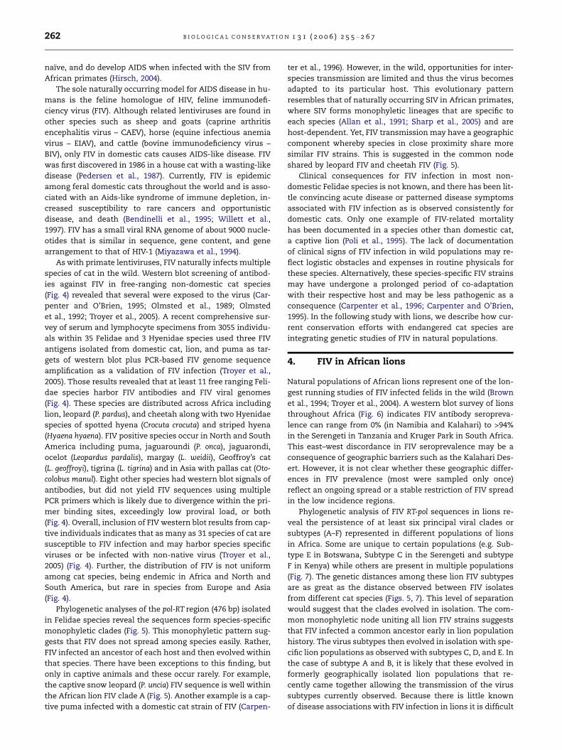
Natural populations of African lions represent one of the lon-
gest running studies of FIV infected felids in the wild (Brown
et al., 1994; Troyer et al., 2004). A western blot survey of lions
throughout Africa (Fig. 6) indicates FIV antibody seropreva-
lence can range from 0% (in Namibia and Kalahari) to >94%
in the Serengeti in Tanzania and Kruger Park in South Africa.
This east–west discordance in FIV seroprevalence may be a
consequence of geographic barriers such as the Kalahari Des-
ert. However, it is not clear whether these geographic differ-
ences in FIV prevalence (most were sampled only once)
reflect an ongoing spread or a stable restriction of FIV spread
in the low incidence regions.
Phylogenetic analysis of FIV RT-pol sequences in lions re-
veal the persistence of at least six principal viral clades or
subtypes (A–F) represented in different populations of lions
in Africa. Some are unique to certain populations (e.g. Sub-
type E in Botswana, Subtype C in the Serengeti and subtype
F in Kenya) while others are present in multiple populations
(Fig. 7). The genetic distances among these lion FIV subtypes
are as great as the distance observed between FIV isolates
from different cat species (Figs. 5, 7). This level of separation
would suggest that the clades evolved in isolation. The com-
mon monophyletic node uniting all lion FIV strains suggests
that FIV infected a common ancestor early in lion population
history. The virus subtypes then evolved in isolation with spe-
cific lion populations as observed with subtypes C, D, and E. In
the case of subtype A and B, it is likely that these evolved in
formerly geographically isolated lion populations that re-
cently came together allowing the transmission of the virus
subtypes currently observed. Because there is little known
of disease associations with FIV infection in lions it is difficult
Fig. 6 – Prevalence of FIV antibodies among lions living in wild populations in Africa. The dark gray portion of the circle
represents seropositives and the light grey represents seroindeterminates, giving conservative and liberal estimates of total
seroprevalence of FIV. The white portion represents FIV negative lions. The number of animals tested for each population is
given in parentheses.
B I O L O G I C A L C O N S E R V A T I O N 1 3 1 ( 2 0 0 6 ) 2 5 5 – 2 6 7 263
to know whether there are now (or ever were) virulence dis-
tinctions among the clades.
One of the most detailed lion population studies is that of
the Serengeti in Tanzania. This large panmictic population
over 3000 individuals has provided many useful insights into
lion pride structure, population dynamics, and transmission
of infectious diseases (Gilbert et al., 1991; Hofmann-Lehmann
et al., 1996; Muller-Graf et al., 1999; Munson et al., 1996; Packer
et al., 2005). Long-term surveillance of lions in the Serengeti
has not revealed any evidence of AIDS like illnesses, de-
creased viability, or decreased fecundity in this population
where seroprevalence among adult lions is 100%. These
observations of (relatively) benign infection are similar to
those for FIV in North American puma (Biek et al., 2006; Biek
et al., 2003) and consistent with the notion that FIV has per-
sisted in these host species longer than in domestic cat and
that significant host adaptation has already occurred (Car-
penter et al., 1996; Carpenter and O’Brien, 1995). However, re-
cently we have shown that lions infected with FIV have
significantly lower CD4 counts than uninfected animals sug-
gesting that there may be sub-clinical immunological effects
associated with FIV-infection in this population (Roelke
et al., in press). At one stage we also suspected that infection
with one clade of FIV might be a natural vaccine against infec-
tion with another or the mutational occurrence of a virulent
deadly FIV variant (Carpenter and O’Brien, 1995). However,
Troyer et al. (2004) inspected the pattern of FIV subtype distri-
bution among 13 Serengeti lion prides and discovered multi-
ple clade infections (i.e., two or three subtypes of virus
present in the same lion) in 43% of the infected lions, an
observation that would argue against acquired immunity by
infection with a single strain.
In 1994, the Serengeti lions became the target of a deadly
infectious disease outbreak that caused whisker twitches,
neurological seizures, and death to over 1000 lions, a third
of the population. FIV was excluded as the primary cause
since several afflicted lions were FIV negative (Roelke-Parker
et al., 1996). The etiological agent was a hyper-virulent form
of a canine distemper virus (CDV) a virus previously known
to infect but seldom cause morbidity in Felidae species
(Roelke-Parker et al., 1996). This time, in the most deadly re-
ported outbreak of the CDV among African carnivore species,
a virulent transmissible CDV variant spread from domestic
dogs living around the Serengeti park to hyena, bat eared
foxes Otocyon megalotis, and to lions (Carpenter et al., 1998).
Although FIV was not the primary explanation for the 1994
Serengeti lion mortality, it may have played a supporting role.
HIV infection in humans causes increased susceptibility to
opportunistic infections of normally benign infectious agents
(like HHV8/Kaposi’s sarcoma, Pneumocystis carnii pneumonia
or cytomegalovirus) as a consequence of CD4 T-lymphocyte
depletion and immune collapse (Levy, 1993). Nearly all the

etis/snoitutitsbus 10.0
001/001/001
99/001/001
48/49/001
67/69/001
19/58/99
0/37/28
26/47/99
E epytbuS( anawstoB )
D epytbuS( acirfA htuoS )
A epytbuS( ,anawstoB ,itegnereS
acirfA htuoS )
B epytbuS( ,retarC orognorogN
,itegnereS adnagU )
C epytbuS( itegnereS )
001/001/001
001/001/001
99/001/001
001/89/59
001/001/001
57/0/38
0/001/001
acF-VIF
001/001/001 FepytbuS( ayneK ) 001/001/001
Fig. 7 – Phylogenetic tree for FIV pol-RT (520 bp included in analysis) and gag (400 bp) nucleotide sequences isolated from lion
populations sampled in Fig. 6. Shown here is the single maximum likelihood (ML) tree. Maximum parsimony (MP), and
minimum evolution (ME; Neighbor joining based on the Tajima Nei algorithm for distance measure) trees gave similar
topologies. Bootstrap values are included at all nodes with bootstrap support (ML/ME/MP). Taxa are colored by the country of
origin of the lion sampled. FIV–FCA is the outgroup strain of domestic cat FIV.
264 B I O L O G I C A L C O N S E R V A T I O N 1 3 1 ( 2 0 0 6 ) 2 5 5 – 2 6 7
lions in the Serengeti carried FIV, and FIV has been shown to
be associated with depleted CD4-lymphocyte counts of
200 cells/ll compared with counts of 800–1000 cells/ll in
uninfected individuals (Roelke et al., in press). One could
speculate that adult lions may have done better in defending
against CDV had they not been afflicted with lentivirus that
depletes their principal defense against viruses, the CD4 lym-
phocyte population. While there were FIV-negative animals
that died, these were all juveniles (Roelke-Parker et al., 1996)
and it is possible that their immature immune system may
have accounted for their susceptibility to the new virus.
The African lion study provides some useful insights on
the impact of infectious disease in endangered populations.
Development of detection methods and effective monitoring
of the health of animals in the wild is problematic, and as
in the case of the CDV outbreak, was documented only due
to the presence of researchers already in place to collect sam-
ples and observe the final outcome. Although this CDV out-
break was marked by relatively high mortality estimates
(Roelke-Parker et al., 1996), subsequent outbreaks were less
virulent (Packer et al., 1999) within the same population. Until
we fully understand the immunological effects of infection
with prevalent FIV strains, the interaction of pathogens such
as CDV and FIV, and the role of the host immune system in
modulating these viral infections, the question of their impor-
tance and collaborations in pathogenesis will continue.
One approach is to continue to adapt and apply new tech-
nologies developed from human biomedical research to con-
servation of wildlife. For example, the massive research effort
into understanding HIV in humans offers new technologies to
apply towards related viruses such as FIV in endangered cat
species. Recognition that FIV causes AIDS-like disease in
domestic cat and that this is a natural model for HIV in hu-
mans, has led to extensive surveillance of FIV in other species
(Biek et al., 2006; Biek et al., 2003; Carpenter et al., 1996; Car-
penter and O’Brien, 1995; Olmsted et al., 1992; Troyer et al.,
2004; Troyer et al., 2005), an essential step in determining
the genetic diversity of the pathogen. Knowledge of key ami-
no acid motifs in the env gene of HIV linked with evading host
immune surveillance (Wyatt and Sodroski, 1998; Yusim et al.,
2002) is in turn targets for comparative genomic research of
virulence in FIV (de Parseval et al., 2006). Understanding
how genetic diversity within MHC in humans is linked with
either resistance to HIV or increased susceptibility (Carring-
ton and O’Brien, 2003) provides an incentive to sequence
MHC in multiple mammalian taxa, including cats (Yuhki
et al., 2003; Yuhki and O’Brien, 1990). The links between HIV
in humans and FIV in endangered cat species illustrate the
utility of biomedical approaches and discoveries that is di-
rectly applicable to elucidating the role of disease in endan-
gered species.
5. Conclusions
We are just now beginning to understand the complex inter-
play of host genetic background, immune competence, and
pathogen evolution that contribute to the course of an epi-
demic when a disease agent enters a new species or popula-

B I O L O G I C A L C O N S E R V A T I O N 1 3 1 ( 2 0 0 6 ) 2 5 5 – 2 6 7 265
tion. The advances in molecular biology, immunology and
genetics provide a diverse collection of effective tools for
unraveling the secrets to survival. As the tools that are used
in human medicine become available to wildlife research,
new approaches and understanding of disease outbreaks
and effective host control of infection are increasingly
possible.
Unfortunately, in wildlife populations, few disease out-
breaks are followed from start to finish and the vast majority
remain completely unobserved and undocumented [but see
Randall et al. on Ethiopian wolves and other papers in this
special edition]. For this reason, our understanding of the ef-
fects of population demographics, genetics, mating and
migration patterns on the spread of (and ultimately clearance
of and/or adaptation to) disease is limited.
These examples of FIPV and FIV illustrate a critical need
for consistent programs designed to monitor and survey
endangered populations. Conservation strategies that incor-
porate biological sampling yield invaluable genetic profiles
of disease and disease resistance within endangered species.
Genomic analyses provide insight into the history of a popu-
lation and its pathogen(s) that can assist in documenting pat-
terns of co-evolution and adaptation. Yet, these examples
underscore the unpredictable aspects of epidemics within
natural populations of threatened or endangered species
and there is need for much more comprehensive and long-
term data. Finally, conservation management would clearly
benefit from a better knowledge of viral evolution, adaptation,
and cross-species infections to plan protected areas, inform
strategies of re-introduction and relocation, and provide
effective interventions in cases of potential disease epidem-
ics. By resolving the root causes of human and animal out-
breaks and their outcomes, we shall continue to learn
lessons such as the cats have shared on SARS and AIDS.
R E F E R E N C E S
Allan, J.S., Short, M., Taylor, M.E., Su, S., Hirsch, V.M., Johnson, P.R.,Shaw, G.M., Hahn, B.H., 1991. Species-specific diversity amongsimian immunodeficiency viruses from African greenmonkeys. J. Virol. 65, 2816–2828.
Ballesteros, M.L., Sanchez, C.M., Enjuanes, L., 1997. Two aminoacid changes at the N-terminus of transmissiblegastroenteritis coronavirus spike protein result in the loss ofenteric tropism. Virology 227, 378–388.
Belov, K., Lam, M.K., Colgan, D.J., 2004. Marsupial MHC class IIbeta genes are not orthologous to the eutherian beta genefamilies. J. Hered. 95, 338–345.
Bendinelli, M., Pistello, M., Lombardi, S., Poli, A., Garzelli, C.,Matteucci, D., Ceccherini-Nelli, L., Malvaldi, G., Tozzini, F.,1995. Feline immunodeficiency virus: an interesting model forAIDS studies and an important cat pathogen. Clin. Microbiol.Rev. 8, 87–112.
Biek, R., Rodrigo, A.G., Holley, D., Drummond, A., Anderson Jr.,C.R., Ross, H.A., Poss, M., 2003. Epidemiology, genetic diversity,and evolution of endemic feline immunodeficiency virus in apopulation of wild cougars. J. Virol. 77, 9578–9589.
Biek, R., Drummond, A.J., Poss, M., 2006. A virus revealspopulation structure and recent demographic history of itscarnivore host. Science 311, 538–541.
Brown, E.W., Yuhki, N., Packer, C., O’Brien, S.J., 1994. A lionlentivirus related to feline immunodeficiency virus:epidemiologic and phylogenetic aspects. J. Virol. 68, 5953–5968.
Carpenter, M.A., O’Brien, S.J., 1995. Coadaptation andimmunodeficiency virus: lessons from the Felidae. Curr. Opin.Genet. Dev. 5, 739–745.
Carpenter, M.A., Brown, E.W., Culver, M., Johnson, W.E.,Pecon-Slattery, J., Brousset, D., O’Brien, S.J., 1996. Genetic andphylogenetic divergence of feline immunodeficiency virus inthe puma (Puma concolor). J. Virol. 70, 6682–6693.
Carpenter, M.A., Appel, M.J., Roelke-Parker, M.E., Munson, L.,Hofer, H., East, M., O’Brien, S.J., 1998. Genetic characterizationof canine distemper virus in Serengeti carnivores. Vet.Immunol. Immunopathol. 65, 259–266.
Carrington, M., O’Brien, S.J., 2003. The influence of HLA genotypeon AIDS. Annu. Rev. Med. 54, 535–551.
CDC, 2004. Update: Severe Acute Respiratory Syndrome –Worldwide and United States. Page 664 in C. f. D. C. a.Prevention, editor. MMWR..
Clark, A.G., Glanowski, S., Nielsen, R., Thomas, P.D., Kejariwal, A.,Todd, M.A., Tanenbaum, D.M., Civello, D., Lu, F., Murphy, B.,Ferriera, S., Wang, G., Zheng, X., White, T.J., Sninsky, J.J.,Adams, M.D., Cargill, M., 2003. Inferring nonneutral evolutionfrom human–chimp–mouse orthologous gene trios. Science302, 1960–1963.
Cooper, G.M., Brudno, M., Stone, E.A., Dubchak, I., Batzoglou, S.,Sidow, A., 2004. Characterization of evolutionary rates andconstraints in three Mammalian genomes. Genome Res. 14,539–548.
Culver, M., Johnson, W.E., Pecon-Slattery, J., O’Brien, S.J., 2000.Genomic ancestry of the American puma (Puma concolor). J.Hered. 91, 186–197.
de Parseval, A., Grant, C.K., Sastry, K.J., Elder, J.H., 2006. SequentialCD134-CXCR4 interactions in feline immunodeficiency virus(FIV): soluble CD134 activates FIV Env for CXCR4-dependententry and reveals a cryptic neutralization epitope. J. Virol. 80,3088–3091.
Drazen, J.M., 2003. SARS – looking back over the first 100 days.New Engl. J. Med. 349, 319–320.
Driscoll, C.A., Menotti-Raymond, M., Nelson, G., Goldstein, D.,O’Brien, S.J., 2002. Genomic microsatellites as evolutionarychronometers: a test in wild cats. Genome Res. 12, 414–423.
Drosten, C., Preiser, W., Gunther, S., Schmitz, H., Doerr, H.W., 2003.Severe acute respiratory syndrome: identification of theetiological agent. Trends Mol. Med. 9, 325–327.
Enard, W., Paabo, S., 2004. Comparative primate genomics. Annu.Rev. Genomics Hum. Genet. 5, 351–378.
Evermann, J.F., Heeney, J.L., Roelke, M.E., McKeirnan, A.J., O’Brien,S.J., 1988. Biological and pathological consequences of felineinfectious peritonitis virus infection in the cheetah. Arch.Virol. 102, 155–171.
Foley, J.E., Poland, A., Carlson, J., Pedersen, N.C., 1997. Risk factorsfor feline infectious peritonitis among cats in multiple-catenvironments with endemic feline enteric coronavirus. J. Am.Vet. Med. Assoc. 210, 1313–1318.
Gao, F., Yue, L., White, A.T., Pappas, P.G., Barchue, J., Hanson, A.P.,Greene, B.M., Sharp, P.M., Shaw, G.M., Hahn, B.H., 1992. Humaninfection by genetically diverse SIVSM-related HIV-2 in westAfrica. Nature 358, 495–499.
Gao, F., Bailes, E., Robertson, D.L., Chen, Y., Rodenburg, C.M.,Michael, S.F., Cummins, L.B., Arthur, L.O., Peeters, M.,Shaw, G.M., Sharp, P.M., Hahn, B.H., 1999. Origin of HIV-1in the chimpanzee Pan troglodytes troglodytes. Nature 397,436–441.
Garrett, L., 1994. The Coming Plague. Penguin, New York.Gilbert, D.A., Packer, C., Pusey, A.E., Stephens, J.C., O’Brien, S.J.,
1991. Analytical DNA fingerprinting in lions: parentage,genetic diversity, and kinship. J. Hered. 82, 378–386.

266 B I O L O G I C A L C O N S E R V A T I O N 1 3 1 ( 2 0 0 6 ) 2 5 5 – 2 6 7
Guan, Y., Zheng, B.J., He, Y.Q., Liu, X.L., Zhuang, Z.X., Cheung, C.L.,Luo, S.W., Li, P.H., Zhang, L.J., Guan, Y.J., Butt, K.M., Wong, K.L.,Chan, K.W., Lim, W., Shortridge, K.F., Yuen, K.Y., Peiris, J.S.,Poon, L.L., 2003. Isolation and characterization of virusesrelated to the SARS coronavirus from animals in southernChina. Science 302, 276–278.
Hahn, B.H., Shaw, G.M., De Cock, K.M., Sharp, P.M., 2000. AIDS as azoonosis: scientific and public health implications. Science287, 607–614.
Heeney, J.L., Evermann, J.F., McKeirnan, A.J., Marker-Kraus, L.,Roelke, M.E., Bush, M., Wildt, D.E., Meltzer, D.G., Colly, L.,Lukas, J., et al, 1990. Prevalence and implications of felinecoronavirus infections of captive and free-ranging cheetahs(Acinonyx jubatus). J. Virol. 64, 1964–1972.
Hill, R.S., Walsh, C.A., 2005. Molecular insights into human brainevolution. Nature 437, 64–67.
Hirsch, V., 2004. What can natural infection of African monkeyswith simian immunodeficiency virus tell us about thepathogenesis of AIDS?. AIDS Rev. 6, 40–53.
Hofmann-Lehmann, R., Fehr, D., Grob, M., Elgizoli, M., Packer, C.,Martenson, J.S., O’Brien, S.J., Lutz, H., 1996. Prevalence ofantibodies to feline parvovirus, calicivirus, herpesvirus,coronavirus, and immunodeficiency virus and of felineleukemia virus antigen and the interrelationship of these viralinfections in free-ranging lions in east Africa. Clin. Diagn. Lab.Immunol. 3, 554–562.
Holmes, K.V., 2003. SARS – associated coronavirus. New Engl. J.Med. 348, 1948–1951.
Johnson, W.E., Eizirik, E., Pecon-Slattery, J., Murphy, W.J., Antunes,A., Teeling, E., O’Brien, S.J., 2006. The late Miocene radiation ofmodern Felidae: a genetic assessment. Science 311, 73–77.
Korber, B., Muldoon, M., Theiler, J., Gao, F., Gupta, R., Lapedes, A.,Hahn, B.H., Wolinsky, S., Bhattacharya, T., 2000. Timing theancestor of the HIV-1 pandemic strains. Science 288, 1789–1796.
Kumanovics, A., Takada, T., Lindahl, K.F., 2003. Genomicorganization of the mammalian MHC. Annu. Rev. Immunol.21, 629–657.
Lau, S.K., Woo, P.C., Li, K.S., Huang, Y., Tsoi, H.W., Wong, B.H.,Wong, S.S., Leung, S.Y., Chan, K.H., Yuen, K.Y., 2005. Severeacute respiratory syndrome coronavirus-like virus in Chinesehorseshoe bats. Proc. Natl. Acad. Sci. USA 102, 14040–14045.
Levy, J.A., 1993. HIV and host immune responses in AIDSpathogenesis. J. Clin. Apher. 8, 19–28.
Li, W., Shi, Z., Yu, M., Ren, W., Smith, C., Epstein, J.H., Wang, H.,Crameri, G., Hu, Z., Zhang, H., Zhang, J., McEachern, J., Field,H., Daszak, P., Eaton, B.T., Zhang, S., Wang, L.F., 2005. Bats arenatural reservoirs of SARS-like coronaviruses. Science 310,676–679.
Luo, S.J., Kim, J.H., Johnson, W.E., van der Walt, J., Martenson, J.,Yuhki, N., Miquelle, D.G., Uphyrkina, O., Goodrich, J.M.,Quigley, H.B., Tilson, R., Brady, G., Martelli, P., Subramaniam,V., McDougal, C., Hean, S., Huang, S.Q., Pan, W., Karanth, U.K.,Sunquist, M., Smith, J.L., O’Brien, S.J., 2004. Phylogeographyand genetic ancestry of tigers (Panthera tigris). PLoS Biol. 2,e442.
Marker, L., O’Brien, J.S., 1989. Captive breeding of the cheetah(Acinonyx jubatus) in North American zoos (1971–1986). ZooBiol. 8, 3–16.
Menotti-Raymond, M., O’Brien, S.J., 1993. Dating the geneticbottleneck of the African cheetah. Proc Natl Acad Sci USA 90,3172–3176.
Miyazawa, T., Tomonaga, K., Kawaguchi, Y., Mikami, T., 1994. Thegenome of feline immunodeficiency virus. Arch. Virol. 134,221–234.
Muller-Graf, C.D., Woolhouse, M.E., Packer, C., 1999. Epidemiologyof an intestinal parasite (Spirometra spp.) in two populations ofAfrican lions (Panthera leo). Parasitology 118 (Pt 4), 407–415.
Munson, L., Brown, J.L., Bush, M., Packer, C., Janssen, D., Reiziss,S.M., Wildt, D.E., 1996. Genetic diversity affects testicularmorphology in free-ranging lions (Panthera leo) of the SerengetiPlains and Ngorongoro Crater. J. Reprod. Fertil. 108, 11–15.
Murphy, W.J., Eizirik, E., Johnson, W.E., Zhang, Y.P., Ryder, O.A.,O’Brien, S.J., 2001a. Molecular phylogenetics and the origins ofplacental mammals. Nature 409, 614–618.
Murphy, W.J., Stanyon, R., O’Brien, S.J., 2001b. Evolution ofmammalian genome organization inferred from comparativegene mapping. Genome Biol. 2, REVIEWS0005..
Murphy, W.J., Pevzner, P.A., O’Brien, S.J., 2004. Mammalianphylogenomics comes of age. Trends Genet. 20, 631–639.
Nowell, K., Jackson, P., 1996. Status Survey and ConservationAction Plan, Wild Cats. International Union for Conservationof Nature and Natural Resources, Gland, Switzerland..
O’Brien, S.J., Johnson, W.E., 2005. Big cat genomics. Annu. Rev.Genomics Hum. Genet. 6, 407–429.
O’Brien, S.J., Yuhki, N., 1999b. Comparative genome organizationof the major histocompatibility complex: lessons from theFelidae. Immunol. Rev. 167, 133–144.
O’Brien, S.J., Roelke, M.E., Marker, L., Newman, A., Winkler, C.A.,Meltzer, D., Colly, L., Evermann, J.F., Bush, M., Wildt, D.E., 1985.Genetic basis for species vulnerability in the cheetah. Science227, 1428–1434.
O’Brien, S.J., Wildt, D.E., Bush, M., Caro, T.M., FitzGibbon, C.,Aggundey, I., Leakey, R.E., 1987. East African cheetahs:evidence for two population bottlenecks?. Proc. Natl. Acad.Sci. USA 84, 508–511.
O’Brien, S.J., Menotti-Raymond, M., Murphy, W.J., Nash, W.G.,Wienberg, J., Stanyon, R., Copeland, N.G., Jenkins, N.A.,Womack, J.E., Marshall Graves, J.A., 1999a. The promise ofcomparative genomics in mammals. Science 286, 458–462.479–481.
Olmsted, R.A., Hirsch, V.M., Purcell, R.H., Johnson, P.R., 1989.Nucleotide sequence analysis of feline immunodeficiencyvirus: genome organization and relationship to otherlentiviruses. Proc. Natl. Acad. Sci. USA 86, 8088–8092.
Olmsted, R.A., Langley, R., Roelke, M.E., Goeken, R.M., Adger-Johnson, D., Goff, J.P., Albert, J.P., Packer, C., Laurenson, M.K.,Caro, T.M., et al, 1992. Worldwide prevalence of lentivirusinfection in wild feline species: epidemiologic andphylogenetic aspects. J. Virol. 66, 6008–6018.
Packer, C., Altizer, S., Appel, M.J., Brown, E.W., Martenson, J.,Roelke-Parker, M.E., Hofmann-Lehmann, R., Lutz, H., 1999.Viruses of the Serengeti: patterns of infection and mortality inAfrican lions. J. Anim. Ecol. 68, 1161–1178.
Packer, C., Hilborn, R., Mosser, A., Kissui, B., Borner, M., Hopcraft,G., Wilmshurst, J., Mduma, S., Sinclair, A.R., 2005. Ecologicalchange, group territoriality, and population dynamics inSerengeti lions. Science 307, 390–393.
Pearks Wilkerson, A.J., Teeling, E.C., Troyer, J.L., Bar-Gal, G.K.,Roelke, M., Marker, L., Pecon-Slattery, J., O’Brien, S.J., 2004.Coronavirus outbreak in cheetahs: lessons for SARS. Curr. Biol.14, R227–R228.
Pedersen, N.C., Ho, E.W., Brown, M.L., Yamamoto, J.K., 1987.Isolation of a T-lymphotropic virus from domestic cats with animmunodeficiency-like syndrome. Science 235, 790–793.
Poli, A., Abramo, F., Cavicchio, P., Bandecchi, P., Ghelardi, E.,Pistello, M., 1995. Lentivirus infection in an African lion: aclinical, pathological, and virologic study. J. Wildlife Dis. 31,70–74.
Poon, L.L., Chu, D.K., Chan, K.H., Wong, O.K., Ellis, T.M., Leung,Y.H., Lau, S.K., Woo, P.C., Suen, K.Y., Yuen, K.Y., Guan, Y., Peiris,J.S., 2005. Identification of a novel coronavirus in bats. J. Virol.79, 2001–2009.
Rathbun, R.C., Lockhart, S.M., Stephens, J.R., 2006. Current HIVtreatment guidelines – an overview. Curr. Pharm. Des. 12,1045–1063.

B I O L O G I C A L C O N S E R V A T I O N 1 3 1 ( 2 0 0 6 ) 2 5 5 – 2 6 7 267
Roelke, M.E., Martenson, J.S., O’Brien, S.J., 1993. The consequencesof demographic reduction and genetic depletion in theendangered Florida panther. Curr. Biol. 3, 340–350.
Roelke, M.E., Pecon-Slattery, J., Taylor, S., Citino, S., Brown, E.W.,Packer, C., VandeWoude, S., O’Brien S, J., in press. T-Lymphocyte profiles in FIV infected wild lions and pumasreveal CD4 depletion. J. Wildlife Dis..
Roelke-Parker, M.E., Munson, L., Packer, C., Kock, R., Cleaveland,S., Carpenter, M., O’Brien, S.J., Pospischil, A., Hofmann-Lehmann, R., Lutz, H., et al, 1996. A canine distemper virusepidemic in Serengeti lions (Panthera leo). Nature 379, 441–445.
Sanchez, C.M., Izeta, A., Sanchez-Morgado, J.M., Alonso, S., Sola,I., Balasch, M., Plana-Duran, J., Enjuanes, L., 1999. Targetedrecombination demonstrates that the spike gene oftransmissible gastroenteritis coronavirus is a determinant ofits enteric tropism and virulence. J. Virol. 73, 7607–7618.
Savarino, A., 2005. Expanding the frontiers of existing antiviraldrugs: possible effects of HIV-1 protease inhibitors againstSARS and avian influenza. J. Clin. Virol. 34, 170–178.
Sharp, P.M., Bailes, E., Robertson, D.L., Gao, F., Hahn, B.H., 1999.Origins and evolution of AIDS viruses. Biol. Bull. 196, 338–342.
Sharp,P.M.,Shaw,G.M.,Hahn,B.H.,2005.Simianimmunodeficiencyvirus infection of chimpanzees. J. Virol. 79, 3891–3902.
Shaw, K., 2006. The 2003 SARS outbreak and its impact oninfection control practices. Public Health 120, 8–14.
Song, H.D., Tu, C.C., Zhang, G.W., Wang, S.Y., Zheng, K., Lei, L.C.,Chen, Q.X., Gao, Y.W., Zhou, H.Q., Xiang, H., Zheng, H.J., Chern,S.W., Cheng, F., Pan, C.M., Xuan, H., Chen, S.J., Luo, H.M., Zhou,D.H., Liu, Y.F., He, J.F., Qin, P.Z., Li, L.H., Ren, Y.Q., Liang, W.J., Yu,Y.D., Anderson, L., Wang, M., Xu, R.H., Wu, X.W., Zheng, H.Y.,Chen, J.D., Liang, G., Gao, Y., Liao, M., Fang, L., Jiang, L.Y., Li, H.,Chen, F., Di, B., He, L.J., Lin, J.Y., Tong, S., Kong, X., Du, L., Hao, P.,Tang, H., Bernini, A., Yu, X.J., Spiga, O., Guo, Z.M., Pan, H.Y., He,W.Z., Manuguerra, J.C., Fontanet, A., Danchin, A., Niccolai, N.,Li, Y.X., Wu, C.I., Zhao, G.P., 2005. Cross-host evolution of severeacute respiratory syndrome coronavirus in palm civet andhuman. Proc. Natl. Acad. Sci. USA 102, 2430–2435.
Troyer, J.L., Pecon-Slattery, J., Roelke, M.E., Black, L., Packer, C.,O’Brien, S.J., 2004. Patterns of feline immunodeficiency virusmultiple infection and genome divergence in a free-rangingpopulation of African lions. J. Virol. 78, 3777–3791.
Troyer, J.L., Pecon-Slattery, J., Roelke, M.E., Johnson, W.,VandeWoude, S., Vazquez-Salat, N., Brown, M., Frank, L.,Woodroffe, R., Winterbach, C., Winterbach, H., Hemson, G.,Bush, M., Alexander, K.A., Revilla, E., O’Brien, S.J., 2005.Seroprevalence and genomic divergence of circulating strainsof feline immunodeficiency virus among Felidae andHyaenidae species. J. Virol. 79, 8282–8294.
Uphyrkina, O., Johnson, W.E., Quigley, H., Miquelle, D., Marker, L.,Bush, M., O’Brien, S.J., 2001. Phylogenetics, genome diversityand origin of modern leopard, Panthera pardus. Mol. Ecol. 10,2617–2633.
Uphyrkina, O., Miquelle, D., Quigley, H., Driscoll, C., O’Brien, S.J.,2002. Conservation genetics of the Far Eastern leopard(Panthera pardus orientalis). J. Hered. 93, 303–311.
Wei, P., Fan, K., Chen, H., Ma, L., Huang, C., Tan, L., Xi, D., Li, C.,Liu, Y., Cao, A., Lai, L., 2006. The N-terminal octapeptide acts asa dimerization inhibitor of SARS coronavirus 3C-likeproteinase. Biochem. Biophys. Res. Commun. 339, 865–872.
Willett, B.J., Flynn, J.N., Hosie, M.J., 1997. FIV infection of thedomestic cat: an animal model for AIDS. Immunol. Today 18,182–189.
Winckler, W., Myers, S.R., Richter, D.J., Onofrio, R.C., McDonald,G.J., Bontrop, R.E., McVean, G.A., Gabriel, S.B., Reich, D.,Donnelly, P., Altshuler, D., 2005. Comparison of fine-scalerecombination rates in humans and chimpanzees. Science308, 107–111.
Wyatt, R., Sodroski, J., 1998. The HIV-1 envelope glycoproteins:fusogens, antigens, and immunogens. Science 280, 1884–1888.
Yuhki, N., O’Brien, S.J., 1990. DNA variation of the mammalianmajor histocompatibility complex reflects genomic diversityand population history. Proc. Natl. Acad. Sci. USA 87, 836–840.
Yuhki, N., Beck, T., Stephens, R.M., Nishigaki, Y., Newmann, K.,O’Brien, S.J., 2003. Comparative genome organization ofhuman, murine, and feline MHC class II region. Genome Res.13, 1169–1179.
Yusim, K., Kesmir, C., Gaschen, B., Addo, M.M., Altfeld, M.,Brunak, S., Chigaev, A., Detours, V., Korber, B.T., 2002.Clustering patterns of cytotoxic T-lymphocyte epitopes inhuman immunodeficiency virus type 1 (HIV-1) proteins revealimprints of immune evasion on HIV-1 global variation. J. Virol.76, 8757–8768.