observations on the reproductive biology of the hydrothermal vent tube worm riftia pachyptila
TRANSCRIPT

Vol. 52: 89-94, 1989 l MARINE ECOLOGY PROGRESS SERIES Mar. Ecol. Prog. Ser.
Published February 16
NOTE
Observations on the reproductive biology of the hydrothermal vent tube worm Riftia pachyptila
S. Craig Cary, Horst Felbeck, Nicholas D. Holland
Marine Biology Research Division, Scripps Institution of Oceanography. La Jolla. California 92093, USA
ABSTRACT: On the Hydronaut Expedition to 13"N on the East Pacific Rise, we made some observations on the repro- ductive biology of Riftia pachyptila relevant to larval disper- sal, symbiont acquisition, and sperm transfer. Two females spawned in a pressure chamber about 15 h after collection. During each 30 min spawning episode, the relatively small (105 pm), lipid-rich eggs were emitted in large numbers from the female gonopores and floated upward in still seawater at about 2 cm min-l. Therefore, it is likely that early develop- ment takes place in deep water well above the vent habitat of the adults. Two males spawned in non-pressurized aquaria about 45 min after reaching the deck of the ship. Semen issuing from the male gonopores contained sperm bundles, each composed of several hundred sperm wlth remarkable detached acrosomes. Each bundle swam vigorously through seawater by the beating of all its flagella In unison Motil~ty was not inhibited by hydrogen sulfide concentratlons greater than those at the vent habitat. After swimmlng for about 15 min, the bundles broke up into individual sperm that were relatively immotile. It is reasonable to assume that sperm bundles swim from the male to the female's tube or body where they adhere by their detached acrosomes before disin- tegrating into individual sperm that subsequently ferhhze the eggs.
Since the discovery of Riftia pachyptila a decade ago (Corliss & Ballard 1977), much has been learned about its physiology and biochemistry (Felbeck et al. 1985, Grassle 1986, Hessler et al. 1988), but many gaps remain in our knowledge of its reproductive biology. Adults of R. pachyptila are sessile and limited to patches sur- rounding hydrothermal vents. The tube worm popula- tion around a given vent is typically limited to a region only tens of meters in diameter and is subject to extinc- tion if the hydrothermal effluent ceases. Moreover, suitable vents are separated from one another by distan- ces on the order of kilometers or tens of kilometers. At present, the early developmental stages of R. pachyptila are unknown, so their mode of transport from one vent habitat to another remains a mystery. A second problem of transport for the hydrothermal vent tube worms is the transfer of sperm from males to females over distances from centimeters to tens of meters. In the present note
O Inter-ResearchIPnnted in F. R Germany
we provide some new information relevant to the trans- port of early developmental stages and to the transfer of sperm from males to females of R. pachyptila.
Materials and methods. All observations were made in November 1987, during the joint French-USA Hy- dronaut Expedition to 13ON on the East Pacific Rise. Following collection by submarine at a depth of 2700 m, males and females of Riftia pachyptila were held in an unpressurized, thermally insulated container during the 3 h ascent. On reaching the surface ship, the worms were removed from their natural tubes and placed in transparent acrylic cylinders of the same diameter. The worms were maintained at 5°C in a transparent acrylic cylinders of the same diameter. The worms were maintained at 5OC in a transparent pressure chamber at 1500 psi (1.0 X 10⁷ Pa), which was less than the pressure at the collection site (4000 psi: 2.8 X 10' Pa), but sufficient to keep them alive for a few weeks (Childress et al. 1984).
Gametes were obtained from 2 spawning females and 2 spawning males of Riftia pacl~yptila. The ascent rate of the eggs was measured at 5 'C by timing their upward passage over several centimeters. Rates were measured under 2 conditions: (1) for 10 eggs in the 10 1 pressure chamber at l500 psi with the circulation turned off and (2) for 10 eggs a t 1 atm in a 500 m1 graduated cylinder with no temperature differential between the inner and outer walls.
For light microscopy (LM), scanning electron micros- copy (SEM) and transmission electron microscopy (TEM), the gametes were fixed for 1 mo at 4OC in buffered glutaraldehyde according to Gould-Somero &
Holland (1975). Rinsing and post-fixation were accord- ing to Holland & Nealson (1978), and subsequent pre- paration for SEM and TEM were according to Holland & Jespersen (1973).
Results and discussion. Two females of k f t i a pachy- ptila spawned in the pressure chamber about 15 h after

90 Mar. Ecol. Prog. Ser. 52: 89-94, 1989
collection. During each spawning episode, which lasted about 30 min, eggs streamed out of the 2 gonopores, evidently mixed with a binding mucus. The spawned eggs were retained in the vestimental cavity for a few minutes, during which time the mucous bin- der appeared to dissolve. Once freed from the mucus, the eggs immediately began to float upward. The ascent rates for eggs were 2 . 2 cm min-' (SD = 0.2; n =
10) at 1500 psi and 2.4 cm min-' (SD = k0 .4 ; n = 10) at 1 atm; these 2 means do not differ significantly (20.05) by Student's t-test. This ascent rate should be only slightly influenced by changes in environmental pressure and temperature as the eggs ascend from the vent habitat (Yayanos et al. 1978, Yayanos & Nevenzel 1978). Based on Eq. (2) in the latter references and the lack of differences in ascent rates between 1 atm and 1500 psi, compressional effects on lipids at 4000 psl should have little influence on the ascent rate of eggs immediately above the vent site.
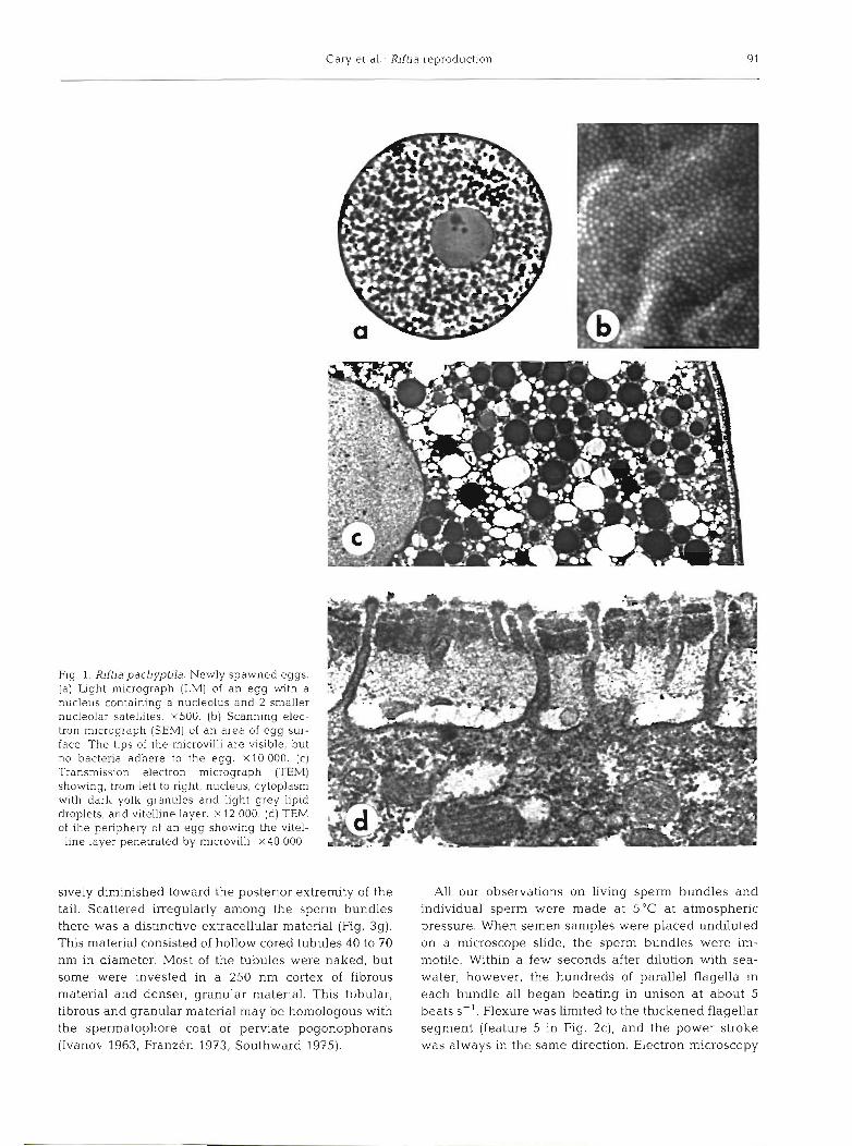
For a sample of 50 eggs from one female, the mean diameter was 105 (SD = + 3.5) pm. The spawned eggs had a nucleolate nucleus (germinal vesicle) and a cyto- plasm packed with yolk granules and lipid droplets (Fig. 1). The membrane-bounded yolk granules were filled with finely granular dense material, and the lipid droplets, which cumulatively occupied almost half the volume of the cytoplasm, no doubt accounted for the buoyancy. The relatively small size of the egg is some indication that a planktotrophic larva (possibly trocho- phore-like) develops. Even so, it is premature to rule out the possibility of lecithotrophic development, since the lipid in the egg represents a considerable reserve of energy The remaining cytoplasm of the egg included abundant free ribosomes, mitochondria, some tubular endoplasmic reticulum and a few Golgi complexes. At the egg surface, microvilli penetrated a fibrous vitelline layer consisting of 4 distinct sub-layers.
We found no bacteria adhering to the outer surface (SEM of 40 eggs) or within the spawned eggs (TEM of 10 eggs), in agreement with previous electron micros- copic studies that revealed no bacteria in eggs of Rftia pachyptila (Cavanaugh et al. 1981, Jones & Gardiner in press). Admittedly, electron mlcroscopy could fail to detect a small population of intracellular bacteria. Even so, it seems unlikely that chemosynthetic symbiotic bacteria are passed to the new generation of R. pachy- ptila via the eggs. The conclusion that later develop- mental stages must pick up their chemosynthetic sym- bionts from free-living bacterial populations is borne out by the recent discovery (Jones & Gardiner in press) that early juveniles of R. pachyptila lack symbiotic bacteria.
The buoyancy of the eggs strongly suggests that the embryonic and larval development takes place in deep water well above the vents where the adult worms live.
An ascent of 2 cm min-' is relatively slow (at this rate, half a week would be needed for an ascent of 100 m). Even so, at the prevailing deep-sea temperature of about 2"C, development should be very slow, and the embryos should reach a n altitude of several hundred meters above the bottom before passing into the larval stage. Interestingly, Southward (1988) also has indirect evidence for a pelagic larval stage in another vesti- mentiferan species.
The buoyancy of the eggs raises the question of how the larvae return to suitable benthic habitats. Possibly, the catabolism of the cytoplasmic lipids during development progressively diminishes the buoyancy until the late embryos or larvae passively sink back toward the bottom. After passively sinking, any given larva would be unlikely to land directly at an active vent - even if one takes the very high fecundity of each female into account. Therefore, it is conceivable that the late larvae, after their descent from midwater, swim near the bottom for an extended period until some of them encounter vent-specific cues to settle. Such cues, which have been discussed previously for hydrother- mal vent bivalves (Lutz et al. 1980), might include elevated temperatures, presence of conspecifics, high densities of bacteria, or high concentrations of heavy metals or of sulfide.
Two males of Riftia pachyptila spawned in non- pressurized aquaria about 45 min after reaching the deck of the ship. Semen issuing from the male gonopores was collected by pipette. The semen con- sisted mainly of sperm bundles, each containing hun- dreds of parallel sperm (Figs. 2 and 3). Our electron microscopic examination of the sperm bundles brought to light some new features not included in the original description of these structures (Gardiner & Jones 1985) - only our new findings will be emphasized here.
Serial fine sections demonstrated that the acrosome is completely detached from the rest of the sperm, and at most only a small amount of acrosomal material occurs at the anterior of the sperm proper. From elec- tron microscopy, it was not evident how adjacent sperm were bound to one another and to the detached acrosomes to form a coherent bundle. Within the cyto- plasm of the sperm head, finely granular material (Fig. 3b) accompanied the anterior fifth of the nucleus, and an elongated mitochondrion ran beside the posterior three-fourths of the nucleus. The flagellum conlprised a 5 pm long anterior region 400 nm in diameter, a 60 km long middle region 300 nm in diameter, and a 5 pm long posterior region about 150 nm in diameter. In the first 2 regions, the rnicrotubules had a typical 9+2 arrangement, and there were cytoplasmic projections protruding laterally from some flagella (Fig. 3d, e). In the last region (the end-piece), the 9+2 pattern was disrupted and the number of microtubules proyres-
Cary et al.: fiftia reproduction
.:.-&t; ..' . -..$:.=*. .p. .*..-*-.. -
; - : .;: - .S-:* : '.* ..,a . . . . W . ' - 'F:
. ..%..::. )* 0 .
"$"' ' *
. b ' L . . .
, v . '. .
Fig. 1. Riftia pacliyptila. Newly spawned eggs. f: (a) Light micrograph (LM) of an egg with a 3 nucleus containing a nucleolus and 2 smaller nucleolar satellites. ~ 5 0 0 . (b) Scanning elec- tron micrograph (SEMI of an area of egg sur- face. The tips of the microvilli are visible, but no bacteria adhere to the egg. x10 000. (c) Transmission electron micrograph (=M] showing, from left to right, nucleus, cytoplasm with dark yolk granules and light grey lipid
of the periphery of an egg showing the vitel- line layer penetrated by microvilh. X40 000
droplets, and vitelline layer. X 12 000. (d) TEM
sively diminished toward the posterior extremity of the tail. Scattered irregularly among the sperm bundles there was a distinctive extracellular material (Fig. 3g). This material consisted of hollow cored tubules 40 to 70 nm in diameter. Most of the tubules were naked, but some were invested in a 250 nm cortex of fibrous material and denser, granular material. This tubular, fibrous and granular material may be homologous with the spermatophore coat of perviate pogonophorans (Ivanov 1963, Franzen 1973, Southward 1975).
All our observations on living sperm bundles and individual sperm were made at 5°C at atmospheric pressure. When semen samples were placed undiluted on a microscope slide, the sperm bundles were im- motile. Within a few seconds after dilution with sea- water, however, the hundreds of parallel flagella in each bundle all began beating in unison a t about 5 beats S- ' . Flexure was limited to the thickened flagellar segment (feature 5 in Fig. 2c), and the power stroke was always in the same direction. Electron microscopy

Mar. Ecol Prog Ser. 52 89-94, 1989
gave no obvious explanations why the flexure should be localized and unidirectional. The vigorous beating drove each sperm bundle through sealvater in a prog- ression of somersaulting loops. The flagellar motility was unaffected by addition of su l f~de (Na2S) up to 4 m M (about 10 to 100 times the environmental concen- trations experienced by adults of hf t ia pachypbla), but motility ceased a t once in 5 mh/l sulfide. After swim- ming for about 15 min, the bundles disaggregated as
Fig 2 Rlfha pachyptda Sperm bundles removed from just inside the gonoducts of males ( a ) SEM of sperm bundles, each composed of 369 (SD = t 40 4) individual sperm, n = 10 For the most con- spicuous bundle, numbers indicate the levels of the cross sectlons shown in Fig 3 ~ 1 0 0 0 (b) LM of sperm bundles in approximately median sagittal section Arrow indicates zone of thickened flagella X 1200 (c) TEM of a sagittal section of the anterior portion of a sperm bundle. Conspicuous features, from top to bottom, are. the detached acrosomes ( l ) , the nuclear regions with uncondensed axial chromatin (2) , the nuclear regions with homogene- ously condensed chromatin (3), the centrioles (4) , and the anterior portions of the tail flagella (5). ~ 5 0 0 0 . (d ) TEM of a sagittal sechon of a detached acrosome (top) and the antenor termination of the sperm proper (bottom). Just anterior to the tip of the nucleus (arrow), a small amount of acrosomal ma- terial is attached to the sperm proper x30 000. ( e ) TEM of a sagittal section showing the basal body and related structures. Arrow ind~cates the flat- tened veslcle, probably a remnant of the proximal
centriole. X30 000
the individual sperm separated from one another and from their detached acrosomes. The resulting indi- vidual sperm were almost immotile except for an occa- sional slight twitch of the flagellurn. Addition of 200 Km ammonium hydroxide did not restore vigorous motility. The insemination of spawned eggs with intact sperm bundles or with disaggregated sperm at atmospheric pressure resulted In no fertilization, posslbly because fertilization in R. pachrptila is pressure-sensitive.

Cary et al.. Riftia reproduction
RC?
Fig. 3 Riftia pachyptila. TEM of sperm bundle cross-sectioned at the levels indicated in Fig. 2a. All magnifications are x20 000 (a) Detached acrosome sectioned at level 1 (b) Transition between nuclear region with uncondensed axial chromat~n (top) und nuclear region with homogeneously condensed chromatin (bottom) sectioned at level 2. Single arrow: zone of finely granular cytoplasn~; double arrow: a mitochondrion. (c) Nuclear region at level 3 in Fig. 2a. (d) Transition between posterior extremity of the nucleus (top left) and anterlor portion of the tall flagella (bottom right) at level 4. Single arrow: flattened vesicle; double arrocv a basal body. (e) Tail flagella sectioned at level 5. ( f ) End pieces of tail sectioned at level 6. (g) Tubular and granular material
loosely associated with the sperm bundle. This section is at level 7
Because the sperm bundles swim so effectively, it is Jones 1985) of individual sperm mixed with the eggs in probable that, after being released by male worms, the anterior oviduct raises the possibility that the sperm they propel themselves through the seawater to nearby may somehow enter the female gonopores and that females. As a sperm bundle contacts a female's epider- internal fertilization may precede spawning by females mis or the lining of her tube, the detached acrosomes of Riftia pachyptila. may possibly react in unison to anchor the bundle in the vicinity of the female gonopores. Subsequently, following the disaggregation of the bundle into indi- Acknowledgements. We are deeply indebted to chief scientist
Anne-blarie Alayse for her cooperation and hospitality during vidual Occur. The the Hydronaut Expedition, We also thank the captains and granular cytoplasm (Fig. 3c, single arrow), which is crews of the RV 'Nadir', the submarine 'Nautile' and the RV associated with the anterior end of the sperm nucleus, 'Thomas G. Thompson' for providing animals and technical
may play a role in sperm ently into the egg, one assistance. Support was provided by the US NSF and the Crinoid Society. Georgia F. Malin and Todd Price asslsted
possible site of fertilization could be within the female's with our electron microscopy~ Our manuscript was much ~es t imental cavity soon after the eggs leave her improved by the criticisms of Robert R. Hessler, Linda 2. gonopores. However, an earlier report (Gardiner & Holland, Jeff Stein, and Art Yayanos.

Mar Ecol. Prog. Ser 52: 89-94, 1989
LITERATURE CITED
Cavanaugh, C. M., Gardiner, S. L., Jones, M. L., Jannasch, H. W.. Waterbury, J. B. (1981). Prokaryotic cells in the hydro- thermal vent tube worm Jbftia pachyptila Jones: possible chemoautotrophic symbionts. Science, N. Y 213: 340-342
Childress. J. J . , Arp, A. J., Fisher, C. R. (1984). Metabolic and blood characteristics of the hydrothermal vent tube-worm Riftia pachyptila. Mar. Biol. 83: 109-124
Corliss, J. B., Ballard, R. D. (1977). Oasis of life in the cold abyss. National Geographic Magazine 152: 440-453
Felbeck, H., Powell, M. A., Hand, S. C., Somero, G. N. (1985). Metabolic adaptations of hydrothermal vent animals. Bull. Biol. Soc. Wash. 6: 261-272
Franzen, A. (1973). The spermatozoon of Siboglinum (Pogonophora). Acta Zool. (Stockh.) 54: 179-192
Gardiner, S. L., Jones, M. L. (1985). Ultrastructure of sper- miogenesis in the vestimentiferan tube worm Riftia pachyptila (Pogonophora: Obturata). Trans. Am. micros- cop. Soc. 104: 19-44
Gould-Somero, M, , Holland, L. (1975). Oocyte differentiation in Urechis caupo (Echiura): a fine structural study. J . Morph. 147: 435-505
Grassle, J . F. (1986). The ecology of deep-sea hydrothermal vent communities. Adv. mar Biol. 23: 301-362
Hessler, R. R., Smithey, W. M,, Boudrias, M. A., Keller, C. H., Lutz, R. A., Childress, J. J . (1988). Temporal change In megafauna at the Rose Garden hydrothermal vent. Deep Sea Res. (in press)
This note was submitted to the editor
Holland, N. D., Jespersen, A (1973). The fine structure of the fertilization membrane of the feather star Cornanthus japonica (Echinodermata: Crinoidea). Tissue and Cell 5: 209-214
HoUand, N. D., Nealson, K. H. (1978). The fine structure of the echinoderm cuticle and the subcutlcular bacteria of echinoderms. Acta Zool. (Stockh.) 59: 169-185
Ivanov, A. V. (1963). Pogonophora. Academic Press, London, p. 1-479
Jones, M. L.. Gardiner, S. L. (in press). Evidence for a transient digestive track in Vestimentifera. Proc. Biol. Soc. Wash.
Lutz, R. A., Jablonski, D., Rhoads, D. C., Turner, R. D. (1980). Larval dispersal of a deep-sea hydrothermal vent bivalve from the Galapagos Rift. Mar. Biol. 57: 127-133
Southward, E. C. (1975). Pogonophora. In: Giese, A. C., Pearse, J . S. (eds.) Reproduction of marine invertebrates, Vol. 2. Academic Press, New York, p. 129-156
Southward, E. C. (1988). Development of the gut and segmen- tation of newly settled stages of lbdgeia (Vestimentifera): implications for relationships between Vestimentifera and Pogonophora. J . mar. biol. Ass. U. K. 68: 465-487
Yayanos, A. A., Benson, A. A., Nevenzel, J. C. (1978). The pressure-volume-temperature (PVT) properties of a lipid mlxture from a plumchrus: implications for buoyancy and sound scatter~ng. Deep Sea Res. 25: 253-268
Yayanos, A. A , Nevenzel, J. C. (1978). &sing-particle hypothes~s: rapid ascent of matter from the deep ocean. Naturw~ssenschaften 65: 255-256
iManuscript received: August 9, 1988 Revised vel-slon accepted: November 18, 1988