observing microorganisms through microscopes. units of measurement metric system the standard unit...
TRANSCRIPT

Observing Microorganisms
Through Microscopes
Units of measurement
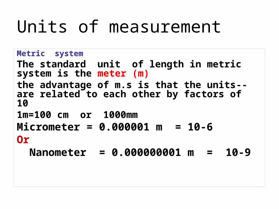
Metric system
The standard unit of length in metric system is the meter (m)
--the advantage of m.s is that the units are related to each other by factors of 101m=100 cm or 1000mm
Micrometer = 0.000001 m = 10-6Or
Nanometer = 0.000000001 m = 10-9

Microscopes
• There are two basic types of microscopes (according to source of illumination) that are commonly used in Microbiology:
light microscopes
and electron microscopes.

illuminator = light source
Condenser = has lenses that direct the light rays through the specimen
Objective lenses = the lenses closest to the specimen
Ocular lens = eyepiece = the image is magnified again by ocular lens

Magnification = enlargement
Resolution = is the ability of the lenses to distinguish fine detail & structure
Refraction index = is a measure of the light bending ability of a medium

•Calculate the magnification. = multiplying the obj. L. power by the ocu.l. power – 10X
•Low power – 10X == 100X
•High power – 40X == 400X
•Oil immersion – 100X == 1000X

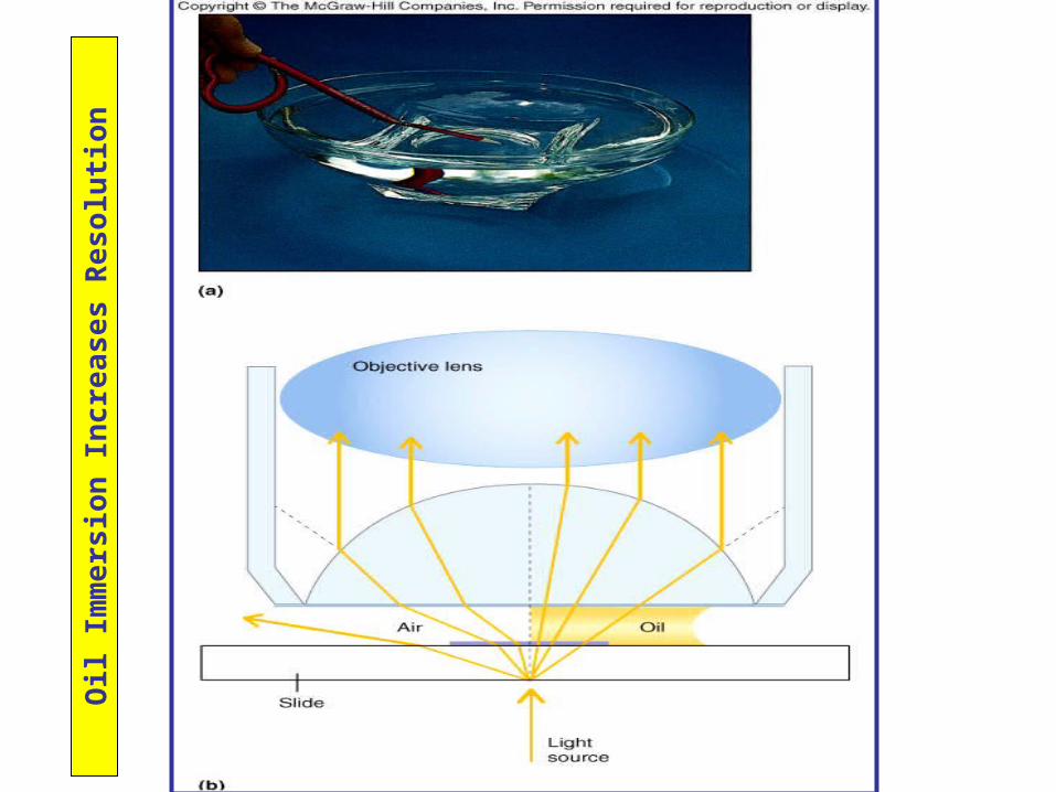
Greater resolution can be achieved by
using oil immersion ,
by filtering out with-blue light ,
and by replacing light with electrons.

Blu
e L
igh
t In
crea
ses
Res
olu
tio
n
Blue filter is inserted between light source and
condenser.
Shorter wavelength
results in higher resolution.
Blue light has shorter
wavelength than other visible
regions of the electromagnetic
spectrum.
Oil
Imm
ers
ion
Incr
ea
ses
Re
solu
tio
n
Oil
Imm
ers
ion
Incr
ea
ses
Re
solu
tion
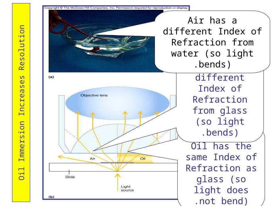
The Mineral Oil has the same Index of Refraction as glass (so light does not
bend).
Air has a different Index of Refraction from glass (so light
bends).
Air has a different Index of Refraction from water (so
light bends).

B- Electron Microscopeabeam of electrons is used –free electrons travel in waves
(100.000)

EM
= In
crea
sed
R
eso
luti
on
Transmission Electron
Microscopy (TEM): electrons are transmitted
through substance .
Scanning Electron Microscopy (SEM): electrons bounce off the surface of
specimen resulting in a more 3-D
image.


Dark field Microscope a- Light Microscopy:

Fluorescent Microscope
•The ability of substance to absorb short wave length (ultraviolet) and give off light at a longer wave length (visible)

•In fluorescence microscopy specimens are first stained with fluorochromes and then viewed through a compound microscope by using an uv light source
•The m.o. appear bright objects against a dark background
•Fluorescence microscopy is used primarily in a diagnostic procedure called f.ab. tequnique

Observation of microorganisms

Observation of microorganisms•Colorless
•A smear must be
• prepared
• stained
• Staining = coloring -Increase contrast of microorganisms
•Fixed = attached
• fixing – kills , fix & preserves various parts of m.o. in their natural state

Preparation of smear for light field Microscope

Organic salts compose of a positive and a negative ion, one of which is colored and is known as the chromophore
In Basic dye: positive ion is coloredCystal violet, methylen blue, malachite green,
safranin.
In Acidic dye: negative ion is colored eosin, acid fuchsine, and nigrosin.
As bacteria are slightly negatively charge at pH7, it colored with basic dye

Types of stains
Simple stain:
Differential stain:
Structural or special stains

• React differently with different kinds of bac.
• More than one dye• Example : as Gram stain, acid fast
Primary dyeMordant Decolorizing stepCounter stain
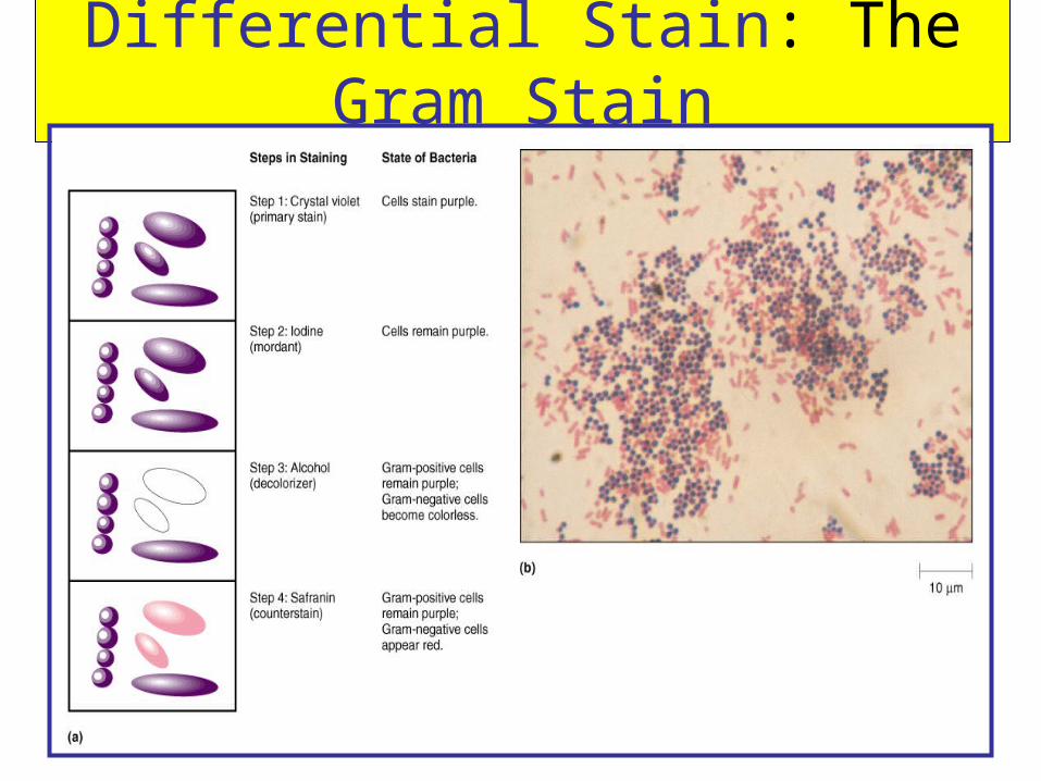
Differential Stain: The Gram Stain

24
Gram Stain
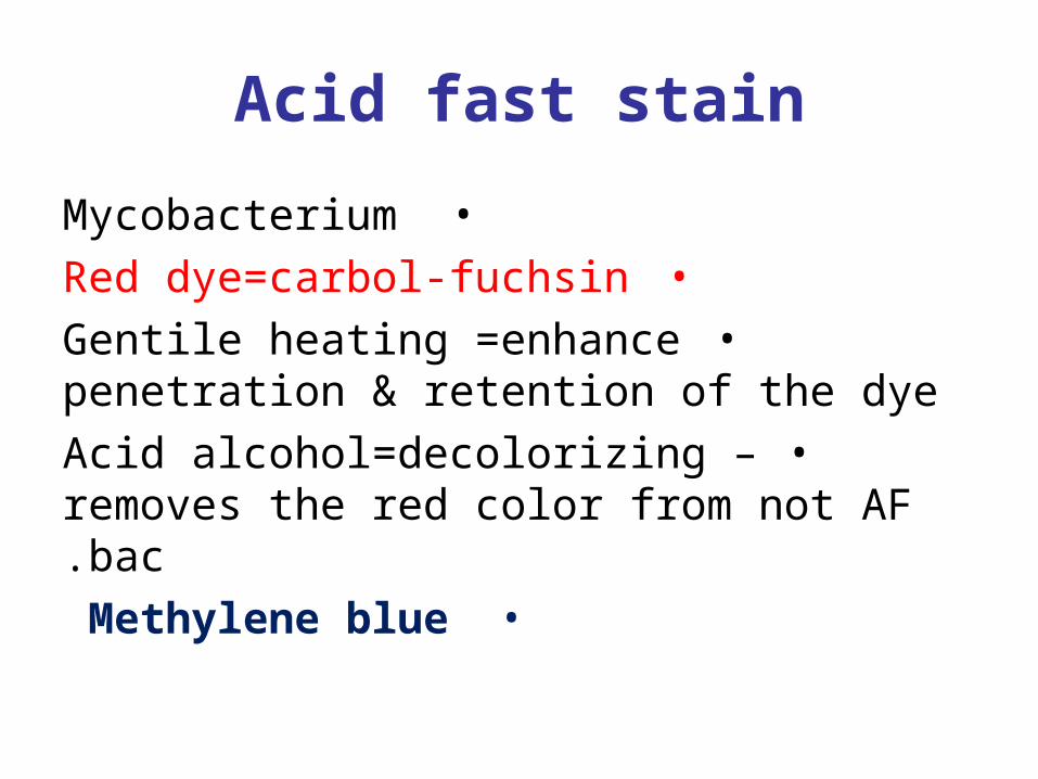
Acid fast stain
•Mycobacterium
•Red dye=carbol-fuchsin
•Gentile heating =enhance penetration & retention of the dye
•Acid alcohol=decolorizing – removes the red color from not AF bac.
•Methylene blue

Aci
d-F
ast
Sta
inin
g
Mycobacterium avium complex (MAC) with acid fast stain often has the characteristic appearance shown here with numerous mycobacteria filling macrophages. Such macrophages may be distributed diffusely or in clusters.
Note that the acid-fast bacteria are found as red clumps of filamentous
cells.
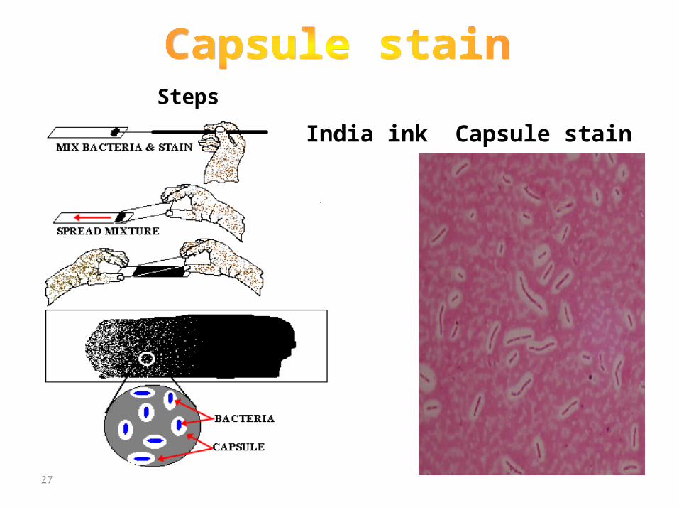
Steps
India ink Capsule stain
27
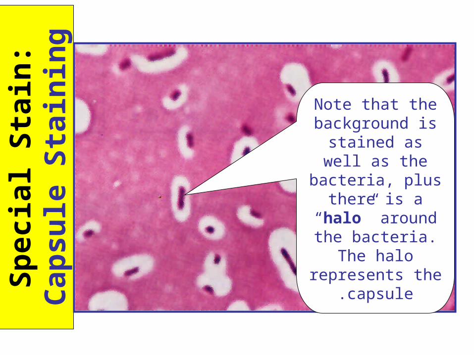
Sp
ecia
l Sta
in:
Cap
sule
S
tain
ing
Note that the background is
stained as well as the bacteria, plus there is a “halo”
around the bacteria. The halo represents
the capsule.
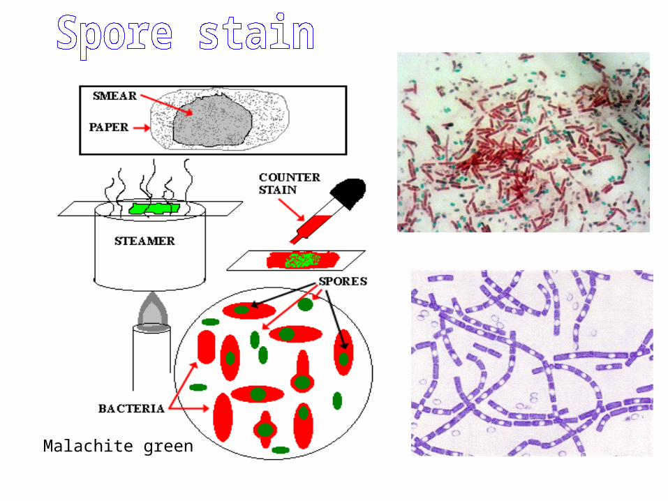
Malachite green
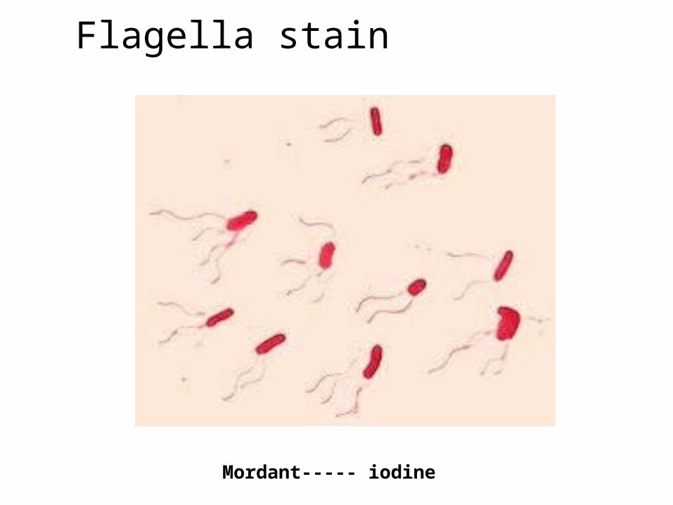
Flagella stain
Mordant----- iodine