obstructive jaundice caused by mucinous cystic tumor of
TRANSCRIPT
Copyright: © 2021 The Author(s). This article has been published under the terms of Creative Commons Attribution-Noncommercial 4.0 International License (CC BY-NC 4.0), which permits noncommercial unrestricted use, distribution, and reproduction in any medium, provided that the following statement is provided.
“This article has been published in Journal of Clinical and Translational Hepatology at https://doi.org/10.14218/JCTH.2020.00123 and can also be viewed on the Journal’s website at http://www.jcthnet.com ”.
Case Report
Journal of Clinical and Translational Hepatology 2021 vol. 9(4) | 598–602 DOI: 10.14218/JCTH.2020.00123
Obstructive Jaundice Caused by Mucinous Cystic Tumor of Gallbladder: A Case Report and Literature ReviewSulai Liu1,2,3#, Zhihua Zhang1,2,3#, Chao Guo1,2,3, Zhangtao Yu1,2,3, Siyuan He1,2,3, Junaid Khan1,2,3, Bo Jiang1,2,3, Yinghui Song1,2,3* and Chuang Peng1,2,3*
1Department of Hepatobiliary Surgery/Hunan Research Center of Biliary Disease, Hunan Provincial People’s Hospital/The First Affiliated Hospital of Hunan Normal University, Changsha, Hunan, China; 2Biliary Disease Research Laboratory of Hunan Provincial People’s Hospital, Key Laboratory of Hunan Normal University, Changsha, Hunan, China; 3Clinical Medical Technology Research Center of Hunan Provincial for Biliary Disease Prevention and Treatment, Changsha, Hunan, China
Received: 18 November 2020 | Revised: 3 January 2021 | Accepted: 19 March 2021 | Published: 6 May 2021
Abstract
Mucinous cystic tumor of the gallbladder is an extremely rare benign tumor, with potential for malignant degenera-tion. Mucinous cystic tumors of the cystic duct are divided into mucinous cystadenoma and mucinous cystadenocarci-noma. Currently, cystadenoma is generally considered to be a precancerous lesion of cystadenocarcinoma. At present, there are few cases reported worldwide, and there are no relevant guidelines for diagnosis and treatment of this dis-ease. This article presents the collected clinical data of a patient with mucinous cystic tumor of the gallbladder who was admitted to the First Affiliated Hospital of Hunan Nor-mal University, with the characteristics of the disease sum-marized in combination with a focused literature review.
Citation of this article: Liu S, Zhang Z, Guo C, Yu Z, He S, Khan J, et al. Obstructive jaundice caused by muci-nous cystic tumor of gallbladder: A case report and litera-ture review. J Clin Transl Hepatol 2021;9(4):598–602. doi: 10.14218/JCTH.2020.00123.
Introduction
The cystic duct mucinous cystic tumor is a rare cystic duct tumor with latent malignant lesions, and represents a spe-cial pathological type in cholangiocarcinoma.1,2 Mucinous cystic tumors of the cystic duct are divided into mucinous cystadenoma and mucinous cystadenocarcinoma.3 Cur-rently, cystadenoma is generally considered to be a precan-cerous lesion of cystadenocarcinoma, so patients in whom this disease is suspected should be decisively operated, with
their intraoperative fast frozen sections used to guide the operation.2,3 The etiology of mucinous cystic gland tumors of the bile ducts is currently unclear. Some believe that this is a congenital disease, caused by fluid retention as a result of inflammatory hyperplasia or obstruction of some abnormal ducts that occurs during embryonic growth; others believe that mucinous cystic tumors of the cystic duct are related to preembryonic intestinal residual or ectopic ovarian tissue.1–3
The patient presented with painless jaundice that had lasted for a duration of 1 month. In this case, the large cystic duct tumor was found to have squeezed the common bile duct to cause obstructive jaundice and dilated intrahe-patic and extrahepatic bile ducts.
Case report
The patient was a 57 year old female, with complaint nausea and vomiting for more than 1 month. She had been treated at a local community hospital 1 month prior to presentation at our hospital. However, after having received intravenous fluids and antibiotics, her symptoms did not alleviate. Labo-ratory examination upon presentation to our hospital showed the following: total bilirubin of 143.9 μmol/L; direct bilirubin of 109.6 μmol/L; alanine aminotransferase of 87.7 U/L; as-partate aminotransferase of 78.25 U/L; alkaline phosphatase of 201 U/L; gamma-glutamyltransferase of 682.0 U/L; carbo-hydrate antigen 19–9 of 252.02 U/mL; cancer antigen 72–4 of 7.76 U/mL; and, negativity for the panel of antinuclear antibodies. Findings for cancer antigen 125, blood routine, and serum C-reactive protein were basically normal. Abdomi-nal computed tomography (Fig. 1A, B) showed obstruction of the lower part of the common bile duct, dilatation of the up-per bile duct, and chronic cholecystitis. Magnetic resonance choliangiopancreatography (Fig. 1C) showed thickening of the lower part of the common hepatic duct with dilatation of the bile ducts inside and outside the liver. For preopera-tive jaundice reduction and cholangiography, percutaneous transhepatic cholangial drainage (referred to as PTCD) was performed. The PTCD angiography (Fig. 1D) showed filling defect in the common bile duct and bile duct dilation.
In order to clarify the nature of the space occupied by the bile duct and relieve the patient’s biliary obstruction, abdominal cavity exploration, biliary exploration, prepara-tion of biliary and enteral drainage were performed. A fro-
Keywords: Mucinous cystic tumor; Jaundice; Gallbladder; Case report; Litera-ture review.Abbreviations: MCN, mucinous cystic neoplasm; PTCD, percutaneous transhe-patic cholangial drainage.#These authors contributed equally to this work.*Correspondence to: Yinghui Song and Chuang Peng, Department of Hepato-biliary Surgery, Hunan Provincial People’s Hospital/The First Affiliated Hospital of Hunan Normal University, Changsha, Hunan 410004, China. Tel: +86-731-83929520, Fax: +86-731-83929520, E-mail: [email protected] (YS) or [email protected] (CP)

Journal of Clinical and Translational Hepatology 2021 vol. 9 | 598–602 599
Liu S. et al: Mucinous cystic tumor of gallbladder
zen section was assessed intraoperatively, and the results were reported as: “Consider gallbladder cyst adenoma”. We then performed an open cholecystectomy, biliary explora-tion, and bile duct repair with shaping and T-tube drainage.
During the operation, a 4.0×2.0 cm mass of the cystic duct protruded into the bile duct lumen (Fig. 2A–D). The mass grew on the wall of the cystic duct with a pedicle, and a few stones were seen in the gallbladder.
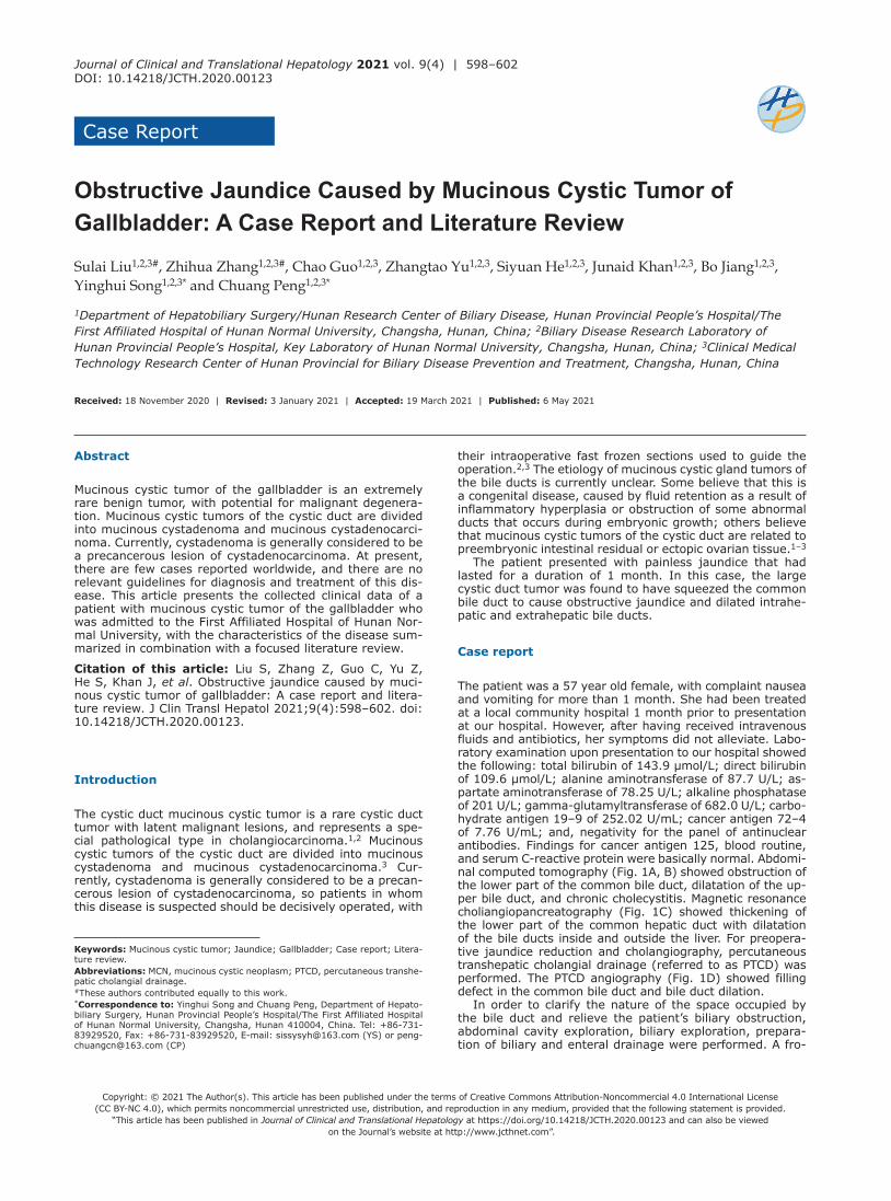
The predischarge inspection showed that the total biliru-bin was 25.8 μmol/L and the direct bilirubin was 18 μmol/L. Computed tomography of the abdomen showed that the dilatation of the bile ducts, inside and outside the liver, was significantly less than before. Postoperative pathology showed that there was a multicystic mass in the cystic duct, 4×2×2 cm in size, multicystic at the cut surface, and con-taining light-yellow, clear liquid in the cyst. The pathological diagnosis was mucinous cystic tumor with mild atypical hy-perplasia with chronic cholecystitis immunohistochemistry of cytokeratin 7 (+), cytokeratin 19 (epithelial +), estrogen receptor (+), progesterone receptor (+), P53 (−), and Ki67 (scattered +) (Fig. 3).
Informed consent
Prior written informed consent was provided from the pa-tient and this study was approved by the Ethics Review
Board of Hunan Provincial People’s Hospital/The First Affili-ated Hospital of Hunan Normal University.
Discussion
Mucinous cystic neoplasms (MCNs) were first reported in pancreatic tissue and, subsequently, there has been much research devoted to investigating pancreatic MCNs. Howev-er, there are still many controversies about pancreatic MCN disease and even less is known about gallbladder MCN. Ac-cording to the authors’ search of the PubMed database, the earliest case of gallbladder MCN was reported by Bishop in The Lancet in 1901,1 and there have been 16 literature reports on gallbladder MCN (Table 1).
Similar to pancreatic MCN, gallbladder MCN can manifest unilocular or multilocular cystic changes, containing septa. In the World Health Organization Classification of Digestive System Tumors (2010 Edition), biliary MCN is listed sepa-rately, as a special tumor of the gallbladder, and is classified into “mucocystic tumors with low-grade or medium-grade epithelium according to the status of intraepithelial neopla-sia. Internal neoplasia (8470/0) (8470/2)”, “Invasive mu-cocystic carcinoma (8470/3)”. The existing literature data divides MCN into at least two types.1 One is non-invasive and has ovarian-like stroma under the epithelium, which is characterized by a high cell density. It appears as a dense
Fig. 1. Preoperative imaging revealed biliary obstruction. (A, B) Abdominal plain computed tomography scan plus enhanced (C) Magnetic resonance choliangio-pancreatography and (D) PTCD angiography showed both intrahepatic bile duct dilation and common hepatic duct dilation, as well as a space-occupying lesion at the confluence of the cystic duct.

Journal of Clinical and Translational Hepatology 2021 vol. 9 | 598–602600
Liu S. et al: Mucinous cystic tumor of gallbladder
collection of spindle-shaped cells lacking cytoplasm and is immune to estrogen and progesterone receptors. This sub-type affects middle-aged women. The other type is more aggressive, has no ovarian-like stroma, and affects men between 75 and 88 years-old. There are others who clas-sify MCN using three subtypes, based on epithelial atypia and infiltration; the subtypes are mucinous cyst-adenoma, non-invasive mucinous cystadenocarcinoma, and invasive mucinous cystadenocarcinoma.2
Both gallbladder MCN and pancreatic MCN are common in women. The difference is that pancreatic MCN often oc-curs in the body and tail of the pancreas, which do not often cause obstructive jaundice.3 In the case of gallbladder MCN, as the tumor increases, some patients will show painful or painless jaundice.4 The overall prognosis of the disease is good, but there is a certain malignant potential. According to a Japanese study encompassing 156 cases of pancreatic MCN resection, the 10-year survival rate after resection was
95% for adenoma and 63% for cancer, among which micro-invasive carcinoma also reached more than 90%.4 Another study showed that the 5-year survival rate of untreated pan-creatic MCN with invasive carcinoma was about 30% and the prognosis was poor.5 Such statistics are still lacking for gallbladder MCN. In pancreatic MCN, the maximum tumor diameter is an independent risk factor affecting malignant transformation, and the level of carbohydrate antigen 19–9 has greater diagnostic significance for male patients.3 In gallbladder MCN, as the tumor size increases, the likelihood of jaundice and malignancy increases together. In our case, the cystic duct tumor was large and it compressed the com-mon bile duct, which then caused obstructive jaundice and intrahepatic bile duct dilation. Additionally, since gallstones were present, the case could have been misdiagnosed as common bile duct stones or Mirizzi syndrome.
Therefore, preoperative examination is particularly impor-tant. For this disease, ultrasound is more sensitive to the in-
Fig. 2. A cystic duct-origin mass was found during the operation to block the common bile duct. (A) 4.0×2.0 cm mass of the cystic duct was seen protruding into the bile duct cavity (white arrow). (B) The mass was found on the wall of the cystic duct (white arrow). (C) The upper common hepatic duct (white arrow) and the lower common bile duct (green arrow) did not show stenosis nor any masses. (D) A cystic duct-origin mass was observed.
Journal of Clinical and Translational Hepatology 2021 vol. 9 | 598–602 601
Liu S. et al: Mucinous cystic tumor of gallbladder
ternal features of the tumor (i.e. separation and fragments) and should be the first choice. Computed tomography can determine the location of the tumor and whether there is infil-tration of surrounding tissues, which can help guide the scope of surgical resection. Magnetic resonance choliangiopancrea-tography can help determine the bile duct compression and involvement, determine the cause of jaundice in patients, and determine whether biliary reconstruction surgery is appropri-ate.6 Assessment of a quick-frozen section during the opera-tion will help guard against the possibility of malignancy.
For asymptomatic patients, such as those who have tu-mors found on physical examination or imaging, one might use the pancreatic MCN endoscopic ultrasound-fine needle aspiration data on fluid collection to evaluate glucose (sensi-tivity of 92%, specificity of 87%, accuracy of 90%) and carci-noembryonic antigen (sensitivity of 58%, specificity of 96%, accuracy of 69%), for evaluation before an invasive opera-tion, since there is always risk of tumor dissemination and surgical complications.3 It is important to comprehensively consider the patient’s sex, age, family history, and surgical conditions. Interestingly, almost all gallbladder MCN patients are female1,7–21 (Table 1). In treatment, surgical resection is recommended for patients with clinical symptoms, such as abdominal pain, bloating, jaundice, or asymptomatic patients with gallbladder stones.22 It is important to send fast frozen sections during the operation to guide the operation method. After the surgical resection, it is recommended to check the confluence of the cystic duct, the wall of the gallbladder, and the common bile duct for other malignant tumors.
In summary, there is currently a lack of consistent evi-
dence for the malignant potential of gallbladder MCN, and there is also a lack of guidelines or consensus in diagnosis and treatment. However, the consensus reached after we compiled the literature is that due to the potential malig-nancy of gallbladder MCN, early diagnosis of such diseases should be paid attention to in clinical work, surgical treat-ment should be actively performed, and changes should be made according to the rapid intraoperative pathological examination results. Operating or expanding the scope of surgery will likely improve the prognosis and reduce recur-rence and malignant transformation.
Funding
This work was financially supported by following funds: Huxiang Youth Talent Support Program (Grant No. 2020RC3066); Postdoctoral Innovation Talents Project (Grant No. 2020RC2064); Hunan Provincial Natural Science Foundation of China (Grant No. 2019JJ50320/2020JJ5610); The Project of Improving the Diagnosis and Treatment Ca-pacity of Hepatobiliary, Pancreas and Intestine Disease in Hunan Province (Xiangwei [2019] Grant No. 118).
Conflict of interest
The authors have no conflict of interests related to this pub-lication.
Fig. 3. Postoperative pathology showed that there was a multicystic mass in the cystic duct. (A) A 100× cyst, lined with a single layer of mucin-producing epithelial cells and showing low-grade dysplasia was observed. (B) Most segments of the 400× cyst wall contained ovarian-like stroma. (C, D) 40× ovarian-like stroma immunohistochemical analysis showed positivity for estrogen receptor (ER) and progesterone receptor (PR).

Journal of Clinical and Translational Hepatology 2021 vol. 9 | 598–602602
Liu S. et al: Mucinous cystic tumor of gallbladder
Table 1. Gallbladder MCN reported cases
Case Year Age Sex Jaundice Abdomi-nal pain Tumor size in cm Carbohydrate
antigen 19-9 Reference
1 1901 42 Female Y Null Size of a child’s head Null 7
2 1930 Null Null Null Null Null Null 8
3 1933 24 Female Y Null 15 Null 9
4 1977 52 Female N Null Null Null 10
5 1989 65 Female Y Y 14 Null 11
6 1994 Null Null Null Null Null Null 12
7 2003 47 Female Y Y 4.6×4.2×4.4 Null 13
8 2003 88 Male Y Y 3.5×3×3 Normal 14
9 2005 38 Female N Y 1.2×0.8×0.8 Null 15
10 2006 75 Female N Y 17 High 16
11 2008 32 Female N Y 12 Null 17
12 2009 50 Female N Y 11×7.5×11.2 Null 18
13 2010 33 Female N Y 0.67 × 0.28 Null 19
14 2014 75 Female Y Y Null Null 1
15 2017 29 Female N Y 3 Null 20
16 2018 70 Female N N 6.7×6.8 ×7.2 High 2
17 2019 70 Female N Y 3×2×1 Null 21
18 2020 57 Female Y N 4.0 x 2.0 High Current study
N, no; Null, not mentioned; Y, yes.
Author contributions
Patient management (CG), drafting of the manuscript (SL, ZZ, JK), statistical analysis (YS, SH), data collection (SL, ZZ, YS, ZY, CP, BJ), and revision of the manuscript for im-portant intellectual content (YS,CP)
Data sharing statement
All data are available upon request.
References
[1] Zevallos Quiroz JC, Jiménez Agüero R, Garmendi Irizar M, Ruiz Montes-inos I, Comba Miranda JW. Mucinous cystic neoplasm of the gallbladder obstructing the common bile duct, a rare entity with a new name. Cir Esp 2014;92(8):567–569. doi:10.1016/j.ciresp.2013.01.015.
[2] Sugawara S, Hirai I, Watanabe T, Tezuka K, Kimura W. A case of mucinous cystic neoplasm of the gallbladder. Clin J Gastroenterol 2018;11(5):428–432. doi:10.1007/s12328-018-0850-8.
[3] Li WL, Xu YD, Han X, Wu WC, Lou WH. Clinical analysis and prognosis fac-tors of malignancy in the patients with mucinous cystic neoplasms of the pancreas. Zhonghua Wai Ke Za Zhi 2020;58(3):225–229. doi:10.3760/cma.j.issn.0529-5815.2020.03.011.
[4] Tada M, Koike K. Pancreatic tumor:progress in diagnosis and treatment. Topics: II. Intraductal papillary mucinous neoplasm of the pancreas (IPMN) /mucinous cystic neoplasm (MCN); 1. Symptoms and surveillance of IPMN and MCN. Nihon Naika Gakkai Zasshi 2012;101(1):51–56. doi:10.2169/naika.101.51.
[5] Yamao K, Yanagisawa A, Takahashi K, Kimura W, Doi R, Fukushima N, et al. Clinicopathological features and prognosis of mucinous cystic neoplasm with ovarian-type stroma: a multi-institutional study of the Japan pancreas so-ciety. Pancreas 2011;40(1):67–71. doi:10.1097/MPA.0b013e3181f749d3.
[6] Xu HX. Contrast-enhanced ultrasound in the biliary system: Potential uses and indications. World J Radiol 2009;1(1):37–44. doi:10.4329/wjr.v1.i1.37.
[7] Bishop ES. An undescribed innocent (?) growth of the gall-bladder. Lancet 1901;158(4063):72–73. doi:10.1016/S0140-6736(01)85060-1.
[8] Kordenat RA. Cystadenoma of the gallbladder report of a case. Wis Med J 1930;29:634–637.
[9] Shambaugh P. Multilocular papillary cystadenoma of the gall bladder. The American Journal of Surgery 1933;22(2):229–231. doi:10.1016/S0002-9610(33)90335-9.
[10] Ishak KG, Willis GW, Cummins SD, Bullock AA. Biliary cystadenoma and cystadenocarcinoma: report of 14 cases and review of the literature. Can-cer 1977;39(1):322–338.
[11] Simmons TC, Miller C, Pesigan AM, Lewin KJ. Cystadenoma of the gallblad-der. Am J Gastroenterol 1989;84(11):1427–1430.
[12] Devaney K, Goodman ZD, Ishak KG. Hepatobiliary cystadenoma and cys-tadenocarcinoma. A light microscopic and immunohistochemical study of 70 patients. Am J Surg Pathol 1994;18(11):1078–1091.
[13] Spector SA, Fernandez VE, Vernon SE, Dunkin B, Livingstone AS. Gallblad-der cystadenoma and common bile duct obstruction. Int J Gastrointest Cancer 2003;34(2-3):151–155. doi:10.1385/IJGC:34:2-3:151.
[14] Terada T, Takeuchi T, Taniguchi M. Hepatobiliary cystadenocarcinoma with cystadenoma elements of the gall bladder in an old man. Pathol Int 2003;53(11):790–795. doi:10.1046/j.1440-1827.2003.01559.x.
[15] Rooney TB, Schofer JM, Stanley MD, Banks SL. Biliary cystadenoma of the gallbladder. AJR Am J Roentgenol 2005;185(6):1571–1572. doi:10.2214/AJR.04.1560.
[16] Waldmann J, Zielke A, Moll R, Schweinsberg TS, Rothmund M, Langer P. Cystadenocarcinoma of the gallbladder. J Hepatobiliary Pancreat Surg 2006;13(6):594–599. doi:10.1007/s00534-006-1129-x.
[17] McCague A, Rosen M, O’Malley K. Laparoscopic cholecystectomy of a poly-poid gallbladder cystadenoma obstructing the common bile duct. Surg Lapa-rosc Endosc Percutan Tech 2008;18(2):209–212. doi:10.1097/SLE.0b013e 3181618b1b.
[18] Sistla SC, Sankar G, Basu D, Venkatesan B. Biliary cystadenocarcinoma of the gall bladder: a case report. J Med Case Rep 2009;3:75. doi:10.1186/1752-1947-3-75.
[19] Gokalp G, Dusak A, Topal NB, Aker S. Cystadenoma originating from the gallbladder. J Ultrasound Med 2010;29(4):663–666. doi:10.7863/jum.2010. 29.4.663.
[20] Moussa M, Douard R, Marzouk I, Kort I, Mekni A. Biliary cystadenoma and cystadenocarcinoma of the gallbladder: A clinical review. Am Surg 2017;83(6):e186–188.
[21] Rivero-Soto RJ, Hossein-Zadeh Z, Coleman J, Ahuja V. A mucinous cystic neoplasm originating from the gallbladder: A case report and literature review. Perm J 2019;23:18–077. doi:10.7812/TPP/18-077.
[22] Arnaoutakis DJ, Kim Y, Pulitano C, Zaydfudim V, Squires MH, Kooby D, et al. Management of biliary cystic tumors: a multi-institutional analysis of a rare liver tumor. Ann Surg 2015;261(2):361–367. doi:10.1097/SLA.0000 000000000543.