obstructive jaundice in autoimmune pancreatitis can be ... ed aip.pdf · obstructive jaundice in...
TRANSCRIPT
lable at ScienceDirect
Pancreatology 16 (2016) 391e396
Contents lists avai
Pancreatology
journal homepage: www.elsevier .com/locate/pan
Original article
Obstructive jaundice in autoimmune pancreatitis can be safely treatedwith corticosteroids alone without biliary stenting
Yan Bi a, Phil A. Hart a, e, Ryan Law a, Jonathan E. Clain a, Michael B. Farnell b,Ferga C. Gleeson a, Michael L. Kendrick b, Mike J. Levy a, Randall K. Pearson a,Bret T. Petersen a, Lisa D. Pisney a, Thomas C. Smyrk c, Naoki Takahashi d,Mark D. Topazian a, Santhi Swaroop Vege a, Suresh T. Chari a, *
a Division of Gastroenterology and Hepatology, Mayo Clinic, Rochester, MN, USAb Division of Surgery, Mayo Clinic, Rochester, MN, USAc Division of Pathology, Mayo Clinic, Rochester, MN, USAd Division of Radiology, Mayo Clinic, Rochester, MN, USAe Division of Gastroenterology, Hepatology, and Nutrition, The Ohio State University, Columbus, OH, USA
a r t i c l e i n f o
Article history:Available online 6 April 2016
Keywords:Autoimmune pancreatitisObstructive jaundicePrednisoneCholangitisERCP (Endoscopic retrogradecholangiopancreatography)Chronic pancreatitis
Abbreviations: AIP, autoimmune pancreatitis; ALPalanine aminotransferase; AST, aspartate aminotraretrograde cholangiopancreatography; TB, total biliru* Corresponding author. Division of Gastroentero
Clinic, 200 First Street SW, Rochester, MN, 55905, Ufax: þ1 507 284 5486.
E-mail address: [email protected] (S.T. Chari
http://dx.doi.org/10.1016/j.pan.2016.03.0171424-3903/© 2016 IAP and EPC. Published by Elsevie
Downloaded from For personal use only
a b s t r a c t
Objective: Autoimmune pancreatitis (AIP) responds dramatically to corticosteroids treatment. Wereviewed our experience to determine the safety and effectiveness of treating obstructive jaundice indefinitive AIP with corticosteroids alone without biliary stenting.Methods: From our AIP database, we retrospectively identified type 1 AIP subjects whose jaundice wastreated with corticosteroids alone without biliary stenting. Their medical records were reviewed andclinical data were evaluated to determine the outcomes.Results: Fifteen AIP subjects (87% male, mean age 68.4 years) were treated with corticosteroids at initialpresentation (n ¼ 8), first (n ¼ 5) or subsequent (n ¼ 2) relapse. Mean values (upper limit of normal, ULN)of liver tests prior to corticosteroids were aspartate aminotransferase (AST) 203.5u/l (4 � ULN), alanineaminotransferase (ALT) 325.8u/l (6 � ULN), alkaline phosphatase (ALP) 567.4u/l (5 � ULN), and totalbilirubin (TB) 5.9 mg/dl (5.9 � ULN). At first follow-up (mean 4 days) the decrease was 54.9% for AST,51.6% for ALT, 33% for ALP and 47.2% for TB (all p < 0.05). After 15e45 days, all patients had normal AST, 3/15 had ALT > 1.5 � ULN, 1/15 had ALP > 1.5 � ULN, 1/15 had TB > 1.5 � ULN. No patient required biliarystent placement, or developed cholangitis or other infectious complications during steroid treatment.Conclusion: Under the supervision of an experienced pancreatologist and with close monitoring of pa-tients, obstructive jaundice secondary to definitive AIP can be safely and effectively managed withcorticosteroids alone, without the need for biliary stenting.© 2016 IAP and EPC. Published by Elsevier B.V. All rights reserved.
Introduction
Reports of steroid-responsive obstructive jaundice weredescribed in the early 1950s, and the possibility of a pancreaticdisease of autoimmune etiology was first postulated by Sarles et al.,
, alkaline phosphatase; ALT,nsferase; ERCP, Endoscopicbin.logy and Hepatology, MayoSA. Tel.: þ1 507 255 5713;
).
r B.V. All rights reserved.
ClinicalKey.com at Italian Associatio. No other uses without permission. C
in 1961 [1], [2]. However, the term autoimmune pancreatitis (AIP)was not introduced until decades later in a review of the etiologyand classification of chronic pancreatitis [3]. Shortly thereafter,Yoshida et al. described the first clinical cohort, including serolog-ical abnormalities and steroid responsiveness, of what we currentlyrefer to as type 1 autoimmune pancreatitis [4].
AIP is a distinctive chronic condition of pancreas, presumablycaused by an autoimmune process. It is a fibro-inflammatory dis-ease with lympho-plasmacytic infiltration and peculiar storiformfibrosis that can cause multi-organ dysfunction. Type 1 AIP isconsidered to be the pancreatic manifestation of IgG4-related dis-ease with elevated serum IgG4, and abundant IgG4-positive cell
n of Gastroenterology (AIGO) June 22, 2016.opyright ©2016. Elsevier Inc. All rights reserved.
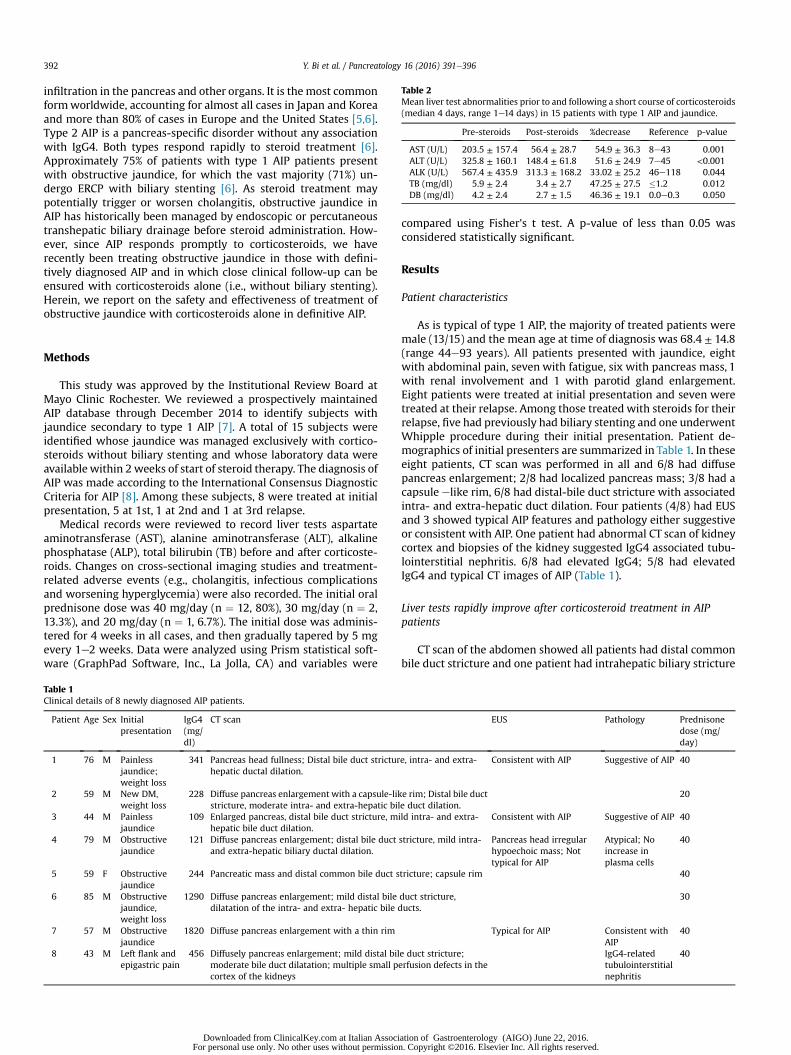
Table 2Mean liver test abnormalities prior to and following a short course of corticosteroids(median 4 days, range 1e14 days) in 15 patients with type 1 AIP and jaundice.
Pre-steroids Post-steroids %decrease Reference p-value
AST (U/L) 203.5 ± 157.4 56.4 ± 28.7 54.9 ± 36.3 8e43 0.001ALT (U/L) 325.8 ± 160.1 148.4 ± 61.8 51.6 ± 24.9 7e45 <0.001ALK (U/L) 567.4 ± 435.9 313.3 ± 168.2 33.02 ± 25.2 46e118 0.044TB (mg/dl) 5.9 ± 2.4 3.4 ± 2.7 47.25 ± 27.5 �1.2 0.012DB (mg/dl) 4.2 ± 2.4 2.7 ± 1.5 46.36 ± 19.1 0.0e0.3 0.050
Y. Bi et al. / Pancreatology 16 (2016) 391e396392
infiltration in the pancreas and other organs. It is the most commonformworldwide, accounting for almost all cases in Japan and Koreaand more than 80% of cases in Europe and the United States [5,6].Type 2 AIP is a pancreas-specific disorder without any associationwith IgG4. Both types respond rapidly to steroid treatment [6].Approximately 75% of patients with type 1 AIP patients presentwith obstructive jaundice, for which the vast majority (71%) un-dergo ERCP with biliary stenting [6]. As steroid treatment maypotentially trigger or worsen cholangitis, obstructive jaundice inAIP has historically been managed by endoscopic or percutaneoustranshepatic biliary drainage before steroid administration. How-ever, since AIP responds promptly to corticosteroids, we haverecently been treating obstructive jaundice in those with defini-tively diagnosed AIP and in which close clinical follow-up can beensured with corticosteroids alone (i.e., without biliary stenting).Herein, we report on the safety and effectiveness of treatment ofobstructive jaundice with corticosteroids alone in definitive AIP.
Methods
This study was approved by the Institutional Review Board atMayo Clinic Rochester. We reviewed a prospectively maintainedAIP database through December 2014 to identify subjects withjaundice secondary to type 1 AIP [7]. A total of 15 subjects wereidentified whose jaundice was managed exclusively with cortico-steroids without biliary stenting and whose laboratory data wereavailable within 2 weeks of start of steroid therapy. The diagnosis ofAIP was made according to the International Consensus DiagnosticCriteria for AIP [8]. Among these subjects, 8 were treated at initialpresentation, 5 at 1st, 1 at 2nd and 1 at 3rd relapse.
Medical records were reviewed to record liver tests aspartateaminotransferase (AST), alanine aminotransferase (ALT), alkalinephosphatase (ALP), total bilirubin (TB) before and after corticoste-roids. Changes on cross-sectional imaging studies and treatment-related adverse events (e.g., cholangitis, infectious complicationsand worsening hyperglycemia) were also recorded. The initial oralprednisone dose was 40 mg/day (n ¼ 12, 80%), 30 mg/day (n ¼ 2,13.3%), and 20 mg/day (n ¼ 1, 6.7%). The initial dose was adminis-tered for 4 weeks in all cases, and then gradually tapered by 5 mgevery 1e2 weeks. Data were analyzed using Prism statistical soft-ware (GraphPad Software, Inc., La Jolla, CA) and variables were
Table 1Clinical details of 8 newly diagnosed AIP patients.
Patient Age Sex Initialpresentation
IgG4(mg/dl)
CT scan
1 76 M Painlessjaundice;weight loss
341 Pancreas head fullness; Distal bile duct stricturhepatic ductal dilation.
2 59 M New DM,weight loss
228 Diffuse pancreas enlargement with a capsule-likstricture, moderate intra- and extra-hepatic bil
3 44 M Painlessjaundice
109 Enlarged pancreas, distal bile duct stricture, mhepatic bile duct dilation.
4 79 M Obstructivejaundice
121 Diffuse pancreas enlargement; distal bile ductand extra-hepatic biliary ductal dilation.
5 59 F Obstructivejaundice
244 Pancreatic mass and distal common bile duct s
6 85 M Obstructivejaundice,weight loss
1290 Diffuse pancreas enlargement; mild distal biledilatation of the intra- and extra- hepatic bile d
7 57 M Obstructivejaundice
1820 Diffuse pancreas enlargement with a thin rim
8 43 M Left flank andepigastric pain
456 Diffusely pancreas enlargement; mild distal bilmoderate bile duct dilatation; multiple small pcortex of the kidneys
Downloaded from ClinicalKey.com at Italian AssociFor personal use only. No other uses without permissio
compared using Fisher's t test. A p-value of less than 0.05 wasconsidered statistically significant.
Results
Patient characteristics
As is typical of type 1 AIP, the majority of treated patients weremale (13/15) and the mean age at time of diagnosis was 68.4 ± 14.8(range 44e93 years). All patients presented with jaundice, eightwith abdominal pain, seven with fatigue, six with pancreas mass, 1with renal involvement and 1 with parotid gland enlargement.Eight patients were treated at initial presentation and seven weretreated at their relapse. Among those treated with steroids for theirrelapse, five had previously had biliary stenting and one underwentWhipple procedure during their initial presentation. Patient de-mographics of initial presenters are summarized in Table 1. In theseeight patients, CT scan was performed in all and 6/8 had diffusepancreas enlargement; 2/8 had localized pancreas mass; 3/8 had acapsule elike rim, 6/8 had distal-bile duct stricture with associatedintra- and extra-hepatic duct dilation. Four patients (4/8) had EUSand 3 showed typical AIP features and pathology either suggestiveor consistent with AIP. One patient had abnormal CT scan of kidneycortex and biopsies of the kidney suggested IgG4 associated tubu-lointerstitial nephritis. 6/8 had elevated IgG4; 5/8 had elevatedIgG4 and typical CT images of AIP (Table 1).
Liver tests rapidly improve after corticosteroid treatment in AIPpatients
CT scan of the abdomen showed all patients had distal commonbile duct stricture and one patient had intrahepatic biliary stricture
EUS Pathology Prednisonedose (mg/day)
e, intra- and extra- Consistent with AIP Suggestive of AIP 40
e rim; Distal bile ducte duct dilation.
20
ild intra- and extra- Consistent with AIP Suggestive of AIP 40
stricture, mild intra- Pancreas head irregularhypoechoic mass; Nottypical for AIP
Atypical; Noincrease inplasma cells
40
tricture; capsule rim 40
duct stricture,ucts.
30
Typical for AIP Consistent withAIP
40
e duct stricture;erfusion defects in the
IgG4-relatedtubulointerstitialnephritis
40
ation of Gastroenterology (AIGO) June 22, 2016.n. Copyright ©2016. Elsevier Inc. All rights reserved.
Y. Bi et al. / Pancreatology 16 (2016) 391e396 393
Prior to treatment, 4 patients had TB between 1e2 � ULN, 5 pa-tients had TB 2e5 � ULN and 6 patients had TB more than 5 � ULN.The mean value (þ/� SD) of liver test values prior to steroidtreatment were as follows: AST 203.5 ± 157.4 u/l (4 � ULN), ALT325.8 ± 160.1 u/l (6� ULN), ALP 567.4 ± 436.0 u/l (5� ULN), and TB5.9 ± 2.4 mg/dl (5.9 � ULN) (Table 2). Follow-up liver tests at amean of 4 days (range 1e14 days) after starting prednisone showedrapid reduction of all liver tests: the decrease was 54.9 ± 36.3% forAST, 51.6 ± 25% for ALT, 33 ± 25.2% for ALP and 47.2 ± 27.5% for TB(Table 2). Fifteen to forty-five days after corticosteroids treatment,all patients had normal AST, 3 patients had ALT more than
Fig. 1. Line graphs demonstrating the liver test changes in each study subject with type 1normal.
Downloaded from ClinicalKey.com at Italian AssociatioFor personal use only. No other uses without permission. C
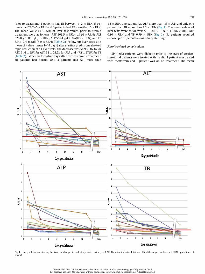
1.5 � ULN, one patient had ALP more than 1.5 � ULN and only onepatient had TB more than 1.5 � ULN (Fig. 1). The mean values ofliver tests were as follows: AST 0.83 � ULN, ALT 1.06 � ULN, ALP0.80 � ULN and TB 0.79 � ULN (Fig. 2). No patients requiredendoscopic or percutaneous biliary stenting.
Steroid-related complications
Six (40%) patients were diabetic prior to the start of cortico-steroids; 4 patients were treated with insulin, 1 patient was treatedwith metformin and 1 patient was on no treatment. The mean
AIP. Dark line indicates 1.5 times ULN of the respective liver test. ULN, upper limits of
n of Gastroenterology (AIGO) June 22, 2016.opyright ©2016. Elsevier Inc. All rights reserved.
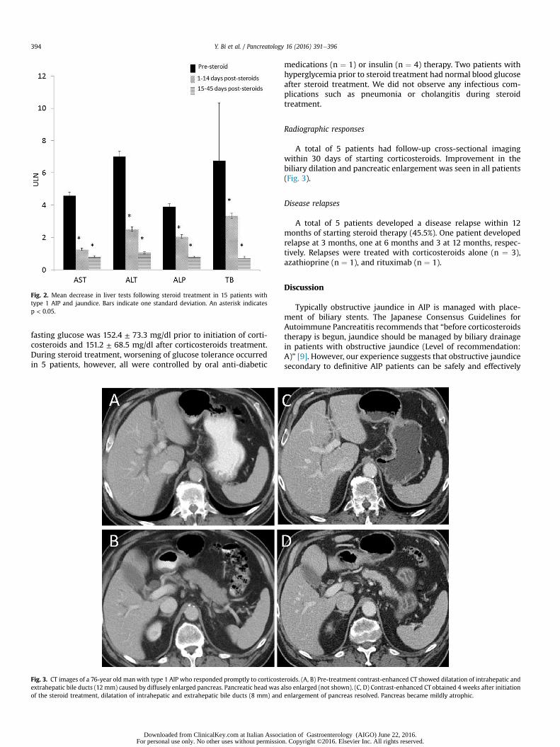
Fig. 2. Mean decrease in liver tests following steroid treatment in 15 patients withtype 1 AIP and jaundice. Bars indicate one standard deviation. An asterisk indicatesp < 0.05.
Y. Bi et al. / Pancreatology 16 (2016) 391e396394
fasting glucose was 152.4 ± 73.3 mg/dl prior to initiation of corti-costeroids and 151.2 ± 68.5 mg/dl after corticosteroids treatment.During steroid treatment, worsening of glucose tolerance occurredin 5 patients, however, all were controlled by oral anti-diabetic
Fig. 3. CT images of a 76-year old manwith type 1 AIP who responded promptly to corticosteextrahepatic bile ducts (12 mm) caused by diffusely enlarged pancreas. Pancreatic head was aof the steroid treatment, dilatation of intrahepatic and extrahepatic bile ducts (8 mm) and
Downloaded from ClinicalKey.com at Italian AssociFor personal use only. No other uses without permissio
medications (n ¼ 1) or insulin (n ¼ 4) therapy. Two patients withhyperglycemia prior to steroid treatment had normal blood glucoseafter steroid treatment. We did not observe any infectious com-plications such as pneumonia or cholangitis during steroidtreatment.
Radiographic responses
A total of 5 patients had follow-up cross-sectional imagingwithin 30 days of starting corticosteroids. Improvement in thebiliary dilation and pancreatic enlargement was seen in all patients(Fig. 3).
Disease relapses
A total of 5 patients developed a disease relapse within 12months of starting steroid therapy (45.5%). One patient developedrelapse at 3 months, one at 6 months and 3 at 12 months, respec-tively. Relapses were treated with corticosteroids alone (n ¼ 3),azathioprine (n ¼ 1), and rituximab (n ¼ 1).
Discussion
Typically obstructive jaundice in AIP is managed with place-ment of biliary stents. The Japanese Consensus Guidelines forAutoimmune Pancreatitis recommends that “before corticosteroidstherapy is begun, jaundice should be managed by biliary drainagein patients with obstructive jaundice (Level of recommendation:A)” [9]. However, our experience suggests that obstructive jaundicesecondary to definitive AIP patients can be safely and effectively
roids. (A, B) Pre-treatment contrast-enhanced CT showed dilatation of intrahepatic andlso enlarged (not shown). (C, D) Contrast-enhanced CT obtained 4 weeks after initiationenlargement of pancreas resolved. Pancreas became mildly atrophic.
ation of Gastroenterology (AIGO) June 22, 2016.n. Copyright ©2016. Elsevier Inc. All rights reserved.

Y. Bi et al. / Pancreatology 16 (2016) 391e396 395
treated using corticosteroids alone without risk of cholangitis orother infectious complications.
In the current study, 7 out of 8 newly diagnosed AIP can beconsidered definitive AIP and 1 probable AIP based on the Inter-national Consensus Diagnostic Criteria (ICDC) [8]. We observed anultra-rapid biochemical response to corticosteroids in 15 type 1 AIPpatients with obstructive jaundice. The liver test reduced by30e55% at 4 days (range 1e14 days) after starting prednisone andthe majority of patients had normal liver tests 2e6 weeks aftercorticosteroid therapy (Fig. 1). Contrast-enhanced CT scan showedsignificant improvement of biliary obstruction and pancreasenlargement (Fig. 3). No patients required biliary stenting and nocases of cholangitis or other infections were observed.
Resolution or improvement of biliary stricture and obstructivejaundice in AIP has been reported previously when treated withsteroids. In one report, three out of seven patients had bile ductstricture improved to almost its normal caliber and 4/7 patients hadrecovery to approximately 30e40% of the normal caliber [10,11].However, the obstructive jaundice resolved in all 5 patients aftersteroids treatment. In another study, resolution of biliary stricturewas found in 18 out of 30 patients (60%) and improvement ofstricture was found in additional 11 (37%) patients [10,11]. In ourpractice, we have observed rapid resolution or improvement ofbiliary structure and jaundice in all patients without need forbiliary stenting. We believe rapid relief from biliary obstructionfrom corticosteroids prevents development of cholangitis and ob-viates the need for ERCP with biliary stent placement or trans-hepatic drainage for these patients. In addition to avoiding the in-dex procedure, follow-up procedures for evaluation of stentplacement and/or removal are unnecessary. Although efforts aremade to minimize ERCP-related complications, this remains arelatively high risk procedure, with potential complicationsincluding pancreatitis hemorrhage, or perforation [12e15]. Like-wise, following placement of a biliary stent complications includingstent migration or occlusion may develop. Due to the nature ofinvasiveness, low cost and effectiveness of corticosteroids on AIP, itis therefore reasonable to consider a steroid therapy alone forobstructive jaundice in AIP patients whose diagnosis is certain orwho have relapse following prior confirmed diagnosis of AIPalthough we have to stress that the steroid treatment should beunder the guidance of an experienced pancreatologist and the pa-tient should be monitored closely clinically and biochemically.While it is possible that the stricture in advanced-stage of scle-rosing cholangiopathy may not respond to steroid due to its fibroticnature therapy, we have not yet seen a patient who has neededbiliary bypass or long-term endoscopic stenting for obstructivejaundice that is not relieved by medical therapy in AIP.
The mechanism of the ultra-rapid liver test and biliary strictureimprovement of AIP is likely multifactorial. An important contrib-uting factor is the bile duct wall inflammation. Studies of surgicalresection specimens in patients with AIP have shown inflammatoryfeatures in the distal bile duct identical to those seen in thepancreas, including lymphoplasmacytic infiltration, storiformfibrosis, and obliterative phlebitis [11,16,17]. Likewise, abundantlymphoplasmacytic infiltration may also be evident in the bile ductbiopsies from distal bile duct strictures in AIP, however oftencannot be seen due to the small amount of tissue acquired [10]. Inaddition, many consider the biliary dilation and resultant jaundicea consequence of extrinsic compression from pancreatic enlarge-ment. This certainly contributes, as the enlargement of pancreashas been shown to significantly decrease even after 2 weeks ofsteroid therapy [18]. “Steroid whitewash” has been previously re-ported in viral hepatitis, wherein liver tests and patient well-beingimprove with corticosteroids despite an infectious etiology of thehepatitis [19]. However, it is not known if a similar phenomenon
Downloaded from ClinicalKey.com at Italian AssociatioFor personal use only. No other uses without permission. C
would be seen in response to corticosteroids in obstructive jaun-dice in AIP.
It is important to recognize that the diagnosis of AIP is chal-lenging and differentiating it from pancreatic cancer and chol-angiocarcinoma is crucial. An ultra-rapid steroid trial of 7 days maypotentially differentiate AIP from malignancies. Differentiating AIPfrom pancreatic cancer is an important however difficult task. Inour study, AIP patients demonstrated an ultra-rapid response ofliver tests to corticosteroids. Therefore, would a “weekend steroidtrial” be useful in differentiating AIP from pancreatic cancer? Un-fortunately, we are not able to definitively answer that question aswe have limited experience with steroid treatment of obstructivejaundice due to causes other than AIP. However, our data are sup-ported by a previous Korean study in which 22 clinically suspectedAIP were challenged to 2 weeks of steroid therapy. The steroidresponse was assessed by improvement of main pancreatic ductnarrowing and/or measurable reduction of a pancreas mass. In thisstudy, all patients (n ¼ 15) who responded to corticosteroids werediagnosed as having AIP, whereas all patients (n ¼ 7) who did notshow a response to corticosteroids were confirmed as havingpancreatic cancer. Complete resection was possible in all (6/6;100%), except one individual who refused surgery [18]. However, itshould be noted that most patients in this trial underwent ERCPand it is unclear how many had biliary stenting at diagnosis. Werecommend in patients with suspicious AIP, especially in cases withindeterminate imaging and not responding to “weekend steroidtrial”, ERCP with biopsy should be performed to excludemalignancies.
We observed 45.5% relapse rate after stopping steroids. It ishigher than our previously report. However, it still aligns well withthe most of published data on AIP relapse (15e60%) [20e23]. It islikely due to the small sample size; higher percentage of patientswith diffuse pancreatic swelling which has been shown as a pre-dictor for AIP relapse (HR, 2.00; P ¼ 0.049) [23].
Our study is limited by small sample size and lack of long-termfollow up. However, it is a proof of principle study showing thatobstructive jaundice in AIP can be safely and effectively treatedwith corticosteroids alone without biliary stenting in a closelymonitored setting under the guidance of an experiencedpancreatologist.
Grant Support
None.
Writing assistance
None.
Author contributions
Chari ST developed the concept. Bi Y, Hart PA, Clain JE, FarnellMB, Gleeson FC, Kendrick ML, Law R, Levy MJ, Pearson RK, PetersenBT, Pisney LD, Smyrk TC, Takahashi N, Topazian MD, Vege SS andChari ST acquired data. Bi Y, Hart P and Chari ST analyzed data anddrafted the manuscript. All authors reviewed and approved thefinal manuscript.
Conflict of interest
The authors have no conflict of interest to report.
n of Gastroenterology (AIGO) June 22, 2016.opyright ©2016. Elsevier Inc. All rights reserved.
Y. Bi et al. / Pancreatology 16 (2016) 391e396396
References
[1] Sarles H, Sarles JC, Muratore R, Guien C. Chronic inflammatory sclerosis of thepancreasean autonomous pancreatic disease? Am J Dig Dis 1961;6:688e98.
[2] Summerskill WH, Jones FA. Corticotrophin and steroids in the diagnosis andmanagement of obstructive jaundice. Br Med J 1958;2:1499e502.
[3] Chari ST, Singer MV. The problem of classification and staging of chronicpancreatitis. Proposals based on current knowledge of its natural history.Scand J gastroenterol 1994;29:949e60.
[4] Yoshida K, Toki F, Takeuchi T, Watanabe S, Shiratori K, Hayashi N. Chronicpancreatitis caused by an autoimmune abnormality. Proposal of the conceptof autoimmune pancreatitis. Dig Dis Sci 1995;40:1561e8.
[5] Sah RP, Chari ST. Autoimmune pancreatitis: an update on classification,diagnosis, natural history and management. Curr Gastroenterol Rep 2012;14:95e105.
[6] Hart PA, Kamisawa T, Brugge WR, Chung JB, Culver EL, Czako L, et al. Long-term outcomes of autoimmune pancreatitis: a multicentre, internationalanalysis. Gut 2013;62:1771e6.
[7] Hart PA, Topazian MD, Witzig TE, Clain JE, Gleeson FC, Klebig RR, et al.Treatment of relapsing autoimmune pancreatitis with immunomodulatorsand rituximab: the mayo clinic experience. Gut 2013;62:1607e15.
[8] Shimosegawa T, Chari ST, Frulloni L, Kamisawa T, Kawa S, Mino-Kenudson M,et al. International consensus diagnostic criteria for autoimmune pancreatitis:guidelines of the international association of pancreatology. Pancreas2011;40:352e8.
[9] Okazaki K, Kawa S, Kamisawa T, Shimosegawa T, Tanaka M. Japaneseconsensus guidelines for management of autoimmune pancreatitis: I. Conceptand diagnosis of autoimmune pancreatitis. J Gastroenterol 2010;45:249e65.
[10] Ghazale A, Chari ST, Zhang L, Smyrk TC, Takahashi N, Levy MJ, et al. Immu-noglobulin g4-associated cholangitis: clinical profile and response to therapy.Gastroenterology 2008;134:706e15.
[11] Nishino T, Toki F, Oyama H, Oi I, Kobayashi M, Takasaki K, et al. Biliary tractinvolvement in autoimmune pancreatitis. Pancreas 2005;30:76e82.
[12] Aliperti G. Complications related to diagnostic and therapeutic endoscopicretrograde cholangiopancreatography. Gastrointest Endosc Clin N Am 1996;6:379e407.
Downloaded from ClinicalKey.com at Italian AssociFor personal use only. No other uses without permissio
[13] Christensen M, Matzen P, Schulze S, Rosenberg J. Complications of ercp: aprospective study. Gastrointest Endosc 2004;60:721e31.
[14] Ong TZ, Khor JL, Selamat DS. Complications of endoscopic retrograde chol-angiography in the post-mrcp era: a tertiary center experience. World J gas-troenterol WJG 2005;11:5209e12.
[15] Suissa A, Yassin K, Lavy A. Outcome and early complications of ercp: a pro-spective single center study. Hepatogastroenterology 2005;52:352e5.
[16] Notohara K, Burgart LJ, Yadav D, Chari S, Smyrk TC. Idiopathic chronicpancreatitis with periductal lymphoplasmacytic infiltration: clinicopathologicfeatures of 35 cases. Am J Surg Pathol 2003;27:1119e27.
[17] Zen Y, Harada K, Sasaki M, Sato Y, Tsuneyama K, Haratake J, et al. Igg4-relatedsclerosing cholangitis with and without hepatic inflammatory pseudotumor,and sclerosing pancreatitis-associated sclerosing cholangitis: do they belongto a spectrum of sclerosing pancreatitis? Am J Surg Pathol 2004;28:1193e203.
[18] Moon SH, Kim MH, Park DH, Hwang CY, Park SJ, Lee SS, et al. Is a 2-weeksteroid trial after initial negative investigation for malignancy useful indifferentiating autoimmune pancreatitis from pancreatic cancer? A prospec-tive outcome study. Gut 2008;57:1704e12.
[19] Ducci H, Motlis J. fulminant hepatitis; recovery. Rev Med Chile 1951;79:590e4.
[20] Kamisawa T, Shimosegawa T, Okazaki K, Nishino T, Watanabe H, Kanno A,et al. Standard steroid treatment for autoimmune pancreatitis. Gut 2009;58:1504e7.
[21] Ryu JK, Chung JB, Park SW, Lee JK, Lee KT, Lee WJ, et al. Review of 67 patientswith autoimmune pancreatitis in korea: a multicenter nationwide study.Pancreas 2008;37:377e85.
[22] Zamboni G, Luttges J, Capelli P, Frulloni L, Cavallini G, Pederzoli P, et al. His-topathological features of diagnostic and clinical relevance in autoimmunepancreatitis: a study on 53 resection specimens and 9 biopsy specimens.Virchows Arch 2004;445:552e63.
[23] Sah RP, Chari ST, Pannala R, Sugumar A, Clain JE, Levy MJ, et al. Differences inclinical profile and relapse rate of type 1 versus type 2 autoimmune pancre-atitis. Gastroenterology 2010;139:140e8. quiz e112e143.
ation of Gastroenterology (AIGO) June 22, 2016.n. Copyright ©2016. Elsevier Inc. All rights reserved.