occurrence of oral lesions associated with hiv/aids in
TRANSCRIPT

OCCURRENCE OF ORAL LESIONS ASSOCIATED WITH HIV/AIDS IN PATIENTS
RECEIVING HAART AT THE COMPREHENSIVE CARE CLINIC,
THIKA DISTRICT HOSPITAL, KENYA
JOHN MBUGUA KIHAMA
MASTER OF SCIENCE
(Public Health)
JOMO KENYATTA UNIVERSITY OF
AGRICULTURE AND TECHNOLOGY
2010

ii
Occurrence of oral lesions associated with HIV/AIDS in patients receiving HAART at
the comprehensive care clinic, Thika District Hospital, Kenya
John Mbugua Kihama
A Thesis Submitted in Partial Fulfilment for the Degree of Master of Science in Public
Health in the Jomo Kenyatta University of Agriculture and Technology
2010
iii
DECLARATION
This thesis is my original work and has not been presented for a degree in any other
university.
Signature …………………………….. Date ……………………
John Mbugua Kihama
This thesis has been submitted for examination with our approval as university supervisors.
Signature……………………………. Date…………………..
Dr. Peter Wanzala
KEMRI, Kenya
Signature……………………………. Date………………………..
Dr. Rose Bosire
KEMRI, Kenya
Signature ……………………………. Date …………………
Dr. Gideon M. Kikuvi
JKUAT, Kenya

iv
DEDICATION
To my wife Mercy, my daughter Fiona, my son Kelvin and my parents Peter and Beth for
their support, encouragement and understanding during the involving process of
preparation of this thesis.
v
ACKNOWLEDGEMENTS
The preparation of this thesis has been a process in which a number of people have been
involved to varying extents. First and foremost I would like to acknowledge my dedicated
supervisors Dr. Peter Wanzala, Dr. Rose Bosire and Dr. Gideon Kikuvi for their invaluable
and selfless guidance, support and encouragement throughout this process. I feel indebted
to the former Chief Dental Specialist, Ministry of Health, Dr. George Ogonji for his
encouragement and support as I prepared to embark on the journey that was Master of
Science in Public Health and the director, CPHR Dr. Yeri Kombe for his guidance during
the initial stages of preparation. My sincere gratitude goes to the staff of Thika District
Hospital comprehensive care clinic, the staff of KEMRI Centre for Public Health Research
and the Training Centre for their support and readiness to assist whenever requested. I
highly appreciate the donation of Chlorhexidine mouthwash (Remidin®) by Sai
Pharmaceuticals for distribution to the study participants. Finally I would like to thank all
the other people who contributed to the success of this process. Without you this would
have been a difficult if not impossible task. May God bless you all.

vi
TABLE OF CONTENTS DECLARATION ............................................................................................................ iii
DEDICATION ................................................................................................................ iv
ACKNOWLEDGEMENTS ............................................................................................. v
TABLE OF CONTENTS ................................................................................................ vi
LIST OF TABLES .......................................................................................................... ix
LIST OF FIGURES ......................................................................................................... x
LIST OF PLATES .......................................................................................................... xi
LIST OF APPENDICES ................................................................................................ xii
LIST OF ABBREVIATIONS AND ACRONYMS ...................................................... xiii
ABSTRACT ................................................................................................................... xv
CHAPTER ONE: INTRODUCTION ............................................................................. 1
1.1 Background .............................................................................................................. 1
1.2 Study Justification .................................................................................................... 3
1.3 Objectives ................................................................................................................ 4
1.3.1 General Objective ............................................................................................. 4
1.3.2 Specific Objectives ........................................................................................... 4
CHAPTER TWO: LITERATURE REVIEW ................................................................ 5
2.1 Overview of Oral Lesions Associated with HIV Infection. ....................................... 5
2.2 Epidemiology of Candidiasis .................................................................................... 8
2.3 Pathogenesis of oral candidiasis ............................................................................... 8
2.4 Clinical manifestations of oral candidiasis ................................................................ 9

vii
2.5 Diagnosis of oral candidiasis .................................................................................. 11
2.6 Correlation of OPC with HIV infection and its progression .................................... 12
2.7 HAART and its impact on OPC .............................................................................. 12
2.8 HAART Regimens ................................................................................................. 14
CHAPTER THREE: MATERIALS AND METHODS ................................................ 16
3.1 Study design ........................................................................................................... 16
3.2 Study Site ............................................................................................................... 16
3.3 Study Population .................................................................................................... 16
3.3.1 Sample Size Determination …………………………………………………….16
3.3.2 Sampling procedure……………………………………………………………..17
3.3.3 Inclusion criteria………………………………………………………………...17
3.3.4 Exclusion criteria………………………………………………………………..18
3.4 Data Collection ...................................................................................................... 18
3.4.1 Administering the Questionnaire……………………………………………….18
3.4.2 Clinical Examination of the oral cavity…………………………………………19
3.4.3 Microscopic examination of specimens for Candida pseudo-hyphae…………..19
3.5 Data Management .................................................................................................. 20
3.5.1 Data storage……………………………………………………………………..20
3.5.2 Data analysis……………………………………………………………………20
3.6 Ethical Considerations ............................................................................................ 20
3.7 Study Limitations ................................................................................................... 21

viii
CHAPTER FOUR: RESULTS ...................................................................................... 22
4.1 Socio-demographic Characteristics ......................................................................... 22
4.2 Clinical characteristics............................................................................................ 25
4.3 HAART Regimens ................................................................................................. 26
4.4 Characteristics of HAART usage ............................................................................ 26
4.5 Prophylaxis and treatment against opportunistic infections ..................................... 27
4.6 Occurrence and distribution of the oral lesions associated with HIV disease. .......... 29
4.7 Occurrence of oral lesions of HIV/AIDS and immune cell changes ........................ 30
4.8 Occurrence of oral lesions and duration of HAART................................................ 32
CHAPTER FIVE: DISCUSSION .................................................................................. 37
CHAPTER SIX: CONCLUSIONS AND RECOMMENDATIONS ............................ 47
6.1 Conclusions ............................................................................................................ 47
6.2 Recommendations .................................................................................................. 47
REFERENCES .............................................................................................................. 49
APPENDICES ................................................................................................................ 59
ix
LIST OF TABLES
Table 1: Prevalence of ‘oral lesions strongly associated with HIV’ in the HAART era;
comparison of published studies ....................................................................... 7
Table 2: Socio-demographic and clinical profile of the study subjects .......................... 23
Table 3: One way ANOVA for mean ages, CD4+ count and duration on HAART
between sexes ................................................................................................. 25
Table 4: Distribution of patients by HAART combination ............................................ 26
Table 5: Distribution of patients on HAART according to duration on HAART and
concurrent medications .................................................................................... 29
Table 6: Distribution of individuals with oral lesions by CD4+ count ........................... 32
Table 7: Distribution of individuals with oral lesions by duration of HAART ............... 34
Table 8: Odds ratios for occurrence of OPC in subjects over 24 weeks on HAART ...... 35
x
LIST OF FIGURES
Figure 1: Distribution of subjects by gender and age groups ........................................ 24
Figure 2: Distribution of concurrent medications used by subjects ............................... 28
Figure 3: Distribution of oral lesions by CD4+ count range ......................................... 31

xi
LIST OF PLATES
Plate 1: Kaposi’s sarcoma on the soft palate in one of the study subjects...................... 30
Plate 2: Pseudo-membranous candidiasis on the dorsum of the tongue in one of the study
subjects. ........................................................................................................... 30
xii
LIST OF APPENDICES
Appendix 1: Oral manifestations of HIV disease in adults ............................................ 59
Appendix 2: List of Some Anti-retroviral Drugs ........................................................... 60
Appendix 3: Consent Information and Consent Form ................................................... 61
Appendix 4: Patient Questionnaire ............................................................................... 66
Appendix 5: Comprehensive Care Clinic Patient Card .................................................. 69
Appendix 6: Adult Data Capture Sheet for Oral Lesions .............................................. 71
Appendix 7: Gram Staining Procedure ......................................................................... 72
Appendix 8: KEMRI/National Ethical Review Committee Study Approval. ................. 73
xiii
LIST OF ABBREVIATIONS AND ACRONYMS
AC Angular Cheilitis
AIDS Acquired Immunodeficiency Syndrome
ANOVA Analysis of Variance
ART Antiretroviral Therapy
CCC Comprehensive Care Clinic
CD4+ Human T helper Cells Expressing CD4 Antigen (T helper cell)
CDC Centres for Disease Control and Prevention
CI Confidence Interval
EEC European Economic Commission
EC Erythematous Candidiasis
ELISA Enzyme-Linked Immunosorbent Assay
HAART Highly Active Anti-retroviral Therapy
HIV Human Immunodeficiency Virus
HIV+ Human Immunodeficiency Virus Positive
IRIS Immune Reconstitution Inflammatory Syndrome
ITROMID Institute of Tropical Medicine and Infectious Diseases
JKUAT Jomo Kenyatta University of Agriculture and Technology
KEMRI Kenya Medical Research Institute
KS Kaposi’s sarcoma
LGE Linear Gingival Erythema
NNRTI Non-Nucleoside Reverse Transcriptase Inhibitor

xiv
NRTI Nucleoside Reverse Transcriptase Inhibitor
OHL Oral Hairy Leukoplakia
OI Opportunistic Infection
OPC Oro-pharyngeal Candidiasis
OR Odds ratio
PI Protease Inhibitor
RNA Ribonucleic Acid
STI Sexually Transmitted Illness
SPSS Statistical Package for Social Sciences
TB Tuberculosis
US United States of America
WHO World Health Organization
xv
ABSTRACT
The prevalence of some easily detectable oral manifestations of HIV/AIDS decreases with
HAART. Their presence may therefore be used as an indicator of the effectiveness of
HAART. The objective of this study was to determine the occurrence and patterns of
HIV/AIDS-related oral lesions in relationship to HAART usage, with a particular focus on
oro-pharyngeal candidiasis (OPC).
In this cross-sectional study, every 5th HIV-positive Comprehensive Care Clinic (CCC)
outpatient on HAART was selected. They underwent oral cavity examination for the
presence of different clinical forms of OPC and other oral lesions associated with HIV
infection. Individual patient medical records were perused for relevant clinical data. Gram
Stain smears from OPC lesions were examined under a light microscope for the presence of
Candida pseudo-hyphae. Data was recorded in structured questionnaires and standard
forms, entered into MS Access and then transferred to Statistical Package for Social
Sciences (SPSS) for analysis. Chi square was used to analyze the statistical significances
of the differences in frequencies and proportions. The data was stratified for periods below
and above 24 weeks on HAART. One-way ANOVA was computed comparing the mean
ages, mean CD4 count and mean durations on HAART between the sexes. Odds ratios
were calculated for the occurrence of OPC in subjects who had been on HAART for 24
weeks or more with adjustments for age, CD4 count, use of antifungal drugs, use of
antibiotics and missed HAART doses marital status and employment status.

xvi
A total of 404 (63% female) patients were examined. Male: female ratio was 1:1.7. The
mean age was 39.8 ± 9.5 years. All the patients examined were on triple HAART therapy.
Patients who had been on HAART for less than 24 weeks were 69 (17.1%) while 335
(82.9%) had been on HAART for more than 24 weeks. Oral lesions were observed in 63
(16%) patients. OPC was the commonest (12%), followed by oral hairy leukoplakia (3%).
Erythematous candidiasis was the most predominant type of oral candidiasis (6%) followed
by pseudo-membranous candidiasis (5%) and angular cheilitis (1%). There was no
statistically significant difference in occurrence or type of oral lesions observed between
those patients who had been on HAART for more or less than 24 weeks (p = 0.12).
Compliance to HAART by the study subjects is satisfactory. The occurrence of oral cavity
lesions of HIV/AIDS in patients on HAART at Thika District Hospital is low and shows a
gradual decrease over time with HAART usage. Clinicians attending to persons with
HIV/AIDS should be capable of diagnosing the oral lesions associated with HIV/AIDS and
may use these lesions as pointers for confirming the immune status of the patients using the
appropriate laboratory testing. Oral health clinicians should routinely examine for oral
opportunistic and neoplastic diseases in patients with HIV/AIDS and offer appropriate
treatment to improve the quality of life of these patients. The rate of occurrence recorded
in this study may be generalized to populations of persons who know and have accepted
their HIV status and are on HAART. More studies, preferably longitudinal, need to be
conducted for reasonable periods of time in order to get a better picture on the patterns of
occurrence of oral lesions in adults on HAART in Kenya.

1
CHAPTER ONE
1. INTRODUCTION
1.1 Background
There is a close relationship between general health/disease and oral mucosal reactions.
Many diseases show specific and/or non-specific changes which may evolve throughout the
course of the disease but the mucosal reaction may also constitute the initial clinical sign of
the disease (Axel1, 1992). The oro-facial region often reflects the presence of various
underlying systemic and generalized disease processes, including various infectious,
metabolic and immune-mediated disorders through specific/non-specific lesions
(Cleveland, 2003). The mouth and pharynx can be easily examined by clinicians with a
wide range of professional training. Many oral lesions are often clearly visible and some
can be diagnosed accurately on clinical features alone (Greenspan JS, 1997). The oral
lesions associated with HIV/AIDS can be fungal, viral or bacterial in origin. HIV-
associated oro-facial lesions have been considered as: clinical indicators of HIV infection in
otherwise healthy, undiagnosed individuals; early clinical features of HIV infection;
clinical markers for the classification and staging of HIV disease; predictors of HIV disease
progression (Coogan et al., 2005). They can therefore be used as entry or end-points in
therapy and vaccine trials and can be determinants of opportunistic infection and anti-HIV
therapy as well as being useful in staging and classification systems (Coogan et al., 2005;
Greenspan et al., 2002). Seven commonly occurring lesions, namely, oral candidiasis

2
(OPC), oral hairy leukoplakia (OHL), Kaposi’s sarcoma (KS), linear gingival erythema
(LGE), necrotizing ulcerative gingivitis, necrotizing ulcerative periodontitis and non-
Hodgkin lymphoma are strongly associated with HIV infection and have been identified
internationally (Appendix 1, EEC-clearing house 1993). These lesions may be present in up
to 50% of people with HIV infection and in up to 80% of those with a diagnosis of AIDS
(Butt et al., 2001; Wanzala and Pindborg, 1995). The lesions parallel the decline in
numbers of CD4+ cells and an increase in viral load, and are independent indicators of
disease progression (Greenspan et al., 2002). Thus, in cases where HIV status is unknown
and where HIV testing is difficult, for example, in some developing countries, certain oral
lesions may provide a strong indication of the presence of HIV infection (Coogan et al.,
2005).
Following the early reports of the oral manifestation of HIV in residents in the developed
world, there have now been many reports of the oral lesions of HIV-infected individuals in
the developing countries. Despite differences in transmission risk behavior, geographical
location, gender distribution, ethnicity, nutritional status and endemic disease, there are few
or no differences between the oral disease of HIV infected individuals in the developing
world when compared to those of the developed world (Patton et al., 2002), particularly as
regards the prevalence and type of common oral lesions observed (EEC-Clearing house
1993).

3
Highly active antiretroviral therapy (HAART) leads to an increase in CD4+ T-cell count,
decrease HIV-RNA viral load, and result in a decreased frequency and severity of
opportunistic disease including HIV-related oral disease (Ho, 1995).
1.2 Study Justification
It has been observed that HIV-infected patients are more likely to be colonized by Candida
than healthy individuals (Mocroft et al., 2003) and that HAART markedly decreases
mortality and morbidity as well as the incidence of AIDS-related opportunistic infections
(Kaplan et al., 2000). When successful, HAART results in undetectable viral load (< 50
copies/mm3) at 16-24 weeks after initiation of therapy. Virological response to successful
HAART shows similar trends with different drug combinations and so far remains the most
reliable laboratory measure of a patient’s immune status. Since the prevalence of
HIV/AIDS-related oral candidiasis decreases with HAART, it has been suggested that OPC
might be a clinically useful marker of patients’ immune recovery when considered
alongside available laboratory markers (Flint et al., 2006). In Kenya, there has been little
work done in this area, but its potential value to the clinical management of HIV/AIDS is
apparent, especially where numerous and frequent measures of CD4+ lymphocyte cell
counts and viral load may not be readily available or affordable in our resource-limited
settings.
4
1.3 Objectives
1.3.1 General Objective
To examine the occurrence and patterns of HIV/AIDS-related oral lesions in relation to
HAART usage among HIV positive adults attending the comprehensive care centre at
Thika District Hospital.
1.3.2 Specific Objectives
1) To determine the characteristics of HAART usage among the study subjects
2) To determine the occurrence of oral lesions, including OPC, associated with
HIV/AIDS among adults on HAART.
3) To determine the relationship between the occurrence of oral candidiasis and
HAART usage after stratifying for periods before and after 24 weeks of therapy
initiation.
1.3.3 Study Variables
Independent varibles: name, age, sex, marital status, employment status, HAART
combination, HAART compliance, duration on HAART, concurrent medication, presence
or absence of HIV/AIDS related oral lesions
Dependent varibles: clinical form and anatomical site of OPC, presence of Candida
pseudo-hyphae or spores on smears

5
CHAPTER TWO
2. LITERATURE REVIEW
2.1 Overview of Oral Lesions Associated with HIV Infection.
The prevalence of oral lesions strongly-associated with HIV/AIDS varies according to
region of the world and cohort examined. In a review of literature done to ascertain the
nature and prevalence of oral lesions in different regions, oral candidiasis was found to be
the most commonly reported oral lesion (Hodgson et al., 2002). Oral hairy leukoplakia
(OHL) appears to be the second most common lesion, with reported prevalence from 0% to
20% in Africa (Mugaruka et al., 1991; Arendorf et al., 1998; Butt et al., 2001), 3–13% in
Asia (Nittayananta and Chungpanich, 1997; Ranganathan et al., 2000), up to 43% in
Mexico (Gillispie and Marino 1993), and from 7% to 30% in the US and Europe (Lamster
et al., 1994; Schmidt-Westhausen et al., 1997; Schuman et al., 1998), with lower
prevalence among women than men. The prevalence of Kaposi’s sarcoma (KS) varies from
0% to 13% in Africa (Tukutuku et al., 1990; Arendorf et al., 1997; Butt et al., 2001) and
from 0% to 38% in the US and Europe (Schmidt-Westhausen et al., 1997; Schuman et al.,
1998).
Linear gingival erythema (LGE) is present in up to 16% of patients in developing nations
(Ranganathan et al., 2000) and 22% in the US (Lamster et al., 1994). Necrotizing
(ulcerative) gingivitis and necrotizing (ulcerative) periodontitis are reported to be present in
up to 16% and 17% of patients in Africa, respectively (Tukutuku et al., 1990), and a 23%
6
prevalence of necrotizing ulcerative periodontitis has been reported in India (Anil and
Challacombe, 1997). In contrast, in the US and Europe, necrotizing (ulcerative) gingivitis
and necrotizing (ulcerative) periodontitis are reported in up to 11% and 19% of HIV-
infected subjects, respectively (Schuman et al., 1998). All the patients in a study done in
Kenya had some form of periodontal disease - gingivitis, necrotizing gingivitis,
periodontitis (Butt et al., 2001).
The few studies of oral lesions in patients on HAART have been conducted in developed
countries and indicate significant differences in the influence of HAART on types and
occurrence of oral lesions (Greenspan et al, 2004; Schmidt-Westhausen et al., 2000). Most
studies in African on HIV-associated oral lesions had been done during pre-HAART era
(Hodgson et al., 2002; Arendorf et al., 1997; Butt et al., 2001; Adurogbangba et al., 2004;
Matee et al. 2000). In a study done in Tanzania, adult patients receiving HAART were
found to have a significantly lower prevalence of oral lesions, particularly oral candidiasis
and oral hairy leukoplakia (Hamza et al, 2006). Table 1 shows the occurrence of oral
lesions associated with HIV/AIDS in different parts of the world. The commonly observed
oral lesions, OPC and OHL, have been shown in separate columns.

7
Table 1: Prevalence of ‘oral lesions strongly associated with HIV’ in the HAART era;
comparison of published studies.
Reference Country Sample size
Any Oral Lesion
OPC (%) OHL (%)
Margiotta et al. (1999)
Italy
104
36%
10%OPC 6%PMC 4%EC
9.6
Schmidt-Westhausen et al. (1997)
Germany
70
43%
31%OPC 24%PMC 6%EC 4%AC
0
Arendorf et al. (1998)
South Africa
600
60%
38% OPC 16% PMC 16% EC 7% AC
20
Wanzala and Pindborg (1995)
Kenya
337
21%
13%EC 1%PMC 1%AC
3
Matee et al. (2000)
Tanzania
192
---- 12%OPC 12%PMC 0%EC 0%AC
6
Hodgson (1997) Zambia
107
---- 25%OPC 19%PMC 6%EC
5
Patton et al. (2000)
USA 299 38% 17% OPC 11
Nittayananta & Chungpanich (1997)
Thailand
124
82%
66% OPC 54%PMC 25%EC 6%AC
13
Key: OPC = oro-pharyngeal candidiasis; PMC = pseudomembranous candidiasis; EC = erythematous
candidiasis; AC = angular cheilitis; OHL = oral hairy leukoplakia

8
2.2 Epidemiology of Candidiasis
Candida is a commensal organism and part of the normal oral flora in about 30% - 50% of
the population, and is capable of producing opportunistic infections (superficial mycosis)
within the oral cavity when appropriate predisposing factors exist. The most frequently
isolated species is Candida albicans, although other species have been isolated in HIV-
infected patients such as C. glabrata, C. tropicalis, C. krusei, C. parapsilosis, C.
guilliermondii and C. dubliniensis (Ranganathan et al., 2006).
2.3 Pathogenesis of oral candidiasis
Various factors trigger pathogenesis, which gives rise to several different clinical forms of
candidiasis (Hodgson et al., 2002). General factors that may lead to clinically evident oral
candidiasis are the immune status of the host, the oral mucosal environment, and the
particular strain of C. albicans with the hyphal form being the one most commonly
associated with pathogenic infection (Lynch et al., 1994). The oral mucosa is continuously
challenged by the resident microbial flora and occasionally by microbial pathogens. It is
therefore armed with several cell populations which individually, or in association, can
produce a protective innate or acquired immune response. These cells include Langerhans’
cells, macrophages, ß- and -T cells (including CD4+ and CD8+ cells), keratinocytes, and
polymorphonuclear lymphocytes. The local response by these cells together with the
systemic response to HIV infection, determine the outcome in relation to oral
manifestations (Louis et al., 2004).
9
The natural history of HIV infection is characterized by progressive, insidious impairment
of the immune system function, with a widely varying rate of progression between patients.
CD4+ lymphocyte cell count and the quantification of viral RNA in blood plasma have
been found to be the main laboratory markers of disease progression (Mellors et al., 1996).
High viral load is currently considered to be one of the main indicators of the progression
of HIV-induced immune suppression (Patton et al., 1999). Advanced clinical stages of
HIV infection have been associated with more frequent occurrence of OPC than the early
stages. In the early stages, OPC affects mainly the oral mucosa, and only involves the
esophageal mucosa in more advanced stages (Imam et al., 1990). The presence of OPC in
subjects with HIV infection has been associated with the occurrence of esophageal
candidiasis and more frequent progression to AIDS (Scully et al., 1991). OPC is an early
oral sign of immunodeficiency and it has been used as a marker of severity in HIV
infection. The occurrence of OPC and the progression of early clinical forms
(erythematous variant and angular cheilitis) to more severe advanced forms
(pseudomembranous variant) are favoured by immune deterioration, as reflected by the
drop in CD4+ lymphocyte cell count (Imam et al., 1990; Ceballos et al., 1996).
2.4 Clinical manifestations of oral candidiasis
Acute or chronic erythematous candidiasis is the most commonly encountered clinical form
of candidiasis in HIV+ individuals. The clinical features of erythematous candidiasis
include reddish areas, mainly located on the back of the tongue and hard palate. This is

10
more frequent in the early stages of HIV infection, in patients with CD4+ counts greater
than 200 cells/µl (Ceballos et al., 1996).
Acute or chronic pseudomembranous candidiasis is the most classical form and comprises
up to 50% of all Candida infections in HIV+ individuals and is significantly more common
in patients with CD4+ lymphocyte cell counts of less than 200 cells/µl. It is characterized
by the presence of white or yellowish-white, soft or gel-like plaques that grow in a
centrifugal pattern and can be eliminated by scraping leaving an erythematous lesion that
can be painful on occasion. The plaques may appear anywhere inside the oral cavity,
although they are most frequent in the buccal mucosa, palate, oro-pharynx and margins of
the tongue. Pain, burning sensation or dysphagia may accompany these presenting features
(Ceballos et al., 1996).
Angular cheilitis is a disease of the lip that presents with reddening, cracks or splitting and
scabs at the corners of the mouth and it tends to be bilateral. It is frequently a mixed
infection caused by gram-positive Cocci and Candida. In HIV+ patients it is typically
chronic or recurrent (Ceballos et al., 1996). Linear gingival erythema (LGE) is an intensely
erythematous condition that clinically manifests as a 2- or 3-mm band, located along the
gingival margin. It appears to be closely related to the sub-gingival colonization by various
species pertaining to the Candida genus (Aguirre et al., 2004). Immunosuppression should
be suspected (and amongst possible causes, HIV infection), when faced with any clinical
form of candidiasis that is both severe and chronic in young people and/ or in individuals

11
with uncommon clinical features without an apparent cause to justify them (Ranganathan et
al., 2006; Hodgson et al., 2002).
2.5 Diagnosis of oral candidiasis
Candidiasis has identifiable clinical characteristics that make its diagnosis easy on visual
examination (Samaranayake, 1992). In addition, candidiasis may be confirmed by a
positive potassium hydroxide (10% KOH) wet mount, Gram-stained smear, culture or
biopsy. A swab of the oral lesions is placed in saline and the evaluation for yeast is carried
out immediately by mixing a drop of this solution with a drop of potassium hydroxide
(KOH) solution on a glass slide. The potassium hydroxide lyses the patient’s cells, making
the yeast easier to see. Alternatively, the swab is rolled onto a slide, fixed (by passing over
a flame) and Gram stained. This slide does not have to be stained or evaluated immediately
therefore making Gram stain a more convenient procedure. Specimens are examined for
the presence of small, round to oval, thin-walled, clusters of budding yeast cells and
branching pseudo-hyphae. The demonstration of pseudo-hyphae in scrapings or smears
from oral and esophageal lesions should be considered significant, provided the clinical
manifestations support the diagnosis (WHO, 1999).
Fungal cultures can also be done but are rarely necessary to make a diagnosis of oral
candidiasis. They are, however, helpful in identification of Candida species or when
colony counts are required. A swab of the oral lesions or an aliquot of a mouth rinse is
inoculated into Sabouraud’s dextrose agar within a few hours of collection and incubated

12
for up to two days at 37° C. Colonies are confirmed as yeast by performing a Gram stain.
In culture, colonies are typically white to cream colored with a smooth wax-like surface
(WHO, 1999). Biopsy specimens of oral lesions may also be used to distinguish certain
forms of leukoplakia and oral herpetic ulcers similar in appearance to certain Candida
variants (Chaisson et al., 1995).
2.6 Correlation of OPC with HIV infection and its progression
The reported prevalence of oral candidiasis varied from 56% to 81% in Asia ( Ranganathan
et al., 2000), 0–94% in Africa (Tukutuku et al., 1990) and 5–92% in the US (Lamster et al.,
1994), largely depending on the prevalence of AIDS diagnoses or individuals with low
CD4+ counts among those studied. Between 20 and 94% of all HIV-infected patients
present with oral candidiasis. In more than 75% of these individuals, some form of
mycosis has been observed at some point during the course of illness (Fine et al., 2003).
Erythematous candidiasis, pseudomembranous candidiasis and angular chelitis are the most
common types of oral candidiasis amongst the HIV-infected patient population and are
significantly predictive of progression to AIDS in these individuals (Schuman et al., 1998)
2.7 HAART and its impact on OPC
HAART has changed the lives and the course of disease in those infected with HIV
(Appendix 2). Since its introduction, there has been a marked reduction in AIDS deaths in
those HIV-infected individuals who accept their status and avail themselves for treatment
(Ho, 1995). The aims of HAART are maximal and durable suppression of the viral load in

13
the peripheral blood; restoration and preservation of immunological function (immune
reconstitution); improvement in quality of life, and reduction in HIV-related morbidity and
mortality. Successful treatment causes a sustained suppression of the virus, resulting in an
undetectable viral load (< 50 copies/mm3) at 16 to 24 weeks after initiation of therapy. A
concomitant rise in the CD4+ lymphocyte cell count is also observed. Increases in CD4+
cell counts in response to HAART confer immunologic reconstitution and a decreased
incidence of opportunistic infections. Treatment failure or HAART failure has been
ascribed to the development of resistance by the virus to the combination of drugs used,
non-compliance with the drug regimen, or suboptimal potency or blood levels of the drug
combination. Studies have shown that viral load falls and stabilizes to less than 500
copies/ml in over 80% of the patients 24 weeks after successful initiation of HAART
regardless of the drug combination or pre-HAART viral loads (Lepria et al., 2001; Philips
et al., 2001).
There is a close association between a patient’s immune state and the presence of OPC;
with an increase in OPC frequency as CD4+ lymphocyte cell counts decreases (Imam et al.,
1990). In HIV-infected patients, OPC has been associated with a less favourable immune
status and more accelerated deterioration of this situation (Ceballos et al., 1996). Phelan et
al., (1997) found that a CD4+ count of less than 200 cells/µl or the presence of OPC at the
time of her data collection increased the risk of death or development of AIDS, and such
prognosis was much worse if both factors were present at the onset of evaluation. This
relationship between OPC and low CD4+ lymphocyte counts may be used to interpret the
14
presence of OPC as an indirect marker of the immune status of HIV-infected patients when
a CD4+ lymphocyte cell count is unavailable (Phelan et al., 1997)
Oral co-morbidities in HIV infection were some of the first manifestations of the disease to
be documented. Classification and staging of HIV with oral lesions included as part of the
criteria has been published (Appendix 1). HAART has been shown to decrease the
prevalence of oral candidiasis in patients with AIDS (Patton et al., 2000). Oral
manifestations of HIV infection might therefore serve as good markers for monitoring not
only restoration of immune function (along with the numerical parameters of viral load and
CD4+ counts), but also HAART failure (Ceballos et al., 1996).
2.8 HAART Regimens
Since HAART is administered for long periods of time, three drug combinations are used
in an attempt to minimize viral resistance to the drugs. According to the ‘Guidelines on
antiretroviral drug therapy in Kenya, 2005’ HIV/AIDS patient are treated on a combination
of three classes of drugs (see Appendix 2); namely nucleoside reverse transcriptase
inhibitor (NRTI), a non-nucleoside reverse transcriptase inhibitor (NNRTI) and a protease
inhibitor (PI). In general, a minimum combination of three drugs from at least two
different classes as in the following combinations is preferred; 2 NRTIs + NNRTI; 2 NRTIs
+ PI/r (ritonavir boosted PI). The first line therapy includes four different combinations of
drugs: Stavudine or Zidovudine + Lamivudine + Nevirapine or Efavirenz. The second line
regimen is used in cases of treatment failure and includes the following drug combination:
15
Didanosine + Abacavir + Lopinavir/ritonavir (Kaletra) or Tenofovir + Abacavir +
Liponavir/ritonavir (Kaletra).

16
CHAPTER THREE
3. MATERIALS AND METHODS
3.1 Study design A cross-sectional study involving HIV/AIDS adult patients on HAART attending a
comprehensive care clinic was carried out.
3.2 Study Site The study site was Thika District Hospital HIV Comprehensive Care Clinic (CCC). Thika
District is one of the seven districts in central province of Kenya. The district has a
population of about 680,000. HIV/AIDS continues to have a devastating impact in this
district, which has the highest disease burden in central province. The HIV prevalence rate
in the district by end of 2007 was 3.6%. Thika District borders highly populated parts of
the adjacent districts whose residents seek health services in its health facilities.
3.3 Study Population
Thika District Hospital is considered a high volume district hospital due to the high
numbers of in- and out-patients. It serves an average of 750 out-patients daily. The CCC
was serving an average of 67 active patients daily by the end of December 2007.
3.3.1 Sample Size Determination
An average prevalence of HIV/AIDS-related oral candidiasis of 50% was assumed (Butt et
al., 2001). The sample size was based on the formula below (Fisher, 1998)
17
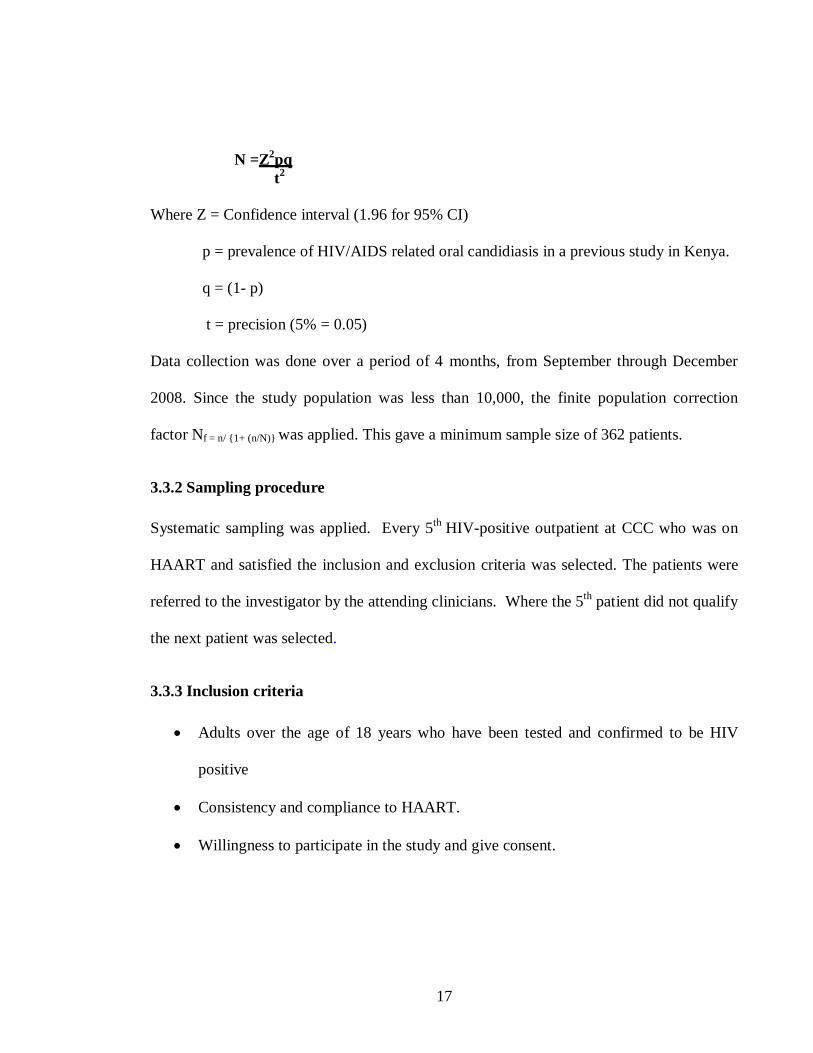
N =Z2pq t2 Where Z = Confidence interval (1.96 for 95% CI)
p = prevalence of HIV/AIDS related oral candidiasis in a previous study in Kenya.
q = (1- p)
t = precision (5% = 0.05)
Data collection was done over a period of 4 months, from September through December
2008. Since the study population was less than 10,000, the finite population correction
factor Nf = n/ {1+ (n/N)} was applied. This gave a minimum sample size of 362 patients.
3.3.2 Sampling procedure
Systematic sampling was applied. Every 5th HIV-positive outpatient at CCC who was on
HAART and satisfied the inclusion and exclusion criteria was selected. The patients were
referred to the investigator by the attending clinicians. Where the 5th patient did not qualify
the next patient was selected.
3.3.3 Inclusion criteria
Adults over the age of 18 years who have been tested and confirmed to be HIV
positive
Consistency and compliance to HAART.
Willingness to participate in the study and give consent.

18
3.3.4 Exclusion criteria
Those not tested and confirmed to be HIV positive.
Those with poor compliance for HAART
Those unwilling to participate in the study or give consent.
3.4 Data Collection
3.4.1 Administering the Questionnaire
All participants were requested to provide voluntary written informed consent before
enrollment. The researcher asked for consent from the patients after explaining to them the
nature of the procedures to be carried out in a simple fashion (Appendix 3).
Socio-demographic data, patients’ medical details including information on other illnesses
and other medication, and the observations of oral cavity examinations were recorded in a
structured questionnaire (Appendix 4) administered by the researcher. Each subject’s
medical records (Appendix 5) were reviewed by the researcher for relevant information
about the date confirmed HIV+, date he/she was started on HAART, HAART combination
and the most recent recording of CD4+ cell count. HIV testing for the subjects had been
done and confirmed at the hospital laboratory using ELISA (Murex HIV Ag/Ab
Combination, Murex Biotech Limited, UK). The CD4+ lymphocyte cell count used had
been carried out in the hospital laboratory through flow-cytometry using fluorescence-
activated cell sorter (FACSCount™, Becton Dickinson WW Inc).

19
3.4.2 Clinical Examination of the oral cavity
Oral cavity examination was carried out by the researcher, a dental surgeon by training,
using established WHO clinical diagnostic criteria for oral pathologic conditions associated
with HIV infection (Appendix 1) and recorded in a standard data capture sheet (Appendix
6). Examination was done in the CCC consultation office using natural daylight and, where
necessary, a headlamp was used. Wooden tongue depressors were used to retract the lips
and depress the tongue for a careful oral and pharyngeal examination. Oral lesions
associated with HIV infection, the different clinical forms of OPC and their anatomic
location was noted. Intra-oral photographs of lesions were taken occasionally with a
Cannon Ixy55 digital camera. Diagnosis of OPC and LGE was based on both clinical
manifestations and detection of organisms on smears. Diagnosis of the Kaposi’s sarcoma
lesion was confirmed through histological examination of an incision biopsy while that of
OHL was confirmed through clinical appearance.
3.4.3 Microscopic examination of specimens for Candida pseudo-hyphae
Specimens from OPC lesions were taken by gently drawing a wooden tongue depressor
across the lesion. The specimens were then transferred onto a clean glass slide, dried in air
and fixed by passing the slide over a flame at the chair-side. The slides were then labeled,
placed in a covered slide rack and later forwarded to the laboratory ready for Gram staining
(Appendix 7) and examination under a light microscope to confirm the presence of
Candida pseudo-hyphae.

20
3.5 Data Management
3.5.1 Data storage
Data was stored in the computer being used, flash disks, compact disks (CDs) and on the
internet. Completed data collection forms and questionnaires were retained as hard copy
back up.
3.5.2 Data analysis
Data was coded and double entry done in MS Access. Data was then transferred from MS
Access to Statistical Package for Social Sciences version 12.0 (SPSS Inc, Chicago, Illinois,
USA) for cleaning and statistical analysis. Data cleaning was done to exclude repeated and
incomplete data by running frequencies in SPSS. Chi-squared test and one way ANOVA
were used to compare differences in proportions of oral lesions between age groups, sexes
and CD4+ ranges. Odds ratios were calculated to examine the relationship between the
occurrence of oral lesions and age, CD4+ count and duration on HAART. Statistical
significance for all analyses was at p ≤0.05.
3.6 Ethical Considerations
Approval to conduct this study was granted by KEMRI/National Ethical Review
Committee (Appendix 8), JKUAT Scientific Committee and the Medical Superintendent,
Thika District Hospital. All participants were requested to provide voluntary written
informed consent before enrollment. Participants were guaranteed of confidentiality by
ensuring that no identifying information was made available to anyone who was not
directly involved in the study at any time during or after the study. Patients were given
21
appropriate advice and/or medication on complaints relating to oral/dental conditions and
referred for health problems that could not be addressed at the center. All procedures done
for the purposes of this study were free of charge to the participants. The results of the
study will be made available to the participating institutions and the individual participants
as appropriate.
3.7 Study Limitations
The CD4+ cell count measurements are done every 6 months for the patients attending
Thika District Hospital CCC. Some of the records available for CD4+ count were 3 or 4
months old and therefore may not have accurately reflected the patient’s immune status at
the time of examination. The data obtained in this cross-sectional study could not be
analyzed to conclusively address issues related to changes in observations in a time-
dependent manner. Additional aspects of risk for these HIV-associated oral lesions may not
have been conceptualized or assessed in this study design.

22
CHAPTER FOUR
4. RESULTS
4.1 Socio-demographic Characteristics
A total of 404 HIV-positive subjects were enrolled into the study, comprising 256 (63.4%)
females and 148 (36.6%) males. The mean age for this study sample was 39.8 ± 9.5 years
(range 21–79years). Majority of the study subjects 213 (52.7%) were married with more of
the males 113 (53.1%) being married compared to the females, 100 (46.9%). Most of the
subjects 289 (71.6%) were in their fourth or fifth decades of life. Majority of the study
subjects were either in part time employment 144 (35.6%) or were unemployed 136
(33.7%). Over two thirds of the study subjects had been on HAART for duration of more
than 24 weeks (Table 2).

23
Table 2: Socio-demographic and clinical profile of the study subjects
Characteristic
& Category
n =
Number (Percentage) of subjects
Males Females
Age
20 – 29 54 10 (18.5%) 44 (81.5%)
30 – 39 157 52 (33.1%) 105 (66.9%)
40 – 49 132 56 (42.4%) 76 (57.6%)
50 + 61 30 (49.2%) 31 (50.8%)
Totals 404 148 (36.6%) 256 (63.4%)
Marital Status
Single 61 8 (13.1%) 53 (86.9%)
Married 213 113 (53.1%) 100 (46.9%)
Others (separated,
divorced, widowed)
130 27 (20.8%) 103 (79.2%)
Employment Status
Full time 124 63 (50.8%) 61 (49.2%)
Part time 144 58 (40.3%) 86 (59.7%)
Unemployed 136 27 (19.9%) 109 (80.1%)
CD4+ count (cells/µl)
0 -200 135 57 (42.2%) 78 (57.8%)
201 – 500 216 73 (33.8%) 143 (66.2%)
501 + 53 18 (34.0%) 35 (66.0%)
Duration on HAART
Below 24 weeks 69 18 (26.1%) 51 (73.9%)
24 weeks and above 335 130 (38.8%) 205 (61.2%)
Totals
Percentages may not add up to exactly 100 due to rounding off

24
Most of the subjects in this study were in their fourth and fifth decades of life with males
having a higher mean age than females. Among the males the peak age group was 40-49
years while the age group 30-39 years was the peak age among the females (Figure 1).
05
1015202530354045
18-29 30-39 40-49 50+
Age group of Subjects
% of Subjects
MaleFemale
Figure 1: Distribution of subjects by gender and age groups
One-way ANOVA showed a significant difference (p =0.01) between the mean ages of
males (42.28 ±9.9) and females (38.43 ± 9.0). Other results for one-way ANOVA are
shown in Table 3.

25
Table 3: One way ANOVA for mean ages, CD4+ count and duration on HAART between sexes
Characteristic
Mean
p-value
Male Female
Age
42.3 ± 10.0
38.4 ± 9.0
0.01
CD4+ cell count
287.5 ± 190.5
317.4 ± 226.3
0.18
Duration on HAART
86.4 ± 59.6
79.8 ± 60.7
0.29
4.2 Clinical characteristics
On the basis of the most recent absolute CD4+ cell count, 135 (33.4%) of the subjects had a
count less than 200 cells/µl, 216 (53.5%) had a count between 200 and 500 cells/µl and 53
(13.1%) had a count greater than 500 cells/µl. The mean CD4+ count was 306.5 ± 214
cells/µl (range; 12 –1416 cells/µl). Males had a lower mean CD4+ cell count compared to
females (Table 3). Participants who had been on HAART for 24 weeks or more had higher
mean CD4+ cell count (331.3 ± 211.8 cells/µl) compared to those who had been on
HAART for less than 24 weeks (185 ± 183.0 cells/µl).

26
4.3 HAART Regimens
Eleven different HAART combinations were recorded in this study. Among those
examined 312 (77.2%) patients were on the first line drug combination: stavudine +
lamivudine + nevirapine. Only three subjects were on a HAART regime containing a
protease inhibitor. The most commonly used classes of drug combination were 2NRTIs +
NNRTI (Table 4).
Table 4: Distribution of patients by HAART combination HAART combination Class of drugs Number of
patients
Proportion (%)
stavudine + lamivudine +
nevirapine
2NRTIs + NNRTI
312
77.2
stavudine + lamivudine +
effavirenz
2NRTIs + NNRTI
57
14.1
stavudine + lamivudine +
tenofovir
2NRTIs + NNRTI
20
5.0
Other Combinations Various classes 15 3.7
NRTI – nucleoside reverse transcriptase inhibitor; NNRTI – non- nucleoside reverse transcriptase inhibitor
4.4 Characteristics of HAART usage
Majority 335 (82.9%) of the study participants had been on HAART for more than 24
weeks while 69 (17.1%) had been on HAART for less than 24 weeks. There was fair
adherence to HAART regimen with 400 (99 %) participants not having missed any doses of
27
HAART drugs in the previous 24 hours. Those who had missed at least one dose in the
previous 3 days were 43 (10.6%). The single most common reason for missing doses was
forgetting in 27 (62.7 %) patients, with 16 (37.2%) patients missing doses for reasons such
as side effects, being away from home and hospitalization. A higher proportion of men
(12.2%) had missed at least one dose of HAART in the previous 3 days compared to
women (9.8%).
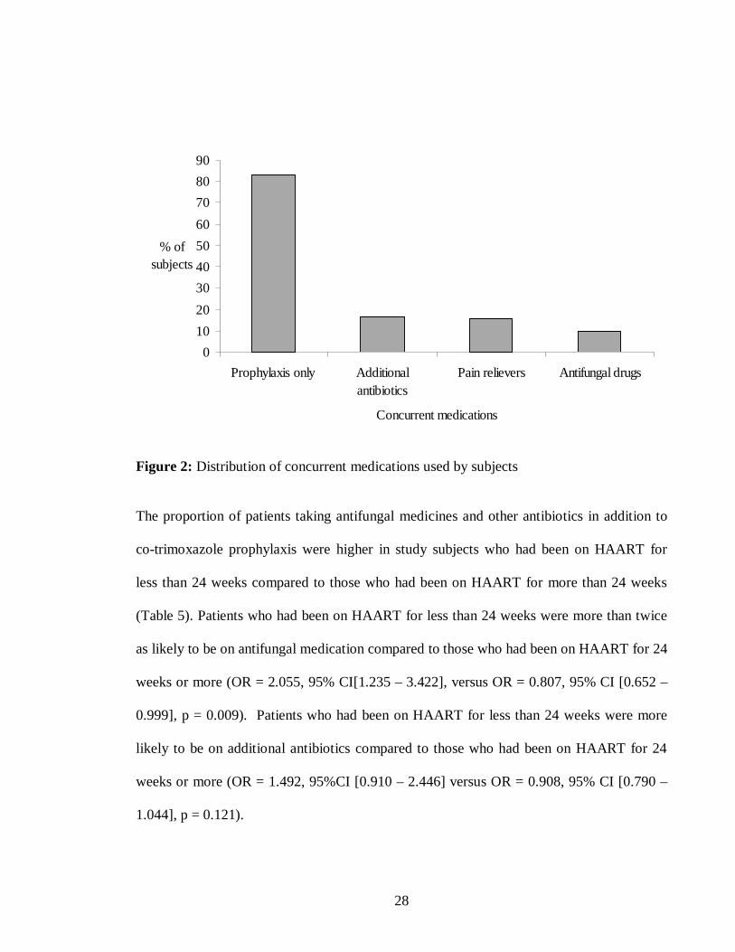
4.5 Prophylaxis and treatment against opportunistic infections
The most commonly documented medication concurrent to HAART in this study was
prophylaxis against opportunistic infections (OIs) using co-trimoxazole 336 (83.2%).
Other medications were additional antibiotics 68 (16.8%), pain relievers 63 (15.6%) and
antifungal drugs 41 (10.1%) as illustrated in Figure 2. Fourteen (3.5%) patients were on
TB treatment, one was suspected to have TB but had not been confirmed and the rest had
no signs of TB. Only 2 (14.3%) patients of those on TB treatment had oral lesions.
28
0102030405060708090
Prophylaxis only Additionalantibiotics
Pain relievers Antifungal drugs
Concurrent medications
% of subjects
Figure 2: Distribution of concurrent medications used by subjects
The proportion of patients taking antifungal medicines and other antibiotics in addition to
co-trimoxazole prophylaxis were higher in study subjects who had been on HAART for
less than 24 weeks compared to those who had been on HAART for more than 24 weeks
(Table 5). Patients who had been on HAART for less than 24 weeks were more than twice
as likely to be on antifungal medication compared to those who had been on HAART for 24
weeks or more (OR = 2.055, 95% CI[1.235 – 3.422], versus OR = 0.807, 95% CI [0.652 –
0.999], p = 0.009). Patients who had been on HAART for less than 24 weeks were more
likely to be on additional antibiotics compared to those who had been on HAART for 24
weeks or more (OR = 1.492, 95%CI [0.910 – 2.446] versus OR = 0.908, 95% CI [0.790 –
1.044], p = 0.121).
29
Table 5: Distribution of patients on HAART according to duration on HAART and
concurrent medications
Characteristic
Category
Duration on HAART
Below 24 weeks
(n = 69)
24 weeks and above
(n = 335)
Subjects taking
antifungal drugs
Yes 13 (18.8%) 28 (8.4%)
No 56 (81.2%) 307 (91.6%)
Subjects taking
antibiotics
Prophylaxis only 53 (76.8%) 283 (84.5%)
Prophylaxis and
additional antibiotics
16 (23.2%) 52 (15.5%)
Subjects on other
treatment
Pain relievers, anti-TB
drugs
17 (24.6%) 60 (17.9%)
4.6 Occurrence and distribution of the oral lesions associated with HIV disease.
A total of 63 (15.5%) patients had oral lesions. Oral candidiasis was the most commonly
observed oral lesion in 44 (10.9%) patients, followed by oral hairy leukoplakia in 12 (3%)
patients, gingivitis in 8 (2%) patients and Kaposi’s sarcoma (Plate 1) in one patient (0.2%).
Among the clinical variants of oral candidiasis, erythematous candidiasis was the most
frequently observed lesion in 21 (5.7%) patients followed by pseudomembranous
candidiasis (Plate 2) in 23 (5.2%) patients, and angular cheilitis in 3 (0.7%) patients.
30
Plate 1: Kaposi’s sarcoma on the soft palate in one of the study subjects.
Plate 2: Pseudo-membranous candidiasis on the dorsum of the tongue in one of the study subjects. 4.7 Occurrence of oral lesions of HIV/AIDS and immune cell changes
Having one or more oral lesions was more common among those with CD4+ counts <200
cells/µl at 26 (19.3%), with 35 (16.2%) of those having CD4+ counts from 200 to 500
cells/µl and 7 (13.2%) of those with CD4+ counts >500 cells/µl presenting with lesions. A
general decline in occurrence of oral lesions was observed as the CD4+ count increased

31
(Figure 3). The odds of having an oral lesion were lower (OR = 0.918) at CD4+ count
above 200 cells/µl than at CD4+ count below 200 cells/µl (OR = 1.170). The odds were
however not statistically significant at 95% CI.
0
2
4
6
8
10
12
14
16
18
20
CD4+ Count Range cells/ul
% of subjects
≤200 201-500 501+
Figure 3: Distribution of oral lesions by CD4+ count range
Having any of the clinical variants of OPC was more common 18 (13.3%) in those with
CD4+ counts < 200 cells/µl; followed by 24(11.1%) in those with CD4+ count between
200 and 500 cells/µl; and 5 (9.4%) in those with CD4+counts >500 cells/µl. The
differences were, however, not statistically significant (p = 0.585). Higher proportions of
PMC, EC, AC, and OHL were observed in the subjects with absolute CD4+ cell count
below 500 cells/µl (Table 6).

32
Table 6: Distribution of individuals with oral lesions by CD4+ count Oral Lesions <200 cells/µl 200-500 cells/µl >500 cells/µl
PMC 6(4.4%) 13(6.0%) 2(3.8%)
EC 10(7.4%) 10(4.6%) 3(5.6%)
AC 2(1.5%) 1(0.5%) 0
OHL 5(3.7%) 5(2.3%) 2(3.8%)
KS 1(0.7%) 0 0
LGE 2(1.5%) 6(2.8%) 0
No Lesion 109(80.7%) 181(83.8%) 46(86.8%)
Total 135(100%) 216(100%) 53(100%)
Percentages within CD4+ range as a proportion of the sample within that range
PMC – pseudomembranous candidasis; EC- Erythematous candididasis; AC-Angular cheilitis,
OHL- Oral Hairy Leukoplakia; LGE- Linear Gingival Erythema; KS- Kaposi’s sarcoma
4.8 Occurrence of oral lesions and duration of HAART
Duration on HAART in this study refers to the period from the initiation of the HAART
regimen up to the time of examination during this study. A higher proportion 17 (24.5%) of

33
oral lesions were observed in those on HAART for duration less than 24 weeks than those
on HAART for more than 24 weeks 51 (15.1%) as illustrated in Table 7. A Chi-squared
test was conducted which showed no statistical significance (p = 0.122) in the occurrence
of oral lesions with duration on HAART. There was also no statistical significance in the
occurrence of any OPC lesion with duration on HAART (p = 0.139). The risk of oral
lesions was less likely in those who had been on HAART for 24 weeks or more (OR =
0.905, 95% CI [0.783 – 1.047]) than in those who had been on HAART for less than 24
weeks (OR = 1.504, 95%CI [0.907 – 2.491]) with p = 0.122.

34
Table 7: Distribution of individuals with oral lesions by duration of HAART
Five subjects had more than one type of oral lesion.
A general decline in the proportions of the different clinical variants of OPC was observed,
with lower values in the group with over 24 weeks of HAART administration (9.9% vs.
15.9%). The odds of having OPC were lower at HAART duration 24 weeks and above
(OR = 0.894, 95% CI [0.749 – 1.067]) compared to the duration below 24 weeks (OR =
1.552, 95% CI [0.883 – 2.726]) with p = 0.139.
Odds ratios were calculated for the occurrence of OPC in subjects with 24 weeks or more
of HAART use with adjustment for age, CD4+ count, use of additional antibiotics, use of
antifungal drugs, missed HAART doses, marital status and employment status. The
Oral Lesions Duration on HAART
Below 24 weeks 24 weeks and above
PMC 4 (5.8%) 17 (5.0%)
EC 6 (8.7%) 17 (5.0%)
AC 1 (1.4%) 2 (0.6%)
OHL 5 (7.2%) 7 (2.1%)
KS 1 (1.4%) 0
Gingivitis 0 8 (2.4%)
Sub-Total (Any oral lesion) 17 (24.6%) 51 (15.2%)
No Oral Lesion 52 (75.4%) 284 (84.8%)
Total 69 (100%) 335 (100%)

35
unadjusted ratio was 0.576, 95% CI (0.275 – 1.205). The ratios were not statistically
significant at 95% CI. The characteristics analyzed showed different effects among the
different strata and probably act as effect modifiers. The stratum specific odds ratio for the
CD4+ count above 500 cells/µl was statistically significant (OR = 0.918, 95% CI [0.845 –
0.998]).
Table 8: Odds ratios for occurrence of OPC in subjects over 24 weeks on HAART
Adjustment
Odds Ratio
[95% CI]
Mantel-Haenszel
Common Odds
Ratio
[95% CI]
Factor
Strata
Age
Below 40 years 0.557
[0.213 – 1.461]
0.589
[0.259 – 1.338]
Above 40 years 0.563
[0.172 – 1.837]
CD4+ cell count
0 – 200 cells/µl 0.456
[0.163 – 1.272]
0.559
[0.265 – 1.181]
201 - 500
cells/µl
0.821
[0.174 – 3.865]
501+ cells/µl 0.918
[0.845 – 0.998]
Additional
antibiotics
Used 0.788
[0.182 – 3.406]
0.603
[0.288 – 1.266]
Not used 0.545
[0.231 – 1.284]
36
Antifungal drugs
Used
0.743
[0.199 – 2.779]
0.883
[0.358 – 1.938]
Not used 0.906
[0.298 – 2.758]
Missed at least one
dose of HAART in
the previous 3 days
Yes 0.625
[0.273 – 1.433]
0.547
[0.256 – 1.166]
No 0.919
[0.767 – 1.101]
Marital Status
Single 1.008
0.760 – 1.337
0.138
[0.275 – 1.197]
Married 0.959
0.765 – 1.202
Others 0.713
0.461 – 1.102
Employment Status
Full time 0.970
0.704 – 1.337
0.187
[0.285 – 1.278]
Part-time 0.946
0.710 – 1.260
Unemployed 0.845
0.637 – 1.122 Unadjusted Odds Ratio: - 0.576, 95% CI [0.275 – 1.205].

37
CHAPTER FIVE
5. DISCUSSION
Following the introduction of specific anti-HIV therapies, there have been well-
documented changes in the frequency and character of the oral complications of HIV
disease. Various studies have reported that HAART has a marked effect on the prevalence
and clinical appearance of oral lesions (Schmidt-Westhausen, 2000; Greenspan et al., 2004,
Powderly et al., 1998). This study showed a higher proportion of HIV-infected females
compared to males similar to other studies in Africa (Butt et al., 2001; Adurogbangba et al.,
2004). The Kenya AIDS Indicator Survey (2007) also showed a higher prevalence of HIV
infection in females (9.2%) as compared to males (5.8%) perhaps because women are
known to be more vulnerable in communities where HIV is spread mainly through
heterosexual transmission (UNAIDS, 2002).
The majority of the subjects in this study were in their fourth and fifth decades of life. This
is in contrast to other studies (Adurogbangba et al., 2004; Chidzonga, 2003) where the age
distribution of the HIV subjects showed a high prevalence in the third and fourth decades of
life. This may be due to fear of social stigma, low uptake of testing and consequently low
uptake of treatment by the lower age groups. A one way ANOVA conducted for mean ages
between sexes showed that male subjects had a higher mean age than female subjects
probably because more females get infected with HIV at younger ages (UNAIDS, 2002).

38
Oral disease and other opportunistic infections may affect the quality of life in HIV-infected
individuals but treatment usually results in a measurable symptomatic improvement.
Adherence to HAART is an essential component of treatment success. A high degree of
adherence to ARV drugs is necessary for optimal virological suppression. Imperfect
adherence is common and results from the present study showed that 43 (10.6%) of the
study subjects had missed at least one dose of HAART in the previous three days. One
survey indicated that one-third of patients missed doses within 3 days of the survey
(Ickovics et al., 1997). Another study has shown that 90–95% of the doses should be taken
for optimal suppression because lesser degrees of adherence are more often associated with
virological failure. This means a patient should not miss HAART doses more than three
times a month in the case of a twice-daily regimen (Peterson, 2000).
Factors associated with poor adherence include a poor adherence counseling, high pill
burden, forgetfulness, mental depression, lack of patient education, inability of patients to
identify their medications, drug toxicity and being too ill (Chesney, 2000). The fairy good
adherence reported in this study may be explained by the measures that have been taken in
the healthcare system and at Thika District Hospital CCC to optimize adherence which
include: provision of affordable and simplified treatment with uninterrupted supply of
HAART; a user-friendly health facility, respecting confidentiality, overcoming stigma and
discrimination; provision for social support by promoting and facilitating peer support and
formation of groups for people living with HIV/AIDS through day care centres and other
39
mechanisms; development of individual treatment plans that fit HAART into patients
lifestyles/daily events and identification of treatment reminders.
This study excluded patients with documented poor compliance to HAART such as those
who had defaulted for long intervals of time. These patients are usually attended separately
to facilitate adherence counseling and would therefore add a bias in sampling. Data on
missed doses of HAART in this study may therefore not accurately represent overall
HAART compliance. Questions were, however, still asked to the study subjects to gauge
their adherence to the drug regimes. Among those who had missed at least a single dose of
HAART the most common reason was forgetting 27 (62.7%) thus the need to intensify
adherence support and counseling in co-ordination with treatment support
persons/organizations. Sixteen patients (37.2%) missed doses for reasons such as side
effects, being away from home and hospitalization. Most of the subjects are part time
workers or unemployed and expected that they would be back to their houses in time to
take their drugs. A higher proportion of men (12.2%) had missed at least one dose of
HAART in the previous 3 days compared to women (9.8%). This may be attributed to the
generally poor health seeking behaviour in men, their traditional role as bread winners
occupying most of their time and the feeling of discomfort taking pills around others. More
than two thirds of the patients were on the same HAART combination; stavudine +
lamivudine + nevirapine. This has been reported to be an effective first line combination
well within the financial capability of resource-limited settings as in Kenya. It is also
available in fixed dose combinations that reduce the pill burden to the patient (Ministry of
Health, Guidelines on antiretroviral drug therapy in Kenya, 2006).
40
The proportion of patients taking antifungal medicines and other antibiotics in addition to
co-trimoxazole prophylaxis in this study were higher in subjects who had been on HAART
for less than 24 weeks compared to those who had been on HAART for more than 24
weeks. This compares favourably with, and can be explained by, the finding that HAART
leads to an increase in CD4+ T-cell count, decreased HIV-RNA viral load, and result in
decreased frequency and severity of opportunistic disease including HIV-related oral
disease (Ho, 1995). Viral load has been shown to fall and stabilize to less than 500
copies/ml in over 80% of the patients 24 weeks after successful initiation of HAART
regardless of the drug combination and pre-HAART viral loads (Lepria et al., 2001; Philips
et al., 2001).
In the current study the occurrence of oral cavity lesions of HIV/AIDS in patients on
HAART appears to be limited to candidiasis, oral hairy leukoplakia, gingivitis and one case
of Kaposi’s sarcoma out of the seven commonly reported oral lesions of HIV/AIDS
(Appendix 1). The observed overall occurrence (15.5%) of oral lesions in this study is in
agreement with the overall proportion of 15.6% reported for Kenyan commercial sex
workers (Wanzala et al., 1989) but far much lower than the 90% overall prevalence
reported for Zairean patients (Tukutuku et al., 1990) and the 60.4% reported in South
Africa (Arendorf et al., 1998). This is probably due to the fact that this study and that by
Wanzala et al., (1989) were carried out among out-patients while the other studies used in-
patients. Oro-pharyngeal candidiasis and OHL are the two most common oral diseases
associated with HIV/AIDS. In the current study the occurrence of OPC and OHL were
41
around 10.9% and 3% respectively similar to the findings of other studies that reported
higher prevalence of OPC than OHL (Eyeson et al., 2002; Patton et al, 2000). Having one
or more oral lesions and any of the clinical variants of OPC appears to be more common
among those with low CD4+ counts compared to those with higher counts. The difference
in occurrence of oral lesions within the different CD4+ count ranges was, however, not
statistically significant (p = 0.585). A lower occurrence of oral lesions was observed in
patients who had been on HAART for 24 weeks or more than in those who had been on
treatment for less than 24 weeks. This probably reflects the improved immune status
conferred by HAART beyond the critical period of 24 weeks. The 3% (n = 404)
occurrence of OHL in the current study is in agreement with 3% (n = 203) in a black
population in a London study involving outpatients on HAART (Eyeson et al., 2002).
Patton et al., (2000) also found a decrease in oral OHL from 26% to 11% after protease
inhibitor therapy. Similar to another study (Patton, 2000), data from this study showed an
association between OHL and poor immune status, CD4 <200 cells/µl, but showed a higher
occurrence of OHL in female sex in contrast to a study by Nittayananta et al., (2001). The
seemingly higher occurrence of OHL in females in this study may be attributed to the
possible exclusion of male homosexuals by Nittayananta et al. (2001) who concentrated on
HIV-infected hetero-sexual subjects and drug abusers. The 0.2% occurrence of Kaposi’s
sarcoma observed in this study falls within the range of 0% to 13% observed in Africa
(Tukutuku et al., 1990; Arendorf et al., 1997; Butt et al., 2001) and from 0% to 38% in the
US and Europe (Schmidt-Westhausen et al., 1997; Schuman et al., 1998).

42
The occurrence of periodontal diseases in this study (2%) is considered remarkably low
compared with other studies reporting levels as high as 78.3% and 100% (Ceballos et al.,
1996; Butt et al., 2001). This may be attributed to the improved immunological status
conferred by HAART usage and the different characteristics of the study samples in various
studies among other reasons. Numerous reports have indicated high prevalence values of
periodontal diseases with HIV infections. The periodontal diseases in HIV/AIDS patients
span a wide spectrum of lesions ranging from conventional gingivitis and periodontitis to
more severe necrotizing ulcerative gingivitis and necrotizing ulcerative periodontitis (EEC-
Clearing House, 1993). The occurrence of periodontal diseases is influenced by some
systemic diseases and an individual patient’s level of oral hygiene. In addition some of the
earlier studies were carried out on patients who were not on HAART while others were on
monotherapy (Chattopadhyay et al, 2005).
This study found a general decline in the occurrence of oral lesions as the CD4+ count
increased. The observed occurrence of oral lesions was 17.8% for 0-200 cells/µl, 15.3% for
200-500 cells/µl and 11.3% for over 500 cells/µl. This is in agreement with other findings
(Imam et al., 1990; Ceballos et al., 1996; Adurogbangba et al., 2004) that the occurrence of
oral lesions, including OPC, is favoured by immune deterioration, as reflected by the drop
in CD4+ lymphocyte cell count. Antiretroviral therapies have a variable effect on plasma
viral load owing to differences in bioactivity, tissue penetration, half-life, ease of
developing resistant strains, tolerance, toxicity and regimen complexity that influence
adherence. Thus, HAART regimens are often not equally successful in reducing viral load

43
and their effect on CD4+ cell count would therefore be variable and indirect (Centres for
Disease Control and Prevention, 1998). It has been reported that low CD4+ cell count is an
important risk factor for OPC and OHL and antiretroviral medications may be protective
for OPC (Greenspan et al., 2001).
Candidiasis is the most common HIV-related oral condition in various populations world
wide including sub-Saharan Africa (Hodgson, 1997; Arendorf et al., 1998). Even in the
absence of clinically evident OPC, the colonization of the oral mucosa by C. albicans in
patients with HIV/AIDS is usually significantly higher than in healthy subjects; this
suggests that the mucosal immune system influences the numbers of C. albicans in the
mouth. In addition, some antiretroviral medications are secreted in the saliva, suggesting
that systemic anti-HIV medications may alter the oral flora through either direct
antimicrobial effects on oral pathogens or a common side effect such as reduced saliva
production (Chattopadhyay et al., 2005). There was no statistically significant difference in
the risk of occurrence of OPC with use or no use of antibiotics and antifungal drugs. In
Kenya all patients on HAART take co-trimoxazole unless contraindicated. This has been
found to be an effective prophylactic agent against bacterial infections and malaria
(Guidelines on antiretroviral drug therapy in Kenya, 2006). The use of co-trimoxazole (as
prophylaxis) in almost all the patients and the improved immunity following use of
HAART may have masked some of the changes associated with the use of additional
antibiotics and antifungal drugs.

44
In this study OPC was observed in 44 (10.9%) patients. Patients on HAART for the
duration of 24 weeks or more had a lower proportion of OPC compared with those on
HAART for a duration of under 24 weeks (9.9% vs. 15.9%). This finding is consistent with
observations in other studies that have shown significant reduction in these oral lesions
associated with HAART usage and linked to the improved immune status of patients
(Schmidt-Westhausen et al., 2000; Patton et al., 2000; Nicolatou-Galitis et al., 2004).
Schmidt-Westhausen et al., (2000) also evaluated the oral lesions of 61 patients pre-
introduction and 24 weeks post-introduction of HAART and showed a drop in the
prevalence of oral lesions from 21.3% to 8.2%. This more pronounced drop may be due to
the fact that they compared the prevalence before and after being initiated on HAART
while in the current study all the subjects were on HAART but for different periods of time.
Among the clinical variants of oral candidiasis, erythematous candidiasis was the most
frequently observed lesion in 21 (5.7 %) patients, pseudomembranous candidiasis in 23 (5.2
%) patients, and angular cheilitis in 3 (0.7 %) patients. A similar pattern was observed by
Umadevi et al., (2007) in a group of patients on HAART, where OPC was present in 8%,
pseudomembranous candidiasis was observed in 4%, one case (2%) had erythematous
candidiasis and one had angular cheilitis. Schmidt-Westhausen et al., (2000) found oral
candidiasis in 66% of patients examined pre- HAART therapy, 10% after 4 weeks of
HAART and no cases of oral candidiasis after 24 weeks of HAART therapy indicating a
progressive decrease in prevalence with treatment. Greenspan et al., (2001), in their
prospective study on 1280 patients over a period of 12 years reported that, after adjusting
45
for CD4+ cell count and HIV viral load, the odds of having OPC were lower in individuals
on HAART (OR = 0.28 [0.12–0.63]) than in individuals who were not on HAART.
In the current study, the odds of having OPC lesions were lower (OR = 0.576, 95% CI
[0.275- 1.205]) for those who had been on HAART for 24 weeks or more compared with
the ones who had been on HAART for less than 24 weeks (OR = 1.736, 95% CI [0.830-
3.630]). This indicates the reduced likelihood of having OPC lesions in those who have
been on HAART for more than 24 weeks. Odds ratios, for OPC and periods above 24
weeks adjusted for age, use of additional antibiotics, use of antifungal drugs and missing at
least one dose of HAART in the previous three days did not show statistical significance at
95% level. This concurs with the observation that exposure to antifungal drugs has little or
no effect on the level of yeast carriage in HIV-positive patients (Mrudula et al., 2006).
While factors such as diminishing cellular immunity, drug interactions, or decreased drug
absorption may account for some of the failure to respond, increasing evidence suggests
that Candida organisms are developing drug resistance (Sangeorzan et al., 1994). Oral
mucosal immunity also interacts with the systemic response to HIV infection to determine
the final outcome in relation to oral manifestations (Louis et al., 2004). Evidence has also
been presented that HAART has an early, immune reconstitution-independent inhibitory
effect on C. albicans secretory aspartyl proteinases in the oral cavities of HIV-infected
patients (Cassone et al., 2002). The low risk of having OPC with a CD4+ count above 500
cell/µl was statistically significant (OR = 0.918, 95% CI [0.845 – 0.998]) in this study
suggesting that immunity at this level of CD4+ count may be significantly protective of

46
oral candidiasis. The number of HIV positive adults on ARVs in Kenya was approximately
140,000 by the end of 2007 (National AIDS and STI Control Programme, Ministry of
Health, Kenya, 2008). If the figures for the occurrence of OPC recorded in this study were
applied, 10.9% or 15, 260 of the adults on ARVs would have OPC at any one time. This
shows the public health significance of clinical OPC.
Data in the current study showed no statistically significant association between the type of
HAART regimen and presence of OPC, most probably due to the fact that most of the
patients 312 (77.2%) were on one type of HAART consisting of stavudine, lamivudine and
nevirapine, and that only three patients had a HAART combination containing PI. These
study settings and findings are similar to those of a study by Hamza et al., (2006). All the
three patients on a HAART combination with a protease inhibitor (PI) in this study had oral
lesions. This may be attributed to the fact that in the current study setting PI – HAART
drugs were used as second line drugs for those who had exhibited poor response to first line
drugs.

47
CHAPTER SIX
6. CONCLUSIONS AND RECOMMENDATIONS
6.1 Conclusions
i. The compliance to HAART by patients is satisfactory but continued efforts are
required to maintain this trend since the success of treatment is related to strict
adherence.
ii. The occurrence of oral cavity lesions in patients with HIV/AIDS on HAART
attending Thika District Hospital CCC is low being limited mainly to candidiasis.
iii. The occurrence of and the morbidity arising from oral lesions associated with
HIV/AIDS decreases over time with proper HAART adherence.
6.2 Recommendations
i. HIV-associated oral lesions have demonstrated usefulness as indicators of disease
progression and as a tool for monitoring HIV infection in conjunction with CD4+
lymphocyte count and plasma viral load. Results from this study, though not
statistically significant, suggest that oral lesions especially OPC, may be considered
to be clinical markers of the effectiveness of HAART by the attending clinicians.
The onset of HIV-associated oral lesions, particularly oral candidiasis, should alert
the clinician to confirm virological failure with the appropriate laboratory testing.
Clinical evaluation should not be a substitute for periodic viral load measurements;
48
however, it could be an additional tool for the clinician, especially in resource-
limited settings, where viral load assays may not be widely available.
ii. Dental care of patients with HIV/AIDS by oral health clinicians should include
periodic oral examinations to monitor their disease progression and to alleviate
symptoms of oral opportunistic and neoplastic diseases, to improve the quality of
life of patients infected with HIV.
iii. Clinicians caring for the HIV-infected persons, often predominantly physicians
should be capable of diagnosing the documented oral lesions associated with
HIV/AIDS, and they should be knowledgeable in the application of that information
for the benefit of their patients.
iv. The interrelationship of oral disease to the entire biologic and social environment in
varying populations must be considered by clinicians to allow for the prediction of
infection and disease progression and to help design strategies to prevent or treat the
varied oral consequences of HIV/AIDS within the limitations of our knowledge and
resources.
v. The rate of occurrence of oral lesions recorded in this study may be generalized to
populations of persons who know their HIV status and who seek care at government
hospitals in Kenya. However, further studies, preferably longitudinal, need to be
conducted for reasonable periods of time in order to get a better picture of the
occurrence of oral lesions in adults on HAART in Kenya.
49
REFERENCES
Adurogbangba MI, Aderinokun GA, Odaibo GN, Olaleye OD and Lawoyin TO (2004).
Oro-facial lesions and CD4 counts associated with HIV/AIDS in an adult population
in Oyo State, Nigeria. Oral Diseases 10: 319–326
Aguirre-urizar JM, Echebarria-goicouría MA and Eguia-del Valle A (2004). Acquired
Immunodeficiency syndrome: Manifestations in the Oral Cavity. Medicina Oral,
Patologia Oral y Cirugia Bucal 9 suppl: s148-57.
Anil S and Challacombe SJ (1997). Oral lesions of HIV and AIDS in Asia: an overview.
Oral Diseases 3 (Suppl. I): S36–S40.
Arendorf TM, Bredekamp B, Cloete C, Wood R and O'Keefe E (1997). Inter-group
comparisons of oral lesions in HIV-positive South Africans. Oral Diseases 3(Suppl
1): S54-S57.
Arendorf TM, Bredekamp B, Cloete CA and Sauer G (1998). Oral manifestations of HIV
infection in 600 South African patients. Journal of Oral Pathology and Medicine
27:176-179.
Axe1l T (1992). The oral mucosa as a mirror of general health or disease. Scandinavian
Journal of Dental Research: 100: 9-16.
Butt FM, Chindia ML, Vaghela VP and Mandalia K (2001). Oral manifestations of
HIV/AIDS in a Kenyan provincial hospital. East African Medical Journal 78:398-
401.

50
Butt FM, Vaghela VP and Chindia ML (2007). Correlation of CD4 count and CD4/CD8
ratio with HIV Infection Associated Oral Manifestations. East African Medical
Journal Vol. 84 No 8 August.
Cassone A., Tacconelli E, De Bernardis F, Tumbarello M, Torosantucci A, Chiani P,
and Cauda R (2002). Antiretroviral therapy with protease inhibitors has an early,
immune reconstitution-independent beneficial effect on Candida virulence and oral
candidiasis in human immunodeficiency virus-infected subjects. Journal of Infectious
Diseases 185:188-195
Ceballos A, Aguirre JM and Bagan SJV (1996). Oral manifestations associated with
human immunodeficiency virus infection in a Spanish population. Journal of Oral
Pathology and Medicine 25: 523–6.
Centers for Disease Control and Prevention (1998). Report of the National Institutes of
Health panel to define principles of therapy of HIV infection and guidelines for the
use of antiretroviral agents in HIV infected adults and adolescents. Morbidity and
Mortality Weekly Report: CDC Surveillance Summaries 47(RR-5):1–82.
Chattopadhyay A, Caplan DJ, Slade GD, Shugars DC, Tien H-C, Patton LL(2005). Risk
indicators for oral candidiasis and oral hairy leukoplakia in HIV-infected
adults. Community Dental Oral Epidemiology 2005; 33: 35–44.
Chaisson RE and Volberding PA (1995). Clinical manifestations of HIV infection. In:
Mandell, Douglas and Bennett’s Principles and Practice of Infectious Diseases
(Mandell GL, Bennett JE, and Dolin R, eds.), 4th Ed. Churchill Livingstone, New
York, pp. 1217–1253.

51
Chesney MA (2000). Factors affecting adherence to antiretroviral therapy. Clinical
Infectious Diseases, 30 (Suppl. 2): S171–S176.
Chidzonga MM (2003). HIV/AIDS oro-facial lesions in 156 Zimbabwean patients at referral
oral and maxillo-facial surgery clinics. Oral Diseases 9: 317–325.
Cleveland D and Joseph Rinaggio (2003). Oral and maxillofacial manifestations of
systemic and generalized disease. Endodontic topics, 4: 69–90
Coogan MM, Greenspan J and Challacombe JS (2005). Oral lesions in infection with
human immunodeficiency virus. In; Bulletin of the World Health
Organization vol.83 no.9 Sept.
EEC-Clearinghouse on Oral Problems Related to HIV Infection and WHO
Collaborating Centre on Oral Manifestations of HIV (1993). Classification and
diagnostic criteria for oral lesions in HIV infection. Journal of Oral Pathology and
Medicine ; 22: 289–91
Eyeson JD, Tenant-Flowers M, Cooper DJ, Johnson NW and Warnakulasuriya KAAS
(2002). Oral manifestations of an HIV positive cohort in the era of highly active anti-
retroviral therapy (HAART) in South London. Journal of Oral Pathology and
Medicine 31: 169–74
Fine DH, Tofsky N, Nelson EM, Schoen D and Barasch A (2003). Clinical implications of
the oral manifestations of HIV infection in children. Dental Clinics of North America
47:159-74.
Fisher A. Laing J. and Townsend J. (1998). Handbook of family Planning Operations
Designs USA: Population Council, USA.

52
Flint SR, Tappuni A, Leigh J, Schmidt-Westhausen AM, and MacPhail L (2006).
Markers of Immunodeficiency and Mechanisms of HAART Therapy on Oral Lesions.
Advances in Dental Research 19:146-151.
Gillispie G and Marino R (1993). Oral manifestations of HIV infection: a Pan-American
perspective. Journal of Oral Pathology and Medicine 22: 2–7.
Greenspan D, Canchola AJ, MacPhail LA, Cheikh B and Greenspan JS (2001). Effect of
highly active antiretroviral therapy on frequency of oral warts. Lancet 357: 1411–2.
Greenspan D, Gange SJ, Phelan JA, Navazesh M, Alves ME, MacPhail LA, Mulligan R
and Greenspan JS (2004). Incidence of oral lesions in HIV-1-infected women:
reduction with HAART. Journal of Dental Research 83:145-150.
Greenspan D, Komaroff E and Alves M (1998). Oral mucosal lesions, CD4 count, viral
load and other covariates in women enrolled in the Women’s Interagency HIV Study.
International Conference on AIDS; 12: 1054
Greenspan JS (1997). Sentinels and signposts: the epidemiology and significance of the oral
manifestations of HIV disease. Oral Diseases 3 Suppl 1: S13-17.
Greenspan JS and Greenspan D (2002). The epidemiology of the oral lesions of HIV
infection in the developed world. Oral Disases 8 Suppl 2:34-9.
Hamza OJM, Mecky IN, Matee, Elison NM, Simon, Kikwilu E, Mainen JM, Ferdinand ,
Frans HMM, Paul EV, and André JAM.( 2006) Oral manifestations of HIV
infection in children and adults receiving highly active anti-retroviral therapy
[HAART] in Dar es Salaam, Tanzania. BioMed Central Oral Health. 6: 12.

53
Ho DD (1995). Time to hit HIV early and hard. New England Journal of Medicine 333:450–
451
Hodgson TA and Rachanis CC (2002). Oral fungal and bacterial infections in HIV-infected
individuals: an overview in Africa. Oral Diseases 8:80-7.
Hodgson TA (1997). HIV-associated oral lesion: prevalence in Zambia. Oral Diseases
3(Suppl.): S46–S50.
Ickovics JR and Meisler AW (1997). Adherence in AIDS clinical trials: a framework for
clinical research and clinical care. Journal of Clinical Epidemiology 50: 385–391.
Imam N, Carpenter ChCJ, Mayer KH and Fisher A (1990). Hierarchical pattern of
mucosal Candida infections in HIV-seropositive women. American Journal of
Medicine 89: 142–6.
Kaplan JE, Hanson D, Dworkin MS, Frederick T, Bertolli J, Lindegren ML, Holmberg
S and Jones JL (2000). Epidemiology of human immunodeficiency virus-associated
opportunistic infections in the United States in the era of highly active antiretroviral
therapy. Clinical Infectious Diseases; 30 Suppl 1:S5-14.
Lamster IB, Begg MD and Mitchell-Lewis D (1994). Oral manifestations of HIV infection
in homosexual men and intravenous drug users. Study design and relationship of
epidemiologic, clinical, and immunologic parameters to oral lesions. Oral Surgery
Oral Medicine Oral Pathology 78: 163–174.

54
Lepria AC, Millerb V, Phillipsa AN, Rabenauc H, Sabina CA and Staszewskib S (2001).
The virological response to highly active antiretroviral therapy over the first 24 weeks
of therapy according to the pre-therapy viral load and the week 4-8 viral load. AIDS
15:47-54
Louis R, Daniel L and Paul J (2004). Immunopathogenesis of Oropharyngeal Candidiasis
in Human Immunodeficiency Virus Infection. Clinical Microbiology Reviews,
October, Vol. 17, No. 4, p. 729-759.
Lynch DP (1994). Oral candidiasis: History, classification, and clinical presentation. Oral
Surgery Oral Medicine Oral Pathology 78:2, 189-93.
Margiotta V, Campisi G and Mancuso S (1999). HIV infection: oral lesions, CD4+ cell
count and viral load in an Italian study population. Journal of Oral Pathology and
Medicine 28: 173–177.
Matee MI, Scheutz F, Mosby J (2000). Occurrence of oral lesions in relation to clinical and
immunological status among HIVinfected adult Tanzanians. Oral Diseases 6: 106–
111.
Mellors JW, Rinaldo CR and Grupta P (1996). Prognosis in HIV infection predicted by
the quantity of virus in plasma. Science 272: 1167–70.
Ministry of Health, Kenya (2006), Guidelines for antiretroviral drug therapy in Kenya; 28-
34
Mocroft A, Ledergerber B, Katlama C, Kirk O, Reiss P, d'Arminio MA, Knysz B,
Dietrich M, Phillips AN and Lundgren JD (2003). Decline in the AIDS and death
rates in the EuroSIDA study: an observational study. Lancet 362:22–29.

55
Mrudula P, Joanne TS, and Coogan MM (2006). Effect of antifungal treatment on the
prevalence of yeasts in HIV-infected subjects. Journal of Medical Microbiology 55;
1279-1284
Mugaruka Z, Perriens JH, Kapita B and Piot P (1991). Oral manifestations of HIV-1
infection in Zairian patients. AIDS 5: 237–238.
National AIDS and STI Control Programme, Ministry of Health, Kenya. (2008). Kenya
AIDS Indicator Survey 2007: Preliminary Report; 25
Nicolatou-Galitis O, Velegraki A, Paikos S, Economopoulou P, Stefaniotis T,
Papanikolaou IS and Kordossis T (2004). Effect of PI-HAART on the prevalence of
oral lesions in HIV-1 infected patients. A Greek study. Oral Diseases 10:145-150.
Nittayananta W, Chanowanna N, Sripatanakul S, Winn T (2001). Risk factors associated
with oral lesions in HIV-infected hetero-sexual and drug abusers in Thailand. Journal
of Oral Pathology and Medicine 30: 224–230.
Nittayananta W and Chungpanich S (1997). Oral lesions in a group of Thai people with
AIDS. Oral Diseases 3: suppl. (1): S41–S45.
Patton LL, McKaig R and Strauss R (2000). Changing prevalence of oral manifestations of
human immunodeficiency virus in the era of protease inhibitor therapy. Oral Surgery
Oral Medicine Oral Pathology Oral Radiology Endodontics 89: 299–304.
Patton LL, McKaig RG, Strauss RP, Lawrence HP and Eron Jr JJ (1999). Oral hairy
leukoplakia and oral candidiasis as predictors of HIV viral load. AIDS 13: 2174–6.

56
Patton LL, Phelan JA, Ramos-Gomez FJ, Nittayananta W, Shiboski CH and Mbuguye
TL (2002). Prevalence and classifcation of HIV-associated oral lesions. Oral
Diseases 8(Suppl. 2): 98–109
Paterson DL (2000). Adherence to protease inhibitor therapy and outcomes in patients with
HIV infection. Annals of Internal Medicine 133:21–30.
Phelan JA, Begg MD and Lamster IB (1997). Oral candidiasis in HIV infection: predictive
value and comparison of findings in injecting drug users and homosexual men.
Journal Oral Pathology and Medicine 26: 237–43.
Phillips AN, Staszewski S, Weber R, Kirk O and Francioli P (2001). HIV Viral Load
Response to Antiretroviral Therapy According to the Baseline CD4 Cell Count and
Viral Load. Journal of American Medical Association, November 28 Vol 286, No. 20
Powderly WG, Landay A and Lederman MM (1998). Recovery of the immune system
with antiretroviral therapy: the end of opportunism? Journal of American Medical
Association 280:72–77
Ranganathan K and R. Hemalatha (2006). Oral Lesions in HIV Infection in Developing
Countries: an Overview. Advances in Dental Research 19:63-68, April.
Ranganathan K, Reddy BVR and Kumarasamy N (2000). Oral lesions and conditions
associated with human immunodeficiency virus infection in 300 south Indian patients.
Oral Diseases 6: 152–157.
Samaranayake LP(1992). Oral mycoses in HIV infection. Oral Surgery Oral Medicine Oral
Pathology 73:171– 180.
57
Sangeorzan JA, Bradley SF, He X, Zarins LT (1994). Epidemiology of oral candidiasis in
HIV-infected patients: colonization, infection, treatment, and emergence of
fluconazole resistance. American Journal of Medicine; 97:339-46.
Schmidt-Westhausen A, Grunewald TH and Reichart PA (1997). Oral manifestations in
70 German HIV-infected women. Oral Diseases 3 (Suppl. I): S28–S30.
Schmidt-Westhausen A, Priepke F, Bergmann FJ and Reichart PA. (2000) Decline in the
rate of oral opportunistic infections following introduction of highly active
antiretroviral therapy. Journal of Oral Pathology and Medicine 29: 336–41.
Schuman P, Ohmit SE and Sobel JD (1998). Oral lesions among women living with or at
risk for HIV infection. American Journal of Medicine 104: 599–564.
Scully C, Laskaris G, Porter SR, Pindborg JJ and Reichart P (1991). Oral manifestations
of HIV infection and their management. I. More common lesions. Oral Surgery Oral
Medicine Oral Pathology 71: 158–66.
Tukutuku K, Muyembe-Tamfum L and Kayembe K (1990). Oral manifestations of
AIDS in a heterosexual population in a Zaire hospital. Journal Oral Pathology and
Medicine 19: 232–234.
Umadevi KMR, Ranganathan K, Pavithra S, Hemalatha R, Saraswathi TR,
Kumarasamy N, Suniti S and Greenspan JS (2007). Oral lesions among persons
with HIV disease with and without highly active antiretroviral therapy in southern
India. Journal of Oral Pathology and Medicine 36: 136–41
UNAIDS (2002). Report on the global HIV/AIDS epidemic. Joint Nation Program on AIDS.
WHO: Geneva, Switzerland.
58
Wanzala P, Maji F and Pindborg JJ (1989). Plummer F. Low Prevalence of Oral Mucosal
Lesions in Hiv-1 Seropositive African Women. Journal of Oral Pathology and
Medicine 18: 416–8.
Wanzala P and Pindborg JJ (1995). Results of a 3-year longitudinal follow-up of women at
high risk of AIDS. In: Oral manifestations of HIV infection: proceedings of the
Second International Workshop on the Oral Manifestations of HIV infection, January
31-February 3, 1993, San Francisco, CA, USA. Greenspan JS, Greenspan D, editors.
Chicago, IL: Quintessence Publishing Co. Inc., pp. 68-72.
World Health Organization, regional office for the Western Pacific, (1999).
Laboratory tests for the detection of reproductive tract infections: pp 3-4

59
APPENDICES
Appendix 1: Oral manifestations of HIV disease in adults Lesions strongly
associated with HIV
infection
Lesions less commonly associated with
HIV infection Lesions seen in HIV infection
Candidiasis
Erythematous
Pseudomembranous
Hairy leukoplakia
Kaposi’s sarcoma
Non-Hodgkin’s
lymphoma
Periodontal disease
Linear gingival
erythema
Necrotizing (ulcerative)
gingivitis
Necrotizing (ulcerative)
periodontitis
Bacterial infections
Mycobacterium avium-intracellulare
Mycobacterium tuberculosis
Melanotic hyperpigmentation
Necrotizing (ulcerative) stomatitis
Salivary gland disease
Dry mouth due to decreased salivary
flow rate
Unilateral or bilateral swelling of the
major salivary glands
Thrombocytopenic purpura
Ulceration NOS (not otherwise specified)
Viral infections
Herpes simplex virus
Human papillomavirus (wart-like
lesions)
Condyloma acuminatum
Focal epithelial hyperplasia
Verruca vulgaris
Varicella zoster virus
Herpes zoster
Varicella
Bacterial infections
Actinomyces Israel
Escherichia coli
Klebsiella pneumoniae
Cat-scratch disease
Drug reactions (ulcerative, erythema
multiforme, lichenoid, toxic
epidermolysis
Epithelioid (bacillary) angiomatosis
Neurologic disturbances
Facial palsy
Trigeminal neuralgia
Fungal infection other than candidiasis
Cryptococcus neoformans
Geotrichum candidum
Histoplasma capsulatum
Mucoraceae (mucormycosis/
zygomycosis)
Aspergillus flavus
Recurrent aphthous stomatitis
Viral infections
Cytomegalovirus
Molluscum contagiosum
Source: EC-Clearinghouse classification, 1993.

60
Appendix 2: List of Some Anti-retroviral Drugs
CLASS OF DRUG MODE OF ACTION, GENERIC AND OTHER NAMES Fusion Inhibitor (FI)
Binds to active site of viral protease enzyme, preventing processing of viral proteins into functional forms
Enfuvirtide (Fuzeon, T20) Nucleoside Reverse- transcriptase Inhibitor (NRTI)
False nucleotides that, when incorporated by reverse
transcriptase, prevent addition of further nucleotides
Abacavir (Ziagen, ABC)* Abacavir + lamivudine (Epzicom Abacavir + lamivudine + zidovudine (Trizivir Didanosine (Videx, ddI)* Emtricitabine (Emtriva, FTC, Coviracil) Zidovudine (Retrovir, AZT, ZDV)* Lamivudine (Epivir, 3TC)* Lamivudine + zidovudine (Combivir) Stavudine (Zerit, d4T)* Tenofovir (Viread, TDF Zalcitabine (Hivid, ddC Emtricitabine + tenofovir DF (Truvada)
Non-nucleoside Reverse- transcriptase Inhibitor (NNRTI)
Binds to reverse transcriptase and inhibit its activity
Delavirdine (Rescriptor, DLV) Nevirapine (Viramune, NVP)* Efavirenz (Sustiva, EFV)*
Protease Inhibitor (PI)
Binds to active site of viral protease enzyme, preventing processing of viral proteins into functional forms
Amprenavir (Agenerase, APV) Atazanavir (Reyataz, ATV) Fosamprenavir (Lexiva, FPV) Indinavir (Crixivan, IDV) Lopinavir/ritonavir (Kaletra, LPV/r)* Saquinavir (Fortovase, SQV, Invirase) Ritonavir (Norvir, RTV) Nelfinavir (Viracept, NFV)
*Available at Thika District Hospital CCC.

61
Appendix 3: Consent Information and Consent Form
Title of Study: Oral Lesions Associated with HIV/AIDS in Patients Receiving HAART
at the Comprehensive Care Centre at Thika District Hospital, Kenya.
You are invited to participate in this research study of the relationship between HIV/AIDS,
ARV therapy and HIV-related diseases of the mouth (oral thrush). You were selected as a
possible participant because you have been tested and confirmed HIV positive. We ask that
you read this form and ask any questions you may have before agreeing to be in the study.
This study is being conducted by Dr. John Kihama from the Institute of Tropical Medicine
and Infectious Diseases, Jomo Kenyatta University of Agriculture and Technology.
Study Purpose
The purpose of the study is to find out if the HIV-related diseases of the mouth can be used
to determine someone’s response to ART. If this turns out to be a good sign of response
then it can be used as a cheaper and less painful way of monitoring patients instead of using
blood.
Study Procedures
If you agree to take part in this study,
1) We shall ask you a few questions whose answers we shall note on paper.
2) We shall briefly examine your mouth for diseases related to HIV infection and then
request to take a photograph of the inside of your mouth without including your
face.
3) We shall gently wipe any diseased part in the mouth with a wooden spatula for
laboratory testing.
62
4) Finally we shall look at your hospital file to get information on date confirmed HIV
positive, date started on HAART, HAART combination, other concurrent
medications and illnesses treated in the near past.
Risks of Study Participation
This study has few known risks. Although we shall write your details on paper no other
person will be allowed to read this information except the researcher. Information likely to
be accessed by other persons directly involved in this study will be coded to make sure such
people cannot identify you.
I wish to confirm that all materials and instruments we shall use for examination in this
study are new, sterile and clean, and that they shall only be used on one person.
Benefits of Participating in our study
You will not be charged for the tests that we shall do although their results can be released
to you on request. Any illness we find in the mouth that needs treatment will be treated free
of charge. You will be referred for treatment of conditions that we are unable to treat in
this facility.
The researcher will also give you advice on maintenance of good mouth hygiene.
Study Costs
Taking part in this study will not involve any payment for those procedures we perform.
Research Related Injury
There are almost no chances of you getting an injury in the course of our study.
Examination of the mouth shall be done gently so as to avoid inducing vomiting or injuring
your mouth. However, in the event that this research activity results in an injury, for
63
example through the our use of the tongue depressor, treatment will be available, including
first aid, emergency treatment and follow-up care as needed at no cost to you.
Confidentiality
The records of this study will be kept private. Any publications or presentations arising
from this study will not include any information that will make it possible to identify you as
a subject. Samples collected for use in the laboratory will be kept under lock and key by the
researcher himself and will not have any identifying information. Although the photographs
of the mouth will be kept, they will only be used by the researcher himself. Your record for
the study may, however, be reviewed by officials from the Institute of Tropical Medicine
and Infectious Diseases (ITROMID, KEMRI) or Jomo Kenyatta University of Agriculture
and Technology. If the records are reviewed, the officials will protect your privacy.
Voluntary Nature of the Study
Participation in this study is voluntary. Your decision whether or not to participate in this
study will not affect your current or future relations with this hospital or the other
institutions involved. If you decide to participate, you are free to withdraw at any time
without affecting those relationships.
Contacts and Questions
The researcher conducting this study is Dr. John Kihama. You may ask any questions you
have now, or if you have questions later, you are encouraged to contact him through
telephone number 0722-360 646, E-mail: [email protected]
If you have any questions or concerns regarding the study and would like to talk to
someone other than the researcher(s), you are encouraged to contact the following;

64
The Director, Institute of tropical medicine and infectious diseases (ITROMID),
Jomo Kenyatta University of Agriculture and Technology,
P. O. Box 62000-00200 Nairobi.
Tel. 067 – 52711,
E-mail: [email protected]
OR
The Director, ITROMID – KEMRI Office,
Kenya Medical Research Institute,
P. O. Box 54840 – 00200 Nairobi.
Tel 020 – 2722541/4,
E-mail: [email protected]
OR
The Secretary/ Chairman
KEMRI Ethical Review Committee
P. O. Box 54840 – 00200 Nairobi
Tel 020- 2722541, 0722- 205901
E-mail: [email protected]
You will be given a copy of this form to keep for your records.
65
Statement of Consent
The above information has been read and explained to me. I have asked questions and
have received answers. I consent to participate in the study.
Subject Names ………………………………………………………
Signature or left thumb print …………………………. Date ………………..
Signature of person taking consent …………………… Date ……………….
Signature of Investigator ……………………………… Date ……………….

66
Appendix 4: Patient Questionnaire PART 1
Serial number: __ __ __
Date (DD/MM/YYYY) __/ __/ __
PART 2
Socio-demographic profile
Participant’s Name ……………………………………
Participant’s Code ……….
Date of Birth (dd/ mm/ yyyy) __/ __/ __
Sex Male [ ]
Female [ ]
Marital status:
Married [ ]
Single [ ]
Other (specify) …………………………………………
Place of residence ……………………………….
Employment status
Full time [ ]
Part time [ ]
Casual [ ]
Unemployed [ ]
Retired [ ]
67
PART 3
HAART Compliance
How many doses have you missed in the last 24 hours?
None [ ] One [ ] Two [ ] Three [ ]
How many doses have you missed in the last 3 days?
None [ ] One [ ] Two [ ] Three [ ]
If you answered yes to the two questions above which of the following best describes your
reason(s) for missing doses?
Side effects [ ]
Forgot [ ]
Inconveniencing frequency/ too many drugs [ ]
Financial constraints [ ]
Others (specify) ………………………..
Information from Treatment Records
Date diagnosed HIV+ (dd/ mm/ yyyy)………………………………..
Date started on HAART (dd/ mm/ yyyy)……………………………..
Most recent CD4+ count ……………… cells/µl
HAART combination
Drug Name once twice thrice
1 [ ] [ ] [ ]
2 [ ] [ ] [ ]
3 [ ] [ ] [ ]

68
Other medications
Pain relievers 1)……………………….
2)……………………….
Antibiotics (other than for TB) 1)………………………..
2)………………………..
3)………………………..
Antifungal medication 1)……………………….
2)……………………….
3)……………………….
Sleeping pills 1)………………………
2)………………………
TB Status No signs of TB [ ]
TB suspected [ ]
On TB Treatment [ ]
Multivitamins 1)……………………….
2)………………………..
Haematinics 1)………………………..
2)………………………..
Other medication (specify) ………………………………………..

69
Appendix 5: Comprehensive Care Clinic Patient Card

70
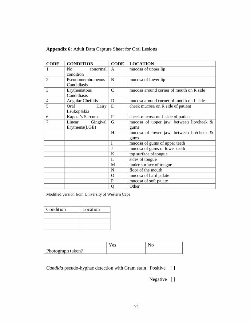
71
Appendix 6: Adult Data Capture Sheet for Oral Lesions
Modified version from University of Western Cape
Yes No Photograph taken? Candida pseudo-hyphae detection with Gram stain Positive [ ] Negative [ ]
CODE CONDITION CODE LOCATION 1 No abnormal
condition A mucosa of upper lip
2 Pseudomembraneous Candidiasis
B mucosa of lower lip
3 Erythematous Candidiasis
C mucosa around corner of mouth on R side
4 Angular Cheilitis D mucosa around corner of mouth on L side 5 Oral Hairy
Leukoplakia E cheek mucosa on R side of patient
6 Kaposi’s Sarcoma F cheek mucosa on L side of patient 7 Linear Gingival
Erythema(LGE) G mucosa of upper jaw, between lip/cheek &
gums H mucosa of lower jaw, between lip/cheek &
gums I mucosa of gums of upper teeth J mucosa of gums of lower teeth K top surface of tongue L sides of tongue M under surface of tongue N floor of the mouth O mucosa of hard palate P mucosa of soft palate Q Other
Condition Location
72
Appendix 7: Gram Staining Procedure
A thin smear was made on a clean glass slide, dried in air and fixed by passing through
flame of a burner.
1) The smear was then covered with crystal violet stain, kept for one minute.
2) The slide was washed with water, then covered with Gram iodine and left to stand
for one minute.
3) The slide was then washed with water.
4) Decolouring was done with acetone/alcohol, rocking the slide gently for 10-15
seconds till the violet colour comes off the slide.
5) Washed with water immediately.
6) Counterstaining was done with saffranin. Counter-stain stands for 30 seconds.
7) Washed with water, blotted dry and examined under the oil immersion lens of a
microscope.
73
Appendix 8: KEMRI/National Ethical Review Committee Study Approval.