oesophageal cancer - all you need to kno oesophagealcancer a4.pdf · normally lined by flat...
TRANSCRIPT
Oesophageal Cancer- All you need to know
Is it really
just Heartburn?

Introduction
The oesophagus is a hollow muscular tube that connects the mouth to thestomach. It is around 25 cm long in adults. The inside of the oesophagus isnormally lined by flat “paving slab” like cells called a stratified squamousepithelium. These sit upon a membrane that separates the lining from the musclelayers of the oesophagus.
Cancer can occur at any point along its length & the structure of theoesophaguscan contribute to cancer progression. For example, unlike the rest of thedigestive tract the oesophagus does not have an outer covering layer, whichmeans that cancer cells can spread more easily and quickly to neighbouringorgans.
There are two predominant types of oesophageal cancer: squamous cellcarcinoma and adenocarcinoma. Squamous cell carcinoma (SCC) arises fromthe normal stratified squamous lining cells. Adenocarcinoma arises in fields oflining cells that have changed shape and size due to long-term exposure tostomach and bile acids. These cells change from looking like “paving slabs”under the microscope, to looking more like piled up columns, a condition knownas Barrett’s oesophagus.
Worldwide the most common form of oesophageal cancer is squamouscell arcinoma. In many developed countries, however, the incidence ofadenocarcinoma exceeds that of SCC. This is especially true of the UK and TheNetherlands but is also true in North America, Australasia and Scandinavia.
Oesophagus(Food pipe)
Lung
Sphincter(band of
muscle
at the
bottom of
oesophagus)
Mouth
Windpipe(trachea)
Inflammationis here, where
acid refluxes
from stomach
Stomach
Diagramshowing theposition ofthe oesophagus

How common in oesophageal cancer?
UK Mortality Rates from Oesophageal Cancer• There were around 7,800 oesophageal cancer deaths in the UK in 2014, that’s 21 deaths every day
• Oesophageal cancer is the sixth most common cause of cancer death in the UK (2014)
• Oesophageal cancer accounts for 5% of all cancer deaths in the UK (2014).• In males in the UK, oesophageal cancer is the fourth most common cause of cancer death, with around 5,200 deaths in 2014
• In females in the UK, oesophageal cancer is the sixth most common cause of cancer death, with around 2,600 deaths in 2014
UK Incidence of Oesophageal Cancer• The United Kingdom has the highest incidence of oesophageal adenocarcinoma in the world: 7.2 per 100,000 in men and 2.5 per 100,000 in women
• There were around 8,900 new cases of oesophageal cancer in the UK in 2014, that’s 24 cases diagnosed every day
• Oesophageal cancer is the 14th most common cancer in the UK (2014)• Oesophageal cancer accounts for 2% of all new cases in the UK (2014)• In males in the UK, oesophageal cancer is the ninth most common cancer, with around 6,000 cases diagnosed in 2014
• In females in the UK, oesophageal cancer is the 14th most common cancer, with around 2,900 cases diagnosed in 2014
Global Incidence of Oesophageal Cancer• There were 456,000 new cases of oesophageal cancer worldwide in 2012. • The majority, 398,000, were squamous cell carcinomas (SCC)• 315000 of those cases were in Central & South-East Asia (210,000 in China alone)• 52,000 were adenocarcinomas • The worldwide incidence of oesophageal SCC is 5.2 per 100,000 but issubstantially higher in males (7.7 per 100,000) than in females (2.8 per 100,000).
• Oesophageal adenocarcinoma has a global incidence of 0.7 per 100,000.
Source: http://www.cancerresearchuk.org/health-professional/cancer-statistics/statistics-by-cancer-type/oesophageal-
cancer#heading-One
Figure 1: Age standardised incidence of oesophageal cancer.
(Adapted from Arnold M et al. 2015
What are the outcomes for people with oesophageal cancer?
Cancer of the oesophagus is the 14th most common cancer in the UK but the6th most common cause of cancer death. In England 40% of all sufferers willsurvive for one year, but by 5 years only 14% will still be alive. This is becausethe majority of patients present with incurable disease; It has either spread toofar from the oesophagus into local organs or secondary tumours (metastasis)have developed at other sites in the body (typically lymph glands, liver andlungs).
The prognosis is dependent on the stage of the cancer and whether or not itcan be cured with the best current treatments.
Less than 40% of patients can be offered curative treatment. These patientshave a 74% chance of surviving for one year after diagnosis compared to 30%if the cancer is too advanced for curative therapy.
What causes oesophageal cancer?
Squamous Cell Carcinoma
Oesophageal squamous cell carcinoma arises through chronic irritation andinflammation of the oesophageal lining. Risk factors vary between countries andcultures but in general it is a disease of poor nutrition, poor–oral hygiene andpoverty. The strongest associations are smoking and alcohol but consumptionof hot beverages, high intake of barbecued meat and human papilloma virusinfection have all been implicated.
Adenocarcinoma
Adenocarcinoma is rare globally but more common in wealthy, industrialisedwestern nations. It is most common in middle-aged, caucasian, obese males witha history of excess alcohol consumption and smoking. Male-pattern obesity (fatcarried around the waist and inside the abdomen) may be responsible forincreased abdominal pressure and therefore acid reflux (often causingheartburn), going some way to explain why adenocarcinoma is seen far morecommonly in men than women.
Adenocarcinoma of the oesophagus is strongly associated withgastro-oesophageal reflux disease (GORD) often described as Heartburn.
GORD is a common disease whereas adenocarcinoma of the oesophagus is not.GORD affects 1 in 10 adults on a daily basis and up to 2 in 10 weekly. Of these afurther 1 in 10 will have Barrett’s oesophagus, the only known precursor foradenocarcinoma, the risk of progression to cancer in this population is around1 in every 1000 patients per year.

How do our genes contribute to Oesophageal Cancer?
Heartburn Cancer UK trustee Professor Rebecca Fitzgerald has led a worldwideeffort to understand the genetic basis of oesophageal adenocarcinoma as partof the International Cancer Genome Consortium. Supported by HCUKVice-Chair Professor Tim Underwood, Prof Fitzgerald and her team havedemonstrated that adenocarcinoma of the oesophagus is a highly disorderedcancer containing many DNA mutations, Several well-known cancer causinggenes have been identified. Unfortunately the complexity of mutations inoesophageal cancer means that no new single gene target has so far beenidentified for new treatments.
However, taking a genome-wide view of oesophageal adenocarcinoma hasidentified six patterns of mutation. These “mutational signatures” give clues asto the underlying causes of the disease and go some way to explaining the hugevariations shown in response to current treatment. They also allow a broadmolecular classification of adenocarcinoma with implications for treatment.
Over 50% of these tumours carry the ‘typical’ mutational signature ofoesophageal adenocarcinoma caused by acid reflux. It is hoped that thesetumours will be amenable to new immunotherapy drugs in the future. Ourunderstanding of the molecular biology of oesophageal cancer is rapidlyprogressing and HCUK trustees are at the forefront of these efforts.
Norman Barrett first described Barrett’s oesophagus in 1950. It is caused bylong-term exposure to stomach acid, bile acids and pancreatic enzymes. Overtime Barretts cells can sometimes change in structure becoming moredisordered, a finding called dysplasia. Doctors classify dysplasia as low gradeor high grade depending on how disorganised the cells appear under amicroscope. If a patient has an upper GI endoscopy (a camera test to view theinside of the oesophagus and stomach) and high-grade dysplasia is found orlow-grade dysplasia is present on two endoscopies 6 months apart doctors willconsider using endoscopic therapies to remove all of the Barrett’s segment andprevent progression to cancer.
It is important to remember that the majority of patients with Barrett’soesophagus will never develop oesophageal cancer.
Figure 2: An internal view of the oesophagus showing an adenocarcinoma
(highlighted yellow) in a field of Barretts oesophagus (highlighted red).

How is oesophageal cancer diagnosed?
The typical symptoms of oesophageal cancer are of difficulty swallowing (foodgetting stuck), at first solids and then sometimes liquids. There may be weightloss and anaemia (a low red blood cell count). Whilst a number of otherconditions can lead to difficulty swallowing, anyone who has this symptomshould see their doctor.
The first test performed is usually an endoscopy (a camera test to view the insideof the oesophagus and stomach) with biopsies (small samples of the lining arepainlessly removed and examined under a microscope).
The majority of patients are diagnosed with oesophageal cancer following areferral from their general practitioner (GP) or another hospital doctor (85%). Avery small number (less than 1%) are identified because they are known tohave Barrett’s oesophagus and undergo regular surveillance endoscopy. Theremaining 14% present as an emergency.
If oesophageal cancer is found how far has it spread?
Once a diagnosis of oesophageal cancer has been made it is important for thepatient and doctors to understand how far the cancer has spread. This allowsaccurate treatment planning and gives an idea of the likely prognosis. A seriesof tests will be performed that are referred to as “staging investigations”.
The first staging investigation will usually be a CT scan of the chest, abdomenand pelvis. The patient lies flat on a bench while they are slowly moved througha large donut shaped x-ray machine. At the same time a dye is injected into avein to help improve the detail of the x-ray pictures. This is often performedstraight after the endoscopy when the cancer is first seen.
The purpose of a CT is to assess the size and spread of the primary cancer andidentify any secondary (metastatic) disease. In those patients where the cancerhas not spread on CT and possible curative treatment is proposed then furtherimaging in the form of Positron Emission Tomography (PET)-CT and EndoscopicUltrasound (EUS) may be performed.
A PET CT is similar to a CT scan but involves a special dye that will show upbrightly on the scan pictures wherever cells in the body are working hard andusing a lot of energy. Cancer cells tend to use a lot of energy and thereforeappear as bright areas on these images.

If oesophageal cancer is found how far has it spread?
Once a diagnosis of oesophageal cancer has been made it is important for thepatient and doctors to understand how far the cancer has spread. This allowsaccurate treatment planning and gives an idea of the likely prognosis. A seriesof tests will be performed that are referred to as “staging investigations”.
The first staging investigation will usually be a CT scan of the chest, abdomenand pelvis. The patient lies flat on a bench while they are slowly moved througha large donut shaped x-ray machine. At the same time a dye is injected into avein to help improve the detail of the x-ray pictures. This is often performedstraight after the endoscopy when the cancer is first seen.
The purpose of a CT is to assess the size and spread of the primary cancer andidentify any secondary (metastatic) disease. In those patients where the cancerhas not spread on CT and possible curative treatment is proposed then furtherimaging in the form of Positron Emission Tomography (PET)-CT and EndoscopicUltrasound (EUS) may be performed.
A PET CT is similar to a CT scan but involves a special dye that will show upbrightly on the scan pictures wherever cells in the body are working hard andusing a lot of energy. Cancer cells tend to use a lot of energy and therefore appear as bright areas on these images.
An EUS is very similar to an endoscopy except the scope that the doctor useswill have a small ultrasound probe on the end which will help to assess the depthof any tumours in the wall of the oesophagus.
In some patients a small operation called a staging laparoscopy will beperformed. This allows doctors to examine the inside of the abdomen to checkfor cancer spread not seen on the scans and to plan surgery.
Figure 3 High Resolution Contrast Enhanced CT scan (Left) and FDG PET CT
showing a thick walled oesophagus and corresponding high metabolic activity).
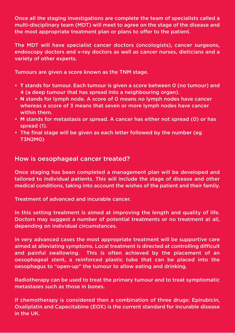
Once all the staging investigations are complete the team of specialists called amulti-disciplinary team (MDT) will meet to agree on the stage of the disease andthe most appropriate treatment plan or plans to offer to the patient.
The MDT will have specialist cancer doctors (oncologists), cancer surgeons,endoscopy doctors and x-ray doctors as well as cancer nurses, dieticians and avariety of other experts.
Tumours are given a score known as the TNM stage.
• T stands for tumour. Each tumour is given a score between 0 (no tumour) and4 (a deep tumour that has spread into a neighbouring organ).
• N stands for lymph node. A score of 0 means no lymph nodes have cancer whereas a score of 3 means that seven or more lymph nodes have cancer within them.
• M stands for metastasis or spread. A cancer has either not spread (0) or has spread (1).
• The final stage will be given as each letter followed by the number (eg T3N2M0)
How is oesophageal cancer treated?
Once staging has been completed a management plan will be developed andtailored to individual patients. This will include the stage of disease and othermedical conditions, taking into account the wishes of the patient and their family.
Treatment of advanced and incurable cancer.
In this setting treatment is aimed at improving the length and quality of life.Doctors may suggest a number of potential treatments or no treatment at all,depending on individual circumstances.
In very advanced cases the most appropriate treatment will be supportive careaimed at alleviating symptoms. Local treatment is directed at controlling difficultand painful swallowing. This is often achieved by the placement of anoesophageal stent, a reinforced plastic tube that can be placed into theoesophagus to “open-up” the tumour to allow eating and drinking.
Radiotherapy can be used to treat the primary tumour and to treat symptomaticmetastases such as those in bones.
If chemotherapy is considered then a combination of three drugs: Epirubicin,Oxaliplatin and Capecitabine (EOX) is the current standard for incurable diseasein the UK.

In a small number of cases the cancer will be suitable for treatment with
Trastuzumab (Herceptin), best known for its role in breast cancer treatment.
Oesophageal tumours are now routinely tested to see whether or not Herceptin
is likely to work.
A range of new treatments, including immunotherapies (treatments that enhance
the body’s own ability to kill cancer), are being tested in clinical trials in the UK
and elsewhere, but these are yet to become standard treatments.
Treatment of curable cancer
If oesophageal cancer is diagnosed early it can be cured. For the earliest stage
cancers the tumour can be removed endoscopically (from the inside of the
oesophagus in a similar way to the camera test used for diagnosis) without the
need for major surgery or other treatments. If any Barrett’s oesophagus is
present this will also be removed completely. More than one endoscopic
treatment may be required.
Squamous cell cancer of the oesophagus often responds well to a combination
of chemotherapy and radiotherapy.
Patients whose tumours have advanced beyond the oesophageal lining (mucosa)
and/or have evidence of cancer cells in local lymph glands, and are fit enough
to undergo aggressive treatment are considered for pre-operative
(neo-adjuvant) therapies followed by surgery. A number of large clinical trials
conducted in the UK, Europe and the USA have proven that this strategy of
giving anti-cancer treatment before surgery offers the best chance of long-term
survival and cure. Uncertainty remains regarding whether this pre-operative
treatment should be chemotherapy alone or chemotherapy and radiotherapy
together. A current clinical trial called NeoAegis is running in the UK and Ireland
and aims to answer this question.
Unfortunately, the survival benefit of neoadjuvant therapy in oesophageal cancer
is limited to only about 20% of patients, at the expense of the many who derive
no benefit at all, and who may be harmed by over-treatment. Current research
being conducted by Prof Underwood and a large UK team is striving to predict
who these patients will be before and during therapy in order to spare patients
unnecessary treatments.
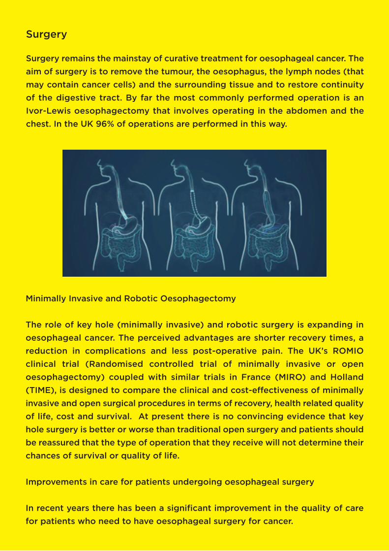
Surgery
Surgery remains the mainstay of curative treatment for oesophageal cancer. The
aim of surgery is to remove the tumour, the oesophagus, the lymph nodes (that
may contain cancer cells) and the surrounding tissue and to restore continuity
of the digestive tract. By far the most commonly performed operation is an
Ivor-Lewis oesophagectomy that involves operating in the abdomen and the
chest. In the UK 96% of operations are performed in this way.
Minimally Invasive and Robotic Oesophagectomy
The role of key hole (minimally invasive) and robotic surgery is expanding in
oesophageal cancer. The perceived advantages are shorter recovery times, a
reduction in complications and less post-operative pain. The UK’s ROMIO
clinical trial (Randomised controlled trial of minimally invasive or open
oesophagectomy) coupled with similar trials in France (MIRO) and Holland
(TIME), is designed to compare the clinical and cost-effectiveness of minimally
invasive and open surgical procedures in terms of recovery, health related quality
of life, cost and survival. At present there is no convincing evidence that key
hole surgery is better or worse than traditional open surgery and patients should
be reassured that the type of operation that they receive will not determine their
chances of survival or quality of life.
Improvements in care for patients undergoing oesophageal surgery
In recent years there has been a significant improvement in the quality of care
for patients who need to have oesophageal surgery for cancer.

Historically oesophagectomy was a high-risk procedure with a 10% (1 in 10)
chance of death in hospital after surgery. In the UK this risk has fallen
dramatically, to less than 2% (1 in 50), and continues to improve.
One of the ways this has been achieved is
through the implementation of Enhanced
recovery.
Enhanced Recovery
Enhanced recovery is a pathway that thespecialist team and the patient followtogether.
Before the operation patients, theirfamilies and carers are taught about thesurgery and what to expect. They arealso told what will be expected of themand what steps they can take before andafter surgery to improve and optimisetheir recovery. Usually this involvesincreasing their activity levels before theoperation (like an athlete training for abig race) and starting to walk again as soon as possible after the operation.Patients are given dietary advice and drink high calorie liquids on the day ofsurgery.
During the operation the surgical team and the anaesthetist work togethertaking steps to minimise the impact of the surgery and to make sure that painis well controlled.
After the operation there will be many drains, catheters and tubes attached tothe patient that will be removed as soon as possible by the medical team.Patients will start to drink as soon as it is safe to do so and will start a pureediet soon afterwards with guidance from the dieticians. Patients will begiven a mobility goal for each day and help from specialist nurses andphysiotherapists to achieve that goal.
Although enhanced recovery and similar projects are continune to improve theexperience of cancer sufferers oesophagectomy remains a major procedureand the consequences may continue to affect the lives of patients long afterthey have left the hospital. Patients must learn a new way of eating meals inorder to avoid problems such as malnutrition. Complications do occur and itcan take many weeks, months and sometimes years to recover.

Future Developments
Despite improving outcomes oesophageal cancer still carries a dismal prognosis.Heartburn Cancer UK has identified oesophageal cancer as a cancer of unmetneed and a research priority. We are supported in this view by Cancer ResearchUK and others.
In July 2017 a group of charities launched the Less Survivable Cancers Taskforce,a collaboration designed to highlight the need for further research and fundingfor the six deadliest cancers including oesophageal cancer.
Immunotherapy
One of the reasons cancer is so hard to treat is that the body struggles torecognise cancer cells and kill them. Immunotherapy is a relatively new approachto treating cancer. Medication is designed to improve the body’s own ability tofind and kill cancer cells.
Cancer cells can often have high numbers of the same molecule on their surface.Immunotherapy drugs called monoclonal antibodies can bind to these moleculesand allow the body’s immune system to recognise them as cancer. Severalimmunotherapy drugs are being trialled in oesophageal cancer.
Unfortunately not all immunotherapies work for all cancers. Researchers aredeveloping tests that will enable doctors to know which drugs will help destroywhich tumours.
In the past, studies have treated all oesophageal cancer as though it is onedisease. It is, however, becoming increasingly clear that oesophageal cancer isfar more complex than that. Some small groups of patients do much better thanother groups using standard therapy.
The next generation of clinical trials will involve stratification of patientsaccording to the molecular features of their tumours and their predictedresponse to therapy.
Immunotherapy is likely to transform the way we think about oesophagealcancer. The increase in survival and reduction in side effects means there is agreat deal of optimism and hope for patients, relatives, carers and those involvedin treating oesophageal cancer. Research funded and supported by charitiessuch as Heartburn Cancer UK are developing a new wave of intelligent,precision-guided treatments in the fight against cancer.
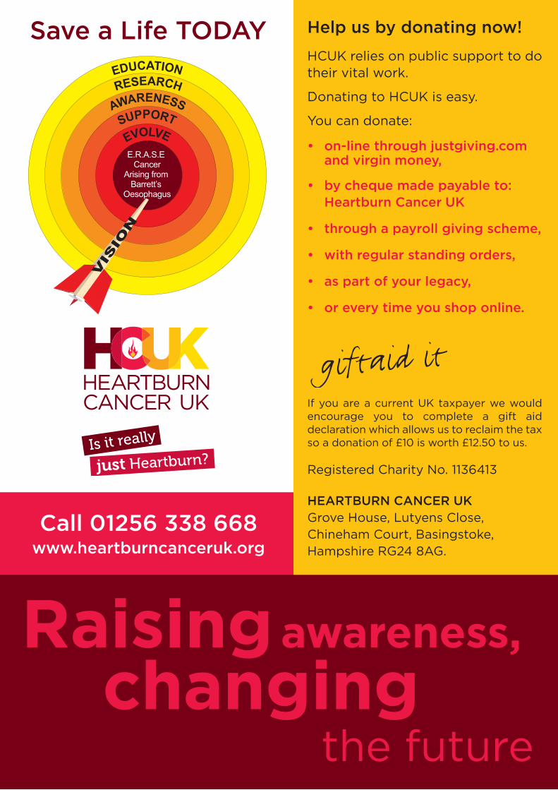
Help us by donating now!
HCUK relies on public support to dotheir vital work.
Donating to HCUK is easy.
You can donate:
• on-line through justgiving.comand virgin money,
• by cheque made payable to: Heartburn Cancer UK
• through a payroll giving scheme,
• with regular standing orders,
• as part of your legacy,
• or every time you shop online.
If you are a current UK taxpayer we wouldencourage you to complete a gift aiddeclaration which allows us to reclaim the taxso a donation of £10 is worth £12.50 to us.
Registered Charity No. 1136413
HEARTBURN CANCER UKGrove House, Lutyens Close,Chineham Court, Basingstoke,Hampshire RG24 8AG.
Call 01256 338 668www.heartburncanceruk.org
Save a Life TODAY
EDUCATION
Reduce fr
om 8
0% to
75%
by
2021
• Le
ad Communication in OC • Provided or consistant/relevant support Fund
Eradica
te C
ance
r Aris
ing f
rom Barrett’s Oesophagus
Lead
Prio
rity
in th
e field of Cancer of the O
esophagealA1 S
top
Shop
for Support and Education
AWARENESS
SUPPORTEVOLVEE.R.A.S.E
CancerArising from
Barrett’s Oesophagus
RESEARCH
Is it really
just Heartburn?
Raisingawareness,
changingthe future