of 4, pp. 1738-1745, 19fw ij purification and some ... · mitochondrial matrix in eukaryotic cells...
TRANSCRIPT
THE JOURNAI. OF BIOLOGICAL CHEMISTRY Vol. 255, No. 4, Issue of February 25, pp. 1738-1745, 19fW Printed m I J S A .
Purification and Some Properties of 6-Aminolevulinate Synthase from the Rat Liver Cytosol Fraction and Immunochemical Identity of the Cytosolic Enzyme and the Mitochondrial Enzyme*
(Received for publication, July 11, 1979)
Masaaki Nakakuki,$ Kohei Yamauchi, Norio Hayashi, and Goro Kikuchig From the Department of Biochemistry, Tohoku University School of Medicine, Sendai 980, Japan
6-Aminolevulinate synthase was purified to near ho- mogeneity from the liver cytosol fraction of rat which had been treated with allylisopropylacetamide and in- sulin. 6-Aminolevulinate synthase was freed from cat- alytically inactive interacting proteins by treatment with papain and was further purified by column chro- matographies. The finally obtained 8-aminolevulinate synthase preparation catalyzed the formation of 73,000 nmol of 8-aminolevulinate/mg of protein/h at 37°C. The purified preparation gave a molecular weight of about 110,000 when estimated by sucrose density gra- dient centrifugation. The enzyme was also shown to be in a dimer form of two identical subunits with a mini- mum molecular weight of 51,000. When estimated by gel filtration, however, the enzyme preparation showed a molecular weight of 70,000, possibly reflecting a par- tial dissociation-association of the dimer form of the enzyme occurring during gel filtration. The purified S- aminolevulinate synthase had an isoelectric point of 8.2, and pH optimum for the reaction was 7.6. These properties were quite similar to those of S-aminolevu- linate synthase preparation partially purified from the rat liver cytosol fraction without applying papain treat- ment. The K,,, values for succinyl-CoA of both enzyme preparations were also very close to each other (11 p ~ ) . However, the K , for glycine (as determined in the presence of 10 mM ATP) in the reaction with the papain- treated enzyme was 7.5 m, whereas the K, value obtained with the papain-untreated enzyme was 2.5 mM.
A specific rabbit antibody against the purified cyto- solic 6-aminolevulinate synthase was prepared. Exper- iments using the antibody revealed that both S-amino- levulinate synthases in the cytosol and the mitochon- dria of rat liver are immunochemically identical, pro- viding further support to the view that S-aminolevuh- nate synthase accumulating in the cytosol fraction is a precursor in transit to the mitochondria.
6-Aminolevulinate synthase, the fist and rate-limiting en- zyme in the heme biosynthesis ( l ) , is usually located in the
* This work was supported in part by Grants 348117 and 358059 from the Ministry of Education, Science and Culture, Japan, by the Yamanouchi Foundation for Metabolic Studies, Japan, and by the Foundation for the Promotion of Research on Medicinal Resources, Japan. The costs of publication of this article were defrayed in part by the payment of page charges. This article must therefore be hereby marked “advertisement” in accordance with 18 U.S.C. Section 1734 solely to indicate this fact. + Present address, Dept. of Gynecology and Obstetrics, Tohoku University School of Medicine, Sendai 980, Japan.
3 To whom correspondence should be addressed.
mitochondrial matrix in eukaryotic cells (2, 3). The level of 6-aminolevulinate synthase in the liver mitochondria can be increased markedly by the administration of porphyrinogenic drugs such as allylisopropylacetamide and 3,5-dicarbethoxy- 1,4-dihydrocollidine to animals, and under these conditions a considerable amount of 6-aminolevulinate synthase also ac- cumulates in the liver cytosol fraction of rat (4-7), mouse (B), guinea pig (7), and chicken (9). The extent of accumulation of baminolevulinate synthase in the cytosol, however, is consid- erably variable according to the species of animals and drugs. For instance, the degree of the enzyme accumulation in the cytosol is smaller in general in DDC’-treated animals as compared with that in AIA-treated animals (8, lo), and in the chick embryo liver, 6-aminolevulinate synthase does not ap- preciably accumulate in the cytosol fraction even after exten- sive induction with either AIA or DDC (11, 12).
The 6-aminolevulinate synthase accumulating in the liver cytosol fraction has been supposed to be a precursor in transit to the mitochondria. Moreover, the intracellular transfer of 8-aminolevulinate synthase from the liver cytosol to the mi- tochondria in the AIA-treated animals appeared to be signif- icantly inhibited by the administration of hemin (9, 13), cobalt protoporphyrin (14), insulin, cyclic AMP, and other sub- stances (15). A similar but relatively slight inhibitory effect of hemin could also be observed in DDC-treated mouse (8). These observations suggest that a unique regulatory mecha- nism acts on the intracellular translocation of S-aminolevuli- nate synthase.
To investigate the suspected intracellular enzyme transfer and its regulation, it is especially useful to have an antibody specific to 6-aminolevulinate synthase. There have been sev- eral studies on this line (7, 16, 17); particularly, Whiting and Elliott (7) prepared a 40-fold purified 6-aminolevulinate syn- thase from rat liver mitochondria and indicated that antibod- ies prepared against this partially purified enzyme preparation inactivated the 6-aminolevulinate synthase in the rat liver cytosol. However, extensive purification of 6-aminolevulinate synthase, which is essential for the preparation of antibody highly specific to 6-aminolevulinate synthase, has been diffi- cult mainly because of its aggregating property; although the native 6-aminolevulinate synthase has a molecular weight of about 110,000 as will be seen later, crude or partially purified 6-aminolevulinate synthase preparations prepared from either the liver cytosol or the mitochondria form aggregates or complexes especially at lower salt concentrations and exhibit quite heterogeneous molecular sizes (70,000 to as large as 650,000 when estimated by gel filtration) according to the
’ The abbreviations used are: DDC, 3,5-dicarbethoxy-l,4-dihydro- collidine; ALA, 6-aminolevulinic acid; AIA, allylisopropylacetamide; SDS, sodium dodecyl sulfate; Hepes, 4-(2-hydroxyethyl)-l-piperazine- ethanesulfonic acid.
1738
by guest on September 9, 2019
http://ww
w.jbc.org/
Dow
nloaded from

6-Aminolevulinate Synthase of Rat Liver 1739
experimental conditions (7, 16, 18, 19). Ohashi and Kikuchi (20) purified about 100-fold a complex form of S-aminolevuli- nate synthase, which originally showed an apparent molecular weight of 320,000 on gel fitration, from rat liver cytosol and indicated that the preparation was composed from three dif- ferent proteins, one catalytically active protein and two cata- lytically inactive proteins. They also showed that the complex form of cytosol 6-aminolevulinate synthase could be dissoci- ated into individual protein components by sucrose density gradient centrifugation or by treatment with papain, and they further purified the enzyme-active protein component by re- peating sucrose density gradient centrifugation (20). On su- crose density gradient centrifugation, the “stripped” enzyme- active protein that is freed from other intreating proteins gave a molecular weight of about 11O,OOO, while a molecular weight of 51,000 was obtained when the protein was subjected to sodium dodecyl sulfate-polyacrylamide gel electrophoresis, suggesting that the stripped 6-aminolevulinate synthase is a dimer of two identical subunits with a molecular weight of 51,000. The apparent molecular weights of the other two catalytically inactive proteins in the complex form of cytosol 8-aminolevulinate synthase were estimated to be 120,000 and 79,000, respectively, by SDS-polyacrylamide gel electropho- resis (20).
However, the yield as well as the purity of the stripped S- aminolevulinate synthase preparation obtained by repeated sucrose density gradient centrifugation were still unsatisfac- tory. In the present study we employed the papain treatment to obtain the cytosolic 8-aminolevulinate synthase freed from other interacting proteins and purified the enzyme-active pro- tein to near homogeneity. Using the highly purified enzyme preparation, we could obtain a rabbit antibody specific to S- aminolevulinate synthase and could demonstrate the immu- nochemical identity of the cytosol S-aminolevulinate synthase with the mitochondrial 8-aminolevulinate synthase.
EXPERIMENTAL PROCEDURES
Materials-AIA was supplied from Nippon Roche Co., Tokyo. Insulin was purchased from Novo Industry, Copenhagen; papain and marker proteins for the molecular weight calibration, from Boehrin- ger, Mannheim; ATP, dithiothreitol, and 4-(2-hydroxyethyl)-l-piper- azineethanesulfonic acid (Hepes), from Sigma Chemical Co., St. Louis; protamine sulfate, from ICN Pharmaceuticals Inc., Cleveland; pyri- doxal phosphate and coenzyme A, from Kyowa Hakko Kogyo Co., Tokyo; dimethyl suberimidate, from Eastman Kodak Co., Rochester; hydroxyapatite, from Nippon Chemical Co., Tokyo; phosphocellulose (P-l l ) , from Whatman Ltd., Springfield Mill; Sephacryl S-200, from Pharmacia Fine Chemicals, Uppsala. Antipain was a gift from Dr. H. Umezawa. Succinyl-CoA synthase was prepared from Escherichia coli as described previously (9). Succinyl-CoA was prepared by a modification of the method of Simon and Shemin (21) according to Zaman et al. (22). Other chemicals were obtained commercially. Rats used in this study were female Std: Wistar strain obtained from Shizuoka Agricultural Cooperative Association for Laboratory Ani- mals, Hamamatsu.
Conditioning ofRats-Rats weighing about 150 g were fasted for 24 h; then AIA (30 mg/100 g of body weight) was injected to animals subcutaneously and, 12 h later, the same dose of AIA was again injected. Three hours after the second AIA administration, insulin (0.2 unit/100 g of body weight) was injected intraperitoneally to bring about a greater accumulation of 6-aminolevulinate synthase in the liver cytosol fraction (cf. Ref. 15). All rats were killed 3 h after the injection of insulin.
Assay of 6-Aminoleuulinate Synthase-6-Aminolevulinate syn- thase was assayed in either System A or System B. System A was used for routine activity measurement during purification. In kinetic experiments the enzyme activity was determined in System B. In System A the reaction mixture contained, in a final volume of 1 ml: 50 pmol of Tris-HCI buffer (pH 7.4), 100 pmol of glycine, 10 pmol of sodium succinate, 10pmol of ATP (neutralized), 0.1 pmol ofpyridoxal- P, 0.1 pmol of CoA, 5 pmol of MgCl,, 5 pmol of EDTA, succinyl-CoA synthase (catalyzing the formation of 5 pmol of succinyl hydroxamate/
h a t 37’C), and an appropriate amount of 6-aminolevulinate synthase. The incubation was carried out in a test tube placed in a shaking water bath incubator at 37°C for 15 or 30 min. The reaction was stopped by adding 0.25 ml of 12.5% trichloroacetic acid. In System B, chemically synthesized succinyl-CoA was employed in place of the succinyl-CoA generating system. System B was supplemented with 10 mM ATP since succinyl-CoA had been known to exert a significant substrate inhibition to rat liver 6-aminolevulinate synthase in the absence of ATP (23). Detailed reaction conditions will be given in the legends to respective figures. 6-Aminolevulinate formed was estimated by a modification of the original method of Mauzerall and Granick (24) as described by Narisawa and Kikuchi (25). One unit of 6- aminolevulinate synthase was defined as the amount of the enzyme catalyzing the formation of 1 nmol of 6-aminolevulinate a t 37°C.
SDS-Polyacrylamide Gel Electrophoresis-SDS-polyacrylamide gel electrophoresis was performed by the method of Weber and Osborn (26) using a 7.5% gel and a current of 6 mA/gel. Gels were stained with Coomassie blue (27).
Sucrose Density Gradient Centrifugation-Centrifugation was performed in a Hitachi RPS 27 swinging bucket rotor a t 27,000 rpm for 36 h in a linear gradient of 5 to 20% sucrose containing 0.4 M NaC1, 0.1 mM pyridoxal-P, 1 m~ dithiothreitol, and 50 mM Tris-HCI buffer (pH 7.2) (the buffer containing the indicated concentrations of NaCI, pyridoxal-P, dithiothreitol, and Tris-HC1 will be referred to as NaTP buffer). Each tube was filled with 35 ml of the gradient and 1.0 ml of the sample which also contained marker proteins was layered on the top. After centrifugation, 2-ml fractions were collected from the top using a Hitachi Gradienter. Molecular weight as well as s value of the enzyme were calculated by the method of Martin and Ames (28).
Isoelectrofocusing-This was performed using a LKB 8100 electro- focusing column (110 ml), according to the Instruction Manual pro- vided by LKB Produkter AB, Bromma. Detailed procedures are given in the legend to Fig. 5.
Preparation of Antibody-6-Aminolevulinate synthase which was purified to near homogeneity (2 mg; specific activity, 73,000 units/mg of protein; see “Results”) was used to immunize two white rabbits (2.5 kg) by the standard procedure (29). Antiserum was collected by bleeding from the ear at 7 days after the second injection of the antigen and used as such in the present study.
Determination of Protein-Protein was estimated by the Lowry method (30) modified by Bensadoun and Weinstein (31) with bovine serum albumin as standard.
RESULTS
Purification of 6-Aminolevulinate Synthase from Rat Liver Cytosol
Preparation of the Cytosol Fraction-One hundred rats were treated with AIA and insulin as described under “Ex- perimental Procedures” and livers were taken and homoge- nized in 2 volumes of 0.01 M potassium phosphate buffer (pH 7.0) containing 0.15 M NaC1,O.l mM pyridoxal-P, 1 mM EDTA, and 1 mM dithiothreitol. The homogenate was centrifuged at 20,000 X g for 20 min, then at 77,000 X g for 120 min to obtain the cytosol fraction.
Ammonium Sulfate Fractionation-Solid sucrose was dis- solved in the cytosol fraction to give a final concentration of 0.75 M: then to this fraction was added a neutralized protamine sulfate solution (50 mg/ml) until the protamine/protein ratio reached 1:20 and insoluble materials formed were removed by centrifugation. Solid ammonium sulfate was added to the supernatant up to 0.35 saturation. Precipitates obtained were dissolved in NaTP buffer to give a final volume of 50 ml (ammonium sulfate fraction).
First Sephacryl Column Chromatography-The two halves of the ammonium sulfate fraction (25 ml each) were loaded separately onto Sephacryl S-200 columns (5 X 100 cm) and eluted with NaTP buffer at a flow rate of 72 ml/h. The 6- aminolevulinate synthase activity was eluted, yielding a single activity peak at the position corresponding to a molecular weight of 240,000, indicating that the cytosol S-aminolevuli- nate synthase remained in a complex form at this step. The
by guest on September 9, 2019
http://ww
w.jbc.org/
Dow
nloaded from
1740 6-Aminolevulinate Synthase of Rat Liver
enzyme-active fractions from the two Sephacryl columns were combined (first Sephacryl fraction).
Papain Treatment and Second Sephacryl Column Chro- matography-Solid ammonium sulfate was added to the First Sephacryl fraction up to 0.5 saturation and precipitates ob- tained were dissolved in 50 mM Tris-HC1 buffer (pH 7.2) containing 100 lll~ glycine, 50 mM sodium succinate, and 0.1 mM pyridoxal-P, to give a final volume of 20 ml. The enzyme solution was dialyzed against the same buffer for 2 h and then added with pyridoxal-P to a final concentration of 10 mM. To this mixture was added papain ( ? h of the protein amount of the first Sephacryl fraction) which had been activated by incubation in a 50 mM Tris-HC1 buffer (pH 7.2) containing 0.1 mM pyridoxal-P, 33 mM cystein, and 33 lll~ EDTA for 5 min at 37”C, and the mixture was incubated at 37°C for 15 min. The reaction was stopped by the addition of antipain (an amount twice that of papain). The presence of indicated concentrations of glycine, sodium succinate, and pyridoxal-P prevented almost completely the inactivation of S-amino- levulinate synthase by the papain treatment; without those additions the S-aminolevulinate synthase activity was lost by nearly 30% during the 15-min incubation with papain. The papain-treated enzyme fraction (about 30 ml) then was ap- plied onto a Sephacryl column (5 X 100 cm) and eluted under the same conditions as for the first Sephacryl column chro- matography. S-Aminolevulinate synthase was eluted showing a single sharp activity peak at the position corresponding to a molecular weight of 70,000, indicating that the active S- aminolevulinate synthase protein was free from any other interacting protein after the papain treatment.
Hydroxyapatite Column Chromatography-The enzyme active fractions after the second Sephacryl column chroma- tography were pooled (second Sephacryl fraction) and diluted 1.6-fold with distilled water, then applied to a hydroxyapatite column (2.6 X 6 cm) which had been equilibrated with 50 mM potassium phosphate buffer (pH 7.0) containing 0.1 m~ pyri- doxal-P and 1 mM dithiothreitol. The column was washed successively with 2 volumes each of the following three buffers at a flow rate of 35 ml/h: Buffer A, 100 m~ potassium phosphate buffer, pH 7.0, containing 0.1 lll~ pyridoxal-P and 1 mM dithiothreitol; Buffer B, 165 mM potassium phosphate buffer, pH 7.0, containing 0.1 m~ pyridoxal-P and 1 mM dithiothreitol; and Buffer C, 300 m~ potassium phosphate buffer, pH 7.0, containing 0.1 m~ pyridoxal-P and 1 mM dithiothreitol. Almost all of the 6-aminolevulinate synthase activity was recovered in the fraction eluted with Buffer C (hydroxyapatite fraction).
Phosphocellulose Column Chromatography-Phosphocel- lulose (4 to 5 g of dry weight), which was equilibrated with 10 mM potassium phosphate buffer (pH 7.0) containing 0.1 lll~ pyridoxal-P and 1 m~ dithiothreitol, was mixed with the hydroxyapatite fraction and the slurry was diluted with dis- tilled water under monitoring with a conductivity meter until a value of 11 to 12 mmho was obtained; this value corresponds to about 100 mM in buffer concentration. The diluted slurry
was stirred for a further 15 min, and then phosphocellulose was collected by centrifugation and resuspended in Buffer A designated above. The phosphocellulose suspension then was packed in a column (2.6 cm in diameter) and was washed successively with 1.5 volumes each of Buffer A, Buffer B’ (175 mM potassium phosphate buffer, pH 7.0, containing 0.1 mM pyridoxal-P and 1 m~ dithiothreitol) and Buffer C’ (400 m~ potassium phosphate buffer, pH 7.0, containing 0.1 mM pyri- doxal-P and 1 m~ dithiothreitol) at a flow rate of 30 ml/h. The S-aminolevulinate synthase activity was eluted mostly with Buffer C’ (phosphocellulose fraction), although about one-fourth of the total activity recovered was also eluted with Buffer B’. The final enzyme preparation had a specific activity of 73,000 units/mg of protein and could be stored at -20°C without appreciable loss of the activity.
A summary of the purification is presented in Table I.
Properties of the Purified S-Aminoleuulinate Synthase Preparation and Comparison with Those of the Papain-
untreated S-Aminoleuulinate Synthase Purity and Stability-The punty of the final enzyme prep-
aration was assessed by SDS-polyacrylamide gel electropho- resis (Fig. 1). The preparation gave one distinct band, but traces of a few minor bands were also visible on the gel, although these minor bands could hardly be seen in the photograph shown in Fig. 1. The purity of this preparation was estimated to be more than 95% from the tracing with a densitometer of the stained gel. The purified S-aminolevuli- nate synthase was stable when kept in 400 mM potassium phosphate buffer (pH 7.0) containing 0.1 m~ pyridoxal-P and 1 mM dithiothreitol at a protein concentration of more than 100 pg/ml. However, the enzyme lost the activity fairly rapidly when it was kept in buffers of lower concentrations or at lower protein concentrations.
Molecular Weight and Subunit Composition-Sucrose density gradient centrifugation of the purified enzyme prepa- ration gave a symmetrical sedimentation pattern of S-amino- levulinate synthase (Fig. 2), and a s value of 6.5 S and a molecular weight of about 110,000 were estimated for S-ami- nolevulinate synthase. However, when estimated by gel fiitra- tion on a column of Sephacryl S-200 (2.6 x 42 cm) with NaTP buffer, a molecular weight of 70,000 was obtained for S-ami- nolevulinate synthase (Fig. 3 ) .
The purified S-aminolevulinate synthase gave a minimum molecular weight of 51,000 when subjected to SDS-polyacryl- amide gel electrophoresis, using bovine serum albumin, cata- lase, ovalbumin, yeast lactate dehydrogenase, chymotrypsin, and myoglobin as marker proteins. Then, cross-linking of S - aminolevulinate synthase by dimethyl suberimidate was at- tempted according to the method of Davies and Stark (32). The final concentration of dimethyl suberimidate was 10 mg/ ml and that of the protein was 100 pg/ml. The reaction was allowed to proceed at pH 8.5 for 3 h at 20°C. For electropho- resis in SDS, the sample was heated at 100°C for 3 min in a 10 mM sodium phosphate buffer (pH 7.0) containing 1% (w/v)
TABLE I Purification of 8-aminolevulinate synthase from liver cytosol of AZA-treated rat
Fraction Volume Total protein Total activitv Specific activity ml mg units units/mg
Crude cytosol fraction 584 23,040 474,600 20.6 Ammonium sulfate fractionation (0-35%) 50 2,600 384,900 148.0 First Sephacryl column chromatography 285 606 368,200 607.6 Second Sephacryl column chromatogra- 125 55.7 270,300 4,853
Hydroxyapatite column chromatography 25 11.9 196,700 16,530 Phosphocellulose column chromatogra- 21 1.67 122,100 73,100
phy (papain treatment)
phy
Yield R
100 81.1 77.6 57.0
41.4 25.7
by guest on September 9, 2019
http://ww
w.jbc.org/
Dow
nloaded from

8-Aminolevulinate Synthase of Rat Liver 1741
- 1 Elution volume (ml ) FIG. 3. Estimation of the molecular weight of purified 8-
aminolevulinate synthase by Sephacryl column chromatog- raphy. Two milliliters of the purified enzyme were loaded onto a column (2.6 X 42 cm) with five marker proteins and eluted a t a flow rate of 12 ml/h. Other conditions are described under “Results.” The marker proteins are horse myoglobin ( M h ) , ovalbumin (OVA). pig heart malate dehydrogenase ( M D H ) , yeast alcohol dehydrogenase (ADH) . and bovine liver catalase.
c “ 4 + -I FIG. 1. SDS-polyacrylamide gel electrophoresis of the puri- fied 8-aminolevulinate synthase. The gel was loaded with 10 pg of protein. Other conditions are described under “Experimental Proce- dures.”
-. ’. 3- FIG. 4. SDS-polyacrylamide gel electrophoresis of purified
Gaminolevulinate synthase after treatment with dimethyl suberimidate. The gel was loaded with 15 pg of protein. Other conditions are described under “Experimental Procedures” and “He- sults.”
TOP Fraction number
Bottom
FIG. 2. Sucrose density gradient centrifugation of purified 8-aminolevulinate synthase. Details are described under “Experi- mental Procedures” and “Results.” Yeast alcohol dehydrogenase (ADH) was used as a marker protein.
SDS and 1% (w/v) 2-mercaptoethanol, followed by overnight dialysis against 10 mM sodium phosphate buffer containing 0.1% SDS a t room temperature. The sample was electropho- resed and stained as described under “Experimental Proce- dures.” As shown in Fig. 4, there were two distinct bands on
the gel, one at the position corresponding to a molecular weight of 51,000 and the other at the position equivalent to twice the subunit molecular weight. The latter band should represent the 6-aminolevulinate synthase in which the sub- units are cross-linked by dimethyl suberimidate. These results confrm that the catalytically active protein of the cytosol 6-
by guest on September 9, 2019
http://ww
w.jbc.org/
Dow
nloaded from

1742 6-Aminolevulinate SJ
aminolevulinate synthase consists of two identical subunits with a subunit molecular weight of 51,000.
For comparison, the stripped form of 6-aminolevulinate synthase was prepared from the liver cytosol fraction without employing papain treatment. Namely, the 6-aminolevulinate synthase preparation after the fist Sephacryl column chro- matography was subjected to sucrose density gradient cen- trifugation. The s value and the molecular weight of the catalytically active protein were estimated to be 6.5 S and 115,000, respectively, which are in good agreement with the respective values of the final preparation of 6-aminolevulinate synthase purified by employing the papain treatment. The peak fraction of the 6-aminolevulinate synthase activity then was concentrated by ammonium sulfate precipitation (0.5 saturation of ammonium sulfate) and subjected to gel fitra- tion in the same way as for Fig. 3. The molecular weight of 6-aminolevulinate synthase was estimated to be 69,000 which is practically equal to that of the papain-treated enzyme.
Isoelectric Point-The papain-treated purified S-amino- levulinate synthase preparation showed a sharp activity peak at pH 8.2 when subjected to isoelectrofocusing (Fig. 5a), indicating that the isoelectric point of the enzyme is 8.2. The papain-untreated 6-aminolevulinate synthase prepared in the same way as in the preceding section, however, exhibited
I O 2 0 30 40 Fraction number
“12
- 10
- 8 I - 6 a I
- 4
- 2
I I I I
I O 20 30 40
Fraction number FIG. 5. Isoelectric focusing of 8-aminolevulinate synthase.
Samples of 6-aminolevulinate synthase were dialyzed against 50 mM Tris-HCI buffer containing 0.1 mM pyridoxal-P and 1 mM dithiothre- itol and focused with a LKB 8100 column of a 110-ml volume in a sucrose density gradient containing 1% ampholytes, pH 4 to IO, and 1 mM dithiothreitol. After 48 h at 400 V, 3-ml fractions were collected
sayed. The pH of each fraction was measured at 0°C. a, with the from the column, and 6-aminolevulinate synthase activity waq as-
enzyme partially purified by employing papain digestion (22,000 units, 2.8 mg as protein); b, with the enzyme partially purified without papain digestion (9,200 units, 2.1 mg as protein).
mthase of Rat Liver
some additional activity peaks in the acidic region besides the one which centered at pH 8.15 (Fig. 5b), and a major activity was in the acidic region, centering at about pH 5. The observed heterogeneous distribution of the enzyme activity seems to have resulted from aggregation of 6-aminolevulinate synthase possibly with other various interacting proteins during the process of electrofocusing since the buffer concentration used in this experiment was quite low (50 mM Tris-HC1 in the sample solution). Nevertheless, it is worth noting that the papain-untreated enzyme preparation showed a distinct peak at pH 8.15 which is very close to pH 8.2, if not the same. We may assume that the isoelectric point of the papain-treated 6-aminolevulinate synthase is practically the same as that of the papain-untreated enzyme. The observed isoelectric point of the rat liver 6-aminolevulinate synthase is somewhat differ- ent from the value of 7.0 reported by Whiting and Granick (17) for 6-aminolevulinate synthase, which was purified from chick embryo liver mitochondria to a specific activity of 20,000 nmol of 6-aminolevulinic acid/mg of protein/h at 37°C.
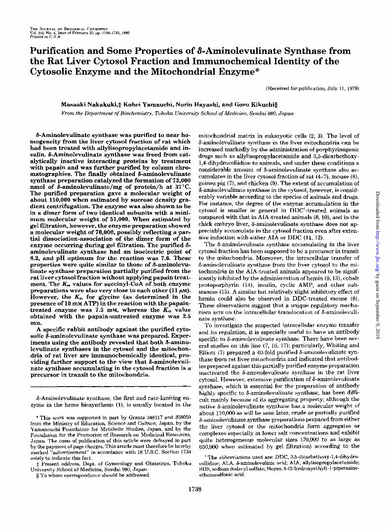
Kinetic Properties-The purified papain-treated &amino- levulinate synthase showed a pH optimum at 7.6 (Fig. 6, closed circle). The pH activity relation of the papain-un- treated 6-aminolevulinate synthase (Fig. 6, open circle) was quite similar to that of the papain-treated enzyme. The K,, values for succinyl-CoA of both the enzymes were also very close to each other (11 IJM, Fig. 7). However, a considerable difference was found in the K , value for glycine between the reactions with the papain-treated and papain-untreated en- zyme preparations as shown in Fig. 8. With the papain-treated enzyme, the K,, value for glycine was 7.5 mM, whereas in the reaction with the papain-untreated enzyme, K,,, for glycine was 2.5 mM. It is apparent that papain-treatment has affected to some extent some catalytic properties of 6-aminolevulinate synthase, although the enzyme activity did not appear to be appreciably reduced by papain-treatment when assayed under the standard conditions in System A as mentioned above.
Immunochemical Comparison of the Cytosolic 6- Aminolevulinate Synthase and the Mitochondrial Enzyme
Ouchterlony double diffusion analysis showed a single pre- cipitin line when the antibody was allowed to react with a purified cytosolic 8-aminolevulinate synthase (specific activ-
t f 6 a
0
r
d 7 8 9
P” FIG. 6. pH activity curves of the papain-treated and the
papain-untreated 8-aminolevulinate synthases. The reaction mixture contained 50 mM Hepes/NaOH buffer, 100 mM glycine, 0.1 mM succinyl-CoA, 0.1 mM pyridoxal-P, 10 mM ATP, and 1 pg as protein of the papain-treated enzyme or 50 pg as protein of the papain-untreated enzyme in a final volume of 1.0 ml. The reaction was started by the addition of succinyl-CoA and stopped with 0.1 ml of 27.5% trichloroacetic acid after IO-min incubation at 30°C. W, papain-treated enzyme; M, papain-untreated enzyme.
by guest on September 9, 2019
http://ww
w.jbc.org/
Dow
nloaded from
6-Aminolevulinate Synthase of Rat Liver 1743
-0.1 0 0.1 0.2 0.3 0.4
[Succinyl-CoA, j~M)-l FIG. 7. Double reciprocal plots of 8-aminolevulinate syn-
thase activity for succinyl-CoA. The papain-treated enzyme (1 pg as protein) and the papain-untreated enzyme (50 pg as protein) were incubated for 5 min at 30°C in the reaction mixtures containing 50 mM Hepes/NaOH buffer (pH 7.6). 100 mM glycine, 0.1 mM pyridoxal- P, 10 mM ATP, and various concentrations of succinyl-CoA, in a final volume of 2.0 ml. The reaction was started by the addition of succinyl- CoA and stopped with 0.2 ml of 27.58 trichloroacetic acid. -, papain-treated enzyme; M, papain-untreated enzyme.
0.7 c / 7
F 0.4k /
I I I 01 I I I I 0 0.1 0.2 d.3 0.4
(Glycine, mM)” FIG. 8. Double reciprocal plots of 6-aminolevulinate syn-
thase activity for glycine. The papain-treated enzyme and the papain-untreated enzyme were assayed at various concentrations of glycine. Other reaction conditions are as in Fig. 6 except that pH was 7.6. M, papain-treated enzyme; W, papain-untreated en- zyme.
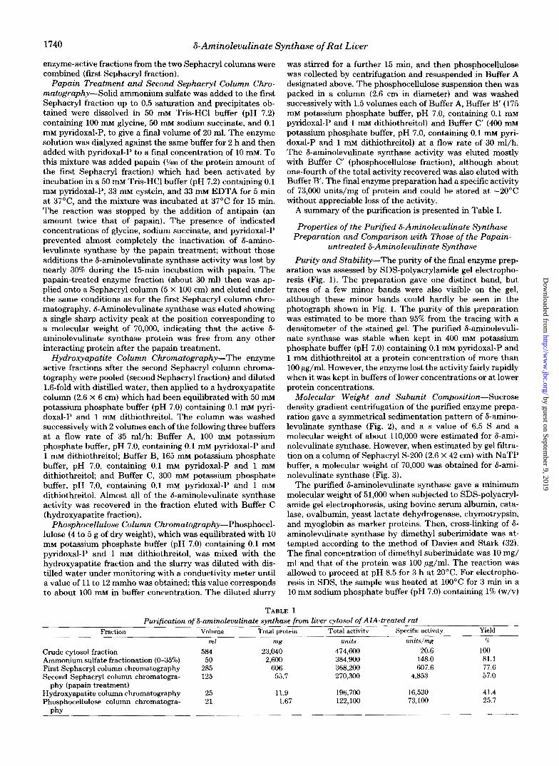
ity, 73,000 units/mg) or a partially purified mitochondrial 6- aminolevulinate synthase (specific activity, 1,200 units/mg) prepared from AIA-treated rats by applying ammonium sul- fate fractionation, hydroxyapatite column chromatography, and sucrose density gradient centrifugation, and the precipitin lines for both preparations completely fused with each other (Fig. 9). In Fig. 10 are shown the results of immunoinhibition experiments with various 6-aminolevulinate synthase prepa- rations, namely, a purified cytosolic 6-aminolevulinate syn-
thase, a crude cytosol fraction, a partially purified mitochon- drial 6-aminolevulinate synthase, and a mitochondrial extract prepared from the AIA-treated rat. With all four different enzyme preparations, the 6-aminolevulinate synthase activity was inhibited to the same extent when a fixed amount of
FIG. 9. Ouchterlony double diffusion analysis of purified cy- tosolic Caminolevulinate synthase and partially purified mi- tochondrial 8-aminolevulinate synthase with antibody pre- pared against purified cytosolic 8-aminolevulinate synthase of rat liver. The center well contained lop1 of antibody against purified cytosolic 6-aminolevulinate synthase prepared as described under “Results.” Wells I , 3, and 5 each contained 14 units of cytosolic 6- aminolevulinate synthase after the phosphocellulose column chro- matography (specific activity, 73,000 units/mg of protein) and Wells 2.4, and 6 each contained 7 units of 6-aminolevulinate synthase from rat liver mitochondria partially purified (specific activity, 1,600 units/ mg of protein) by the method described under “Results.” Agar plate was placed at 2°C for 24 h.
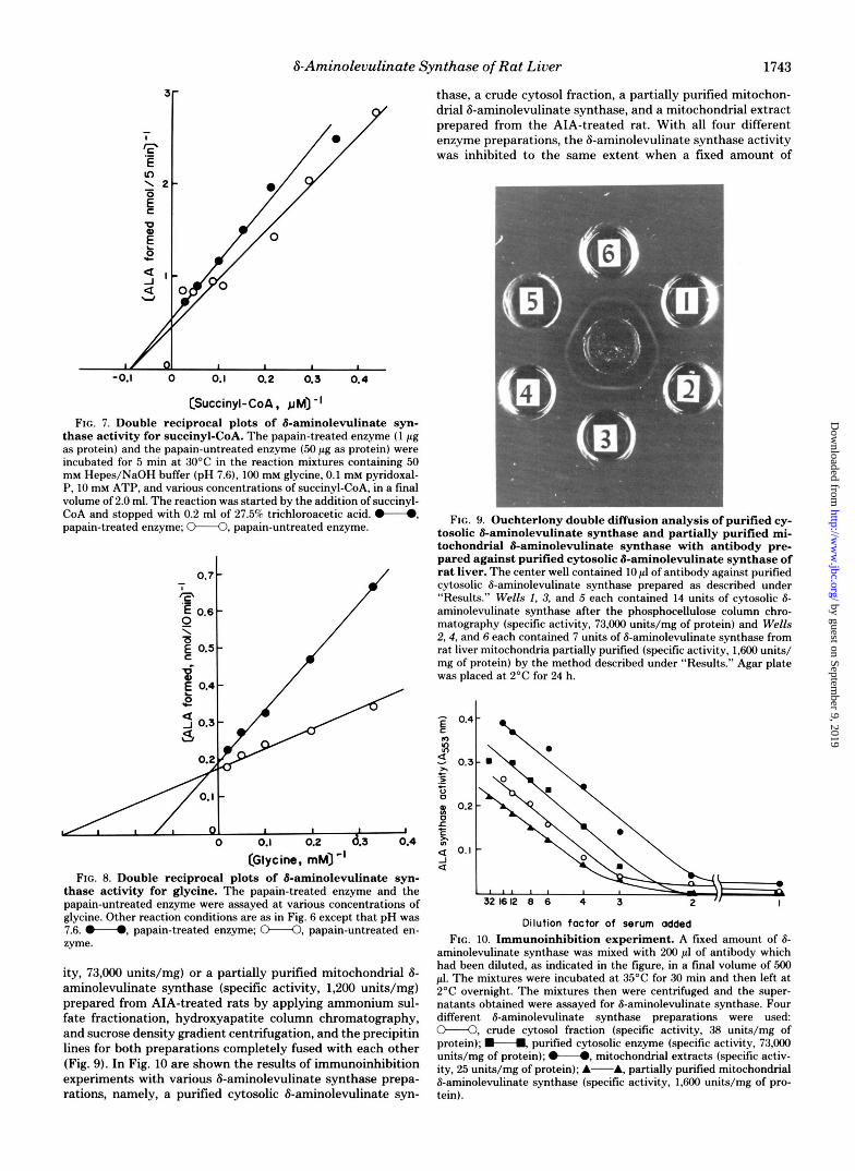
Dilution factor of serum added FIG. 10. Immunoinhibition experiment. A fixed amount of 8-
aminolevulinate synthase was mixed with 200 pl of antibody which had been diluted, as indicated in the figure, in a final volume of 500 pl. The mixtures were incubated at 35°C for 30 min and then left at 2°C overnight. The mixtures then were centrifuged and the super- natants obtained were assayed for 8-aminolevulinate synthase. Four different 6-aminolevulinate synthase preparations were used: M, crude cytosol fraction (specific activity, 38 units/mg of protein); H, purified cytosolic enzyme (specific activity, 73,000 units/mg of protein); M, mitochondrial extracts (specific activ- ity, 25 units/mg of protein); A-A, partially purified mitochondrial 6-aminolevulinate synthase (specific activity, 1,600 units/mg of pro- tein).
by guest on September 9, 2019
http://ww
w.jbc.org/
Dow
nloaded from

1744 8-Aminolevulinate Synthase of Rat Liver
antiserum was added; in other words, the slopes of decrease of the enzyme activity resulting from the addition of varied amounts of antiserum were quite parallel for all four prepa- rations. Control serum exerted practically no inhibitory effect on the 8-aminolevulinate synthase activity.
DISCUSSION
In the present study the cytosol 8-aminolevulinate synthase was purified to a purity of more than 95% and the purified 6- aminolevulinate synthase preparation exhibited a specific ac- tivity of 73,000 units/mg of protein/h. The most highly puri- fied preparation of liver 6-aminolevulinate synthase so far reported is the enzyme prepared by Whiting and Granick (17) from the chick embryo liver mitochondria which gave a spe- cific activity of 20,000. Assuming the purity of our enzyme preparation to be about 95%, the really pure 8-aminolevulinate synthase may have a specific activity of about 77,000 and this would provide a mean to estimate an actual concentration of 8-aminolevulinate synthase in the cell. Recently Davies and Neuberger have reported a higher value of 130,000 to 170,000 as the specific activity of 8-aminolevulinate synthase purified from Rhodopseudomonas spheroides (33).
The purified 8-aminolevulinate synthase gave a molecular weight of about 110,000 when estimated by sucrose density gradient centrifugation. Also the purified 8-aminolevulinate synthase was evidenced to be a dimer of two identical subunits with a subunit molecular weight of 51,000. This value is in good agreement with the values reported for the enzymes from chick embryo liver mitochondria (17), rat liver cytosol (20), and R. spheroides (34). However, when estimated by gel filtration, the purified 6-aminolevulinate synthase gave a mo- lecular weight of about 70,000; that is smaller than the value obtained by sucrose density gradient centrifugation. It should be noted that the 8-aminolevulinate synthase preparation, which was partially purified from the liver cytosol fraction by sucrose density gradient centrifugation instead of employing papain treatment, also gave a value of about 70,000 when estimated by gel filtration. Also, we have observed in an independent experiment not reported here that, when a crude extract of liver mitochondria from AIA-treated rat was sub- jected to gel filtration, 8-aminolevulinate synthase is eluted usually showing a molecular weight of about 110,000 (cf . Ref. 18), but sometimes an additional activity peak appears at the position corresponding to a molecular weight of about 70,000, and the relative content of the enzyme showing the activity peak at 70,000 becomes significantly greater as the enzyme is subjected to purification procedures. The apparently smaller molecular weight estimated for 6-aminolevulinate synthase by gel filtration may probably be a reflection of a partial disso- ciation-association of the dimer form of the enzyme occurring during gel filtration.
The rabbit antiserum prepared against 6-arninolevulinate synthase purified from the cytosol fraction reacted with the mitochondrial 6-aminolevulinate synthase equally well, indi- cating that both 8-aminolevulinate synthases in the cytosol and the mitochondria are immunochemically identical. This provides strong support to the view that 6-aminolevulinate synthase in the cytosol is the precursor in transit to the mitochondria. The mechanism by which 6-aminolevulinate synthase is translocated from the cytosol fraction to the mitochondria is not known. Recently Ohashi and Shinohara (35) reported that, when a Iz5I-labeled complex form of cyto- solic 6-aminolevulinate synthase was incubated with intact mitochondria in vitro, all three protein components of the complex form of 6-aminolevulinate synthase were recovered in the inner membrane-matrix fraction and suggested that 6- aminolevulinate synthase may be incorporated into the mi-
tochondria in a form of complex. Their experimental results, however, do not necessarily evidence that 8-aminolevulinate synthase was actually incorporated into the matrix side of the mitochondria. A kinetic study with combined use of the im- munochemical method and the radioisotope method has been carried out in this laboratory to obtain a concrete evidence for the intracellular translocation of 6-aminolevulinate syn- thase, as well as the occurrence of regulation acting on this process; the results are given in the accompanying paper (36).
Whiting and Granick (37) prepared a rabbit antibody against the purified chick embryo liver mitochondrial 6-ami- nolevulinate synthase with a specific activity of 20,000. Using this antibody, Whiting (11) studied synthesis of 6-aminolevu- linate synthase in vitro with polyribosomes isolated from postmitochondrial supernatants of chick embryo liver and indicated that the enzyme synthesized in vitro had a minimum molecular weight of 70,000. Whiting suggested that this pro- tein is a “pro form” of the 6-aminolevulinate synthase usually found in the liver. If so, the pro form of 6-aminolevulinate synthase may be processed quickly after the synthesis, possi- bly before being released from polysomes since we could find only the entity with a minimum molecular weight of 51,000 in the liver cytosol fraction. Whether a similar pro form of 6- aminolevulinate synthase occurs or not in the rat liver remains to be examined.
Acknowledgments-We are grateful to the Nippon Roche Com- pany, Tokyo, for providing us with allylisopropylacetamide, and to Dr. Hamao Umezawa, Institute of Microbial Chemistry and National Institute of Health, Tokyo, for his generous supply of antipain.
Addendum-Ater submitting the present paper we noticed a paper by Paterniti and Beattie (38) showing that 6-aminolevulinate synthase was purified from rat liver mitochondria to a specific activity of 2,000 nmol of 6-aminolevulinic acid/mg of protein/h at 30°C and that the partially purified mitochondrial enzyme exhibited an isoelectric point of 4.5. This value is close to a value of about 5 that is one of the isoelectric points observed for a partially purified papain-untreated cytosolic 6-aminolevulinate synthase and which was supposed to be resulted from interaction of 6-aminolevulinate synthase with other interacting proteins in the enzyme preparation employed.
1.
2.
3.
4.
5.
6.
7.
8.
9.
10.
11. 12.
13.
14.
15.
16.
REFERENCES
H. J., ed) Vol. 5, pp. 77-141, Academic Press, New York
Biophys. Acta 178,408-411
Biochem. J. 114,455-461
Granick, S., and Sassa, S. (1971) in Metabolic Pathways (Vogel,
Zuyderhoudt, F. M., Borst, P., and Huijing, F. (1969) Biochim.
McKay, R., Druyan, R., Getz, G. S., and Rabinowitz, M. (1969)
Hayashi, N., Yoda, B., and Kikuchi, G. (1969) Arch. Biochem.
Scholnick, P. L., Hammaker, L. E., and Marver, H. S . (1969) Proc.
Beattie, D. S., and Stuchell, R. N. (1970) Arch. Biochem. Biophys.
Whiting, M. J., and Elliott, W. H. (1972) J. Biol. Chen. 247,
Igarashi, J., Hayashi, N., and Kikuchi, G. (1976) J. Biochem. 80,
Ohashi, A,, and Kikuchi, G. (1972) Arch. Biochem. Biophys. 153,
Kikuchi, G., Kim, H. J., Watarai, K., Tomita, Y., and Ohashi, A.
Whiting, M. J. (1976) Biochem. J. 158,391-400 Tomita, Y., Ohashi, A,, and Kikuchi, G. (1974) J. Biochem. 75,
Hayashi, N., Kurashima, Y., and Kikuchi, G. (1972) Arch. Bio-
Igarashi, J., Hayashi, N., and Kikuchi, G. (1978) J. Biochem. 84,
Kim, H. J., and Kikuchi, G. (1974) Arch. Biochem. Biophys. 164,
Scholnick, P. L., Hammaker, L. E., and Marver, H. S. (1972) J.
Biophys. 131,83-91
Natl. Acad. Sci. U. S. A. 63,65-70
139,291-297
6818-6826
1091-1099
34-46
(1973) Enzyme 16,258-266
1007-1015
chem. Biophys. 148, 10-21
997-1000
293-304
Biol. Chem. 247,4126-4131
by guest on September 9, 2019
http://ww
w.jbc.org/
Dow
nloaded from

6-Aminolevulinate Synthase of Rat Liver 1745
17. Whiting, M. J., and Granick, S. (1976) J. Biol. Chem. 251, 1340-
18. Hayashi, N., Yoda, B., and Kikuchi, G. (1970) J. Biochem. 67,
19. Ohashi, A,, and Kikuchi, G. (1977) Arch. Biochem. Biophys. 178,
20. Ohashi, A., and Kikuchi, G. (1978) J. Biochem. 85,239-247 21. Simon, E. J., and Shemin, D. (1953) J. Am. Chem. SOC. 75, 2520 22. Zaman, Z., Jordan, P. M., and Akhtar, M. (1973) Biochem. J . 135,
23. Ohashi, A., and Shinohara, H. (1978) Biochem. Biophys. Res.
24. Mauzerall, D., and Granick, S. (1956) J. Biol. Chem. 219,435-446 25. Narisawa, K., and Kikuchi, G. (1966) Biochim. Biophys. Acta
26. Weber, K., and Osborn, M. (1969) J. Biol. Chem. 244,4406-4412 27. Fairbanks, G., Steck, T. L., and Wallach, D. F. H. (1971) Bio-
28. Martin, R. G., and Ames, B. N. (1961) J. Biol. Chem. 236, 1372-
1346
859-861
607-616
257-263
Commun. 80,370-376
123,596-605
chemistry 10,2606-2617
1379 29. Chase, M. W. (1967) Methods Immunol. Zmmun. 1, 197 30. Lowry, 0. H., Rosebrough, N. J., Farr, A. L., and Randall, R. J .
31. Bensadoun, A., and Weinstein, D. (1976) Anal. Biochem. 70,241-
32. Davies, G. E., and Stark, G. R. (1970) Proc. Natl. Acad. Sci. U.
33. Davies, R. C., and Neuberger, A. (1979) Biochem. J. 177,649-659 34. Nandi, D. L., and Shemin, D. (1977) J. Biol. Chem. 252, 2278-
35. Ohashi, A., and Shinohara, H. (1978) Biochem. Biophys. Res.
36. Yamauchi, K., Hayashi, N., and Kikuchi, G. (1980) J. Biol. Chem.
37. Whiting, M. J., and Granick, S. (1976) J. Biol. Chem. 251, 1347-
38. Paterniti, J. R., Jr., and Beattie, D. S. (1979) J. Biol. Chem. 254,
(1951) J. Biol. Chem. 193,265-275
250
S. A . 66,651-656
2280
Commun. 83, 76-82
255, 1746-1751
1353
6112-6118
by guest on September 9, 2019
http://ww
w.jbc.org/
Dow
nloaded from

M Nakakuki, K Yamauchi, N Hayashi and G Kikuchithe mitochondrial enzyme.
liver cytosol fraction and immunochemical identity of the cytosolic enzyme and Purification and some properties of delta-aminolevulinate synthase from the rat
1980, 255:1738-1745.J. Biol. Chem.
http://www.jbc.org/content/255/4/1738.citation
Access the most updated version of this article at
Alerts:
When a correction for this article is posted•
When this article is cited•
to choose from all of JBC's e-mail alertsClick here
http://www.jbc.org/content/255/4/1738.citation.full.html#ref-list-1
This article cites 0 references, 0 of which can be accessed free at
by guest on September 9, 2019
http://ww
w.jbc.org/
Dow
nloaded from