of flavins in enzyme-catalyzed reactions? - mh … stereochemistry of flavins vol. 25, no.22, 1986...
TRANSCRIPT
Biochemistry 1986, 25, 6807-68 16 6807
REFERENCES Aas, M., & Bremer, J. (1968) Biochim. Biophys. Acta 164,
Alger, J. R., Behar, K. L., Rothman, D. L., & Shulman, R.
Bates, M. W., Krebs, H . A., & Williamson, D. H. (1968)
Berry, M. N., Gregory, R. B., Grivell, R. A., & Wallace, P.
Blackshear, P. J., & Alberti, K. G. M. (1974) Biochem. J. 138,
Cahill, G. F. (1981) J. Parent. En?. Nutr. 5 , 281-287. Canioni, P., Alger, J . R., & Shulman, R. G. (1983) Bio-
Cross, T. A., Pahl, C., Oberhansli, R., Aue, W. P., Keller, U.,
Cross, T. A., Muller, S., & Aue, W. P. (1 985) J. Magn. Reson.
Heussler, A., Ganz, P., & Gaumann, T. (1975) J. Labelled
Hird, F. J. R., & Symons, L. P. (1962) Biochem. J. 84,
Lynen, F., Henning, U., Bublitz, C., Sorbo, B., & Kroplin-
Mayes, P. A., & Laker, M. E. (1981) Biochem. SOC. Trans.
157-1 66.
G. (1984) J. Magn. Reson. 56, 334-337.
Biochem. J . 110, 655-661.
G. (1983) Eur. J . Biochem. 131, 215-222.
107-1 1 7.
chemistry 22, 4974-4980.
& Seelig, J . (1984) Biochemistry 23, 6398-6402.
62, 87-98.
Compd. 1 1 , 37-42.
2 1 2-2 1 6.
Rueff, L. (1958) Biochem. Z. 330, 269-295.
9, 339-341.
McGarry, J. D., & Foster, D. W. (197 1) J . Biol. Chem. 264,
McGarry, J. D., & Foster, D. W. (1 980) Annu. Rev. Biochem.
Ohgaku, S., Brady, P. S., Schumann, W. C., Bartsch, G. E., Margolies, J. M., Kumaran, K., Landau, S. B., & Landau, B. R. (1982) J. Biol. Chem. 257, 9283-9289.
Owen, 0. E., & Schramm, V. L. (1981) Biochem. SOC. Trans
Reo, N. V., Siegfried, B. A,, & Ackerman, J. J. H. (1984) J. Biol. Chem. 259, 13664-13667.
Schumann, W. C., Hemmelgarn, E., & Landau, B. R. (1978) Arch. Biochem. Biophys. 190, 345-350.
Siegfried, B. A., Reo, N. V., Ewy, C. S., Shalwitz, R. A,, Ackerman, J. J. H., & McDonald, J. M. (1985) J. Biol. Chem. 260, 16137-16142.
Siess, E. A., Kientsch-Engel, R. I . , & Wieland, 0. H. (1982) Eur. J . Biochem. 121, 493-499.
Stevens, A. N., Iles, R. A., Morris, P. G., & Griffiths, J. R. (1982) FEBS Lett. 150, 489-493.
Stromski, M. E., Arias-Mendoza, F., Alger, J. R., & Shulman, R. G. (1986) Magn. Res. Med. 3, 24-32.
Wieland, 0. H. (1968) Adu. Metab. Disord. 3, 1-47. Williamson, D. H. (1 979) Biochem. SOC. Trans. 7 , 13 13-1 320.
1149-1159.
49, 395-420.
9, 342-344.
Absolute Stereochemistry of Flavins in Enzyme-Catalyzed Reactions?
Dietmar J. Manstein and Emil F. Pai* Department of Biophysics, Max Planck Institute for Medical Research, 0-6900 Heidelberg, Federal Republic of Germany
Lawrence M. Schopfer and Vincent Massey Department of Biological Chemistry, The University of Michigan, Ann Arbor, Michigan 481 09
Received April 28, 1986; Revised Manuscript Received July 10, 1986
ABSTRACT: The 8-demethyl-8-hydroxy-5-deaza-5-carba analogues of F M N and FAD have been synthesized. Several apoproteins of flavoenzymes were successfully reconstituted with these analogues. This and further tests established that these analogues could serve as general probes for flavin stereospecificity in enzyme- catalyzed reactions. The method used by us involved stereoselective introduction of label on one enzyme combined with transfer to and analysis on a second enzyme. Using as a reference glutathione reductase from human erythrocytes for which the absolute stereochemistry of catalysis is known from X-ray studies [Pai, E. F., & Schulz, G. E. (1983) J. Biol. Chem. 258, 1752-17581, we were able to determine the absolute stereospecificities of other flavoenzymes. W e found that glutathione reductase (NADPH), general acyl-CoA dehydrogenase (acyl-CoA), mercuric reductase (NADPH) , thioredoxin reductase (NADPH) , p-hydroxy- benzoate hydroxylase (NADPH), melilotate hydroxylase (NADH) , anthranilate hydroxylase (NADPH) , and glucose oxidase (glucose) all use the re face of the flavin ring when interacting with the substrates given in parentheses.
F A D and FMN are ubiquitous coenzymes. They are ex- tremely versatile redox catalysts, taking part in radical, car- banion, or hydride-transfer mechanisms (Hamilton, 197 1; Hemmerich, 1976; Bruice, 1980; Walsh, 1980). They therefore occupy a central position in enzyme-catalyzed redox
+Supported, in part, by Grant GM-1106 from the US. Public Health Service. This work has also been submitted by D.J.M. to the University of Heidelberg in partial fulfillment of the requirements for the Ph.D. degree.
0006-2960/86/0425-6807$0 1 .50/0
chemistry. At present, there are far more than 100 different flavoenzymes known, most of them members of the class of oxidoreductases (Enzyme Nomenclature, 1984).
Enzymatic (Jorns & Hersh, 1974; Fisher & Walsh, 1974; Hersh & Walsh, 1981; Thorpe & Williams, 1976) as well as bioorganic model studies (Briistlein & Bruice, 1972; Loechler & Hollocher, 1980) have made it clear that positions C4a and N5 are the key loci of interaction between flavins and sub- strates. A common mechanism that has been proposed for many flavoenzymes involves the transfer of the equivalent of
0 1986 American Chemical Society

6808 B I oc H E M I S T R Y M A N S T E I N E T A L .
Chart I R R I I
si -face
B re -face
R
a hydride ion to N5 of the isoalloxazine ring system. As the two faces of the flavin ring are prochiral (Chart I), the question arose whether only one of them is used by any particular enzyme and if so which one.
The labile nature of the resulting N-H bonds made it im- possible to use label transfer to and from common flavin nu- cleotides to determine stereospecificities of flavoenzymes. In a first attempt to overcome this problem, 5-deaza-5-carba analogues of FAD and FMN were synthesized (O'Brien et al., 1970; Hersh & Jorns, 1975; Spencer et al., 1976) in order to make the prochiral center inert to solvent exchange. Further studies conclusively showed that there is direct hydrogen transfer from substrates to bound 5-deazaflavin] coenzymes (Hersh & Jorns, 1975; Hersh et al., 1976; Jorns & Hersh, 1975, 1976; Fisher et al., 1976). Scrambling of label due to a rapid transfer from reduced to oxidized 5-deazaflavin molecules, however, did not allow determination of relative stereospecificities (Spencer et al., 1976).
Recently, it has been shown that analogous exchange be- tween the oxidized and reduced forms of the riboflavin part of cofactor F420 from methanogenic bacteria (7,8-dide- methyl-8-hydroxy-5-deazariboflavin) is several orders of magnitude slower than that of 5-deazariboflavin (Jacobson & Walsh, 1984). Taking advantage of this effect, first relative (Yamazaki et al., 1980) and later absolute stereochemistries of NAD+:FMN oxidoreductase from Beneckea harveyi and of several cofactor F420 dependent enzymes were determined (Yamazaki et a]., 1985).
These findings made the use of 8-demethyl-8-hydroxy-5- deaza-5-carba-FAD and the corresponding FMN compound respectively the first choice when trying to develop a reasonably fast method of analyzing the absolute stereochemistry of flavin prosthetic groups. This analogue combines the redox properties of cofactor F420 with structural features as close as possible to those of native riboflavin nucleotides, permitting easier reconstitution of apoproteins with the analogue.
The first flavoenzyme for which the absolute stereochemistry of its prosthetic group became known was glutathione re- ductase from human erythrocytes. X-ray crystallography established that the nicotinamide ring of its substrate NADPH
' Abbreviations: AcPyAD', oxidized 3-acetylpyridine adenine di- nucleotide; AcPyADP+, oxidized 3-acetylpyridine adenine dinucleotide phosphate: Sdeazaflavin, 5-deaza-5-carbaisoalloxazine: 8-OH-flavin, 8-demethyl-8-hydroxyisoalloxazine; 8-SH-flavin, 8-demethyl-8- mercaptoisoalloxazine; 8-OH-5-deazaflavi11, 8-demethyl-8-hydroxy-5- deaza-5-carbaisoalloxazine; 8-OH-5-deaza[5-)H]FADH2, reduced 8- OH-5-deaza-FAD with one tritium label at carbon-5; Tris-HCI, tris- (hydroxymethy1)aminomethane hydrochloride; MES, 2 - ( N - morpho1ino)ethanesulfonic acid; DTT, dithiothreitol; EDTA, ethylene- diaminetetraacetic acid; HPLC, high-pressure liquid chromatography.
interacts with the re face of the isoalloxazine ring of its prosthetic group FAD (Pai & Schulz, 1983). On reducing 5-deaza-FAD-reconstituted general acyl-CoA dehydrogenase with NaB3H4, Ghisla et al. (1984) found that about 90% of tritium label had been incorporated into one side of the flavin ring. Combination of these results should allow determination of absolute stereospecificities by performing stereoselective labeling of the flavin analogue in general acyl-CoA de- hydrogenase and reoxidizing it in glutathione reductase. Depending on whether the label would be released or whether it would stay at the flavin ring, general acyl-CoA de- hydrogenase should use the re or si face, respectively. Then for any flavoenzyme to which the respective 8-OH-5-deaza cofactor can be bound and reduced or reoxidized by the corresponding substrate or substrate analogues, determination of absolute stereospecificity should be possible.
EXPERIMENTAL PROCEDURES
Materials 5-Amino-o-cresol was obtained from TCI/Tokyo Kasei and
4-chlorouracil from Lancaster Synthesis. D-Ribose was from Sigma and trimethylorthoformate from Aldrich. NAD (grade I11 from yeast), NADH (grade I11 from yeast), and Naja naja venom were purchased from Sigma. NADP+, NADPH, and 3-acetylpyridine adenine dinucleotides were obtained from P-L Biochemicals. Aquasolve scintillation fluid and NaB3H4 (lots 1749-181 and 1953-227; 8 mCi/mg) were from NEN. [ 1- 3H]Glucose (25 Ci/mmol) was from ICN Chemical and Radioisotope Division and was used after dilution to 400 mCi/mmol. Ultrapure-grade guanidine hydrochloride was from Schwarz/Mann. All other chemicals were of the highest purity commercially available.
Methods 8-OH-5-deazariboflavin. 1-Deoxy-1- [(3-hydroxy-4-
methylphenyl)amino]- ribitol was prepared by reacting 12.5 g of 5-amino-o-cresol with 15.5 g of D-ribose in 200 mL of absolute ethanol under nitrogen. The mixture was stirred at reflux for 1 h, then cooled down to room temperature, and diluted with another 150 mL of absolute ethanol. A total of 8 g of NaBH, was added in small portions to reduce the Schiff base. Then, the golden yellow solution was stirred for three more hours. The pH was adjusted to 5.0 with concentrated HC1, and 400 mL of well-degassed water was added. A total of 150 mL of AG 50W-X8 cation-exchange resin (1 00-200 mesh, H+ form) was brought into this solution, and the re- sulting slurry was gently stirred for 15 min. Further purifi- cation was performed at 4 OC in the dark. The mixture was layered on top of another 130 mL of ion-exchange resin packed into a column (2.5 X 54 cm). After being washed with 1.5 L of degassed water, the product was eluted with 1% ammo- nium hydroxide. Fractions containing the desired compound were combined and concentrated on a rotary evaporator at 40 "C to give 23 g (88% yield) of a light brown, amorphous material. This was further converted to 8-OH-5-deazaribo- flavin as published by Ashton and Brown (1980).
8-OH-5-deaza-FAD. 8-OH-5-deaza-FAD was prepared from 8-OH-5-deazariboflavin and ATP with riboflavin ki- nase/FAD synthetase partially purified from Brevibacterium ammoniagenes (Spencer et al., 1976; Manstein & Pai, 1986). About 80 mg of protein was dissolved in 150 mL of buffer containing 10 mM MES, 5 mM ATP, 30 mM MgCI2, 1 mM CaC12, and 1 mM DTT. The pH was adjusted to 5.9 with 1 M KH2P0,. After addition of 12 mg of 8-OH-5-deazaribo- flavin, the mixture was incubated at 25 OC for 24-28 h. Progress of the conversion was monitored by HPLC (LKB

A B S O L U T E S T E R E O C H E M I S T R Y O F F L A V I N S V O L . 2 5 , N O . 2 2 , 1 9 8 6 6809
to another centrifuge tube containing prewashed charcoal. This step was repeated 3-4 times. Guanidine was finally removed by applying the solution to a Sephadex G-25 column equilibrated with 0.1 M potassium phosphate-0.3 mM EDTA, pH 7.6. Protein-containing fractions were identified by measuring the optical absorbance at 280 nm.
The apoprotein of glutathione reductase from human erythrocytes was prepared by modifying a procedure described by Fritsch (1982). A total of 0.5 mL of 25-50 pM enzyme solution in 100 mM potassium phosphate, pH 7.0, 200 mM KCl, 1 mM EDTA, and 1.4 mM 2-mercaptoethanol cooled to 4 OC was mixed with 0.6 mL of 10 mM EDTA-1.4 mM 2-mercaptoethanol, pH 5.0, saturated with (NH4)2S04. To adjust the pH to 3.0, 55 pL of 1 M HCI was added. After being incubated on ice for 20 min, the apoprotein was pelleted in an Eppendorf centrifuge at 128OOg for 6 min. The pellet was washed 4 times with 800 pL of 2.8 M (NH4)2S04, 10 mM EDTA, and 1.4 mM 2-mercaptoethanol, pH 3.0. Finally, the apoprotein was taken up in 150 fiL of 100 mM Tris-HCl buffer, pH 8.7, 10 mM EDTA, and 0.5 mM DTT.
Reconstitution of Apoenzymes with 8-OH-5-deazaf2auins. Unless indicated otherwise, reconstitution of the respective apoenzymes with the 8-OH-5-deaza analogues was achieved by adding a 1.5-2 molar excess of flavin analogue to the apoenzyme dissolved in the appropriate buffer. The apo- enzyme solutions used in this step were 20-60 pM. In order to remove surplus nucleotide and to exchange buffers, if necessary, the reaction mixtures were passed through Sephadex G-25 columns (1 X 25 cm).
Reconstitution of pig kidney general acyl-CoA de- hydrogenase apoenzyme was performed by incubating a 20 pM solution of apoprotein in 50 mM potassium phosphate43 mM EDTA, pH 7.6 (buffer A), with 1-2 equiv of 8-OH-5- deaza-FAD at 4 OC for 18 h. Surplus flavin was removed by adding this solution to a pellet of prewashed charcoal to yield a suspension 0.5% (weight/volume) in charcoal. After being incubated at 4 O C for 1 min, this solution was centrifuged at 12800g for 2 min. The supernatant was transferred to another centrifuge tube and the procedure repeated 2-3 times until the shoulder at 429 nm due to the visible absorption maximum of the free form of 8-OH-5-deazaflavin could no longer be detected. The peak of 8-OH-5-deaza-FAD bound to general acyl-CoA dehydrogenase (403 nm) does not interfere.
Test of Stereospecificity. General acyl-CoA dehydrogenase reconstituted with 8-OH-5-deaza-FAD was reduced by incu- bating 13 nmol of enzyme in 1 mL of buffer A with 0.5 mg NaB3H4 at 0 OC for 1 h. After this, no residual oxidized enzyme could be detected in absorption spectra of the reaction mixture. Residual borohydride was destroyed by adding 3 mg of sodium pyruvate before the solution was chromatographed over a G-25 fine column (1 X 20 cm) equilibrated with buffer A. Chiral 8-OH-5-deaza [5-3H] FADHz was released by heating the protein to 100 OC for 1 min followed immediately by cooling in ice and centrifuging for 1 minute at top speed in an Eppendorf centrifuge at 4 O C .
Glucose oxidase was tested as an alternative way of ste- reoselectively labeling enzyme-bound 8-OH-5-deaza-FAD. A total of 10 nmol of reconstituted enzyme was incubated with 14 pmol of [l-3H]-~-glucose (0.05 mci ) at 25 "c. Progress of the reaction was followed spectrophotometrically. After reduction was complete, the solution was passed over a Sephadex G-25 fine column (1 X 20 cm). A total of 9.5 nmol of 8-0H-5-dea~a[5-~H] FADHz enzyme with a specific activity of 27 000 cpm/nmol was eluted from the column. The labeled flavin analogue was released by heating the enzyme to 100
modules) on a C-18 column (Abimed Analysen-Technik GMBH, Shandon ODS Hypersil, 5 ym, 0.46 X 25 cm). Products of the FAD synthetase reaction were developed at a flow rate of 2.5 mL/min with a linear gradient from 90% solvent A (50 mM potassium phosphate, pH 6.0) to 45% solvent B (50 mM potassium phosphate buffer, pH 6.0, plus 50% CH3CN) that took 6 min to reach the final conditions. The absorbance at 260 nm was followed.
After the reaction was completed, protein that had already been denatured was removed by centrifuging for 15 min at 18000g. The supernatant was filtered through an Amicon PM-10 membrane. The resulting clear solution was concen- trated to approximately 15 mL on a rotary evaporator at 40 "C. It was then purified by applying 900-pL aliquots to a preparative HPLC column (Latek, Heidelberg; RPI 8, l0-pm HL; 1 X 25 cm). Elution was performed by a linear gradient at 5 mL/min. Solvent A was 50 mM triethylammonium acetate, pH 7.5; solvent B was 50 mM triethylammonium acetate, pH 7.5, plus 50% CH3CN. The gradient ran from 0% to 45% of solvent B in 18 min.
8-OH-5-deaza-FMN. The FMN analogue was obtained from the modified FAD by hydrolysis with Naja naja venom.
Enzymes and Apoenzymes. The following enzymes and corresponding apoenzymes were prepared as previously de- scribed: flavodoxin (Mayhew & Massey, 1969; Wassink & Mayhew, 1975) and D-lactate dehydrogenase (Olson & Massey, 1979) from Megasphera elsdenii, L-lactate oxidase from Mycobacterium smegmatis (Choong et al., 1975; Sullivan et al., 1977), glucose oxidase from Aspergillus niger (Swoboda & Massey, 1965; Swoboda, 1969), melilotate hydroxylase from Pseudomonas sp. (Strickland & Massey, 1973; Detmer et al., 1984), p-hydroxybenzoate hydroxylase from Pseudomonas
fluorescens (Entsch et al., 1976, 1980), D-amino acid oxidase from pig kidney (Brumby & Massey, 1966; Massey & Curti, 1966), and glutathione reductase from human erythrocytes (Krohne-Ehrich et al., 1977).
The following enzymes were provided as generous gifts: pig kidney general acyl-CoA dehydrogenase (Thorpe et al., 1979) by Dr. C. Thorpe (University of Delaware), apoenzymes of mercuric reductase from Pseudomonas aeruginosa PA 09501 carrying the plasmid pVSl (Fox & Walsh, 1982) and a mutant of the native enzyme with Cys-135 replaced by a serine (Schultz et al., 1986) by Dr. C. T. Walsh (Massachusetts Institute of Technology) and Dr. S. Miller (University of Michigan), and (from colleagues at the University of Mich- igan) anthranilate hydroxylase from Trichosporum cutaneum (Powlowski & Dagley, 1982) by Dr. J. Powlowski, thioredoxin reductase from Escherichia coli (O'Donnell & Williams, 1984) by Dr. C. H. Williams, Jr., and spinach ferredoxin-NADP' reductase (Zanetti & Curti, 1980) by Dr. M. Ludwig. Spinach ferredoxin reductase apoprotein was prepared as described by Zanetti et al. (1982). Apoproteins of anthranilate hydroxylase and of general acyl-CoA dehydrogenase were resolved as described by Mayer and Thorpe (1981).
The apoprotein of thioredoxin reductase was prepared by a slight modification of a procedure described by O'Donnell and Williams (1984). Native enzyme was incubated with 5 M guanidine in 0.1 M potassium phosphate-0.3 mM EDTA, pH 7.6, at 4 OC. One minute after addition of 1.65 mL of 8 M guanidine to 1 mL of a 50-100 pM solution of thioredoxin reductase, this solution was transferred to a centrifuge tube containing a pellet of prewashed charcoal (Sigma Norit A) sufficient to make the resulting suspension 0.5% (dry weight/volume) in charcoal. The tube was centrifuged at 4 "C and 12800g for 3 min. The supernatant was transferred
6810 B I O C H E M I S T R Y M A N S T E I h E T A L .
O C for 1 min, immediately followed by cooling on ice. Sub- sequently, denatured protein was removed from the solution by centrifugation.
Analogues that had been stereoselectively labeled by one of the procedures described were bound to the apoform of the enzyme, whose stereochemistry was to be tested. Reoxidation was started by adding the appropriate nicotinamide nucleotide and followed spectrophotometrically. After completion of the reaction, an aliquot of the solution was passed through a Sephadex G-25 column with a void volume of 5 mL. Each fraction was analyzed for radioactivity, and its absorption spectrum was recorded. In order to concentrate most data relevant to a specific enzyme, details for individual enzymes will be given under Results.
Instrumentation. Optical spectra were recorded on Cary 2 19 or Shimadzu UV-260 spectrophotometers. Fluorescence spectra were measured on a ratiorecording fluorometer built by Dr. D. Ballou and G. Ford of the University of Michigan. Radioactivity was determined with a Beckman LS 7800 liquid scintillation counter. Selected vials were calibrated internally. The average counting efficiency was about 40%. HPLC was performed on an LKB instrument equipped with a Model 21 50 pump, UV detector 2 15 1, solvent programmer 2 152, and a Shimadzu C-Rl a integrator. Stopped-flow kinetics were measured with a temperature-controlled instrument interfaced with a Nova 2 (Data General) minicomputer system (Beaty & Ballou, 1981).
RESULTS Synthesis of 8-OH-5-deazaj7avins. When the Schiff base
formed upon condensation of 5-amino-o-cresol with D-ribose was reduced with NaBH,, 1 -deoxy- 1 - [ (3-hydroxy-4-methyl- phenyl)amino]-~-ribitol was obtained in pure form and in good yield. Conversion of this compound to 8-OH-5-deazariboflavin according to Ashton and Brown (1980) gave 321 mg of golden yellow product.
By modifying the procedure for the purification of the FAD-synthesizing activity from Brevibacterium ammonia- genes, we were able to obtain an enzyme preparation of in- creased stability that allowed complete conversion of 8-OH- 5-deazariboflavin to the FAD level (Manstein & Pai, 1986). The purity of the product was >95% as judged by HPLC analysis.
In trying to improve the yield of the enzymatic reaction, we found that p H is an important variable to optimize. In the conversion of 8-OH-5-deazariboflavin, maximal activity was obtained at pH 5.9. Below pH 5.8, denaturation of the enzyme started to be a problem; above this pH, the rate of the enzy- matic reaction rapidly decreased, consistent with only the neutral forms of flavins being accepted as substrates. Curi- ously, addition of 1 m M CaC1, to the reaction mixture was found to have a pronounced stabilizing effect on the enzyme (Manstein & Pai, 1986).
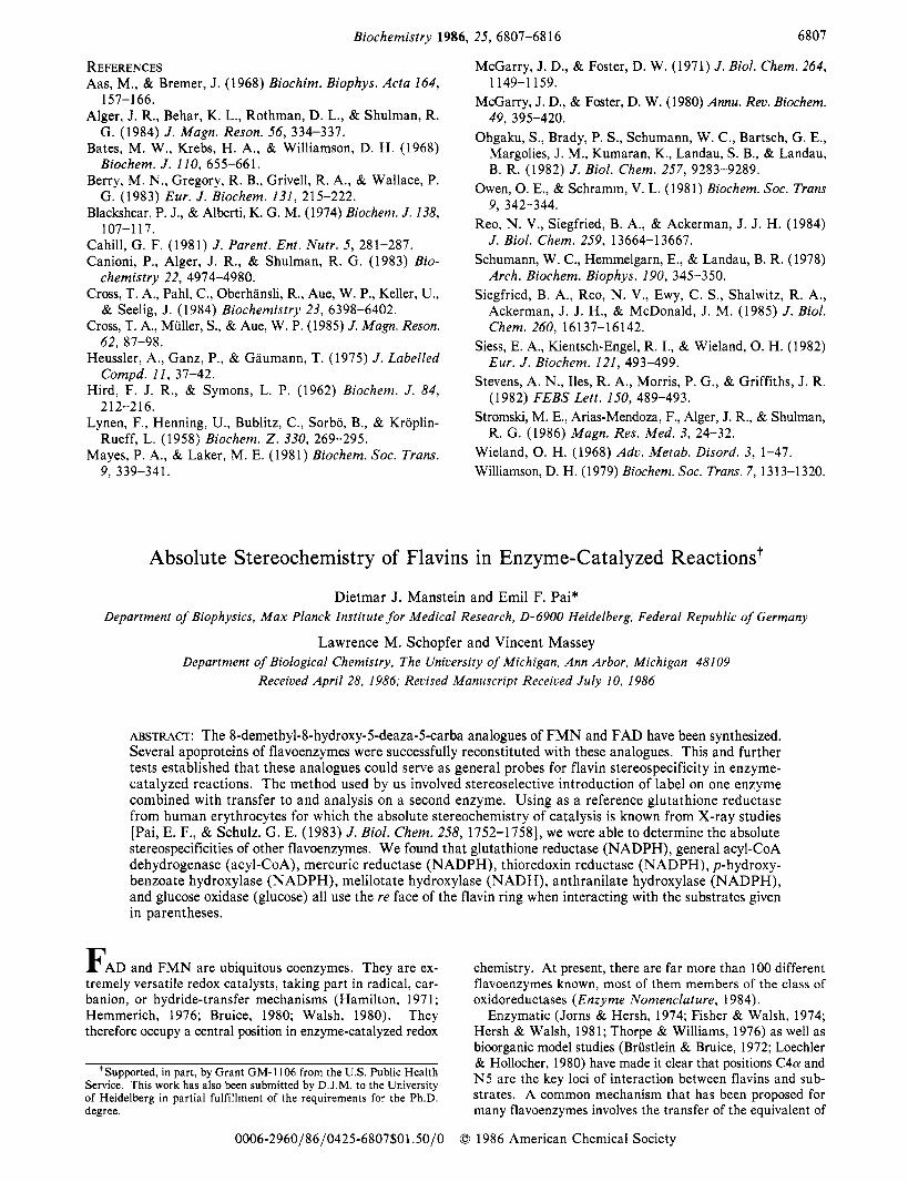
Chemical and Spectral Properties of 8-OH-5-deazaflauin Analogues. The redox properties of 8-OH-5-deazariboflavin were found to be virtually identical with those of 7,8-dide- methyl-8-OH-5-deazariboflavin described by Jacobson and Walsh (1984). A spectrophotometric titration (Figure 1) gave a PKA of 6.1 for 8-OH-5-deaza-FAD. For both 8-OH-5- deaza-FMN and 8-OH-5-deazariboflavin, a pKA value of 6.0 was found.
When 8-OH-5-deaza-FAD a t pH 7.6 was treated with Naja naja venom, the maximum of the visible absorption peak shifted from 430 to 426 nm (Table I). Its intensity increased by 8%, an amount comparable with the value of 11% observed on hydrolysis of FAD to F M N (Bessey et al., 1949).
300 400 500 -Wavelength (nrni-
FIGURE 1: Effect of pH on absorbance of oxidized 8-OH-5-deaza- FAD. 8-OH-5-deaza-FAD was dissolved in a mixed buffer system of acetate, 2-(N-morpholino)ethanesulfonic acid, phosphate, and pyrophosphate, 10 mM each; 2 M HCI was used to bring the pH to 3.9. Then, small volumes of 5 M NaOH were added, and spectra and pH were recorded after each addition. The curve with the lowest absorption at 430 nm was recorded at pH 3.9; subsequent curves with increasing absorbance at 430 nm were recorded at pH values of 4.6, 5.0, 5.7, 6.1, 6.4, 6.5, 6.9, 7.6, and 10.5. The inset shows corrected for dilution a linearized plot of the optical density a t 430 nm as a function of pH.
Table I: Absorption Maxima and Extinction Coefficients of 8-OH-Sdeazaflavins
Xlnm ~,,, . ..._ . flavin (nm) (cm-’ M-’)
8-OH- 5-deazariboflavin 425 52 900“ 8-OH-5-deaza-FMN 426 41 2OOb 8-OH-5-deaza-FAD 430 43 6OOb 8-OH-5-deaza-FADH, 326 16 OOOb
“ I n 0.1 N NaOH (Ashton & Brown, 1980). buffer A as solvent.
For the native coenzymes, these changes are accompanied by an about 10-fold increase in fluorescence. This can be explained by release of self-quenching due to intramolecular interactions between the planar purine and isoalloxazine systems (Weber, 1950). A similar self-quenching was not observed with 8-OH-5-deaza-FAD at neutral pH, suggesting that this kind of “hairpin” complexation can only occur be- tween uncharged subunits. Ghisla and Mayhew (1980) ob- tained similar results investigating 8-demethyl-8-OH-FAD. Further support comes from the fact that in both the FMN and the FAD forms the pK, of the 8-hydroxy substituent is nearly identical, which again is not expected if the purine and isoalloxazine rings interact. Upon reduction to the dihydro form, fluorescence was almost completely lost.
Reconstitution Experiments and Results of Stereospecificity Tests. With the exception of Old Yellow Enzyme, all flavo- enzymes tested so far showed tight binding of the respective oxidized 8-OH-Sdeazaflavin cofactor. No loss of analogue was observed upon gel filtration, and the spectral features remained constant (Table 11). This included the apoproteins of D-lactate dehydrogenase (EC 1.1.99.6), D-aminO acid ox- idase (EC 1.4.3.3), and ferredoxin-NADP’ reductase (EC 1.18.1.2) as FAD enzymes and L-lactate oxidase (1.13.12.4) and flavodoxin as F M N enzymes beside those flavoenzymes for which the stereochemistry has been elucidated, and more details are given below. With some of these proteins, asso- ciation with the reduced form of the analogue has been tested,
A B S O L U T E S T E R E O C H E M I S T R Y O F F L A V I N S
Table 11: Absorption Maxima and Extinction Coefficients of Proteins Reconstituted with 8-OH-5-deazaflavins"
Xmax emax protein source (nm) (cm-l M-I)
33 700 general acyl-CoA dehydrogenase
glutathione reductase mercuric reductase mercuric reductase
(ser-l35)* thioredoxin reductase glucose oxidase p-hydroxybenzoate
hydroxylase +50 fiM p-hydroxybenzoate
melilotate hydroxylase +20 pM melilotate
anthranilate hydroxylase +IO0 pM anthranylic acid +IO0 fiM salicylic acid
ferredoxin-NADP' oxidoreductase
L-lactate oxidase D-hCtate dehydrogenase D-amino acid oxidase flavodoxin
pig kidney
human erythrocytes P . aeruginosa P . aeruginosa
E . coli A . niger P . fluorescens
P. sp
T . culaneum
spinach
Mc. smegmatis Mg. elsdenii pig kidney M P . elsdenii
403
434 437 432
414 413 435
443
433 433 440 440
442 432
446 430 432 430
29 000 nd nd
31 000 32 000 39 500
40 000
36 600 41 000 40 200 42 400
45 500 36 800
nd 36 500 39 000 29 600 u
" In buffer A as solvent. bSingle-site mutant with Cys-135 replaced by Ser-135.
too. In all cases it was tightly bound. General Acyl-CoA Dehydrogenase (EC I .3.99.3). Recon-
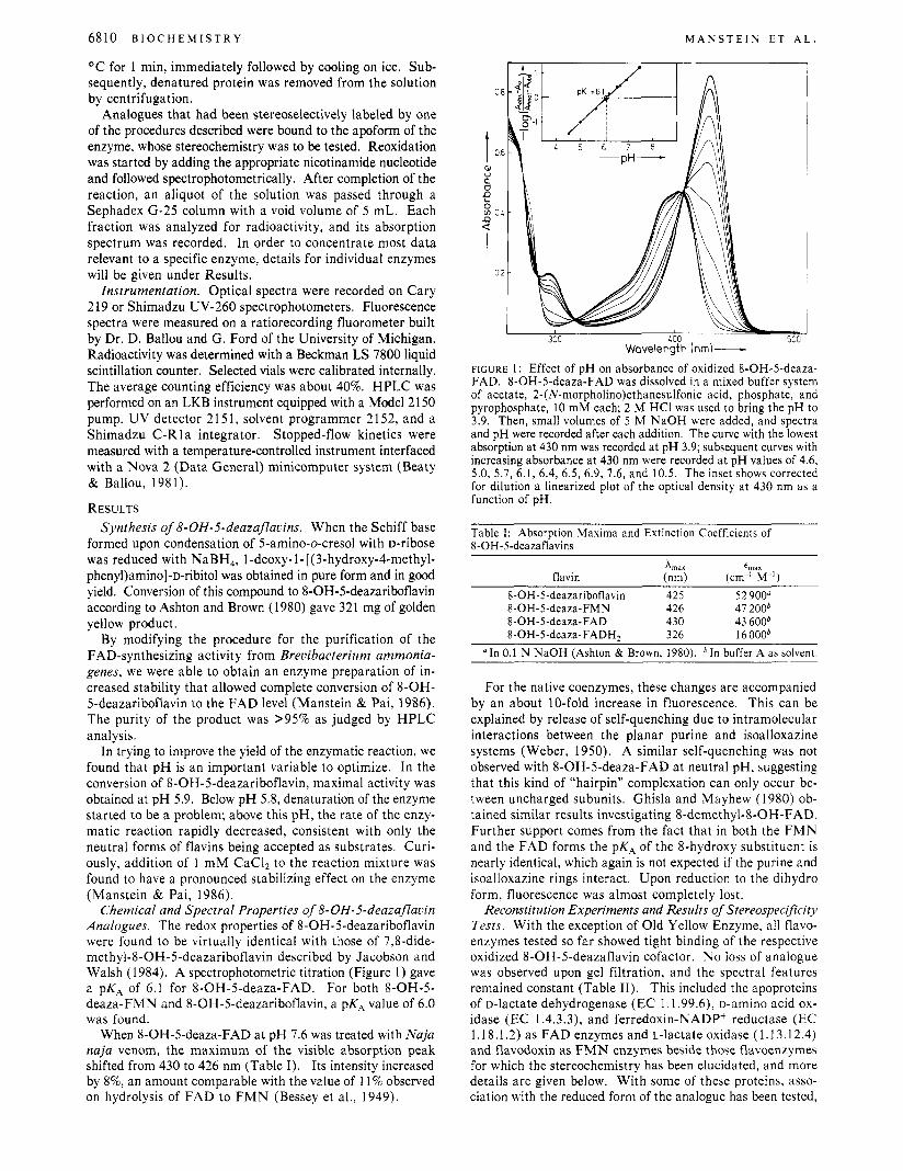
stitution of pig kidney general acyl-CoA dehydrogenase apo- protein with 8-OH-5-deaza-FAD yielded a spectrum (Figure 2) showing a marked decrease in extinction coefficient, t403
= 33 700 cm-' M-' (see also Table 11). Addition of octano- yl-CoA (20 pM) or acetoacetyl-CoA (50 pM) perturbed the oxidized flavin chromophore, leading to an intensification and 5-nm red shift of the absorbance peak. Neither reduction nor long wavelength band formation was detectable upon addition of the thio ester substrates. This reflects the considerably more negative oxidation-reduction potential of 8-OH-5-deaza-FAD compared to FAD.
In contrast to enzyme reconstituted with 8-OH-FAD or 8-SH-FAD, which shows a marked tendency to lose its re- spective chromophore (Thorpe & Massey, 1983), the 8-OH- 5-deaza-FAD form of general acyl-CoA dehydrogenase was stable and could be stored at 4 "C for at least 1 month. This is consistent with the finding that the apoenzyme preferentially binds neutral isoalloxazine species. While the PKA values of 4.8 for free 8-OH-FAD (Ghisla & Mayhew, 1976) and 3.8 for free 8-SH-FAD (Moore et al., 1979), respectively, are too low to be elevated into the neutral pH range upon binding to the dehydrogenase, 8-OH-5-deaza-FAD (pKA = 6.1 ; Figure 1) can be found in its neutral form.
Reconstituted enzyme was reduced as described above. The resulting spectrum is also given in Figure 2. As was the case with enzyme reconstituted with 5-deaza-FAD, NaB3H4 re- duced the bound flavin stereoselectively (Ghisla et al., 1984), introducing the tritium label at the re side of the flavin ring, as will be documented below.
Glutathione Reductase (EC I .6.4.2). When 8-OH-5-dea- za-FAD was incubated with the apoprotein of glutathione reductase from human erythrocytes, the reconstitution reaction was clearly biphasic. Rapid initial attachment of the flavin to the protein moiety was followed by a slow rearrangement. Such a behavior has also been described for the rebinding of FAD to the apoprotein (Staal et al., 1969). Spectral data for the species obtained after incubation for 1 h are given in Table
V O L . 2 5 , N O . 2 2 , 1 9 8 6 6811
-Wavelength (nm) - FIGURE 2: Optical absorption spectra of 14.5 WM pig kidney general acyl-CoA dehydrogenase reconstituted with oxidized (-) or NaBH,-reduced (--) 8-OH-5-deaza-FAD in 50 mM phosphate buffer-0.3 mM EDTA, p H 7.6, a t 4 OC.
11. The reconstituted enzyme had no detectable reductase activity, which is consistent with the results of Chan and Bruice (1 977), who reported that 5-deazaflavins cannot transfer electrons to thiols, and also with the finding of Krauth-Siege1 et al. (1985) that glutathione reductase when reconstituted with 5-deaza-FAD has no reductase activity anymore. How- ever, the addition of a 4-fold excess of NAD' to a solution containing reduced 8-OH-5-deaza enzyme led to the complete reoxidation of the flavin chromophore within the time of mixing. One equivalent of NADPH reduced about 50% of the enzyme-bound 8-OH-5-deaza-FAD. Excess reductant did not significantly increase the amount of reduced enzyme. A very similar result was obtained when glutathione reductase reconstituted with 5-deaza-FAD was titrated with NADPH (R. L. Krauth-Siegel, S. Ghisla, and E. F. Pai, unpublished results). The fact that the reduced form of the analogue binds tightly to the apoenzyme and the competence of 8-OH-5-de- azaglutathione reductase in catalyzing transhydrogenation are of major importance since glutathione reductase is the only flavoenzyme for which the stereochemistry of catalysis has been independently established with the native enzyme (Pai & Schulz, 1983). This knowledge is the basis for all inter- pretations of the results described below.
A 540-pL aliquot of buffer A containing 4.5 nmol of ste- reoselectively labeled 8-OH-5-deaza [ S 3 H ] FADH,, obtained by reduction of 8-OH-5-deaza general acyl-CoA de- hydrogenase with NaB3H4 as described above, was added to 6.5 nmol of glutathione reductase apoenzyme in 250 pL of the same buffer. The solution was incubated for 60 min at 4 OC in the dark before it was mixed with 20 WL of an 11 mM solution of NAD+. Reoxidation was more than 95% complete within the time of mixing. A total of 600 pL of this solution was passed through a Sephadex G-25 fine column, and frac- tions of 900 pL were collected. Only 9% of the radioactivity coeluted with the protein, the rest of the radioactivity was found in fractions containing pyridine nucleotides (Figure 3B).
As further proof that the label is bound to the FAD ana- logue before reoxidation with NAD', we repeated the ex-

6812 B I O C H E M I S T R Y M A N S T E I N E T A L .
i 0
t E
c - 2 a
2cooo
1500C
toow
500C
IMO
t
? 0
Y E
e
a U , 20000
15wO
loo00
S O X
low ib 15
~ F r o c t i o n s - FIGURE 3: (A) Sephadex G-25 gel filtration profile of glutathione reductase reconstituted with 8-0H-5-dea~a[5-~H]FADH~. The FAD analogue was stereoselectively labeled by NaB3H4 reduction while bound to general acyl-CoA dehydrogenase and then transferred to glutathione reductase apoenzyme as described in the text. (B) Gel filtration profile after completely reoxidizing this solution by the addition of a large excess of NAD’. Fractions containing 900 pL were collected: (-) Azso; (--) Ad36.
periment under otherwise identical conditions but did not add NAD+. Again, 600 pL of the solution containing the still- reduced 8-OH-5-deaza analogue bound to glutathione re- ductase was applied to the gel filtration column. This time, 80% of the label was found to coelute with the protein (Figure 3A).
Mercuric Reductase (EC 1.6.x.x). Incubation of 8-OH- 5-deaza-FAD with 1.2 equiv of apoenzyme at 4 OC for 1 h lead to a 7-nm blue shift of the flavin absorption maximum. As had been found with glutathione reductase, association between modified flavin and apoprotein was a two-step process. Again, only transhydrogenase activity can be restored by re- constitution with 8-OH-5-deaza-FAD. A slightly different spectral change (Table 11) and reduced transhydrogenase activity were observed when the analogue was bound to a mutant enzyme that had the “proximal” cysteine, Cys-135, replaced by a serine. The reduced flavin is bound tightly to both native and mutant enzymes, and for reconstitution, the same protocol can be used as with the oxidized analogue.
A total of 4 nmol of chiral8-0H-5-dea~a[5-~H]FADH~ was incubated with 10 nmol of mercuric reductase apoenzyme a t 20 OC for 60 min; 500 nmol of NAD+ dissolved in 10 pL of buffer A was added, and then 750 pL of this solution was applied to a gel filtration column and 900-kL fractions were collected. A total of 84% of the counts coeluted with the pyridine nucleotide pool; only 16% was found in fractions containing protein.
The stereospecificity test was also performed with the Ser-135 mutant enzyme of mercuric reductase. A total of 5 nmol of the labeled analogue was incubated with 18 nmol of apoprotein at 20 O C for 5 min. Ten minutes after the addition of NAD+, no further changes in the visible spectrum of the enzyme-bound chromophore could be detected. When frac-
tions from the gel filtration column were tested for radioac- tivity, only 7% of the label was found to coelute with the protein; 93% was found in the small molecule fractions.
Thioredoxin Reductase (EC 1.6.4.5). Apoprotein of E. coli thioredoxin reductase immediately recombined with oxidized or reduced 8-OH-5-deaza-FAD. As with glutathione reductase and mercuric reductase, only transhydrogenase activity was restored. Reconstitution of the apoenzyme with 8-OH-5- deaza-FADH, yielded the spectrum of the fully reduced chromophore. Contrary to what had been found with gluta- thione reductase and mercuric reductase, the intensity of the visible absorption peak of the flavin chromophore came back to only -80% when the reduced form of the enzyme was reoxidized by addition of NAD+. This was accompanied by a shift of the maximum from 414 nm to 396 nm with a shoulder a t 414 nm still observable. However, when this enzyme was denatured, the flavin released was completely oxidized; therefore, the spectral differences should not be due to incomplete reoxidation. In order to check whether the denaturation procedure caused further oxidation of the chromophore, the same kind of treatment was performed with fully reduced enzyme. No reoxidation of the flavin analogue could be observed. When AcPyADP’ was used instead of NAD+, reoxidation was much faster, but the resulting spec- trum still appeared as described above.
After 5 nmol of chiral 8 - 0 H - 5 - d e a ~ a [ 5 - ~ H ] F A D H ~ in buffer A had been incubated with a 5-fold excess of thioredoxin reductase apoprotein a t 4 O C for 10 min, 200 nmol of Ac- PyADP’ was added to reoxidize the reconstituted enzyme. A 300-pL aliquot was passed through the G-25 fine column, and 900-pL fractions were collected. A total of 9% of the radio- activity was found to coelute with the protein, 91% with the pyridine nucleotides.
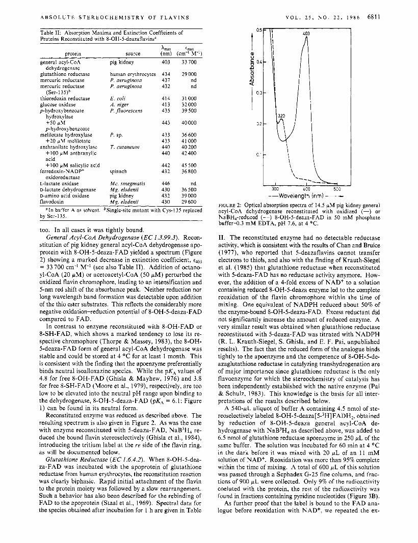
Glucose Oxidase (EC 1 . I .3.4). At 25 O C it took 20 min until the apoprotein of A. niger glucose oxidase had completely combined with 8-OH-5-deaza-FAD. The reaction could be followed spectrophotometrically when an excess of apoprotein was added. As with the 5-deaza-FAD-reconstituted enzyme (Fisher et al., 1976), addition of D-glucose caused rapid bleaching of the flavin peak. The spectra indicated that re- duction of the bound flavin went nearly to completion (Figure 4). In further analogy of the 5-deaza-FAD enzyme, no oxygen was consumed when D-ghCOSe was used to reduce the recon- stituted enzyme, indicating that reduction was stoichiometric, not catalytic. When glucose oxidase was reconstituted with reduced 8-OH-5-deaza-FAD and passed through a Sephadex (3-25 column, no reoxidation could be detected. Even after addition of 2 equiv of gluconolactone, the enzyme stayed in its reduced form.
In order to describe the kinetics of the reduction of 8-OH- 5-deaza-FAD-reconstituted glucose oxidase by substrate in more detail, stopped4 ow experiments were performed (Figure 4), measuring the changes of absorbance a t 410, 350, and 320 nm. All three wavelengths gave the same results. The rate of reduction was proportional to D-glucose concentration up to the highest concentrations that could be used. The corre- sponding second-order rate constant was 24.7 M-’ s-’. Also with the native glucose oxidase a similar lack of evidence for the formation of a Michaelis complex in the first step of the reductive half-reaction has been reported. With the native enzyme, the second-order rate constant under similar condi- tions is - lo4 M-’ s-’ (Gibson et al., 1964).
8-OH-5-deaza-FAD bound to glucose oxidase apoprotein could be reduced by incubation with [ l-3H]-~-glucose. When the now stereospecifically labeled flavin was transferred to 30
A B S O L U T E S T E R E O C H E M I S T R Y O F F L A V I N S
100 200 300 LOO 500
\ ~~~ \L I
LOO 500 661 -Wavelength hm)-
FIGURE 4: Reduction by D-glucose of 8-OH-5-deaza-FAD bound to glucose oxidase. (-) Absorption spectrum of 4 WM 8-OH-5-dea- za-FAD-reconstituted glucose oxidase in 50 mM phosphate buffer43 mM EDTA, pH 7.6, at 25 O C . Addition of D-glucose to a final concentration of 35 mM led to the formation of spectrum (--). The inset shows a plot of the rate of the half-reaction, E,, + glucose - EFd*P, as determined by mixing 8-OH-5-deaza-FAD-reconstituted glucose oxidase with D-glucose in the stopped-flow apparatus.
nmol of mercuric reductase apoprotein and stereospecificity was analyzed as described above with 100 nmol of NAD' for reoxidation, only 7% of the tritium was found in the protein peak, the rest being associated with small molecules.
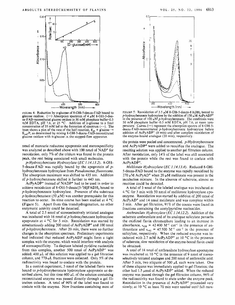
p-Hydroxybenzoate Hydroxylase (EC 1.14.1 3.2). 8-OH- 5-deaza-FAD was rapidly bound by the apoprotein of p - hydroxybenzoate hydroxylase from Pseudomonas fluorescens. The absorption maximum was shifted to 435 nm. Addition of p-hydroxybenzoate shifted it further to 443 nm.
AcPyADP' instead of NADP+ had to be used in order to achieve reoxidation of 8-OH-5-deaza [ 5-3H] FADHz bound to p-hydroxybenzoate hydroxylase. Presence of the substrate p-hydroxybenzoate (50 pM) was another prerequisite for this reaction to occur. Its time course has been studied a t 4 O C (Figure 5). Apart from this transhydrogenation, no other enzymatic activity could be detected.
A total of 2.5 nmol of stereoselectively tritiated analogue was incubated with 16 nmol of p-hydroxybenzoate hydroxylase apoprotein a t 4 "C for 5 min. Reoxidation was started by simultaniously adding 200 nmol of AcPyADP' and 100 nmol of p-hydroxybenzoate. After 20 min, there were no further changes in the absorption spectrum. Preliminary experiments had indicated that reduced AcPyADP might form a tight complex with the enzyme, which would interfere with analysis of stereospecificity. To displace labeled pyridine nucleotide from this complex, another 500 nmol of AcPyADP' was added; 400 p L of this solution was applied to a gel filtration column, and 770-pL fractions were collected. Only 5% of the radioactivity was found to coelute with the protein.
In a control experiment, 5 nmol of the labeled flavin were bound to p-hydroxybenzoate hydroxylase apoprotein as de- scribed above, but this time 600 pL of the solution containing reconstituted enzyme was passed directly through a gel fil- tration column. A total of 90% of the label was found to coelute with the enzyme. Now fractions containing most of
V O L . 2 5 , N O . 2 2 , 1 9 8 6 6813
I
20' n
I I 300 L 00 500 -Wavelength Inm)-
FIGURE 5: Reoxidation of 3.5 WM 8-OH-5-deaza-FADH2 bound to p-hydroxybenzoate hydroxylase by the addition of 150 WM AcPyADP' in the presence of 100 WM p-hydroxybenzoate. The conditions were 50 mM phosphate buffer-0.3 mM EDTA, pH 7.6, at room tem- perature. Curves (-) represent the absorption spectra of 8-OH-5- deaza-FAD-reconstituted p-hydroxybenzoate hydroxylase before addition of AcPyADP' (0 min) and after complete reoxidation of the enzyme-bound analogue (20 min), respectively.
the protein were pooled and concentrated. p-Hydroxybenzoate and AcPyADP' were added to reoxidize the analogue. The resulting solution was applied to another gel filtration column. After reoxidation, only 14% of the label was still associated with the protein while the rest was found to coelute with AcPy ADP'.
Melilotate Hydroxylase (EC I .14.13.4). Reduced 8-OH- 5-deaza-FAD bound to the enzyme was rapidly reoxidized by 270 p M AcPyAD' when 20 pM melilotate was present in the incubation mixture. In the absence of substrate, almost no reaction could be detected.
A total of 3 nmol of the labeled analogue was incubated a t 4 O C for 5 min with 50 nmol of melilotate hydroxylase apo- enzyme. Reoxidation was started by addition of 200 nmol of AcPyADf and 14 nmol melilotate and was complete within 3 min. After gel filtration, 91% of the counts were found in fractions containing the acetylpyridine nucleotides.
Anthranilate Hydroxylase (EC 1.14.12.2). Addition of the substrate anthranilate and of its analogue salicylate perturbs the oxidized flavin chromophore leading to an increase in absorbance, = 42400 M-' cm-I in the presence of an- thranilate and t442 = 45 500 M-' cm-' in the presence of salicylate, respectively. When the reduced enzyme was in- cubated with 2.7 m M AcPyADP' a t 10 O C in the presence of substrate, slow reoxidation of the enzyme-bound flavin could be obtained.
A total of 18 nmol of anthranilate hydroxylase apoenzyme was incubated a t 10 O C in the presence of 9 nmol of stereo- selectively tritiated analogue and 200 nmol of anthranilic acid. After 5 min, two aliquots of 500 pL each were taken. One of these aliquots was immediately applied to gel filtration; the other had 1.5 pmol of AcPyADP' added. When the reduced enzyme was passed through the gel filtration column, 96% of the radioactivity was found to elute under the enzyme peak. Reoxidation in the presence of AcPyADP' proceeded very slowly; a t 10 O C a t least 70 min were needed until full reox-
68 14 B I o c H E M I s T R Y M A N S T E I N E T A L .
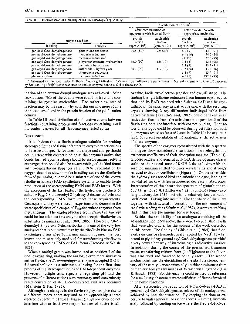
Table 111: Determination of Chirality of 8 - 0 H - 5 - d e a ~ a [ 5 - ~ H ] F A D H ~ ~ distribution of tritiumb
after reconstitution of apoprotein with labeled flavin
after reoxidation with appropriate nucleotide
protein nucleotide protein nucleotide fraction fraction fraction fraction enzyme used for
labeling analysis (cpm x IO3) (cprn x IO3) (cpm x 10') (cpm x io3) gen acyl-CoA dehydrogenase glutathione reductase 39.5 (80)c 9.9 (20) 4.2 (9) 43.0 (91) gen acyl-CoA dehydrogenase mercuric reductase 16.1 (16) 84.0 (84)
gen acyl-CoA dehydrogenase p-hydroxybenzoate hydroxylase 36.0 (90) 4.0 (10) 1.2 ( 5 ) 22.3 (95) gen acyl-CoA dehydrogenase melilotate hydroxylase 3.3 (9) 33.7 (91) gen acyl-CoA dehydrogenase anthranilate hydroxylase 38.7 (90) 4.3 (10) 13.7 (24) 43.3 (76) gen acyl-CoA dehydrogenase thioredoxin reductase 6.4 (9) 63.7 (91) glucose oxidase' mercuric reductase 14.5 (7) 192.5 (93)
gen acyl-CoA dehydrogenase mercuric reductase" 2.0 (7) 27.0 (93)
"Performed as described under Methods. bAfter gel filtration. 'Values in parentheses are percentages. "Mutant enzyme with Cys-135 replaced by Ser-135. e [ l-3H]Glucose was used to reduce enzyme-bound 8-OH-5-deaza-FAD.
idation of the enzyme-bound analogue was achieved. After reoxidation, 76% of the counts were found in fractions con- taining the pyridine nucleotides. The rather slow rate of reaction may be the reason why with this enzyme more counts than usual are found in the protein fraction of the gel filtration column.
In Table I11 the distribution of radioactive counts between fractions containing protein and fractions containing small molecules is given for all flavoenzymes tested so far.
DISCUSSION It is obvious that a flavin analogue suitable for probing
stereospecificities of flavin cofactors in enzymic reactions has to have several specific features: its shape and charge should not interfere with correct binding at the enzyme's active site; bonds formed upon labeling should be stable against solvent exchange; there should also be no scrambling of the kind found with Sdeazaflavins (Spencer et al., 1976); reoxidation by oxygen should be slow to make handling easier; the riboflavin form of the analogue should be a substrate of one of the known riboflavin kinase/FAD synthetase systems to enable facile production of the corresponding FMN and FAD forms. With the exception of the last feature, the hydrolysis products of cofactor F420, 7,8-didemethyl-8-hydroxy-5-deazariboflavin and the corresponding FMN form, meet these requirements. Consequently, they were used in experiments to determine the stereospecificities of a couple of F,,,-dependent enzymes from methanogens. The oxidoreductase from Beneckea harveyi could be included, as this enzyme also accepts riboflavins as substrates (Yamazaki et al., 1980, 1985). However, 7,8-di- demethyl-8-hydroxy-5-deazariboflavin is one of the very few analogues that is not turned over by the riboflavin kinase/FAD synthetase from Brevibacterium ammoniagenes, the best known and most widely used tool for transforming riboflavins to the corresponding FMN or FAD forms (Jacobson & Walsh, 1984).
When a methyl group was introduced at position 7 of the isoalloxazine ring, making the analogue even more similar to native flavin, the B. ammoniagenes enzyme accepted 8-OH- 5-deazariboflavin as a substrate, allowing for the first time probing of the stereospecificities of FAD-dependent enzymes. However, multiple tests especially regarding pH and the presence of different cations were necessary until conveniently rapid conversion of 8-OH-5-deazariboflavin was obtained (Manstein & Pai, 1986).
Although the changes in the flavin ring system give rise to a different redox chemistry and to an appreciably altered electronic spectrum (Table I, Figure l), they obviously do not interfere with at least two major features of native isoall-
oxazine, facile two-electron transfer and overall shape. The finding that glutathione reductase from human erythrocytes that had its FAD replaced with 5-deaza-FAD can be crys- tallized in the same way as native enzyme, with the resulting crystals showing X-ray diffraction indistinguishable from native patterns (Krauth-Siegel, 1982), could be taken as an indication that at least the substitution at position 5 of the flavin ring does not interfere with correct binding. That no loss of analogue could be observed during gel filtration with all enzymes tested so far and listed in Table I1 also argues in favor of correct orientation of the analogue at the active sites of these enzymes.
The spectra of the enzymes reconstituted with the respective analogues show considerable variations in wavelengths and extinction coefficients of their absorption maxima (Table 11). Glucose oxidase and general acyl-CoA dehydrogenase clearly stabilize the neutral state of 8-OH-5-deazaflavins with ab- sorption maxima shifted to lower wavelengths and strongly reduced extinction coefficients (Figure 1). On the other side, the hydroxylases tested bind the anionic analogue, leading to red-shifted peaks with less pronounced decrease in absorption. Interpretation of the absorption spectrum of glutathione re- ductase is not so straightforward as it combines long-wave- length absorption (434 nm) with a relatively low extinction coefficient. Taking into account also the shape of the curve together with structural information on the environment of the flavin binding site (Schulz et al., 1982), it seems more likely that in this case the anionic form is bound.
Besides the availibility of an analogue combining all the advantages mentioned above, there were two published results that were also crucial for the success of the work described in this paper. The finding of Ghisla et al. (1984) that 5-de- azaflavin can be stereoselectively labeled by NaB3H4 when bound to pig kidney general acyl-CoA dehydrogenase provided a very convenient way of introducing a radioactive marker. In addition, during the course of the present work another route, transferring tritium from [ 1 - 3 H ] g l ~ c ~ ~ e to the flavin, was also tried and found to be equally useful. The second anchor point was the elucidation of the absolute stereochem- istry of the catalytic mechanism of glutathione reductase from human erythrocytes by means of X-ray crystallography (Pai & Schulz, 1983). So, this enzyme could be used as reference for elucidating absolute stereospecificities of flavins involved in enzymic reactions.
After stereoselective reduction of 8-OH-5-deaza-FAD in general acyl-CoA dehydrogenase, release of the analogue was achieved by heat denaturation. By keeping the time of ex- posure to high temperature rather short (- 1 min), immedi- ately followed by cooling on ice where the free 8-OH-5-de-
A B S O L U T E S T E R E O C H E M I S T R Y O F F L A V I N S V O L . 2 5 , N O . 2 2 , 1 9 8 6 6815
question of accessibility of the flavin ring to solvent or other small molecules.
Flavodoxin might be a first test case for this approach. Inspection of its molecular structure on a graphics system, using the coordinates deposited with the Protein Bank, Brookhaven National Laboratory, by Dr. M. Ludwig, suggests the presence of a small channel that should allow access of small molecules to the si side of the isoalloxazine ring.
ACKNOWLEDGMENTS We are grateful to S. Ghisla for many very helpful dis-
cussions. We thank all the colleagues mentioned in the text who provided us with samples of purified enzymes. D.J.M. and E.F.P. thank K. C. Holmes for continuous support.
Registry No. ATP, 56-65-5; 8-OH-5-deaza-FMN, 104324-32-5; 8-OH-5-deaza-FAD, 104324-33-6; 8-OH-5-deaza-FADH2, 104324- 34-7; E C 1.6.4.2, 9001-48-3; E C 1.3.99.3, 9027-65-0; EC 1 . 6 . ~ . ~ , 67880-93-7; E C 1.6.4.5, 9074-14-0; EC 1.1.3.4, 9001-37-0; E C 1.14.13.2, 9059-23-8; EC 1.14.13.4, 37256-72-7; EC 1.14.12.2, 37256-68-1; E C 1.18.1.2, 9029-33-8; E C 1.13.12.4, 9028-72-2; E C 1.1.99.6,9028-36-8; E C 1.4.3.3, 9000-88-8; 8-OH-5-deazariboflavir1, 7 14 15-45-7; 1 -deoxy- 1 - [ (3-hydroxy-4-methylphenyl)amino] -~-ribitol, 77994-63-9; 5-amino-o-cresol, 2835-95-2; D-ribose, 50-69-1.
REFERENCES Ashton, W. T., & Brown, R. D. (1980) J . Heterocycl. Chem.
Beaty, N., & Ballou, D. P. (1981) J . Biol. Chem. 256,
Bessey, 0. A., Lowry, 0. H., & Love, R. H. (1949) J . Biol.
Bruice, T. C. (1980) Acc. Chem. Res. 13, 256-262. Brumby, P. E., & Massey, V. (1966) Biochem. Prep. 12,
Briistlein, M., & Bruice, T. C. (1972) J . Am. Chem. SOC. 94,
Chan, R. L., & Bruice, T. C. (1977) J . Am. Chem. SOC. 99,
Choong, Y. S., Shephard, M. G., & Sullivan, P. A. (1975)
Detmer, K., Schopfer, L. M., & Massey, V. (1984) J . Biol.
Entsch, B., Ballou, D. P., & Massey, V. (1976) J . Biol. Chem.
Entsch, B., Husain, M., Ballou, D. P., Massey, V., & Walsh,
Enzyme Nomenclature (1984) Academic, New York. Fisher, J., & Walsh, C. (1974) J . Am. Chem. SOC. 96,
Fisher, J., Spencer, R., & Walsh, C. (1976) Biochemistry 15,
Fritsch, K.-G. (1982) Diplomarbeit, Freie Universitat Berlin. Fox, B., & Walsh, C. T . (1982) J . Biol. Chem. 257,
Ghisla, S., & Mayhew, S. G. (1976) Eur. J . Biochem. 63,
Ghisla, S . , & Mayhew, S. G. (1980) Methods Enzymol. 66,
Ghisla, S., Thorpe, C., & Massey, V. (1984) Biochemistry 23,
Gibson, Q. H., Swoboda, B. E. P., & Massey, V. (1964) J . Biol. Chem. 239, 3927-3934.
Hamilton, G. A. (1971) in Progress in Bioorganic Chemistry (Kaiser, E. T., & KEzdy, T. J., Eds.) pp 83-137, Wiley- Interscience, New York.
Hemmerich, P. (1 976) in Progress in the Chemistry of Organic Natural Products (Herz, W., Grisehach, H., & Kirby, G.
17, 1709-1712.
10634-1 0643.
Chem. 180, 755-769.
29-4 1.
6548-6549.
6721-6730.
Biochem. J . 145, 37-45.
Chem. 259, 1523-1 538.
251, 2550-2563.
C. (1980) J . Biol. Chem. 251, 1420-1429.
4345-4346.
1054-1 064.
2498-2503.
373-390.
241-253.
3 1 54-3 1 6 1.
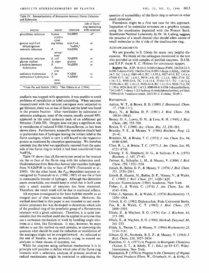
Table IV: Stereochemistry of Interaction between Flavin Cofactors and Substrates
side of flavin ring interacting
enzyme source substrate with substrate glutathione reductase
general acyl-CoA dehydrogenase
mercuric reductase
thioredoxin reductase glucose oxidase p-hydroxybenzoate
hydroxylase
melilotate hydroxylase anthranilate hydroxylase
human erythro- cytes
pig kidney
P. aerugi- nosa
E . coli A . niger P .
Juores- cens
P . sp. T.
cuta- neurn
NADPH
acyl-CoA, NaBH4b
NADPH
NADPH glucose NADPH
NADH NADPH
re"
re
re
re re re
re re
"From Pai and Schulz (1983). bSee Ghisla et al. (1984).
azaflavin was trapped with apoprotein, it was possible to avoid problems of reoxidation or label scrambling. When enzymes reconstituted with the reduced analogues were subjected to gel filtration, there was no loss of flavin and the label was found in the protein fraction. After reoxidation by substrates or substrate analogues, most of the counts, usually around 90%, appeared in the small molecule peak of an additional gel filtration (Table 111). Oxygen does not play a significant role in reoxidation of reduced 8-OH-5-deazaflavins, as has been shown above. Furthermore, unspecific reoxidation should lead to preferential loss of hydrogen leaving the tritium label at the flavin analogue, which in turn is still bound to the respective protein as monitored by absorption spectroscopy. We therefore conclude that the label was specifically removed from the same side of the flavin ring to which it had been transferred from NaB3H,.
Table IV shows that allflauoenzymes tested so far interact via the re face of the flavin ring with the substrates used. Oxidoreductase from Beneckea hameyi also transfers hydrogen from NAD(P)H to the re face of FMN (Yamazaki et al., 1980). On the other hand, the F420-dependent enzymes in- vestigated by Yamazaki et al. (1980, 1985) all use the si face in stereospecific transfer of hydrogen. Although this distinction seems remarkable, one should keep in mind that in both cases only a small number of enzymes has been examined. Therefore;the result could still be due to statistical effects.
All enzymes investigated so far have hydride transfer as an integral part of their reaction mechanism. However, the method described in this paper is not intended to test mech- anistic proposals but was developed to determine which side of the prochiral ring of the prosthetic group of a flavoenzyme interacts with a given substrate. Therefore, it is quite con- ceivable that this method could also be applied to enzymes that use a carbanion mechanism or work by handling single elec- trons at a time, like flavodoxin. It may, however, be more tedious to use this method on such proteins, as answering the question what should be used for reduction or reoxidation of the analogue might not be straightforward. As indicated at the end of Results, we are presently trying to extend our analyses to these classes of enzymes, too.
While for enzymes using carbanion mechanisms it is in principle still possible to define which side of the flavin ring interacts with a substrate, analyses of proteins involved in radical mechanisms might be restricted to addressing the

6816 B I O C H E M I S T R Y
W., Eds.) pp 421-527, Springer-Verlag, Wien. Hersh, L. B., & Jorns, M. S. (1975) J . Biol. Chem. 250,
Hersh, L. B., & Walsh, C. (1980) Methods Enzymol. 66,
Hersh, L. B., Jorns, M. S., Peterson, J., & Currie, M. (1976)
Jacobson, F., & Walsh, C. (1984) Biochemistry 23,979-988. Jorns, M. S., & Hersh, L. B. (1974) J . Am. Chem. SOC. 96,
Jorns, M. S., & Hersh, L. B. (1975) J . Biol. Chem. 250,
Jorns, M. S., & Hersh, L. B. (1976) J . Biol. Chem. 251,
Krauth-Siegel, R. L. (1982) Ph.D. Thesis, University of
Krauth-Siegel, R. L., Schirmer, R. H., & Ghisla, S. (1985)
Krohne-Ehrich, G., Schirmer, R. H., & Untucht-Grau, R.
Loechler, E. L., & Hollocher, T. C. (1980) J . Am. Chem. SOC.
Manstein, D. J., & Pai, E. F. (1986) J . Biol. Chem. (in press). Massey, V., & Curti, B. (1966) J . Biol. Ckem. 241,
Mayer, E. J., & Thorpe, C. (1981) Anal. Biochem. 116,
Mayhew, S. G., & Massey, V. (1966) J . Biol. Chem. 244,
Moore, E. G., Ghisla, S., & Massey, V. (1979) J . Biol. Chem.
O’Brien, D., Weinstock, L., & Cheng, C. (1970) J. Heterocycl.
8728-8734.
277-287.
J . Am. Chem. SOC. 98, 865-867.
401 2-40 14.
3620-3628.
4872-4881.
Heidelberg.
Eur. J . Biochem. 148, 335-344.
(1977) Eur. J . Biochem. 80, 65-71.
102, 7312-7334.
341 7-3423.
227-229.
794-802.
254, 8173-8178.
M A N S T E I N E T A L .
Chem. 7, 99-105.
Chem. 259, 2243-2251. O’Donnell, M. E., & William
Olson, S . T., & Massey, V
Pai, E. F., & Schulz, G. E. 47 14-4724.
1752-1 7 58.
, C. H., Jr. (1984) J . Biol.
(1 979) Biochemistry 18,
1983) J . Biol. Chem. 258,
Powlowski, J., & Dagley, S. (1982) in Flavins and Flavo- proteins (Massey, V., & Williams, C. H., Jr., Eds.) pp 339-341, Elsevier/North-Holland, New York.
Schultz, P. G., Au, K. G., & Walsh, C. T. (1985) Biochemistry
Schulz, G. E., Schirmer, R. H., & Pai, E. F. (1982) J. Mol.
Spencer, R., Fisher, J., & Walsh, C. (1976) Biochemistry I 5
Staal, G. E. J., Visser, J., & Veeger, C. (1969) Biochim.
Strickland, S., & Massey, V. (1973) J . Biol. Chem. 248,
Sullivan, P. A., Choong, Y. S., Schreuers, W. A., Cutfield, J. F., & Shephard, M. G. (1977) Biochem. J . 165,375-383.
Swoboda, B. E. P. (1969) Biochim. Biophys. Acta 175,
Swoboda, B. E. P., & Massey, V. (1965) J . Biol. Chem. 240,
Thorpe, C., & Massey, V. (1983) Biochemistry 22,
Thorpe, C., & Williams, C. H., Jr. (1976) J . Biol. Chem. 251,
Thorpe, C., Matthews, R. G., & Williams, C. H., Jr . (1979)
Walsh, C. (1980) Acc. Chem. Res. 13, 148-155. Wassink, J. H., & Mayhew, S. G. (1975) Anal. Biochem. 68,
Weber, G. (1950) Biochem. J . 47, 114-121. Yamazaki, S., Tsai, L., Stadtman, T. C., Jacobson, F. S . , &
Walsh, C. (1980) J . Biol. Chem. 255, 9025-9027. Yamazaki, S., Tsai, L., Stadtman, T. C., Teshima, T., Nakaji,
A., & Shiba, T. (1985) Proc. Natl. Acad. Sci. U.S.A. 82,
Zanetti, G., & Curti, B. (1980) Methods Enzymol. 69,
Zanetti, G., Cidaria, D., & Curti, B. (1982) Eur. J. Biochem.
24, 6840-6848.
Biol. 160, 287-308.
1043-1 053.
Biophys. Acta 185, 39-48.
2944-2952.
365-379.
2209-2215.
2972-2978.
7 7 26-7 7 28.
Biochemistry 18, 33 1-337.
609-6 16.
1364-1 366.
250-254.
126, 453-458.