of loop films for prosected - eric · an evaluative investigation of silent loop films in the...
TRANSCRIPT

DOCUMENT RESUME
ED 029 796 24 SE 006 819
By-Welser. John R.An Evaluative Investigation of Silent Loop Films in the Teaching of Anatomy. Final Report.Purdue Univ.. Lafayette. Ind.Spons Agency-Office of Education (DHEW). Washington. D.C. Bureau of Research.Bureau No-BR-7-E-143Pub Date Apr 69Grant- 0EG-0-8-014300-0206-010Note- 68p.EDRS Price MF-$0.50 HC-$3.50Descriptors-Anatomy. Biology. College Science. Comparative Analysis. Conventional Instruction. Filmstrips.Instruction. Instructional Media. Zoology
Investigated were (1) the acceptance and effectiveness of silent film loops as ateaching and review aid. (2) the possible substitution of loop films for prosectedand/or fresh dissection materials. and (3) the comparative costs of loop films used inthe presentation of five units of a gross anatomy course and six units of an appliedanatomy course. The students in each of the two ciasses. were divided into threerandomly selected groups: Differences between the treatment groups were evaluatedutilizing objective tests. student opinion questionnaires, time records. instructionalcosts, and loop film production costs. Results for the anatomy units indicated (1)there were no significant difference apparent between treatment groups immediatelyfollowing the presentation of a subject unit. (2) the addition of film loops to a methodof presentation appeared to aid retention. (3) a savings of time was apparent forthe treatment group having loops as their guide in a technique-oriented exerdse, (4)students liked the loops as aid but had doubts about their effectiveness as a primarymeans of instruction. (5) there were no significant differences in achievement as aresult of the different procedures. (6) students preferred the film loops for reviewover active dissection or viewing the prosected cadavers: (RS)
"'".:

Cf
CY's
C\1
LL-1
FINAL REPORT
Project No. 7-E-143
Grant No. 0EG-0-8-014300-0206-010
U.S. DEPARTMENT OF HEALTH, EDUCATION & WELFARE
OFFICE OF EDUCATION
THIS DOCUMENT HAS BEEN REPRODUCED EXACTLY AS RECEIVED FROM THE
PERSON OR ORGANIZATION ORIGINATING IT. POINTS OF VIEW OR OPINIONS
STATED DO NOT NECESSARILY REPRESENT OFFICIAL OFFICE OF EDUCATION
POSITION OR POLICY.
612- 47
AN EVALUATIVE INVESTIGATION OF SILENT LOOP FILMS
IN THE TEACHING OF ANATOMY
April 1969
U.S. DEPARTMENT OFHEALTH, EDUCATION, AND WELFARE
Office of EducationBureau of Research
Final Report
Project No. 7-E443
Grant No. 0EG-0-8-014300-0206410
An Evaluative Investigation of Silent Loop Films
In the Teaching of Anatomy
John R. Welser
Purdue University
West Lafayette, Indiana
April 1969
The, =March reported herein was performed pursuant to
a grant with the Office of Education, U.S. Department of
Health, Education, and Welfare. Contractors undertaking
such projects under Government sponsordhip are encouraged
to express freely their professional judgement in the con-
duct of the project. Points of view or opinions stated
do not, therefore, necessatily represent official Office
of Education position or policy.
U.S. DEPARTMENT OFHEALTH* EDUCATION, ANp WELFARE
Office of EducationBureau of Research
11

TABLE OF CONTENTS
PAGE
ACKNOWLEDGEMENTS 4
SUMMARY 5
INTRODUCTION 7
RELATED RESEARCH 3
OBJECTIVES 9
METHODS
BASIC ANATOMY 10
APPLIED ANATOMY 11
RESULTS AND FINDINGS
BASIC ANATOMY 12
APPLIED ANATOMY 14
PRODUCTION COST 15
CONCLUSIONS AND RECOMMENDATIONS 17
REFERENCES 19
APPENDIXESA. DIRECTION, HANDOUT, QUIZ AND OPINION SHEETS
USED IN BASIC ANATOMYB. DIRECTION, HANDOUT, QUIZ AND OPINION SHEETS
USED IN APPLIED ANATOMYC. LISTING OF LOOP FILMSD. LOOP FILM PRODUCTION COSTE. OPINION DATA SUMMARY SHEETS
LIST OF TABLES1. ANALYSIS OF2. ANALYSIS OF
FIRST YEAR3. ANALYSIS OF4. ANALYSIS OF5. ANALYSIS OF6. ANALYSIS OF
21
37
61
65
67
QUIZ 2 TEST SCORES, U.G. TRACT, FIRST YEAR . . 12
QUIZ 2 TEST SCORES, LUMBO-SACRAL PLEXUS,
QUIZ 2 TEST SCORES, BRACHIAL PLEXUS, FIRST YEARVARIANCE, TIME, DOG DEFLESHING, FIRST YEAR . . .
VARIANCE, TIME, BRACHIAL PLEXUS, REVIEW . . .
VARIANCE, TIME, LUMBO-SACRAL PLEXUS, REVIEW . .
12
12
131414

ACKNOWLEDGEMENTS
The author wishes to recognize the contribution of ability, time andeffort of: Professor Algernon R. Allen, Director of Medical Illustration,School of Veterinary Science and Medicine, Purdue University, in theproduction of loop films and Professor Allen R. Starry, Director, Measure-ment and Research Center, Purdue University, in the analysis of data.
I I
-4-

In the future, veterinary anatomy will be taught to more students inless time with a shortage of qualified staff. The number of prosected andfresh dissection specimens that can be made available for student study islimited by both time and money. Silent super 8 mm "single concept" loopfilms may help alleviate some of the problems a...1 serve as a valuableteaching aid in medical schools.
We felt that loop films were effective teaching aids; however, moredefinite data was needed before further expansion of production of loopfilms could be undertaken. The objectives of this project were: (1) Toevaluate the acceptance and effectiveness of silent loop films as ateaching and review aid, (2) To determine whether loop films could besubstituted for prosected and/or fresh dissection materials, and (3) Tocompare the economy of loop films to prosected and fresh dissectionmaterials.
"Single concept" loop films were used in the presentation of fiveunits of the gross anatomy course and six units of the applied anatomycourse. The students in each of the two classes were divided into threerandomly selected groups. In basic anatomy the acceptance and effective-ness of loop films as a primary teaching tool and as a substitute for theprosected guide cadaver was tested. The subject matter was presented totreatment group one in the conventional manner (dissection guide, prosectedcadaver) student dissection of a cadaver), with treatment group two theloop films were substituted for the prosected cadaver, and with treatmentgroup three loop films were substituted for both the prosected cadaver andstudent dissection. In applied anatomy, the acceptance and effectivenessof loop films as review aids and whether or not loop films could be substi-tuted for fixed or fresh prosections were evaluated. The review materialwas presented to group one using loop films and a handout, to group twowith a prosected fixed cadaver plus a handout and to group three with aprosected fresh cadaver plus a handout. Differences between the treatmentgroups in both basic and applied anatomy were evaluated utilizing objectivetests, student opinion questionaires, time records, instructional costsand loop film production costs.
In the basic anatomy units, no significant differences were apparentbetween treatment groups immediately following presentation of a subjectunit; however, the addition of loop films to a method of presentationappeared to aid retention in this study. A saving of time was apparentfor the treatment group having loop films as their guide in a techniqueoriented exercise. The students felt that loop films were extremely use-ful in the correlation of structure with function and as review aids.They also felt that loop films were good teaching aids and could serve asa substitute for the prosected cadaver. However, the students expresseddoubts about the effectiveness and a strong dislike for loop films as aprimary teaching tool (as a substitute for dissection).
In applied anatomy, analyses revealed that the different methods oftreatment resulted in no significant differences in achievement. Studentopinion indicated that super 8 mm loop films were preferred for review

muwarowANP ,4",r0VMFATO.
over active dissection or viewing the prosected fixed cadavers and could
serve as a substitute for the prosected fresh cadaver. As a review aid
loop films will save either the instructor's time in preparation or the
student's time in dissection.
If loop films are used two to three times per year to replace cadavers,
they are relatively inexpensive. However, considering the time involvedto make a good loop film, decisioas concerning the ritost relevant subjeLts
for loop films must be made. As a result of this study, loop films whichwe produce in the future will be in the areas that correlate structurewith function or films which will serve as review aids.

INTRODUCTION
The fast increasing volume and complexity of material to be learnedin modern veterinary science, as in other fields, requires more efficientand improved teaching techniques. Historically, anatomy has occupied acentral position in the veterinary curriculum; however, due to the advance-ment of fhe other preclinical and clinical sciences and the resultingchange in emphasis, this is no longer true. It was recommended, for exam-ple, at the Second PAO/WHO International Meeting on Veterinary Educationthat, in the interest of saving instructional time, anatomy teaching shJuldbe further reduced from its present average of 650 to 480 instructionalhours(1). Such reductions seem likely to be adopted.
Despite efforts to recruit and train larger numbers of veterinaryanatomists, demand in this country continues to exceed the supply. At dhe1966 American Association of Veterinary Anatomists Meeting, Venzke(2)reported that there was an average shortage of 1.5 PMY per department. Thenumber of veterinary school graduates has risen from 192 in 1948 to 860 in1965, with a majority of the increase coming from admission of more stu-dents into the existing professional schools. This trend is expected to
continue(3). Therefore, in the future, veterinary anatomy will be taughtto more students in less time and with a continuing shortage of qualified
staff. It is necessary to analyze and evaluate carefully the customaryand the new approaches to anatomical teaching, as well as the time require-ments and effectiveness of each.
At the School of Veterinary Science and Medicine, Purdue University,the teaching of veterinary gross anatomy closely parallels the program at16 of file other 18 veterinary schools(4). A basic anatamy course is taughtto first year professional students, followed by an applied anatomy coursewhich is taught to third year professional students. To date, both courseshave relied heavily on prosecfed and fresh dissection materials to aid inthe presentation of anatomy. Prosected specimens have the disadvantagesof lacking natural color, of smelling strongly of formalin, of being timeconsuming to prepare, and of having to be renewed every two years or so.Fresh specimens, while having the natural color and consistency, are veryshort-lived. With the advent of new legislation, dogs for fresh dissectionare becoming more difficult to obtain and more expensive. The price hasrisen from $5.00 to $50.00 a piece for a 6 month old dog. Cows, pigs, and
horses which we must use for comparison of anatomical structure have in-creased substantially in price due to the increase in beef and pork pricesand the increased popularity of pleasure horses. Thus, both time andmoney limit the number of prosected and fresh dissected specimens avail-
able for each class period.
-7-

RELATED RESEARCH
Much of medicine is visual or visualizable and is concerned basi-cally with the material from which motion pictures are made. Earlierresearch in the use of educational films has shown them to be effectivein teaching and that they may permit the saving of time. Stein(5), forexample, reported no significant difference between studentr who weretaught typing by using 16 mm film loops and students taught convention-ally. Similar 1.esults were reported by Vander Meer(6) in a study con-ducted with 9th graders and by Snow(7) who compared filmed vs. livephysics lecture demonstrations. Vander Meer(8) also reported that goodfilms can be used as the sole means for teaching some kinds of factualmaterial and performance skills. Film loops can aid inexperienced in-structors or can reduce the burden on experienced instructors. Harby(9)found that film loops projected in daylight proved as effective as liveinstructor demonstrations in the teaching of simple skills, and Murnin(10)reported that using loop films, an instructor with a minimum amount oftraining and experience can teach skills effectively. It has been dhownby Rimland(11) that repetition of a film results in subi%antial incre-ments in learning. If a film is short, it can be repeated more easilyand according to Ruhe(12), "short films better answer the emotional andintellectual needs of individualistic medical staff instructors than doconventional long teaching films." Leveridge(13) reported that byshowing and discussing films, fhe instructor can do a better job ofteaching than he can possibly do in describing a phenomenon verbally.Thus it appears that dhort silent films that can be discussed, that per-mit repetition, and are economical to produce would be of great servicein teaching anatomy, just as in other instructional fields. Silent 8 mm"single concept" loop films appear to answer the above requirements andlend themselves well to teaching in medical schools.
The Single Concept Teaching Film Conference for Medicine(20) re-vealed the familiar group use of film in medical instruction and stressedindividual and small group use of films for self instruction. Markee(14)reported the successful long term use of short 16 mm films in the teachingof anatomy at Duke University. Huber(15) pointed out that the motion ofmodels and specimens in a motion picture gives the viewer the impressionof depth which is an important factor in the study of anatomy. Ham(16)
suggested that movies have their greatest success if they are brief andif they emphasize one or two points to supplement a teaching sequence.This is borne out by Postlethwait(17), who uses silent single conceptloop films to illustrate specific concepts in botany. West and McKim(18),who have utilized 8 mm sound loops, reported that programmed cinematicself-instruction was effective. The educational communication projectat Teachers College, Columbia, under the direction of Forsdale(19) hasmade loop films of several types among which are: a) skill teaching
films, b) drill films, c) motivation films, d) films of phenomena, and
e) situation films. At the Single Concept Film Conference(20), eightof the ten speakers indicated a great need for exploration into the partthat films integrated with other media can play in increasing efficiencyand quality of medical education.
A011001.

.
The Department of Veterinary Anatomy and the Medical Illustration
Section of the School of Veterinary Science and Medicine of Purdue
University, with the advice of Dr. Postlethwait, has produced 14 "single
concept 8 mm loop films. These films were utilized as teaching aids
during 1965-66 in gross and applied anatomy courses. It was felt that
these were well accepted by students and quite effective. The films
were used to replace prosected specimens, to correlate structure with
function, and to serve as review aids. It appeared that less staff
time was spent in going over the same material with individual students,
leaving more time to answer speciac questions. Since films can be
easily reproduced, more could be made available, and it has seemed that
students derive more from the films than the previously used prosected
material. However, no adequate evaluation data was collected then.
OBJECTIVES
From reviewing the literature and our experience using them it
appeared that silent single concept loop fiLms could help alleviate some
of the current problems in the field of medical education and serve as
a valuable teaching adjunct. Before further expansion of the production
of single concept loop films could be justified, data as to their teaching
effectiveness, student acceptance, time involvement for staff and student,
as well as their economy had to be collected. It was decided to test loop
films in the basic anatomy course for their usefulness as primary teaching
tools or aids as compared to the more conventional methods, and in the
applied anatomy course for their usefulness as review aids. The primary
objectives of this study may be briefly stated as follows:
In basic anatomy
1. To evaluate the acceptance and effectiveness of silent
loop films as a primary teaching aid.
2. To determine whether loop films can be substituted for
prosected guides or student cadaver dissection.
3. To compare the relative costs of loop films to fixed
dissection materials.
In applied anatomy
1. To evaluate the acceptance and effectiveness of silent
loop films as a review aid for anatomy.
2. To determine if loop films can be substituted for fresh
and/or fixed dissection material in anatomical review.
3. To compare the economy of loop films to fresh and/or
fixed dissection materials.
-9-

METHODS
Basic Anatomy
The basic anatomy course is taught to 60 first year veterinary stu-dents who have had little previous exposure to anatomy. In basic anatomythe acceptance and effectiveness of loop films as primary teaching aidsand as a substitute for the prosected guide cadaver was tested. Loopfilms were used in the presentation of five units of the basic anatomycourse.
I. Canine female urogenital tractII. Canine limb innervation - brachial plexus
III. Canine limb innervation - lumbo-sacral plexusIV. Skeletal preparationV. Prehension in the domestic species
For each unit the students were divided randomly into three groupsof approximately twenty students each. In the case of Units I, II, and
III, three methods of presentation were utilized. (See Appendix A for
direction sheets and for handouts.)
Group 1. Conventional method (dissection guide, prosected cadaver,and student dissection on a cadaver)
Group 2. Dissection guide, student dissection on a cadaver withloop films substituted for the prosected cadaver
Group 3. Dissection guide with loop films as the primary learningaid (substituted for the prosected cadaver and studentdissection on a cadaver)
The groups were rotated among the methods of presentation as theyproceeded from one of the three units to the next. Thus at the end of
Unit III each student had experienced all three methods of presentation.Students in all groups were tested on recognition of structure and corre-lation of structure with function immediately following eadh unit (quiz 1)and one month later (quiz 2). (See Appendix A for the quiz sheets.)During each presentation records of each student's time were kept. An
opinion questionaire vas filled out following the presentation of Unit III.(See Appendix A for the questionaire.)
With Unit IV, the defleshing of a dog for skeletal preparation, eachof the three student groups had a different guide for the activity. (See
Appendix A for the direction sheet and the handout.)
Group I. Loop filmsGroup 2. HandoutGroup 3. Instructor
An accurate record of each student's time spent defleshing the dogwas kept and the investigator solicited comments from the students on
their guide method following the activity.
All students had access to the loop films in Unit V, prehension inthe domestic species. (See Appendix A for the directions and opinion
-10-
sheet.) The students were directed to dissect in the normal manner usingthe loop films for structure-function correlation. The students recordedthe number of times they observed each film and answered additional opin-ion questions.
Applied Anatomy
In applied anatomy, which is taught to third year veterinary studentswho have had basic anatomy, the acceptance and effectiveness of loop filmsas a review aid was evaluated. The hypothesis that loop films could b!substituted for fixed or fresh dissection materials in review was alsotested. The following six units were presented to three groups of approx-imately twenty randomly selected students.
I. Canine female urogenital tractII. Canine limb innervation - brachial plexus
III. Canine limb innervation - lumbo-sacral plexusIV. Surgical approaches - pectoral limb IV. Surgical approaches - pectoral limb IIVI. Surgical approaches - pelvic limb
For all six units the following three methodsof presentation wereutilized. (See Appendix B for the direction sheets and the handouts.)
Group 1. Loop films plus handout or textGroup 2. Fixed cadaver plus handout or textGroup 3. Fresh cadaver plus handout or text
The student groups were rotated among the methodsof presenation asthey proceeded from one of the six units to the next. Thus at ehe comple-tion of the study each student would have had two units presented usingloop films, two using the fixed cadaver and two using the fresh cadaver.The students recorded the amount of time they spent on each of the unitsand filled out the opinion questionaire following the completion of thesixth unit. A pre-quiz was given prior to each mat's presentation todetermine any differences in the quality of groups (see Appendix B for thepre-quizzes) as well as a post-quiz one week following each unit (seeAppendix B for the post-quizzes). The quizzes were objective and con-structed to emphasize principles, recognition of structure as well ascorrelation of structure with function.
The loop films were produced in conjunction with the Veterinary Medi-cal Illustration and Visual Aids Section, under the direction of ProfessorAl Allen (see Appendix C for the listing of loop films). One series wasproduced in black and white, the rest in color. (See Appendix D for loopfilm production cost chart.) The series of films on innervation to thecanine limbs (brachial plexus and lumbo-sacral plexus) were adapted from"Functional Anatomy of the Nerves to the Appendages" by R.P. Worthman,D.V.M., Washington State University. Three super 8 mm color copies of the16 mm film were purchased from Calvin Productions, Kansas City, Missouriand cut into 9 loops per copy and loaded into cartridges. (See AppendixC for the length of each film.) For cost comparison, the cost of fixedand fresh dissection materials was made available from departmental records.
-11-
3

RESULTS AND FINDINGS
Basic Anatomy
Quiz 1 and quiz 2 results of each treatment group in Units I, II,and III were analyzed using a one way analysis of variance design. In
the case of quiz 1, given immediately following the presentation of eachunit, no significant differences between treatment groups were found
the 0.05 level. With quiz 2, given one month later to test retention,significant differences between groups were obtained in two out of three
units. Table 1 shows the quiz 2 mean test scores for the three treatmentgroups (1-traditional, 2-loop films substituted for the prosected cadaver,3-loop films as the primary teaching aid) in Unit I - canine urogenital
tract. Following a one way analysis of variance on the means, treatments
Table 1.
Analysis of Quiz 2 Test Scores, U.G. Tract, First Year
Treatment groups 1 2 3
Mean 12.40 13.85 14.79
F= 4.18* *p1:0.05
were found to differ at the 0.05 level. Table 2 illustrates the threetreatment groups mean test scores for quiz 2 of Unit III, lumbo-sacral
plexus. An F test again dhowed these means to be different at the 0.05level.
Table 2.
Analysis of Quiz 2 Test Scores, Lumbo-sacral Plexus, First Year
Treatment groups 1 2 3
Mean 12.80 15.42 15.26
F= 6.19* *K0.05
Variance analysis of quiz 2 mean test scores in Unit II, brachial plexus,showed no significant differences at the 0.05 level (see Table 3).
Table 3.
Analysis of Quiz 2 Test Scoles, Brachial Plexus, First Year
Treatment groups 1 2 3
Mean 13.10 14.70 14.32
F= 1.88* *NS
Although the mean test scores illustrated in Table 3 showed no signi-ficant difference, they did indicate a possible trend of higher mean testscores for the treatment groups (2 and 3) which were exposed to loop films.
In the case of Units I and II this trend showed up as a significant dif-ference in favor of these two treatment groups (2 and 3).
-12-

While there appeared to be no significant difference in the resultsof the learning procedures immediately following the presentations, theaddition of loop films, whether substituted for the prosected cadaver oras the primary learning aid, appeared to benefit retention. An increasein retention is supported by Rimland(11), who found that repetition of afilm results in substantial increments in learning over time. Since locp
films are short the students can repeat them several times, having theeffect of repeating the total lesson and thus presumably aiding retention.The finding of no significant difference between treatment groups immedi-ately following instruction is in agreement with the findings of Stein,5),Vander Meer(6) and Snow(7).
Each student recorded the amount of time he spent in each learningexercise of Units I, II and III. A one way analysis of variance of themean times indicated no significant differences between the groups.
Unit IV, dog deaeshing for skeletal preparation, is a techniqueoriented exercise. The objective is to remove as much flesh from the dogskeleton in as short a time as possible in preparation for boiling. Asis apparent from Table 4, the treatment group with the loop films .(1) asa guide performed the task at a faster rate than the groups with a hand-
out (2) or the group having an instructor (3) as a guide. This finding
Table 4.
Analysis of Variance, Time, Dog Defleshing, First Year
Treatment groups 1 2 3
Mean time 2.97 3.76 3.33
F= 10.55* -:cp < 0.05
is supported by Stein(5) ,Vander Meer(6) and Harby(9) who found that loopfilms can teach skills as effectively as instructor demonstrations. Thesuperiority of loop films as a technique guide might be attributed to theease of access to loop films and their ability to show the whole procedure
in a very short time. The group using the handout lacked any visual imageof what they were trying to accomplish while the group with the instructorwould be hindered by the number of questions other members of fhe treatmentgroup asked and the resultant lack of individual attention. In the ceseof the loop films any student could see any part of any film within a veryshort time.
In Unit V, prehension in the domestic species, the students observedthe six loop films on prehension an average of 1.66 times each. A seriesof opinion questions accompanied the directions for Unit V, which was thelast loop film unit. These are summarized in Appendix E, summary dheet 2.An earlier opinion sheet filled out after the completion of Unit III isalso summarized in Appendix E, summary sheet 1. In response to the ques-tion, "Did the loop films help you in the correlation of structure withfunction," 95% replied "yes" with 91% of the students rating this as beingvery valuable or valuable to them. Sixty-eight percent of the studentsfelt that the addition of loop films illustrating function helped to makethe structure of organs clearer to them. This is supported by the findings
-13-
,

I
of Huber(15). Two other questions concerning the usefulness of loop films
in the correlations of structure with function (summary sheet 1, #6 and
summary gheet 2, #C-3) emphatically supported the above data. On opinion
sheet 1, 91% of the students liked loop films as a teaching aid, with 83%
feeling they learned as fast and 76% stating they learned as well. The
above percentages are interesting in light of Twyford's(21) findings that
students can predict with reasonable accuracy whether they are learning or
not. Better than three fourths of the students stated that loop films are
easier or as easy to understand as ehe prosected animal. However, when
asked how they thought loop films might be best used, 95% of the students
rated loop films "fair to poor" as a primary teaching tool. Eighty-seven
percent of the students felt that loop films would make a "good to excel-
lent" review aid. In reply to question #7, summary sheet 1, which required
an essay response, 48% of the students mentioned the usefulness of loop
films as a substitute for the prosected guide.
Applied Anatomy
One way analyzes of variance of the pre-quizzes, which were given
prior to each of the six units, showed that there were no significant
differences between treatment group means at the 0.05 level; indicating
that prior knowledge of the subject matter should not be a factor affecting
the outcome of the different methods of presentation. Likewise, an anal-
ysis of variance between the test scores of the three treatment groups fol-
lowing the presentations of each of the six units again revealed no
significant differences. Thus in the presentation of the material of each
of the six units there was no appreciable advantage to any of the three
different methods of presentation.
The students were asked to record their time spent in the learning
process for each of the units. Variance analysis of treatment group mean
time for the six units revealed two (Units II - brachial plexus and III -
lumbo-sacral plexus) with significant differences. As is apparent from
Tables 5 and 6, the treatment group requiring the most time in both cases
was group 1, (loop films plus handout).
Table 5.
Analysis of Variance, Time, Brachial Plexus, Review
Treatment groups 1 2 3
Mean time 15.75 .9.47 9.31
F= 20.09* *p<0.05
Table 6.
Analysis of Variance, Time, Lumbo-sacral Plexus, Review
Treatment groups 1 2 3
Mean time 15.12 8.44 8.13
F= 21.84* *pc-0.05
-14-

A list of each of the loop films and its running time appears in Appendix
C. For comparison, the total film running time of each unit is as follows:
Unit I - urogenital 7'25"; Unit II - bradhial plexus 12'35"; Unit III -
lumbo-sacral plexus 13'25"; Unit IV - pectoral limb I 6'15"; Unit V -
pectoral limb II 6'35"; Unit VI - pelvic limb 6'. As is apparent the
running time of the loop films used in the presentation of Units II and
III is almost twice that used in the other four Units (I, IV, V, VI).
This may account for the increased time taken by treatment group in
Units II and III. The students in all units whl.ch had loop films as a
method of presentation (treatment group 1) averaged between 1.3 and 1.,
viewings of the units' films. Thus if a unit's loop films are long,perhaps the student could review the subject matter faster by same other
method. However, if the loop films used in a unit are short, as was the
case in four of the units in this study, there seems to be no significant
difference in the time spent learning.
A factor not considered in this study was instructor time (approxi-
mately 25 minutes per demonstration) spent in the preparation of fresh
and fixed prosected cadavers for each unit. Many students in groups II
and III mentioned that if they had had to do the dissections themselves
their time spent in the unit mould have been increased three to four
times. This is supported by opinion dheet 3, question #7, to which 72%
of the students replied that loop films mould be a faster method of review
than active dissection. In response to the question, "Do you like loop
films as a review aid," 81% answered "yes". When asked to choose which
they preferred for review only 42% indicated loop films with 54% of the
students choosing prosected fresh specimens and 4% choosing prosected
fixed specimens. Eighty-four percent of the students felt that loop films
were as easy or easier to understand than the prosected cadaver. It is
interesting to note that neither the freshmen nor the juniors considered
loop films as a novelty.
The age old complaints against the smell of formalin fixed tissue
and the rapid deterioration of fredh tissue were mentioned numerous times
in the comment section, with several students stating that they preferred
loop films over prosections or dissection for these reasons. Several
students mentioned they preferred fresh cadavers because they thought
that the natural feel of tissues aided their learning. However, West and
Stickley(24) point out that with the knowledge explosion we suould str4.ve
for maximal learning in minimal time and it is the aim of cinematicself-instruction to increase breadth, even though some depth is sacri-
ficed in the process.
PRODUCTION COSTS
Due to the variability in the types of loop films (super 8 mm vs.
8 mm, black and white vs. color), method of production (direct vs. inter-
negative vs. optical print master), length of loop films (12 ft. - 61 ft.),
amount of original film used, amount of art work and titles included,
complexity of the aubject, number of release prints ordered and man hours
required, it is impossible to come up with an average cost per loop. The
loop film production chart in Appendix D illustrates eight procedures for
producing super 8 mm loop films approximately four minutes long using the
-15-

prices that are currently available to us. Upon first examination it
appears easiest and cheapest to shoot and edit the original film and to
print super 8 mm loop films (see Appendix D, procedure 1). However,
the quality of the original film deteriorates with handling and all
splices and scratches are reproduced in the final product, lowtring the
overall quality. If multiple copies are going to be made and a higherquality final product is desired, then the use of an internegative or aprint master is preferred (see Appendix D, procedures 2-7). A high
quality film is important in teaching. Therefore, for the relativelysmall additional cost (approximately $10.00 per loop) we prefer to repa:o-
duce our films using an internegative or print master. With procedure#4, which we preferred, a black and white work print is edited and a 16
mm copy of the final product is available for class room projection. The
chart does not include the man hours spent in the actual planning, settingup, shooting, editing and art work but rather outlines those proceduresthat were done by a commercial lab. Based on our experience an estimateof five man days total is given for the time involved in the productionof a four minutes loop film. The salaries for the necessary man hoursadded to the price of materials and processing provides a very rough costfigure of $60.00 per super 8 mm loop film if at least six copies of the
loop film are ordered.
Where the instructor feels that color is not a factor in fhe presen-tation of subject matter, some monies can be saved in production by using
black and white film. This method is outlined as procedure #8 on thecost chart, and was used in the production of six of fhe films for this
project.
Dogs, fixed or fresh, for dissection cost between $20-$25 apiecewhile horses used for dissection range from $30-$100 and cows are from
$130 up. In cost comparison it is assumed that fresh specimens will notlast beyond two weeks and fixed tissue will not last over two years. If
a procedure is only illustrated once per year and one dog is used for the
demonstration the loop films will have paid for themselves within threeyears. This is not considering the increased use that would take placefor review and in ansillary areas if the loop films were made available.In the case of the larger animals it may be less expensive to produce a
loop films for the initial use. A straight comparison, one dog = onedemonstration = one loop films use, is not fair since a cadaver could beused for more than one demonseration as long as it is within the giventime limits. However, loop films are very convenient and they may be used
numerous times throughout the year, each time avoiding the necessary
preparation needed by the prosected cadavers.
-16-

v
CONCLUSIONS AND RECOMMENDATIONS
In the basic anatomy units, no significant differences betweentreatment groups were apparent immediately following presentation of asubject unit; however, the addition of loop films to a method of presenta-tion appeared to aid retention in this study. A saving of time wasapparent for the treatment group having loop films as their guide in atechnique oriented exercise. The students felt that loop films wereextremely useful in the correlation of structure with function and asreview aids. They also felt that loop films were good teaching aids andcould serve as a substitute for the prosected cadaver. However, thestudents expressed doubts about the effectiveness and a strong dislikefor loop films as a primary teaching tool (as a substitute for dissection).
In applied anatomy analyses revealed that the different methods oftreatment resulted in no significant differences in achievement. Studentopinion indicated that super G mm loop films were preferred for reviewover active dissection or viewing the prosected fixed cadavers and couldserve as a substitute for the prosected fresh cadaver. As a review aidloop films will save either the instructor's time in preparation or thestudents' time in dissection.
If loop films are used two to three times per year to replacecadavers they are relatively inexpensive. However, considering the timeinvolved to make a good loop film (five man days of labor and a total of2-2k months from start to finish to allow for processing) decisionsconcerning the most relevant subjects for loop films must be made. As aresult of this study, loop films which we produce in the future will bein the areas that correlate structure with function or films which willserve as review aids.
17/Ig

REFERENCES
1. Food and Agriculture Organization of the United Nations. FAO/WHO
Expert Panel on Veterinary Education. Report of the Second
Meeting, August 12, 1965.
2. Venzke, W.G.: Survey of Anatomists, read before American Association
of Veterinary Anatomists, Louisville, Kentucky, July 11, 1966.
3. Joint Committee on Veterinary Education. Manpower in Veterinary
Education. American Veterinary Medical Association, Chicago, Ill.
4. Julian, L.M.: The Teaching of Anatomy in the Veterinary Curriculum.
Amer. Journal of Veterinary Research. 26:2, March, 1965.
5. Stein, Sara Christine: An experimental study of the use of motion
picture film loops in the instruction of beginning typewriting.
Unpublished Doctor's Dissertation, University of Southern
California, Los Angeles, 1958.
6. Vander Meer, A:W.: Relative Effectiveness of Instruction by Films
Exclusively, Films Plus Study Guides, and Standard Lecture
Methods. Document No. 269-7-13 in Instructional Film Research
Reports. Vol. 11, U.S. Naval Training Device Center, Post
Washington, L.I., N.Y.
7. Snow, R.E., J. Tiffin and W. Seibert: Individual Differences and
Instructional Film Effects. J. Educ. Psychol. 56:315-26, Dec.,
1965.8. Vander Meer, A:W.: Instructional Effect of the Film "How to Operate
the Army 16 mm Sound Projector". Document No. 269-7-29 in Instruc-
tional Film Research Reports. Vol. 11, U.S. Naval Training Device
Center, Post Washington, L.I., N.Y.
9. Herby, S.F.: Evaluation of the Procedure for Using Daylight Projec-
tion of Film Loops in Teaching Skills. Document No. 269-7-25 in
Instructional Film Research Reports. Vol. 11, U.S. Naval Training
Device Center, Post Washington, L.I., N.Y.
10. Murnin, J.A., W. Hayes and S.F. Harby: Daylight Projection of Film
Loops as the Teaching Medium in Perceptual Motor Skill Training.
Document No. 269-7-26 in Instructional Film Research Reports.
Vol. 11, U.S. Naval Training Device Center, Post Washington, L.I.,
N.Y.
11. Rimland, B.: Effectiveness of Several MethodsDocument No. 269-7-45 in Instructional Film
Vol. 11, U.S. Naval Training Device Center,
N.Y.12. Ruhe, D.S.: Short Motion Films for Medical School Classroom Instruc-
tion. J. Med. Educ. 28:49-83, 1953.
13. Leveridge, L.L.: Films for Medical Education. J.Med.Educ.
38:307-314, April, 1963.
14. Markee, J.: The Integration of Single Concept Films into the Medi-
cal Curriculum. Papers and Proceedings of Single Concept Teaching
Film Conference. Communicable Disease Center, Atlanta, Ga.
April, 1965.15. Huber, J.F.: Potential of Programmed Single Concept Film in the
Teaching of Anatomy. Papers and Proceedings of Single Concept
Teaching Film Conference. Communicable Disease Center, Atlanta,
Ga. April, 1965.
of Repetition of Films.Research Reports,Post Washington, L.I.,
-19-

16. Ham, T.: Case Teaching Methods. Papers and Proceedings of Single
Concept Teaching Film Conference. Communicable Disease Center,
Atlanta, Ga. April, 1965.
17. Postlethwait, S.: Personal Communication. Department of Botany and
Plant Pathology. Purdue University, Lafayette, Indiana.
18. West, T.C. J.W. McKim and W.T. Stickley: The 8 mm Film as a Dynamic
Instructional Media in Bio-medical Education. J. Biol. Photogr.
Assoc. 33:161-168, November, 1965.
19. Forsdale, L.: 8 mm Film: A New Tool for Research and Education.
Papers and Proceedings of Single Concept Teaching Film Conferene.
Communicable Disease Center, Atlanta, Ga. April, 1965.
20. Papers and Proceedings of Single Concept Teaching Film Conference.
Communicable Disease Center, Atlanta, Ga. April, 1965.
21. Twyford, L.: Film Profiles. Document No. 269-7-23 in Instructional
Film Research Reports. Vol. 11, U.S. Naval Training Device Center,
Post Washington, L.I., N.Y.22. Martin, D.S.: A Proposed Infectious Disease Teaching Aid Library;
J. Med. Educ. 39:374-376, April, 1964.
23. North, A.F.: Learning Clinical Skills Through the Use of Self-Teaching
Films. J. Med. Educ. 42:177. 1967.
24. West, T.C. and W.T. Stickley: Reinforcement Experiment in Laboratory
Pharmacology by Film: A Model for Cinematic Self-Instruction in
Medical Education. J. Med. Educ. 40:990. 1965.
-20-

APPENDIX A
Direction, Handout, Quiz and Opinion
Sheets Used in Basic Anatomy

Loop Film Trial
First Year
Female Uro-Genital System
Unit IDirections
Depending on your group assignment study the female urogenitalsystem of the dog utilizing the handout, Miller pp. 200-205 and themedia assigned.
Group l. Dissect in the regular manner making full use of thedissection guide, prosected animal and handout.
Group 2. Use the loop films as you would normally use the pro-sected animal. Read the book or handout and doyour dissection on the cadaver.
Group 3. Use the loop films in place of active dissection on acadaver. While you read the book or handoutobserve the loop films.
-22-

Loop Film Trial
First Year
Female Uro-Genital SystemUnit I
Primary Learning Aid
Utilizing the media provided identify the right and left kidneys(noting their position in the body) ureter, bladder and urethra. Thesestructures make up the urinary portion of the urogenital system.
The components of the genital portion are right and left ovaries,oviducts, uterine horns, uterus body, cervix and vagina. Note theovarian bursas which are the uteri openings into the peritoneal cavity.The fimbrae surrounding these openings guide the released egg into theoviducts.
The uterus and ovaries are suspended on the abdominal cavity byligaments. The suspensory ligaments run from the ovaries to a strongattachment in the transversalis fossia medial to the 13th rib. Theproper ligament continues the suspensory ligament and connects the ovaryand the uterus. The braod ligament is a double peritoneal fold attachingthe ovaries and the uterus to the lateral body wall. Its chief functionis to carry the vessels and nerves. In the free border of this peritonealfold is the round ligament which is a feeble fibromuscular cord. Itarises near the ovary and runs down to and through the vaginal ring.
Blood is supplied to the uterus via the uterine and ovarian arterieswhich anastomose in the broad ligament near the ovary. The right and leftovarian arteries are direct branches of the aorta. The right and leftuterine arteries are brahches of the urogenital arteries which in turn arebranches of the right and left internal iliacs which come off of the aorta.
Venous drainage is via correspondingly named veins. The right andleft uterine veins drain into urogenital veins to internal iliac veins tothe post cava. The left ovarian vein drains into the left renal and theninto the post cava.
-23-

Loop Film Trial
First Year
Female Uro-Genital SystemUnit IQuiz I
1. List the blood supply to the uterus.
2. List the venous drainage of the ovary.
3. The ligaments associated with the bitch's reproductive tract are:
4. Identify the tagged structures:
a.
b.
C.
d.
e.

Loop Film Trial
First Year
Female Uro-Genital System
Unit IQuiz II
1. The ligament attaches the ovary to the uterus.
2. The ovarian vein empties into the renal vein
whereas the ovarian vein dumps into the post cava.
3. The suspensory ligament attaches the
4. The
to the .
and arteries
anastomose near the ovary.
5. Identify:
a.
b.
C.
d.
e.
-25-

Loop Film Trial
First Year
Brachial PlexusUnit II
Directions
Group 1. Dissect the nerves of the brachial plexus in the regular man-
ner making full use of Miller's dissection guide pp. 167-169
and the prosected animal. Attempt to evaluate the effects
of a lesion of the nerves on function.
Group 2. Dissect the nerves of the brachial plexus in the regular man-
ner making full use of Miller's dissection guide pp. 167-169.Use the loop films as you would use the prosected animal.
Attempt to evaluate the effect of a lesion of the nerveson function.
Group 3. Use the loop films in place of active dissection on a cadaver.Read Miller's dissection guide pp. 167-169 and observe theloop films for the paths of the nerves. Attempt to evaluate
the effect of a lesion of the nerves on function.
-26-

Loop Film Trial
First Year
Brachial PlexusUnit IIQuiz I
1. What spinal nerves make up the brachial plexus?
2. The nerve serves the extensors of the digits.
3. The median and arise by a common trunk and supply
(extensors, flexors) of the carpus and thus the
(cranial, caudal) side of the forearm.
4. The musculocutaneous nerve serves what area?
5. The nerve supplies the supraspinatus muscle.
6. Identify:
a.
b.
C.
d.
7. On the following accompanying diagram draw in the distribution of theradial nerve.

Loop Film Trial
First Year
Brachial PlexusUnit IIQuiz II
1. What spinal nerves make up ehe brachial plexus?
2. Atrophy of the supra and infra spinatus might be caused by a lesionto nerve.
3. The radial nerve serves what area?
4. The caudal side of the forearm is served by what nerves
5. Identify:
a.
b.
C.
d.
e.
6. On the accompanying diagram draw in the distribution of the mediannerve.
-28-

fa,
Loop Film Trial
First Year
Lumbo-Sacral Plexus
Unit IIIDirections
Group I. Dissect in the regular manner making full use of the dissec-tion guide and the prosected animal. Attempt to evaluatethe effect of a lesion of the nerves on function.
Group 2. Dissect in the regular manner using the loop films as you woulduse the prosected animal. Attempt to evaluate the effectof a lesion of the nerves on function.
Group 3. Use the loop films in place of active dissection on a cadaver.Read the dissection guide and observe the loop films forthe paths of the nerves. Attempt to evaluate the effect ofa lesion of the nerves on function.
-29-

Loop Film Trial
First Year
Lumbo-sacral PlexusUnit IIIQuiz I
1. What spinal nerves make up the lumbo-sacral plexus?
2. The obturator serves groups of muscles.
3. Injury to the obturator nerve results in:
4. The femoral nerve serves group of muscles.
5. The saphenous branch of the femoral nerve serves what cutaneous area?
6. What area(s) and group(s) of muscles are supplied by the fibular nerve?
7. What area(s) and group(s) of muscles are supplied by the tibial nerve?
8. Describe the effects of a lesion to the sciatic nerve.
9. A dog with a lesion of the femoral nerve would be unable to
(extend or flex) the stifle.
10. On the accompanying diagram draw the distribution of the femoral nerve.
-30-

Loop Film Trial
First Year
Lumbo-Sacral PlexusUnit IIIQuiz II
1. Identify the tagged nerve What is its function?
2. Identify the tagged nerve What is its function?
3. Describe the effect of a lesion of the obturator nerve.
4. Describe the effect of a lesion of the tibial nerve.
5. What nerve serves the quadriceps femoris?
6. Identify the tagged nerve
7. Loss of sensation to the medial side of the stifle is most likely
caused by nerve.
8. Inability to fix the stifle is associated with
nerve.
9. Identify the tagged nerve.
10. Identify the tagged nerve.
-31-

Loop Film Trial
First Year
DefleshingUnit IV
Directions
The objective of this exercise is to remove as much of the flesh andvisceral structures from the bony structure of the dog as possible inorder to prepare a skeleton. Depending on your group assignment use themedia assigned as your guide. Keep an accurate record of the time spentdefleshing.
Group 1.
Group 2.
Group 3.
Loop Films. Observe the loop films before and during thedefleshing procedure.
Handout. Follow the step wise procedure listed in the hand-out for defleshing.
Instructor. The instructor will instruct you as to theprocedure for defleshing.
-32-

. 1.01111,11110,1,.17-~d0071,11114 uow,r,0-4.3.4..rx,
Loop Film Trial
First Year
Skeletal PreparationUnit IV
Directions
1. Obtain a freih exsanguinated dog.
2. Remove the viscera.
3. Place the dog on its side and start on the front limb.
a. Make parallel incisions on either side of the spine of the scapularemoving the musculature from the lateral surface of the scapula.(Do not detach the scapula).
b. Follow down the leg removing the flesh over the scapulo-humoraljoint.
c. Cut around the leg just below this joint and strip the leg (musclesand skin together) down and off the phalanges.1. Use care to remove the skin and musculature in and around the
joints and phalanges.d. After cleaning the front limb remove it from the body with a medial
parallel incision to the body of the scapula.
4. Repeat the same procedure on the opposite front limb.
5. Roll the dog up on its back making a cut in the groin region from theanus to the flank.
a. Clean the musculature around the hip joint and half the pelvis.(Do not detach the hind limb).
b. Continue down the hind limb stripping it of skin and muscle in amanner similar to the front leg.
c. After cleaning the limb remove it by cutting the joint capsule andround ligament.
6. Repeat the same procedure on the opposite hind limb.
7. Cut around the base of the tail and make a longitudinal incision alongthe coccygeal vertebrae. Strip the skin and what musculature you canfrom the tail.
S. Split the skin down the backbone to the head.
a. Starting at the tail region make a parallel incision to the dorsalspines of the vertebrae loosing and removing the musculature fromone side of the vertebral column.
b. Continue to roll the skin and muscle tawards the head removing itover the ribs as well.
-33-

9. Repeat the procedure on the opposite side.
10. Hang the dog up by its pelvis.
11. Cut and roll the musculature and skin from around the cervical
vertebrae up to the head.
12. Split the skin to the tip of the nose.
13. Cut and pull the skin and musculature from the head down off the nose.
14. Clean the musculature from between the thoracic spines of the
vertebrae.
15. Clean the musculature from the intercostal space by cutting down the
posterior surface of one rib and anterior surface of the corresponding
rib.
16. Remove any excess musculature that may still be clinging to the bones.
17. Wrap each leg separately in cheese cloth.
18. Put the body and legs in a bag.
-34--

Loop Film Trial
First Year
Prehension and Opinion Sheet
Unit VDirections
'Dissect the oral cavity and related structures in the regularmanner following Miller's dissection guide. Upon completion of dissec-tion observe the loop films on prehension in the domestic species andmake correlations of structure with function.
1. Record the number of times you viewed each film. cat
Pig dog horse grasscow grass horse and cow grain
2. Answer the following opinion questions.a. Did the loop films help you in the correlation of structure
with function? Yes No
How valuable were they to you in correlations?Very valuable Valuable Not essential No value
b. Did the loop films help make the structure of organs clearer?Yes No
How valuable were they in this respect?Very valuable Valuable Not essential No value
c. Do you think loop films might be best used:1. as the primary teaching tool
Excellent Good Fair Poor
2. as a primary teaching aidExcellent Good Fair Poor
3. in correlations of structure with functionExcellent Good Fair Poor
4. as the primary review toolExcellent Good Fair Poor
5. as a primary review aidExcellent Good Fair Poor
-35-

Loop Film Trial
First Year
Opinion Questions
1. Did you like loop films as a teaching aid?
2. Were the loop films as easy, easier or harder to understand than theprosected animal?
3. Did you learn as fast?
4. Did you learn as well?
5. How would you compare the loop films to active dissection(effectiveness and speed of learning)?
6. Did loop films help you in the correlation of structure and function?How much?
7. How do you think loop films might be best used?
8. Do you consider loop films as a novelty?
-36-
APPENDIX B
Direction, Handout, Quiz and Opinion
Sheets Used in Applied Anatomy
-37-11

Loop Film Trial
Review
Uro-Genital SystemUnit I
Directions
Depending on your group assignment review the urogenital system ofthe dog utilizing the handout and the media assigned.
Group 1. Loop Films. Observe the loop films thinking of the function ofeach named structure and noting its location in relationshipto other structures. Keep track of the number of times youobserve each film.
Group 2. Fixed Cadaver. Following the handout observe the structuresnamed; think of the functions of each and note their locationin the animal. Keep track of your total time reading thehandout and observing the cadaver.
Group 3. Fresh Cadaver. Following the handout observe the structuresnamed; think of the function of each and note their locationin the animal. Keep track of your total time reading thehandout and observing the cadaver.

Loop Film Trial
Review
Uro-Genital System
Unit IReview Handout
Utilizing the media provided identify the right and left kidneys(noting their position in the body) ureter, bladder and urethra. Thesestructures make up the urinary portion of the urogenital system.
The components of the genital portion are right and left ovaries,oviducts, uterine horns, uterus body, cervix and vagina. Note the ovarianbursas which are the uteri openings into the peritoneal cavity. Thefimbrae surrounding these openings guide the released egg into the ovi-ducts.
The uterus and ovaries are suspended on the abdominal cavity by liga-ments. The suspensory ligaments run from the ovaries to a strong attach-ment in the transversalis fossia medial to the 13th rib. The proper liga-ment continues the suspensory ligament and connects the ovary and uterus.The broad ligament is a double peritoneal fold attaching the ovaries andthe uterus to the lateral body wall. Its chief function is to carry thevessels and nerves. In the free border of this peritoneal fold is theround ligament which is a feeble fibromuscular cord. It arises near theovary and runs down to and through the vaginal ring.
Blood is supplied to the uterus via the uterine and ovarian arterieswhich anastomose in the broad ligament near the ovary. The right and
left ovarian arteries are direct branches of the aorta. The right and leftuterine arteries are branches of the urogenital arteries which in turnare branches of the right and left internal iliacs which come off the aorta.
Venous drainage is via correspondingly named veins. The right andleft uterine veins drain into urogenital veins to internal iliac veins tothe post cava. The left ovarian vein drains into the left renal and theninto the post cava.
-39-

Loop Film Trial
Review
Uro-Genital SystemUnit IPre-Quiz
1. Give the blood supply to the uterus.
2. Give the blood drainage of the ovary.
3. Name the ligaments associated with the bitch's reproductive tract.
4. Identify:
a.
b.
C.
d.
e.

Loop Film Trial
Review
Uro-Genital SystemUnit I
Post-Quiz
1. Identify:
a.
b.
C.
d.
e.
2. The ligament attaches from the ovary to the
uterus.
3. The ovarian vein empties into the renal vein
whereas the ovarian vein dumps into the post
cava.
4. The suspensory ligament attaches the ovary to .
5. The and arteries
anastomose near the ovary.
-41-
11W,,,...=re

Loop Film Trial
Review
Brachial Plexus
Unit IIDirections
Depending on your assignment review the nerves of the brachial plexusutilizing the handout and media assigned.
Group 1. Loop Films. Using the loop films as your review aid note thelocation, course, area as well as structures supplied and thefunction of the nerves illustrated. Keep track of the number
of times you observe each film and time spent reading thehandout.
Group 2. Fixed Cadaver. Using the handout locate eadh of the named nerves.Note the nerves' course, area as well as structures supplieland effect of a lesion. Keep track of your total time spentreading the handout and observing the cadaver. (Be accurate!)
Group 3. Fresh Cadaver. Using the handout locate each of the named nerves.Note the nerves' course, area as well as structures suppliedand effect of a lesion. Keep track of your total time spentreading the handout and observing the fresh specimen.(Be accurate!)
-42-

Loop Film Trial
Review
Brachial PlexusUnit II
Review Handout
The Major Nerves of the Forelimb
The nerves of the forelimb arise from the brachial plexus. Thebrachial plexus is formed by the ventral branches of the last 3 Cervicaland the first 2 Thoracic nerves (C6-7-3, T1-2).
SUPRASCAPULAR NERVE - 6th (7) Cervical components.
Motor - to supraspinatus and infraspinatus muscles.
Signs of Paralysis - The loss of extensor action by these muscles onthe shoulder is difficult to detect. These muscles serve largely aslateral ligaments for this joint. Atrophy is pronounced and the spineof the scapula becomes prominent (Sweeny).
AXILLARY NERVE - (6) 7th (8) Cervical components.
Motor - to certain flexors of the shoulder (teres major, teres minor,deltoideus) part of the subscapularis muscle).
Sensory - from the skin of the dorsolateral aspect of the true arm orbrachium.
Signs of Paralysis - Small area of cutaneous desensitization on thelateral side of the arm, but there is no pronounced loss of flexion ofthe shoulder joint. (Flexion of the shoulder apparently can be accom-plished by the synergistic action of such muscles as the long head ofthe triceps and the latissimus dorsi.)
Test - Flexor Reflex (shoulder) weakened in axillary paralysis.
RADIAL NERVE - 7th 8th Cervical, 1st 2nd Thoracic components.The entire radial nerve may be injured by fractures of the first rib ortraumatic avulsion of its roots from the spinal cord.
Motor - to all the extensor muscles of the elbow, the carpus, and thedigits.
Sensory - from the skin on the dorsal and lateral parts of the fore-arm and the dorsal aspect of the paw.
Signs of Paralysis - The leg can bear no weight when the entire radialnerve is injured. This is primarily due to the paralysis of the exten-sors of the elbow. This joint remains flexed when walking. When thenerve is injured distal to the branches which supply the triceps mus-cle, the paralysis is much less marked. Fractures of the humerus may
-43-

easily involve this part of the radial nerve. The elbow can beextended, but there is a tendency to knuckle over onto the dorsalside of the paw when walking. After a ttme, the paralysis may bedifficult to detect, but the cutaneous desensitization is diagnostic.
Test - Extensor Thrust Reflex, Supporting and PlacingReactions are absent in radial paralysis.
MUSCULOCUTANEOUS NERVE - 7th Cervical component.
Motor - to special flexors of the elbow joint (biceps and brachialismuscles).
Sensory - from the skin on the medial side of the forearm. An anas-tomotic branch joins the median nerve and is distributed with it.
Signs of Paralysis - Paralysis of this nerve causes little change ingait. There appears to be a slight straightening of the angle of theelbow joint. With some difficulty, the elbow can still be flexed (aswhen raising the paw to the edge of the table). This ability isprobably due to the flexor action on the elbow joint by the extensormuscles of the carpus and digits which originates on the humerus.Skin is desensitized on the medial side of the forearm.
Test - Flexor Reflex (elbow) weakened in musculo-cutaneous paralysis.
MEDIAN AND ULNAR NERVES - 8th Cervical, 1st 2nd Thoracic components.
Motor - to all the flexor muscles of the carpus and digits.
Sensory - (together with the anastomotic branch of the musculocuta-neous nerve) from the skin and pads on the volar side of the paw.Sensation from the skin on the caudal side of the forearm and thedorso-lateral aspect of the 5th digit is mediated solely by the ulnarnerve.
Signs of Paralysis - Loss of both these nerves causes little altera-tion of gait. There is some sinking of the carpus and fetlock due tothe loss of tone to the flexors of these joints. Injury to the ulnarnerve does cause desensitization in the areas which are suppliedsolely by it. The volar aspect of the paw is completely desensitizedonly when 3 nerves, the median, the ulnar, and the musculocutaneousare injured. Active flexion of the carpus is lost.
Reference: Demonstration of Specific Nerve Paralyses in the Dog, byR.P. Worthman; J.A.V.M.A. 131: 174-178 (1957).
-44-

Loop Film Trial
Review
Brachial Plexus
Unit IIPre-Quiz
1. What spinal nerves make up the brachial plexus?
2. The suprascapular supplies what two muscles?
3. The extensors of the elbow carpus and digits are supplied.by the
4. The median and
and they supply the
carpus and thus the
the forearm.
5. Identify:
a.
b.
C.
d.
nerve.
nerves arise by a common trunk
(extensors) flexors) of the
(cranial, caudal) side of
-45-

Loop Film Trial
Review
Brachial PlexusUnit IIPost Quiz
1. What spinal nerves make up the brachial plexus?
2. Paralysis of the suprascapular nerve results in atrophy of what two
muscles?
3. Lack of a placing reflex in one of the pectoral limbs indicates the
nerve is injured.
4. The musculocutaneous nerve is motor to what muscles?
5. The and nerves are motor
to the flexor muscles of the carpus and digits.
6. Identify:
a.
b.
C.
d.

Loop Film Trial
Review
Lumbo-sacral PlexusUnit IIIDirections
Depending on your group assignment review the nerves of the lumbo-sacralplexus utilizing the handout and the media assigned.
Group 1. Loop Films. Using the loop films as your review aid note thelocation, structures, and area supplied as well as thefunction of the nerves illustrated. Keep track of thenumber of times you observe each film.
Group 2. Fixed Cadaver. Using the handout locate each of the named nerves.Note the nerves' location, structures, and area supplied aswell as the effect a lesion would have. Keep track of yourtotal time spent reading the handout and observing the cadaver.
Group 3. Fresh Cadaver. Using the handout locate each of the named nerves.Note the nerves' location, structures, and area supplied aswell as the effect a lesion would have. Keep track of yourtotal time spent reading the handout and observing the freshspecimen.

Loop Film Trial
Review
Lumbo-sacral Plexus
Unit IIIReview Handout
1. Femoral N. - Supplies quadriceps femoris M. (Extensor of stifle).This muscle is essential to the supporting functions of the whole limb.(If the stifle is not fixed, the hock and hip also collapse and thelimb can bear no weight = femoral nerve paralysis, azoturia, patellarluxation.) Its saphenous branch is sensory to the medial side of the
leg and motor to the sartorius M.
2. Obturator N. - Supplies adductors of the thigh. Vulnerable course in
pelvic cavity of dhaft of ilium (obturator paralysis - give symptoms).
3. Sciatic N. - Sensory and motor to rest of limb. It courses medial to
the trochanteric fosse. (Femoral pinning, hip luxations, etc.) Injury
to it would affect both of the following nerves as well as the motor
supply to the caudal muscles of the thigh.
a. Tibial N. - one of two terminal divisions of sciatic N. It supplies
the muscles located on the caudal aspects of the true leg (extensorsof the hock and flexors of the digits) as well as sensation to plan-tar side of pes. (Liable to "injury" by procaine penicillin, etc.injections into caudal thigh muscles.)
b. Common Peroneal (Fibular) N. - the other terminal division of sciaticN. It supplies the muscles located on the anteriolateral aspect ofthe true leg (flexors of the hock and extensors of the digits) aswell as sensation to the dorsal side of the pes. (Because of itssuperficial course on the lateral side of the stifle joint, it maybe vulnerable to injury by pressure - "downer" cows knuckle over atthe fetlock when they are forced to stand. It is also liable toinjury by injections into the caudal thigh muscles.) The nerve may
be blocked at the stifle, but this would include Lts motor branches.
-48-

=1,1,,I. V11521.
Loop Film Trial
Review
Lumbo-sacral Plexus
Unit IIIPre-Quiz
1. What spinal nerves make up the lumbo-sacral plexus?
2. The femoral nerve serves muscle a lesion which
would result in the animal being unable to the
stifle.
3. Name two branches of the sciatic.
4. A lesion of the sciatic nerve would have what effect?
5. Identify:
a.
b.
C.
-49-

Loop Film Trial
Review
Lumbo-sacral PlexusUnit IIIPost Quiz
1. What spinal nerves make up the lumbo-sacral plexus?
2. Injury to the results in inability to fix the
stifle.
It is the main motor supply to muscle(s).
Its branch is the nerve.
3. A synonym for the fibular nerve is nerve.
It is one of the two terminal branches of the
nerve.
The fibular nerve supplies muscles on the aspect
of the true leg which are .
4. A lesion of the tibial nerve mould have what effect?
5. From the slides identify:
a.
b.
c .
6. On the tagged specimens identify the nerve serving the tagged muscles
or area.
a.
b.
C.
-50-

Fr-,
Loop Film Trial
Review
Surgical Approaches - Pectoral Limb IUnit IV
Directions
Depending on your group assignment review the listed surgicalapproaches to the pectoral limb utilizing the media assigned. Keeptrack of your total time spent and answer the opinion questions.
Surgical Approaches Ref. Piermattei & Greeley1. Humerus - medial approach pp. 462. Humerus - lateral approach pp. 423. Radius - medial approach pp. 64
Group 1. Loop Films. Using the loop films and the pages listed in thetest study the structures involved in the listed surgicalapproaches.Time spent
Group 2. Fixed Cadaver. Using the fixed cadaver and the pages listedin the text study the structures involved in the listedsurgical approaches.Time spent
Group 3. Fresh Cadaver. Using the fresh cadaver and the pages listedin the text study the structures involved in the listedsurgical approaches.Time spent
-51-
.,

Loop Film Trial
Review
Surgical Approaches - Pectoral Limb IUnit IVPre-Quiz
1. Identify the tagged structures.
a.
b.
C.
d.
2. The nerve is found on the medial side of the
olecranon.
3. The muscle runs from the humerus to the head.
4. The large group of muscles located caudal to the humerus are the
-52-

Loop Film Trial
Review
Surgical Approaches - Pectoral Limb I
Unit IVPost Quiz
1. Identify the tagged structures,
a.
b.
C.
d.
MIIMI111
2. The process of the ulna is removed in one
approach to the elbow.
3. In approaching the humerus from the lateral side you make an
incision along the caudal border of muscle.
4. Name one of the two muscles separatod in a medial approach to the
distal humerus.
-53-

Loop Film Trial
Review
Surgical Approaches - Pectoral Limb II
Unit VDirections
Depending on your group assignment review the listed surgical
approaches to the pectoral limb utilizing the media assigned. Keep
track of your total time spent and answer the opinion questions.
Surgical Approaches Ref. Piermattei & Greeley
4. Radius - lateral approach pp. 66
5. Elbow joint - trans-olecranon approach pp. 54
6. Ulna - caudal approach pp. 58
Group 1. Loop Films. Using the loop films and the pages listed in the
text study the structures involved in the listed surgical
approaches.Time spent
Group 2. Fixed Cadaver. Using the fixed cadaver and the pages listed
in the text study the structures involved in the listed
surgical approaches.Time spent
Group 3. Fresh Cadaver. Using the fresh cadaver and the pages listed
in the text study the structures involved in the listed
surgical approaches.Time spent
-54-

Loop Film Trial
Review
Surgical Approaches - Pectoral Limb IIUnit VPre-Quiz
1. Identify the tagged structures.
a.
b.
C.
d.
2. The muscle lies on the anterior surface of
the radius.
3. The (supinator or pronator) is located on the lateral side of the
forearm?
4. The (extensor or flexor) carpi ulnaris lies on the medial side of the
ulna?

Loop Film Trial
Review
Surgical Approaches - Pectoral Limb II
Unit VPost Quiz
1. Identify the tagged structures.
a.
b.
C.
d.
..111.11Mr
2. The vein obliquely crosses the distal end of the
medial side of the forearm.
3. To expose the lateral side of the radius you must separate what two
muscles?
Loop Film Trial
Review
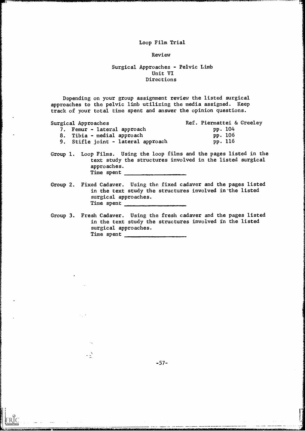
Surgical Approaches - Pelvic LimbUnit VI
Directions
Depending on your group assignment review the listed surgicalapproaches to the pelvic limb utilizing the media assigned. Keeptrack of your total time spent and answer the opinion questions.
Surgical Approaches Ref. Piermattei & Greeley
7. Femur - lateral approach pp. 104
8. Tibia - medial approach pp. 106
9. Stifle joint - lateral approach pp. 116
Group 1. Loop Films. Using the loop films andtext study the structures involvedapproaches.Time spent
the pages listed in thein the listed surgical
Group 2. Fixed Cadaver. Using the fixed cadaver and the pages listedin the text study the structures involved inthe listedsurgical approaches.Time spent
Group 3. Fresh Cadaver. Using the fresh cadaver and the pages listedin the text study the structures involved in the listedsurgical approaches.Time spent
-57-
r-

Loop Film Trial
Review
Surgical Approaches - Pelvic LimbUnit VIPre-Quiz
1. Identify the tagged structures.
a.
b.
C.
d.
2. muscle covers the lateral surface of the
pelvic limb. (outlined on the cadaver)
3. True or False - Fascia lata and the stifle joint capsule are the
same structure.
4. A (flexor, extensor) of the hock lies on the posterior surface of
the tibia.
-58-

Loop Film Trial
Review
Surgical Approaches - Pelvic Limb
Unit VIPost Quiz
1. Identify the tagged structures.
a.
b.
C.
d.
/1111===
2. To expose the femur you must separate what two major muscles?
3. lies over the stifle joint
capsule.
4. The muscle lies along the anterior surface of
the tibia.
-59-

Loop Film Trial
Review
Opinion Questions
Throughout this past semester you have all been assigned to the testgroup which utilized loop films as a review aid in applied anatomy. Pleaseanswer the following questions to the best of your ability!
1. Did you like the loop films as a review aid?
2. In which capacity would loop films work the best?
a. as a primary teaching tool
b. as a review aid
c. as a primary teaching aid
3. Which did you like the best for review?
a. prosected fixed specimen
b. prosected fresh specimen
c. loop film
Why?
4. Were the loop films as easy, easier, or harder to understand thanehe prosected animal?
5. Did you feel you learned as fast or faster from theloop films?
6. Did you feel you learned as well from the loop films?
7. How would you compare the loop films to active dissection (effectivenessand speed of learning)?
8. Did loop films help you to correlate structure with function?
9. Do you consider loop Ma's a novelty?
10. Comments!!-60-

APPENDIX C
Listing of Loop Films
-61-

Listing of Loop Films
Super 8 mm Color Loop Films -- Produced on this Grant
Surgical Approaches - Pelvic Limb
1. Surgical Anatomy2. Surgical Anatomy3. Surgical Anatomy
Femur Lateral ApproachTibia Medial ApproachDistal Femur and Stifle Joint
Lateral ApproL.ch
Surgical Approaches - Pectoral Limb
1. Surgical Anatomy2. Surgical Anatomy3. Surgical Anatomy4. Surgical Anatomy5. Surgical Anatomy6. Surgical Anatomy
Length of Film
Humerus Medial ApproachHumerus Lateral ApproachRadius Lateral ApproachRadius Medial ApproachElbow Joint Transolecranon ApproachUlna Caudal Approach
1'50"1'35"
2'35"
2' 5"1'35"2'35"2' 5"3 5"
1'25"
The following films were adapted from "Functional Anatomy of the Nervesto the Appendages" by R.P. Worthman, D.V.M., Washington State University.Super 8 mm color copies were purchased from Calvin Productions, KansasCity, Missouri.
Nerves of the Brachial Plexus
1. Ulnar and Median Nerve2. Suprascapular Nerve3, Radial Nerve4. Musculocutaneous Nerve
Nerves of the Lumbo-Sacral Plexus
1. Sciatic Nerve2. Femoral Nerve3. Fibular Nerve4. Tibial Nerve5. Obturator Nerve
4' 5"2'10"3'55"2'25"
3'48"2'57"2'32"2' 3"2'
ea&Super 0 mm Black and White Loop Films Produced on this Grant
Skeletal Preparation
1. Skeletal Preparation Defleshing Part 1 2'30"
2. Skeletal Preparation Defleshing Part 2 3'30"
3. Skeletal Preparation Defleshing Part 3 1150"
4. Skeletal Preparation Defleshing Part 4 4'15"
Locomotion in ehe Domestic Aninals
1. Dog Walking 1'51"
2. Dog Running 2'10"
-62-

Listing of Loop Films
Super 3 mm Color Loop Films Produced Previously and Used in This Study
Canine Female Urogenital Tract Length of Film
1. Topographical Anatomy and Approach 2'15"2. Genital Tract 1'30"3. Arteries and Venous Supply 2'45"4. Ligaments 1'15"5. Associated Structures 1'40"
Prehension in the Domestic Animals
1. Horse and Cow Eating Grain 1'20"2. Cow Eating Grass 1'13"3. Horse Eating Grass 0'33"4. Pig Eating Grain 1'42"5. Cat Eating Meat and Cat Food 1'25"6. Dog Eating Meat and Dog Food 1'43"
63/01

APPENDIX D
Loop Film Production Cost

MP PIM ISODOCTIOV COSTS
IMO& =MATZOSDirect
April S. 1968Procarss 1
Iaternegative2 3 4
Contact Prise Mester
5 6 7
SIM8
VIIIIIIII1111
1. Shoot SCO Original 2:1 - 200 ft. 9 .0659/ft.
la. Slack Vbite Plus x lavereal200 ft. 9 0414/ft.
13.18 13.16 13.18 13.18 13.18 13.18 13.18
S. 282. Process Original 100 - 200 ft. 9 .045/ft. 9.00 9.00 9.00 9.00 9.00 9.00 9.00
2a. Slack & Mite Processing200 ft. $ .035/ft.
7.4103. Color Reversal Work Print one ligbt --
200 ft. 9 .097/ft. 19.40 19.40 19.40 19.40(3) 15 1 V Reversal Mork Print one light
200 ft. 9 .06/ft.12.00 12.00 12.00
4. 16 aso Sigh Contrast Positive Slack and Sbitefor Titles Stock and Processing.048/ft. (50' min) 2.39 2.39 2.39 2.39 2.39 2.39 2.39
5. Ccaform Original film to V.!. - II roll,100 ft. total $10.00/bour - 3 hrs. for 100' 30.00 30.00 30.00 30.00 30.00 30.00 30.00
6. Make 16 um Interneg. 100 ft. @ .35/ft. 35.00 35.00 35.00(6) mak. 16 en Contect Priatmaster -- 100 ft. 9 .16/ft. 21.00 28.00 28.00 24.00
6a. 16 so & V Costume Printmester 100 ft.# .12/ft.
7. Soper Answer Print from Inter:seg. 61 ft. I .096/ft. 5.86 5.86 5.867. 16 am Answer Print fro. Interneg. - 110 ft. 0 .106/ft. 11.66 11.668. Super Answer Pent free Coot. Print Super -
61 ft. 9 .144/ft.1.7$ 1.7$ 8.7$ $.70k
(8) 16 ma Answer Print from Cont. Print -- 110 ft.@ .158/ft, 17.38 17.38 17.38
9. Super Release Print from Iaterneg.5 x 61 op 366 ft. I .076/ft. 27.82 27.82 27.82
(9) Super I Mosso Priat from Coatect Print5 slam 366 ft. @ .121/ft. 44.28 s' 44.2$ 44.2$ 44.2$ 44.21
10. Super Cartridge II 1.ading - 6 9 1.50 sack .00 9 00 9 9.00 9.00 9 00 9.00 9 OD'TOM 92.84 151.65 163.31 155.91 164.03 181.41 174.01 121.97
Cbart illustrates preductise of 6film** all splices amd scratches
:11:4rep 3,4,6,7 bavp.a.III gm_11COLOr SMOCK UMMAIIOLO
super a copies of 61' ia least!' approximately 4 slates. Procedure 1 edit orlabialshow la Me product. Protadures 4 & 7 work Id* black eat *Ito votkpalst.
vierclam now projection.5 ma plats

APPENDIX E
Opinion Data Summary Sheets
-67-

Loop Film Trial
First Year
Opinion Data Summary Sheet 1
Total of 58 students replying. Replies given in percentages.
1. Did you like the loop films as a teaching aid?Yes 91%No 7%
2. Were the loop films as easy, easier or harder to understand than theprosected animal?As easy 22% Easier 577 Harder 21%
3. Did you learn as fast?Yes 38%No 12%
4. Did you learn as well?Yes 76%No 22%
5. How would you compare the loop films to active dissection (effective-ness and speed of learning)?Effectiveness Speed of Learning
loop films more effective 477 loop films faster 667loop films less effective 26% loop films slower 77
loop films same as dissection 317 loop films same asdissection 287
6. Did loop films help you in the correlation of structure and function?How much?
Yes 78% Very much 597
No 2% Much 26%
7. How do you think loop films might best be used?487 with dissections and a book59% as a correlation of structure and function237 as a review aid
8. Do you consider loop films a novelty?Yes 31%No 69%*
* 337 mentioned they thought they were a good teaching aid.
-68-

Loop Film Trial
First Year
Opinion Data Summary Sheet 2
1. Average number of times each film was viewed.cat 1.3 pig 1.4 dog 1.76 cow grass 2.1
horse grass 1.63 horse and cow grain 1.3
total number of times loop films viewed - averaged 1.66
2. Total student replies given in percentages.a. Did the loop films help you in the correlation of structure
with function? Yes 957 No 57
How valuable were they to you in correlations?Very valuable Valuable Not essential No value
53% 38% 7% 2%
b. Did the loop films help make the structure of organs clearer?Yes 687 No 32%
How valuable were they in this respect?Very valuable Valuable Not essential No value
287 407 217 12%
c. Do you think loop films might be best used:1. as the primary teaching tool
Excellent Good Fair Poor
0% 5% 24% 71%
2. as a primary teaching aidExcellent Good Fair Poor
3% 41% 41% 14%
3. in correlation of structure with functionExcellent Good Fair Poor
4. as the primary review toolExcellent Good Fair Poor
3470 41% 21% 3%
5. as a primary review aidExcellent Good Fair Poor
4770 40% 10% 3%
-69-

Loop Film Trial
Review
Opinion Data Summary Sheet 3
Data is given in percentages with 52 students replying.
1. Did you like the loop films as a review aid?Yes 01% No 14% No opinion 67
2. In which capacity would loop films work the best?a. as a primary teaching tool 2%b. as a review aid 54%c. as a primary teaching aid 44%
3. Which did you like the best for review?a. prosected fixed specimen 47b. prosected fresh specimen 54%c. loop film 42%
Why?1. Prosected fresh cadavers are more like the actual surgery,
can feel the fresh tissue.2. Loop films are good for reviewing, bringing out highlights.
4. Were the loop films as easy, easier, or harder to understand than theprosected animals?a. loop films as easy as prosected 40%b. loop films easier than prosected 44%c. loop films harder than prosected 157
5. Did you feel you learned as fast or faster from loop films?a. as fast from loop filmsb. faster from loop films 50%c. slower from loop films 17%
6. Did you feel you learned as well from loop films?Yes 69% No 31%
7. How would you compare the loop films to active dissection (effective-ness and speed of learning)?Opinions varied though a majority felt there was little differencein the effectiveness. However 72% felt it was a faster method oflearning than dissection and only 12% felt it would not be a satis-factory review aid.
8. Did loop films help you to correlate structure with function?Yes 92% No 6%
9. Do you consider loop films a novelty?Yes 21% No 797
10. Comments!A concensus of the comments showed the students in favor of loop
films as a review aid especially if it got them out of dissection.They felt they were excellent at correlating structure with function.
-70-