of lucentis therapy for retinal disorders: what we have ...retinatoday.com/pdfs/0817_supp2.pdf ·...
TRANSCRIPT

Powerful. Precise. Proven.
Supplement to July/August 2017
SPONSORED BY NOVARTIS PHARMA AG
10 YEARS of Lucentis Therapy
for Retinal Disorders: What We Have Learned
Lucentis provides powerful efficacy and precise action that
is supported by extensive clinical trial and real-world evidence.

2 SUPPLEMENT TO RETINA TODAY JULY/AUGUST 2017
Powerful. Precise. Proven.
LUCENTIS PROVIDES POWERFUL AND TARGETED EFFICACY TO IMPROVE AND MAINTAIN VISION Lucentis Provides Rapid Visual Acuity Gains Across Indications
Patients included in the pivotal ANCHOR study of Lucentis in nAMD achieved rapid visual acuity gains, with a mean gain at 1 week of 5 letters (Figure 1).8 In the HARBOR study of Lucentis in nAMD, most of the vision gain in the Lucentis 0.5 mg pro re nata (PRN) arm was achieved within the first 3 months.9,10 In a retrospective sub-analysis of HARBOR,11 of 1,057 patients analysed, 266 (25.2%) were early 15-letter responders (gained ≥15 letters from baseline at month 3). Over 80% of early 15-letter responders receiving PRN treatment maintained ≥15-letter gains at month 24.
In DME, in the RESTORE study, rapid and clinically relevant improvements in BCVA were observed as early as month 1.12 In the RISE study, patients had gains of 5 letters after just 1 week (Figure 1).13 In the DRCR.net Protocol I study, clinically meaningful visual acuity gains of approximately 7 letters were achieved within 2 months with Lucentis 0.5 mg PRN with deferred laser.14
In the BRAVO and CRUISE studies of Lucentis 0.5 mg in patients with BRVO and CRVO, significant vision gains were seen after 7 days in both studies (7.5 and 9.0 letters, respectively; Figure 1).15-18
Patients with myopic CNV enrolled in the RADIANCE study also achieved early visual gains with the group where retreatment was based on visual acuity stabilization gaining 4 letters at week 1, and 12.5 letters at month 3 (Figure 1).19
The MINERVA study assessed the use of Lucentis in patients with CNV associated with diseases other than nAMD and myopic CNV. In this study, patients with CNV caused by rare diseases
treated with Lucentis gained a mean 9.5 letters at 2 months, compared with patients given sham who lost 0.4 letters over the same period.20
Early Visual Gains With Lucentis Can Be Sustained Across Indications
In the HARBOR study of Lucentis in nAMD, mean change in BCVA from baseline in the Lucentis 0.5 mg PRN arm was 8.2 letters at 12 months and maintained at 7.9 letters at 24 months. The mean injec-tions were 13.3 over 2 years (5.6 injections in the second year).9,10
10 Years of Lucentis Therapy for Retinal Disorders:
What We Have Learned Lucentis provides powerful efficacy and precise action that is supported by extensive clinical trial and real-world evidence.
BY PRAVIN U. DUGEL, MD
Figure 1. Visual gains at 1 week in clinical trials of Lucentis.
Prior to the advent of anti-vascular endothelial growth factor (VEGF) therapy, visual outcomes were often poor for patients with one of the range of retinal disorders that are now routinely treated with anti-VEGF agents. Patients with neovascular age-related macular degeneration (nAMD) could expect to lose a significant proportion of their central vision and become legally blind if left untreated.1 Even when treated with the previous stan-dard of care, verteporfin photodynamic therapy, most nAMD patients experienced clinically significant vision loss within a few years of diagnosis.2
It has been over 10 years since Lucentis (ranibizumab; Novartis Pharma AG, Basel, Switzerland; Genentech/Roche, South San Francisco, CA) was approved as the first anti-VEGF therapy for ocular use in Europe and the United States. In addition to that initial approval in nAMD, Lucentis is now also indicated for use in diabetic macular edema (DME), central retinal vein occlusion (CRVO), branch retinal vein occlusion (BRVO), and choroidal neovascularization (CNV) secondary to pathological myopia (myopic CNV). Most recently, in November 2016, the European Commission approved Lucentis for the treatment of visual impairment due to CNV, extending its use to a range of rare retinal conditions with a pathological CNV component. “The approval of Lucentis has been sight-saving around the world,” says Pravin U. Dugel, MD. “The positive impact that it has had has been enormous.” Data from Europe, Australia, and the United States show a 46% to 72% decrease in the incidence of blindness since the introduction of Lucentis.3-7
This supplement provides an overview of the evidence and experience gained over the past decade with use of Lucentis.
JULY/AUGUST 2017 SUPPLEMENT TO RETINA TODAY 3
Powerful. Precise. Proven.
“In nAMD, the HARBOR study shows us that as long as regu-lar monitoring is maintained, the number of injections can be customized according to the patient’s needs with maintenance of excellent vision,” says Dr. Dugel. “Customized care should be the aim in nAMD due to the inherent variability of the disease.”
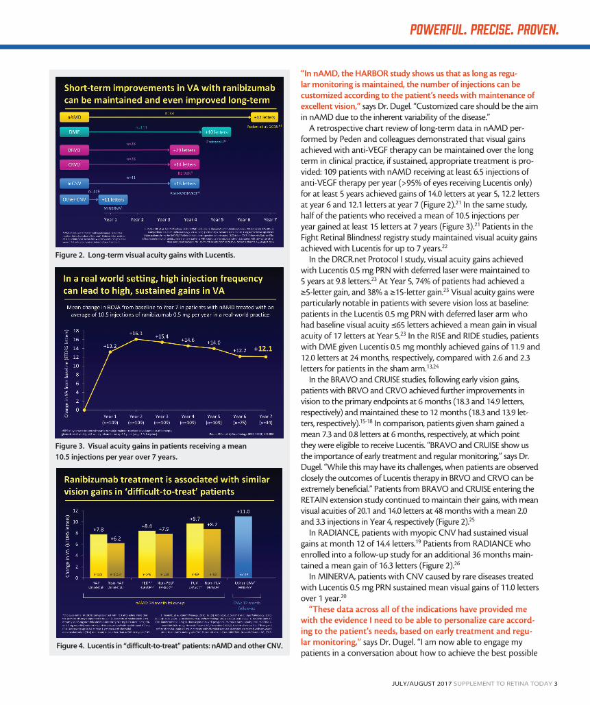
A retrospective chart review of long-term data in nAMD per-formed by Peden and colleagues demonstrated that visual gains achieved with anti-VEGF therapy can be maintained over the long term in clinical practice, if sustained, appropriate treatment is pro-vided: 109 patients with nAMD receiving at least 6.5 injections of anti-VEGF therapy per year (>95% of eyes receiving Lucentis only) for at least 5 years achieved gains of 14.0 letters at year 5, 12.2 letters at year 6 and 12.1 letters at year 7 (Figure 2).21 In the same study, half of the patients who received a mean of 10.5 injections per year gained at least 15 letters at 7 years (Figure 3).21 Patients in the Fight Retinal Blindness! registry study maintained visual acuity gains achieved with Lucentis for up to 7 years.22
In the DRCR.net Protocol I study, visual acuity gains achieved with Lucentis 0.5 mg PRN with deferred laser were maintained to 5 years at 9.8 letters.23 At Year 5, 74% of patients had achieved a ≥5-letter gain, and 38% a ≥15-letter gain.23 Visual acuity gains were particularly notable in patients with severe vision loss at baseline: patients in the Lucentis 0.5 mg PRN with deferred laser arm who had baseline visual acuity ≤65 letters achieved a mean gain in visual acuity of 17 letters at Year 5.23 In the RISE and RIDE studies, patients with DME given Lucentis 0.5 mg monthly achieved gains of 11.9 and 12.0 letters at 24 months, respectively, compared with 2.6 and 2.3 letters for patients in the sham arm.13,24
In the BRAVO and CRUISE studies, following early vision gains, patients with BRVO and CRVO achieved further improvements in vision to the primary endpoints at 6 months (18.3 and 14.9 letters, respectively) and maintained these to 12 months (18.3 and 13.9 let-ters, respectively).15-18 In comparison, patients given sham gained a mean 7.3 and 0.8 letters at 6 months, respectively, at which point they were eligible to receive Lucentis. “BRAVO and CRUISE show us the importance of early treatment and regular monitoring,” says Dr. Dugel. “While this may have its challenges, when patients are observed closely the outcomes of Lucentis therapy in BRVO and CRVO can be extremely beneficial.” Patients from BRAVO and CRUISE entering the RETAIN extension study continued to maintain their gains, with mean visual acuities of 20.1 and 14.0 letters at 48 months with a mean 2.0 and 3.3 injections in Year 4, respectively (Figure 2).25
In RADIANCE, patients with myopic CNV had sustained visual gains at month 12 of 14.4 letters.19 Patients from RADIANCE who enrolled into a follow-up study for an additional 36 months main-tained a mean gain of 16.3 letters (Figure 2).26
In MINERVA, patients with CNV caused by rare diseases treated with Lucentis 0.5 mg PRN sustained mean visual gains of 11.0 letters over 1 year.20
“These data across all of the indications have provided me with the evidence I need to be able to personalize care accord-ing to the patient’s needs, based on early treatment and regu-lar monitoring,” says Dr. Dugel. “I am now able to engage my patients in a conversation about how to achieve the best possible
Figure 2. Long-term visual acuity gains with Lucentis.
Figure 3. Visual acuity gains in patients receiving a mean
10.5 injections per year over 7 years.
Figure 4. Lucentis in “difficult-to-treat” patients: nAMD and other CNV.

4 SUPPLEMENT TO RETINA TODAY JULY/AUGUST 2017
Powerful. Precise. Proven.
results and the level of treatment burden likely to be associated with gaining those outcomes.”
The efficacy of Lucentis 0.5 mg has been demonstrated across nAMD disease subtypes, including PED (mean gain of 8.4 letters at 24 months in the 54.5% of patients in HARBOR with PED),27,28 PCV (mean gain of 9.7 letters at 24 months in DRAGON),29 and RAP (mean gain of 7.8 letters at 24 months in CATT; Figure 4).30 In the MINERVA study, visual gains were seen across all CNV etiologies included in the trial,20 and nearly half (49%) of patients achieved ≥15-letter gains by month 12.20 “We often forget that all of these diseases are enormously variable and they do not always behave in a given pattern,” says Dr. Dugel.
In the DRCR.net Protocol S study, patients with DME and prolif-erative diabetic retinopathy (PDR) who received Lucentis as needed gained a mean 7.9 letters at 24 months,31 while BRIGHTER and CRYSTAL demonstrated that visual gains could be achieved with Lucentis in patients with BRVO and CRVO regardless of the pres-ence or absence and the severity of macular ischemia (Figure 5).32,33
Lucentis Demonstrated Equivalent or Superior Efficacy Compared to Alternative Treatments
“Since the approval of Lucentis, no alternative treatment has shown better outcomes, either by itself or in combination, which shows what a very high bar it has set,” says Dr. Dugel. The only large clinical trials to directly compare ranibizumab and aflibercept, the only other anti-VEGF agent currently approved for ocular condi-tions, are the identically designed VIEW 1 and VIEW 2 studies, in patients with nAMD.34 In year 2 of VIEW 1 and 2, the capped PRN regimen with mandatory dosing at least every 12 weeks allowed a comparison of both agents under the same dosing regimen. The mean change in BCVA from baseline to week 96 was similar in all three study arms: 7.9 letters in the Lucentis 0.5 mg monthly arm, 7.6 letters in the aflibercept 2.0 mg monthly arm, and 7.6 letters in the aflibercept 2.0 mg bimonthly arm, with 4.7, 4.1 and 4.2 injec-tions, respectively, in the second year of the study (Figure 6).35 The majority of this vision gain was achieved in the first 3 months and was sustained over the course of the study.
An analysis performed on the Fight Retinal Blindness! registry examined data from treatment-naive patients with nAMD who started treatment with ranibizumab or aflibercept between the end of 2013 and the start of 2015.36 Findings at 1 year showed that differences in visual gains were not statistically significant between drugs. The number of injections given over 12 months to patients completing the study was also similar between groups: in patients who completed 12 months of therapy, those treated with Lucentis received 8.1 injections while those treated with aflibercept received 8.0 injections.36
In another analysis on the same registry, patients who received Lucentis according to a PRN or treat-and-extend regimen for at least 12 months before switching to aflibercept (PRN or treat-and-extend) and being observed for at least 12 months showed no mean gain in visual acuity 12 months after the switch.37 Mean visual acuity was 63.4 letters before the switch versus 63.3 letters 12 months after the switch (P=.17). There was a modest decrease
in the number of injections (7.4 in the 12 months preceding the treatment switch versus 6.6 in the 12 months after).37 This registry evidence suggests no additional benefit of switching from Lucentis to aflibercept.
“As physicians, our decisions really need to be data driven. I do not know of any robust scientific study providing convinc-ing data to show that one drug is better than the other, or that switching from one to the other in a particular disease or disease subtype is more beneficial for the patient,” says Dr. Dugel. “For example, subanalysis of the VIEW data clearly shows that the loca-tion of fluid below the pigment retinal epithelium versus within the retina has more of a bearing on outcomes than the investigational agent used.38 As a result, I do not feel compelled to change my clinical behavior.”
Comparing outcomes with Lucentis against laser treatment, the RESTORE study showed that patients with DME given Lucentis 0.5 mg PRN achieved mean gains of 6.8 letters at month 12 of the study, compared with 0.9 letters in the laser arm.12 Real-world evi-dence from the Maggiore study reports equivalence of Lucentis and dexamethasone in DME, with mean gains at 12 months of 7.6 and 4.3 letters, respectively.39
Figure 5. Lucentis in “difficult-to-treat” patients: DME and RVO.
Figure 6. Ninety-six-week results of VIEW 1 and 2.

JULY/AUGUST 2017 SUPPLEMENT TO RETINA TODAY 5
Powerful. Precise. Proven.
In BRVO, patients in the BRIGHTER study given Lucentis 0.5 mg PRN gained 14.8 letters at 6 months compared with 5.5 letters in patients receiving laser therapy,33 while head-to-head trials of Lucentis and dexamethasone in BRVO and CRVO (COMRADE B and C) dem-onstrates superior benefits of Lucentis in both indications.40,41
Lucentis Provides Visual Gains With Less-Frequent-Than-Monthly Treatment
Real-world evidence in nAMD shows that frequent Lucentis injec-tions enable many patients to sustain high gains in visual acuity over time, with patients receiving a mean 10.5 injections per year maintain-ing visual gains of over 12 letters over 7 years (Figure 3).21 The number of injections required per year is similar between Lucentis and afliber-cept, according to real-world evidence from an observational registry and a database of US physician-level claims data (Figure 7).36,42
“When talking about treatment burden, it is important to under-stand that injections are not really the indicator of treatment bur-den—the clinic visit is. It takes just a few seconds to do an injection, but to attend a clinic visit, whether or not an injection is given, is burdensome because of time lost from work, the requirement for travel and so on,” says Dr. Dugel. “For this reason, treat-and-extend regimens, which offer a reduced treatment burden compared with monthly monitoring, are becoming increasingly popular. I think there is some very good evidence that this regimen can also provide excel-lent results, in some cases as good as with monthly monitoring.”
Studies incorporating treat-and-extend regimens have shown that, in nAMD, Lucentis can provide visual gains with extended treatment intervals.43 In the TREX-AMD study, patients with nAMD given Lucentis 0.5 mg according to a treat-and-extend regimen had similar outcomes to patients treated monthly: a mean gain of 10.5 letters at 12 months with 10.1 injections, compared with 9.2 letters with 13.0 injections.43 Nearly half (45%) of patients achieved a treat-ment interval of ≥8 weeks, and 18% achieved the maximum interval of 12 weeks.43 A meta-analysis of over 24,000 patients with nAMD
treated with either a treat-and-extend or a PRN regimen found that visual outcomes were significantly better with treat-and-extend in the first 2 years of treatment and numerically better in the third year.44
In DME, the requirement for injections tends to decrease over time. In the DRCR.net Protocol I study, patients in the Lucentis 0.5 mg PRN with deferred laser arm received a median of nine injections in year 1, 2 in year 3, and none in year 5, whilst maintaining vision, suggesting an underlying disease-modifying effect.23,45 “In DME with diabetic retinopathy (DR), Lucentis has the potential to be disease modifying, and I think we are just starting to realize the enormity of that potential to change the course of a dis-ease which a leading cause of blindness in the world,” says Dr. Dugel.
In the open-label extension of RISE and RIDE, around 25% of patients required no further injections during the extension period of up to 54 months, whilst maintaining vision.46 A subanalysis of data from the RESTORE and RETAIN studies in DME showed that approximately a quarter of the patients who received Lucentis in both studies were able to achieve visual acuity gains of at least 10 letters with 8 or fewer injections.47-49 A treat-and extend regimen may also be beneficial in patients with DME. In the RETAIN study of patients with DME, 83% of patients given Lucentis 0.5 mg according to a treat-and-extend regimen through year 2 achieved treatment intervals of ≥2 months, with 44% achieving intervals of 3 months (at which the interval was capped according to the protocol).50
Similarly, in RVO and mCNV, the long-term treatment burden may be low. In RETAIN, 53% of patients with BRVO and 57% of patients with CRVO received no further injections after the first year.25 Of 41 patients with mCNV from RADIANCE who enrolled into a follow-up study, 34 (83%) required no further treatment between month 12 and month 48.51 “In other rare CNV-based dis-eases, the behavior of the neovascular membrane appears to be quite different than in patients with typical nAMD,” says Dr. Dugel. “In a lot of these rare diseases, such as myopic CNV or idiopathic neovascular membrane formation, Lucentis appears to be disease modifying and, in my experience, it is rare for continued injections to be required.”
DESIGNED TO PROVIDE PRECISE, TARGETED ACTIONLucentis is a small, 48 kDa antibody fragment that specifically
binds all isoforms of VEGF-A with high affinity and was specifically designed for ocular use.52-55 The small molecular size of Lucentis enables it to rapidly penetrate the retina to reach the choroid, its site of action.56-58 Preclinical investigations have demonstrated that Lucentis does not enter or disrupt nontarget cells such as retinal pigment epithelium.57,59,60
As an antibody fragment, Lucentis does not contain the Fc domain of the antibody, which is responsible for systemic transport and recycling.61-63 As a result of the lack of the Fc domain, Lucentis does not bind to the Fc receptors that mediate transport across the blood-retinal barrier,61-63 and is retained in the eye with an ocular half-life of 9 days.64 Any Lucentis that does reach the systemic cir-culation is quickly broken down, with a half-life of approximately 2 hours64 (in comparison to the systemic half-life of aflibercept estimated at 5 to 6 days following intravenous administration65),
Figure 7. Comparison of treat-and-extend and PRN regimens in
patients with nAMD in a real-world setting.
6 SUPPLEMENT TO RETINA TODAY JULY/AUGUST 2017
Powerful. Precise. Proven.
thus minimizing systemic exposure. Serum Lucentis concentrations are predicted to be approximately 90,000-fold lower than vitreal Lucentis concentrations.53
“I think what we have learnt over the past decade is that the eye is not an isolated organ, and when you inject anything into the eye it has a systemic impact,” says Dr. Dugel. “That systemic impact is worthy of being studied, particularly since we are injecting patients who are elderly and who are sick, and we are injecting over a long period of time.”
A prospective, open-label trial of 151 patients with AMD, DME, or RVO given three monthly anti-VEGF injections showed that sys-temic exposure to Lucentis was much lower than to aflibercept, and Lucentis resulted in smaller decreases in plasma-free VEGF.66 Mean serum concentrations of aflibercept were higher than its reported half-maximal inhibitory concentration (IC50) for VEGF inhibition (0.068 nM) at most time points, while for Lucentis, mean serum concentrations were equal to or greater than its IC50 (0.060 nM) at 3 and 24 hours, but below this level thereafter.66
In a randomized, prospective study, patients receiving intravitreal injection of Lucentis experienced no significant changes to median plasma VEGF levels 7 days and 1 month after injection (P=.776 and P=.670, respectively), while median plasma VEGF levels were signifi-cantly decreased in patients injected with aflibercept at these time points (P<.001 for both). At 7 days, nearly 90% of patients injected with aflibercept had VEGF levels below the minimum detectable dose, with over 25% of patients injected with aflibercept still having undetectable levels of plasma VEGF levels at 1 month.67
“We do not have clinical evidence to show that these findings translate into differences in safety profile between Lucentis and aflibercept, since performing a large enough study to demonstrate significant differences in rare safety events such as stroke, heart attack, or death would not be feasible,” says Dr. Dugel. “However, for me at least, it gives me confidence that the drug that I have cho-sen will clear the system faster.”
SAFETY AND EFFICACY PROFILE IS PROVEN BY CLINICAL TRIAL AND REAL-WORLD EVIDENCE
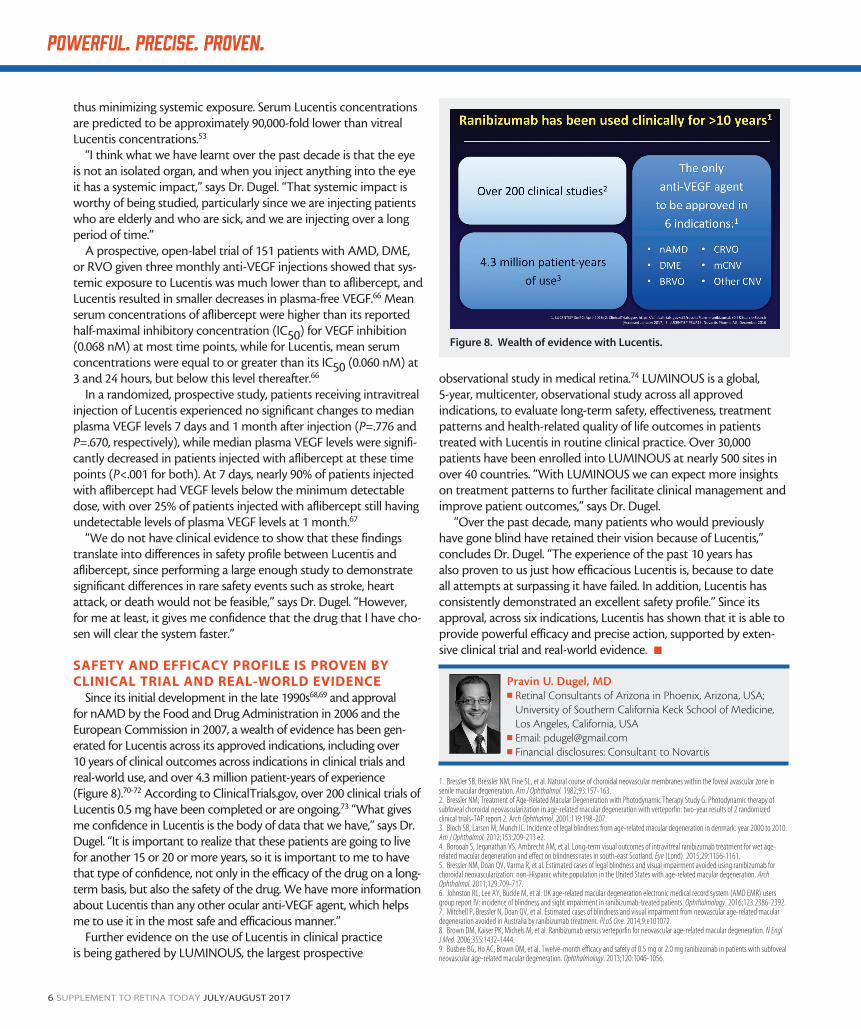
Since its initial development in the late 1990s68,69 and approval for nAMD by the Food and Drug Administration in 2006 and the European Commission in 2007, a wealth of evidence has been gen-erated for Lucentis across its approved indications, including over 10 years of clinical outcomes across indications in clinical trials and real-world use, and over 4.3 million patient-years of experience (Figure 8).70-72 According to ClinicalTrials.gov, over 200 clinical trials of Lucentis 0.5 mg have been completed or are ongoing.73 “What gives me confidence in Lucentis is the body of data that we have,” says Dr. Dugel. “It is important to realize that these patients are going to live for another 15 or 20 or more years, so it is important to me to have that type of confidence, not only in the efficacy of the drug on a long-term basis, but also the safety of the drug. We have more information about Lucentis than any other ocular anti-VEGF agent, which helps me to use it in the most safe and efficacious manner.”
Further evidence on the use of Lucentis in clinical practice is being gathered by LUMINOUS, the largest prospective
observational study in medical retina.74 LUMINOUS is a global, 5-year, multicenter, observational study across all approved indications, to evaluate long-term safety, effectiveness, treatment patterns and health-related quality of life outcomes in patients treated with Lucentis in routine clinical practice. Over 30,000 patients have been enrolled into LUMINOUS at nearly 500 sites in over 40 countries. “With LUMINOUS we can expect more insights on treatment patterns to further facilitate clinical management and improve patient outcomes,” says Dr. Dugel.
“Over the past decade, many patients who would previously have gone blind have retained their vision because of Lucentis,” concludes Dr. Dugel. “The experience of the past 10 years has also proven to us just how efficacious Lucentis is, because to date all attempts at surpassing it have failed. In addition, Lucentis has consistently demonstrated an excellent safety profile.” Since its approval, across six indications, Lucentis has shown that it is able to provide powerful efficacy and precise action, supported by exten-sive clinical trial and real-world evidence. n
1. Bressler SB, Bressler NM, Fine SL, et al. Natural course of choroidal neovascular membranes within the foveal avascular zone in senile macular degeneration. Am J Ophthalmol. 1982;93:157-163.2. Bressler NM; Treatment of Age-Related Macular Degeneration with Photodynamic Therapy Study G. Photodynamic therapy of subfoveal choroidal neovascularization in age-related macular degeneration with verteporfin: two-year results of 2 randomized clinical trials-TAP report 2. Arch Ophthalmol. 2001;119:198-207.3. Bloch SB, Larsen M, Munch IC. Incidence of legal blindness from age-related macular degeneration in denmark: year 2000 to 2010. Am J Ophthalmol. 2012;153:209-213 e2.4. Borooah S, Jeganathan VS, Ambrecht AM, et al. Long-term visual outcomes of intravitreal ranibizumab treatment for wet age-related macular degeneration and effect on blindness rates in south-east Scotland. Eye (Lond). 2015;29:1156-1161.5. Bressler NM, Doan QV, Varma R, et al. Estimated cases of legal blindness and visual impairment avoided using ranibizumab for choroidal neovascularization: non-Hispanic white population in the United States with age-related macular degeneration. Arch Ophthalmol. 2011;129:709-717.6. Johnston RL, Lee AY, Buckle M, et al. UK age-related macular degeneration electronic medical record system (AMD EMR) users group report IV: incidence of blindness and sight impairment in ranibizumab-treated patients. Ophthalmology. 2016;123:2386-2392.7. Mitchell P, Bressler N, Doan QV, et al. Estimated cases of blindness and visual impairment from neovascular age-related macular degeneration avoided in Australia by ranibizumab treatment. PLoS One. 2014;9:e101072.8. Brown DM, Kaiser PK, Michels M, et al. Ranibizumab versus verteporfin for neovascular age-related macular degeneration. N Engl J Med. 2006;355:1432-1444.9. Busbee BG, Ho AC, Brown DM, et al. Twelve-month efficacy and safety of 0.5 mg or 2.0 mg ranibizumab in patients with subfoveal neovascular age-related macular degeneration. Ophthalmology. 2013;120:1046-1056.
Pravin U. Dugel, MDn Retinal Consultants of Arizona in Phoenix, Arizona, USA;
University of Southern California Keck School of Medicine, Los Angeles, California, USA
n Email: [email protected] Financial disclosures: Consultant to Novartis
Figure 8. Wealth of evidence with Lucentis.

JULY/AUGUST 2017 SUPPLEMENT TO RETINA TODAY 7
Powerful. Precise. Proven.
10. Ho AC, Busbee BG, Regillo CD, et al. Twenty-four-month efficacy and safety of 0.5 mg or 2.0 mg ranibizumab in patients with subfoveal neovascular age-related macular degeneration. Ophthalmology. 2014;121:2181-2192.11. Stoller GL, Kokame GT, Dreyer RF, et al. Patterns of early and delayed visual response to ranibizumab treatment for neovascular age-related macular degeneration. JAMA Ophthalmol. 2016;134:545-553.12. Mitchell P, Bandello F, Schmidt-Erfurth U, et al. The RESTORE study: ranibizumab monotherapy or combined with laser versus laser monotherapy for diabetic macular edema. Ophthalmology. 2011;118:615-625.13. Nguyen QD, Brown DM, Marcus DM, et al. Ranibizumab for diabetic macular edema: results from 2 phase III randomized trials: RISE and RIDE. Ophthalmology. 2012;119:789-801.14. Elman MJ, Aiello LP, et al; Diabetic Retinopathy Clinical Research N. Randomized trial evaluating ranibizumab plus prompt or deferred laser or triamcinolone plus prompt laser for diabetic macular edema. Ophthalmology. 2010;117:1064-1077 e35.15. Brown DM, Campochiaro PA, Bhisitkul RB, et al. Sustained benefits from ranibizumab for macular edema following branch retinal vein occlusion: 12-month outcomes of a phase III study. Ophthalmology. 2011;118:1594-1602.16. Brown DM, Campochiaro PA, Singh RP, et al. Ranibizumab for macular edema following central retinal vein occlusion: six-month primary end point results of a phase III study. Ophthalmology. 2010;117:1124-1133 e1.17. Campochiaro PA, Brown DM, Awh CC, et al. Sustained benefits from ranibizumab for macular edema following central retinal vein occlusion: twelve-month outcomes of a phase III study. Ophthalmology. 2011;118:2041-2049.18. Campochiaro PA, Heier JS, Feiner L, et al. Ranibizumab for macular edema following branch retinal vein occlusion: six-month primary end point results of a phase III study. Ophthalmology. 2010;117:1102-112 e1.19. Wolf S, Balciuniene VJ, Laganovska G, et al. RADIANCE: a randomized controlled study of ranibizumab in patients with choroidal neovascularization secondary to pathologic myopia. Ophthalmology. 2014;121:682-692 e2.20. Novartis Data on File. Efficacy and safety of ranibizumab 0.5 mg in patients with choroidal neovascularization associated with any cause other than nAMD and myopic CNV: 12-month results from MINERVA. Novartis Pharma AG, August 2016.21. Peden MC, Suner IJ, Hammer ME, Grizzard WS. Long-term outcomes in eyes receiving fixed-interval dosing of anti-vascular endothelial growth factor agents for wet age-related macular degeneration. Ophthalmology. 2015;122:803-808.22. Gillies MC, Campain A, Barthelmes D, et al. Long-term outcomes of treatment of neovascular age-related macular degeneration: data from an observational study. Ophthalmology 2015;122:1837-1845.23. Elman MJ, Ayala A, Bressler NM, et al. Intravitreal Ranibizumab for diabetic macular edema with prompt versus deferred laser treatment: 5-year randomized trial results. Ophthalmology. 2015;122:375-381.24. Brown DM, Nguyen QD, Marcus DM, et al. Long-term outcomes of ranibizumab therapy for diabetic macular edema: the 36-month results from two phase III trials: RISE and RIDE. Ophthalmology. 2013;120:2013-2022.25. Campochiaro PA, Sophie R, Pearlman J, et al. Long-term outcomes in patients with retinal vein occlusion treated with ranibi-zumab: the RETAIN study. Ophthalmology. 2014;121:209-219.26. Novartis Data on File. Long-term follow-up of East Asian patients from the RADIANCE clinical trial: a retrospective cohort study. 16 October 2015.27. Schlottmann P. Ranibizumab and PED: exploring the evidence. Presentation at: APVRS Congress; July 31-August 2, 2015; Sydney, Australia.28. Sarraf D, London NJ, Khurana RN, et al. Ranibizumab treatment for pigment epithelial detachment secondary to neovascular age-related macular degeneration: post hoc analysis of the HARBOR study. Ophthalmology. 2016;123:2213-2224.29. Novartis Data on File. Ranibizumab 0.5 mg in Chinese patients with polypoidal choroidal vasculopathy: results of the 2-year DRAGON study. Novartis Pharma AG, December 2016.30. Daniel E, Shaffer J, Ying GS, et al. Outcomes in eyes with retinal angiomatous proliferation in the Comparison of Age-Related Macular Degeneration Treatments Trials (CATT). Ophthalmology. 2016;123:609-616.31. Gross JG, Glassman AR, et al; Writing Committee for the Diabetic Retinopathy Clinical Research Network. Panretinal photocoagu-lation vs intravitreous ranibizumab for proliferative diabetic retinopathy: a randomized clinical trial. JAMA. 2015;314:2137-2146.32. Larsen M, Waldstein SM, Boscia F, et al. Individualized ranibizumab regimen driven by stabilization criteria for central retinal vein occlusion: twelve-month results of the CRYSTAL study. Ophthalmology. 2016;123:1101-1111.33. Tadayoni R, Waldstein SM, Boscia F, et al. Individualized stabilization criteria-driven ranibizumab versus laser in branch retinal vein occlusion: six-month results of BRIGHTER. Ophthalmology. 2016;123:1332-1344.34. Heier JS, Brown DM, Chong V, et al. Intravitreal aflibercept (VEGF trap-eye) in wet age-related macular degeneration. Ophthalmology. 2012;119:2537-2548.35. Schmidt-Erfurth U, Kaiser PK, Korobelnik JF, et al. Intravitreal aflibercept injection for neovascular age-related macular degenera-tion: ninety-six-week results of the VIEW studies. Ophthalmology. 2014;121:193-201.36. Gillies MC, Nguyen V, Daien V, et al. Twelve-month outcomes of ranibizumab vs. aflibercept for neovascular age-related macular degeneration: data from an observational study. Ophthalmology. 2016;123:2545-2553.37. Barthelmes D, Campain A, Nguyen P, et al. Effects of switching from ranibizumab to aflibercept in eyes with exudative age-related macular degeneration. Br J Ophthalmol. 2016;100:1640-1645.38. Waldstein SM, Simader C, Staurenghi G, et al. Morphology and visual acuity in aflibercept and ranibizumab therapy for neovascu-lar age-related macular degeneration in the VIEW trials. Ophthalmology. 2016;123:1521-1529.39. Callanan DG, Loewenstein A, Patel SS, et al. A multicenter, 12-month randomized study comparing dexamethasone intravitreal implant with ranibizumab in patients with diabetic macular edema. Graefes Arch Clin Exp Ophthalmol. 2017;255:463-473.40. Hattenbach LO, Feltgen N, Bertelmann T, et al. Head-to-head comparison of ranibizumab PRN versus single-dose dexamethasone for
branch retinal vein occlusion (COMRADE-B) [published online ahead of print March 2, 2017]. Acta Ophthalmol. doi: 10.1111/aos.13381.41. Hoerauf H, Feltgen N, Weiss C, et al. Clinical efficacy and safety of ranibizumab versus dexamethasone for central retinal vein occlusion (COMRADE C): a european label study. Am J Ophthalmol. 2016;169:258-267.42. Ferreira A, Sagkriotis A, Olson M, et al. Treatment frequency and dosing interval of ranibizumab and aflibercept for neovascular age-related macular degeneration in routine clinical practice in the USA. PLoS One. 2015;10:e0133968.43. Wykoff CC, Croft DE, Brown DM, et al. Prospective trial of treat-and-extend versus monthly dosing for neovascular age-related macular degeneration: TREX-AMD 1-year results. Ophthalmology. 2015;122:2514-2522.44. Kim LN, Mehta H, Barthelmes D, et al. Metaanalysis of real-world outcomes of intravitreal ranibizumab for the treatment of neovascular age-related macular degeneration. Retina. 2016;36:1418-1431.45. Ferrara N, Adamis AP. Ten years of anti-vascular endothelial growth factor therapy. Nat Rev Drug Discov. 2016;15:385-403.46. Boyer DS, Nguyen QD, Brown DM, et al. Outcomes with as-needed ranibizumab after initial monthly therapy: long-term outcomes of the phase III RIDE and RISE trials. Ophthalmology. 2015;122:2504-2513 e1.47. Figueira J. An exploration of ranibizumab injection frequency in patients with diabetic macular edema: a post-hoc analysis of the RESTORE and RETAIN study data. Poster presented at: ESASO Retina Academy; November 13-15, 2014; Istanbul, Turkey.48. Margaron P. High visual acuity response with reduced number of ranibizumab injections in patients with diabetic macular edema: a post-hoc analysis of the RESTORE study. Presented at: ARVO; May 4-8, 2014; Orlando, Florida. 49. Novartis Data on File. RETAIN – TE high-gainers post-hoc analysis. December 2014.50. Prunte C, Fajnkuchen F, Mahmood S, et al. Ranibizumab 0.5 mg treat-and-extend regimen for diabetic macular oedema: the RETAIN study. Br J Ophthalmol. 2016;100:787-795.51. Novartis Data on File. Long-term follow-up of East Asian patients from RADIANCE. Novartis Pharma AG, August 2015.52. Ferrara N, Damico L, Shams N, Lowman H, Kim R. Development of ranibizumab, an anti-vascular endothelial growth factor antigen binding fragment, as therapy for neovascular age-related macular degeneration. Retina. 2006;26:859-870.53. Lucentis Summary of Product Characteristics. December 2016. Available at: http://www.ema.europa.eu.54. Steinbrook R. The price of sight--ranibizumab, bevacizumab, and the treatment of macular degeneration. N Engl J Med. 2006;355:1409-1412.55. Yang J, Wang X, Fuh G, et al. Comparison of binding characteristics and in vitro activities of three inhibitors of vascular endothelial growth factor A. Mol Pharm. 2014;11:3421-3430.56. Gaudreault J, Fei D, Beyer JC, et al. Pharmacokinetics and retinal distribution of ranibizumab, a humanized antibody fragment directed against VEGF-A, following intravitreal administration in rabbits. Retina. 2007;27:1260-1266.57. Julien S, Biesemeier A, Taubitz T, Schraermeyer U. Different effects of intravitreally injected ranibizumab and aflibercept on retinal and choroidal tissues of monkey eyes. Br J Ophthalmol. 2014;98:813-825.58. Mordenti J, Thomsen K, Licko V, et al. Efficacy and concentration-response of murine anti-VEGF monoclonal antibody in tumor-bearing mice and extrapolation to humans. Toxicol Pathol. 1999;27:14-21.59. Klettner A, Tahmaz N, Dithmer M, et al. Effects of aflibercept on primary RPE cells: toxicity, wound healing, uptake and phagocy-tosis. Br J Ophthalmol. 2014;98:1448-1452.60. Stewart MW. Pharmacokinetics, pharmacodynamics and pre-clinical characteristics of ophthalmic drugs that bind VEGF. Expert Rev Clin Pharmacol. 2014;7:167-180.61. Goebl NA, Babbey CM, Datta-Mannan A, et al. Neonatal Fc receptor mediates internalization of Fc in transfected human endothe-lial cells. Mol Biol Cell. 2008;19:5490-5505.62. Kim H, Robinson SB, Csaky KG. FcRn receptor-mediated pharmacokinetics of therapeutic IgG in the eye. Mol Vis. 2009;15:2803-2812.63. Lee TY, Tjin Tham Sjin RM, Movahedi S, et al. Linking antibody Fc domain to endostatin significantly improves endostatin half-life and efficacy. Clin Cancer Res. 2008;14:1487-1493.64. Lucentis EPAR Scientific Discussion. Available at: www.ema.europa.eu/docs/en_GB/document_library/EPAR_-_Scientific_Dis-cussion/human/000715/WC500043550.pdf. Published 2007. Accessed March 28, 2017. 65. Avery RL, Castellarin AA, Steinle NC, et al. Systemic pharmacokinetics following intravitreal injections of ranibizumab, bevaci-zumab or aflibercept in patients with neovascular AMD. Br J Ophthalmol. 2014;98:1636-1641.66. Avery RL, Castellarin AA, Steinle NC, et al. Systemic pharmacokinetics and pharmacodynamics of intravitreal aflibercept, bevaci-zumab, and ranibizumab [published online ahead of print January 18, 2017]. Retina. doi: 10.1097/IAE.0000000000001493. 67. Zehetner C, Kralinger MT, Modi YS, et al. Systemic levels of vascular endothelial growth factor before and after intravitreal injection of aflibercept or ranibizumab in patients with age-related macular degeneration: a randomised, prospective trial. Acta Ophthalmol. 2015;93:e154-159.68. Baca M, Presta LG, O’Connor SJ, Wells JA. Antibody humanization using monovalent phage display. J Biol Chem. 1997;272:10678-10684.69. Muller YA, Chen Y, Christinger HW, et al. VEGF and the Fab fragment of a humanized neutralizing antibody: crystal structure of the complex at 2.4 A resolution and mutational analysis of the interface. Structure. 1998;6:1153-1167.70. IMS Padds monthly (data extracted on 8 December 2016). Novartis Pharma AG, December 2016.71. LUCENTIS® Summary of product characteristics; April 2016.72. Novartis Data on file. Lucentis PSUR 13. November 2016.73. ClinicalTrials.gov. www.clinicaltrials.gov. Searched January 2017.74. ClinicalTrials.gov. Observe the effectiveness and safety of ranibizumab in real life setting (LUMINOUS). https://clinicaltrials.gov/ct2/show/NCT01318941. Updated May 20, 2016. Accessed March 28, 2017.
Note: Before prescribing, consult full prescribing information.Presentation: Vial: Ranibizumab. Each vial contains 2.3 mg of ranibizumab in 0.23 mL solution. Pre-filled syringe: Ranibizumab. Each pre-filled syringe contains 1.65 mg of ranibizumab in 0.165 mL solution.Indications: •Treatment of neovascular (wet) age-related macular degeneration (AMD). •Treatment of visual impairment due to choroidal neovascularization (CNV). •Treatment of visual impairment due to CNV secondary to pathologic myopia (PM). •Treatment of visual impairment due to diabetic macular edema (DME). •Treatment of visual impairment due to macular edema secondary to retinal vein occlusion (branch RVO or central RVO). Dosage and administration: •The recommended dose is 0.5 mg (0.05 mL) given as a single intravitreal injection. The interval between two doses injected into the same eye should not be shorter than 1 month. Wet AMD, DME, RVO, CNV, and CNV secondary to PM: •Treatment is initiated with one injection per month until maximum visual acuity is achieved and/or there are no signs of disease activity. •Thereafter, monitoring and treatment intervals should be determined by the physician and should be based on disease activity as assessed by visual acuity and/or anatomic parameters. •Monitoring for disease activity may include clinical examination, functional testing or imaging techniques (e.g. optical coherence tomography or fluorescein angiography). •While applying the treat-and-extend regimen, the treatment interval should be extended by two weeks at a time for wet AMD and central RVO, or by one month at a time for DME and branch RVO. •LUCENTIS® and laser photocoagulation in DME or in branch RVO: LUCENTIS® has been used concomitantly with laser photocoagulation in clinical studies. When given on the same day, LUCENTIS® should be administered at least 30 minutes after laser photocoagulation. LUCENTIS® can be administered in patients who have received previous laser photocoagulation. •LUCENTIS® must be administered by a qualified ophthalmologist using aseptic techniques. Broad-spectrum topical microbicide and anesthetic should be administered prior to the injection. •Not recommended in children and adolescents. Contraindications: Hypersensitivity to ranibizumab or to any of the excipients, patients with active or suspected ocular or periocular infections, patients with active intraocular inflammation. Warnings and precautions: •Intravitreal injections have been associated with endophthalmitis, intraocular inflammation, rhegmatogenous retinal detachment, retinal tear and iatrogenic traumatic cataract. Therefore proper aseptic injection tech-niques must be used. Patients should be monitored during the week following the injection to permit early treatment if an infection occurs. •Transient increases in intraocular pressure (IOP) have been seen within 60 minutes of injection of LUCENTIS®. Sustained IOP increases have also been reported. Intraocular pressure and the perfusion of the optic nerve head must be monitored and managed appropriately. •There is a potential risk of arterial thromboembolic events following intravitreal use of VEGF inhibitors. A numerically higher stroke rate was observed in patients treated with ranibizumab 0.5 mg compared to ranibizumab 0.3 mg or control; however, the differences were not statistically significant. Patients with known risk factors for stroke, including history of prior stroke or transient ischemic attack should be carefully evaluated by their physicians as to whether LUCENTIS® treatment is appropriate and the benefit outweighs the potential risk. •Available data do not suggest an increased risk of systemic adverse events with bilateral treatment. •As with all therapeutic proteins, there is a potential for immunogenicity with LUCENTIS®. •LUCENTIS® has not been studied in patients with active systemic infections or in patients with concurrent eye conditions such as retinal detachment or macular hole. •Should not be used during pregnancy unless the expected benefit outweighs the potential risk to the fetus. For women who wish to become pregnant and have been treated with ranibizumab, it is recommended to wait at least 3 months after the last dose of ranibizumab before conceiving a child; use of effective contracep-tion is recommended for women of child-bearing potential; breastfeeding is not recommended. •Following treatment patients may develop transient visual disturbances that may interfere with their ability to drive or use machines. Patients should not drive or use machines as long as these symptoms persist. Interactions: No formal interaction studies have been performed. Adverse drug reactions: •Very common (≥10%): intraocular inflammation, vitritis, vitreous detachment, retinal hemorrhage, visual disturbance, eye pain, vitreous floaters, conjunctival hemorrhage, eye irritation, foreign body sensation in eyes, lacrimation increased, blepharitis, dry eye, ocular hyperemia, eye pruritus, intraocular pressure increased, nasopharyngitis, headache, arthralgia. •Common (1 to 10%): retinal degeneration, retinal disorder, retinal detachment, retinal tear, detachment of the retinal pigment epithelium, retinal pigment epithelium tear, visual acuity reduced, vitreous hemorrhage, vitreous disorder, uveitis, iritis, iridocyclitis, cataract, cataract subcapsular, posterior capsule opacification, punctuate keratitis, corneal abrasion, anterior chamber flare, vision blurred, injection site hemorrhage, eye hemorrhage, conjunctivitis, conjunctivitis allergic, eye discharge, photopsia, photophobia, ocular discomfort, eyelid edema, eyelid pain, conjunctival hyperemia, stroke, influenza, urinary tract infection*, anemia, anxiety, cough, nausea, allergic reactions (rash, pruritus, urticaria, erythema). •Uncommon (0.1 to 1%): blindness, endophthalmitis, hypopyon, hyphema, keratopathy, iris adhesions, corneal deposits, corneal edema, corneal striae, injection site pain, injection site irritation, abnormal sensation in eye, eyelid irritation.•Serious adverse events related to intravitreal injections include endophthalmitis, rhegmatogenous retinal detachment, retinal tear and iatrogenic traumatic cataract.
*observed only in the DME populationPacks and prices: Country-specific.Legal classification: Country-specific.
LUCENTIS® indications may vary from country to country.Physicians should refer to their national prescribing information.

Note: Before prescribing, consult full prescribing information.
Presentation: Vial: Ranibizumab. Each vial contains 2.3 mg of ranibizumab in 0.23 mL solution. Pre-filled syringe: Ranibizumab. Each pre-filled syringe contains 1.65 mg of ranibizumab in 0.165 mL solution.Indications: ♦Treatment of neovascular (wet) age-related macular degeneration (AMD). ♦Treatment of visual impairment due to choroidal neovascularization (CNV). ♦Treatment of visual impairment due to CNV secondary to pathologic myopia (PM).♦Treatment of visual impairment due to diabetic macular edema (DME). ♦Treatment of visual impairment due to macular edema secondary to retinal vein occlusion (branch RVO or central RVO). Dosage and administration: ♦The recommended dose is 0.5 mg (0.05 mL) given as a single intravitreal injection. The interval between two doses injected into the same eye should not be shorter than 1 month. Wet AMD, DME, RVO, CNV, and CNV secondary to PM: ♦Treatment is initiated with one injection per month until maximum visual acuity is achieved and/or there are no signs of disease activity. ♦Thereafter, monitoring and treatment intervals should be determined by the physician and should be based on disease activity as assessed by visual acuity and/or anatomic parameters. ♦Monitoring for disease activity may include clinical examination, functional testing or imaging techniques (e.g. optical coherence tomography or fluorescein angiography). ♦While applying the treat-and-extend regimen, the treatment interval should be extended by two weeks at a time for wet AMD and central RVO, or by one month at a time for DME and branch RVO. ♦LUCENTIS® and laser photocoagulation in DME or in branch RVO: LUCENTIS® has been used concomitantly with laser photocoagulation in clinical studies. When given on the same day, LUCENTIS® should be administered at least 30 minutes after laser photocoagulation. LUCENTIS® can be administered in patients who have received previous laser photocoagulation. ♦LUCENTIS® must be administered by a qualified ophthalmologist using aseptic techniques. Broad-spectrum topical microbicide and anesthetic should be administered prior to the injection. ♦Not recommended in children and adolescents. Contraindications: Hypersensitivity to ranibizumab or to any of the excipients, patients with active or suspected ocular or periocular infections, patients with active intraocular inflammation. Warnings and precautions: ♦Intravitreal injections have been associated with endophthalmitis, intraocular inflammation, rhegmatogenous retinal detachment, retinal tear and iatrogenic traumatic cataract. Therefore proper aseptic injection techniques must be used. Patients should be monitored during the week following the injection to permit early treatment if an infection occurs. ♦Transient increases in intraocular pressure (IOP) have been seen within 60 minutes of injection of LUCENTIS®. Sustained IOP increases have also been reported. Intraocular pressure and the perfusion of the optic nerve head must be monitored and managed appropriately. ♦There is a potential risk of arterial thromboembolic events following intravitreal use of VEGF inhibitors. A numerically higher stroke rate was observed in patients treated with ranibizumab 0.5 mg compared to ranibizumab 0.3 mg or control; however,
the differences were not statistically significant. Patients with known risk factors for stroke, including history of prior stroke or transient ischemic attack should be carefully evaluated by their physicians as to whether LUCENTIS® treatment is appropriate and the benefit outweighs the potential risk. ♦Available data do not suggest an increased risk of systemic adverse events with bilateral treatment. ♦As with all therapeutic proteins, there is a potential for immunogenicity with LUCENTIS®. ♦LUCENTIS® has not been studied in patients with active systemic infections or in patients with concurrent eye conditions such as retinal detachment or macular hole. ♦Should not be used during pregnancy unless the expected benefit outweighs the potential risk to the fetus. For women who wish to become pregnant and have been treated with ranibizumab, it is recommended to wait at least 3 months after the last dose of ranibizumab before conceiving a child; use of effective contraception is recommended for women of child-bearing potential; breast-feeding is not recommended. ♦Following treatment patients may develop transient visual disturbances that may interfere with their ability to drive or use machines. Patients should not drive or use machines as long as these symptoms persist. Interactions: No formal interaction studies have been performed. Adverse drug reactions: ♦Very common (≥10%): intraocular inflammation, vitritis, vitreous detachment, retinal hemorrhage, visual disturbance, eye pain, vitreous floaters, conjunctival hemorrhage, eye irritation, foreign body sensation in eyes, lacrimation increased, blepharitis, dry eye, ocular hyperemia, eye pruritus, intraocular pressure increased, nasopharyngitis, headache, arthralgia. ♦Common (1 to 10%): retinal degeneration, retinal disorder, retinal detachment, retinal tear, detachment of the retinal pigment epithelium, retinal pigment epithelium tear, visual acuity reduced, vitreous hemorrhage, vitreous disorder, uveitis, iritis, iridocyclitis, cataract, cataract subcapsular, posterior capsule opacification, punctuate keratitis, corneal abrasion, anterior chamber flare, vision blurred, injection site hemorrhage, eye hemorrhage, conjunctivitis, conjunctivitis allergic, eye discharge, photopsia, photophobia, ocular discomfort, eyelid edema, eyelid pain, conjunctival hyperemia, stroke, influenza, urinary tract infection*, anemia, anxiety, cough, nausea, allergic reactions (rash, pruritus, urticaria, erythema). ♦Uncommon (0.1 to 1%): blindness, endophthalmitis, hypopyon, hyphema, keratopathy, iris adhesions, corneal deposits, corneal edema, corneal striae, injection site pain, injection site irritation, abnormal sensation in eye, eyelid irritation. ♦Serious adverse events related to intravitreal injections include endophthalmitis, rhegmatogenous retinal detachment, retinal tear and iatrogenic traumatic cataract.
*observed only in the DME populationPacks and prices: Country-specific.Legal classification: Country-specific.
LUCENTIS® indications may vary from country to country. Physicians should refer to their national prescribing information.
Novartis Pharma AG, CH-4002 Basel, Switzerland ©2017 Novartis Pharma AG March 2017 GLOPH/LUC/0668
607385_BRAVE PRESS_BMC_9x10,75_0803.indd 1 10/03/2017 15:35
GLOPH/LUC/0672April 2017