of no. 5, 268, 28, of 21130-21136,1993 pp. 1993 by for u.s ... · characterization of two forms of...
TRANSCRIPT

THE JOURNAL OF BIOLOGICAL CHEMISTRY Q 1993 by The American Society for Biochemistry and Molecular Biology, Inc.
Vol. 268, No. 28, Issue of October 5, pp. 21130-21136,1993 Printed in U.S.A.
Characterization of Two Forms of Human Factor Va with Different Cofactor Activities*
(Received for publication, April 19, 1993, and in revised form, June 18, 1993)
Jan RosingS, Harry M. Bakker, M. Christella L. G . D. Thomassen, H. Coenraad Hemker, and Guido Tans From the Department of Biochemistry, Cardiovascular Research Institute, University of Limburg, Maastricht, The Netherlands
Factor Va is an essential cofactor in factor Xa-cata- lyzed prothrombin activation. Purified human factor Va appears to consist of a heavy chain (M, 105,000) and a light chain doublet with M. = 74,000 and = 71,000. We separated factor Va by chromatography on a Mono-S column into two fractions, designated factors Val and Vaz. Factor Val contains the light chain with M, = 74,000, and factor Vaz exclusively contains the light chain with M, = 71,000. The two forms of factor Va express different cofactor activities when pro- thrombin is activated at low phospholipid concentra- tions or on membranes containing low amounts of phos- phatidylserine in phosphatidylcholine. Compared with factor Vaz, much higher amounts of factor Val are required for factor Xa. Va complex formation at the membrane surface. Once incorporated into the pro- thrombinase complex, factors Val and Vaz are equally active in prothrombin activation. This indicates that the two forms of factor Va do not differ in their ability to promote the catalytic activity of factor Xa or to interact with prothrombin. Direct binding experiments show that the different cofactor activities are ex- plained by a greatly impaired ability of factor Val to bind to negatively charged membranes. Factor V is also separated into two protein peaks after chromatog- raphy on a Mono-S column. Upon incubation with thrombin, the first peak yields factor Val and the sec- ond peak factor Vaz. The same two forms of factor Va were generated when freshly prepared plasma samples or platelet suspensions were treated with thrombin. This shows that the heterogeneity of the light chain domain is an intrinsic property of both plasma and platelet factor V. It is hypothesized that the heteroge- neity is caused by small differences in the carboxyl- terminal C2 domain of factor V that are introduced as the result of post-ribosomal processing.
Activated blood coagulation factor V (factor Va) plays an essential role as cofactor in the conversion of prothrombin into thrombin by the serine protease factor Xa. Factor Va accelerates factor Xa-catalyzed prothrombin activation four to five orders of magnitude by causing a 1000-fold increase of the catalytic capacity ( kcat) of factor Xa (1,2) and by promot-
* This work was supported by Program Grant 900-526-192 from the Dutch Organization for Scientific Research (NWO). The costs of publication of this article were defrayed in part by the payment of page charges. This article must therefore be hereby marked “aduer- tisement” in accordance with 18 U.S.C. Section 1734 solely to indicate this fact.
$ To whom correspondence should be addressed Dept. of Biochem- istry, University of Limburg, P. 0. Box 616, 6200 MD Maastricht, The Netherlands. Tel.: 31-43-881678; Fax: 31-43-670988.
ing the binding of both factor Xa and prothrombin to proco- agulant membranes (1,3, 4).
Factor Va is generated from the pro-cofactor, factor V, which circulates in plasma as a single chain glycoprotein with M, of about 330,000 (5, 6). Factor V has little or no procoag- ulant activity (1) and is converted into factor Va after prote- olysis of specific peptide bonds, which, among others, can be catalyzed by thrombin (5-7). Thrombin-activated factor V is a two-chain molecule (7), that consists of a heavy and a light chain held together by a tightly bound calcium ion (8,9). The heavy chain is derived from the amino-terminal region of factor V and has an Mr of 105,000. The light chain originates from the carboxyl-terminal region and appears on SDS-poly- acrylamide gels as a doublet with apparent M, of 71,000 and 74,000, both in case of human (7) and bovine factor Va (10, 11).
There is as yet no good explanation for the heterogeneity of the light chain of factor Va. It is likely, however, that the structural basis for the light chain doublet has to be sought in its carboxyl-terminal region, since large parts of the amino- terminal region of the light chain (with M, up to 62,000) appear as single bands after SDS-gel electrophoretic analysis (11, 12).
In this paper, we show that the heterogeneity of the car- boxyl-terminal domain of the light chain region of factor Va is already observed in factor V present in fresh plasma and platelet samples. We further report the separation of factor Va into two fractions that are homogeneous with respect to their light chain composition. The factor Va preparation with the M, 71,000 light chain is characterized by a more efficient incorporation in the prothrombinase complex than the factor Va that contains the light chain with M, = 74,000. This difference in cofactor function appears to be due to different binding affinities for procoagulant membranes.
EXPERIMENTAL PROCEDURES
Materials-Bovine serum albumin, chicken egg albumin (ovalbu- min), soybean trypsin inhibitor (type IS), bovine brain PS,’ egg-yolk PC, Russel’s viper venom, Hepes, and Tris were purchased from Sigma. DOPC and DOPS were obtained from Avanti Polar Lipids, Pelham, AL. S2238 (D-Phe-(pipecoly1)-Arg-pNA), S2337 (Ile-Glu- (piperidyl)-Gly-Arg-pNA), and I2581 (N-dansyl-(p-guanidine)-phen- ylalanine-piperidide hydrochloride) were supplied by AB Kabi Diag- nostica, Stockholm, Sweden. PPACK (D-Pro-Phe-Arg-CH,Cl) was obtained from Calbiochem, and p-NPGB was from Nutritional Bio- chemicals. Column materials and FPLC equipment used for protein
’ The abbreviations used are: PS, phosphatidylserine; PC, pbos- phatidylcholine; DOPS, 1,2-Dioleoyl-sn-glycero-3-phosphoserine; DOPC, 1,2-dioleoyl-sn-glycero-3-phosphocholine; BSA, bovine serum albumin; FPLC, fast liquid protein chromatography; PAGE, poly- acrylamide gel electrophoresis;p-NPGB,p-nitrophenyl-p’-guanidino- benzoate hydrochloride; RVV-X, purified factor X activator from Russel’s viper venom.
21130

Heterogeneity of Human Factor Vu 21131
purification were purchased from Pharmacia, Uppsala, Sweden. Proteins-The human coagulation factors used in this study were
purified from fresh frozen plasma. Human prothrombin and factor X were purified according to DiScipio et al. (13). Human thrombin was prepared from prothrombin activation mixtures by the method of Pletcher and Nelsestuen (14). Human factor Xa was obtained from purified factor X after activation with RVV-X and isolation from the activation mixture by affinity chromatography on soybean trypsin inhibitor-Sepharose (15). RVV-X was purified from Russel’s viper venom according to Schiffman et al. (16). Human factor V was purified essentially as described by Dahlback ( 5 ) with minor modifi- cations (17). Factor Va was prepared by incubating factor V (0.3 mg/ ml) for 20 min with 30 nM thrombin in a buffer containing 10 mM Hepes pH 7.5,50 mM NaCl, and 5 mM CaCl2. After activation, 60 nM PPACK was added to inhibit thrombin. The factor Va preparation was subsequently applied to a 1-ml prothrombin-CL4B-Sepharose column and eluted with a linear gradient of 50-500 mM NH4Cl in 10 mM Hepes pH 7.5. Factor Va eluted at 260 mM NH4Cl. The factor Va-containing fractions were pooled and diluted in a buffer containing 25 mM Hepes pH 7.5, 100 mM NH4Cl, 5 mM CaC12, and 5 mg/ml BSA. Prothrombin, factor Xa, factor V, and factor Va were stored at
judged by SDS-PAGE according to Laemmli (18). -80 “C. Protein preparations were homogeneous and >95% pure as
Separation of the Two Forms of Factor Va (Val and Vad-The factor Va obtained from the prothrombin-CL4B-Sepharose column (see previous paragraph) was further subjected to fast protein liquid chromatography (FPLC) on a Mono-S column (HR 5 / 5 ) at room temperature. The flow rate during the whole purification procedure was 0.5 ml/min. After application of 0.6-mg factor Va, the column was washed with 15 ml of a buffer containing 25 mM Hepes pH 7.5, 50 mM NHICI, and 5 mM CaC12. No factor Va activity eluted during the application and wash procedure. The Mono-S column was then developed with 15 ml of a linear gradient (50-1000 mM NH4Cl) in the same buffer. Factor Va activity eluted from the column in two well separated protein peaks at 450 and 750 mM NH4CI. The first factor Va peak contained the light chain with M, = 74,000 (factor Val), and the second peak consisted of factor Va with a light chain with M, = 71,000 (factor Vaz). The two factor Va-containing peaks were pooled separately and contained pure and homogeneous factor Va as judged by SDS-PAGE according to Schagger and von Jagow (19). In a large number of factor Va preparations isolated from different batches of human plasma, the amount of protein present in peak 1 was about half that present in peak 2.
Separation and identification of different factor V forms present in purified factor V preparations and in plasma and platelet samples were accomplished by the same procedure described in the legends to the figures.
Gel Electrophoretic Techniques-Factor Va preparations and the activation patterns of factor V activated with thrombin were analyzed by SDS-PAGE in the presence of Tricine according to Schagger and von Jagow (19). This electrophoretic technique has the advantage that the separation of protein bands in the gel is hardly affected by the ionic strength of the samples.
Protein Concentrations-Protein concentrations were determined according to Lowry et al. (20). Molar thrombin and factor Xa concen- trations were determined by active site titration with p-NPGB (21, 22). Prothrombin concentrations were determined after complete activation of prothrombin with Echis carinatus venom and quantita- tion of thrombin with p-NPGB. Factor V concentrations were esti- mated from the absorbance at 280 nm using an AZW 1% of 8.9 (6). Factor Va concentrations were determined as described by Lindhout et al. (3).
phospholipids dissolved in CHC13/CH30H (1/1, v/v) were mixed in a Phospholipid Vesicle Preparations-Appropriate quantities of
glass tube and dried under a mild flow of Nz. The phospholipids were suspended in 2 ml of buffer (25 mM Hepes pH 7.5, 175 mM NaCl) and vigorously vortexed for 1 min. The phospholipid suspension was subsequently sonicated for 10 min at 0 “C using an MSE Mark I1 150 W ultrasonic disintegrator set at 8 pm peak-to-peak amplitude. Phos- pholipid concentrations were determined by phosphate analysis (23).
Factor Va Assay-Factor Va was quantitated by measuring its cofactor activity in factor Xa-catalyzed prothrombin activation at saturating concentrations of factor Xa, phospholipid vesicles, and prothrombin and at a limiting amount of factor Va (17). The factor Va concentration in the assay mixture was calculated from the rate of prothrombin activation using a turnover number of 6000 mol of prothrombin activated/min/mol of factor Xa. Va complex (24).
It should be emphasized that the quantitation of the two forms of
factor Va (Val and Vaz) that are studied in this paper is correct despite the fact that there are reaction conditions at which they express different cofactor activities in prothrombin activation. This is inherent to the fact that the high concentrations of factor Xa, prothrombin, and phospholipids used in the assay system nullify the differences in cofactor activities (see “Results,” Table I).
Kinetic Data Analysis-Complex formation between membrane- bound factors Xa and Va was determined by measuring the rate of prothrombin activation in the presence of phospholipid vesicles at a fixed limiting concentration of factor Xa and varying amounts of factor Va. Factor Va, factor Xa, and phospholipid vesicles were preincubated for 5 min at 37 “C in 25 mM Hepes pH 7.5, 175 mM NaCl, 2 mM CaClZ, and 5 mg/ml BSA. Activation was started by addition of prothrombin (preincubated at 37 ‘C in the same buffer). Rates of prothrombin activation were determined with the chromo- genic substrate S2238 (2). The apparent K d for dissociation of the membrane-bound factor Xa. Va complex (KwJ and the prothrombin- converting activity of this complex at [factor Va]+m (VV,) were obtained from a plot of the rate of prothrombin activation as a function of the factor Va concentration that was fitted to the equation for a single site binding isotherm (hyperbola) in the computer pro- gram “Enzfitter.”
The kinetic parameters of factor Xa-catalyzed prothrombin acti- vation ( K , for prothrombin and V,. of prothrombin activation) were determined by measuring the rate of thrombin formation at varying prothrombin concentrations in the presence of a fixed phospholipid concentration, a limiting amount of factor Xa, and a saturating concentration factor Va under conditions described in the previous paragraph. The kinetic parameters were obtained by fitting the data to the Michaelis-Menten equation using the Enzfitter computer pro- gram.
Binding Studies-Binding parameters for factor Va-membrane association were determined on planar phospholipid bilayers. The concentrations of membrane-bound factor Va were determined by ellipsometry (25), and the free factor Va concentrations were deter- mined with the functional assay described above. Amounts of phos- pholipid-bound factor Va were plotted as a function of the free factor Va concentration, and the binding parameters ( K d and number of membrane binding sites) were obtained by fitting the data to the equation for a single site binding isotherm in the computer program Enzfitter.
RESULTS
Separation of Two Forms of Factor Va by FPLC on a Mono- S Column-Purified human factor V was converted into factor Va with thrombin. Factor Va was subsequently separated from the activation fragments by chromatography on a pro- thrombin-Sepharose column (cf. Ref. 26). SDS-gel electro- phoretic analysis showed that this procedure yielded a pure factor Va preparation that consisted of a heavy chain with M, of 105,000 and the characteristic light chain doublet migrating in the 71,000-74,000 molecular weight region (Fig. 1, lane 2).
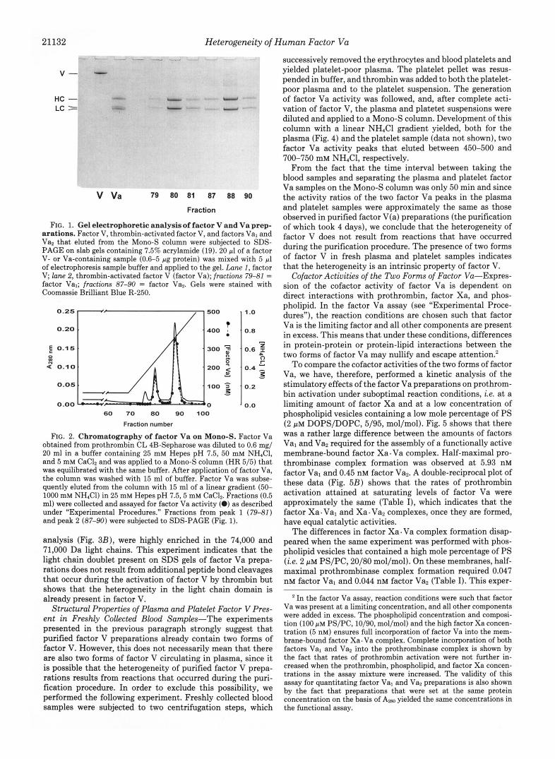
Although anion exchangers are usually employed in the purification of factor V and Va, it appears that the light chain of factor Va is a basic protein with a high affinity for cation exchangers (3, 11). Fig. 2 shows that factor Va also bound to a Mono-S column. Subsequent elution with a NH,C1 gradient yielded two protein peaks with factor Va activity. SDS-PAGE analysis showed that the first peak consisted of factor Va molecules with a light chain of M, = 74,000 (Fig. 1, fractions 79-81) and that the factor Va that eluted in the second peak contained the light chain with M, = 71,000 (Fig. 1, fractions 87-90). In the text that follows, we will designate the factor Va present in the first peak, factor Val, while the factor Va present in the second peak will be called factor Vaz.
Separation of Two Forms of Factor V on a Mono-S Col- umn-Nonactivated human factor V showed a chromato- graphic behavior that strongly resembled factor Va. Factor V also bound t o the Mono-S column and eluted as a double peak when the column was developed with an NH&l gradient (Fig. 3A). Incubation of the protein fractions with thrombin yielded two factor Va activity peaks, which, as shown by SDS-PAGE
21132 Heterogeneity of Human Factor V u
V -
HC - LC =
V Va 79 ao a1 a7 aa 90
Fraction
FIG. 1. Gel electrophoretic analysis of factor V and Va prep- arations. Factor V, thrombin-activated factor V, and factors Val and Vaz that eluted from the Mono-S column were subjected to SDS- PAGE on slab gels containing 7.5% acrylamide (19). 20 pl of a factor V- or Va-containing sample (0.6-5 pg protein) was mixed with 5 pl of electrophoresis sample buffer and applied to the gel. Lane 1, factor V; lane 2, thrombin-activated factor V (factor Va); fractions 79-81 = factor Val; fractions 87-90 = factor Vas. Gels were stained with Coomassie Brilliant Blue R-250.
0.20 I 0.15
0 OD
4“ 0.10
I 400
300 9 0 c
200 Y d
100 7 - 0
’ 0.8
’
0.6 2 z 0 r
0.4 x 3:
- ’
- ’ 0.2
. 0.0 60 70 80 90 100
Fraction number
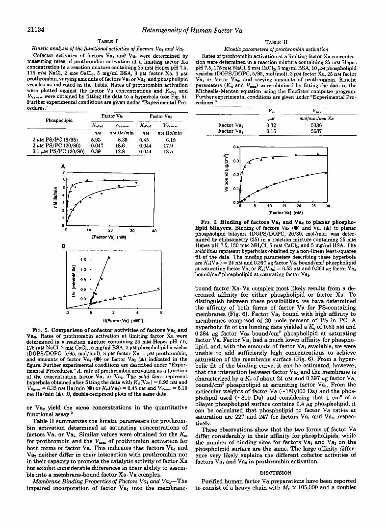
FIG. 2. Chromatography of factor Va on Mono-S. Factor Va obtained from prothrombin CL 4B-Sepharose was diluted to 0.6 mg/ 20 ml in a buffer containing 25 mM Hepes pH 7.5, 50 mM NHIC1, and 5 mM CaClZ and was applied to a Mono-S column (HR 5/51 that was equilibrated with the same buffer. After application of factor Va, the column was washed with 15 ml of buffer. Factor Va was subse- quently eluted from the column with 15 ml of a linear gradient (50- 1000 mM NH,Cl) in 25 mM Hepes pH 7.5,5 mM CaCl2. Fractions (0.5 ml) were collected and assayed for factor Va activity (0) as described under “Experimental Procedures.” Fractions from peak 1 (79-81) and peak 2 (87-90) were subjected to SDS-PAGE (Fig. 1).
analysis (Fig. 3B) , were highly enriched in the 74,000 and 71,000 Da light chains. This experiment indicates that the light chain doublet present on SDS gels of factor Va prepa- rations does not result from additional peptide bond cleavages that occur during the activation of factor V by thrombin but shows that the heterogeneity in the light chain domain is already present in factor V.
Structural Properties of Plasma and Platelet Factor V Pres- ent in Freshly Collected Blood Samples-The experiments presented in the previous paragraph strongly suggest that purified factor V preparations already contain two forms of factor V. However, this does not necessarily mean that there are also two forms of factor V circulating in plasma, since it is possible that the heterogeneity of purified factor V prepa- rations results from reactions that occurred during the puri- fication procedure. In order to exclude this possibility, we performed the following experiment. Freshly collected blood samples were subjected to two centrifugation steps, which
successively removed the erythrocytes and blood platelets and yielded platelet-poor plasma. The platelet pellet was resus- pended in buffer, and thrombin was added to both the platelet- poor plasma and to the platelet suspension. The generation of factor Va activity was followed, and, after complete acti- vation of factor V, the plasma and platetet suspensions were diluted and applied to a Mono-S column. Development of this column with a linear NH,Cl gradient yielded, both for the plasma (Fig. 4) and the platelet sample (data not shown), two factor Va activity peaks that eluted between 450-500 and 700-750 mM NH4Cl, respectively.
From the fact that the time interval between taking the blood samples and separating the plasma and platelet factor Va samples on the Mono-S column was only 50 min and since the activity ratios of the two factor Va peaks in the plasma and platelet samples were approximately the same as those observed in purified factor V(a) preparations (the purification of which took 4 days), we conclude that the heterogeneity of factor V does not result from reactions that have occurred during the purification procedure. The presence of two forms of factor V in fresh plasma and platelet samples indicates that the heterogeneity is an intrinsic property of factor V.
Cofactor Activities of the Two Forms of Factor Vu-Expres- sion of the cofactor activity of factor Va is dependent on direct interactions with prothrombin, factor Xa, and phos- pholipid. In the factor Va assay (see “Experimental Proce- dures”), the reaction conditions are chosen such that factor Va is the limiting factor and all other components are present in excess. This means that under these conditions, differences in protein-protein or protein-lipid interactions between the two forms of factor Va may nullify and escape attention?
To compare the cofactor activities of the two forms of factor Va, we have, therefore, performed a kinetic analysis of the stimulatory effects of the factor Va preparations on prothrom- bin activation under suboptimal reaction conditions, i.e. at a limiting amount of factor Xa and at a low concentration of phospholipid vesicles containing a low mole percentage of PS (2 PM DOPS/DOPC, 5/95, mol/mol). Fig. 5 shows that there was a rather large difference between the amounts of factors Val and Va2 required for the assembly of a functionally active membrane-bound factor Xa . Va complex. Half-maximal pro- thrombinase complex formation was observed at 5.93 nM factor Val and 0.45 nM factor Vaz. A double-reciprocal plot of these data (Fig. 5B) shows that the rates of prothrombin activation attained at saturating levels of factor Va were approximately the same (Table I), which indicates that the factor Xa. Val and Xa. Va2 complexes, once they are formed, have equal catalytic activities.
The differences in factor Xa. Va complex formation disap- peared when the same experiment was performed with phos- pholipid vesicles that contained a high mole percentage of PS (i.e. 2 p M PS/PC, 20/80 mol/mol). On these membranes, half- maximal prothrombinase complex formation required 0.047 nM factor Val and 0.044 nM factor Va2 (Table I). This exper-
In the factor Va assay, reaction conditions were such that factor Va was present at a limiting concentration, and all other components were added in excess. The phospholipid concentration and composi- tion (100 p~ PS/PC, 10/90, mol/mol) and the high factor Xa concen- tration (5 nM) ensures full incorporation of factor Va into the mem- brane-bound factor Xa. Va complex. Complete incorporation of both factors Val and Vaz into the prothrombinase complex is shown by the fact that rates of prothrombin activation were not further in- creased when the prothrombin, phospholipid, and factor Xa concen- trations in the assay mixture were increased. The validity of this assay for quantitating factor Val and Van preparations is also shown by the fact that preparations that were set at the same protein concentration on the basis of Am yielded the same concentrations in the functional assay.

FIG. 3. A, chromatography of factor V on Mono-S. 5 ml of purified factor V (5 mg) in 10 mM Hepes pH 7.5, 50 mM NaCl was applied to a Mono-S column (HR 5/5). The column was washed with 15 ml of a buffer containing 25 mM Hepes pH 7.5,50 mM NH,CI, and 5 mM CaC12 and subsequently developed with 30 ml of a linear gradient from 50 to 1000 mM NH4CI in 25 mM Hepes pH 7.5 and 5 mM CaC12. Samples from the column fractions were incubated for 1 h at 37 "C with thrombin (final concentration, 25 nM) and the factor Va generated (0) was assayed as described under "Experimen- tal Procedures." B, gel electrophoretic analysis of factor Va generated in the factor V fractions that eluted from the Mono-S column. Aliquots (50 pl) from the column fractions 64, 66, 72, 76, 78, and 82) were activated with thrombin (Fig. 3A) and subsequently mixed wih 12.5 pl of gel electrophoresis sample buffer (19). 20 pl of the gel samples were then applied to a 7.5% slab gel and sub- jected to SDS-PAGE (19). Sm is a lane with starting material (factor V) acti- vated with thrombin; M. is a lane with molecular weight markers. The proteins on the gel were stained with Coomassie Brilliant Blue R-250.
Heterogeneity of Human Factor Vu
A 1 .o
0.8
E 0.6 0 OD 2 0.4
0.2
0.0
0
B
1 .o
0 0.8 ; .
0
0.6 * nl 2
0.4 . Y
0.2 . Y E
0.0
20 40 60 80 100
Fraction number
Sm
0.10 E 2 0 m
0.05
0.00 L
0 20 40 60
Fraction number
FIG. 4. Chromatography of thrombin-activated plasma on a Mono-S column. Platelet-poor plasma derived from blood collected in citric acid was brought to 5 mM free CaC12 and incubated for 10 min at 37 "C with thrombin (final concentration 10 nM). The fibrin clot that was formed was removed, and 1 ml of the serum was diluted five times with column buffer (25 mM Hepes pH 7.5, 5 mM CaC12) and applied to a Mono-S column (HR 5/5). The column was washed with 13 ml of a buffer containing 25 mM Hepes pH 7.5, 150 mM NH4C1, and 5 mM CaCI2, and the proteins bound to the column were subsequently eluted with 15 ml of a salt gradient from 50 to 1000 mM NH4C1 in 25 mM Hepes pH 7.5 and 5 mM CaC12. Column fractions (0.5 ml) were assayed for factor Va activity (0) as described under Experimental Procedures."
21133
1 .o
0.8 - 0.6 $
0 r
zz
P
0.4 - 0.2
0.0
t 92,000
t 67,000
a- 43,000
c 30,000
c 21,500
c 14,400
64 66 72 76 78 82 Mr
Fraction
iment indicates that the different efficiencies by which factors Val and Van incorporate in the prothrombinase complex are likely compensated by an increased binding affinity and an increased number of binding sites for factors Xa and Va on membranes containing 20 mole percent of PS (3, 27, 28-31). This argumentation predicts that differences between factors Val and Van can also be observed on membranes with 20 mole percent of PS, provided that the experiment is performed at a lower phospholipid concentration. Indeed titration of a limited amount of factor Xa with factors Val or Va2 on 0.1 PM PS/PC (20/80, mol/mol) vesicles again showed a differ- ence between the two forms of factor Va (Table I). A t this phospholipid concentration, a 9-fold higher concentration of factor Val was required to obtain half-maximal rates of pro- thrombin activation (0.389 nM factor Val versus 0.044 nM factor Van).
With respect to the different functional properties of factors Val and Van, the possibility has to be ruled out that factor Val lacks functional activity and that the cofactor activity of factor Val is due to a small (-8%) contamination with factor Van. This possibility is excluded by the observations that ( a ) the amounts of factor Val and factor Van required for factor Xa.Va complex formation on 2 pM Ps/Pc (20/80) were the same (Table I), and ( b ) equal protein amounts of factors Val
21134 Heterogeneity of Human Factor Vu TABLE I
Kinetic analysk of the functional activities of Factors Val and Va, Cofactor activities of factors Val and Va, were determined by
measuring rates of prothrombin activation at a limiting factor Xa concentration in a reaction mixture containing 25 mM Hepes pH 7.5, 175 mM NaCl, 2 mM CaC12, 5 mg/ml BSA, 3 PM factor Xa, 1 p~ prothrombin, varying amounts of factors Val or Va,, and phospholipid vesicles as indicated in the Table. Rates of prothrombin activation were plotted against the factor Va concentrations and and VV.,, were obtained by fitting the data to a hyperbola (see Fig. 5 ) . Further experimental conditions are given under “Experimental Pro- cedures.”
Factor Val Phospholipid
Factor Va,
KMv.1 VV.-- KHVIZ V V ~ - - nM nM IIa/min nM nM IIa/min
2 p M Ps/Pc (5/95) 5.93 6.35 0.45 6.15 2 pM Ps/Pc (20/80) 0.047 18.6 0.044 17.9 0.1 p M Ps/Pc (20/80) 0.39 12.8 0.044 13.5
A 7 , 1 . I
0 ’ I
[Factor Val (nM) 0 10 20 30 40
B
-2 0 2 4
l/[Factor Val (nM.’)
FIG. 5. Comparison of cofactor activities of factors Val and Van. Rates of prothrombin activation at limiting factor Xa were determined in a reaction mixture containing 25 mM Hepes pH 7.5, 175 mM NaCl, 2 mM CaC12, 5 mg/ml BSA, 2 p~ phospholipid vesicles (DOPS/DOPC, 5/95, mol/mol), 3 pM factor Xa, 1 p~ prothrombin, and amounts of factor Val (0) or factor Va, (A) indicated in the figure. Further experimental conditions are described under “Exper- imental Procedures.” A, rate of prothrombin activation as a function of the concentration factor Val or Va2. The solid lines represent hyperbola obtained after fitting the data with &(Val) = 5.93 nM and VV, = 6.35 nM IIa/min (0) or K,+(Va2) = 0.45 nM and VV, = 6.15 nM IIa/min (A). B, double-reciprocal plots of the same data.
or Va2 yield the same concentrations in the quantitative functional assay.2
Table I1 summarizes the kinetic parameters for prothrom- bin activation determined at saturating concentrations of factors Val or Va2. Similar values were obtained for the K,,, for prothrombin and the V,, of prothrombin activation for both forms of factor Va. This indicates that factors Val and Va2 neither differ in their interaction with prothrombin nor in their capacity to promote the catalytic activity of factor Xa but exhibit considerable differences in their ability to assem- ble into a membrane-bound factor Xa. Va complex.
Membrane Binding Properties of Factors Val and Va2”The impaired incorporation of factor Val into the membrane-
TABLE I1 Kinetic parameters of prothrombin activation
Rates of prothrombin activation at a limiting factor Xa concentra- tion were determined in a reaction mixture containing 25 mM Hepes pH 7.5,175 mM NaCl, 2 mM CaCl,, 5 mg/ml BSA, 10 pM phospholipid vesicles (DOPS/DOPC, 5/95, mol/mol), 3 PM factor Xa, 25 nM factor Val or factor Vaz, and varying amounts of prothrombin. Kinetic parameters (K,,, and V& were obtained by fitting the data to the Michaelis-Menten equation using the Enzfitter computer program. Further experimental conditions are given under “Experimental Pro- cedures.’’
P M mol/min/mol Xa Factor Val 0.32 5386 Factor Vaz 0.16 5687
0 5 10 15 20 25 30
[Factor Val (nM)
FIG. 6. Binding of factors Val and Va, to planar phospho- lipid bilayers. Binding of factors Val (0) and Va2 (A) to planar phospholipid bilayers (DOPS/DOPC, 20/80, mol/mol) was deter- mined by ellipsometry (25 ) in a reaction mixture containing 25 mM Hepes pH 7.5, 150 mM NH,Cl, 5 mM CaCl2, and 5 mg/ml BSA. The solid lines represent hyperbolas obtained by a non-linear least-squares fit of the data. The binding parameters describing these hyperbola are &(Val) = 24 nM and 0.397 pg factor Val bound/cm2 phospholipid at saturating factor Val or Kd(Vaz) = 0.53 nM and 0.364 pg factor Vas bound/cm2 phospholipid at saturating factor Va,.
bound factor Xa-Va complex most likely results from a de- creased affinity for either phospholipid or factor Xa. To distinguish between these possibilities, we have determined the affinity of both forms of factor Va for PS-containing membranes (Fig. 6). Factor Vaz bound with high affinity to membranes composed of 20 mole percent of PS in PC. A hyperbolic fit of the binding data yielded a Kd of 0.53 nM and 0.364 pg factor Va2 bound/cm2 phospholipid at saturating factor Va. Factor Val had a much lower affinity for phospho- lipid, and, with the amounts of factor Val available, we were unable to add sufficiently high concentrations to achieve saturation of the membrane surface (Fig. 6). From a hyper- bolic fit of the binding curve, it can be estimated, however, that the interaction between factor Val and the membrane is characterized by a Kd of about 24 nM and 0.397 pg factor Val bound/cm2 phospholipid at saturating factor Val. From the molecular weights of factor Va (-180,000 Da) and the phos- pholipid used (-800 Da) and considering that 1 cm2 of a bilayer phospholipid surface contains 0.4 pg phospholipid, it can be calculated that phospholipid to factor Va ratios at saturation are 227 and 247 for factors Val and Vaz, respec- tively.
These observations show that the two forms of factor Va differ considerably in their affinity for phospholipids, while the number of binding sites for factors Val and Va2 on the phospholipid surface are the same. The large affinity differ- ence very likely explains the different cofactor activities of factors Val and Va2 in prothrombin activation.
DISCUSSION
Purified human factor Va preparations have been reported to consist of a heavy chain with M, = 105,000 and a doublet

Heterogeneity of Human Factor Vu 21135
light chain with M, = 71,000 and = 74,000 (7). The data presented in this paper show that it is possible to bind factor Va to a Mono-S column and to separate two forms of factor Va by developing the column with an NH&1 gradient. The peak eluting at the low salt concentration consisted of factor Va (Va,) with M, = 74,000 light chain, whereas factor Va with M , FZ 71,000 light chain (factor Van) eluted at a much higher salt concentration. It is likely that the factor Va bound to the Mono-S column via its light chain, since this subunit is considered to be a basic protein that exhibits a high affinity for cation exchangers (3, 11). The elution profile from the Mono-S column is, therefore, indicative of a charge difference between the light chains of factors Val and Vaz, in the sense that the light chain of factor Van is more positively charged than the light chain of factor Val.
The interaction of factor Va with procoagulant membranes appears to depend on both hydrophobic interactions (32-35) and on electrostatic interactions between the positively charged light chain and negatively charged phospholipids (27, 30, 36). Considering that electrostatic interactions are impor- tant for factor Va-membrane association and that there may be a considerable charge difference between the light chains of the two factor Va forms, we hypothesized that factor Val may exhibit a lower affinity for negatively charged membranes than factor Van. Direct binding experiments show that this is indeed the case. Factor Vaz binds with a high affinity (Kd = 0.53 nM) to PS-containing membranes (Ps/Pc, 20/80), whereas the affinity of factor Val for such membranes is much less (Kd = 24 nM). The lipid/factor Va ratios (n) at protein saturation were approximately the same (n = 227 for factor Val and n = 247 for factor Van), which shows that the membrane surface contains the same number of binding sites for both forms of factor Va. It is difficult to compare these binding parameters with literature values, since the interac- tion of human factor Va with membranes is not well docu- mented, whereas a wide range of binding parameters ( K d = 0.05-10 nM and n = 42-250) is reported to describe the interaction of bovine factor Va with PC membranes that contain 25 mole percent of PS (27, 29-31, 36).
Factor Va plays an important role in both the assembly and expression of the catalytic activity of the prothrombinase complex on procoagulant membrane surfaces (1-4). Since proper cofactor function requires the binding of factor Va to the membrane, it is to be expected that the different affinities of factors Val and Van for negatively charged membranes will result in different efficiencies to act as a cofactor in prothrom- bin activation. Kinetic experiments showed that the cofactor function of factor Val is indeed impaired. Much higher amounts of factor Val were required for full assembly of the prothrombinase (factor Xa. Va) complex at the membrane surface. Once formed, the factor Xa- Val and Xa. Van com- plexes exhibit the same catalytic activity in prothrombin activation. This can be inferred from the fact that the kinetic parameters (kat and K,) for prothrombin activation are sim- ilar (Table 11). Differences in efficiencies by which factors Val and Van assemble into a membrane-bound factor Xa. Va complex were observed only on membranes that contain low amounts of PS or when prothrombin is activated at low phospholipid concentrations. This is not surprising if one considers that factor Xa. Va complex formation on the phos- pholipid surface is a reflection of the concentrations of sur- face-bound factors Va and Xa (3, 37). At high mole percent- ages of PS and at high phospholipid concentrations, differ- ences between factors Val and Van will be nullified when the concentration of factor Va binding sites exceeds the Kd values for the membrane-factor Va complexes, since this condition
results in virtually complete binding of both forms of factor Va.
The heterogeneity in the light chains of factor Va is not a unique property of factor Va but is already present in factor V. It appears that the purified factor V preparations and even freshly prepared plasma samples and platelet suspensions contain two forms of factor V, which, after activation with thrombin, are converted into factors Val and Van. This shows that the heterogeneity is an intrinsic property of factor V and is not caused by reactions occurring during the purification procedure or to additional peptide bond cleavages that take place when factor V is activated with thrombin.
Our experiments give no direct insight to the structural basis underlying the difference between the light chain regions of the two forms of factor V. However, combination of infor- mation presented in this paper with data recently reported by Ortel et al. (12) strongly suggests that the observed heteroge- neity originates from the carboxyl-terminal C2 domain of the light chain region of factor V. Ortel et al. (12) isolated recom- binant human factor V and showed that activation with thrombin resulted in the formation of a light chain doublet. Activation of mutant molecules that lack the carboxyl-ter- minal C2 domain (i.e. amino acid residues 2037-2196) yield a factor Va molecule with a single truncated light chain with M, = 62,000. Deletion of the C2 domain caused an essentially complete loss of the ability of the mutant factor Va molecule to bind to negatively charged membranes. This indicates that the occurrence of two forms of factor Va (Val and Van) with different affinities for procoagulant membranes results from the presence of two light chains with structural differences in the C2 domain. The fact that heterogeneity is also observed in the factor V present in freshly prepared platelet and plasma samples and in recombinant factor V (12) strongly suggests that the heterogeneity is due to differences in post-ribosomal processing or post-ribosomal modification (e.g. glycosylation) of the two factor V molecules. Whether the presence of two forms of factor V in plasma has a physiologicdl function remains to be clarified.
Acknowledgments-We thank Rita Janssen and Dr. George Wil- lems for performing the binding experiments on the ellipsometer.
REFERENCES 1. Nesheim. M. E.. Taswell. J. B.. and Mann. K. G. (1979) J. Biol. Chem.
264,10952-10962 '
, , ~ ~~
2. Rosing, J., Tans, G., Govers-Riemslag, J. W. P., Zwaal, R. F. A,, and Hemker. H. C. (1980) J. Biol. Chem. 256.274-283
3. Lindhout, T., Govers-Riemslag, J. W. P., van de Waart, P., Hernker, H. C.,
4. van Rijn, J. L. M. L., Govers-Riemslag, J. W. P., Zwaal, R. F. A,, and and Rosing, J. (1982) Biochemistry 21,5494-5502
5. Dahlback, B. (1980) J. Clin. Inuest. 66,583-591 Rosing, J. (1984) Biochemistry 23,4557-4564
6. Kane, W. H., and Majems, P. W. (1981) J. Biol. Chem. 2 6 6 , 1002-1007 7. Suzuki, K., Dahlback, B., and Stenflo, J. (1982) J. Biol. Chem. 267,6556-
8. Esmon, C. T. (1979) J. Biol. Chem. 264,964-973
10. Nesbeim, M. E., Foster, W. B., Hewick, R., and Mann, K. G. (1984) J. Biol. 9. Guinto. E. R., and Esmon, C. T. (1982) J. Biol. Chem. 267,10038-10043
11. Odegaard, B., and Mann, K. (1987) J. Biol. Chem. 2 6 2 , 11233-11238 12. Ortel, T. L., Devore-Carter, D., Quinn-Allen, M., and Kane, W. H. (1992)
13. DiScipio, R. G., Hermodson, M. A., Yaks, S. G., and Davie, E. W. (1977)
14. Pletcher, C. H., and Nelsestuen, G. L. (1982) J. Biol. Chem. 2 5 7 , 5342-
~~I~ ~ ~~~
6564
Chem. 269,3187-3196
J. Biol. Chem. 267,4189-4198
Biochemistry 16,698-706
5345 15. Bock, P. E., Craig, P. A., Olsen, S. T., and Singh, P. (1989) Arch, Biochem.
16. Schiffman, S., Theodor, J., and Rapaport, S. J. (1969) Biochemistry 8 , Biophys. 273,375-388
l w 7 - 1 A M
17. Tans, G., Rosing, J., Thomassen, M. C. L. G. D., Heeb, M. J., Zwaal, R. F.
18. Laemmli, U. K. (1970) Nature 227.680-685
_". * " Y
A., and Griffin, J. H. (1991) B h d 77,2641-2648
19. Schiipger, H., and von' Jagow, G. (1987) Anal. Biochem. 166,368-379 20. Lowry, 0. H., Rosebrough, N. J., Farr, A. L., and Randall, R. J. (1951) J.
Biol. Chem. 193,265-275 21. Chase, T., Jr., and Shaw, E. (1969) Biochemistry 8, 2212-2224 22. Smith, R. L. (1973) J. Biol. Chem. 248,2418-2423
21136 Heterogeneity of Human Factor Va 23. Bottcher, C. J. F., van Gent, C. M., and Pries, C. (1961) Anal. Chim. Acta
24. Tans, G.. J a ~ ~ n - C l a e s ~ n , T., Hemker, H. C., Zwaal, R F. A., and Rosing,
25. Cuypers, P. A,, Cornel, J. W., Janssen, M. P., Kop, J. M. M., Hermens, W.
26. Guinto, E. R., and Esmon, C. T. (1984) J. Bwt. Chem 269,13966-13992 27. van de Waart, P., Bruls, H., Hemker, H. C., and Lindhout, T. (1983)
24,203-207
J. (1991) J. Bid. Chem. 266,21864-21873
T., and Hemker, H. C. (1983) J. Bid. Chem. 268.2426-2431
B ~ h e m i s ~ ~ 22.2427-2432 28. Nelseatuen, G: L., and Broderius, M. (1977) Biochemistry 16,4172-4177 29. Bloom, J. W., Nesheim, M. E., and Mann, K. G. (1979) Biochemktry 18,
449-4425
30.
31. 32. 33.
34.
35.
36. 37.
Pusey, M. L., Mayer, L. D., Jason Wei, G Bloomfield, V. A,, and Nelses-
Krishnaswamy S. and Mann K. G. (1988) J. Bwl. Chem. 263 5714-5723 Higgina, D. L 'ant? Mann, K.'G. (1983) J. BwL Chem. 268.6.503-6508 Lecompte MYF., Krishnaswamy, S., Mann, K. G., Nesheim, M. E., and
Krie , d. C. Isaacs, 3. S., Yerng, S . S., Esmon, C. T., Bailey, H., and
Kalafatis M Jenny, R. J., and Mann, K. G. (1990) J. Bwl. Chem.'266,
tuen, G. L. (1982) Biochemistry 21,526i-5269
Gltler 6. (1987) J. Ewl. Chem. 282,1935-1937
Jotnson, A. E. (1987) Biochemistry 26,103-109
215f%??1 id9 Pusey, M. L., and Nelsestuen G. L. (19%) Biochenkitry 23,6202-6210 Krishnaswamy, S Jones, K.'C., and Mann, K. G. (1988) J. Bid. Chem.
"" ~ _"" 263,3823-383i