of0 lipids of sarcinalutea - journal of bacteriologyjb.asm.org/content/89/3/768.full.pdf · lipids...
TRANSCRIPT
JOURNAL OF BACTERIOLOGY, Mar., 1965 Vol. 89 No. 3Copyright 0 1965 American Society for Microbiology Printed in U.S.A.
Lipids of Sarcina luteaIII. Composition of the Complex Lipids
CHARLES K. HUSTON, PHILLIP W. ALBRO, AND GERALD B. GRINDEYPhysical Defense Division, U.S. Army Biological Laboratories, Fort Detrick,
Frederick, Maryland
Received for publication 9 November 1964
ABSTRACTHUSTON, CHARLES K. (Fort Detrick, Frederick, Md.), PHILLIP W. ALBRO, AND GER-
ALD B. GRINDEY. Lipids of Sarcina lutea. III. Composition of the complex lipids. J.Bacteriol. 89:768-775. 1965.-The complex lipids from a strain of Sarcina lutea were iso-lated and separated into fractions on diethylaminoethyl cellulose acetate and silicicacid columns. These fractions were monitored in several thin-layer chromatographysystems. The various lipid types were characterized by their behavior in thin-layersystems and by an analysis of their hydrolysis products. The fatty acid composition ofthe column fractions was determined by gas-liquid chromatography. A number of com-ponents (13) were separated by thin-layer chromatography and characterized. Themajor components were polyglycerol phosphatide (17.0%), lipoamino acids (15.1%),phosphatidyl glycerol (13.8%), and an incompletely characterized substance (15.0%).Minor constituents included phosphatidyl inositol (5.5%), phosphatidic acid (4.2%),phosphatidyl serine (2.0%), and phosphatidyl choline (1.0%). No phosphatidyl etha-nolamine was observed.
Few bacterial phospholipids have been studiedthoroughly by modern analytical techniques.Lovern (1957) described many bacterial phos-pholipids as simple fatty acid esters of phos-phorylated carbohydrates. Ethanolamine is themost commonly reported nitrogen-containingcomponent, whereas choline is found in somespecies (Asselineau and Lederer, 1960). Recentreports have also described the isolation oflipoamino acid complexes from the complexlipids of bacteria (MacFarlane, 1962a, b; Hunterand James, 1963; Ikawa, 1963).We have reported (Huston and Albro, 1964)
that Sarcina lutea contains a complex mixture ofhighly polar lipids comprising approximately 23%of the total extractable lipid. The present com-munication is on the composition of this polarfraction.
MATERIALS AND METHODS
Whatman diethylaminoethyl (DEAE) cellulosewas obtained from Scientifica, Clifton, N.J., andconverted to the acetate form, according to Rouseret al. (1961). Silicic acid was obtained from Mal-linckrodt Chemical Works, St. Louis, Mo., andsieved to 100/200, 100/140, and 160/200 mesh.Inorganic reagents, solvents, and Hyflo Super-Cel were obtained from Fisher Scientific Co.,Silver Spring, Md., and were the highest gradesavailable. Chloroform, hexane, and propionicacid were redistilled before use. The methanol,
acetone, benzene, and diisobutyl ketone were gas-chromatographically pure. Synthetic phospha-tidyl ethanolamine, phosphatidyl serine, andphosphatidyl choline (all dipalmitoyl) were A-grade from Calbiochem. Lecithin and cephalinstandards were isolated from soy phosphatides bysilicic acid column chromatography, according toFleischer et al. (1962), and examined for homoge-neity by thin-layer chromatography (TLC)according to Horrocks (1963). All other lipidstandards used were the highest grades availablefrom Mann Research Laboratories, New York,N.Y., or Applied Science Laboratories, Inc., StateCollege, Pa. Rhodamine 6G and ninhydrin wereobtained from Eastman Organic Chemicals,Rochester, N.Y. TLC apparatus and materialswere obtained from Brinkmann Instruments, Inc.,Great Neck, N.Y.
Culture conditions. S. lutea ATCC 533 wascultured at 25 C for 24 hr in tryptic soy broth(Difco) under forced aeration. The preparedmedium was found to contain less than 0.009%lipid.
Cells were harvested, as previously described(Huston and Albro, 1964), immediately after thegrowth; all operations were performed below 5 C.
Lipid extraction. Extraction of lipids and isola-tion of the "complex lipid" fraction was accom-plished as previously described (Huston andAlbro, 1964). Nonlipid contaminants that were notremoved by the Folch washing procedure wereseparated on Sephadex (Wells and Dittmer, 1963)and on DEAE cellulose columns (Rouser et al.,1961). These contaminants amounted to 8 to 14%
768
on May 27, 2018 by guest
http://jb.asm.org/
Dow
nloaded from
COMPLEX LIPIDS OF SARCINA LUTEA
of the complex lipid fraction from silicic acidchromatography and consisted of glycerol, nitrog-enous bases, and peptides.Column chromatography. Four classes of lipids
were isolated on DEAE cellulose acetate columns,by the method of Rouser et al. (1961). "Dipolarionic" and neutral lipids were eluted with chloro-form-methanol (7:1, v/v),"basic" lipids with chlo-roform-methanol (7:3, v/v), "acidic" lipids withglacial acetic acid, and "highly acidic" lipids withchloroform - methanol - concentrated ammoniumhydroxide (80:20:1, v/v/v). The fraction elutedwith chloroform-methanol (7:1) was resolved intoneutral lipids, lecithins, and lysolecithins on asilicic acid-silicate column (Rouser et al., 1961).Free fatty acids were removed from the "acidic"lipid fraction on silicic acid (Fleischer et al., 1962).
Phosphatidic acid and polyglycerol phosphatide(cardiolipin type) were isolated by elution from anacid-washed silicic acid column with 5% methanolin chloroform. Phosphatidic acid travelled withthe solvent front, whereas the polyglycerol phos-phatide eluted much later. After rechromatogra-phy, each of the fractions gave a single spot onthin-layer plates. None of the other componentscould be isolated in pure form by column chroma-tography.
TLC. Silica gel G plates prepared according toStahl (1958), or silica gel G plates impregnatedwith 10% sodium acetate (Horrocks, 1963), weredeveloped in the ascending manner in unlinedtanks to resolve the various complex lipids and tomonitor column fractions. Chloroform-methanol-water (65:25:4, v/v/v; Horrocks, 1963), chloro-form-methanol-14% aqueous ammonia (17:7:1,v/v/v), and diisobutyl ketone-acetic acid-water(8:5:1, v/v/v; Lepage, 1964) were used as solventsystems. The spots were detected with (i) aqueousRhodamine 6G, (ii) iodine vapors, (iii) a molyb-date reagent (Dittmer and Lester, 1964), (iv)Dragendorff's reagent for choline and quaternaryammonium compounds, (v) 0.25% ninhydrin inacetone buffered to pH 5.5 with acetate, (vi) io-dine-sodium azide reagent for sulfolipids (Block,Durrum, and Zweig, 1958), and ammoniacal silvernitrate for phosphatidic acid, phosphatidylglycerol, polyglycerol phosphatide, and inositides.
In some cases, the silicic acid-impregnatedfilter-paper system of Marinetti (1962) was usedto aid in the identification of lipids for which nostandards were available.
Quantitation of TLC-resolved material wasperformed according to Amenta (1964).
Hydrolysis and paper chromatography. Water-soluble products, produced by hydrolysis of thevarious fractions and individual componentsaccording to Ikawa (1963), were analyzed byascending chromatography in n-butanol-aceticacid-water (2:1:1, v/v/v; Fink, Cline, and Fink,1963), or in phenol-n-butanol-formic acid-water(50:50:3:10, v/v/v/v; Goldfine, 1962); and bydescending chromatography in n-butanol-aceticacid-water (4:1:5, v/v/v; Block, Le Strange, andZweig, 1952), or n-butanol-propionic acid-water
according to Benson and Maruo (1958). Phosphateesters were detected with Hayes-Isherwood rea-gent, amino compounds with 0.4% ninhydrin inwater-saturated n-butanol, carbohydrates with2.5% aniline hydrogen phthalate, polyhydroxycompounds with ammoniacal silver nitrate, andquarternary ammonium salts with Dragendorff'sreagent. Whatman no. 1 paper was used for ascend-ing chromatography and Whatman no. 4 paper fordescending chromatography.
Acetonation of vicinal hydroxyl groups. Samplessuspected of containing either phosphatidylglycerol or polyglycerol phosphatide were ace-tonated by the procedure of Benson and Maruo(1958). Samples before and after acetonation wereanalyzed by TLC on silica gel G in chloroform-methanol-water (65:25:4, v/v/v). Detection wasas described above.
Chemical methods. Inorganic phosphorus wasdetermined by the procedure of Fiske and Subba-Row (1925), total phosphorus by the Allen (1940)modification, amino nitrogen according to Leaand Rhodes (1955), and total nitrogen accordingto Miller and Miller (1948). Acetals and aldehydeswere qualitatively sought with azobenzenephenyl-hydrazine-sulfonic acid (Feigl, 1954). Fatty acidswere determined by saponification, methanolysis,and weighing. Fatty acid methyl esters were pro-duced for gas chromatography as previouslydescribed (Huston and Albro, 1964).
Gas-liquid chromatography (GLC). Gas-liquidchromatography of fatty acid methyl esters wascarried out as described previously (Huston andAlbro, 1964) after separation of the normal fattyacid and hydroxy acid esters on silicic acid col-umns (Kishimoto and Radin, 1963).
Carbohydrates were separated as their tri-methylsilyl (TMS) derivatives (Bentley et al.,(1963) on a column (121.92 by 0.635 cm) of 3%SE-52 on 60/80 mesh Chromosorb W at varioustemperatures.
Infrared analysis. Infrared spectra of variousfractions and constituents were examined as thinfilms on KBr or AgCl plates with a Perkin-Elmermodel 21 recording infrared spectrophotometer.
RESULTS
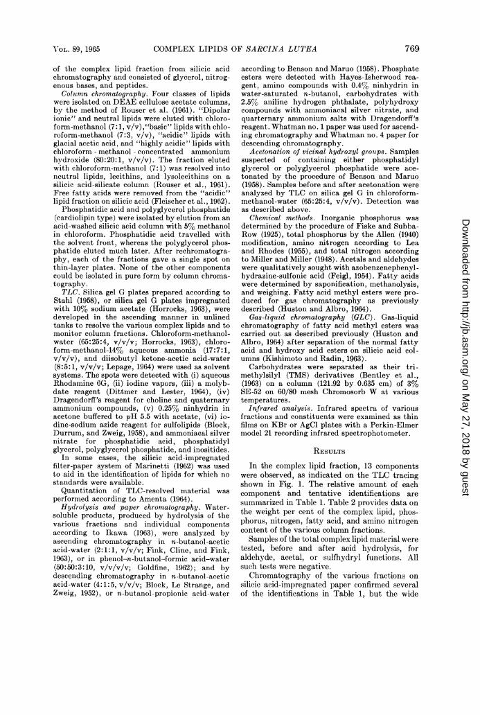
In the complex lipid fraction, 13 componentswere observed, as indicated on the TLC tracingshown in Fig. 1. The relative amount of eachcomponent and tentative identifications aresummarized in Table 1. Table 2 provides data onthe weight per cent of the complex lipid, phos-phorus, nitrogen, fatty acid, and amino nitrogencontent of the various column fractions.Samples of the total complex lipid material were
tested, before and after acid hydrolysis, foraldehyde, acetal, or sulfhydryl functions. Allsuch tests were negative.Chromatography of the various fractions on
silicic acid-impregnated paper confirmed severalof the identifications in Table 1, but the wide
769I OL. 89, 1965
on May 27, 2018 by guest
http://jb.asm.org/
Dow
nloaded from
770 HUSTON, ALBRO, AND GRINDEY
FIG. 1. Thin-layer chromatography of Sarcinalutea complex lipids. Solvent system: chloroform-methanol-14% aqueous ammonia (17:7:1, v/v/v).Spots: (1) total complex lipid, (2) neutral-"di-polar ionic" fraction, (8) "basic" fraction, (4)"acidic" fraction, (5) "highly acidic" fraction, (6)authentic phosphatidyl ethanolamine, (7) soy leci-thin, and (8) authentic phosphatidyl serine.
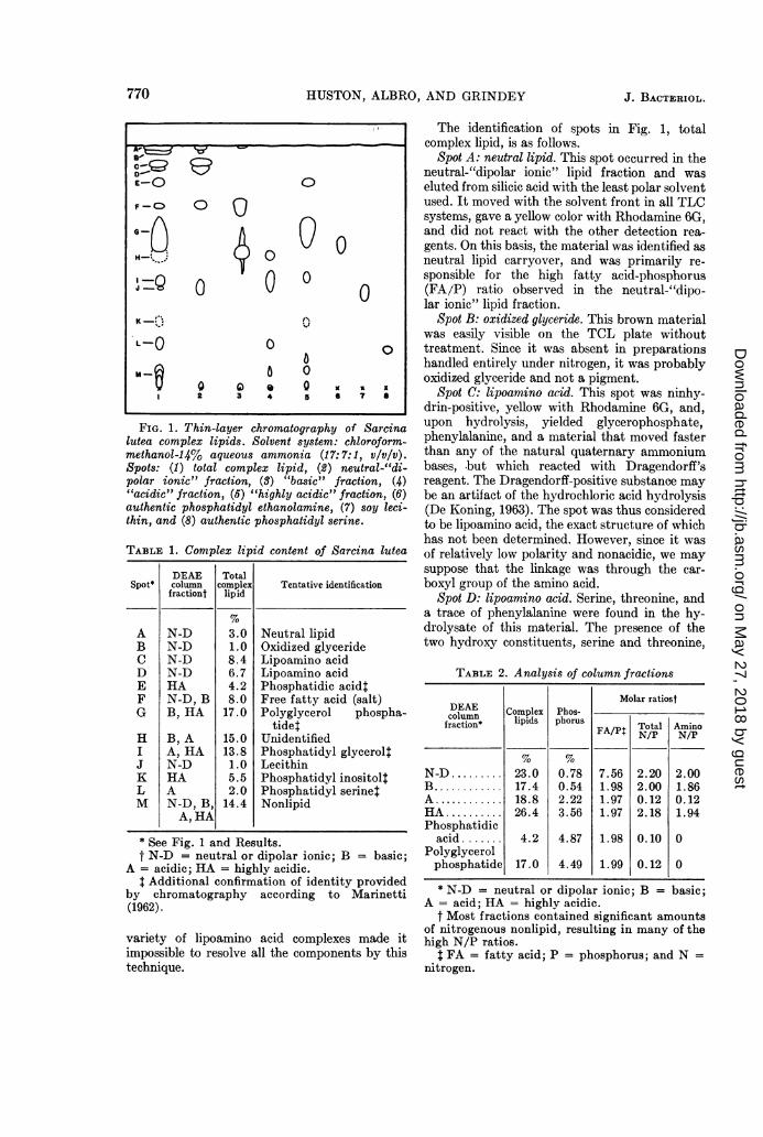
TABLE 1. Complex lipid content of Sarcina lutea
DEAE TotalSpot* column complex Tentative identification
fractiont lipid
A N-D 3.0 Neutral lipidB N-D 1.0 Oxidized glycerideC N-D 8.4 Lipoamino acidD N-D 6.7 Lipoamino acidE HA 4.2 Phosphatidic acid:F N-D, B 8.0 Free fatty acid (salt)G B, HA 17.0 Polyglycerol phospha-
tidetH B, A 15.0 UnidentifiedI A, HA 13.8 Phosphatidyl glycerol:J N-D 1.0 LecithinK HA 5.5 Phosphatidyl inositoltL A 2.0 Phosphatidyl serinetM N-D, B, 14.4 Nonlipid
A, HA
* See Fig. 1 and Results.t N-D = neutral or dipolar ionic; B = basic;
A = acidic; HA = highly acidic.t Additional confirmation of identity provided
by chromatography according to Marinetti(1962).
variety of lipoamino acid complexes made itimpossible to resolve all the components by thistechnique.
J. BACTERIOL.
The identification of spots in Fig. 1, totalcomplex lipid, is as follows.
Spot A: neutral lipid. This spot occurred in theneutral-"dipolar ionic" lipid fraction and waseluted from silicic acid with the least polar solventused. It moved with the solvent front in all TLCsystems, gave a yellow color with Rhodamine 6G,and did not react with the other detection rea-gents. On this basis, the material was identified asneutral lipid carryover, and was primarily re-sponsible for the high fatty acid-phosphorus(FA/P) ratio observed in the neutral-"dipo-lar ionic" lipid fraction.
Spot B: oxidized glyceride. This brown materialwas easily visible on the TCL plate withouttreatment. Since it was absent in preparationshandled entirely under nitrogen, it was probablyoxidized glyceride and not a pigment.
Spot C: lipoamino acid. This spot was ninhy-drin-positive, yellow with Rhodamine 6G, and,upon hydrolysis, yielded glycerophosphate,phenylalanine, and a material that moved fasterthan any of the natural quaternary ammoniumbases, but which reacted with Dragendorff'sreagent. The Dragendorff-positive substance maybe an artifact of the hydrochloric acid hydrolysis(De Koning, 1963). The spot was thus consideredto be lipoamino acid, the exact structure of whichhas not been determined. However, since it wasof relatively low polarity and nonacidic, we maysuppose that the linkage was through the car-boxyl group of the amino acid.
Spot D: lipoamino acid. Serine, threonine, anda trace of phenylalanine were found in the hy-drolysate of this material. The presence of thetwo hydroxy constituents, serine and threonine,
TABLE 2. Analysis of column fractions
Molar ratiostDEAE Complex___Phos_column Cope Phs
fraction* lipids phorus FA/P Total AminoFA/P N/P N/P
% '%N-D ......... 23.0 0.78 7.56 2.20 2.00B......... 17.4 0.54 1.98 2.00 1.86A ......... 18.8 2.22 1.97 0.12 0.12HAo......... 26.4 3.56 1.97 2.18 1.94Phosphatidic
acid ....... 4.2 4.87 1.98 0.10 0Polyglycerolphosphatide 17.0 4.49 1.99 0.12 0
* N-D = neutral or dipolar ionic; B = basic;A = acid; HA = highly acidic.
t Most fractions contained significant amountsof nitrogenous nonlipid, resulting in many of thehigh N/P ratios.
t FA = fatty acid; P = phosphorus; and N =nitrogen.
A -
E-O 0
F-O 0 0
zQ O 0 0K-@w*;e 31
I, 2 V
.L-0~~~~~~~~~~~~~~~~.0
m- 0 0~~~
12 3 4 ~~5 6 ? a
on May 27, 2018 by guest
http://jb.asm.org/
Dow
nloaded from

COMPLEX LIPIDS OF SARCINA LUTEA
in view of the failure of this material to reduceammoniacal silver nitrate, suggests the pos-sibility that the ester linkage occurs through theOH group of the amino acids. Since this materialmoved much faster than ordinary phosphatidylserine, a structure other than the usual acylatedglycerol phosphate skeleton is postulated. And,since this spot was ninhydrin-positive, the linkagewas probably not through the NH2 group. Theamino acid-containing lipids of S. lutea gaveindications of great diversity in structure, butcomplete characterization of each type wasbeyond the scope of this study.
Spot E: phosphatidic acid. This material washighly acidic, easily eluted from silicic acid with5% methanol in chloroform, and contained nonitrogen. It had a FA/P molar ratio of 1.98 anda phosphorus content of 4.87 %, which corre-sponds almost exactly with the calculated valuefor diisopentadecanoyl phosphatidic acid. It gavea weak reaction with the molybdate reagent anda strong reaction with a modified Hayes-Isher-wood reagent, thus reflecting its oxidizing nature.Only glycerophosphate and fatty acids werefound in the hydrolysis products. These data, plusthe observed RF values in various TLC systems,identified this spot as phosphatidic acid.
Spot F: salt of free fatty acid. The fatty acidnature of this material was ascertained by itsnegative reaction with the two molybdate spraysand with ninhydrin when developed in the acidor neutral TLC systems, and by its extractionfrom the complex lipid mixture with aqueous0.1 M borax. When developed in the ammoniacalsolvent system, this spot held sufficient ammoniato give a transient ninhydrin reaction. Qualitativeanalysis of this material (Wiig, Line, and Flagg,1954) indicated the absence of calcium, magne-sium, and ammonium salts. Sodium ions werefound, but could easily be contaminants. Therelative size of the spot was larger in older prep-arations, indicating its formation by slow auto-hydrolysis of more complex lipid compounds.
Spot G: polyglycerol phosphatide. On the basis ofTLC data and column chromatographic prop-erties, as well as its reaction with various of thedetection reagents, spot G could have been eithera bis-phosphatidic acid salt or a cardiolipin typeof polyglycerol phosphatide. Hydrolysis of thismaterial, after isolation on silicic acid, gavediphosphoglycerol (RF 0.13 in butanol-aceticacid-water, 2:1:1, v/v/v), and diglycerol phos-phate [RF 0.17 in the descending system ofBenson and Maruo (1958)]. The spot had aphosphorus content of 4.49%, which would beexpected for tetraisopentadecanoyl cardiolipin ofthe structure described by MacFarlane and Gray(1957). The FA/P molar ratio of 1.99 indicates
Q-~-CD
00oo
n ,
'P 8
1 2 3 4 5f
. '.
0
0
0
0
6 7 9
FIG. 2. Thin-layer chromatography of phos-phatidyl glycerol and phosphatidyl inositol. Solventsystem: chloroform-methanol-water (65:25:4, vlv/v). Spots: (1) phosphatidyl glycerol fraction fromsilicic acid column, (2) material from spot 1 afteracetonation, (3) total complex lipids, (4) "highlyacidic" fraction, (6) inositol, (6) oleic acid, (7)authentic phosphatidyl ethanolamine, (8) soy leci-thin, and (9) authentic phosphatidyl serine. Solidlines outline spots detected with molybdate reagent;dotted lines outline spots visible only after exposureto iodine vapors; double lines outline spots detectedwith alkaline silver nitrate. Spots 1 and 2 are thesame concentration.
that the structure is probably of the cardiolipintype rather than a bis-phosphatidic acid, whichwould have a FA/P ratio of 4:1. Further evidencefor the cardiolipin structure was provided by thenearly negligible nitrogen content, failure of thematerial to reduce ammoniacal silver nitrate(bis-phosphatidic acid reacts), and the extremetailing on silicic acid columns.
Spot H: unidentified. This material, uponhydrolysis, produced a wide variety of con-stituents, including glycerophosphate, serine, themethyl ester of alanine, methyl ethanolamine,and an as yet unidentified fast-moving Dragen-dorff-positive substance (De Koning, 1963). Theappearance of this material primarily in the"basic" lipid fraction suggests that the nitrog-enous moieties are linked through their carboxylfunctions. It has, however, not been structurallycharacterized because it has been impossible toisolate it in a pure form.
Spot I: phosphatidyl glycerol. This material wasidentified by its acidic character, RF values in thevarious TLC systems [especially the two-dimen-sional system of Lepage (1964)], loss of ability to
771VOL. 89, 1965
on May 27, 2018 by guest
http://jb.asm.org/
Dow
nloaded from

HUSTON, ALBRO, AND GRINDEY
react with alkaline silver nitrate after acetona-tion (Fig. 2), and its response to the variousdetection reagents. It strongly resisted hydrolysis,probably because of the stability of the glycerol-phosphate bond (Olley, 1956). Hydroxyl absorp-tion at 2.87 and at 9.43 A was very prominent inthe infrared spectrum of this material.
Spot J: lecithin. Spot J was identified as lecithinby its nonretention on DEAE cellulose acetate,RF values in the TLC systems, reaction withDragendorff's reagent, and the production ofglycerophosphate and what appears to be (2-chloroethyl) trimethylammonium chloride (DeKoning, 1963) upon hydrolysis.
Spot K: phosphatidyl inositol. Although thismaterial is reported as comprising 5.5% of thecomplex lipid (Table 1), this value must be takenas a maximal one. GLC of the TMS derivative ofinositol released by a 48-hr hydrolysis of thismaterial in 6 N aqueous HCl at 110 C indicateda phosphatidyl inositol content of less than 1 %.However, complete hydrolysis to free inositol isseldom achieved (Hanahan, 1960), and the highervalue may be more nearly correct. Identificationwas based on the material's acidic nature, RFvalues in various TLC systems, ability to reduceammoniacal silver nitrate, and recovery of inositolfrom hydrolysates. The entire fraction in whichthis material appeared had a FA/P ratio of 1.96,and the RF values were fairly high. It is thereforeprobable that this material was a monophospho-,monoinositol structure.
Spot L: phosphatidyl serine. The concentrationof this material varied widely between batches ofphospholipid material. It behaved as authenticphosphatidyl serine in various TLC systems, inits reactions with the detection reagents, and inproducing serine upon hydrolysis. The reason forits variable quantitation is unknown.
Spot 31: water-soluble nonlipid. Much, but notall, of this material was removed from the complexlipid mixture in the methanol fraction fromDEAE cellulose acetate. Paper chromatographyof this material prior to hydrolysis revealed thepresence of choline, serine, phenylalanine, alanine,glycerol, and inorganic phosphate. No fatty acidswere recovered from acid hydrolysates of thismaterial.Lipoamino acid components. Cultures (24-hr)
of S. lutea were quite rich in amino acid-con-taining lipids. Alanine, phenylalanine, serine,proline, leucine, isoleucine, valine, arginine,methyl alanine, tyrosine, and threonine weredetected in various batches of the complex lipids.WVhereas some of these may be nonlipid contam-inants, there was evidence that serine, alanine,phenylalanine, leucine, tyrosine, and threonine
were structural components of the lipid constit-uents examined. Phosphoryl-serine, -choline, and-threonine have been observed on paper chroma-tograms of various hydrolysates. No positivebiuret reaction was observed with any of thepreparations.
Cells allowed to stand at 4 C for weeks tomonths yielded a complex lipid fraction es-sentially free of amino nitrogen and extremely lowin total nitrogen. In these samples, almost all ofthe nitrogenous material was riemoved by theFolch washing procedure. The only complexlipids found in significant amounts in thesepreparations were phosphatidic acid, poly-glycerol phosphatide, and phosphatidyl glycerol.
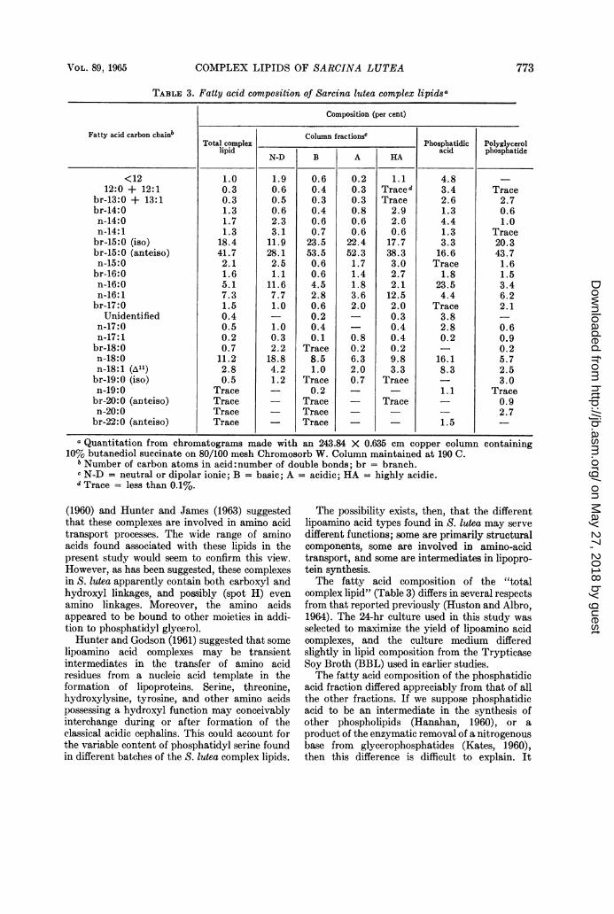
Fatty acid composition. The fatty acid composi-tion of the various complex lipid fractions issummarized in Table 3.
Spectral data. The infrared spectra of thevarious complex lipid fractions differed sub-stantially in only two regions. The "basic" lipidsabsorbed in the 6 to 6.8 , region, character-istic of the NH group of amino acids (Silversteinand Bassler, 1964), whereas the "acidic" lipidsdid not. The "acidic" lipids, on the other hand,absorbed conspicuously at 8.25 ,u, characteristicof secondary amides (Silverstein and Bassler,1964), but the "basic" lipids did not.
DISCUSSIONThe complex of lipids of S. lutea contains fairly
large quantities of lipoamino acids, phosphatidylglycerol, and polyglycerol phosphatide (Table 1).Lecithin, phosphatidyl serine, and phosphatidylinositol, common among plant and animal phos-pholipids, were present only in small amounts,whereas phosphatidyl ethanolamine was notdetected at all. Akashi and Saito (1960) havepreviously reported that Sarcina phospholipidsconsist principally of phosphatidic acid. Phos-phatidyl ethanolamine has been reported as themajor phospholipid of Serratia miiarcescens (Kates,Adams, and MIartin, 1964), Escherichia coli (Law,1961), Azotobacter agilis, and Agrobacteriumtumefaciens (Kaneshiro and 'Marr, 1962). Thephospholipids of some lactic acid bacteria, how-ever, have been described by Ikawa (1963) ascontaining no serine, ethanolamine, or choline.Instead, the principal ninhydrin-positive lipids ofthese organisms yielded lysine and alanine uponhydrolysis.
Lipoamino acid complexes amotunted to 15.1 %of the total complex lipids from S. lutea. Thesecomplexes are very labile to enzymatic degrada-tion and may be lost from cells unless enzymaticprocesses are stopped immediately after harvest-ing (MacFarlane, 1962a). Gaby, Wolin, and Zajac
772 J. BA~CTERIOL.
on May 27, 2018 by guest
http://jb.asm.org/
Dow
nloaded from
COMPLEX LIPIDS OF SARCINA LUTEA
TABLE 3. Fatty acid composition of Sarcina lutea complex lipidsa
Composition (per cent)
Fatty acid carbon chainb Colujmn fractionscTotal complex Phosphatidic Polyglycerol
lipid N-D B A HA acid phosphatide
<12 1.0 1.9 0.6 0.2 1.1 4.8 _12:0 + 12:1 0.3 0.6 0.4 0.3 Traced 3.4 Trace
br-13:0 + 13:1 0.3 0.5 0.3 0.3 Trace 2.6 2.7br-14:0 1.3 0.6 0.4 0.8 2.9 1.3 0.6n-14:0 1.7 2.3 0.6 0.6 2.6 4.4 1.0n-14:1 1.3 3.1 0.7 0.6 0.6 1.3 Tracebr-15:0 (iso) 18.4 11.9 23.5 22.4 17.7 3.3 20.3br-15:0 (anteiso) 41.7 28.1 53.5 52.3 38.3 16.6 43.7n-15:0 2.1 2.5 0.6 1.7 3.0 Trace 1.6br-16:0 1.6 1.1 0.6 1.4 2.7 1.8 1.5n-16:0 5.1 11.6 4.5 1.8 2.1 23.5 3.4n-16:1 7.3 7.7 2.8 3.6 12.5 4.4 6.2br-17:0 1.5 1.0 0.6 2.0 2.0 Trace 2.1
Unidentified 0.4 - 0.2 0.3 3.8n-17:0 0.5 1.0 0.4 - 0.4 2.8 0.6n-17:1 0.2 0.3 0.1 0.8 0.4 0.2 0.9br-18:0 0.7 2.2 Trace 0.2 0.2 0.2n-18:0 11.2 18.8 8.5 6.3 9.8 16.1 5.7n-18:1 (A1l) 2.8 4.2 1.0 2.0 3.3 8.3 2.5br-19:0 (iso) 0.5 1.2 Trace 0.7 Trace 3.0n-19:0 Trace 0.2 1.1 Tracebr-20: 0 (anteiso) Trace - Trace - Trace - 0.9n-20:0 Trace - Trace - 2.7br-22:0 (anteiso) Trace - Trace - - 1.5 -
a Quantitation from chromatograms made with an 243.84 X 0.635 cm copper column containing10% butanediol succinate on 80/100 mesh Chromosorb W. Column maintained at 190 C.
b Number of carbon atoms in acid: number of double bonds; br = branch.- N-D = neutral or dipolar ionic; B = basic; A = acidic; HA = highly acidic.d Trace = less than 0.1%.
(1960) and Hunter and James (1963) suggestedthat these complexes are involved in amino acidtransport processes. The wide range of aminoacids found associated with these lipids in thepresent study would seem to confirm this view.However, as has been suggested, these complexesin S. lutea apparently contain both carboxyl andhydroxyl linkages, and possibly (spot H) evenamino linkages. Moreover, the amino acidsappeared to be bound to other moieties in addi-tion to phosphatidyl glycerol.Hunter and Godson (1961) suggested that some
lipoamino acid complexes may be transientintermediates in the transfer of amino acidresidues from a nucleic acid template in theformation of lipoproteins. Serine, threonine,hydroxylysine, tyrosine, and other amino acidspossessing a hydroxyl function may conceivablyinterchange during or after formation of theclassical acidic cephalins. This could account forthe variable content of phosphatidyl serine foundin different batches of the S. lutea complex lipids.
The possibility exists, then, that the differentlipoamino acid types found in S. lutea may servedifferent functions; some are primarily structuralcomponents, some are involved in amino-acidtransport, and some are intermediates in lipopro-tein synthesis.The fatty acid composition of the "total
complex lipid" (Table 3) differs in several respectsfrom that reported previously (Huston and Albro,1964). The 24-hr culture used in this study wasselected to maximize the yield of lipoamino acidcomplexes, and the culture medium differedslightly in lipid composition from the TrypticaseSoy Broth (BBL) used in earlier studies.The fatty acid composition of the phosphatidic
acid fraction differed appreciably from that of allthe other fractions. If we suppose phosphatidicacid to be an intermediate in the synthesis ofother phospholipids (Hanahan, 1960), or aproduct of the enzymatic removal of a nitrogenousbase from glycerophosphatides (Kates, 1960),then this difference is difficult to explain. It
VOL. 89, 1965 773
on May 27, 2018 by guest
http://jb.asm.org/
Dow
nloaded from
HUSTON, ALBRO, AND GRINDEY
suggests that phosphatidic acid may be syn-thesized independently of the other phospholipids.Of the various fractions shown in Table 3, onlythe "basic" lipid and the "acidic" lipid fractionsclosely resemble each other in fatty acid composi-tion. They also constitute similar percentages ofthe total complex lipid (17.4 and 18.8%, respec-tively). It is not unlikely, therefore, that thesematerials are formed from similar starting ma-terials, e.g., diglycerides, and differ primarily onlyin the nature and linkage of the nitrogenousconstituent.The polyglycerol phosphatide of S. lutea
appears to be a cardiolipin. This type has beenreported in Staphylococcus aureus (MacFarlane,1962b), whereas the bisphosphatidic acid type hasbeen reported in Bacillus polymyxa (Matches,Walker, and Ayres, 1964).
Further discussion on the possible metabolicroles of the various complex lipids of S. luteawill have to await studies on the distribution ofthese components within the cell and character-ization of the individual metabolic pathways foreach component.
LITERATURE CITEDAKASHI, S., AND K. SAITO. 1960. A branched-satu-
rated C,5 acid (sarcinic acid) from Sarcina phos-pholipids and a similar acid from several micro-bial lipids. J. Biochem. (Tokyo) 47:222-229.
ALLEN, R. J. L. 1940. The estimation of phospho-rus. Biochem. J. 34:858-865.
AMENTA, J. S. 1964. A rapid chemical method forquantification of lipids separated by thin-layerchromatography. J. Lipid Res. 5:270-272.
ASSELINEAU, J., AND E. LEDERER. 1960. Chemistryand metabolism of bacterial lipides, p. 337-406.In K. Bloch [ed.], Lipide metabolism. JohnWiley & Sons, Inc., New York.
BENSON, A. A., AND B. MARUO. 1958. Plant phos-pholipids. I. Identification of the phosphatidylglycerols. Biochim. Biophys. Acta 27:189-195.
BENTLEY, R., C. C. SWEELEY, M. MAKITA, ANDW. W. WELLS. 1963. Gas chromatography ofsugars and other polyhydroxy compounds. Bio-chem. Biophys. Res. Commun. 11:14-18.
BLOCK, R. J., E. L. DURRUM, AND G. ZWEIG. 1958.A manual of paper chromatography and paperelectrophoresis, 2nd ed., p. 131. Academic Press,Inc., New York.
BLOCK, R. J., R. LE STRANGE, AND G. ZWEIG.1952. Paper chromatography. Academic Press,Inc., New York.
DEKONING, A. J. 1963. Identification of an artifactin the hydrolysis of choline-containing phos-pholipids. J. Chromatog. 12:264-266.
DITTMER, J. C., AND R. L. LESTER. 1964. A simple,specific spray for the detection of phospholipidson thin-layer chromatograms. J. Lipid Res.5:126-127.
FEIGL, F. 1954. Spot tests, vol. 2. Organic applica-tions, p. 152. Elsevier Publishing Co., New York.
FINK, K., R. CLINE, AND R. FINK. 1963. Paperchromatography of several classes of com-pounds. Correlated RF values in a variety ofsolvent systems. Anal. Chem. 35:389-398.
FISKE, C. H., AND Y. SUBBAROW. 1925. The colori-metric determination of phosphorus. J. Biol.Chem. 66:375-400.
FLEISCHER, S., G. BRIERLEY, H. KLOUWEN, ANDD. B. SLAUTTERBACK. 1962. Studies of the elec-tron transfer system. XLVII. The role of phos-pholipids in electron transfer. J. Biol. Chem.237:3264-3272.
GABY, W. L., H. L. WOLIN, AND I. ZAJAC. 1960.The role of phospholipids in the uptake of aminoacids by Ehrlich Ascites carcinoma cells. CancerRes. 20:1508-1513.
GOLDFINE, H. 1962. The characterization andbiosynthesis of an N-methylethanolamine phos-pholipid from Clostridium butyricum. Biochim.Biophys. Acta 59:504-506.
HANAHAN, D. J. 1960. Lipide chemistry, p. 106-133. John Wiley & Sons, Inc., New York.
HORROCKS, L. A. 1963. Thin-layer chromatographyof brain phospholipids. J. Amer. Oil Chemists'Soc. 40:235-236.
HUNTER, G. D., AND GODSON. 1961. Later stagesof protein synthesis and the role of phospho-lipids in the process. Nature 189:140-141.
HUNTER, G. D., AND A. T. JAMES. 1963. Lipo-amino-acids from Bacillus megaterium. Nature198:789.
HUSTON, C. K., AND P. W. ALBRO. 1964. Lipids ofSarcina lutea. I. Fatty acid composition of theextractable lipids. J. Bacteriol. 88:425-432.
IKAWA, M. 1963. Nature of the lipids of somelactic acid bacteria. J. Bacteriol. 85:772-781.
KANESHIRO, T., AND A. G. MARR. 1962. Phospho-lipids of Azotobacter agilis, Agrobacterium tume-faciens, and Escherichia coli. J. Lipid Res. 3:184-189.
KATES, M. 1960. Lipolytic enzymes, p. 212-214. InK. Bloch [ed.], Lipide metabolism. John Wiley& Sons, Inc., New York.
KATES, M., G. A. ADAMS, AND S. M. MARTIN.1964. Lipids of Serratia marcescens. Can. J.Biochem. Physiol. 42:461-479.
KISHIMOTO, Y., AND N. S. RADIN. 1963. Microde-termination, isolation and gas-liquid chroma-tography of 2-hydroxy fatty acids. J. Lipid Res.4:130-138.
LAW, J. H. 1961. Lipids of Escherichia coli. Bac-teriol. Proc., p. 129.
LEA, C. H., AND D. N. RHODES. 1955. The ninhy-drin reaction of unhydrolyzed phospholipids.Biochim. Biophys. Acta 17:416-423.
LEPHAGE, M. 1964. The separation and identifica-tion of plant phospholipids and glycolipids bytwo-dimensional thmn-layer chromatography.J. Chromatog. 13:99-103.
LOVERN, J. A. 1957. The phosphatides and glyco-lipids, p. 376-392. In W. Ruhland [ed.], Hand-
774 J. BACTERIOL.
on May 27, 2018 by guest
http://jb.asm.org/
Dow
nloaded from
COMPLEX LIPIDS OF SARCI1VA LUTEA
buch der Pflanzenphysiologie, vol. 7. Springer-erlag, Berlin.
MACFARLANE, M. G. 1962a. Characterization oflipoamino-acids as 0-amino-acid esters of phos-phatidyl glycerol. Nature 196:136-138.
MAcFARLANE, M. G. 1962b. Lipid components ofStaphylococcus aureus and Salmonella typhi-murium. Biochem. J. 82:40P-41P.
MAcFARLANE, M. G., AND G. M. GRAY. 1957.Composition of cardiolipin. Biochem. J. 67:25P-26P.
MARINETTI, G. A. 1962. Chromatographic separa-
tion, identification, and analysis of phospha-tides. J. Lipid Res. 3:1-20.
MATCHES, J. R., H. W. WALKER, AND J. C. AYRES.1964. Phospholipids in vegetative cells andspores of Bacillus polymyxa. J. Bacteriol. 87:16-23.
MILLER, G. L., AND E. E. MILLER. 1948. Deter-mination of nitrogen in biological materials.Anal. Chem. 20:481-488.
OLLEY, J. 1956. Quantitative paper chromatogra-phy of lipid constituents, p. 49-55. In G. Popjak
and E. LeBreton [ed.], Biochemical problems oflipids. Butterworths Scientific Publications,London.
ROUSER, G., A. J. BAUMAN, G. KRITCHEVSKY, D.HELLER, AND J. S. O'BRIEN. 1961. Quantitativechromatographic fractionation of complex lipidmixtures. J. Amer. Oil Chemists' Soc. 38:1-11.
SILVERSTEIN, R . M., AND G. C. BASSLER. 1964.Spectrometric identification of organic com-
pounds, p. 66-67. John Wiley & Sons, Inc., NewYork.
STAHL, E. 1958. Dunnschict-chromatography, eineadsorptions chromatographische Schnellme-thode, mit besonderer Beruieksichtigung der Un-tersuchung von Lipiden. Fette, Seifen, Anstrich-mittel 60:1027-1032.
WELLS, M. A., AND J. C. DITTMER. 1963. The useof Sephadex for the removal of nonlipid con-
taminants from lipid extracts. Biochemistry 2:1259-1263.
WIIG, E. C., W. R. LINE, AND J. F. FLAGG. 1954.Semimicro qualitative analysis, p. 160-164. D.Van Nostrand Co., Inc., New York.
VOL. 89, 1965 775
on May 27, 2018 by guest
http://jb.asm.org/
Dow
nloaded from