on some contributors to uncertainties in gyn igbt...
TRANSCRIPT

8/2/2012
1
On Some Contributors to Uncertainties in GYN IGBT
Duke Experience
Oana Craciunescu, PhD, DABR
Duke University Medical CenterDepartment of Radiation Oncology
• NO FINANCIAL RELATIONSHIPS TO DISCLOSE
Overview
• Do we need MRI for each fraction?
• Role of vaginal balloon packing systems
• Intelligent dose summation
• Do we need MBDCA for GYN?
• In-vivo dosimetry for treatment delivery verification

8/2/2012
2
Do we need MRI for each fraction?
• UNCLEAR, if use of MRI beyond the first fraction is useful in clinical practice.
• Or is the HRCTV changing from FX to FX?
• Or it is not changing in reality, but it is contoured differently?
• Duke experience with MR-based planning
– Variation among fractions
– Clinical/meaningful differences in dose to target
J Chino et al., WCB 2012
Methodology
• We retrospectively identified 8 PT (39 FXs)
• CT and MRI for each FX
• Contoured as per the GEC-ESTRO for target delineation (HR CTV, IR CTV) and OAR.
• Planning for each fraction was done in BrachyVision 8.10 (Varian Corp., Palo Alto, CA)
• Subset of 35 HRCTVs retrospectively re-contoured in one sitting (blinded to original HRCTVs)
• The total time from insertion to TX was also recorded.
J Chino et al., WCB 2012
Accord Across Fractions
J Chino et al., WCB 2012

8/2/2012
3
Slight Difference Across Fractions
J Chino et al., WCB 2012
Discord Across Fractions
J Chino et al., WCB 2012
HRCTV Volume Variation Between FXs
• Among individual women,
– range = 37 - 73% of the woman’s median value, (10-28 cc absolute
difference), median range = 47% (absolute range 11 cc)
• Recontoured HRCTVs:
– range = 16-76% of the woman’s median value, (2-20 cc absolute difference), median range = 33% (absolute range 8 cc)
• HRCTV D90 Median dose = 663cGy,
• HRCTV D90 doses range was 4-13% of the woman’s median value (26-86cGy absolute difference).
• The median time of procedure = 3.7 hrs (IQR 3.3 – 4.1 hrs).
J Chino et al., WCB 2012

8/2/2012
4
So, do we need MR for each FX?
• Yes, based on our data.
• The HRCTV volumes were variable between fractions, and resulted in variability in the plans developed to meet GEC-ESTRO dose goals.
• This variability was less with retrospective “single sitting” re-contouring but was still up to 76% of the median volume in some women.
• However, MRI based TP also requires significant time and access to resources and resources, which should be factored into the decision to implement for all fractions.
J Chino et al., WCB 2012
Overview
• Do we need MRI for each fraction?
• Role of vaginal balloon packing systems
• Intelligent dose summation: to deform, or not to deform?
• Do we need MBDCA for GYN?
• In-vivo dosimetry for treatment delivery verification
Role of vaginal balloon packing system
• With such still long treatment procedures there is increased need for very good, controllable, customized packing
• RadiaDyne Alatus Balloon
• Advantages
– Pushes away normal tissue
– Compatible with T&R and T&O
– Fiducials for depth verification
– Balloon can expand and conform
to patient anatomy and applicator
– Improved dosimetry
• Balloon role in maintaining implant stability
8/2/2012
5
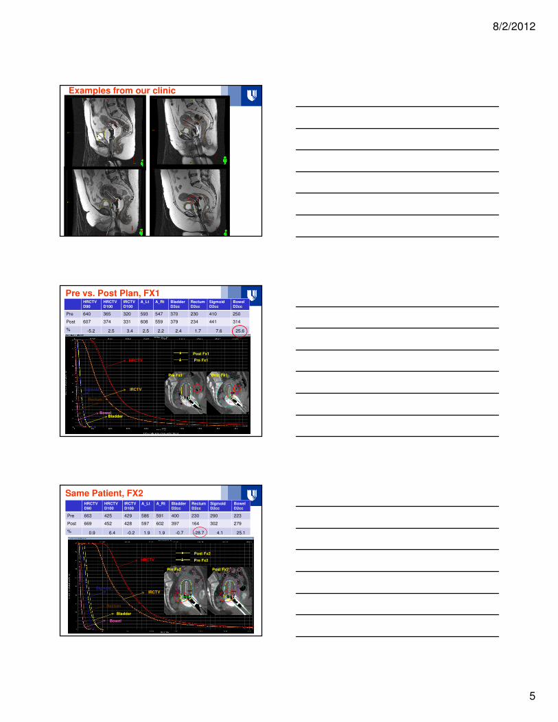
Examples from our clinic
Pre vs. Post Plan, FX1HRCTVD90
HRCTVD100
IRCTVD100
A_Lt A_Rt BladderD2cc
RectumD2cc
SigmoidD2cc
BowelD2cc
Pre 640 365 320 593 547 370 230 410 250
Post 607 374 331 608 559 379 234 441 314
% -5.2 2.5 3.4 2.5 2.2 2.4 1.7 7.6 25.6
Pre Fx1
Post Fx1
HRCTV
IRCTV
BladderBowel
Rectum
Sigmoid
Pre Fx1 Post Fx1
Same Patient, FX2HRCTVD90
HRCTVD100
IRCTVD100
A_Lt A_Rt BladderD2cc
RectumD2cc
SigmoidD2cc
BowelD2cc
Pre 663 425 429 586 591 400 230 290 223
Post 669 452 428 597 602 397 164 302 279
% 0.9 6.4 -0.2 1.9 1.9 -0.7 -28.7 4.1 25.1
Post Fx2
Pre Fx2HRCTV
IRCTV
Bladder
Bowel
Rectum
Sigmoid
Post Fx2Pre Fx2 Post Fx2
8/2/2012
6
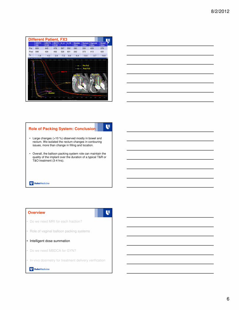
Different Patient, FX3
HRCTV
IRCTV
Sigmoid
BowelBladder
Rectum
HRCTVD90
HRCTVD100
IRCTVD100
A_Lt A_Rt BladderD2cc
RectumD2cc
SigmoidD2cc
BowelD2cc
Pre 609 445 478 567 552 280 330 429 379
Post 598 426 492 526 601 262 373 413 420
% -1.8 -4.3 2.9 -7.2 8.9 -6.4 13.0 -3.7 10.8
Pre Fx3
Post Fx3
Pre Fx3 Post Fx3
Role of Packing System: Conclusion
• Large changes (>10 %) observed mostly in bowel and rectum. We isolated the rectum changes in contouring issues, more than change in filling and location.
• Overall, the balloon packing system role can maintain the quality of the implant over the duration of a typical T&R or T&O treatment (3-4 hrs).
Overview
• Do we need MRI for each fraction?
• Role of vaginal balloon packing systems
• Intelligent dose summation
• Do we need MBDCA for GYN?
• In-vivo dosimetry for treatment delivery verification

8/2/2012
7
Intelligent Dose Summation:
Proof of Concept
• Summation of doses relies currently on point EQD2 dose additions of target and OAR metrics form the EBRT and BT plans.
• To correctly sum doses in 3D we have to account for:
– Applicator position relative to anatomy, or lack of (in EBRT)
– OAR deformation (differential filling in most OARs of interest)
– Account for radiobiologic differences between EBRT and BT
– Account for dose inhomogeneities in IMRT plans
• Investigate DEFORMABLE IMAGE REGISTRATION (DIR)
Clinical Commercial Software
• VelocityAI (Velocity Medical, Atlanta, GA)
• RTx (Mirada Medical Ltd., Oxford, UK)
• MIM Software™ (Cleveland, OH)
– Free form intensity-based deformable registration algorithm– Piper JW. Evaluation of An Intensity-Based Free-Form Deformable Registration Algorithm.
Medical Physics. June 2007;34(6):2353-2354.
• Others…??
`
Registration Process
Crude Dose
HDR5 HDR4 HDR3 HDR2 HDR1
∑rHDR
∑dHDR
One for each Target, OARs
EQD2Target(αααα/ββββ = 10)OARs(αααα/ββββ = 3)
∑rHDR
∑dHDR
On HDR1 CT
r(∑rEBRT+∑rHDR)d(∑dEBRT+ ∑dHDR)
r(∑dEBRT+ ∑dHDR)
DVH – TargetFrom EQD210
DVH – OARFrom EQD23
∑rEBRT
∑dEBRT
One for each Target, OARs
EBRT1
(Primary)
EBRT2
(Boost)
∑rEBRT)
∑dEBRT
On one of the CTs

8/2/2012
8
Registration Rules
• EBRT: Rigid registration first, then deformable
• BT (HDR): Rigid first based on applicator if same tandem length/angle, or uterus if different. NOT based on BONE!
Applicator Based
Uterus BasedApplicator BasedBony Anatomy Based
• When applicator/uterus off from FX to FX, mask uterus (HU = 1000)
Mask
Rigid
Contour
Deformable
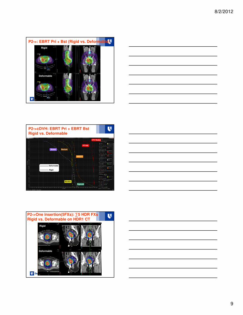
Duke Clinic Examples
• P1: EBRT + 5 HDR FXs (5 HDR insertions): same applicator
– EBRT: 45 3D conformal to pelvis and PA nodes
– HDR: T&R 27.5 Gy in 5.5 Gy/fx
• P2: EBRT + 5 HDR FXs (1 HDR insertion)
– EBRT: 45 Gy pelvis + integrated boost 55 Gy to nodes + 12.6 Gy
sequential boost
– HDR: T&R 27.5 Gy in 5.5 Gy/fx
• P3: EBRT+ 5 HDR FXs (5 HDR insertions): mix of applicators
– EBRT: 45 Gy 3D conformal
– HDR: T&R 27.5 Gy in 5.5 Gy/fx (2 FX T&O and 3 FXs T&R)
8/2/2012
9
P2->: EBRT Pri + Bst (Rigid vs. Deformable)
Rigid
Deformable
P2->cDVH: EBRT Pri + EBRT Bst
Rigid vs. Deformable
Rigid
Deformable
Bowel
PTV68
CTV Nodes
HRCTV
Bladder
Rectum
Sigmoid
Deformable
Rigid
P2->One insertion(5FXs): ∑5 HDR FXs
Rigid vs. Deformable on HDR1 CT
Rigid
Deformable

8/2/2012
10
P2->cDVH: One insertion(5FXs) ∑ 5HDR FXs
Rigid vs. Deformable on HDR1 CTRigid
Deformable
Rigid
HRCTV
IRCTV
Bladder
Bowel
Rectum
Sigmoid
-8%
- 2%
P2->r(∑dEBRT + ∑dHDR) for Target(HRCTV)
r(∑dEBRT + ∑dHDR)
∑dHDR
r∑dEBRT on HDR1 CT
Circle of trust
P2/DVH: r(∑dEBRT +∑dHDR) for Target(HRCTV)
Cumulative (EQD2)HRCTV D90 = 91.94 Gy
HRCTV D100 = 73 Gy
D90: + 4% vs. current practice D100: +4.9% vs. current practice

8/2/2012
11
P1-Five insertion(5FXs): EBRT+5 HDR FXs
(Deformable on EBRT CT)
P1/DVH: d(∑dEBRT + ∑dHDR) for OARs
Cumulative (EQD2)Bladder D2cc = 66 Gy
- 3.8 % vs. current practice
Bladder
P3->Five insertion(5FXs): 2 FX T&O, 3 FX
T&R (Deformable on HDR1 CT)
Rigid
Deformable

8/2/2012
12
P3->DVH (Crude)-One insertion(2 T&O; 3 T&R)
Rigid vs. Deformable Rigid
Rigid
Deformable
HRCTV
Bladder
Bowel
Rectum
Sigmoid
4% d
6% d
Intelligent Dose Summation
• Posible aid to keep uncertainties in check
• Requires DIR
– Very challenging for GYN cervix cancer patients due to change in
topography between the EBRT and HDR
• We have to understand the limitations of the DIR algorithm used and control each step.
• We have to generate more data to understand patient-dependent variations.
Overview
• Do we need MRI for each fraction?
• Role of vaginal balloon packing systems
• Intelligent dose summation
• Do we need MBDCA for GYN?
• In-vivo dosimetry for treatment delivery verification

8/2/2012
13
TG-43 dosimetry formalism
• Defines water as reference medium
• Tissue is not water-equivalent
• Variable/dynamic scattering conditions
• Doesn’t account for source/applicator shielding
BV ACUROS® (BrachyVision, Varian):
Grid-Based Boltzman Solver (GBBS)
• Alternative to MC with similar accuracy, but increased efficiency
• Deterministic solver of the differential linear Boltzmann transport equation (LBTE)
• Accounts for the effect of applicators (use of solid applicators libraries)
• Accounts for the different tissue types
• Accounts for the effects of the patient boundaries
Existent studies• Libby, Ter-Antonyan, Schneider, Brachytherapy , vol 10 (S1, S66),
2011 – abstract, not too many details
• Mikell et al, 2012, IJROBP, T&O– +/- overriding patient contour to 1 g/cm3 muscle
– +/- overriding contrast material to muscle or bone
– majority of metrics < 5%: A, B, D2 cc bladder, ICRU bladder, regardless of the masking of balloon contrast
– BV-ACUROS-based dose calculations have minimal clinical impact;

8/2/2012
14
Establish relevance of using BV-
ACUROS for HDR Cervical Cancer Patients
• 5 patients (T&R)
• 22 fractions
• Clinical plans calculated with TG43 and BV-ACUROS
• HRCTV D90 and D100 [%RX]
• Manchester Point A [%RX]
• Bladder, Rectum, Sigmoid, Bowel D0.1 cc, D2cc, D10cc [Gy]
• Solid Model Applicator
Target: TG43 vs. BV-ACUROS™
Mean % diff < 2%
OARs: TG43 vs. BV-ACUROS™
Mean % diff < 2%

8/2/2012
15
Our Conclusions
• Like MD Anderson Group, Mikell et al, we concluded that BV-
ACUROS-based dose calculations have minor clinical impact in GYN cases that use non-shielded applicators.
• Await the recommendations of TG 186 for MBDCA and continue to
generate data to include in larger multi-institution comparisons.
Overview
• Do we need MRI for each fraction?
• Role of vaginal balloon packing systems
• Intelligent dose summation: to deform, or not to deform?
• Do we need MBDCA for GYN?
• In-vivo dosimetry for treatment delivery verification
Role and need for in-vivo dosimetry
• With complex and lengthy planning and treatment procedures required by IGBT there is increased need for validation of delivered doses;
• Investigate the use of Presage Dosimetry for HDR procedures:
– develop system to read small dosimeters;
– calibrate dosimeters at Ir-192 energy and body temperature;

8/2/2012
16
Imaging Set Up
LED
Light Source
Presage
Jig Camera
Lens
M Oldham’s Dosimetry Lab, Duke Univ.
Experiment Set Up for iPD Calibration at Ir-192 Energy
iPresage Dosimeters
Water at 36-37oC
Catheter for Ir-192 source
GMP iX Afterloader (Varian)
T&R Patient: 1 iPresage Dosimeter on
Alatus Balloons (Bladder Side)
Mean Dose Bladder (TPS) = 269.4 cGyPoint Dose Bladder (TPS) = 272.9 cGy
(mid way through iPD)
iPD Reading = 280 cGy (3.7 %)
2cm
2mm

8/2/2012
17
T&R Patient: 1 iPresage Dosimeter on
Alatus Balloons (Rectum Side)
Mean Dose Rectum (TPS) = 174.6 cGyPoint Dose Rectum (TPS) = 172.7 cGy
(mid way through iPD)
iPD Reading = 167 cGy (-3.5%)
Conclusion
• Contouring still the largest contributor to uncertainties
– Aid the process further with functional imaging
• Applicator stability within the planning time frame
– Optimize insertion, imaging, planning and treatment flows to
reduce the overall time
• For GYN with unshielded applicators, TG43-based calculation introduces only minimal uncertainty when compared to MBDCA (BV-ACUROS)
• Intelligent dose summation very challenging but maybe worth having clear defined protocols to assess impact , if any, on local control and late toxicities
• In-vivo dosimetry: yes, if available
Acknowledgments
• Duke Brachytherapy
– Jing Cai, PhD
– Junzo Chino, MD
– Jackie Maurer, PhD
– Jesica Sanchez-Mazon, MS
– Beverly Steffey, MS
• Duke Dosimetry Lab
– Mark Oldham, PhD
– Adria Vidovic
– Titania Juang