on the cloacal region of anura
TRANSCRIPT
Ann. Univ. Stell. Vol. 35, Sect. A, No.4 (1959)
ON THE CLOACAL REGION OF ANURA III Particular of Larva l Ascaphus
by
D. E. VAN ntJK
Department of Zoology, Universit y of Natal,
Pietermaritzburg
(With 44 text.figures)
Thesis accepted for the Degree of Doctor of Science at the University of Stellenbosch
Promotor: Professor C. G. S. de Villiers
Submitted: January, 1959
ABSTRACT The ontogeny of the cloacal region of Ascaphus is described from limited
larval material (beginning approximately at the stage of hind-limb bud development). A comprehensive series of Bulo angusticeps larvae and late pre-larval embryos were used for comparison . The adult and/ or late larval conditions of the cloacal region in Ascaphu,~, Bulo, Bombina, Leiopelma, Rana and Xenopus arc compared.
The rods of Noble supporting the cloaca in Ascaphus and the tendinous sheet connecting these with the epipubis arc shown to be modifications of an interfemoral ligament present, with thickened lateral margins, in all the Annra studied. The cloacal lips differentiate early in metamorphosis in Ascaphus and Eu/o and bear similar relations to the interfemoral ligament in these and other A nura, so that they arc apparently homologous. The posterior part of the urodaeum is lengthened in the adult male Ascaphus to form the "tail" (phallus) .
The hind-limb anlagen of Ascaphus appear directly beneath the spinal myomeres and immediately behind the posterior tips of the abdominal muscle cords. In Ascaphus, Bulo and Bombina the abdominal muscles (metamerically disposed in A8ca-phu8 and Eu/o) are initially attached posteriorly to the spinal myomeres but are separated from them anteriorly. lt is probable that the mm. compressorcs cloacae are derived from the hind·limb anlagen. In all Anura examined, including members of all the South African families, the a . ischiadica and n. ischiadicus have a small muscle (designated m. circumflexor arteriae) associated with them; it is presumably capable of compressing the artery against the nerve.
The cloacal region of Ascaphus appears to be less specialized than that of Rana and Xenopus, contrary to what is generally believed for the last two genera. Bu/o, particularly. and Bonrbina have undergone less specialization.
169
CONTENTS
Introduction 173 Enunciation of the Problems 173 Acknowledgements 174 Material 174 Techniques 175
Ascaphus, Leiopelnw, Xenopus and Rana Sections 175 Bufo angusticeps material (and comparative series ) 177
Procedure 178 Description of the Stages in Asca11hus 179
Stage 47 179 Stage ± 50 180 Stage 51 181 Stage 52 184 Stage 56 185 Stage 58 191 Stage 60 196 Stage 62--63 199 Stage 63 202 Stage 64 204
The DevCIopment of the Proctodaeum and Associated Structures in Ascaphu8 210
Structures of Ectodernwl Origin 210 Proctodaeal Invagination 210
Proctodaeal Aperture 210 Proctodaeal-Urodaeal Junction 210
Structures of Endodernwl Origin 210 Coprodaewn and Urodaeum 210
Aperture of Nephric Ducts 210 Urodaeal Diverticulum 211 Bladder 211
Structures of Mesodermal Origin 211 Coelomic Cavity 211 Splanchnic and Somatic ]\{esoderm 212 Visceral Muscle Layers 212
Circular Muscle Layer 212 Longitudinal Muscle Layer 212
Anlagen of Pelvic Limbs and Girdle 213 Pelvic Limbs 213 Pelvic Girdle 213 Epipubis 214 Rods of Noble and Interfemoral Ligament 214.
Somatic l\lusclcs 215 Abdominal Muscle Cords (Rectus Muscles) 215 Obliquus and Transversus Muscles 216 Epipubic Muscles and Anlagen of the Epipuhis 216 Femorococcygeal and Crurococcygeal Muscles 216 Fcmoropelvic and Cruropelvic Muscles 217
170
CONTENTS (continued)
Coccygcopelvic Muscles 217 Cloacal Comprcssor Muscles 217 Arterial Circumflcxor Muscles 217 Unstriated fibres of the mm. compressores cloacae 218
Cavernous Tissue 218 Lymph SI)uces 218
Gliding P lanes 218 Ellithelial Structures 219
l)roctoliuea l Glands 219 Horny Spines 219
The Proctodaeum and Associated Structures in Adult Anura 219 Structures of Ectodermal Origin 219
Proctodaea! In\'agination 219 Proctodaeal Aperture and Proctodaeal-Urodaeal Junction 219 Proctodaeal Glands 222 Horny Spines 222
Structures of Endodermal Origi1l 222 Structures of Mesodermal Origi1l 222
Coelomic Cavity 222 Visceral !Uu sc!e Layers 223 Pelvic Skeletal Structures 223
Lin ea Alba 223 Epipubis 223 Pelvic Girdle 224 Inte rfemoral Li gament 224
Somatic Muscles 224 Epipubic Muscles 224 Cloacal Compressor Muscles 226 Arterial Circumficxor Mu scles 229 Unstriated Fibrcs of thc mm. comprcssores cloacae 229
Cavcrnous Tissue 232 Discussion 232 Summary and Conclusions 240 Bibliography 245
171
INTRODUCTIQK
This investigation was undertaken at the suggestion of my promotor. Prof. C. G. S . de Villiers, and to his encouragement, particularly in the initial stages of the work, and his inspiration, its completion is largely due.
During the course of an investigation of the anatomy of the cloacal region of the adult male Ascaphu8, including some details of the adult female and larvae, it was decided to attempt to determine the ontogeny of the cloacal structures from the limited Ascaph1ts larval material available. The present pape r represents the results of this attempt.
As suggested by the title of the paper (van Dijk. 1955) in which the work on the adul t male was recorded - "The 'Tail' of A8caphus: A Historical Resume and new Histological-Anatomical Details" - an attempt was made to review the literature on the cloacal region of Ascaphu8.
As the paper was completed (and accepted for the i).'f.Sc. degree) in March 1953, Bhaduri's work on the urinogenital system of the Anura (Bhaduri. 1953) which included an investigat ion of Asca1Jhus, and RitJand's studies (1955a and 1955b) on the post-cranial skeleton, nervous and muscular systems of Ascaph1ts were thus not discussed.
ENUNCIATION OF THE PROBLE;\IS
According to classic investigations the muscular layers of the mesenteron of Chordata are derived from the splanchnic mesoderm, which constitutes the mesial wall oC the splanch notome of each side, and the trunk and abdominal musculature develops in the somatic mesoderm, which constitutes the lateral wall of the splanchnotome of each side. By an investigation of the development of the (visceral) muscle layers of the coprodaeum at the coprodaeal-proctodaeal junction, and of the (somatic) abdominal muscle layers in the region of the proctodaeal aperture, the origin of the muscle-lining of the proctodaeum might be determined. The development of the tail musculature and of the muscles of the hind-limb might be relevant to this problem in view of their proximity to the proctodaeum. The relation of the coelom, posterior lymph sacs, pelvic girdle a nd urostyle, and of the blood vessels and nerves of the cloacal region, to the cloaca during development might provide important information on the development of the proctodaeum.
In Ascaphu8 the development of the cavernous tissue of the cloaca , of the (post-pubic) rods of Noble supporting the cloaca, and the muscle layers apparently dorsally striated and ventrally unstriated and associated with these rods (cL van Dijk, 1955), merit investigation. The developmental origin of the mm. eaudalipuboischiotibiales and (mm. pyriformes) and of the epipubis and mm. epipubici, might be relevant in view of the association of the former with the tail and hind·limb in the region of cloaca, and in view of the epipubis forming the posterior attachment of the abdominal muscles anteroventral to the cloacal allerture (though separated from the latter by the pelvic girdle).
The distribution, forms and funct ions of the anterior slips of the mm. compressores cloacae extending from these muscJes around the ischiadic
173
nerves and arteries in R(J1Iu, Ascaphu-s and Xenopus, present problems which require investigation, particularly as Leio1Jiema has no such slip arisinz; from the m. compressores cloacae (van Dijk, 1955) .
With a knowledge of the development of the proctodaeum of the Anllra the evolution of the cloaca and copulatory organs of the Amniota might be capable of solution .
ACKNOWLEDGEMENTS
At the completion of my studies fo r the degree of Doctor of Science I would like to express my indebtedness to all, zoologists and non-zoologists alike, from whom I have received inspiration, learnt techniques and gained knowledge.
The inspiration of working both under Dr. de Villiers, and also with the re,scarch students he has taught. has been very great. The staff of the Zoological Institute, Stellenbosch (Dr. C. G. S. de Villiers, Dr. C. A. du ToiL Dr. C. S. Grobbelaar and Dr. M. E. Malan ) have all helped to make my years of work there of the happiest.
The facilities of the Zoology Department, University of Cape Town were used during 1956; those of the University of Natal, Pietermaritzburr; duri ng 1957 and 1958. The use of these facilities is much appreciated.
~lATERIAL
The larval Ascaphus material examined was available at the Zoological Institute, Stellenbosch, largely in the form of serial sections or remnants imbedded in paraffin-wax, these all being derived from studies by de Vos during 1938 (on Leiopelmaj and van Eeden (1951 ). The six larvae used by de Vos were Haemalum bulk-stained and sectioned transversely. Those sections which were counterstained by her had usually been so treated with Eosin, occasionally with Light-Green, Bismarck Brown or van Gieson's Picrofuchsin; those not done by her were counterstained with Eosin during this study. The thicknesses of the sections in each series were not recorded by de Vos and were not always constant. The larvae of van Eeden, sixteen in number . were bulk-stained with Borax-Carmine and the sections (transverse except 1 sagittal ser ies) were counterstained with Azan. The thickness of the sections cut by van Ecden, and those cut in the present study, were recorded and were usually uniform for each series. In addition to the material sectioned or imbedded by de Vos and van Eeden four Ascaphus embryos from Mason County, Washington (A. M. N. H. Nos. 50583 and 50586, 1930) were treated In the same manner as van Eeden's specimens, giving 3 fronlal and 1 transverse series cut at 10 /-, .
Thus of Ascaphlls 1 embryos, 22 larvae, and (from the studies of de Villiers, 1933, and van Dijk, 1955) 3 adult males and 2 adult females were available as serial sections.
For comparative purposes 17 Cacosternum capensc larvae, 9 Bombin(t pachypus larvae, 1 Plethodon sp. adult, 1 Siphonops (mnlllatus adult, 1 Amby.s toma maculalum (=A mblystoma) adult. 1 Salmo furio juvenile. 5 Periopt halmll~
174

koclrcuteri. small adults, 1 Honw sapiens embryo (late indifferent/ female? stage), and 200 specimens of Bu/o ang1t.'jticeps (representing all the recognisable stages) were sectioned. There were also available 1 series of a Leiopclma hochstetteri adult male (from Wagner, 1934a and 1934b) , 1 series each of Rana graYl adult male and female, 2 of Xenopus laevi..9 larvae and 1 juvenile Xenopus 1aevis, 1 of a Bl~fo gariepetlsis juvenile, 3 of Breviceps juveniles and a postmetamorphic Arthroleptela series.
Material utilized for dissection includcd numerous Xenopus /uevis adults, and one adult of each of Rana angolcnsis, Pyxicephalu-8 natalensis, Phrynobatrachus sp., Heleophryne sp., Phrynomerus sp., Bu/o regulari8 and Chir(}muntis xerampelina,
TECHNIQUES
The techniques used frequently differed from those in general practice. They are therefore described at some length .
.'\scaphus, Leiopelma, Xenol)lls alld Hana sections
The average thickness of the sections in each of de Vas's series was determined by means of the micrometer fine-focus of a microscope (checked against other microscopes). It was also done by assuming dermal glands to be spherical, measuring their diameters (by means of an eye-piece micrometer) and comparing these diameters with the number of sections which the glands occupied or in which they appeared, the terminal sections in which the glands appeared usually being assumed to be half-occupied. The second of these methods was found to be reliable, giving consistent results.
Drawings on thin paper at enlarGements of 50 diameters, were made (usually of every 4th section) by means of a vertical slide projector, Zeiss Luminar lenses being used as they gave wide, flat and bright images. The drawings were rendered transparent with xylene, each was adjusted in position to correspond with those before (and sometimes after) it, and basc-lines for reconstruction were then markcd by drilling orientation holes through the piles of papers, Graphic reconstructions were then made (see Pusey, 1939) of left lateral aspects (denoted norma lateralis sinistral. and sometimes dorsal aspects (denoted norma dorsalis). Use was also made of liver base-lines in dOing lateral reconstructions fro m van Eeden's series and from those cut from his wax blocks (which contained liver slices). These base-lines were found to be of little use for adjusting the orientation of successive sections, but useful in establishing a general trend in direction over long intervals. Liver base·lines were most useful in orientating where a skeletal structurc appeared or disappeared in a series of sections, thereby affecting the extent to which cach section stretched during mounting. Sections through thc pelvic girdle often stretched less than those in front of, or bchind, them, and the cloaca tended to stretch away from the tail in those sections in which it was not held by skin to it. (For liver base-line technique see van Eeden, 1951, and Schepers. 1938.)
For truer dorsal reconstructions the degree of skewness (sagittalness) of the transverse sections was estimated by measuring the distance between
175
distinct bilateral structurcs (such as the ilia) and the distance by which one of these preccded the othcr in the series of sections (number of sections occupied x section thickness). The distances so measured gave the tangent (or cotangent) of the angle of skewness, so that the true mid-line of the recon· struction could be drawn and the mid-line of each drawing could be aligned to this. For some distorted scctions two mid-lines were drawn through each drawing and the structures on which each mid-line was based (ventral and dorsal respectively) were then reconstructed separately.
Photomicrographs were made of significant sections in all the series reconstructed, thc levels of the sections bein:; indicated on the reconstructions. Photomicrographs were also made of instructive sections from series not reconstructed, these illustrating, for instance, statements not figured in the 1955 work, the scope of which was reduced for publication. Use was also made of photomicrographs instead of drawings for reconstructions, and proved to be time-saving and otherwise vastly superior where orientation difficulties were not prohibitive. Such difficultics with photomicrographs arosc from the need to use the cheaper, relatively opaquc, photographic paper, instead of film plates or film-type papers which would havc madc possible simultaneous comparison of a greater number of photographs.
Where orientation difficulties were not great (e.g. where the notochord could be assumed to be straight, or in dorsal vicws) reconstructions were also done successfully by projecting the image of each section onto the reconstructing board, adjusting it to a base-line (e .g. notochord for lateral, and mid-line for dorsal, views), and to a vertical or horizontal line; then proceeding as usual with the image substituted for the usual drawing or photomicrograph.
Certain of the sections of Prof. de Villiers's specimens of adult Ascaphu8 male and female, which had been stained with Haemalum and van Gieson's Picrofuchsin, were restained with 1 % aq. Light Green or van Gieson's Picrofuchsin followed by 1 % aq. Light Green. The same treatment was given to sections through the cloacal region of the other adult female Ascaplws, which had becn stained with Borax-Carmine-Azan, and to sections of the Xenopus adult aftcr rcmoval of the Eosin which had been used as counterstain to Haemalum. The Acid Fuchsin (of the van Gieson's Picrofuchsin ) stained the collagenous connective tissue red, while the Light Green was taken up by non-collagenous connective tissue and by muscle. The Jreater affinity for Picric Acid (of the van Gieson's Picrofuchsin) of striated muscle, and, to a lesser extent, of unstriated muscle, resulted in a grass-green tinJe (Light Green + Picric Acid) in the musclcs as compared with a blue-green colour in the non-collagenous connective tissuc. The Light Green was employed for ! to 1 minute and differentiated in water to givc the correct overall colour on the section (apprOximately 15 secs. to 1 min .). Van Gieson - LiJht Green , Picro-Indigo-Carmine, Picro-Nigrosin and Rettercr's Alum Carmine were employed on sections through the cloacal region of the male adult Ascaphus and on the female adult Ascaphus sectioncd for that work.
The developmental stages of Ascaphus of which reconstructions were made have been numbered according to the criteria used for Xenopus laevis in the Hubrecht Laboratory publication "Normal Table of Xenopus laevis ( Daudin)" (Nieuwkoop and Faber, 1956). For the· stages up to sta:;e 57 rcference was made to the hind-limb development, for the later sta:;es the
176
conditions of the opercula and fore -limbs were used. (The hind-limbs of the later stages of dc Vos's material were incomplcte distally.)
Bufo angusticejlf. lIl uteriul (and ROlllbina, Cacosternum. Ambystolllu, Plethodoll, S il)hollOI'S, Salmo, l>eriol)hthallllus and Homo)
The Bombina, Salmo and Periophthalmus specimens after fixation in Bouin's fluid, and the rest, except Buro, after formalin fixation, were all treated much as described below for Buro.
The Bulo angusticeps material was identified by refercnce to de Villiers (1929) and Noble (1926) initially; subsequently by the time of the year at which oviposition took place, the form of the egg-strings, and also by the dark pigmentation of the eggs and larvae (apparently darker than that of any other South African anuran) . The material was killed and fixed in Lenhossek's Picric Acid - ~iercuric Chloride - Acetic Acid - Alcohol solution. washed after 24 hours. decalcificd (in some cases) in 5% Nitric Acid in 70 % alcohol, stained in Grenacher's Borax-Carmine (except alternate early stages, which were Haelllalulll bulk-stained), dehydrated (sometimes only to 96% alcohol). clcared in Methyl Benzoate-Celloid in, impregnated for a half-hour in Benzol Wax at about 30°C, impregnated in vacuo in 52 Q C paraffin wax and imbedded and sectioned in fresh 52° C wax. T he animals, whole or with only a part of the tail missing, were sectioned frontally most often, transversely least often, the relative numbers (excluding the earliest stages) being 74 frontally, 54 sagittally, 39 transversely, i.e. approximately 4 Frontal: 3 Sagittal: 2 Transverse series of Bulo.
Counterstaining of thc Bulo angusticeps material was done with Heidenhain's Azan (except the Haemalum-stained early stages, for which Eosin was used). In addition 1 % aq. Light Green was used for most of the older, and some of the younger, spccimens. It was found that it facilitated recognition of striated muscle, non-collagenous connective tissue and nervc fibres . Striated muscle stains a characteristic orange-brown (similar to Bismarck Brown in hue) because of its affinity for both Orange G (from the Azan) and Light Green; non-collagenous connective tissue is stained bluegreen. and nerve fibres a characteristic grey-green. The blue of collagenous connective tissue (Aniline Blue from the Azan) and the pink of unstriated muscle fibres (Borax-Carm ine) are little affected by Light Green unless this is cmployed for too long. The use of Light Green derived from a suggestion of Prof. de Villiers that it be used as a counterstain to Haemalum. Safranin ( = "Safranelin'''?) was tried as a stain for non-collagenous fibres (Maskor, 1953), but seemed unsuitable for immature anman tissue.
Limited use was made of base-lines, these being produced by boring holes at right angles to the plane of sectioning in wax blocks and filling the resulting holes in the sections with Indian ink. Wax models such as those described in the earlier paper (van Dijk, 1955), and photomicrographic stereograms similar to the graphic stereogram used in that paper (op. cit., fig . 29), were used to visualize thc milieu of the cloaca and associated structures. The photomicrographic stereograms were produced by distorting photomicrographs by means of specially constructed anamorphote lens systems (which have since become familiar in connexion with "cinemascope").
177
DisSt-octions of members of each of the families of Sout h Africa n Anura
After a number of different approaches to the cloacal region of Xenopus had been attempted, median incisions through the skin were made, extending half the way along the back above the cloacal aperture in all the specimens (Xenopus and others) dissected . This was followed by a median incision through the membrane over the urostyle. It was then usually possible to separate the fat containing the coccygeal lymph hearts and the septum iliacum mediale of one side, intact; this separation being achieved by carefuily loosening these from the m. coccygeo-iliacus, and hooking them laterally whilst the urostyle and m. coccygeo-i1iacus were hooked over to the opposite side. This dissection exposed the cloaca in dorsal and lateral view. Using a hooked needle, it was possible, in numerous Xenopus, to locate the dorsal aorta and puil it sideways so that the fluorescein injection technique used by de Graaff (1957) could be applied, the injection taking place after the dorsal aorta had been returned to its natural position with the needle in it. Thus blood-flow in the aa. iliacae could be watched under ultraviolet light.
PROCEDURE
As complete a series of Ascaphus as could be obtained from the available material, was made. Similarly, as complete a series of Bulo angusliceps was prepared as was possible from the specimens sampled at intervals duringdevelopment. Extra specimens of Bu/o at each sampling were preserved in wax ready for sectioning, should it be necessary. The comparative material listed above was thcn prcpared.
Reconstructions of all the discernible features of as much of the pelvic region as pOSSible, were made of all the Ascaphus stages that could be reconstructed. Of the Bu/o angu-sticep-s material, reconstructions of the salient features of the entire animal were made at the stages which marked the first appearance, and which illustrated the development of, the proctodaeum, the permanent gut lumen (unoecluded by yolk), the pronephric ducts, the abdominal muscles, the limb-buds, and the cloacal muscles.
Conclusions on the Ascaphus material were arrived at, illustrated and noted . These conclusions were checked on the Bulo angusticeps material and illustrated with further notes. Only then was the relevant literature thoroughly examined. This was done in an attempt at avoiding "anticipatory set", which produces subjectivity of approach, particularly in perception. Finally the material was re~examined together with the other animals prepared.
In presenting the results an attempt. has been made to reproduce the evidence on which descriptive statements were based. Photomicrographs serve both to illustrate features and to check on the necessarily somewhat subjective reconstructions.
1t must be noted that, while the Ascaphus material has been described at length and the Bulo material cursorily, the preparation and examination of Bulo angusticeps stages represented more than half of the work involved in this study. Without recourse to Bulo angu-sticeps, representing the typical anuran condition, the Ascaphus material could not have been adeqUately interpreted or described .
178
N"otc on Terminology
Gadow's (1887) terminology for the cloaca of Amniota has been adopted. The cloaca is thus considered to consist of a terminal ectodermal proctodaeum , a 1troda81~m into which the urinary and genital ducts and the bladder open , a nd a coprodaeum receiving the intestinum. The use of this terminology implies that the urodaeum and coprodneum are accepted as being of endodermal origin, for these parts of the cloaca are distinguishable in Amniota internal to the cloacal membrane (wh ich represents the limit of the ectodermal proctodaeum) before this membrane perforates. That the urodaeum and coprodaeum are of endodermal origin in the Anura, appears to be established by evidence, presented below, that the urinogenital ducts open into an cndodermal part of the cloaca.
DESCRIPTlON OF T HE STAGES IN ASCAPHUS
Stage 47 ( fi gs. ] and 2)
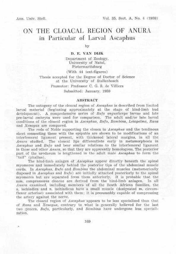
The nephric ducts open into the urodaeum at the postcrodorsal margin of the coelom (fig. 1) . Behind the urodaeum the proctodaeum is largely isolated from the inner, splanchnic, wall of the splanchnotome. The coelom extends furthest posteriorly along the gut laterally (fig. 2) , ventrally not reaching as far posterior as the openings of the nephric ducts. From their ventral s ituation in the l::ody.wall anteriorly the abdom inal muscles pass dorsally caudad, separating the hind-limb buds partially from the coelom anteriorly (f ig. 2, left), and completely from the myomeres of the tail anterodorsally (fig. 1 ), attaching to these myomeres at the level of the nephric aperture.
....\gI) It> ... ';:0
FigS. 1 and 2. A~C{lphu8 embryo. Stage 47. Cloacal region ( norma dorsa lis).
Photomicrographs of frontal sections x 50.
179
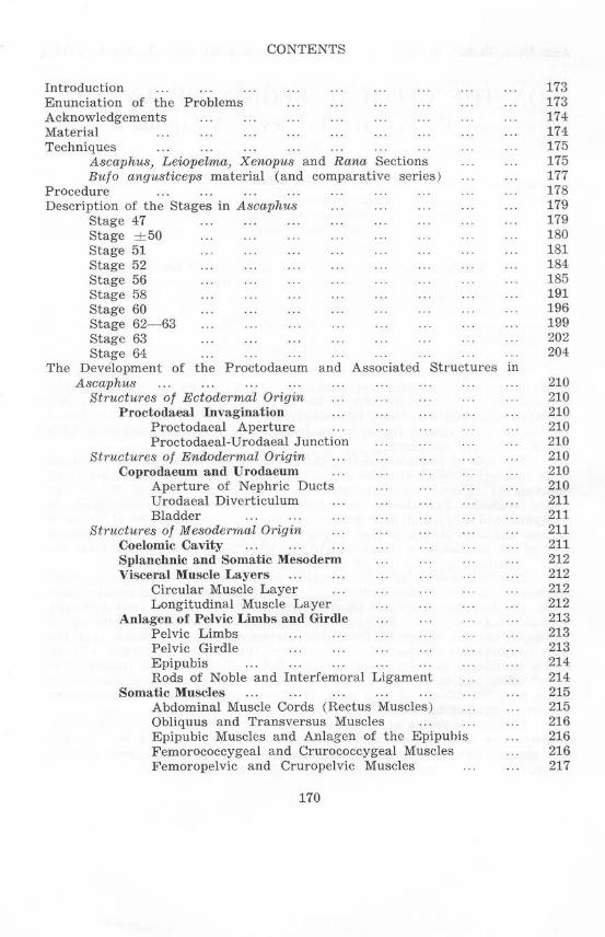
Stage ± SO (figs, 3 and 4)
The relations of the segments of the abdominal muscles to the myomeres of the tail and trunk are well shown in the specimen at this stage. The posterior-most segment of the abdominal muscle is applied to the ventrolateral face of the myomere corresponding to the 9th spinal nerve (here referred to as the 9th spinal myomere). The second abdominal muscle segment from the posterior end is lateral to the coelom and ventrolateral to the 8th spinal myomere. The posterior-most abdominal segment is anteriorly mediad to that in front of it, but is lateral to it further posteriorly. The 9th and subsequent spinal myomeres curve ventrally round those behind them and extend dorsally on the inner faces of the latter. A few muscle fibres occur mediad to the posterior tip of the posterior-most abdominal myomere.
The coelom extends to lIear the posterior edge of the 9th spinal myomere posteriorly; medially it extends between the 9th spinal myomeres further dorsally than the level of the top of the last abdominal myomere. Laterally the coelom extends posteriorly almost to the posterolateral margin of the 8th spinal myomere dorsally. extending beyond this margin further ventrally.
FigS. 3 and 4. Ascap/ws larva, Stage ± 50 (both figs. from one specimen).
Posterior abdominal region x 25.
- lrom
. "",tn vertebra
Fig. 3. Norma lateralis sinistra.
Graphie reeonstruction from tra nsverse sections.
180
The 8th spinal and abdominal myomeres are thus separated by coelom at this stage.
Hind-limb buds are visible at this stage and a femur anlage is present ncar the base of each . The concentrations of cells of each limb anlage extend ant eroven trally to just anterior to the' posterior limit of the coelom. Laterally each anlage extends anterodorsally over the posterior tip of the 9th spinal myomer e onto the lateral face of the 10th spinal myomere. From this part of each limb anlage the ilium and associated structures will differentiate.
Mediodorsal and mediovcntral to the hind-limb buds are ceU aggregations. the latter clearly constituting the anlagen of the rods of Noble.
The hypochord in the specimen is much thicker than in all other larvae examined, and thc difference may not be developmental, but specific.
The myomeres and nerves of the specimen were numbered from the vertebral arches, the most anterior shown in fig. 3 being found, by counting, t o be the 9th. At this stage the 9th arch is well-developed, but dorsally incomplete.
Fig. 4. Norma posterioris. Photomicrographs of transverse sections.
(The numbers refer t(l levels in fig. 3.)
Stage 51 (fi gs. 5 and 6 )
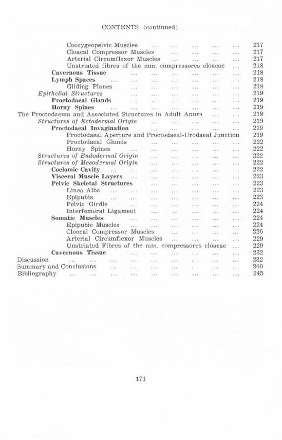
Thc proctodaea] aperture is situated on the dorsal surface of a flap of skin which covers the hind·limb buds ventrally and protrudes beyond them posteriorly. The aperture is roughly triangular, wit h the base ncar thc posterior tip of the ventral flap, and the apex anterodorsal to this. Just anterior to the apex of the proctodaea] aperture a ventral projection from the tail attaches to the dorsal surface of the proctodaeum; anterior to this point the proctodaeum, and the flap of skin ventral to it, are suspended from the tail . Anteriorly the furthest limits of the proctodaeum are marked dorsally by the opening of the nephric ducts, vcntrally by a thickening which represents the bladder. The coelomic cavity does not extend ventrally beyond the bladder, but dorsally the median urinogcnital aperturc is now a short distance
181
from the end of the coelom, which extends to the level of the 12th spinal nerve. The circular muscle layer present around the coprodaeum is continued in the urodaeal region ; but it is interrupted dorsally where the nephric ducts join and enter the urodaeum, and ventrally at the bladder. Posterior to the nephric ducts the dorsal wall of the gut slopes down steeply, the circular muscle laycr again becoming continuous dorsally where this slope decreases, which is still within the coelomic cavity laterally and dorsally. Dorsally the circular muscle layer extends caudad to the end of the coelomic cavity and a short distance beyond, reaching approximately the posterior limit of the bases of the hind-limb buds. The circular muscle layer a lso becomes continuous posterior to the bladder anlage which is beyond the ventral limit of the coelomic cavity; but it does not extend as far into the extra-coelomic tissue ventrally as dorsally. Longitudinal muscles are not clearly distinguishable, and would thus seem to be less extensive than the circular layers.
A dorsal median r idge of the gut epithelium, extending from the Urinogenita l aperture to t he anterior limit or the bases of the hind-limb buds. is present and constitutes a characteristic feature observable in adults, in which it extends to the cloacal aperture.
Figs. 5 and 6. Ascaphu8 larva, Stacre 51 (both figs. from one specimen) . Cloacal region x 25.
I .11
~Inm::;:~~:th ::: vmebra
spinal nerw:
hearts
"",,,n,lrQ' skin flop
Fig. 5. Norma latcralis sinistra .
Graphic reconstruction from transverse sections.
182
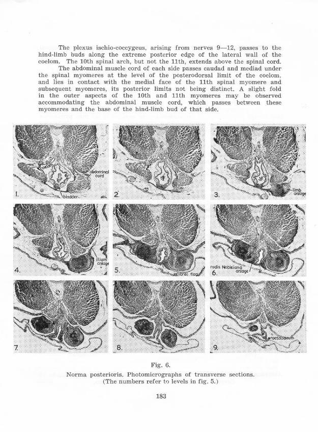
The plexus ischio-coccygeus, arising from nerves 9-12, passes to the hind-limb buds along the extreme posterior edge of the lateral wall of the coelom. The 10th spinal arch, but not the 11th, extends above the spinal cord.
The abdominal muscle cord of each side passes caudad and mediad under the spinal myomeres at the level of the posterodorsal limit of the coelom. and lies in contact with the medial face of the 11th spinal myomere and subsequent myomeres, its posterior limits not being distinct. A slight fold in the outer aspects of the 10th and 11th myomeres may be observed accommodating the abdominal muscle cord, which passes between these myomeres and the base of the hind-limb bud of that side.
Fig. 6.
Norma posterioris. Photomicrographs of transverse sections. (The numbers refer tf) levels in fig. 5. )
183
The kidneys and their ducts extend to thc level of the 10th spinal myomere.
At the level of the urostyle 4 coccygeal lymph hearts are present laterally near the anterior edges of the 11th to 14th spinal myomeres.
At the ventral limits of the bases of the hind-limb buds, and along the medial borders of these bases, are thickenings; these ventr ally representing the (post-pubic) rods of Noble, and dorsally the fibrous connective tissue which extends to the urostyle from thc posterior tips of these rods in the adult, particularly thc female adult.
Thc anlagen of the muscles of thc hind limbs are becoming distinguishable around pro-cartHage in the limb-buds; the anlagen of the mm. pyriformcs and mm . caudaiipuboischiotibiales are indicated by extensions from the myogenic tissue of the limb-buds towards the urostyle.
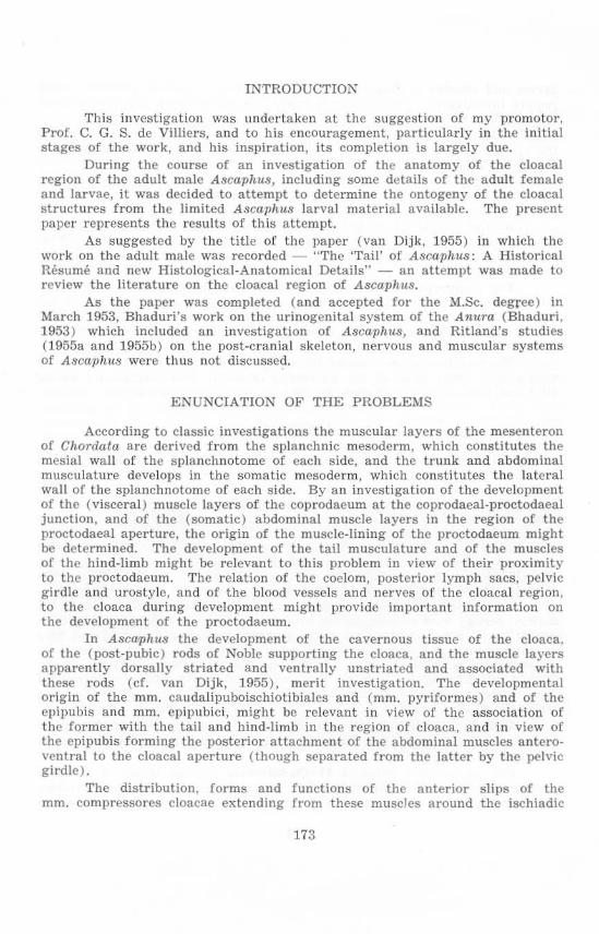
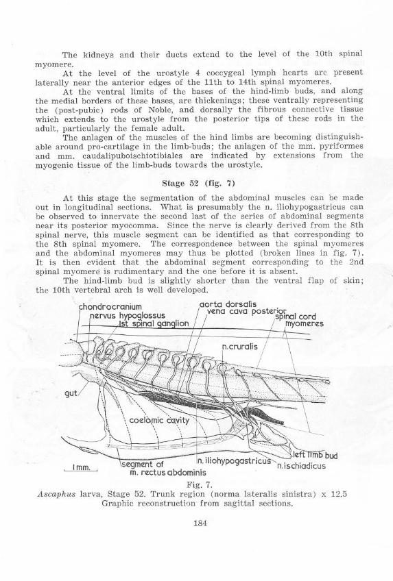
Stage 52 (fig. 7)
At this stage thc segmentation of the abdominal muscles can be made out in longitudinal sections. What is presumably the n. iliohypogastricus can be observed to innervate the second last of the series of abdominal segments near its posterior myocomma. Since the nerve is clearly derived from the 8th spinal nerve, this muscle segment can be identified as that corresponding to the 8th spinal myomere. The correspondence between the spinal myomeres and the abdominal myomeres may thus be plotted (broken lines in fig. 7). It is then evidcnt that the abdominal segment corresponding to the 2nd spinal myomere is rudimentary and the one before it is absent.
The hind-limb bud is slightly shorter than the ventral flap of skin; the 10th vertebral arch is well developed.
l"~;.~~dorsalis cava
Fig. 7. Ascaphus larva, Stage 52. Trunk region (norma lateralis sinistra ) x 12.5
Graphic rcconstruction from sagittal ::;cctions.
184
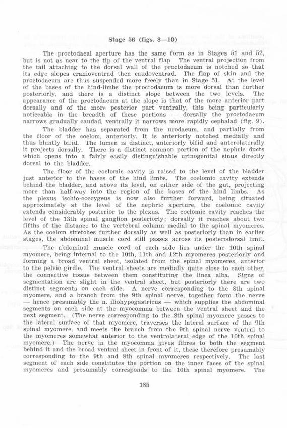
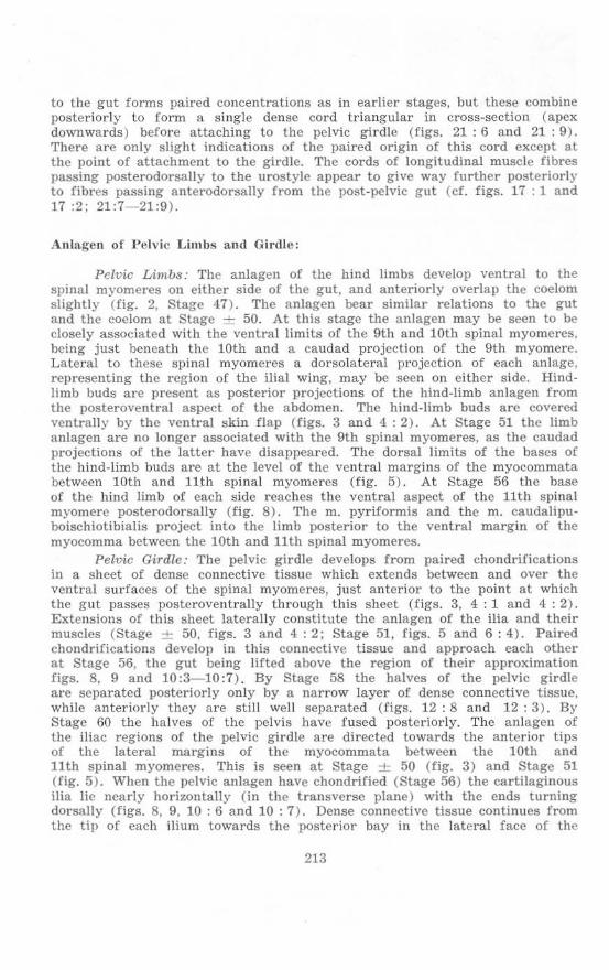
Stage 56 (figs. 8-JO)
The proctodaeal apcrture has thc same form as in Stages 51 and 52, but is not as near to the tip of the ventral flap. The ventral projection from the tail attaching to thc dorsal wall of the proctodaeum is notched so that its edge slopes craniovcntrad then caudoventrad. The flal) of skin and the proctodaeum are thus suspended more freely than in Stage 51. At the level of the bases of the hind-limbs the proctodaeum is more dorsal than further posteriorly, and there is a distinct slope between the two levels. The a ppearance of the proctodaeum at the slope is that of the mOre anter ior part dorsally and of the more posterior part ventrally, this being particularly noticeable in the breadth of these portions - dorsally the proctodaeum narrows gradually caudad, ventrally it narrows more rapidly cephalad (fig. 9).
The bladder has separated from the urodaeum. and partially from the floor of the coelom. anteriorly. It is anteriorly notched medially and thus bluntly bifid. The lumen is distinct, anteriorly bifid and anterolaterally it prOjects dorsally. There is a distinct common portion of the ncphrie ducts which opens into a fairly easily distinguishable urinogenital sinus directly dorsal to the bladder.
The floor of the coelomic cavity is raised to the level of the bladder just anterior to the bases o[ the hind limbs. The coelomic cavity extends behi nd the bladder, and above its level, on either side of the gut, projecting more than half-way into the region of the bases of the hind limbs. As the plexus ischio-coccygeus is now also further forward. being situated ap proximately at the level of the nephric aperture, the coelomic cavity extends considerably posterior to the plexus. The coelomic cavity reaches the level of the 13th spinal ganglion posteriorly; dorsally it reaches about two fifths of the distance to the vertebral column medial to the spinal myomercs. As the coelom stretches further dorsally as well as posteriorly than in earlier stages, the abdominal muscle eord still passes across its posterodorsal limit.
The abdominal muscle cord of each side lies under the 10th spinal myomere, being internal to the 10th, 11th and 12th myomeres posteriorly and formi ng a broad ventral sheet, isolated from the spinal myomeres. anterior to the pelvic g-irdle. The ventral sheets are medially quite close to each other, the connective tissue between them constituting the linea a lba. Si!Jns of segmentation are slight in the ventral sheet, but posteriorly there are two distinct segments on each side. A ne rve corresponding to the 8th spinal myomere, and a branch from the 9th spinal nerve, together form the nerve - hence presumably the n. iIiohypogastricus - which supplies the abdominal segments on each side at the myocomma between the ventral sheet and the next segment. (The nerve corresponding to the 8th spinal myomere passes to the lateral surface of that myomere, t raverses the lateral surface of the 9th spinal myomere, and meets the branch from the 9th spinal nerve ventral to the myomeres somewhat anterior to the ventrolateral edge of t he 10th spinal myomere.) The nerve in the myocomma !Jives fibres to both the segment behind it and the broad ventral sheet in front of it, these therefore presumably corresponding to the 9th and 8th spinal myomeres respcetively. The last segment of each side constitutes the portion on the inner faces of the spinal myomercs and presumably corresponds to the lOth spinal myomere. The
185
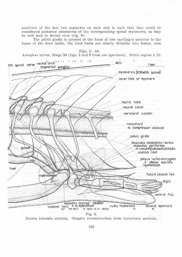
positions of the last two segments on each side is such that they could be considered posterior extensions of the corresponding spinal myomeres, as may be well seen in dorsal view (fig. 9 ) .
The pelvie girdle is present in the form of two cartilages anterior to the bases of the hind limbs; the hind limbs a re clearly divisible into femur, crus
Figs. 8-10. Ascaphus larvae, Stage 56 (figs. 8 and 9 from one specimen). Pelvic region x 25.
9\\hh:"';i"~a~I~""";'~~~~\,~"~h~'~' ~' ~'~' ~I:i ~'=:' :"~~~":::' :"::::~:::":::~)""im' yo<"".<>(Oh~!; :;oo!) : I ) nntr limit of myomtrt
I .
live.r
rudis I 91.-::1112\ P l4\l'I\1I>
Fig. 8.
.j"lturol tubt
_ nturol cono!
"ve.rte. bra! column
notochord m. comprtssor doocot:
pdvic gird!t
rectus
i i cottom
plexus ischfo.roccygeu~ + ploZX\Js socralis
ligamentum
future cloacal lips
flop
optrturt
Norma lateralis sinistra. Graphie reconstruction from transverse sections.
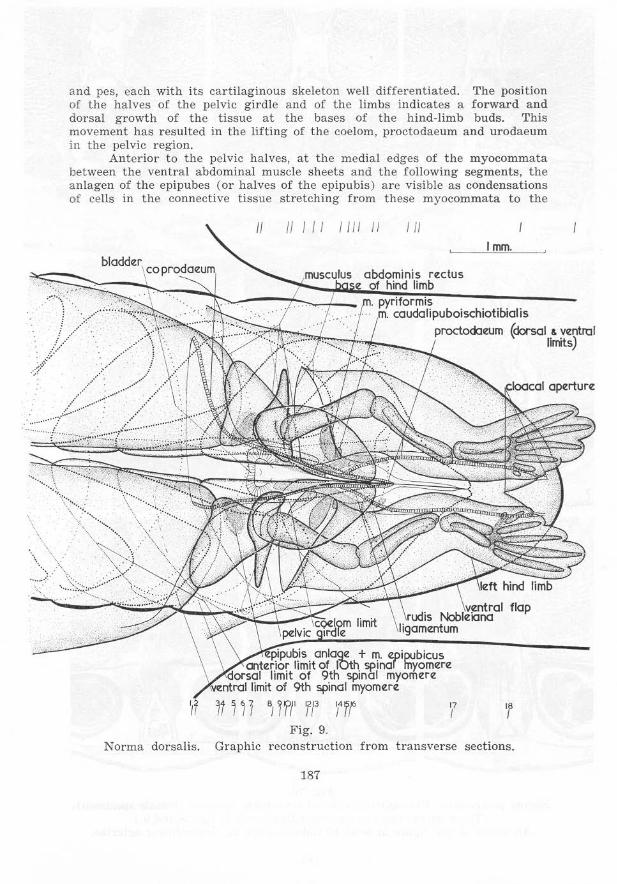
and pes, each with its cartilag inous skeleton well differentiated. The position of the halves of the pelvic girdle and of the limbs indicates a for ward and dorsal growth of the tissue at the bases of the hind-limb buds. This movement has resulted in the lifting of the coelom, proctodaeum a nd u rodaeum in the pelvic region.
Anterior to t he pelvic halves, at the medial edges of the myocommata between the ventral abdominal muscle sheets and the following segmen ts, the anlagen of the epipubes (or halves of the epipubis) are visible as condensations of cells in the connective t issue stretching from these myocommata to the
/I I II 1111 II III 1 rom.
; / •...... ".:'::·~C"'~+[~Sv~~t~d~C~al:":do:'~iPUbOiSChiotibiatiS .. .... ,. 'dorsal & ventrol \' Jirrits)
. ""
""
..... .. .
jill j 'I1f1' fl' i'1f F ig. 9.
" I
'J,:k><'co' aj)f!rturtl
','
Norma dorsalis. Graphic reconstruction from transverse sect ions.
187
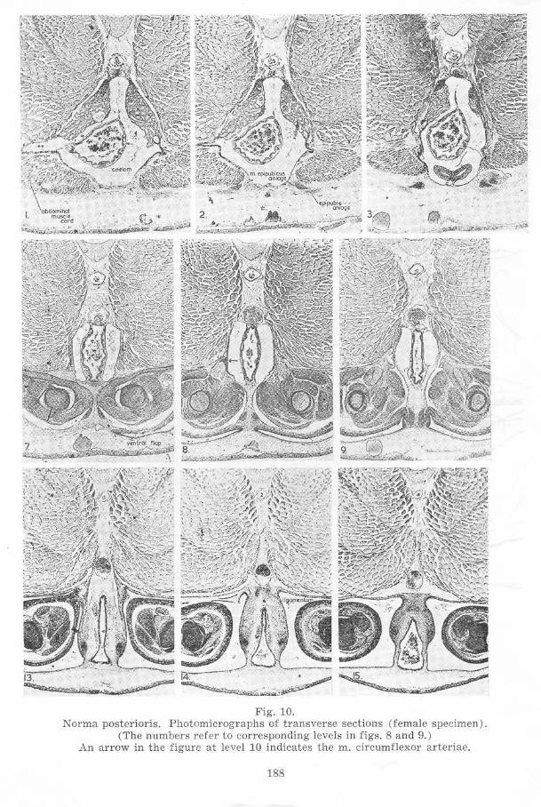
Fig. 10. Norma poster ioris. Photomicrographs of t r ansverse sections (female specimen).
(The numbers refer to corresponding levels in figs. 8 and 9.) An arrow in the figure at level 10 indicates the m. circumflexor arteriae.
188
189
pelvic halves. Dorsal to the epipubic anlagen, and considerably more distinct, are the mm. epipubici.
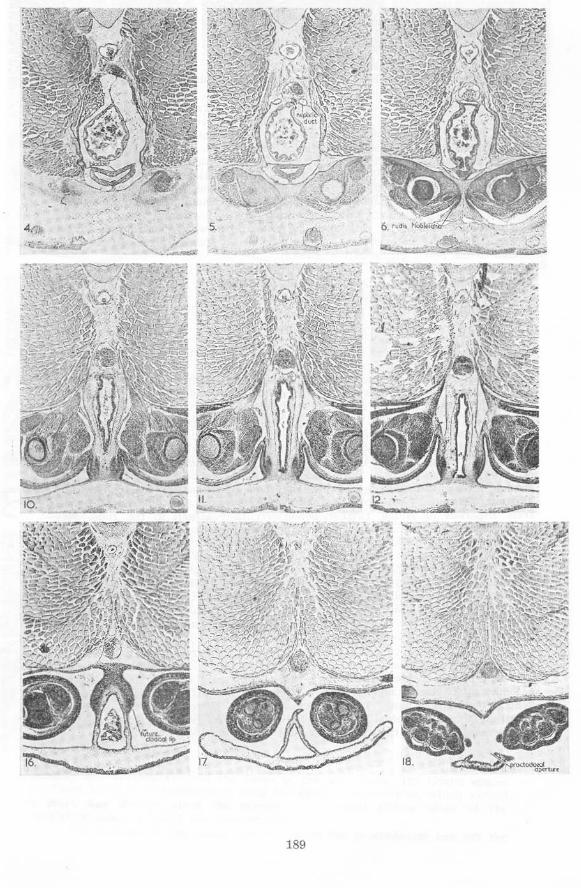
An aggregation of cells continues posteriorly from the epipubic anlage of cach side medioventral to the pelvic girdle half, becoming the (post-pubic) rod of Noble posteriorly. These rods thus begin anteriorly medioventral to the posterior third of the pelvic girdle. Behind the pelvic girdle the rods of the two sides approach each other beneath the proctodaeum, parting again as the proctodaeum slopes posteroventrally between them and tu rning mediodorsally at their posterior tips. The rods extend about two thirds of the way across the sides of the sloping portion of the proctodaeum. At their posterior tips the rods approach not only each other in the horizontal plane, but also a si milar aggregation of cells forming a ligament which lies dorsal to the cloaca. From the anterior tips of the rods dense connective tissue stretches medial to the pelvic girdle halves dorsally.
In the region of the tips of the rods of Noble the skin laterodorsal to the proctodaeum is thicker than further posteriorly, these thicker walls representing the future cloacal lips. The median dOrsal ridge of the urodaeum and proctodaeum is absent posterior to this point, but it is distinct somewhat anterior to it.
Near the posterior end of the bases of the hind limbs the mm. caudalipuboischiotibiales a re located, lying dorsomedial-Iateroventrally on either side of the dorsal half of the proctodaeum. Just anterior to the mm. caudalipuboischiotibiales the mm. pyriformes have their origins at the same horizontal and lateral levels. Their direction is more lateral than that of the former muscles, and they are longer, since they reach to the femurs. The mm. pyriformes just reach the level of the coelomic cavity anteriorly.
The mm. compressores cloacae have developed, stretching across the medial faces of the posterior segments of the abdominal muscles dorsally and t>assing on either side of the gut ventrally to approach the dorsal surfaces of the rods of Noble. Posteriorly the mm. pyriformes and the mm. caudalipuboischiotibiales lie between the mm. compressores cloacae and the abdominal muscles. Anteriorly the mm. compressores cloacae end some distance from the rods of Noble, further posteriorly they attach to the rods. Just posterior to the hind-limb bases the mm. compressores cloacae become indistinct. Posterior to the bladder the mm. compressores cloacae become continuous ventrally in the pelvic girdle region. Anteriorly slips of muscle continuous with the mm. compressores cloacae extend anterolaterally, a dorsal slip stretching along the lower half of the medial faces of the posterior abdominal segment of each side and a ventral slip curving around the arteria ischiadica and the nervus ischiadicus at the point at which the artery twists eaudolaterad over the nerve. The ventral slip of muscle is here named m. circumflexor arteriae to facilitate reference.
The circular muscle layer of the gut can be detected approximately as far postcriorly as the mm. compressores cloacae, but it is incomplete ventrally posterior to the coelom. Posterior to the coelom there are few indications of a longitudinal muscle layer. Just posterior to the pelvic girdle, and to the ventrally complete mm. compressores cloacae, there are two cords of longitudinal muscle ventral to the gut. Indications of lonrritudinal muscle arc also visible dorsolateral to the gut at this level.
]90
The lymph sacs of the limbs have developed and extend from the hindlimb bases ventrally, and media lly both cephalad and caudad, dorsal to the rods of Noble. The rods of Noble are firmly adherent to the ventral flap of skin posteriorly; anterior to the bases of the hind limbs there are small lymph spaces ventral to t he rods separating them partially from the ventral skin .
Veins on either side of Lh e proctodaeum just an terior to the future cloacal lips drain considerabl e s inuses around the proctodaeum in this region.
Th e gonads have developed, a nd the sex of some specimens may be determined at this stage.
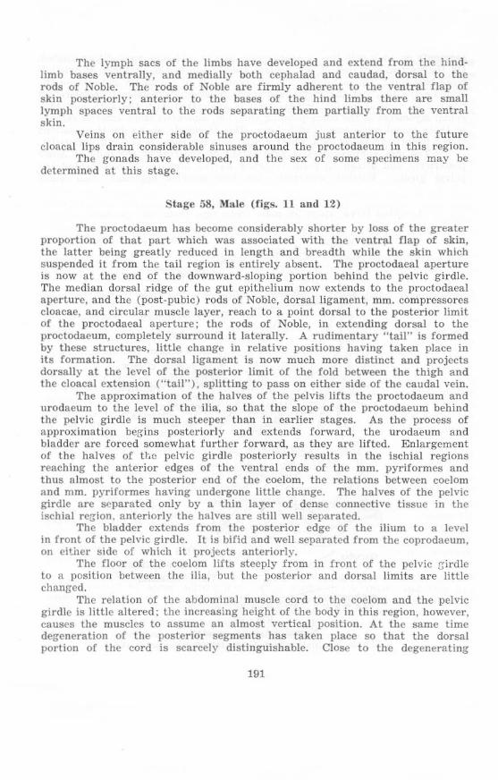
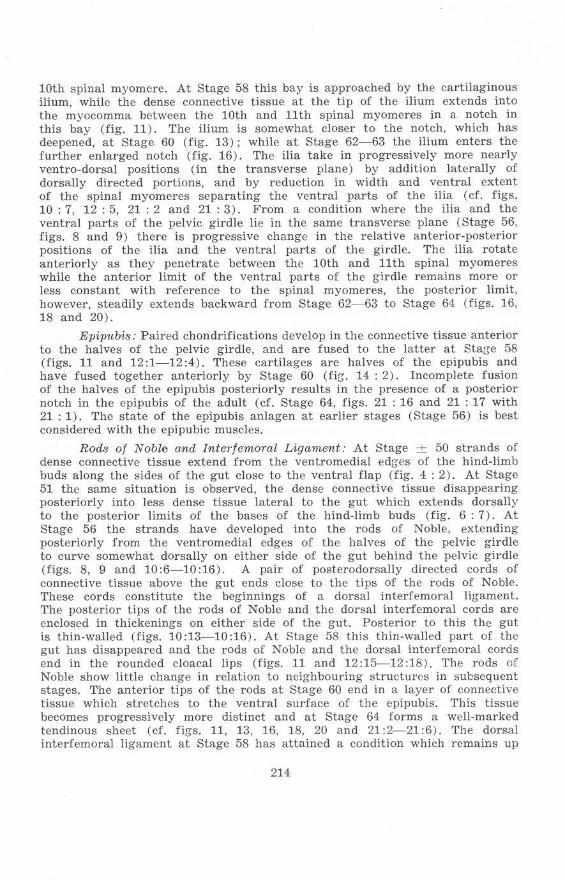
St.llge 58, Mal e (figs. 1 Land 12)
The proctodaeum has become considerably shorter by loss of the greater proportion of that part which was associated with the ventral flap of skin, the la tter being greatly reduced in length and breadth while the skin which suspended it from the tail region is entirely absent. The proctodaeal aperture is now at the end of the downward-s loping portion behind the pelvic girdle. The median dorsal ridge of th e gut epithelium now extends to the proctodaeal aperture, and the (post-pubic) rods of Noble, dorsal ligament, mm. compressores cloacae, a nd circular muscle layer , r each to a point dorsa l to the poste rior limit of t he proctodaeal aperture; the rods of Noble, in extending dorsal to the proctodaeum, completely surround it laterally. A rudimentary "tail" is formed by these structures, little cha nge in relative positions having taken place in it s formation. The dorsal ligament is now much more distinct and projects dorsally at the level of the posterior limit of the fold between the thigh and the cloacal exte nsion ("tail"), splitting to pass on eithe r side of the caudal vein.
The approximation of the halves of the pelvis lifts the proctodaeum and urodaeum to the level of the ilia, so that the slope of the proctodaeum behind the pelvic gi rdle is much steeper than in earlier stages. As the process of approximation begins posteriorly and extends forward, the urodaeum and bladder are forced somewhat further forward, as they are lifted. Enlargement of the halves of it,e pelvic girdle posteriorly results in the ischial regions reaching the anterior edges of the ventral ends of the mm . pyriformes and thus a lmost to the posterior end of the coelom, the relations between coelom and mm. pyriformes having undergone little change. The halves of the pelvic girdle are separated only by a thin layer of dense connective tissue in the ischia l region. anteriorly the halves are I:ItiJI well separated.
The bladder extends from the posterior edge of the ilium to a level in front of the pelvic girdle. It is bifid and well separated from the coprodaeum, on cit.ilcr side of which it projects anteriorly.
The floor of the coelom lifts steeply from in front of the pelvic r:;-irdle to a position between th e ilia, but the posterior and dorsal limits are little changed.
Th e relation of the abdominal muscle cord to the coelom and the pelvic girdle is litlle altered ; the increasing height of the body in this region, however, causes the muscles to assume an a lmost vertical position. At the same time degeneration of the posterior segments has taken place so that the dorsal portion of the cord is scarcely disti nguisha ble. Close to the degenerating
191
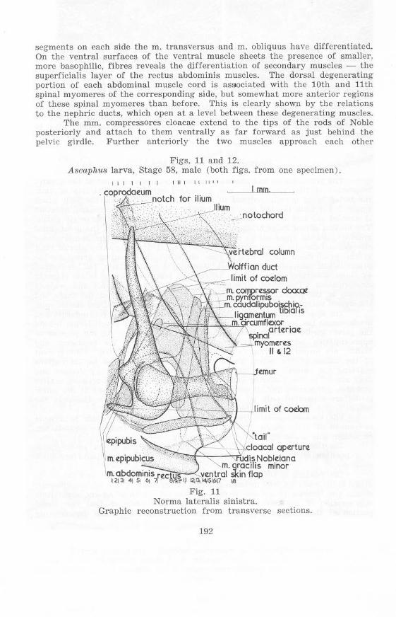
segments on each side the m. transversus and m. obliquus have d ifferentiated. On the ventral surfaces of the ventral muscle sheets the presence of smaller, more basophilic, fibres reveals the ctifferentiation of secondary muscles - the superficialis layer of the rectus abdominis muscles. The dorsal degenerating portion of cach abdom inal muscle cord is aS90ciated with the 10th and 11th spinal myomeres of the corresponding side, but somewhat more anterior regions of these spinal myomeres than before. This is clearly shown by the relations to the nephric ducts, which open at a level between these degenerating muscles.
The mm. compressores cloacae extend to the tips of the rods of Noble posteriorly and attach to them ventrally as far forward as just behind the pelvic girdle. Further anteriorly the two muscles approach each other
Figs. 11 and 12. A8caphu8 larva, Stage 58, male (both figs. from one specimen ) .
I I I I I I I I II 1 I L I L "
I rom. for jlium
column
\~_'NoIrt;an dlet
(~~~~2~~~~2~~limit of codom
I aperture
" Fig. 11
Nonna lateralis sinistra. Graphic reconstruction from transverse sections.
192
--.-- -Fig. 12.
Norma posterioris. Photomicrographs of transverse sections. Arrows in the figure at levels 2- 6 indicate the atrophying abdominal muscle cord, and at level 7 indicate the m. compressor cloacae and the m. circumflexor
arteriae.
194
,0 . ...
'QjQ'~ ... . . b . .
16. 18.
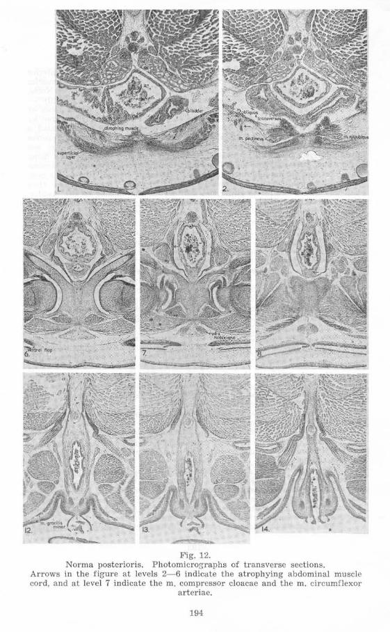
dorsally and dorsoiaterally at the bases of the hind limbs, the lymph spaces produced being- in communication with the sacci interfcmoraies, which extend a short way distally along the femurs. The dorsal gliding plane of the cloacal extension ("tail") has also developed.
The increase in the lymph spaces around the proctodaeum has left the
195
blood vessels and the nerves (12th) of this structure suspended between the posteromedial edges of the mm. pyriformes and the dorsolateral edges of the mm. compressores cloacae in the cloacal extension.
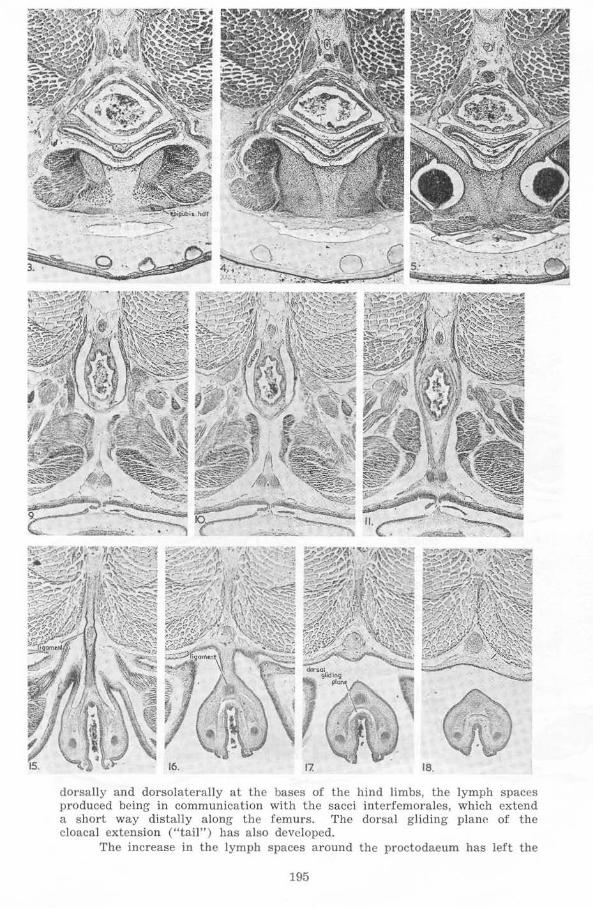
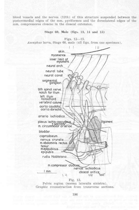
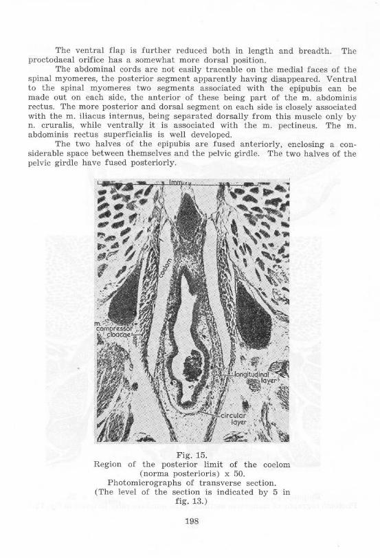
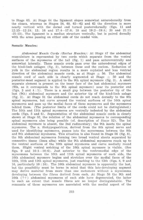
Stage 60. l\I.a le (figs. la, 14 Ilnd 15)
Figs. 13- 15. Ascaph l18 larva, Stage 60, ma le (a ll figs. from one specimen ) .
I I I II
skin _______ .~;=~~=j::::;:=-,_ myomeres "l!
innr:r limit of myomere.
neural arch~
neural tubr: nr:ural canal
scgmr:nta\. gong IOn
9th spinal nf:rvf: notch for ilium Idt ilium notochord
Vf:rtr:brol coIu oorla caudolis aorta dorsalis
orteria ischiodico
plo:us iSCl'lio'OOCC~~·~.:-~~~t~~{~~ . ~ 5Qcroli m. c,rcumflr:xar orlr:rrQ(.
bloddotr_ coprodootum nr:rvus crurolis _ m.obdominis rr:ctus_
m.r:pipubicus _ fl1:mur _~~~~ r:pipubis
rudis Nablr:iano
I mm. \ ,
Fig. 13.
I
\
Pelvic region (norma lateralis sinistra) . Graphic reconstruction from transverse sections.
196
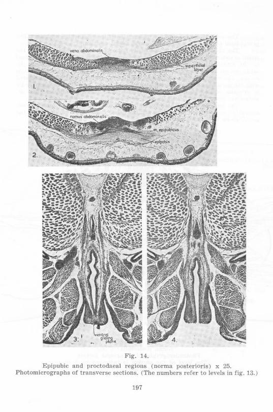
Fig. 14.
Epipubic and proctodaeal regions ( norma posterioris) x 25. Photomicrographs of transverse sections. (The numbers refer to levels in fig. 13.)
197
The ventral flap is further reduced both in length and breadth. The proctodaeal orifice has a somewhat more dorsal position.
The abdominal cords are not easily traceable on the medial faces of the spinal myomeres, the posterior segment apparently having disappeared . Ventral to the spinal myomeres two segments associated with the epipubis can be made out on each side, the antcrior of these being part of the m. abdominis rectus. The more posterior and dorsal segment on eaeh side is closely associated with the m. iliacus intern us, being separated dorsally from this muscle only by n. cruralis, w hile ventrally it is associated with the m . pectineus. The m. abdominis rectus superficialis is well developed.
The two halves of the epipubis are fused anteriorly, enclosing a considerable space between themselves and the pelvic girdle. The two halves of the pelvic girdle have fused posteriorly.
Fig. 15. Region of the posterior limit of the coelom
(norma posterioris) x 50. Photomicrographs of transverse section.
(The level of the section is indicated by 5 in fig. 13. )
198
With the formation of a vena abdominaJis (showing signs of a dual origin) dorsal to the anterior tip of the epipubis and to the linea alba further anteriorly, the vascular system in the pelvic region has attained essentially the adult form.
Anterior to the ventral lip of the proctodaeal orifice a ventral gliding plane has developed; it is incomplete midventrally.
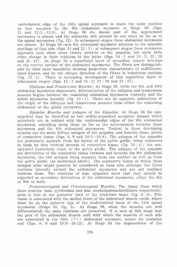
Stage 62-6:J, Male (figs. 16 and 17)
The proctodaeal aperture is somewhat more dorsal, particularly its upper rim, and there is a more marked fold between the cloacal extension ("tail") and the tail. The mm. compressores cloacae now reach further dorsally, to beyond the aorta eaudalis, and are separated from each ventrolatcral edge of the vertebral column by a short tendinous band only. Anteriorly the mm. compressores cloacae have shifted forward to a position lateral to the urinogenital aperture. The mm. circumflexores arteriarum arising from the mm. compressores cloacae project beyond the anterior edges of the latter, the change in position being made possible by greater separation distally of the two muscles on each side. There is a forward shift of the plexus sacralis relative to the ilium.
The ilium now projects somewhat antcrodorsally instead of posterodorsally or vertically upwards from the acetabulum. This change is associated with a shift of the ilium posteriorly relative to the ventrolateral face of the 10th spinal myomere, while the dorsal tip of the ilium enters the notch at the posterior edge of the myomere, which notch is now deeper and hence extends further anterior. The tip of the ilium approaches the transverse process of the 10th, the sacral, vertebra, which lies lateral to, but not in contact with . the arch of the vertebra. The secondary muscles associated with the transverse process are well·developed.
The abdominal muscle cord is represented by the m. abdominis rectus originating on the cpipubis. The epipubis is connected to the rods of Noble by a tendinous sheet of connective tissue which has two thickened portions revealing its paired origin.
Posteriorly the rods of Noble are distinctly continuous with the dorsal ligament, which now reaches to the vertebral column. Associated with the rods of Nobl e cavernous tissue has begun to develop; this tissue surrounds the rods behind the pelvis and also lies lateroventral and medioventral to the rods in this region. The mm. compressorcs cloacae have the same transverse niveau with the rods as has the cavernous tissue. Between the rods just anterior to the ventral rim of the cloacal orifice there are fibres of the mm. compressores cloacac. These are well isolated from differentiated circular and longitudinal muscle layers.
There is a urodaeal diverticulum where the common ncphric duct opens into the urodaeum . The wall of the urodaeum and proctodaeum is little folded in comparison with the other larvae, and there is no median dorsal ridge. Just within the proctodaeal aperture, and cephalad, cloacal glands have begun to develop anteriorly. They are visible as thickenings in the epithelium.
Ventrally the cloacal extension, rods of Noble and mm . graciles minores have separated from the skin except just anterior to the proctodaea! aperture.
199
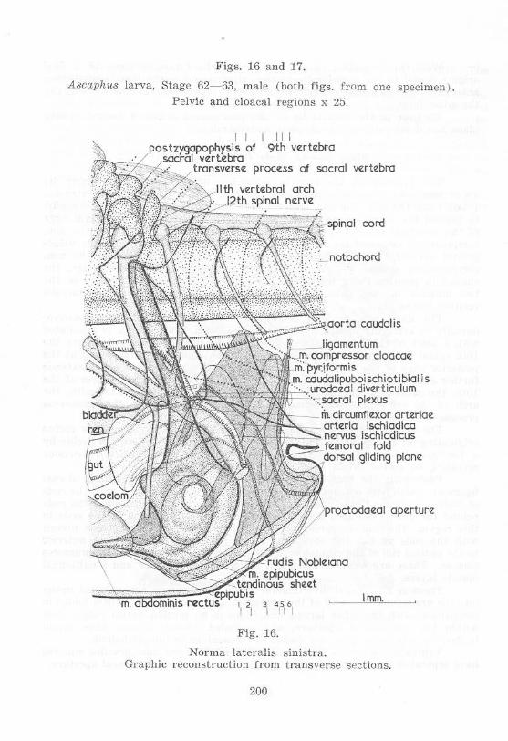
Figs. 16 and 17.
Ascaphus larva, Stage 62-63, male (both figs. from one specimen) .
Pelvic and cloacal regions x 25.
I I II I
P;~~y.~~~~Of 9th vertebra :S~CC'7:--7,L proce:ss of sacral ve:rte:bra
caudolis
doaax
lis
arte:riae
plane:
)p"",to,da,al aperture
:~:~~~~~~~N~:obl' __ --",,",-_ sheet Imm
II ~I 1 4,511>,
Fig. 16.
Norma lateralis sinistra. Graphic reconstruction from transverse spctions.
200
o
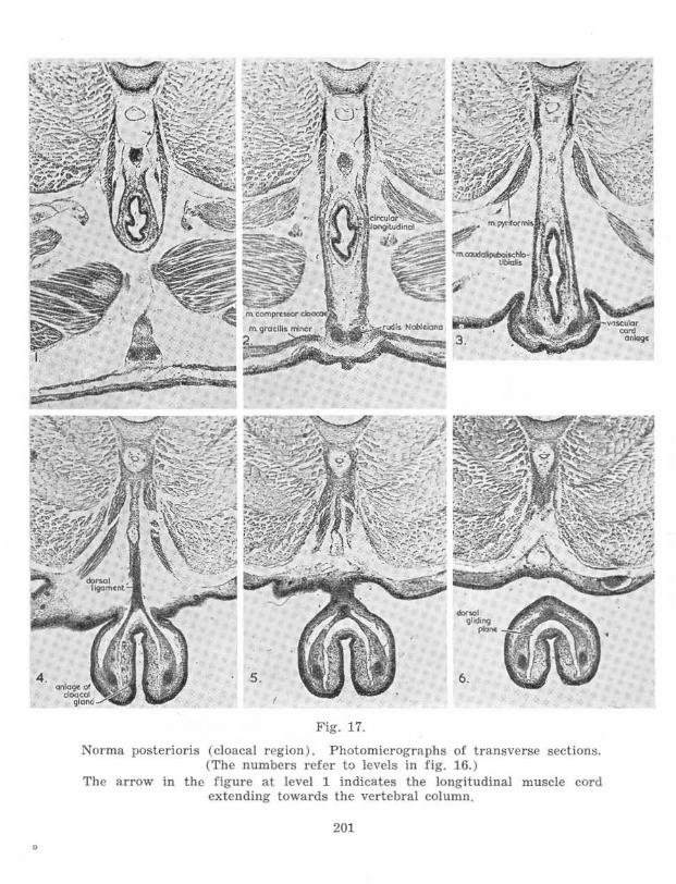
Fig. 17.
Norma posterioris (cloacal region). P hotomicrographs of transverse sections. (T he numbcrs refer to lcvels in fig. 16. )
The arrow in the figure at levcl 1 indicates the longitudinal muscle cord extending towards the vertebral colu mn.
201
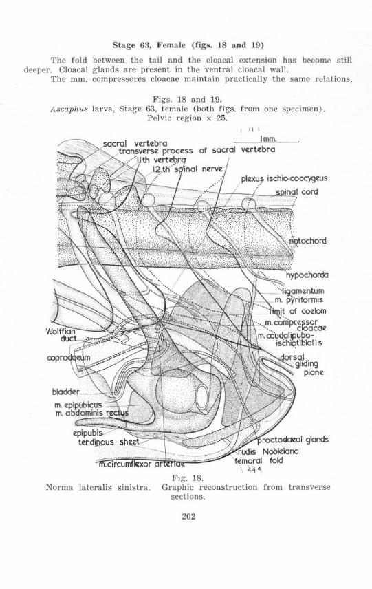
Stage 63, l~ema le (figs. 18 and 19)
The fold between the ta il and the cloacal extension has become still deeper . Cloacal glands are present in the ventral cloacal wall.
The mm. compressores cloacae maintain practically the same relations,
Figs. 18 and 19. Ascaphu8 larva, Stage 63, 1emale (both figs. from one specimen) .
Pelvic region x 25. I II I
Imm._
,tfP(rO" todo<ol glonds
Fig. 18. Norma la teralis sinistra. Graphic reconstruction from t ransverse
sections.
202
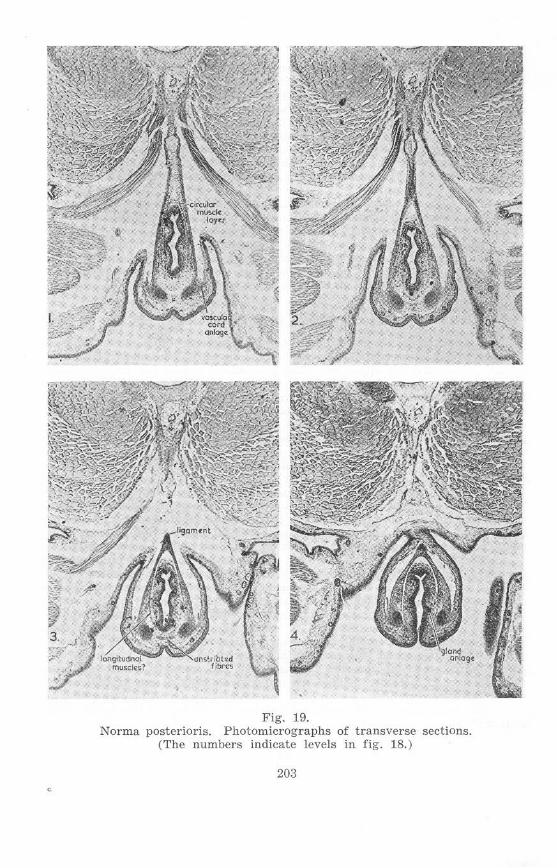
Fig. 19. Norma postcrioris. Photomicrographs of transverse sections.
(The numbers indicate levels in fig. 18.)
203
but in doing so have considerably lengthened antero-posteriorly, since the ischial region has become considerably larger. The origins of the mm. pyriformes are only slightly postcrior to the ischia, and slightly posterior to a point at which the hypochorda becomes dorso-ventrally thinner.
On each side the dorsal portion of the ilium projects much more anteriorly, passing from a position lateral to the posterior extrcmity of the lateral face of the 10th spinal myomere to penetrate the very decp notch at the posterior margin of this myomere. It closely approaches the transverse process, now fused to the sacral vertebra. The plexus sacralis is displaced further anteriorly relative to the ilium.
The 12th neural arch rcachcs almost to the top of the spinal cord . The hypochorda is considerably smaller posterior to a point just behind the ganglion of the 14th spinal nerve than further anteriorly.
There is little change in the epipubis, tendinous sheet, rods of Noble, and dorsal ligament. Between the rods of Noble just anterior to the proctodaeal glands is a portion of the mm . compressores cloacae. The relation of this portion of muscle to the circular musclc layer is not clear.
Stage 64, Female (figs. 20 a nd 21)
The tail myomeres are considerably shorter dorso-ventrally, the ventral portions having been reduced. The proctodaeal aperture is considerably fUrther dorsal, particularly its upper rim. The proctodaeal region has enlarged considerably so that the dorsal ligament is an terodorsally directed, instead of first being directed dorsally then curving posterodorsally nearer the vertebral column. This enlargement of the proctodaeum is partly due to the decrease of the tail, which is also more widely separated from the cloacal extension, providing more spacc for the proctodaeum. Cloacal glands are now distinct on the lateral walls of the cloaca.
The rods of Noble are longer and more sigmoid, the shape in thc adult female being quite similar.
The mm. pyriformes and mm . caudalipuboischiotibiales are now close to the vertebral colUmn and the former are dorsal to the ischia. The hypochorda has become thinner posteriorly from a point just in front of the level of the ganglion of the 14th spinal nerve (sec previous stage), and it is at this point that the mm. pyriformes approach the vertebral column most closely.
The anterior edges of the mm. compressores cloacae have advanced further anteriorly. Since this is particularly so dorsally, the anterior edges are now nearly vertical. The plexus sacralis is relatively further cephalad and the ilium projects somewhat more anteriorly.
The mm. compressores cloacae are well developed in the rcgion between the rods of Noble. 'l'he isolation of their fibres from the circular and longitudinal muscle layers is considerable in this region. Near the posterior end of the pelvic girdle the longitudinal muscles of the gut attach to the girdle ventral to these muscles and approach the urostyle dorsal to them (cf. also figs. 22 and 23). Behind the pelvic girdle they do not extend far as well-differentiated entities.
The 12th neural arch reaches the level of the top of the spinal cord at this stage.
204
Figs. 20 and 21.
Ascuphus larva, Stage 64, female (both figs. from one specimen ) . Pelvic region x 25.
, ,
.... . ....
Fig. 20.
Norma lateralis sinistra. Graphic reconstruction from transverse sections.
205
Fig. 21. Norma posterioris. Photomicrographs of transverse sections.
(The number refer to levels in fig . 20.) The arrows in the figure at levels 2 and 3 indicate the m. circumflexor arteriae,
206
at level 8 the arrow indicates the ventral longitudinal muscle cord, and the arrows at levels 16 and 17 indicate a myocomma between myomerps of the
m. abdominis rectus.
207
O"ROOAEUM SLAOOE
fOLD Cf" SKIN
UNSTRlATED MJSClE
,.
NINC Cf" BLAIXlER COIVIPRESSOR ClQ(ICAE
NNe; Cf" URQ(,£NITAL SINUS
COAEAL DlVERTIO..LUM
fflAJ\J CUCT iJ..LERlAN CUCT
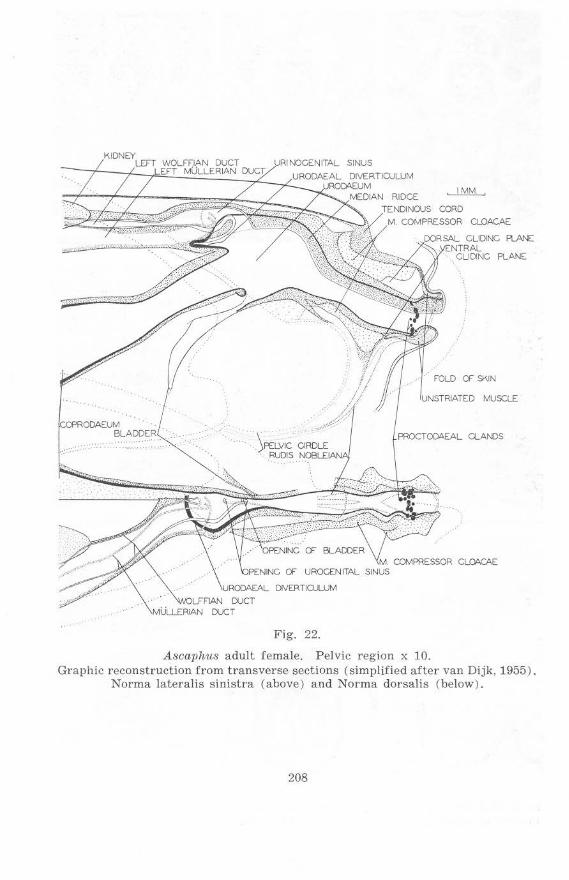
Fig. 22.
Ascaphus adult female. Pelvic region x 10. Graphic reconstruction f rom transverse sections (simplified after van Dijk, 1955).
Norma lateralis sinistra (above) and Norma dorsalis (below).
208
PELVIC
"CO<;
KIDNEY
WQI..InAN DUCT
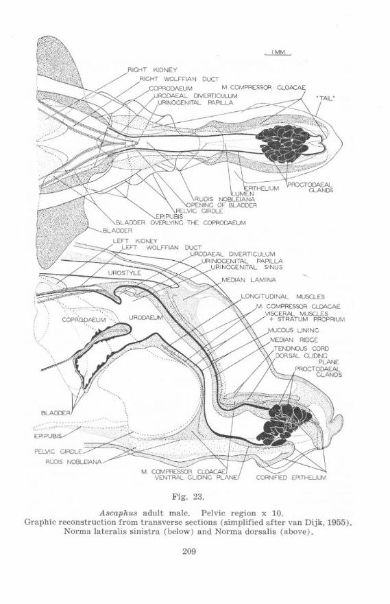
Fig. 23.
A8caphu8 adult male. Pelvic region x 10. Graphic reconstruction from transverse sections (simpl ified after van Dijk, 1955).
Norma lateralis sinistra (below) and Norma dorsalis (above).
209
to the ventrolateral edge of the 10th spinal myomere at Stages 62- 63 and 63. The nephric ducts shift dorsally during development as a consequence of a similar movement of the gut in this region.
UrOOaeal Diverticulum: At Stage 62-63 a slight anterodorsal diverticulum has developed anterior to the excretory aperture, and this develops slowly in subsequent stages (figs. 16, 18, 20; compare figs. 21 : 3 and 21 : 4 ).
Bladder: The anlage of the bladder is present in Stage 51 as a thickening of the ventral wall of the urodaeum, joining the latter to the floor of the coelomic cavity (figs. 5 and 6:1- 6:3). It does not involve endoderm (gut epithelium) at this stage. At Stage 56 (figs. 8. 9, and 10:3- 10:6) the bladder has developed a lumen, this presumably bei ng a diverticulum of the urodaeum (and hence presumably lined with endoderm) projecting into the previously solid mesodermal anlage. Anteriorly the bladder is free from the gut dorsal to it, and from the floor of the coelom ventral to it, and is externally and internally bifid with anterodorsally directed horns (figs. 9 and 10:3- 10:5). Further posteriorly the bladder is attached to thc floor and side-walls of the coelom by membranes (figs. 10:3- 10:6). The bladder retains its eonnexions with the coelomic walls while it increases in size in subsequent stages. At Stages 58 the lumen of the bladder begins to show convoluted outlines anteriorly (fig . 12 : 2), by stage 64 the anterior region of the bladder shows many e pithelial folds.
Structures of Me80dermal Origin
Coelomic Cavity: At Stage 47 the coelom does not extend dorsally as far posterior as the level of the aperture of the nephric ducts (fig. 1), while laterally it extends poste riorly a short distance beyond the anterior margins of the hind-limb buds (fig. 2). At Stage -+- 50 the relations of the coelom to the tissue or the hind-limb buds is similar. The coelom may be seen to extend posteriorly to a level not far from the posteroventral edge of the 9th spinal myomere (fig. 3). At Stage 51 thc coelom reaches to the level of the bases of the hind-limb buds and extends con::liderably posterior to the level of the aperture of the nephric ducts, reaching to the posteroventral edge of the 10th spinal myomere (figs. 5 a nd ti: 5). The coelom extends furthe r posteriorly at Stage 56. reaching a level well within the region of the bases of the hind Jimbs and slightly beyond the posteroventral margin of the 10th spinal myomere (figs. 8, 9, 10 : 9 and 10 : 10). At StaITe 58 the posterior edge of the coelom is still approximately level with the posteroventral margin of this myomere, at Stages 62-63 and 63 it has moved posteriorly half-way towards the posteroventral margin of the 11th spinal myomere and has come to correspond in extent wi t h the posterior limit of the pelvic gi rdle. At Stage 51 the coelom docs not extend far into the tissue between the series of spinal myomeres of the two sides; at Stage 56 it extends approximately 2/5 of the way from the ventral limits of these myomeres to the vertebral column (ct. figs. 5 and 8. and 6:1- 6:5 and 10:1-10:10). In late r stages the coelom extends somewhat fU rther upwards. Lateral to the spinal myomeres the posterior limit of the eoelom lies close to, and nearly parallel with, the late ral margins of t he myocomma between the 8th and 9th spinal myomeres, anterior to th em at Stage ± 50, posterior to them at Stage 56 (cf. figs . 3 and 8).
211
SI)lanclmic lind Somatic Mesoderm : Splanchnic and somatic mesoderm are continuous with each other at the posterior limit of the coelom and a sharp distinction cannot be made betwccn them. Nevertheless examination of sections close to the posterior limit of the coelom (fig. 15) does reveal that their individuality might be rcpresented in the muscle layers a r ising in association with them. Somatic striated muscles laterally arc distinguishable from splanchnic unstriated muscles mediad to them: whethcl' this distinction is valid in the post-coelomic tissue, which contains both these types of muscles, is uncertain.
Viscera l lUuscle Layers:
Ctrcular Muscle Layer: Posterior to the bladder in Stage 51 the circular muscle layer of the gut is complete for a short distance; further postcrior it is incomplete ventrally although present dorsally almost as far caudad as the point at which the limb buds project from the abdomen (figs. 5, 6: 5 and 6: 6). Ventrally, laterally and dorsally the circular muscles extend into the tissue behind the coclom. At Stagc 56. and subsequcnt stages, the relations arc little changed (figs. 10:6- 10:13); the circular musclc layer is still complete behind the coelom although the latter extends further posterior than in earlier stages; the dorsa l part of the circular muscle layer still extends to approximately the same level.
Longitudinal Muscle Layer: The longitudinal muscle layer of the gut is not as distinct as the circular layer at any level of any s tage. This is partiy due to the circular muscle lay<'r being more easily distinguished by virtue of its arrangement being more easily detected in transverse sections: but the longitudinal layer does seem to be less developed. particularly posteriorly. The longitudinal muscle laycr, in the early stages, is distinguished also from the coelomic epithelium only with difficulty. while posterior to the coelom it is often so close to the mm. compressorcs cloacae as to be difficult to detect. At Stage 56 the longitudinal muscle layer cannot be detected with certainty posterior to the coelom. At Stage 58 the longitudinal muscle layer is detectable dorsolateral to the gut (see figs. 12: 11 and 12: 12) almost to the level of the posterior tip of the ventral flap. At Stages 60 and 62- 63 the fibres are detectable dorsolateral to the gut as far posterior as just anterior to the c loacal aperture (figs. 13 and 14: 3, cf. 14 : 4: 16 and 17 : 2, cf. 17 : 3). At Stages 63 and 64 , particul arly the former, there arc suggestions of the longitudinal layer extending further ventrally at the corresponding levels. The situation is complicated in these stages by increasing development of a gliding plane dorsal to the cloaca and apparently between the circular and longitudinal muscle layers. At Stage 58 there are paired concentrations of longitudinal muscle fibres between the gut and the pelvic girdle near the posterior edge of the latter (figs. 12: 8 and 12: 9). At the same level longitudinal muscle fibres turn I>osterodorsally dorsolateral to the gut and approach the vertebral column. These concentrations of longitudinal muscle fibres dorsal and ventral to the gut become better developed in subsequent stages until in Stage 64 it is seen that a substantial proportion of the longitudinal fibres attach to the urostyle above and the pelvic girdle below (cf. figs. 22 and 23). At Stage 64 the longitudinal muscle layer ventral
212
t.o the gut forms pai red concentrations as in earlier stages, but these combine posteriorly to form a single dense cord triangular in cross-section (apex downwards) before attaching t.o t.he pelvic girdle (figs. 21: 6 and 21: 9). There arc only slight indications of the paired origin of this cord except at the pOint of attachment to the girdle. The cords of longitudinal muscle fibres passing posterodorsally to the urostyle appear to give way further posteriorly to fibres passing anterodorsally from the post-pelvic gut (cf. figs. 17 : 1 and 17 :2: 21:7-21:9).
Anlagen of Pelvic Limbs and Gir(lIc:
Pelvic LAmbs: The anlagen of the hind limbs develop ventral to the spinal myomeres on either side of the gut, and anteriorly overlap the coelom sl ightly (fig. 2, Stage 47). The anlagen bear similar relations to the gut and the coelom at Stage ± 50. At this stage the anlagen may be seen to be closely associated with the ventral limits of the 9th and 10th spinal myomeres. being just beneath the 10th and a caudad projection of the 9th myomere. Lateral to these spinal myomeres a dorsolateral projcction of each anlage, representing the region of thc i1ial wing, may be seen on cither side. Hindlimb buds are present as posterior projections of the hind-limb anlagen from the posteroventral aspect of the abdomen. The hind-limb buds are covered ventra ll y by the ventral skin flap (figs. 3 and 4 : 2). At Stage 51 the limb anlagen are no longcr associated with the 9th spinal myomeres, as the caudad pt'ojections of the latter have disappeared. The dorsal limits of the bascs of the hind·limb buds are at the level of the ventral margins of the myocommata bet.ween 10th and 11th spinal myomeres (fig. 5). At Stage 56 the base of the hind 11mb of each side reaches the ventral aSI}(lct of the 11th spinal myomere posterodorsally (fig. 8). The m. pyriformis a nd the m. caudalipuboischiotibialis project into the limb posterior to the ventral margin of the myocomma between the 10th and 11th Sl)inal myome res.
Pelvic Girdle: The pelvic girdle develops from paired chondrifications in a sheet of dcnse connective tissue which extends between and over the ventral surfaces of the spinal myomere!!, just anterior to the point at which the gut passes posteroventrally through this sheet (figs. 3, 4 : 1 and 4 : 2). Extensions of this sheet laterally constitute the anlagen of the ilia and their muscles (Stage ± 50. figs. 3 and 4: 2; Stage 51, figs. 5 and 6: 4). Paired chondrificat ions develop in this connective tissue and approach each other at Stage 56, the gut being lifted above the region of their approximation figs. 8, 9 and 10 :3- 10 :7 ) . By Stage 58 the halvcs of the pelvic gi rdle are separated posteriorly only by a narrow layer of dense connective tissue, while anteriorly they are still well separatcd (figs. 12: 8 and 12: 3). By Stage 60 the halves of the pelvis have fused posteriorly. The anlagen of the iliac regions of the pelvic girdle are directed towards the anterior tips of the lateral margins of the myocommata between the 10th and 11th spinal myomeres. This is seen at Stage + 50 (fig. 3) and Stage 51 (rig. 5). When the pelvic anlagen have chondrified (Stage 56) the cartilaginous ilia lic nearly horizontally (in the transverse plane) with the ends turning dorsally (figs. 8, 9, 10 : 6 and 10 : 7 ). Dense connective tissue continues from the tip of each ilium towards the posterior bay in the lateral face of the
213
10th spinal myomere. At Stage 58 this bay is approached by the cartilaginous ilium, while the dense connective tissue at the tip of the ilium extends into the myocomma bctwecn the 10th and 11th spinal myomeres in a notch in this bay (fig. 11). The ilium is somewhat closer to the notch, which has deepened, at Stage 60 (fig. 13); while at Stage 62-63 the ilium enters the further enlarged notch (fig. 16). The ilia take in progressively more nearly ventro-dorsal positions (in the transverse plane) by addition laterally of dorsally directed portions, and by reduction in width and ventral exten t of the spinal myomeres separating the ventral parts of the ilia (cf. figs. 10: 7, 12: 5, 21: 2 and 21 : 3). From a condition where the ilia and the ventral parts of the pelvic girdle lie in the same transverse plane (Stage 56. figs . 8 and 9) there is progressive change in the rela.tive anterior-posterior positions of the ilia and the ventral parts of the girdle . The ilia rolate anteriorly as they penetrate between the 10th and 11th spinal myomeres while the anterior limit of the ventral parts of thc girdle remains more or less constant with r eference to the spinal myomeres, the posterior limit, however, steadily extends backward from Stage 62- 63 to Stage 64 (figs. 16, 18 and 20) .
Epipubis: Paired chondrifieations develop in the connective tissue anterior to the halves of the pelvic girdle, and arc fused to the latter at Sta.';e 58 (figs. 11 and 12:1-12 :4). These cartilages are halves of the epipubis and have fused together anteriorly by Stage 60 (fiIT . 14: 2) . Incomplete fusion of the halves of the epipubis posteriorly rcsults in the presence of a posterior notch in the cpipubis of the adult (cf. Stage 64, figs. 21 : 16 and 21 : 17 with 21 : 1 ). The state of the epipubis anlagen at earlier stages (Stage 56) is best considered with the epipubic muscles.
Rods 0/ Noble and lnter/emoral Ligament: At Stage ± 50 strands of dense connective tissue extend from the ventromedial edgcs of the hind-limb buds along the sides of the gut close to the ventral flap (fig. 4 : 2). At Stage 51 the same situation is observed, the dense connective tissue disappearing posteriorly into less dense tissue lateral to the gut which extends dorsally to the posterior limits of the bases of the hind-limb buds (fig. 6 : 7). At Stage 56 the strands have developed into the rods of Noble, extending posteriorly from the ventromedial edges of the halves of the pelvic girdle to curve somewhat dorsally on either side of the gut behind the pelvic girdlc (figs. 8, 9 and 10 :6-10 :16) . A pair of posterodorsalIy directed cords of connective tissue above the gut ends close to the tips of the rods of Noble. These cords constitute the beginnings of a dorsal interfemoral ligament. The posterior tips of the rods of Noble and the dorsal interfemoral cords are enclosed in thickenings on either side of the gut. Posterior to this the gut is thin-walled (figs. 10:13-10 :16). At Stage 58 this thin-walled part of the gut has disappeared and the rods of Noble and the dorsal interfemoral cords end in the rounded cloacal lips (figs. 11 and 12:15- 12:18). The rods of Noble show little change in relation to neighbouring structures in subsequent stages. The anterior tips of the rods at Stage 60 end in a layer of connective tissue which stretches to the ventral surface of the epipubis. This tissue becomes progressively more distinct and at Stage 64 forms a well-marked tendinous sheet (ef. figs. 11, 13, 16, 18, 20 and 21:2- 21:6). The dorsal interfemoral ligament at Stage 58 has attained a condition which remains up
214
to Stage 63. At Stage 64 the ligament slopes somewhat anterodorsally from the cloaca, whereas in Stages 58, 60, 62- 63 and 63 the direction is more nearly vertical with the dorsal end turned posterodorsally (figs. 11 and 12 :15- 12:18; 13; 16 and 17 :4- 17:6; 18 and 19:2-19:4; 20 and 21:11 -21:15). The ligament is a median structure ventrally, but is paired dorsally with the arms passing on either side of the caudal vein.
Somatic j\luscles:
Abdominal Muscle Cords (Rectus Muscles): At Stage 47 the abdominal musculature is represented by two cords which separate from the ventral surfaces of the rnyomcres of the tail (fig. 1) and pass anteroventrally and somewhat laterally. These muscle cords pass over the anterodorsal edges of the hind-limb anlagen (fig. 2), between these and the coelom. Reduction of yolk in the abdominal region results in a more cephalad and less ventrad direction of the abdominal muscle cords, as at Stage -+- 50. The abdominal muscle cord of each side is clearly segmented at Stage -+- 50 and the posterior-most segment is applied to thc 9th spinal myomere (fig_ 3). A small separate element is present on the inner face of the last abdominal myomere (9th, as it corresponds to the 9th spinal myomere) near its posterior end (figs. 3 and 4 : 1 ). There is a small gap between the posterior tip of the last (9th ) abdominal myomere and the anterior tip of the hind-limb anlage (fig. 3). At Stage 51 the abdominal cords do not reach dorsally to the 9th spinal myomeres, but curve around the ventral surfaces of the 10th spinal myomeres and pass up the medial faces of these myomeres and the myomeres behind them. (The posterior limits of the cords could not be distinguished.) The 10th and 11th spinal myomeres are ventrally indented by the abdominal cords (figs. 5 and 6). Segmentation of the abdominal muscle cords is clearly shown at Stage 52, the relation of the abdominal myomeres to corresponding spinal myomeres also being possible (cf. description of Stac:e 52). The 1st abdominal myomere is absent, the 2nd rudimentary; the 9th meets the spinal myomeres. The n . iliohypogastricus, derived from the 8th spinal nerve and used for identifying myomeres, passes into the myocomma between the 8th and 9th abdominal myomeres. This situation is also found in Stage 56 (fig. 8), the 8th abdominal myomeres forming two broad ventral sheets separated by connective tissue (linea alba) while the 9th abdominal myomeres reach up to the ventral surfaces of the 10th spinal myomeres and curve medially round them. Slight ventral notching of the 10th spinal myomere is visible. (See figs. 9 and 10 :1- 10:6). Just anterior to the ventromedial edge of the myocomma between the 10th and 11th spinal myomeres of each side, the 10th abdominal myomere begins and stretches over the medial faces of the 10th, 11th and 12th spinal myomeres, just reaching to the 13th (figs. 8, 9 and 10, particularly 10 : 16). The 10th abdominal myomere is apparently the last, but the possibility must be borne in mind that this late-developing segment may derive material from more than one metamere without a myocomma developing between the fibres derived from each. At Stage 58 the 9th and 10th (? + ) abdominal myomeres of each side have degenerated, the latter to such an extent that muscle fibres are no longer dlstinguishable. The remnants of these myomeres are associated with the medial aspect of the
215
ventrolateral edge of the 10th spinal myomere in much the same position as that occupied by the 9th abdominal myomere at Stage 56 (figs. 11 and 12:1- 12 :5). At Stage 60 the dorsal part of the degenerated myomeres is absent and the remnants still present do not reach as far as to the spinal myomeres (fig. 13). In subsequent stages these abdominal myomeres are absent. At Stage 58 each 8th abdominal myomere attaches to the epipubie cartilage of that side (figs. 11 and 12 : 1) ; at subsequent stages these myomeres approach each other more closely anterior to the epipubis, but show little othcr change in their relations to the latter (figs. 14 : 1 and 14 : 2; 21: 16 and 21 : 17). At Stage 58 a superficial layer of secondary muscle develops on the ventral surface of the abdominal myomeres. The fibres arc distinguishable by their more basophilic staining properties, characteristic of undifferentiated muscle, and by the oblique direction of the fibres in transverse sections (fig. 12: 1). There is increasing development of this superficial layer in subsequent stages (figs. 14: 1 and 14 : 2; 21: 16 and 21: 17).
ObUquus and Transversus Muscles.- At Stage 58, while the 9th and 10th abdominal myomeres degenerate, differentiation of the obliquus and transversus muscles begins between the remaining abdominal myomeres ventrally and the spinal myomcres dorsally (ilg. 12 : 1). There are no apparent indications of the origin of the obliquus and transversus muscles from either the remaining abdominal or the spinal myomeres.
Epipubic ~l1tscles and Anlagen of the Epipubis.- At Stage 56 the mm. epipubici may be identified as two widely-separated myogenic masscs which anteriorly are in contact with thc ventromedial edges of the 9th abdominal myomeres, extending along these as far as the myocommata between these myomeres and the 8th abdominal myomcres. Ventral to these developing muscles are the more diffuse anlagen of the epipubis, and bencath these, shects of connective tissue (fi gs. 8, 9 and 10:1-10:4). The anlagen of the epipubis are posteriorly separate from the halves of the pelvic girdle, being connected to them by thin vertical strands of connective tissue (fig. 10 : 4); the mm. epipubici posteriorly reach to the pelvic girdle. The anlagen of thc epipubis are derivatives of the connective tissue between and beneath the 9th abdominal myomeres, the two anlagen being separate from one another as well as from the pelvic girdle (as mentioned above). The connective tissue in which these anlagen arise might possibly be considered as linea alba although the fibres continue laterally around the abdominal myomeres and are not confined betwecn them. The rclations of mm . epipubici show that they should be regarded as secondary derivatives of the abdominal myomeres, either the 8th or 9th or both.
Femorococcygeal and Crurococcygeal Muscles.- The tissue from which these muscles (mm. pyriformes and mm. caudalipuboischiotibiales respectively) arise is that at the posterior limit of the hind-limb bases (fig . 6: 6). This tissue is associated with the medial faces of the abdominal muscle cords, where these lie on the anterior tips of the medioventral faces of the 11th spinal myomeres (Stage 51, fig. 5). At Stage 56, whcn the muscles are well differentiatcd, the same relations are preserved. It is seen at this stage that the part of the abdominal muscle cord with which the muscles of each side are associated is the 10th (?+) abdominal myomere, nearer its posterior end (figs. 8, 9 and 10:9-10:12) . At Stage 58 the degeneration of the
216
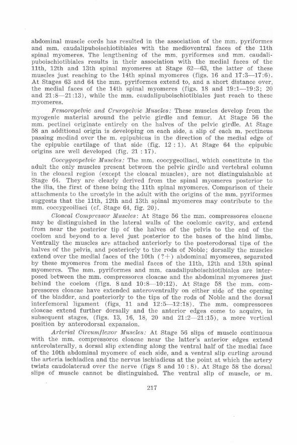
abdominal muscle cords has resulted in the association of the mm. pyriformes and mm. eaudalipuboischiotibiales with the medioventral faces of thc 11th spinal myomeres. The lengthening of the mm . pyriformes and mm. caudalipuboischiotibiales results in their association with the mcdial faces of the 11th, 12th and 13th spinal myomeres at Stage 62- 63, the latter of these muscles just r eaching to the 14th spinal myomeres (figs. 16 and 17:3- 17:6) . At Stages 63 and 64 the mm. pyriformes extend to, and a short distance over, the medial faces of the 14th spinal myomeres (figs. 18 and 19 :1-19:3; 20 and 21 :8- 21 :13 ), while the mm . caudalipuboischiotibiales just reach to these myomeres.
Ji'emoropelvic and Cruropelvic Muscles: These muscles develop from the myogenic material around the pelvic girdle and femur. At Stage 56 the mm. pectinei originate entirely on the halves of the pelvic girdle. At Stage 58 an additional origin is developing on each side, a slip of each m. pectineus passing mediad over the m. epipubicus in the direction of the medial edge of the cpipubic cartilage of that side (fig. 12: 1). At Stage 64 the epipubic origins are well dcveloped (fig. 21 : 17 ).
Coccygcopelvic Muscles : The mm. coccygeoiliaci, which constitute in the adult the only muscles present between the pelvic girdle and vertebral column in the cloacal region (except the cloacal muscles), are not distinguishable at Stage 64. They are clearly derived from the spinal myomeres posterior to the ilia, the first of thcse being the 11th spinal myomeres. Comparison of their attachments to the urostyle in the adult with thc origins of the mm. pyriformes suggests that the 11th, 12th and 13th spinaL myomeres may contribute to the mm. coccygeoiliaci (cf. Stage 64, fig. 20).
Cloacal Compressor Muscles: At Stage 56 the mm. compressores cloacae may be distinguished in the lateral walls of the coelomic cavity, and extend from near the posterior tip of the halves of the pelvis to the end of the coelom and beyond to a level just posterior to the bases of the hind limbs. Ventrally the muscles are attached anteriorly to the posterodorsal tips of the halves of the pelvis, and posteriorly to the rods of Noble; dorsally the muscles extend over the medial faces of the 10th (? + ) abdominal myomeres, separated by these myomeres from the medial faces of the 11th, 12th and 13th spinal myomeres. The mm. pyriformes and mm . caudalipuboischiotibiaics are interposed between the mm. compressores cloacae and the abdominal myomeres just behind the coelom (figs. Sand 10:8-10:12). At Stage 58 the mm. compressores cloacae have extended anteroventrally on either side of the opening of the bladder, and posteriorly to the tips of the rods of Noble and the dorsal interfemoral ligament (figs. 11 and 12 :5---12:18). The mm . compressores cloacae extend further dorsally and the anterior edges come to acquire, in subsequent stages, (figs. 13, 16, 18, 20 and 21 :2-21 :15), a more vertical position by antcrodorsal expansion.
Arterial Circum/lexor Muscles: At Stage 56 slips of muscle continuous with the mm . compressoroo cloacae near the latter's anterior edges extend anterolaterally, a dorsal slip extending along the ventral half of the medial face of the 10th abdominal myomere of each side, and a ventral slip curling around the arteria ischiadica and the nervus ischiadicus at the point at which the artery twists caudolaterad over the nerve (figs 8 and 10 : 8). At Stage 58 the dorsal slips of muscle cannot be distinguished. The ventral slip of muscle, or m.
217
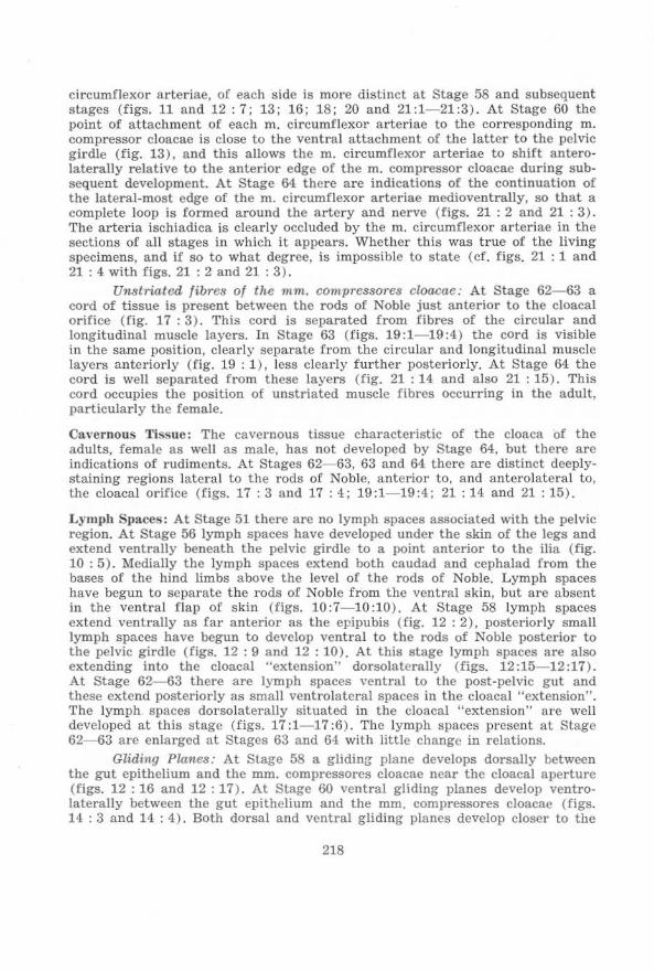
circumfiexor arteriae, of each side Is more distinct at Stage 58 and subsequent stages (figs. 11 and 12 : 7; 13; 16; 18; 20 and 21:1- 21:3). At Stage 60 the point of attachment of each m. circumflexor arteriae to the corresponding m. compressor cloacae is close to the ventral attachment of the latter to the pelvic girdle (fig. 13). and this allows the m. circumfiexor a rteriae to shift anterolaterally relative to the anterior edge of the m. compressor cloacae during subsequent development. At Stage 64 there are indications of the continuation of the lateral-most edge of the m. circumfiexor arteriae medioventrally, so that a complete loop Is formed around the artery and nerve (figs. 21 : 2 and 21 : 3) . The arteria ischiadica is clearly occluded by the m. circum rIexor arteriae in t he sections of all stages in which it appears. Whether this was true of the living specimens, and if so to what degree, is impossible to state (cf. figs, 21 : 1 and 21: 4 with figs. 21 : 2 and 21 : 3).
Unstriated fibres of the mm. compressores cloacae: At Stage 62- 63 a cord of tissue is present between the rods of Noble just anterior to the cloacal orifice (fig. 17 : 3). This co rd is separated from fibres of the circular and longitudinal muscle layers. In Stage 63 (figs. 19:1- 19:4) the cOrd is visible in the same position, clearly separate from the circular and longitudinal muscle layers anteriorly (fig. 19 : 1), less clearly further posteriorly. At Stage 64 the cord is well separated from these layers (fig. 21 : 14 and also 21 : 15). This cord occupies the position of unstriated muscle fibres occurring in the adult, particularly the female.
Clwernous Tissue: The cavernous tissue characteristic of the cloaca of the adults, female as wel1 as male, has not developed by Stage 64, but there arc indications of rudiments. At Stages 62- 63, 63 and 64 there are distinct deeplystaining regions lateral to the rods of Noble, anterior to, and anterolateral to, the cloacal orifice (figs. 17: 3 and 17: 4; 19:1- 19:4; 21: 14 and 21: 15).
Lympb Silaces: At Stage 51 there a re no lymph spaces associated with the pelvic region. At Stage 56 lymph spaces have developed under the skin of the legs and extend ventral1y beneath the pelvic gird.le to a point anterior to the ilia (fig. 10: 5). Medially the lymph spaces extend both caudad and cephalad from the bases of the hind limbs above the level of the rods of Noble. Lymph s paces have begun to separate the rods of Noble from the ventral skin, but are absent in the ventral flap of skin (figs. 10:7-10:10). At Stage 58 lymph spaces extend ventrally as far anterior as the epipubis (fig. 12 : 2), posteriorly small lymph spaces have begun to develop ventral to the rods of Noble posterior to the pelvic girdle (figs. 12 : 9 and 12 : 10). At this stage lymph spaces are also extending into the cloacal "extens ion" dorsolateral1y (figs. 12:15- 12:17). At Stage 62-63 there arc lymph spaces ventral to the post-pelvic gut and these extend posteriorly as small ventrolateral spaces in the cloacal "extension". The lymph spaces dorsolateral1y s ituated in the cloacal "extension" are well developed at this stage (figs. 17:1- 17:6). The lymph spaces present at Stage 62-63 are enlarged at Stages 63 and 64 with little change in relations.
Gliding Planes,' At Stage 58 a glidinG' plane develops dorsally between the gut epithelium and the mm. compressores cloacae near the cloacal aperture (figs. 12 : 16 and 12: 17). At Stage 60 ventral gliding planes develop ventrolaterally between the gut epithelium and the mm. compressores cloacae (figs. 14 : 3 and 14 : 4). Both dorsal and ventral gliding planes develop closer to the
218
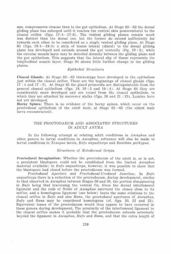
mm. compressores cloacae than to the gut epithelium. At Stage 62-63 the dorsal gliding plane has en la rged until it r eaches the ventral skin posterolateral to the cloacal orifice (figs. 17:4- 17:6). The ventral gliding planes remain much less di stinct than the dorsal one, but the former do extend sufficiently far towards each other to be considered as a single ventral gliding plane. At Stage 63 (figs . 19:1- 19 :4) a strip of tissue lateral (distal) to the dorsal gliding plane has developed and cxtends around the gut ventrally (fig-. 19 : 3), while the circular muscle layer may be detccted dorsally between the gliding plane and the gut epithelium. This suggests that the lateral slip of tissue represents the long itudina l muscle layer. Stage 6-1 shows li ttle further change in the glid ing planes.
Epithelial Stmctures
Cloacal Glands: At Stage 62- 63 thickenings have developed in the epithelium just within the cloacal orifice. These are the beginnings of cloacal glands (figs. 17:" and 17 : 5). At Stage 63 the gland primordia arc distinguishable fro m the general cloacal epithelium (figs. 18, 19 : 3 and 19 : 4). At Stage 64 they are considerably more developed and a rc raised from the cloacal epithelium, to which they are attached by narrower stalks (figs. 20 and 21 : 15). Lumina have not yet developed. lIor n~' SI)ines: There is no evidence of the horny spines. which occur on the proctodaeal epithelium of t he adult male, at Stage 62- 63 (the oldest male larva reconstructed).
THE PROCTODAEUM AND ASSOCIATED STRUCTURES IN ADULT ANURA
1n the foilowing attempt at relating adult conditions in Ascaphus and other genera to larval conditions in Ascaphus, reference will also be made to larval conditions in Xenopus laevis, Bnlo angus/icC1)S and Bombina pachypus.
Structtlres 01 Ectodermal Origin
l'I'octodaeal Illvag inat ion : Whet her the proctodaeum of the adult is, or is not, a persistent blastopore could not be established from the limited Ascaphus material available; in B'~/o allgllsticeps, however, it was possible to show that the blastopore had closed before the proctodaeum was fo rmed.
Proctodaeal Aperture alld Proctodaeal-Urodaeal Junction: In Bulo allgusticeps there is a reduction of the proctodaeum, durinG' development, similar to that observed in AscapkuIJ between Stages 56 and 58, the portion d isappeari ng in Bulo being that traversing the ventral fin. Since the dorsal interfemoral ligament and the rods of Noble of Asca-phus surround the cloaca close to its orifice, and a homologous ligament (sec below) bears the same relations t o the cloacal orifice in Bulo and also Rana, the proctodaeal apertures of Ascaphus, Btllo and Rana may be considered homologous (cf. figs. 20, 22 and 23). Equivalent losses of the I)roctodaeum would thus appear to have occurred in these genera during development, The proximity of the interCemoral ligament to the cloacal or ifice makes it probable that the proctodaeu m extends anteriorly beyond the ligament in Ascaphlls, Bn/o and Rana, and that the extra length of
219
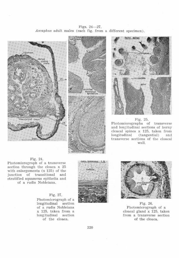
Figs. 24--27. AscapJIIIs adult males (each fig. from a different s lX!cimen).
Fig. 24. Photomicrorrraph of a transverse section through the cloaca x 25 with enlargements (x 125) o r the junction of transitional and stratified squamous epithelia and
of a rudis Nobleiana .
Fig. 27. Photomicrograph of a longitudinal section of a rudis Noblciana x 125, taken from a longitudinal section
of the cloaca.
220
Fig. 25. Photomicrographs of transverse and lon[:, itudinal sections of horny cloacal spines x 125, taken from longitudinal (tangential) and transvcrse sections of the cloacal
wall.
Fig. 26. Photomicrograph of a
cloacal g land x 125, taken from a transverse section
of the cloacn.
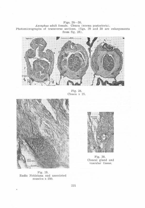
Figs. 28- 30. Ascaphus adult female. Cloaca (norma posterioris).
Photomicrographs of transverse sections. (figs. 29 and 30 are enlargements from fig. 28).
Fig. 19. Rudis Nobleiana and associated
muscles x 250.
Fig. 28. Cloaca x 25.
221
Fig. 30. Cloacal gland a nd
vascular tissue.
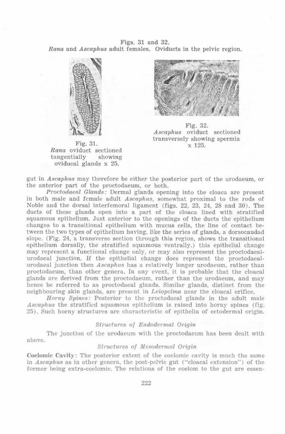
Figs. 31 and 32. Rana and Ascaphus adult females. Oviducts in the pelvic region.
Fig. 31. Rana oviduct sectioncd tangentially showing
oviducal glands x 25.
Fig. 32. Ascaphus oviduct sectioned transversely showing spermia
x 125.
gut in A scaphu8 may thcrefore be either the posterior part of the urodaeum, or the antcrior part of the proctodaeum, or both.
ProctodaeaZ Glands: Dermal g lands opening into the cloaca are present in both male and female adult Ascaphl18, somewhat proximal to the rods of Noble and the dorsal interremoral ligament (figs. 22, 23, 24, 28 and 30). The ducts of these glands open into a part of the cloaca lined with stratified squamous epithelium, Just anterior to thc openings of the ducts the epithelium changes to a transitional epithelium with mucus cells, the line of contact betwcen the two types of epithelium having, like the series of glands, a dorsocaudad slope, (Fig. 24, a transverse section through this region, shows the transitional epithelium dorsally, the stratified squamous ventrally.) this epithelial change may represent a functional change only, or may also represent the proctodacalurodaeal junction. If the epithelial change does represent the proctodaealurodaeal junction then AsCa1Jhus has a relatively longer urodaeum, rather than proctodaeum, than other genera. In any event, it is probable that the cloacal glands are derived from the proctodaeum, rather than the urodaeum, and may hence be referred to as proctodaeal glands_ Similar glands, distinct from the neighbouring skin glands, arc present in Leiopeima near the cloacal orifice.
HONlY Spines: Posterior to the proctodaeal glands in the adult male AscaphU8 the stratified squamous epithelium is raised into horny spines (fig. 25). Such horny structures arc characteri>.:tic of epithelia of ectodermal origin.
Structures oj Endodermal Origin The junction of the urodacum with the proctodaeum has been dealt with
above. Structures oj Mesodermal Origin
Coelomic Cavity: The posterior extent of the coelomic cavity is much the same in Ascaphu8 as in other genera, the post-pelvic gut ("cloacal extension") of the fOrmer being extra-coelomic. The relations of the coelom to the gut are essen-
222
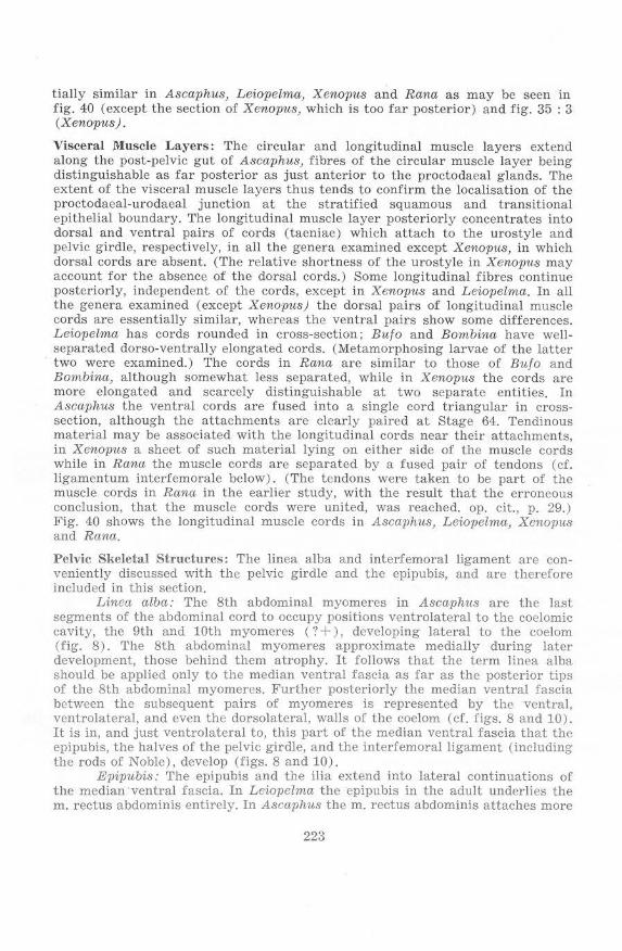
tially similar in Ascaphus, Leiopelma, Xenopus and Rana as may be seen in fig. 40 (except the section of Xeno1nls, which is too far posterior) and fig. 35 : 3 (Xenopus).
Viscera l !'I[uscle La;rers: The circular and longitudinal muscle layers extend along t he post-pelvic gut of Asca1Jhus, fibres of the circular muscle layer being distinguishable as far posterior as just anterior to the proctodaeal glands. 1'he extent of the viscer al muscle layers thus tends to confirm the localisation of the proctodaeal-urodaeal junction at the stratified squamous and transitional epithelial boundary. The longitudinal muscle Jaycr posteriorly concentrates into dorsal and ventral pairs of cords (taeniae) which attach to the urostyle and pelvic girdle, respectively, in all the genera examined except Xenopus, in which dorsal cords are absent. (The relative shortness of the urostyJe in Xenopus may account for the absence of the dorsal cords.) Some longitudinal fibres continue posteriorly, independent of the cords, except in X enopus and Leiopelma. In all the genera examined (except Xenopus) the dorsal pairs of longitudinal muscle cords are essentially similar, whereas the ventral pairs show some differences. Leiopelma has cords rounded in cross-section; Rulo and Bombina have wellseparated dorso-ventrally elongated cords. (Metamorphosing larvae of the latter two wcre examined.) The cords in Rana are similar to thOse of Bulo and Bombina, although somewhat less separated, while in Xenopus the cords are more elongated and scarcely distinguishable at two separate entities. In Asca-phus the ventral cords are fused into a single cord triangular in erosssection, although the attachments are clearly paircd at Stage 64. Tendinous material may be associated with the longitudinal cords near their attachments, in Xenopus a sheet of such material lying on either side of the muscle cords while in Rana the muscle cords are separated by a fused pair of tendons (cf. ligamentum interfemorale below). (The tendons were t aken to be part of the muscle cords in Rana in the earlier study, with the result that the erroneous conclusion, that the muscle cords were united, was reached. op. cit., p. 29.) Fig. 40 shows the longitudinal muscle cords in Ascapitus, Leiopdma, Xenopus and Rana.
Pelvic Skeletal Structures: T he linea alba and interfemoral ligament are conveniently discussed with the pelvic girdle and the epipubis, and arc therefore included in this section.
Linea alba: The 8th abdominal myomeres in Ascaphus are the last segments of the abdominal cord to occupy positions ventrolateral to the coelomic cavity, the 9th and 10th myomeres (? + ), developing lateral to the coelom (fig. 8). The 8th abdominal myomeres approximate medially during later development, those behind them atrophy. It follows that the term linea alba should be applied only to the median ventral fascia as far as the posterior tips of the 8th abdominal myomeres. Further posteriorly the median ventral fasc ia between the subsequent pairs of myomeres is represented by the ventral, ventrolateral, and cven the dorsolateral, walls of the coelom (cf. figs. 8 and 10). It is in, and just ventrolateral to, this part of the median ventral fascia that the epipubis, the halves of the pelvic girdle, and the interfemora! ligament (including the rods of Noble), develop (figs. 8 and 10).
Epipubis: The epipubis and the ilia extend into lateral continuations of the median ventral fascia. In Lciopclma the epipubis in the adult underlies the m. rectus abdominis entirely. In Ascaphus the m. rectus abdominis attaches more
223
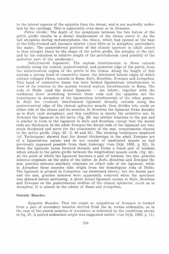
to the lateral aspects of the epipubis than the dorsal, and is not markedlY underlain by the cartilage. This is apparently cvcn more so in Xtmopus.
Pelvic Girdle: The depth of the symphysis between the two halves of the pelvic girdle results in a dorsal displacement of the cloaca above it. As the tail atrophies during metamorphosis, the cloaca, which had opened at the base of the fully-formed tail, becomes shorter (very little so in A8caphu8, particularly the male). The posterodorsal position of the cloacal aperture in adult An1~ra is thus brought about by the shape of the pelvic girdle, the atrophy of the tail. and by the reduction in relative length of the proctodaeum (and possibly of the posterior part of the urodaeum).
lnterlemoral Ligament: The septum interfemorale in Rana extends medially along the ventral, posteroventral, and posterior edge of the pelvis, from the anteroventral region of the pelvis to the cloaca. Along the length of this septum a strong band of connective tissue, the thickened lateral edges of which contain collagen fibres, extends in Rana, Rulo, Bombilla, XenOJlus and Leiopelma. This band of connective tissue has been termed ligamentum inlerfemorale in view of its relation to the septum termed septum interfemorale in Raila. The rods of Noble (and the dorsal ligament - sec below), together with the tendinous sheet stretching between these rods and the epipubis are the homologues in Ascaphus of the ligamentum interfemorale of the other genera. In Bu/o the (ventral) interfemoral ligament dorsally extends along the posteroventral edgc of the cloacal sphinctcr muscle, then dividcs into cords on either side of the cloaca and its muscles. In Bombina the ligament forks dorsally as in Rulo (and Ascaph1t3) , and this condition is clearly the primitive one. In Xenopus the ligament in the larva (fig. 36) has similar relations to the gut and is similar in form to the ligament in Bulo and Ronlbina, except that the dorsal ends are thickened. In the adult Xenopus the dorsal ends of the ligament are very much thickened and serve for the attachment of the mm. compressores cloacae to the pelvic girdle (figs, 35 : 3. 40 and 33). The staining techniques employed (cf. Techniques) showed that the dorsal thickenings in the adult Xenopus arc of a ligamentous nature and do not consist oC unstrinted muscle as had previously appeared possible f rom their histology (van Dijk. 1955, p. 52). In Rana the ligament turns forward dorsally and forms a fused pair of tendons which attach to the pelvic girdle between the longitudinal muscle cords (fig. 40). At the point at which the ligament becomes a pair of tendons, the mm. graciles minores originate on the sides of the latter. In Bit/a, Bombina. and XetWPUS the mm. graciles minores similarly originate on either side of the ligament, while in A8Caphus these muscles ta ke origin from the homologous rods of Noble. The ligament is present in Leiopeima (as mentioned above), but the dorsal part a nd the mm. graciles minores wcre apparently removed when the specimen was ski nned before sectioning. A short dorsal ligament occurs in Bu/c, Bombina and Xenopus on the posterodorsal midline of the cloacal sphincter , much as in A 8caphu8. It is absent in the adults of Rana and Leio11elma.
Somatic Muscles:
Epipubic Muscles: That the s ingle m. epipubicus of Xenopus is formed from a pair of secondary muscles derived from the m. rectus abdominis, as in the cnse of the paired muscles of Ascaphus, is indicated by the conditions shown in fig. 37. A paired abdominal origin was suggested earlier (van Dijk. 1955. p. 11)
224
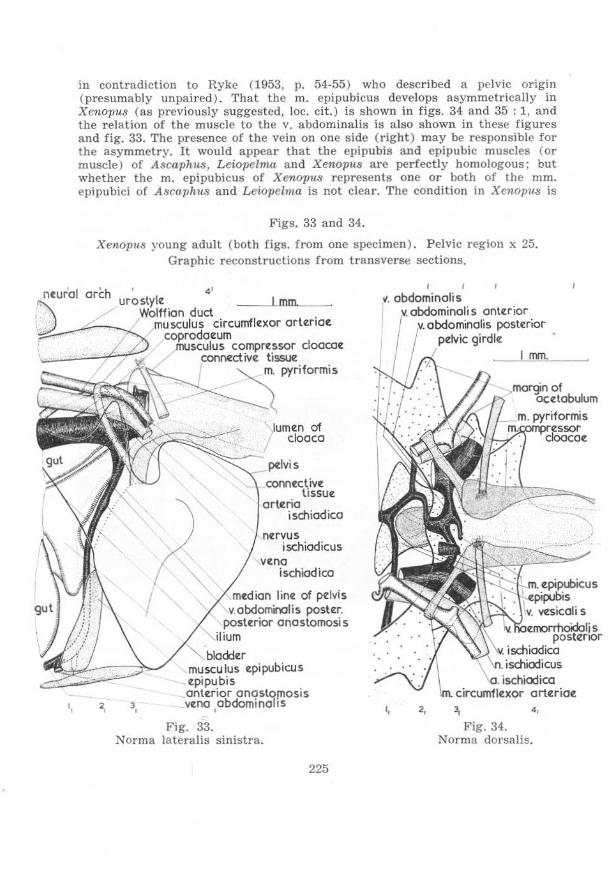
'.
in contradiction to Ryke (1953, p. 54-55) who described a pelvic origin (presumably unpaired). That the m. epipubicus develops asymmetrically in Xenopus (as previously suggested, lac. cit.) is shown in figs. 34. and 35 : 1, and the relation of the muscle to the v. abdominalis is also shown in these figures and fig. 33. The presence of the vein on one side (right) may be responsible for the asymmetry. It would appear that the epipubis and epipubic muscles (or muscle) of Ascaphlls, Leiopelma and Xenopus are perfectly homologous; but whether the m. epipubicus of Xenopus represents one or both of the mm. epipubici of Ascaphus and Le-iopelma is not clear. The condition in Xenopus is
Figs. 33 and 34.
Xenopus young adult (both figs. from one specimen). Pelvic region x 25. Graphic reconstructions from transverse sections.
!
" duct
mu sculus circumfle:xor orte:riae: coprodae:um
rtlJsculus compre:ssor cloacoe: COIlI"Iective tiSSU'Z
m. pyriformis
\lumen of / cloaca
~'o--
COMe:ctive: ti ssue:
orte:ria ischiooico
ne:rvus ischiadicus
ve:na ischiodico
me:dion linez of pelvis v.abdominalis poste:r.
poste:rior anastomosis . ium
musculus e:pipubicus e:pipubis ontuior anastofJIosiS ve:no abdominolts
•
Fig. 33 . Norma lateralis sinistra.
225
0.
onle:rior ,'.'Jtx'o'O~'JIi5 poste:rior
girdle: I mm.
ischiodica circumfle:xor orte:ricx
' . Fig. 34.
Norma dorsalis.
i
Ii

clearly derived by specialization from a condition like that in the Ascaphidac. The presence of small cell-aggregations in Bu/o tadpoles is similar sites to those at which the epipubis anlagen develop in Ascaphus, is noteworthy. In Bombina tadpoles a sUbstantial cord of connective tissue passes from the linea alba to the pelvic girdle, from which it is isolated ventrally, in a position reminescent of t he m. epipubicus of Xenopus. These features of Bu/o and Bombina might indicate that the epipubis was of wide distribution among primitive Anura.
CloacnZ Compressor Muscles: Distinction may be made between three reg ions in these muscles, the anterior-most of which is dealt with separately in the next section under the name of Arterial Circumflcxors. The other two divisions were termed mm. compressores cloacae and m. sphincter ani cloacails by Gaupp (1904) (cL van Dijk, 1955, p. 30). The m. sphincter ani cloacalis comprises that posterior part of the cloacal compressor muscles which is attached neither to urostyle nor pelvic girdle, but forms a complete ring around the cloaca. It is absent in Leiopelma, moderately developed in Rana, somewhat better in Bu/o and very well developed by anterior extension in Xenopus and by posterior extension in Ascaphus (see fig. 33 for Xenopus). The whole post-pelvie part of the cloacal compressor muscles in Ascaphus actually represents the m.
Figs. 35- 37. X enopus (each fig. from a different specimen). Photomicrographs of transverse
sections x 25. Norma posterioris.
Fig. 35. Young adult (same specimen as figs. 33 and 34).
Pelvic region . (The numbers refer to levels in figs . 33 and 34. )
226
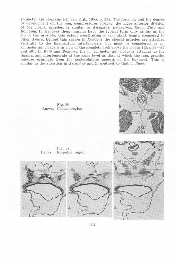
sphincter ani cloacalis (cf. van Dijk, 1955, p. 31). The form of, and the degree of develOI)ment of, the mm. compressores cloacae, the more anterior division of the cloacal muscles, is si milar in Ascaphus, Leiopelma, Ran«, Bulo and Bom.bina, In Xenopi/s these muscles have the typical form only as far as the tip of the urostyle, this extent constituting a very short length compa red to other Atntra. Behind this region in Xenopus the cloaca l muscles are attached ventrally to the ligamentum interfemorale, but must be considered as m. SI)hincter ani cloacalis in view of the complete areh above the cloaca (figs. 33-35 and 40). ]n Bulo and Bombina the m. sphincter ani cloacalis attaches to the ligamentum interfemorale at the same level as that at which the mm. graciles minorcs originate from the posterolateral aspects of the ligament . This is sim ilar to the situation in Ascaphus and in contrast to that in Rana.
I.
Fig. 36. Larva. Cloacal region.
Larva. Fig. 37. Epipubic region.
3. ~ .
227
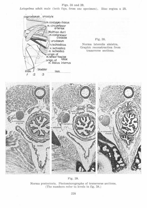
Figs. 38 and 39. L eiopelma adult male (both figs. from one specimen ) . Iliac region x 25.
" , Ic~ostYIe:
'-;;::m. COCC)'g0:>.iliQQJS _..;=:::. ,. m. circunfle:xor
crte:riae: Wolff ian duct
,: .JTI.compre:ssor cloocac
urodae:um n. ischiadicus
\ ___ v. isch iadica ~a. ischiadica
orign of ". ," m. tmsor fasciae: . '. .. or igin of latae: ~ m. iliacus inttlTlJS
12 i3
Fig. 39.
Fig. 38.
Norma lateralis sinistra. Graphic reconstruction from
transverse sections.
Norma posterioris. Photomicrographs of transverse sections. (The numbers refer to levels in fig. 38. )
228
Arterial Oircltmflexor Muscles: At Stage 56 in Ascaphus the arterial cireumflexor muscles are represented on either side by a slip of muscle detaching from the m. compressor cloacae and dividing into ventral and dorsal portions. By Stage 58 the dorsal portion is no longer distinguishable and the m. circumflexor arteriae is represented only by the ventral portion. At Stage 64 each m. circumfJexor arteriae shows a tendency to form a complete loop (cf. fi!js. 21 : 2 and 21 : 3 with 21 : 1 and 21 : 4). In the adult Ascaphlts, and some species of Rana, complete loops are not formed by these muscles. In Xenopu.s, Bombina and Bufo complete loops are formed, as was observed in serial sections (figs. 33, 34 and 35: 3). Dissections of representatives of all the South African anuran families (cf. Materials) revealed that the muscles were present in aU cases, and in every case, including Rana al1golensis, a complete loop appeared to be formed. The muscle is smaU and the dorsal dissection necessary does not expose it s ufficiently to allow the completeness, or otherwise, of the loop to be established with absolute certainty. Sections of Rana grayi showed that the very small m. circumflcxor arteriae of each side forms an almost complete loop. Lciopelma differs from all the other Anura examined in that the m. cireumflexor arteriae of each side has no connexion with the m. compressor cloacae (figs. 38 and 39) and is clearly derived from the dorsal portion of a slip of muscle extending from the m. compressor cloacae at an earlier stage similar to Stage 56 in Ascaphus. In Leiopehna, as in all the other Anura examined, the m. circumflexor arteriae curves around the arteria ischiadica at the point at which this artery twists laterally over the nervus ischiadicus. It is for this reason, and because the muscle is not associated with the m. compressor cloacae in Leiopelma, that the muscle has been termed m. circumflexor arteriae. The occlusion, or partial occlusion, of the a. ischiadica between the n. ischiadicus medioventral to it and the m. circumflexor arteriae dorsolateral to it, seems the only function which contraction of the muscle could perform in Leiopelma and the other Anura. That the neighbouring coccygeal lymph hearts and their ostia may be influenced, as suggested by Gaupp (1904. p. 364), by contraction of the muscle, is unlikely beca.use of variation in relations of these structures to the muscle. In sections the artery is seen to be partially or completely occluded, but this may be a postmortem effect. Dissections of prescrved and of lightly narcotized (ethyl Urethane) specimens showed some constriction of the artery in every case. The relative volumes of blood flowing in the arteriac iliacae of numerous Xenopus adults under light narcosis (ethyl urethane), was studied by injection of flUorescein under ultraviolet light, such injections taking place into the dorsal aorta while it was in a position representing as nearly as possible its natural one (cC. Techniques). One m. circumflexor arteriae was cut after an initial injection, and a second injection then given, in every speCimen. Changes in the blood-flow were frequently traced to slight movements of the lcgs occurring during the second injection, and attempts at staking the animal more firmly tended to stop the circulation to the legs. Consistent results could therefore not be obtained. The technique, however, holds promise.
Unstriated fibres of the 1nfn. compressores cloacae: Unstriated fibres are present betwcen the rods of Noble in the female (and, to a much lesser extcnt. the male ) Ascaphus, and these fibres have the staining reactions of unstriatcd muscle. Since these fibres are closely associated with the mm. compressores cloacae (figs. 28, 29 and 21 : 14) it would seem that they are derived from these muscles. The mm. compressores cloacae, widening. have given risc to
229
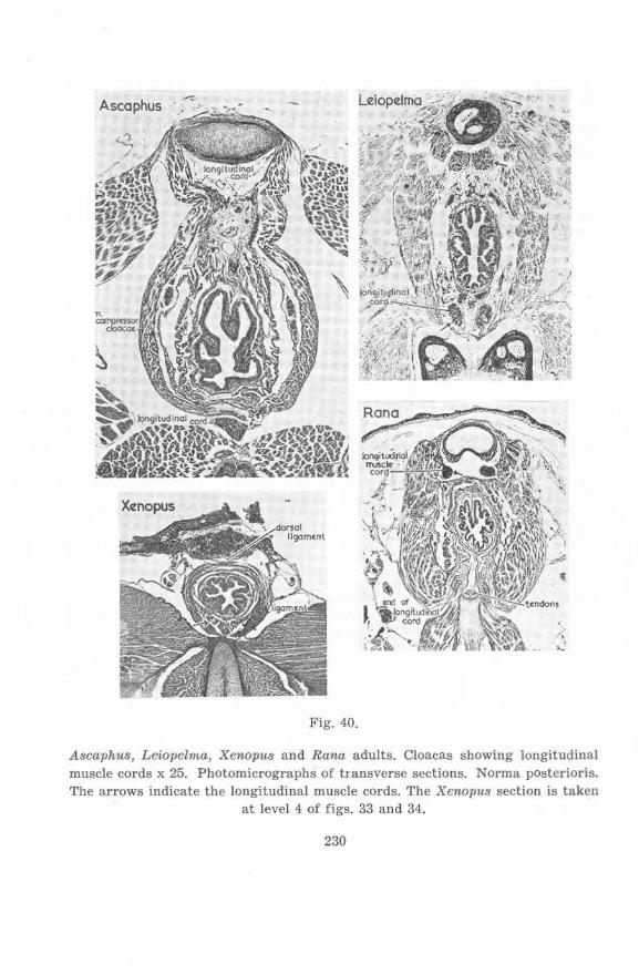
Ascaphus
Fig. 40.
Ascaphus, Leiopelma, Xenopu.s and Rana adults. Cloacas showing longitudinal muscle cords x 25. Photomicrographs of tI ansverse sections. Norma posterioris. The arrows indicate the longitudinal muscle cords. The Xenopus section is taken
at level 4. of figs . 33 and 34..
230
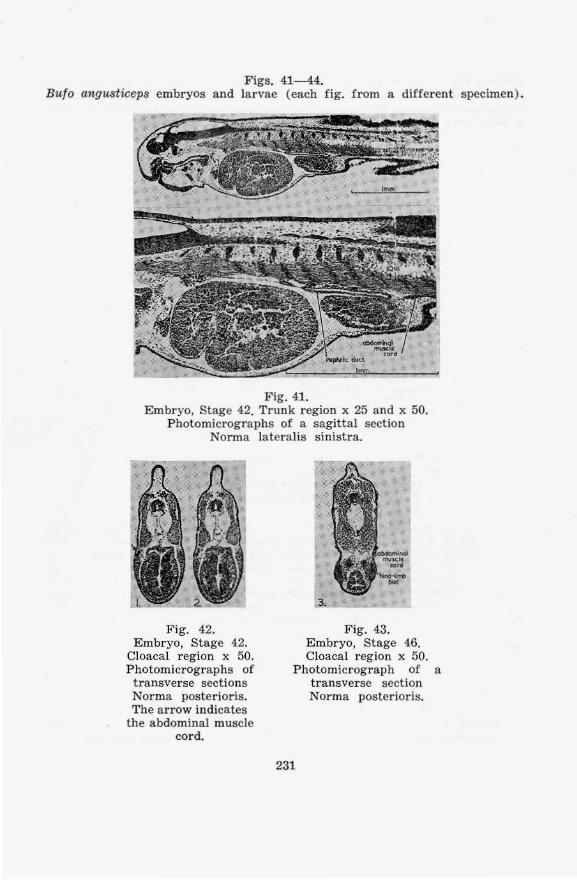
Figs. 41-44. Bufo ang usticeps embryos and la rvae (each fig. from a different specimen ) .
Fig. 41. Embryo, Stage 42. Trunk region x 25 and x 50.
Photomicrographs of a sagittal section Norma lateralis sinistra.
Fig. 42. Embryo, Stage 42.
Cloacal region x 50. Photomicrographs of transverse sections Norma posterioris. The arrow indicates
the abdominal muscle cord.
Fig. 4.3. Embryo, Stage 46. Cloacal region x 50.
Photomicrograph of a transverse section Norma postcrior is.
231
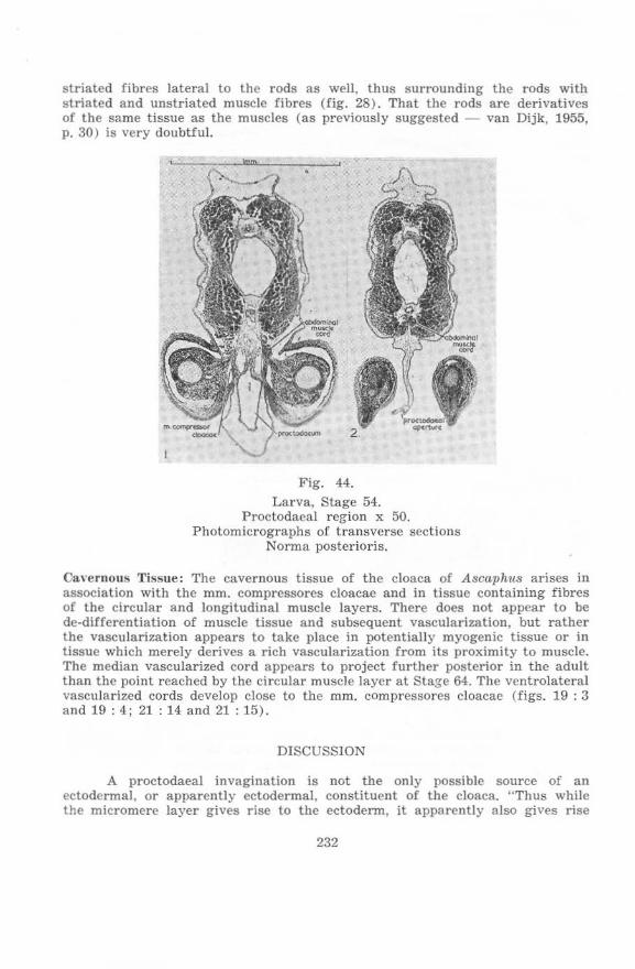
striated fibres lateral to the rods as well, thus surrounding the rods with striated and unstriated muscle fibres (fig. 28). That the rods are derivatives of the same tissue as the muscles (as previously suggested - van Dijk, 1955, p. 30) is very doubtful.
Fig. 44.
Larva, Stage 54. Proctodaeal region x 50.
Photomicrographs of t r a nsverse sections Norma post crioris.
CavernOIlS Tissue; The cavernous tissue of the cloaca of Ascaphus arises in association with the mm. comprcssores cloacae and in tissue containing fibres of the circular and longitudinal muscle layers. There does not appear to be de-differentiation of muscle tissue and subsequent vascularization, but rather the vascu larization appears to take place in potentially myogenic tissue or in tissue which merely derives a rich vascularization from its proximity to muscle. The median vascularized cord appears to project further posterior in the adult than the pOint reached by the circular muscle layer at Stage 64. The ventrolateral vascularized cords dcvelop close to the mm. compressores cloacae (figs. 19 : 3 and 19 : 4 ; 21 : 14 and 21 : 15).
DlSCUSSION
A proctodaeal invagination is not the only possible source of an ectodermal, or apparently ectodermal, constituent of the cloaca. "Thus while the micromere layer gives rise to the ectodenn, it apparcntly also gives rise
232

to a part of the lining of the archenteron, as LWOFF (Bull. de la Soc. imper. des Naluralistes de Moscou, T. 8, 1894) maintained some years ago" (Wilson, 1901, p. 128) . Rugh (1943) gives a description of the neurenteric canal of Rana pipiens and observes that "The cells of the floor of the neurocoel, of the floor of the neurenteric canai, and of the roof of the archenteron are of the same type and are, in all probability, continuous in early developmcnt" (p. 307). Earlier Erlanger (1891) had come to the conclusion "dass der After aus dem ventralsten Theile des Urmundes hervorgeht, wuhrend der dorsalste den Neuroporus und den Canalis neurenteric us bildet" (p. 251). Whether the proctodaeum originates as an invagination of ectoderm, or whether it originates as a persistent blastopore (or part thereof) in the Anura has been the subject of several studies. Schanz (1887). Morgan (1890) and Erlanger (1891) have given reviews of earlier papers. The formation of the anus from a persistent blastopore (or part thereof) has been reported (or numerous animals: by Shipley (1885) for Petromyzon; Caldwell (1885) for Ccratodus; Sedgwick (1884) and Johnson (1884) for Triturus cristatus, Johnson and Sheldon (1886) for Triturus sp., Morgan (1890) for Ambystoma and Brachet (1902) for Ambystoma tigrinum (Siredon piscilormis); among the Anura by Spencer (1885) for Rana t emporarw, Kupffer (1887) for Rana temporaria (?) and Alytes obstetricans, Gasser (1882) for Alytes obstetrican.s, and Pasteels (1942 ) for Discoglossus pictus and Xenopwi laevis. The formation of the proctodaeum from a proctodaeal pit independent of the blastopore has been reported by Hatschek (1881 ) for Amphioxus, Scott (1880) for the lamprey; among Anura by Stricker (1860) for Bulo cinereus, Bambeke (1868) for Pelobates luscus, Goette (1875) for Bombina igneus, Schultze (1884) for Rana lusca, Durham (1886 ) for Rana sp., Sidebotham (1888) for Rana temporaria, :Morgan (1889 and 1890) for Rana halecina and Bulo lentiginosus, Erlanger (1891 ) for Rana lusca and R. esculenta, Bulo variabilis and B. vulgaris and Bombina igneus, by Ziegler (1892) for Rana sp., Brachet (1902) for Rana temporaria, Marchetti (1918) for Bulo vulgaris, Rondinini (1928) for Bulo vulgariS, Vogt (1929) for Bombina, Kagawa (1932) for Bulo vulgaris japonicus, Schechtman (1939) for Hyla regUla and Pasteels (1942) for Rana lusca and Runa esculenta, Bulo vulgaris and Hyla arborea.
Studies of Bytinski-Salz (1936), Risley (1939) and Schechtman (1939) were undertaken to elucidate the mechanism of proctodaeum formation and cloacal plate perforation . Schechtman (op. cit.) working on Hyla regilla suggested the following mechanism: "The vcntral lip mesoderm induces the proctodaeal pit in that portion of the ectoderm which happens to come into contact with it. The invagination of the pit brings it into contact with the entoderm of the hind-gut. The latter completes the process of allus formation by inducing the perforation of the proctodaeal pit" (p. 49-50).
Risley (1939) remarked of anus formation in urodeles: "It is of particular interest that, under experimental conditions, a secondary anus may appear in conjunction with secondary tails even though no blastopore formation or gastrulation has occurred" (p. 114) . This suggests that the formation of the "anus" from a part of the blastopore is not necessarily the primitive condition.
Pasteeis summarizes the devclopment of the proctodaeum as follows: "Le blastopore prescnte chez les Anoures deux modes d'evolution: chez les uns ( Discoglossus, Xenopus), it se transforme en anus sans subir d'occlusion transitoire ; chez les autres (Rana, Bulo, Hyla, probablement Bombinator) , iI s'obture de f~on precoce, l'anus apparaisant it la suite d'une perforation
238
secondaire .... Ces differences ne correspondent qu'a une variante chronologique de la meme cinematique" (1942, p. 25).
In the descriptions of the formation of the proctodaea I pit, the position. in relation to the mesoderm, at which the cloacal membrane is formed, is rarely mentioned. Schanz (1887) docs note of the proctodaeal pit that it had been supposed that "ein Stiick dcs Urdarms wiichse ihm cntgegen, ... " (p. 10), and Marchetti (1918) observes that "Le cellule sticotropiche del fondo delle due fossette si vanno incontro, e per potersi raggiungere e fondere scacciano iI mesoderma interposto" (p, 237) ,
The proctodaeum develops as an ectodermal fossa in Bulo angusticeps. The region of the cloaca into whi ch the bladder and the nephric ducts opcn is appa rently endodermal in Ascaphus (fig. 1 ) and Bulo (figs. 41 , 42 and 43 ), as is inferred from the presence of yolk in the cloaca l walls. According to Gadow's (1887) terminology this region may be described, by definition. as urodaeum. The urodaeum of Ascaphlls and Bulo, then , is apparently endodermal. Shumway (1942) uses Gadow's terminology ( Ame ricanised ) in describing the cloaca of the chick: "No urinary bladder is formed but the cloaca is ultimately divided into three regions: an an terior portion, the coprodeum. into which the rectum entcrs ; an intermediate part. the urodeum, into which the urogcnital ducts enter, and the terminal proctodeum" (p. 147). From this description it is clear wh at should be understood by cloaca a nd rectum. Unfortunately, other authors have been less precise. Huettner (19<19) !<tates of the young larval Rana: "The cloaca being continuous with the proctodaeum, is therefore partially formed by ectoderm. Its inner portion, which is continuous with the rectum, is formed from entoderm. Later on the urinary bladder is developed from the entodermal region of the cloacal portion of the hind-gut" (p. 134 ). Th is description does make it clear that the urodaeum is endodermal, at least ventrally, in Huettner's opi nion. The region of the cloaca into which the nephric ducts open, is variously referred to as "cloaca", "rectum" and "proctodeum" in "NORMAL TABLE OF XENOPUS LAEVIS ( DAUDIN)" edited by Nieuwkoop and Faber (1956). Thus the Wolffian ducts are said to opcn into "reclal diverticula" which originate as follows: "The rectal diverticula are indicated for the first time at STAGE 32, at which they appear as tiny slits in the dorsal wall of the proctodeum" (p. 132). It is further noted (p. 135, 0». cit.) that "Th e urinary bladder develops as a ventral diverticulum of the rectum at STAGE 55 ... ". On p. 157 it is stated: "At its posterior end the rectum is continued beyond the last coil of the ileum, and then becomes the cloaca, The cloaca receives the ureters which cnter by two separate openings lying close to one another in its dorsal wall. On the ventral side the cloaca communicates with the urinary bladder."
A study of O'Connor (1940) on Pleurodeles walt ii, Triturus taeniatus and Ambystoma indicated that the posterior part of t he gut might attract the nephric ducts during development and that the terminal parts of the ducts in adults might have a cloacal origin. Bijtel (1948), working on in Triturus alpcstris, found that grafts of presumptive cloacal material attracted the nephric ducts to it during deVelopment, She refers to this material as "materiel cloacal", but refers to that which develops from it as a secondary proctodaeum. "II est patent que Ie proctodaeum secondaire a exerce dans cc cas, donne en exemple, une action attractive sur l'uretere primaire". Bijel noted one limitation: "Si regrettable que ce soit, la transplantation d'un pcu de materiel sometique est inevitable", It is also important to emphasize that the exact limits of the
231
proctodaeum , in an animal in which it does not develop as a proctodaeal pit (as appears to be the case in urodeles), a re difficult to determine. In Xen01'U8 the cloaca is described as developing diverticula which grow anter iorly in the direction of the nephric ducts, later fusing to these (Nieuwkoop and Faber, editors, see above).
There is little literature on the development of the cloacal muscles in chordates. Lineback (1920) investigated the origin and development of the longitudinal muscle layer of the colon of Homo and refers to ea rlier remarks on the development of the visceral muscles in mammals; but in neither his own work, nor, apparently, in the works cited by him, is the relation of these muscles to the coelom described. The later development of the somatic muscles of the gut of Honw has been studied by Popowsky (1899), but the origins of these muscles would seem to be obscure. In Tachyglossus the sphincter cloacae appears to be a dermal muscle as it is continuous with the dermal muscles of the abdomen ( Ruge, 1895). ( Ruge's illustration appears in Wiedersheim's Vergleichende Anatomie der Wirbeltiere and other textbooks. )
Goette (1875) observed the relation of the abdominal muscle cords to the pelvic region . "Das hinter dem Becken geJegene Stiick ltisst er teils atrophieren, leils soli aus ihm das M. ischiococcygeus werden," to quote Lignitz (1897), p. 32. The m. ischiococcygeus is, in Gaupp's nomenclature (1896), the m. compressot' cloacae. The abdominal mu scle cords ("die altesten Bauchmuskeln" - Kaestner) were subsequently studied by Kaestner (1892, 1893a and 1893b ), Wieders heim (1892 ), Field (1894) and Wild (1 897 ), while Lignitz (1897) referred to their findi ngs (except those of Wild) critically. Field a nd Lignitz were interested in the development of the fore·limbs. Maurer (1892), investigating the devclopment of the trunk and abdomina l muscles of urodeles, does not appear to have observed simi la r relations to the pelvic region in thc case of the urodelan abdominal musclcs as occur in the Anura. Green (1931) mentioned that "'The 'pri mary abdominal muscle' preceding M. rectus abdominis at first passes through th e pelvic basin to a point posterior to this. During the closure of the pelvis it moves fo rward and becomes attached to the pubic symphysis ( p. 1287, of Rana) . Recently !tyke (1953) described the development of the somatic muscles of the trunk of Xenopus, stating in his introduction (p. 2): "1 do not intcnd reviewing the literature prior to MAURER, as it has been full y dealt with by this author in his above-mentioned works. Suffice it to say, that these include the publicationsof MECKEL (1828), DUG£:S (1835), STANNIUS (18M), HUXLEY (1873), GOETTE (1875), SCHNElDER (1879) and KASTNER (1892)." The works of Maurcr referred to are those of 1892 and 1895. Goette is later mentioned by Ryke in eonnexion with the m. sternohyoideus and appears in his bibliography, the other works mentioned (other tha n Maurer 's) are not citcd in the bibliography. Wiedersheim (1892), Kaestner (1893a and 1893b), Wild (1897), Lignitz (1897) and Green (1931 ) a re not referred to at all. Ryke observed the abdominal muselc cords in the pelvic region: "An interesting point in the development of the m. rectus abd. is the occurencc (siC) of a backwardly directed strip reaching the root of the t ail (fiIT. II ) (SiC). It runs along the outer side of the cloaca and possibly represents t he m. rectus JateraJis of some fishes. During metamoq)hosis this muscle strip atrophies" (p. 19). The m. rectus lateralis is situated much further dorsally than the abdominal muscle cords, being found at the level of the horizontal myoseptum.
The significance of the abdominal muscle cords for the interpretation of
235

the development of the pelvic muscles is summarized by Wild (1897, p . 33) as fo llows: "Kastner selbst sagt, dass es ganz verkehrt sci, den Versuch zu machen, den Wiedersheim und er selbst unternommen hatten aus einem wohl differenzierten Abschnitte der Bauchmuskulatur die Muskulatur der hinteren Extremitat abzuleiten . Auf densclben Umstand macht auch Field ... aufmerksam: 'Oer bereits differenzierte Bauchmuskel habe mit der Bildung der Extremitatenmuskulatur nichts zu schaffen'." Field (1894) and Lignitz (1897) advanced evidence that the fore-limbs are dcrived from the spinal myomeres and not from the abdominal muscle cords. Kaestner (1893a and 1893b) and Wild (1897) could not advance much evidence for the derivation of the hind-limb anlagen [ rom the spinal myomeres, but were convinced that the hind-limb anlagen were not derived from the abdominal muscle ccrds. The present study confirms that the hind-limb anlagen are not derived from the abdominal muscle cords and removes one obstacle to the derivation of these anlagen (and hence also the cloacal muscles) from the spinal myomeres: The abdominal muscle cores are not interposed between the hind-limb anlagen and the spinal myomeres at Stage ± 50 of Ascaphus. 'l'his is quite clear although the specimen is by no means a perfect one. It is difficult to understand the relationship of the abdominal cords at Stage 47 to the same muscles at Stage ± 50, but subsequent development is easily followed. The hind-limb anlagen develop independently of the abdominal muscles in Ascaphu8, but differentiation of the cloacal compressor musclcs occurs aftcr the abdominal muscle cords have extended backwards to beyond the pelvic region (Stage 51).
When the cloacal compressor muscles first appear in the Ascaphu8 material examined (Stage 56), it is clear that they are not associated with the skin, and are therefore not dermal muscles; but they are quite closely associated with the abdominal muscles and the mm. pyriformes and mm . caudaJipuboischiotibiales dorsally, and with the pelvic girdle anteroventrally. It is possible that the cloacal compressor muscles may be late derivatives of the same myogenic tissue which gave rise to the late-developing pelvic parts of the abdominal cords. In Buto the abdominal muscle cords are complete when the hind-limb a nlagen are barely indicated (figs. 41 and 42), and the anlagen subsequently develop ventral to the abdominal cords (fig. 43). When the abdominal muscle cords atrophy posteriorly the anterior regions of the mm. compressores cloacae, and, particularly, the mm . circumflexores arteriarum, are closely associated with the atrophying muscles. There is a marked increase in development of the mm. compressores cloacae while the abdominal muscle cords are atrophying in Ascaphus (Stages 58- 60) and, particularly, in Bufo (cf. fig. 44. showing little development of the mm. compressores cloacae a nd maximally deveioped abdominal cords). That thcre may be a causal association between the atrophying of the abdominal cords and the differentiation of the mm. compressores cloacae, is a possibility. Goette (1875) may have been correct in his derivation of the ';M. ischiococcygeus" from the abdominal muscle cords. Kaestner (1893a) does not consider the derivation of Goette likely: "Nach meinen Untersuchungen geht jencs hinterste Sti.ick bei Froschlarven ganz zu Grunde. Bombinator igneus scheint sich demnach etwas anders zu verhalten" (p. 280, discussing the abdominal cords). It is worth mentioning that the obliquus and transversus muscles also differentiate, between the abdominal cords and the spinal myomeres, while the abdominal cords are atrophying (Stage 58 of Ascaphus) , their differentiation not beginning in Bufo until atrophy of the abdominal cords is con-
236
siderabJy advanced. In the comparison which can thus be made bctween the differentiation of the cloacal compressors and the obliquus and transversus muscles it is of importance to note that Ryke's (1953) interpretation of the abdominal muscle cords of Xenopus as constituting all the abdominal muscle fayers anterior to the pelvis (loc. cit., fig . 6, p. 14; fig. 27, p. 39) seems to gain no support whatever from conditions in Ascaphus and Buto, and is at variance with Kaestner's interpretation of the obliquus and transversus muscles : "Die erwiihnte Muskelschicht liegt iiberall lateral - bezw. dorsalwarts vorn Rectus abdominis, sU.isst, wenigstens anfangs, nirgends mit ihm zusammen, sondern es findet sich ein muskeJfreier Zwischenraum zwischen der lateralen Grenze des Rectus und der media\en der neuen Muskelschicht" (1893a, p. 283).
If the cloacal compressor muscles are neither derived by division of the abdominal muscle cords when these have already formed (which derivation seems most unlikely), nor whilc these cords are developing or atrophying, another mode of origin from the abdominal muscle cords is still possible. It is possible that, like the mm. epipubici, they are derived in association with, if not from, the myocommata between segments of the abdominal cords. (The possibility that myocommata can contain myogcnic elements must be admitted, since the proximity of the epipubic muscles to myocommata in Ascaphus suggests a possible histogenetic relationship.) It is probable that the mm. compressores cloacae are pelvic muscles unconnected with the abdominal muscles.
Experimental evidence, such as that obtained by Byrnes (1898), Lewis (1910) and Detwiler (1918 and 1929), has been that "There is no demonstrable contribution of the somites to the musculature of the limb in amphibians". (Nicholas, J . S., in "Analysis of Development" edited by Willier, Weiss and Hamburger, 1955, p. 431.) From the works of Lewis and Detwiler cited, there is evidence that extirpation of somites in the anterior region of urodeles results in the absence of ventrolateral abdominal muscles, but that the fore-limb still develops. Byrnes (op. cit.) found in both Ambystoma and Rana that suppression of the abdominal muscles is effected by treatment not affecting the hind-limb musculature. It would be interesting to learn to which category the cloacal compressor muscles belong on this criterion. While negative experimental evidence has questionable morphological Significance, positive findings would be, at least, suggestive. There is evidence (based on carbon marking technique) that not only the limb buds, but also part of the ventrolateral muscles may form independently of the somites, at least in Aves (Rawles and Straus, 1948; Straus and Rawles, 1953). Detwiler (1955). however, finds the ventrolateral muscles of u rodeles to be derived from the somites in studies based on extirpation and xenoplastic grafts. Since the abdominal muscles have already appeared in the earliest Ascaphu8 stage available (Stage 47), the study of their origin was unavoidably beyond the scope of the present investigation.
An investigation of Salmo and Periophth«lmu8 showed the absence of striated cloacal muscles. Paired longitudinal muscles stretch from the pelvic girdle to the anal-fin skeleton in both of these teleosts, but these are widely separated in the cloacal region in both genera.
The thickening of the septum interfemoraJe which has here been named ligamentum interfemorale, and which in Ascaphus is modified to form the rods of Noble ventrally, was observed by Gaupp (1896) in Rana. Describing the m. gracilis minor, he wrote : "Dieser schmale, ;;anz am medial-hinteren Rande des Oberschenkels gelegene Muskel entspringt mit dem der anderen Seite von einem
237
sehnigen Streifen, der den hinteren Rand der Beckenschcibe deckt, vorn an der Insertion des Rectus abdominis, hinten an der Spina pelvis posterior befestigt ist. An dem hinteren Ahschnittc dicses Streifcns, bis zum Ursprunge des Gracilis minor, cntspring-en die hinteren Biindel des Sph-incter ani" (p. 182). Grobbelaar (1922) did not mention this "sehniger Strcife", but described the m. gracilis minor as originating on the ischium, close to the symphysis, in Xenopus. Bigalke (1926) divided the m. gracilis minOr (1'1. cutaneous femoris) into a Caput superius and Caput inferius, referred to Gaupp's description and stated: "Bei B. vulgaris aber entspring-t das Caput sUI>erius, zusammen mit dem der anderen Seite, mit dUnnem Ursl>rung gerade unterhalb der Afteroffnung von der hinteren l<~lache des M. sphincter ani cloacalis und nimmt gJcich an Breite betrachtlich zu" (p. 335). In both Xen01JUs and Bulo the ligament is nevertheless present. In Xenopus the ventral and dorsal parts were reconstructed and labelled "connective tissue" befo re their significance was realized (fig. 33). In Ascaph11S the rods of Noble bear essentially similar relations to the cloaca and the mm. g-raciles minorcs as do the dense edges of the ligamentum interfemoralc in other A num. Since the possession of these rods has been regarded as evidence of great specialization in AsC(1)hus, this genus must now be considered as being less specialized than has been maintained. The glands of the oviduct in which spermia arc to be found in A8caphus are essentially similar to the oviducal glands of Ran«, as may be seen from figs. 31 and 32. Here specialization for internal fertilization has not produced any marked morphological changes.
Szarski (1957), discussing the larval state in Amphibia, observed: "All amphibian and fish larvae havc a similarly constructed tail fin, consisting of a skin fold." 'I'he shortening of the proctodaeum during metamorphosis, which occurs in the Anura, would thus also take place in fish larvae when the tail fin becomes reduced. This may be seen, for example, in Stages 27 and 28 of the development of Gasterostcus acu/catus (Swarup, 1958).
Hula exhibits no marked specializations in the cloacal region which distinguish it from the other Anura studied. Green (1931) remarked that ';the oldest definitely accepted Anuran is of Upper Jurassic age (Eobatrachus agilis March; Moodie 1912)" and "8obatrachlUI is structurally almost indistinguishable from modern Bufonids" (p. 1259). ln respect of the cloacal region there is no reason to believe that this is not because the bufonids arc rather primitive Anura. Rana and XenOllU8 reveal several spccializations in the cloacal region, while Botnbina resembles Bulo quite closely. Szarski's evidence that Rana shows greater specialization than Bltlo and Bombina in the vascular system (1948) is significant in the light of conditions in the cloacal reg ion. Ascaphus and Leiopcllllu show specializations from the bufonid condition in the cloacal region, and these are typically in divergent directions. An example is afforded by the enlargement and modification of the m . sphincter ani cloacalis in the male Ascap}ws and the absence of this muscle in Lciopelma.
In Ascaphus and Leiopelma there is one more presacral vertebra than in typical Anura such as Bulo, and this is regarded as a primitive feature. Green (1931) offers evidence of this and remarks: "The phylogenetic forward extension of the ilia is represented during larval development" (p. 1288). Smit (1953) states: " If we consider that during the evolution of the Anura a forward shifting of the ilio·sacral articulation has taken place (Watson, 1939--1940, and Piveteau, 1937), ... then the urostyle must represent original trunk vertebrae, as suggested by Piveteau (1937), and not caudal vertebrae" (p. 127-128). Despite the dif-
238
ference in sacral vertebrae in the Ascaphidae (in which it is the 10th vertebra) and in Bu,fa and Rana (in which it is the 9th ) the plexus ischiadicus and the innervations of the pelvic muscles, including the mm. compressores cloacae, is essentially the same. Information in this connexion has been given for Ascallhtl,s by Ritland (1955a and b), and van Dijk (1955), for Lciopclma by Stephenson (1952) and Ritland (1955b). for Rana by Gaupp (1899) and others; and for Bulo by Bigalke (1926). Goodrich (1930) remarked of movcments of the hind·limb: "For instance, in the frog the straining backwards of the pelvic plexus shows that the base of the hind-limb has moved backwards with the elongation of the pelvic g irdle" (p. ]38). The innervation of the cloaca by the 11th spinal nerves in Rana, by the 11th, 12th, and somctimes the 13th (according to Ritland) spinal nerves in AscaphllS, and by 11th to 13th nervcs in Leiopelma (as appears to be the case), suggests a process of transposition (Rosenberg, 1875 and ]907) by which the innervations have bcen shifting to more anterior nerves during phylogeny. On the other hand it is clear that the hindlimbs shift backwards relative to the spinal nerves during ontogeny. Comparison of Stages 56 and 64. of Ascaphu8 (figs. 8 and 21) shows the pelvis moving from a position ventral to the 12th spinal nerve to one behind the 13th. 'l'his movement is even more strikingly illustrated by comparing Stage 56 with Ritland'S (1955a) fig . 11 and Stephenson's (1952) fig. 2, when it may be seen that the origin of the 12th nerve, which marks the original level of the pelvic girdle, is far forward on the urostyle in both A,scaphus and Lciopeima. The same movement of the pelvis clearly takes place in all Anltru during development. The cloacal nerves of tetrapods are apparently rarely presacral, and it seems likely that the 11th vertebra is the posteriormost sacral vertebra to have occurred in an anuran with the ascaphid arrangement of the plexus isehiadicus. The relation of the sacral plexus to the sacrum in Urodcla is well illustrated by Davidoff (] 884). The 13th spinal nerve is apparcntly rarely ret!tined in the adult Ascaphu8, although Ritlund has detected it (pp. 166-167 and fig. 11, 1955a) and reports that it suppJies the cloaca. In none of the larvae reconstructed in the present study, nor in the adults reconstructed previously (1955) does the 13th nerve participate in the plexus isehiadieus.
Of the tail muscles of the larval Anura, only those in the region of the urostyle are retained in the adult, becoming the mm. cocygeo-i1iaci. Since these muscles only become differentiated at metamorphosis, when the cloaca has almost achieved its adult form, their development is of little consequence to cloacal differentiation. The differentiation of the spina l myomeres early in metamorphosis has here been studied in Ascuphus and the comparative material (Bulo). As the youngest AscapltllS e mbryo studied (Stage 47) has well-differentiated spinal myomeres, the carlier development of the spinal myomeres is not relevant to the present study, which is centred on A,scaphus. It may be mentioned, however, that there have been a number of studies on the origin of the tail and the early stages of its subsequent development. Notable amon3" the workers in this field is Bijtel, whose thesis (1929) and some of whose subsequent works (Bijtel 1930, 1931, 1936 and 1958) have been consulted in the present study. Other works consulted are those of Chuang Hsiao-Hui (1947), Nakamura (1942, 1947 and 1952) and Bijtel and Woerdeman (1928), as well as others also mentioned in other connexions (e.g. Marchetti, 1919; Rondinini , 1928).
Of the Amphibia, the anurans Ascaphtt,s and Nectophrynoide,s, the salamandroid Urodela, and all the Gymnophiona practice internal fertilization.
239
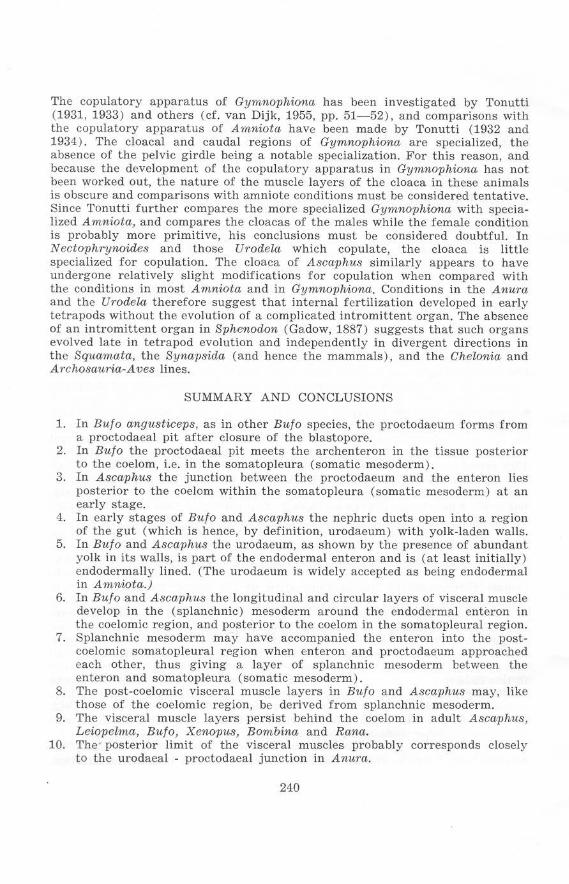
The copulatory apparatus of Gymnophiona has been investigated by Tonutti (l931, 1933) and others (cf. van Dijk, 1955, pp. 51-52), and comparisons with the copulatory apparatus of A1nniota have been made by Tonutti (1932 and 1934). The cloacal and caudal regions of Gymnophiona are specialized, the absence of the pelvic girdle being a notable specialization. For this reason, and because the development of the copulatory apparatus in Gymnophiona has not been worked out, the nature of the muscle layers of the cloaca in these animals is obscure and comparisons with amniote conditions must be considered tentative. Since Tonutti further compares the more specialized Gymnophiona with specialized Amniota, and compares the cloacas of the males while the female condition is probably more primitive. his conclusions must be considered doubtful. In Nectophrynoides and those UrodeZa which copulate, the cloaca is little specialized for copulation. The cloaca of Ascaphus similarly appears to have undergone relatively slight modifications for copulation when compared with the conditions in most Amniota and in Gymnophiona. Conditions in the Anura and the Urodela therefore suggest that internal fertilization developed in early tetrapods without the evolution of a complicated intromittent organ. The absence of an intromittent organ in Sphcnodon (Gadow, 1887) suggests that such organs evolved late in tetrapod evolution and independently in divergent directions in the Squamata. the Synapsida (and hence the mammals), and the Chelonia and Archosauria-Aves lines.
SUMMARY AND CONCLUSIONS
1. In Bulo angusticeps. as in other Hulo species, the proctodaeum forms from a proctodaeal pit after closure of the blastopore.
2. In Bulo the proctodaeal pit mcets the archenteron in the tissue posterior to the coelom, I.e. in the somatopleura (somatic mesoderm).
3. In Ascaphus the junction between the proctodaeum and the enteron lies posterior to the coelom within the somatopleura (somatic mesoderm) at an early stage .
4. In early stages of Bufo and Ascaphus thc nephric ducts open into a region of the gut (wh ich is hence. by definition, urodaeum) with yolk-laden walls.
5. In Hulo and Ascaphus the urodaeum. as shown by the presence of abundant yolk in its walls, is part of the endodermal enteron and is (at least initially) endodermally lined. (The urodaeum is widely accepted as being endodermal in Amniota.)
6. In Bulo and Ascaphus the longitudinal and circular layers of visceral muscle develop in thc (splanchnic ) mesoderm around the endodermal enteron in the coelomic region, and posterior to the coelom in the somatopleural region.
7. Splanchnic mesoderm may have accompanied the enteron into the postcoelomic somatopleural region when enteron and proctodaeum approached each other, thus giving a layer of splanchnic mesoderm between the enteron and somatopieura (somatic mesoderm).
8. The post-coelomic visceral muscle layers in Bulo and Ascaphus may, like those of the coelomic region, be derived from splanchnic mesoderm .
9. The visceral muscle layers persist behind thc coelom in adult Ascaphu8, Leiopelma, Hulo, Xenopus, Bombina and Rana.
10. The posterior limit of the visceral muscles probably corresponds closely to the urodaeal - proctodaea! junction in Anura.
240
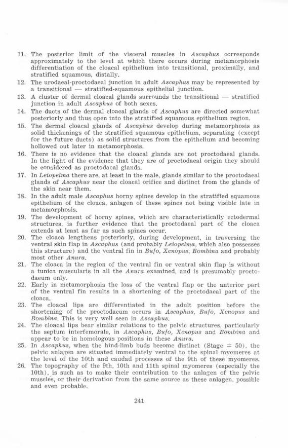
11. The posterior limit of the visceral muscles in Ascaphus corrcsponds approximately to the level at which there occurs during metamorphosis differentiation of the cloacal epithelium into transitional, proximally, and stratified squamous, distally.
12. The urodaeal-proctodaeal junction in adult Ascaphus may be represented by a transitional - stratified·squamous epithelial junction.
13. A cluster of dermal cloacal glands surrounds the transitional - stratified junction in adult Ascaphus of both sexes.
14. The ducts of the dermal cloacal g lands of Ascaphll8 are directed somewhat posteriorly and thus open into thc stratified squamous epithelium region.
15. The dermal cloacal glands of AscapllUs dcvelop during metamorphOSiS as solid thickenings of the stratified squa mous epithelium, separating (except for the future ducts) as solid structures from the epithelium and becoming hollowed out later in metamorphosis.
16, There is no evidence that the cloacal glands are not proctodaeal glands. In the light of the evidence that they arc of proctodaeal origin they should be considered as proctodaeal glands,
17, In Leiopelma there a re, at least in the male, glands similar to the proctodaea I glands of A scaphus near the cloacal orifice and distinct from the glands of the skin near them,
18. In the adult male A8caphus horny spines develop in the stratified squamous epithelium of the cloaca, anlagen of these spines not being visible late in metamorphosis.
19. The development of horny spines, which are characteristically ectodermal structures, is further evidence that the proctodaeal part of the cloaca extends at least as far as such spines occur.
20. The cloaca lengthens posteriorly, during development, in traversing the ventral skin flap in Ascaphu8 (and probably Leiopelmu, which also possesses this structure ) and the ventral fin in Bulo, Xenopus, Bombina and probably most other Anura.
21 . The cloaca in the region of the ventral fin or ventral skin flap is without a tunica muscularis in all the Anura examined, and is presumably proctodaeum only.
22. Early in metamorphosis the loss of the ventral flap or the anterior part of the ventral fin results in a shortening of the proctodaeal part of the cloaca.
23. The cloacal lips are differentiated in the adult pOsition before t he shortening of the proctodaeum occurs in Ascaphlls, Bu/o, Xe-nO.,,118 and Bombina. This is very well seen in A8caphu8.
24. The cloacal lips bear similar relations to the pelvic structures, particularly the septum interfemorale, in Ascaphu8, Bulo, Xenopus and Bombina and appear to be in homologous positions in these Anura.
25. In Ascaphus, when the hind·limb buds become distinct (Stage ± 50 ), the l)elvie anlarren are situated immediately ventral to the spinal myomeres at the level of the 10th and caudad processes of the 9th of these myomeres.
26. The topography of the 9th, 10th and 11th spinal myomercs (espeCially the 10th), is such as to make their contribution to the anlaa-en of the pelvic muscles, or their derivation from the same source as these anlagen, possible and even probable.
241
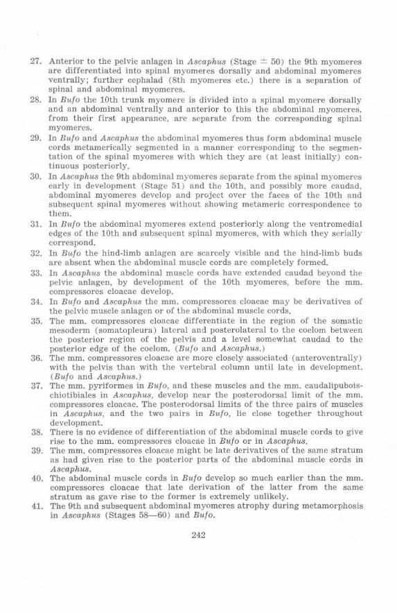
27. Anterior to the pelvic anlagen in Ascaph1t.S (Stage ± 50) the 9th myomeres are differentiated into spinal myomeres dorsally and abdominal myomeres ventrally; further cephalad (8th myomeres etc.) there is a separation of spinal and abdominal myomeres.
28. In Hulo the 10th trunk myomere is divided into a spinal myomere dorsally and an abdominal ventrally and anterior to thii:! the abdominal myomcres. from their first appearance, are separat e from the corresponding spinal myomeres.
29. In Hulo and Ascaphus the abdominal myomercs thus form abdominal muscle cords metamerically segmented in a manner corresponding to the segmentation of the spinal myomeres with which they arc (at least initially) continuous posteriorly.
30. In Ascaph11s the 9th abdominal myomeres separate fro m the spina l myomcrcs early in development (Stage 51 ) and the 10th, and possibly more caudad, abdominal myomeres develop and project over the faces of the 10th and subsequent spinal myom eres without showing metameric correspondence to them.
31. In Bltlo the abdominal myomcres extend posteriorly along the ventromedial edges of the 10th and subsequent spinal myomercs, with which they serially correspond.
32. In Bulo the hind-limb anlagen arc scarcely visible and the hind-limb buds arc absent when the abdominal muscle cords are completely formed.
33. In AscaphtM the abdominal muscle cords have extended caudad beyond the pelvic anlagen, by development of the 10th myomeres, before the mm. compressores cloacae develop.
34. In Bulo and Ascaph~ the mm. compressores cloacae may be derivatives of the pelvic muscle anlagen or of the abdominal muscle cords.
35. The mm. compressores cloacae differentiate in the region of the somatic mesoderm (somatopleura) lateral and posterolateral to the coelom between the posterior region of the pelvis and a level somewhat caudad to the posterior edge of the coelom. (Hulo and Ascaphus.)
36. The mm. compressores cloacae are more closely associated (anteroventrally) with the pelvis than with the vertebral column until late in development. (Bulo and Ascaphus.)
37. The mm. pyriformes in Bulo, and these muscles a nd the mm. caudalipuboischiotibiales in Ascaphus, develop near the poste rodorsal limit of the mm. comprcssores cloacae. The posterodorsal limits of the three pairs of muscles in Ascaphu.s, and the two pairs in Bulo, lie close together throughout development.
38. 'fh ere is no evidence of differentiation of the abdominal muscle cords to give rise to the mm. compressores cloacae in B1~/o or in Ascaphu.s.
39. The mm. compressores cloacae might be late derivatives or the same stratum as had given rise to the posterior parts of the abdominal muscle cords in Ascaphus.
40. The abdominal muscle cords in Bulo develop so much earlier than the mm. compressores cloacae that late derivation of the latter from the same stratum as gave r ise to the former is extremely unlikely.
41. The 9th and subsequent abdominal myomeres atrophy during metamorphosis in A"scaphus (Stages 58-60) and Bulo.
24.2
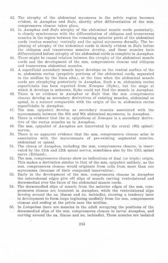
42. The atrophy of the abdominal myomeres in the pelvic region becomes evident, in Ascaphus and Bit/oJ shortly after differentiation of the mm. compressores cloacae takes place.
43. In Ascaphus and Bli/O atrophy of the abdominal muscle cords posteriorly, is closely synchronous with the differentiation of obliquus and transversus muscles in the region between the remaining anterior parts of the abdominal cords (rectus muscles) ventrally a nd the spinal myomeres dorsally. The be· ginning of atrophy of the abdominal cords is clearly evident in Bulo before the obliquus and transversus muscles develop, and these muscles have differentiated before atrophy of the abdominal cords is complete in Ascaphus.
44. There might be causal rclation between the atrophy of the abdominal muscle cords and the development of the mm. compressores cloacae and obliquus and transversus abdominal muscles.
45. A 8ullerficlal secondary muscle layer develops on the ventral surface of the m. abdominis rectus (prepelvic portions of the abdominal cords, separated in the midllne by the linea alba), at the time when the abdominal muscle cords are atrophying posteriorly in Ascaphus. Such a m. abdominis rectus superficialis has been rel>orted from Xenopus (Ryke), but the stage at whieh it develops is unknown. Ryke could not find the muscle in Ascaphus.
46. There is no evidence in AsCal)}ms or Bula that the mm .compressores cloacae develop as secondary derivatives of existing muscles, abdominal or spinal, in a manner comparable with the origin of the m. abdominis rectus superficialis in Ascaphus.
47. The mm. epipubici develop as secondary muscles associated with the myocommata between the 8th and 9th abdominal myomeres, in Ascaphus.
48. There is evidence that the m. epipubicus of Xenopus is a secondary derivative of the rectus muscles as in Ascaphus.
49. The mm. epipubici of A8Caph1t8 are innervated by the crural (9th spinal) nerves.
50. There is no apparent evidence that the mm. compressores cloacae arise in association with the myocommata of pre·existing segmental muscles, abdominal or spinal.
51. The cloaca of Ascaphus, including the mm. compressores cloacae, is innervated by the 11th and 12th s llinal nerves, sometimes also by the 13th spinal nerve (Ritland).
52. The mm. com pressores cloacae show no indications of dual (or triple) origin. This makes a derivation similar to that of the mm. epipubici unlikely, as the mm. compressores cloacae would originate from cells from more than one myocomma (because of their coml)Qund innervation).
53. Early in the development of the mm. compressores cloacae in Ascaphus the anterodorsal edges give off slips of muscle curving ventrolaterad and dorsomediad over the faces of the abdominal muscle cords.
54. The dorsomediad slips of muscle from the anterior edges of the mm. com· pressores cloacae arc transient in AsCal)hus, while the ventrolaterad slips develop around the aa. iliacae and nn. ischiadici, showing a tendency later in development to form loops beginning medially from the mm. compressores cloacae and ending at the pelvis near the midline.
55. In Leiopelma there are muscles in the adult occupying the positions of the dorsomediad slips of the mm. compressores cloacae in larval AscaphtlS, and curving a round the aa. iJiacae and nn. ischiadici. These muscles are isolated
243
from the mm. compressores cloacae and hence have been given a separate name, mm. circumflexores arteriarum. This name is also applied to the homologous muscles in Ascaphus etc. (dorsales may be appended to distinguish the Lciopelma, ventrales the Ascaphus, condition) .
56. In all the families of Anura found in South Africa mm. circumflex ores arteriarum occur and appear to form complete or nearly complete loops extending from the mm. compressores cloacae around the aa. iliacae and nn. ischiadici to the pelvis.
57. In Ascaphus and Rana (some species only, apparently, e.g. R. temporaria according t o Gaupp) the mm. circumflexores arteriarum form half-loops only, the lateral parts being absent.
58. The mm. circumflexores arteriarum have no known function, but they have the same relation to the aa. iliacae and nn. isehiadici in all the Anura examined, whether they are separate from the mm. compressores cloacae as in LeiopeZma or joined as in other Anura.
59. The aa. iliacae may be compressed against the nn. ischiadici by the mm. circumflexores arteriarum.
60. The posterior parts of the mm. compressores cloacae meet each other midventrally and mid-dorsally to form a sphincter, the m. sphincter ani eloacalis, in Rana, Bulo, Bombina, Xenopus, Ascaphus and probably most other Anura.
61. A sphincter ani eloacalis is absent in Leiopelma, the cloacal sphincter in this gen us consisting of a circular visceral muscle layer.
62. The teleosts Salmo and Periophthalmus, have no striated cloacal (anal) sphincters.
63. The absence of a striated cloacal sphincter in LeiopeZma may be a primitive feature.
64. The m . sphincter ani cloacalis is enlarged in the male Ascaphus to form the striated muscle layer of the "cloacal extension".
65. The bilateral origin of the m. sphincter ani cloaealis may be reflected in the presence of fascial sheets in the dorsal and ventral midlines, such fasciae formin:; thickened cords on the surfaces of the muscles.
66. The cords on the dorsal and ventral midlines of the m. sphincter ani cloacalis form part of a ligament lying in the septum interfemorale, and hence have together been named ligamentum interfemorale.
67 . Dorsal to the cloaea the ligamentum interfemorale consists of a single dorsal ligament attached to the urostyle, and penetrated by the v. caudalis at least in the larvae, in Ascaphu8, Bula, Bombina and Xenopus.
68. A dorsal ligament is absent above the cloaca in Rana and Leiopelma. 69. Ventral to the cloaca the lateral edges of the ligamentum interfemorale
are thickened into tendinous cords in Bula, Bombina, Xenopus, Rana and Leiopelma, while in Ascaphll,'j these thickenings are represented by the rudes Nobleianae (rods of Noble, "post-pelvic" rods, "post-pubic" rods, "NobeJian bones", etc.).
70. The ligamentum interfemorale stretches from the pubic region along the ventral, posteroventral and posterior edge of the pelvis to the cloaca in Bulo, Bombina and Rana.
71. In X ,,;nopus, Leiopelma and Ascaphus the ligamentum interfemorale reaches the cpipubis anteriorly ( = tendinous sheet in Ascaphus).
211
..
72. In the male Ascaplws the rods of Noble are widely separated from the posterior edge of the pelvis and reach to a level just anterior to the cloacal orifice.
73. The ligamentum interfemorale of Bujo and Xenopus, and the rods of Noble of Ascaphus fork posteriorly to extend on either side of the cloaca near its orifice.
74. In Xenopus the fork of the ligamentum interfemorale bclow the cloaca is greatly thickened and extended forwards, a nd serves to attach the mm. cloacae to the pelvis.
75. In Rana the end of the ligamentum interfemorale below the cloaca is turned forward. and it attaches to the pelvis.
76. In Hu/o, Bombina, Xenopus and Rana the mm. g raciles minores originate from the ligamentum interfemorale just below the cloaca. In Ascnphus these muscles originate from the rods of Noble in a comparable position.
77. The cloacal orifices of Ru/o, Rombina and Xenopus bear the same relations to the ligamentum interfemorale as does that of Ascaphus to the rods of Noble: these orifices are hence homologous.
78. In Leiopclma, Bu/o, Bombina and also Rann (ct. van Dijk, 1955) there are two cords of longitudinal visceral muscle attached to the urostyle, and two to the pelvis, in the cloacal region.
79. In Xenopus there arc no longitudinal visceral muscle cords attached to the urostyle, and the cords to the pelvis are closely associated with each other.
80. I n Ascaphus there are two longitudinal visceral muscle cords attached to the urostyle, but there is only one cord attached to the pelvis, although this has a dual origin.
81. The vascularized tissue in the cloacal region of Ascaphus develops in association with the visceral muscle layers and the mm. eompressores cloacae, but does not arise from de-differentiated muscle tissue.
BIBLIOGRAPHY
See also van Dijk (1955).
Bambeke, C. van, 1868. Recherches sur Ie developpement du Pelobate bruno Mem . eour. Acad. R. Belg., 34 : ? See Erlanger, 1891.
Bhaduri, J . L. 1953. A study of the urinogenital system of Salientia. Proc. zool. Soc . Bengal, 6: 1- 111.
Bigalke, R. 1926. Zur Myologie der Erdkrote (Buto vulgaris, Laurenti). Z. Anat. EntwGesch., 82 : 287- 353.
Bijtel. J. H. 1929. Over de vorming van den staart bij Amphibien. Thesis, Groningen.
Bijtel, J. H. 1930. Beitrage zur Schwanzentwicklung der Amphibien. Anat. Anz., 71: 87-93.
Bijtel, J . H . 1931. tiber die Entwicklung des Schwanzes bei Amphibien. Arch. Entw~Iech. Org .. 125: 448---486.
Bijtel. J. H . 1936. Die Mesodermbildungspotenzen der hinteren Medullarplattenbezirke bei Amblystoma mexican urn in Bczug auf die Schwanzbildung. Arch. EntwMech. Org., 134: 262-282.
Bijtei, J. H . 1948. Deviation experimentale de l'uretere primaire par la greffe de materiel cloacal chez Ie Triton. Experien tia, 4: 389--391.
245
Bijtc1, J. H . 1958. The mode of growth of the tail in urodele larvae. J. Embryo!. exp o Morph., 6: 466-478.
Bijlci, J. H. and Woerdeman, M. W. 1928. On the development of the tail in the amphibian embryo. Proc.roy. Akad. Sci. Amsterd. , 31: 1030- 1040.
Braehet, A. 1902. Recherches sur I'ontogenese des Amphibiens Urodeles et Anoures (Siredon pisciformis - Rana temporaria). Arch. Bio!., 19 : 1- 243.
Byrnes, E. F. 1898. l-Jxperimental Studies on the Development of Limb-Muscles in Amphibia. J. Morph., 14: 105-14.0.
ByUnski-Salz, H. 1936. Lo sviluppo della coda degli Anfibi. R.C. Accad. Lincei, 2J, : 34-88.
Caldwell, see Morgan (1890) . Chuang Hsiao-Hui. 1947. Defekt- und Vitalfiirbungsversuche zur Analyse der
Entwicklung der kaudalen Rumpfabschnitte und des Schwanzes bei Urodelen. Arch. Entw.Mech. Org., 143 : 19-125.
Davidoff, M. 1884. Ober die VarieUiten des Plexus lumbosacralis von Salamandra maculosa. Morph. J b., 9: 401- 413.
de Graaff, A. R. 1957. Investigations into the distribution of blood in the heart and aortic arches of Xenopus laevis (Daud.). J. exp o BioL, 34: 143-172.
de Villiers, C. G . S. 1929. Some observations on the breeding habits of the Anura of the Stellenbosch Flats, in particular of Cacosternum capense and Bufo angustieeps. Ann. T ransv. lI.'1us., 13: 123- 141.
de Villiers, C. G. S. 1934. On the Morphology of the Epipubis, the Nobelian Bones and the Phallic Organ of Ascaphus truei Stejnegcr. Anat. Anz., 78 : 23- 47.
dc Vos, C. M. 1938. The zonal and sternal skeleton of the Liopelmidae (Anura). Anat. Anz. , 87: 5<1- 81.
Detwiler, S. R. 1918. Experiments on the Development of the Shoulder Girdle and the Anterior Limb of Amblystoma punctatum. J . expo Zool., 25 : 499- 538.
Detwiler, S. R. 1955. Experiments on the origin of the ventrolateral trunk musculature in the urodele (Amblystoma). J . expo Zoo!., 129: 45- 75.
Durham, see Morgan (1890) . Erlanger, R. v. 1891. Ueber Blastoporus der anuren Amphibicn, sein Schicksal
und seine Beziehungen wm bleibenden After. Zool. Jb., 4: 239- 254. Field, H. H. 1894. Die 'Vornierenkapsel, ventrale Muskulatur und Extremitatenan
lagen bei den Amphibien . Anat. Anz., 9: 713-724. Gadow, H. 1887. Remarks on the cloaca and copulatory organs of Amniota.
Philos. T rans., 178 : 5- 37. Gasser, E . 1882. Zur Entwicklung von Alytes obstetricans. S. B. naturw. Ges.
Marburg, 5: 73-104. Gaupp, E. 1896. A. Ecker's und R. Wiedersheim's Anatomie des Frosches. I
(3rd ed.), Braunschweig. Gaupp, E. 1899. A. Ecker's und R. Wiedersheim's Anatomie des Frosches. II.
(3rd cd.), Braunschweig. Goette, A. 1875. Die Entwickelungsgeschiehte dcr Unkc, Bombinator igneus,
Rosel. Leipzig. Goodrich, E . S. 1930. Studies on the Structure and Development of Vertebrates.
Macmillan and Co., London. Green, T. L. 1931. On the pelvis of the Anura; a study in adaptation and
recapitulation. Proe. zool. Soc . Lond., 1259- 1290.
246
Grobbelaar, C. S. 1924. Beitrage zu einer anatomischen Monographie von Xenopus laevis (Daud.). Ergebn. Anat. Entwgesch., 72: 131-168.
Hatschek, see Morgan (1890). Huettner, A. F. 1949. Fundamcntals of Comparative Embryology of the
Vertebrates. (2nd ed.), Ncw York. Johnson, sce Morgan (1890). Johnson and Sheldon, see Morgan (1890). Kaestner, S. 1892. Ueber die allgemeine Entwicklung der Rumpf- und Schwanz
muskulatur der Wirbeltiere. Arch . Anat. PhysioL Lpz., Kaestner, S. 1893a. Die Entwickelung der Extremitiitcn- und Bauchmuskulatur
bei den anuren Amphibien . Arch. Anat. Physiol. Lpz., ?: 257-291. Kaestner, S. 1893b. Ueber die Entstehung der Extremitatenmuskulatur bei den
anuren Amphibien. Verh. anat. Ges. Jena. Kagawa, T. 1932. Obcr die Ausbildung des Afters und des Canalis neurentericus
bei Bufo vulgaris japonicus. Okayama Igk., 44 : 2405-2418 (in Japanese). Jap, J. med. Sci. Anat., 5 : (12) (Abstract in German).
Kupffer, C. 1887. Ueber den Canalis neurentericus der Wirbeithiere. S. B. Ges. Morph. Physiol. Munchen, 3 : See Schanz (1887).
Lewis, W. H. 19.10, The relation of the myotomes to the ventro-lateral musculature and to the anterior limbs in Amblystoma. Anat, Rcc., 4: 183- 190.
Lignitz, W, .1897. Die Entwicklung des Schulterglirtels beim Frosch. InauguralDissertation, Leipzig,
Lineback, P. E. 1920. Studies on the Longitudinal Musele of the Human Colon, with Special Reference to the Development of the Taeniae. Contr. EmbryoL Carneg. Instn ., 21: 35--44.
Lwoff, see Wilson (1901). Marchetti, L. 1919. Sui destino del blastoporo durante la formazione del Canalis
neurentericus e del Proctodaeum nel Bufo vulgaris. Arch. itaL Anat. EmbrioL, 17: 216-238.
Maurer, F . 1892. Der Aufbau und die Entwicklung der ventralen Rumpfmuskulatu r bci den urodelen Amphibicn und deren Beziehung zu den gleichen J\Iuskeln der Selaehier und Teleostier. Morph. J b ., 18: 76-179.
Maurer, F. 1895. Die Entwickiung des Muskelsystems und der elektrischen Organe, Hertwig's Handb. vergl. expo Entw. Wirb., Jena, S : 1-80.
Moodie, R. L. 1912. An American Jurassic F'rog. Amer. J. SeL, 34 : Morgan, T . H. 1889. See Morgan (1890). Morgan. T. H. 1890. On the Amphibian Blastopore. Stud. biol. Lab. John Hopkin's
Hosp., 4: 355-377. Nakamura, O. 1942. Die Entwicklung der hinteren Korperhiilfte bel Urodelen.
Annot. zool. jap., 21: 169-236. Nakamura, O. 1947. Determination and Differentiation in the Development oC
the Urodele Tail. Exp. Morph., 3: (In Japanese, Author's Resume in English consulted) .
Nakamura, O. 1952: Extirpation Experiments of the Presumptive Rudiments in the Caudal Region of the Newt. Annot, zool. jap., 25: 105- 112.
Nicholas, J. S. 1955. Limb and Girdle. Chapter 4 of Analysis of Development, edited by Willier, '\'eiss and Hamburgcr, Saunders and Co., Philadelphia. pp. 429--439,
Nieuwkoop, P. D, and Faber, J., editors. 1956. Normal Table of Xenopus laevis (Daudin), North·Holiand Publishing Company, Amsterdam.
247
Noble, G. K. 1926. The importance of larval characters in the classification of the South African Salientia. Amer. :Mus. NoviL, 237 : 1- 10.
O'Connor , R. J. 1940. An experimcntal s tudy of the developme nt of the amphibian cloaca. J. AnaL Lond., 14-: 301- 308.
Pasteels, J. 1942. Fenneture du blastopore, anu s et intesti n caudal chez les Amphibiens a noures. Acta necrl. mOrph., 5: 11- 25.
Piveleau, J. 1931. Pa leontologie de Madagascar XXIH. - Un Amphibien du Trias Inferieur. Essai sur l'Origi ne et I'Evoluti on des Amphi biens anoures. Ann. Palcont .. 26: 133-177.
Popowsky, J. 1899. Zur Enlwicklungsgeschichte der Dammuskulatul' beim Menschcn. Arb, anal. Inst., Wiesbaden, 12 : 13- 48.
Pusey, H. K. 1939. Methods of Reconstruction from r.,'l icroscopic Sections. J. R. micro Soc., 59: 232-244.
Rawles, M. E. and Straus, W. L. 1948. An experimenta l analysis of the develop· ment of the trunk musculatu re and ribs in the chick, Anat . Rec., 100: 755. (Abstract.)
Risley, P. L. 1939. An Experimental Study of Anus Formation in Urodela. J . expo Zool., 83 : 113- 128.
Ritiand, R. M. 1955a. Stud ies on the post-cranial morphology of Ascaphus truei. l. Skeleton an d spinal nerves, J. Morph., 91: 119-178.
Ritiand, R. M. 1955b. Studies on the posl-cranial morphology of Ascaphus truei. II. Myology. J. Morph., 91: 215--282.
Rondinini, R. 1928. Particolarita formative in alcuni organi primitivi e sviluppo della coda nelle larve di Bufo vulgaris. Arch. ita1. Anat. Embriol., £5 : 98-130.
Rosenberg, E. 1875. tiber die Entwickelung del' Wirbelsaule und das Centrale Carpi des Menschen, Morph. J b., 1: 83-197.
Rosenber;, E. 1907. Bemerkungen tiber den Modus des Zustandckommens der Regionen an del' Wirbelsaule des Menschen. Morph. Jb., 36: 609- 659.
Ru:;e, G, 1895. Die Hautmuskulatur der Monotremen und ihre Beziehungen zu dem Marsupial- und Mammarapparate, Je naische Denkschr., 5. See Wiedcrs· heim's Vergleichende Anatomie der Wirbeltiere.
Rugh, R. 1943. The Neurenteric Canal in the Frog, Runa pipiens. Proc. Soc . expo Biol., 52: 304-307.
Ryke, P. A. J. 1953. The ontogenetic developmen t of the somatic musculature of the trunk of the Aglossa l Anuran Xenopus laevis (Daudin ) . Acta. 1.001., 34 : 1- 70.
Schanz. F . 1881, Das Schicksal des Blastoporus bei den Amphihien. Jena. Z. Naturw. , 21: 1- 12.
Schechtman, A. M. 1939. Expe ri ments on Anus Formation in a Frog Egg . Proc. Soc. expo BioI., 4-1 : 48- 49.
Schepers, G. W. H. 1938. The External Morphology of the Brain of Tcs l ndo geometrica. J. Anat. Lond., 12: 535- 555.
Scott. see Morgan (1890). Schultze, see Morgan(1890). Sedgwick, see Morgan (1890). Shipley, see Morgan (1890) . Shum way, W. 1942, Introduction to Vertebrate Embryology (4th ed. ) , John
Wiley and Sons, New York.
248
Sidebotham, see Morgan (1890). Smit, A. L. 1953. The ontogenesis of the Vertebral Column of Xenopus laevis
with Special Referencc to the Segmentation of the r.Ietotic Region of the Skull. Ann . Univ. Stellenbosch, 29: 79- 136.
Spencer, see Morgan (1890). Stephenson, E. M. 1952. T he Vertebral Column and Appendicular Skeleton of
Leiopehna hochstetteri Fitzinger. Trans. roy. Soc. N. Z., 79: 601-613. Straus, \\'. L . and Rawles, M. E. 1953. An experimental study of the origin of
the trunk musculature and ribs of the chick. Amer. J. Anat., 92: 471-509. Swarup, H. 1958. Stages in the Development of the Stickleback Ga~tero~teus
aculeatus (L .), J . Embryo. expo Morph., 6: 373- 383. Szarski, H. 1957. The Origin of the Larva and Metamorphosis in Amphibia.
Amer. Nat., 91: 283-301. Tonutti, E. 1931. Beitrag zur Kenntnis der Gymnophionen XV. Das Genital·
system. Morph . Jb., 68 : 151-292. Tonutti, E . 1932. Vergleichend-morphologische Sludie liber die Phylogenie des
Enddarmes und des Kopulationsorganes der mannlichen Amnioten, aus· gehend von den Gymnophionen. Morph . Jb., 70: 101- 130.
Tonutti. E. 1933. Beitrag zur Kenntnls der Gymnophionen XIX. Untersuchungen der Kopulationsorgane be l weiteren Gymnophionenarten. Morph. Jb. , 72: 155-211.
Tonutti, E. 1934. Einheitliche Ablcitung der Wirbcitierkopulationsorgane. E rganzungsh. Anat. Anz ., 78: 148-156.
van Dijk, D. E. 1955. The "Tail" of Ascaphus: A Histor ical Resume and new Histological.Anatomical Details. Ann . Univ . Stellenbosch, 31: 1- 71 .
van Eeden, J. A. 1951. The Development of the Chondrocranium of Ascaphus truei Stejneger with Special Reference to the Relations of the Palatoquadrate to the Neurocranium. Acta zoo\., 32: 41- 176.
Vogt, see Pasleels (1942). Wagner, D. S. 1934a. Liopelma Studies Nr. 1. The structure of the inner ear in
relation to the reduction of the middle ear in the Liopelmidae (Noble). Anat. Anz., 79 : 20- 36.
Wagner, D. S. 1934b. Liopelma Studies Nr. 2. On the cranial characters of Liopelma hochstetteri. AnaL Anz., 79: 65- 77.
Watson, D. M. S. 1939. The Origin of Frogs. Trans. roy. Soc. Edinb., 60: 195-231.
Wiedersheim, R. 1892. Das Gliedmassenskelett der Wirbeltiere. Jena. Wild, M. 1897. Beitrag zur Kenntnis der Entwicklung der hinteren Extremitaten
der anuren Amphibien. Inaugural·Dissertation, Leipzig. Wilson, H. V. 1901. Closure of Blastopore in the normaliy placed Frog Egg. Anat.
Anz., 20: 123-128. Ziegler, F . 1892. Zur Kenntnis del' Oberflacherbilder der Rana-Embryonen . Anat.
Anz .. 7: 211- 215.
249