on the morphology 0 the caudal gills 0 the larva of ... · part i. deals with the general...
TRANSCRIPT
J-J), l917
(From the Proceedings of the Linnean Society of New South Wales,
1917, Vol. xlii , Part 1, March 28th.]
ON THE MORPHOLOGY 0 THE CAUDAL GILLS 0 THE LARVA OF ZYGOPTERID DRAGONFLIES.
INTRODUCTION, PART i. (GENERAL MORPHOLOGY), ANTI PAR'rii.
(STUDIES OF THE SEPARATE TYPES).
By R. J. TILLYARD, F.L.S., LINNEAN
MACLEAY FELLOW OF THE SOCIETY IN ZOOLOGY.
(Plates i.-vi.; and 32 Text-figs.).
INDEX. PAW: INTRODUCTION ... 31
PART NIonrramoov OF TOE ILLS.
Historical Summary ... ... •., ... ... ,,. Material Studied ... Methods of Study ... ... ... ... ,.. Nature of the Caudal Gills ... ... ... ... The Hypodermis and Cuticle ... ... The Basal Pieces and Breaking,foint ... ... The Alveolar Meshwork ... ... ... ... ... The Internal Lamina- ... ... ... ... ... 'I'lle Blood-Canals ... ... .., ... The Nervous System '... The Tracheal System ,.. ... ... ••• ... ... ...
PART ii. --STUDIES OF Till.: SEPARATE GILL-TYPES.
34
39
44
46
48
50
57
60
Fiu
69
A. The Saccoid Type ... ••• ..• ... ... ... 72 II. The Triquetro-Quadrate Type C. The Lamellar Type ... ... ... •.. ... ••• 83
a. The Vertical Lamellar Sub-type (1.) The Lestid Form ... .,. ... ... 85 (ii.) The Agrionid Form ..• ... ••• 89
a The Constricted Lamella ... ... ... 92 /3 The Nodate Lamella ... ... ... ... 93 y The Subnodate Lamella ... ... 96 8 The Denodate Lamella .,. ... .•• ... 98
b. The Horizontal Lamellar Subtype ... ••. ... 100 D. The Reduced (Non-functional) Type ... ... ... 103
Bibliography ... .., ... ... ... ... ... ... 107
Explanation of Abbreviations used in Text-figures and Plates ... 109 Explanation of Plates" ... .. ... ... ••• •.. ... 110
INTRODUCTION.
In the year 1913, I began an extended study of the various
organs which function as gills in the body of the Dragonfly larva.
As is well known, one great division or sub-order of the (Menotti,
32 CAUDAL GILLS OF ZYGOPTERID LARV/E, BY R. J. TILLYARD. 33
(the Anisoptera) possesses larvm which breathe chiefly—one may say, almost entirely—by means of delicate gills situated in
the rectum. The second sub-order (the Zygoptera) is remarkable
in possessing lame in which the caudal processes are very con-
spicuously developed. These processes vary much in size and
shape, but in general they serve as one of the principal means of
respiration, though by no means the only one. These organs are
now generally known as the caudal gills, though it is by no means
certain that they function as such in all cases, since in some
genera they appear to have undergone reduction from disuse. During 1913-14, I completed my study of the morphology of the
rectal gills of Anisoptera, in a paper which was 'sent to the
Linnean Society of London in November, 1914, but which,
through the unforeseen delays caused by the war, has only
recently appeared in print.* A sequel to this, dealing with the
physiology of the same organs, appeared in the Proceedings last
year (Vol. xl., Part 3, pp.422-437, Plate xlvii.). - With the com-
pletion of these two papers, I passed on to the study of the
Zygoptera. The state of affairs in connection with this sub-order offers a remarkable contrast with that existing for the Anisoptera.
In the latter, the unique beauty and high complexity of design of the rectal tracheal gills have been sufficient to attract the
attention of workers from many fields of Biology, and the study
of these organs stands well advanced. But, in the Zygoptera,
the condition is one of comparative neglect, so that at present
no general study of the morphology of the gills has been
attempted, nor is there as yet any clear idea as to how respira-tion is carried on. As the case stands at present, it would
appear that at least five parts of the body of a Zygopterid larva
may function as organs of respiration, viz., (1) the general in-
tegument, (2) the spiracles (on certain occasions only), (3) the rectum, albeit lacking in the highly specialised gills of the An-
isoptera, (4) paired lateral abdominal processes or gills (in
certain Calopterygida; only), and (5) the so-called "caudal gills."
* "On the Rental Breathing-Apparatus of Anisopterid Larvae." Journ. Linn. Soc. London, Zool., xxxiii., No.223, 1916, pp.127-196, Plates xviii.-
xxii,
To deal with all five of these possibilities would take us beyond
the limits of a single paper. Leaving aside entirely the con-sideration of the functions of the general integument, as well as
that of the spirtieles, which, even if they are really permeable, can only be used for breathing in air, and not in water, we still have three sets of organs demanding our attention. Specially
projecting folds of the rectum, assumed to have a respiratory function in cases . where direct experiments have shown the ex-istence of regular movements of impulsion and expulsion of water in the rectal cavity, have so far only been found in the ease of a
few genera, though it does not follow that they may not be uni-
versally present in the sub-order, if carefully sought for. Paired
lateral processes or gills are known to be confined to a few genera in the subfamilies Epallaginm and Thorium. There remain, then, for
caudatygilts,a, the universally present organs known as
the caudal gills, clearly of the highest importance to a ,right understanding of the respiratory processes of the lame of this
sub-order. I have, therefore, decided that the first step neces-
sary in the solution of this latter problem is a careful study of
the morphology of these organs. This paper is confined entirely
to that one object, and will only touch upon other aspects of the
problem when their introduction appears to be necessary for the development of the main purpose of the paper.
I had originally intended to confine my study to Australian
forms. However, it soon became apparent that the triguetral form of gill, which does not occur in any Australian genus, must be included ' in any general study of these organs. I therefore sought to obtain gills of this form from various European and
American correspondents. Dr. P. P. Calvert, of the University
of Pennsylvania, Philadelphia, U.S.A., very kindly provided me with the required material, in the form of fixed larvae of Calop-tergac and Heta-rina. To him, I desire to express my best thanks and appreciation of his kindness. I desire, also, to thank Pro-fessor W. A. Haswell, F.R.S., Professor of Biology in the Uni-
versity of Sydney, and Dr. S. J. Johnston, Lecturer in Biology,
for much valuable advice concerning the technical difficulties which have been met with duLARVAEhe course of my work.
4

BY R. J. TILLYARD. 35 34 CAUDAL GILLS OF ZYGOPTICRID LARVA,
The scope of the present paper is sufficiently large to make it advisable that it should be divided into four parts. Part i. deals with the General Morphology of the Gills, and includes also a
Historical Summary of the work of preVions authors, a list of the
material studied, and a short account of the biological methods
used. Part ii. deals only with the Morphology of the Separate
Gill-Types. Part iii. deals with the Ontogeny, and Part iv. with
the Phylogeny of the Caudal Gills. It was originally intended
to publish the paper as a complete whole. However, certain
problems connected with Part iv. have made it imperative that
the Ontogeny should be studied in much greater detail than was
originally intended; in fact, it will be necessary to study by sections all instars of the growing larva from the time of hatching
up to the attainment of the complete gill-form. To include this
would mean a delay of at least six months more. As Parts i.
and ii. include the principal results of more than eighteen months' work, it seems best to publish these without further delay, leaving
Parts iii. and iv. to appear together later on. In the Bibliography, placed at the end of Part ii., there will
be found all the publications known to me which deal with the
problems of respiration in Zygopterid larvae. Most of these have
little bearing upon the actual problem of the morphology of the
caudal gills, but it seemed advisable to offer as complete a list as
possible. References to the Bibliography are given in brackets
in heavy type.
PART i.—GENERAL MORPHOLOGY OF THE CAUDAL GILLS.
HISTORICAL SUMMARY.
The first author to give a definite name to the caudal append-
ages of Zygopterid larvae was Reaumur(26), who called them fins
("nageoires"), but did not commit himself to any opinion of
their possible functions. A few years later, Roesel von Rosen-
hof (30) spoke of them as "rudderfeathers" ("Ruder-Federn ")-
again, without making any suggestion as to their function. Both
these authors were evidently speaking of the common type of
gill found in Lestidee and most Agrionidce, which I shall desig-
nate in this paper as the lamellar type. The first suggestion
that I can find as to their supposed respiratory function seems to have been thrown out by Carus(7), who, while describing the blood-circulation in these organs in the larva of an Agrionid, applied, amongst other terms, the name gill-like leaflets ("kiemen-artigen Blattchen") to them. This may, or may not, have con-
veyed a hint that he thought of them as possible gills. Some
twenty-five. years later, we find Dufour(9, 10) and Hagen (12)
using names for them which show us that they accepted their
respiratory function without question. The former—to whom,
by the way, we must credit undoubtedly the first discovery of
rectal folds, with a possible respiratory function, in the larvae of Calopteryx—termed the lamellar appendages of Agrionid larval extermll or caudal gills ("branchies exterieures on caudales"),
adding (in agreement with Reaumur) that they were also in the nature of fins. This author's disagreement with the observation
of Reatnnur on the question of the position of the spiracles is well-known, and the fact that we now know that Reaumur was
completely in the right does not add to our confidence in Dufour's
capacity . for judgment. Hagen, who accepted Dufour's .deter-
initiation of the rectal folds in Calopteryx larvae as gills, speaks of the lamellar appendages of Agrionid larval as tad-gills or caudal gitls ("Schwanzkiemen"), the name which appears to
have remained in common use over since.
It would be out of place here to attempt to give a complete
list of the authors who have used the term caudal gills for these organs since Hagen first invented it. It see m s to have come
into general use, not only in scientific treatises, but also in text-
books, encyclopedias, and works of a popular nature. The
reason for this ready acceptance would appear to be the form of
the organs themselves, in which the richly-branching tracheae at
once suggest a respiratory function. We must note, however, the observations which appeared from time to time as to the
well-known ability of Agrionid larva to live without their caudal gills. Such observations may be found in von Rosenhof (30),
Hagen(12), Sharp(35), Tillyard (37) and others. Taken together,
they amount to a growing recognition that the caudal gills
could not possibly be the only organs of respiration for Zygop-

36 CAUDAL GILLS OF ZYGOPTERID LARVIE,
terid larva, since their loss causes little or no inconvenience
to the growing larva. This has led to the beginnings of
the study of the rectum of Zygopterid larvm,- as a possible
respiratory organ. On this question, Ris(29) appears to have
favoured a negative attitude, though Calvert (6) has quite recently
made observations on the larvae of Calopteryx and litherina,
which support the original views of Dufour and Hagen. I may add that unpublished experiments of my own on the larvae of
Diphlebia and Austrolestes agree closely with Calvert's results.
This question must, however, remain over to be dealt with on a
future occasion, as much more work needs to be done before we
can generalise with any prospect of finality.
If we turn to the more immediate problem of this paper, the
morphology of the caudal gills, we find very little work published
on it. Leaving out of account the numerous descriptions of the
external form of the gill, in various genera and species (a large
number of which have been described, chiefly from Europe and North America), I am only able • to indicate one exhaustive
study . of the morphology of a caudal gill, viz., that by Ris(28) on
the large bladder-like gills of Pseudoph,cea. This is the form of
gill which will be dealt with in this paper under the name of
saccus or saccoid gitl. As Ris' account is by far the most im-
portant piece of work so far published on these organs, a full
comparison between his results and my own will be given in.
the section devoted to Saccoid Gills. Quite recently, Calvert(6,
p.391) has sectioned the peculiar gills of Thaumatoneura, and
given a short but excellent account of their internal structure,
agreeing in many points with that given by Ris for Pseudopluea.
IF have not been able to find any detailed account of the mor-
phology of the triquetral gills of the subfamily Calopterygime,
nor of the commoner lamellar gitls of the Lestidai and most
Ay/ionic/a?. Nor is there, as far as I know, any published work,
in which a comparative study of the various forms of caudal
gills known to exist has been undertaken, with a view to indi-cating the phylogenetic course of development of these organs.
The only paper that can claim to offer any ontogenetic results is that by Balfour-Browne(1). This paper, however, does not go
BY R. 4, TILLYARD,
beyond noting the changes in the external form of the gill during
the growth of the larva. Before a satisfactory solution of the phylogenetic problem can be attempted, the ontogeny of the
internal structures of the gill must be fully understood. These
gaps in our knowledge it will be my chief endeavour to fill in the course of this paper.
MATERIAL STUDIED.
For the purposes of this paper, a very large amount of material
has been gathered together, from many widely separated locali-
ties. In this connection, I desire to record my deep appreciation
of the valuable help afforded me by Mr. F. W. Carpenter, M.A.,
Science Master at Sydney Grammar School. He has accom-
panied me on many of my collecting expeditions, and it has only
been through his zeal and energy that many of the rarest and
most valuable larval forms have been obtained. In particular, I
desire to thank him for the discovery of a fine series of larva! of Diphlebia lestdides from Wentworth Falls, N.S.W., and many larvae of _Areotaticta canescens from Heathcote, N.S.W. Both these larvae are rock-dwellers, and can only be obtained by wading
into very cold water, and lifting out a large number of heavy rocks. Mr. Carpenter has also wielded the dredge-net for Inc
with great success, and most of the specimens taken in this
manner at National Park must he credited to his energy. To my brother, the late Lieut. S. J. Tillyard, 5th Royal Berks., I am indebted for a fine series of fixed larvm of, Diphlebia coplue-vides, from rocky streams at Kuranda and Yungaburra, N.
Queensland, as well as for my only two specimens of the rare larva of Yososticta solida. My thanks are also due to Professor P. P. Calvert, of Philadelphia, U.S.A., for larvae of Calopteryx and Htheriva, to Mr. G. Howes, of Dunedin, N.Z., for larvm of Austroleste,s coleusonis and Xanthocuentis zelandica, and to Mr. J. Bridwell, of Honolulu, Hawaii, for a number of larvw of Agrion asteli(e, a very peculiar form with reduced caudal gills.
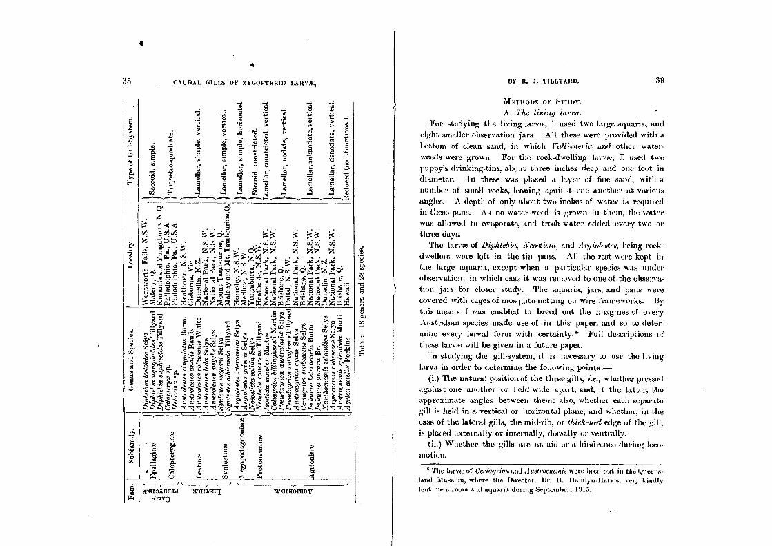
I append herewith a tabular list of all the forms studied. The names given under the type of gill-system are those used in this paper, and will be found defined in their respective sections,
I
• 38 CAUDAL GILLS or ZYGOPTERID LARVA:,
-4 -4 1 -4 --4 .,.>
-4 c 2 "a o o
.9. 8 If. o A. •••••
. e) ,r,"} 0" 17 7.; p. p. r"..;.,. ":3 ti) 0 . . a; .4 -g . I) " ,;. 0
4.. 4, ee C 0
-
0 ai -e in...
o, 0. *C "1:: 03 1 "g 5 TY g E E E ft lf, ..g -
0-, .; "a IA 8 g 0 g "U 31 ''i; a, ' ...T. 1.4- 5 ..5, .21 -16 a ..5. .... .... ..c$ .z• 2
o 1, Tp. -iii I; a> 8 .i' g E 5 8 3 E E E cO) c2 Et 4 4 1 1 I 4 4 4 V, ____-.„..., ,..___„_____.,,,....., ,..........,„,, 4 ,..._.____„,,......„,, r4
s vary CSC H ca
.4,z$113 c4 r6 r/5 rb S 444
.ei"'VZ"4ai c".'" ''="= .. ‘2" j',4 71 4) 7,50 c -4 a g oo".80t•a
It4 o N p_ ,p,;1 .-47 ) :4 l4
v:4 7 7 t ° =4) Y.7;1 TaiScrit14441114 ;li t
.1 1 11 1 11 LI Hill I 111 „zt 3,1 rA .69
...„—_,.....,.... ,...„—,—,......., ,........,___,..._, C a 5' 2?, 0
c 1.".. ...- ' 2' l''. ° 2 g 'r: ..'. C 0
1 ;;;', ▪ , c t c
,, ..`li 0 ° 0
ce 0 ic 0 I. .6 .7..... tli I C. a a) P... N
:z;C..) .4 W ..."-, it, -1, gr.:trial:11CM "NATIIKII
-MVO
BY, R. .1. TILLYARD. 39
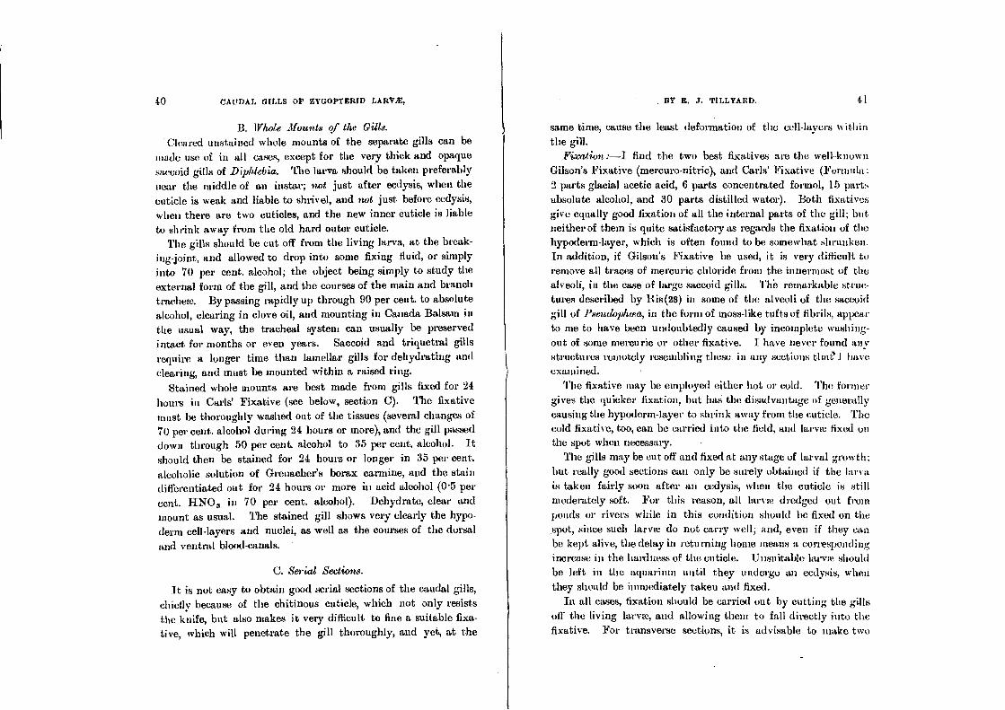
METHODS OP STUDY.
A. The living larva. For studying the living larva, I used two large aquaria, and
eight smaller observation - jars. All these were provided with a
bottom of clean sand, in which Valligneria and other water-
weeds were grown. For the rock-dwelling larvae, I used two
puppy's drinking-tins, about three inches deep and one foot in diameter. In these was placed a layer of fine sand, with a
number of small rocks, leaning against one another at various
angles. A depth of only about two inches of water is required
in these pans. As no water-weed is grown in them, the water was allowed to evaporate, and fresh water added every two or
three days. • The larva! of Diphtebia, Xealtieta, and Avg iolestes, being rock-
dwellers, were left in the tin pans. All the rest were kept in
the large aquaria, except when a particular species was under
observation; in which case it was removed to one of the observa-tion jars for closer study. The aquaria., jars, and pans were
covered with cages of mosquito-netting on wire frameworks. By
this means I was enabled to breed out the imagines of every
Australian species made use of in this paper, and so to deter-
mine every larval form with certainty.* Full descriptions of
these larva will be given in a future paper.
In studying the gill-system, it is necessary to use the living
larva in order to determine the following points:-
(i.) The natural position of the three gills, i.e., whether pressed against one another or held wide apart, and, if the latter, the
approximate angles between them; also, whether each separate gill is held in a vertical or horizontal plane, and whether, in the case of the lateral gills, the mid-rib, or thickened edge of the gill, is placed externally or internally, dorsally or ventrally.
(ii.) Whether the gills are an aid or a hindrance during loco-
ittotion.
The lame of ueriageion and"' uetrocue»a* were bred out in the Queens-land Museum, where the Director, Dr. Hamlyn-Harris, very kindly lent me a room and aquaria during September, 1916.
'yp
e o
f G
ill-
Sy
ste
m.
S C
Tota
l :-
18 g
ener
a an
d 2
8 s
pec
ies.
BY R. J. TILLYARb. 41 40 CAUDAL GILLS OP ZYGOPTERID LARV1E,
B. Whole Mounts of the Gills.
Ch. :tired unstained whole mounts of the separate gills can be
made use of in all cases, except for the very thick and opaque
saccoid gills of Diphiebia. The larva should be taken preferably
near the middle of an instar; not just after ecdysis, when the
cuticle is weak and liable to shrivel, and not just before ecdysis,
when there are two cuticles, and the new inner cuticle is liable
to shrink away from the old hard outer cuticle. The gills should be cut off from the living larva, at the break-
ing-joint, and allowed to drop into some fixing fluid, or simply
into 70 per cent. alcohol; the object being simply to study the
external form of the gill, and the courses of the main and branch
trachete. By passing rapidly up through 90 per cent. to absolute
alcohol, clearing in clove oil, and mounting in Canada Balsam in
the usual way, the tracheal system can usually be preserved
intact for months or even years. Saccoid and triquetral gills
require a longer time than lamellar gills for dehydrating and
clearing, and must be mounted within a raised ring.
Stained whole mounts are best made from gills fixed for 24 hours in Carls' Fixative (see below, section C). The fixative
must be thoroughly washed out of the tissues (several changes of 70 per cent. alcohol during 24 hours or more), and the gill passed down through 50 per cent, alcohol to 35 per cent, alcohol. It
should then be stained for 24 hours or longer in 35 per cent.
alcoholic solution of Grenacher's borax carmine, and the stain differentiated out for 24 hours or more iu acid alcohol (0.5 per
cent. HNO 3 in 70 per cent. alcohol). Dehydrate, clear and
mount as usual. The stained gill shows very clearly the hypo-derm cell-layers and nuclei, as well as the courses of the dorsal
and ventral blood-canals.
C. Serial Sections.
It is not easy to obtain good serial sections of the caudal gills,
chiefly because of the chitinous cuticle, which not only resists
the knife, but also makes it very difficult to fine a suitable fixa-
tive, which will penetrate the gill thoroughly, and yet, at the
same time, cause the least deformation of the cell-layers within
the gill.
Fixation :—I find the two best fixatives are the well-known
Gilson's Fixative (mercuro-nitric), and Carls' Fixative (Formula:
2 parts glacial acetic acid, 6 parts concentrated formol, 15 parts
absolute alcohol, and 30 parts distilled water). Both fixatives
give equally good fixation of all the internal parts of the gill; but neither of them is quite satisfactory as regards the fixation of the hypoderm-layer, which is often found to be somewhat shrunken.
In addition, if Gilson's Fixative be used, it is very difficult to
remove all traces of mercuric chloride from the innermost of the
alveoli, in the case of large saccoid gills. Th'e remarkable struc-
tures described by Ris(28) in some of the alveoli of the saccoid
gill of Pseudophcea, in the form of moss-like tufts of fibrils, appear
to me to have been undoubtedly caused by incomplete washing-
out of sonic mercuric or other fixative. I have never found any
structures remotely resembling these in any sections thaE*1 have
examined.
The fixative may be employed either hot or cold. The former
gives the quicker fixation, but has the disadvantage of generally
causing the hypoderm-layer to shrink away from the cuticle. The cold fixative, too, can be carried into the field, and larva fixed on the spot when necessary.
The gills may be cut off and fixed at any stage of larval growth;
but really good sections can only be surely obtained if the larva
is taken fairly soon after an cedysis, when the cuticle is still
moderately soft. For this reason, all larva dredged out from
ponds or rivers while in this condition should be fixed on the
spot, since such larva do not carry well; and, even if they can
be kept alive, the delay in returning home means a corresponding
increase in the hardness of the cuticle. Unsuitable larva should
be left in the aquarium until they undergo an ecdysis, when
they should be immediately taken and fixed.
In all cases, fixation should be carried out by cutting the gills
off' the living larva, and allowing then, to fall directly into the
fixative. For transverse sections, it is advisable to make two
42 CAUDAL GILLS OF ZYGOPTERID LARVAE,
cuts across the larva with a pair of fine scissors; the first across
the three gills at a level about one-third of their total length
front the base, and the second across the seventh abdominal
segment of the larva. For this method, I am indebted to Dr.
Calvert, of Philadelphia. Its great advantage is that in one
series one can study the general structure of the gills, while in
the other series one combines the structure of the rectum, which is of great importance in the general question of the respiration of these larvae, with the structure of the basal portions of the
gills themselves. For longitudinal sections, I find it best to
sever the gills at the breaking-joint. If the gills are too long, they may be cut across the middle. The last three segments of
the larval abdomen may also be cut off separately, if it is desired to study the rectum and basal pieces in longitudinal sections.
If it is desired to employ the fixative hot, it should be heated in a test-tube to just on boiling-point. Some of the hot fixative
is then poured into a large watch-glass, over which the larva is
held, while its gills are cut off with the scissors. The cut-off
portions usually sink immediately; if any of them float, they
should be at once submerged by means of a camel's-hair brush.
Leave the gills in the fixative until it has cooled to about the
temperature of the room. They should be transferred to 70 per
cent. alcohol (either directly, from Curls' Fixative, or indirectly,
from Gilson's Fixative, through grades of 17, 35, and 50 per
cent). Ili addition, if Gilson's Fixative be employed, the mercuric
chloride must be thoroughly removed by means of iodised alcohol, in the usual manner. In both cases, wash well in several changes
of 70 per cent. alcohol over a period of 24 hours. If used cold, the fixative should be poured into a crystal dish,
and the gills cut off and allowed to fall into it as described above.
The dish must then be covered over, and the gills left in time
fixative for front 12 to 24 hours (not longer) according to the
state of the cuticle. Transfer to 70 per cent. alcohol as describe,_;
above, and wash well.
8w-telling :—Bef ore dehydrating, the gills should always be
subjected to a certain amount of softening, by immersion in a
BY •R. TILLYARD. 43
solution of soft soap in 70 per cent. alcohol. A good rule to
follow is to make the time of softening equal to the time of fixation, e.g., in the majority of cases, 24 hours for both. The
soap will remove the fat from the cells of the fat-body in the
larval abdomen, but will not affect the gill-tissues in the slightest,
except perhaps by a removal of fat front a wandering fat-Cell or
two in the• blood-canals. In the case of very tough gills, taken
from larvae just before ecdysis, a little warming (by standing the
solution on the bath) will expedite the softening.
After softening, the gills must be thoroughly washed in several
changes of 70 per cent. alcohol over a period of at least two days. The last change, when poured off into a small tube and shaken up, should not show the slightest sign of forming a lather.
Dehydration:—Pass up into 90 per cent. alcohol for 6 hours, then into absolute alcohol for 6 hours at the most.
Embedding studying the rectal gills of Anisoptera, ex-
cellent sections were obtained by single-embedding in paraffin. This method is riot recommended for the caudal gills of Zygoptera.
Owing to the thickness of the cuticle, the method of doubte-embedding should always be employed. Some of the larger and
tougher gills resist considerably the penetration of celloidin. Gills of ordinary thickness should be left 24 hours in each suc-
cessive stage, beginning with a 1:1 mixture of alcohol and ether,
and passing up through per cent., per cent., and 5 per cent.
celloidin. Very tough gills may be left 4$ hours in each suc-
cessive stage of celloidin solution. The hardening of the cclloidin
block in chloroform vapour must be very carefully carried out, the block being cut out and shaped when, if anything, it is slightly harder than it would be allowed to beconje if it contained
softer tissues, but still not hard enough to cause any contraction
in the gill. Hardening is then completed by immersion in liquid
chloroform, and the hardened block is infiltrated with, and em-
bedded in, paraffin in the usual manner. The cutting and
mounting of the sections present no features of special difficulty-, other than those usually attendant on dealing with somewhat tough material.

44 cAtibAL GILLS Of'' tYGOP't'hRlb LAttvit, BY R. J. Tfm.yAaD. 45
Staining:—The gill-tissues, exeepting perhaps the hypoderm-
layer, take up stains very slowly. If a series of sections, running
from the rectal region of the abdomen into the basal third of the gills, be arranged on one side, and stained in luematexylin, it
will be found that the body-sections become deeply over-stained
before the gill-seetions are sufficiently stained to allow of differ-entiation. Consequently, sections of gills require to be left in
the stain for a long time, until the hypoderm-layer is strongly
uverstained. In the subsequent differentiation in acid alcohol,
the hypoderm may be allowed to remain slightly overstained, in order to obtain the best differentiation in the internal tissues.
Two double-stains may be recommended as almost equally good
for gill-sections, viz. :—
(1) Ehrlich's Htematoxylin with Eosin. The Eosin only stains
the cuticle and the endotraehem.
(2) Heidenhain's Iron Htematoxylin with Orange G. The
Ifiematoxylin should be used as a purely nuclear stain, the
Orange G as a general cytoplasmic stain. The combination of
the twb differentiates oat the endotraehem and the outer layer of
cuticle in black, the nuclei in brown or greyish-black, and the
cytoplasm in dull orange. For the purposes of this paper, about 40 series of sections have
been prepared and stained by one or other of the above methods .
Many of these sets are serial transverse sections taken right
through from the region of the rectum to near the end of the
gill, and thus running into more than one thousand sections of
10/4 thickness. Much labour may be saved by embedding the
three cut-off ends of the gills close together in one block. In the
case of very long gills Austrolestes), the three eut-off ends
may be arranged close together alongside the piece. containing
the rectal region and the gill-bases, thus reducing the number of
sections to one-half.
NATURE OF TUE CAUDAL GILLS.
In all Zygopterid larvm, the caudal gills are three in number,
viz., a single unpaired median gill, placed dorsally, and a pair of
laterat gills, placed latero-ventrally, to right and left of the
median gill respectively. The median gill is bilaterally symme-trical about the mid-sagittal plane of the larva. The lateral gills are asymmetrical in themselves, but the two gills are symmetri-
eally plaeed, to right and left, with respeet to the mid-sagittal plane.
The median gill is formed from the appendix darsalis of the larva. This is a median outgrowth from the eleventh abdominal
tergite. In position, it corresponds with the telson of Crustacea, and may be eonsidered analogous with that organ. It is not,
however, a true homologue of the telson, since there is strong evidence that the appendix dorsalis of the Odonate larva is a more recent development than the archaie cerei.
The lateral gills are formed from the two cowl, of the larva. These are the original abdominal appendages of the last or
eleventh segment, i.e., outgrowths from the bipartite sternite of
that segment. They are, therefore, the true homologues of the uropods of Crustacea.
Although the cerci, and the appendix dorsalis (when present), pi other insect-larvm, are frequently many-jointed, yet in the
Dragonfly larva they are always either unjointed or only two-
jointed. Throughout the Sub-Order Anisoptera, where caudal
gills are not developed, these three processes are always eornpara-
tively short, hard, and unjointed, forming together a strong anat pyramid, whieh guards the anal opening, and can be opened or
closed at will. In the Zygoptera, as we shall see later, the two-
jointed forms appear to be the older, and we are able to establish the descent of certain unjointed forms from older two-jointed
forms. There are, however, other unjointed forms which show
no evidence of an ancestral two-jointed form. Further, redue-
tions from a two-jointed to an unjointed form appear to have
taken place along several separate lines of descent. Hence we
cannot divide the gills into two main types according to the
number of joints, but must search for a more natural method of classifying them.
Each gill is attached to a short basal piece, viz., the reduced remnant of the eleventh tergite in the case of the median gill,

46 CAUDAL GILLS OF ZYGOPTERID LARNE, BY R. J. TILLYARD. 47
the bipartite remnant of the bipartite eleventh sternite in the
ease of the two larval gills. The muscles which control the movements of the gills are small but strong, and are inserted
into the walls of these basal pieces. The nature of the joint
between the basal piece and the gill is such that, if the gill be
seized by an enemy, it can be cast off without injury to the basal
piece. Such a joint is known as a breaking joint (8, 18).
Morphologically, each caudal gill is a hollow outgrowth of the
body-wall, lined by a tough, chitinous integument or cuticle,
beneath which lies a continuous layer of hypoderm-cells. The
space enclosed by the gill-walls always carries one or more large
longitudinal traehem, from which more or less numerous branch-
trachew pass outwards to all points of the gill-wall. Within the
gill, also, there are always developed two blood-canals, or pro-
longations of the Immoccele. One of these is placed dorsally,
the other ventrally. These two eanals become confluent distally
within the gill, so that there is a continuous circulation of blood_ corpuscles, passing into the gill and out of it again. The trachea
and blond-eanals are supported in a more or less strongly devel-
oped meshwork of alveolar tissue, which fills up all the rest of
the space in the interior of the gill.
The Hypodermic and Cuticle.
The wall of the gill, like that of the body of the larva, is
formed of a tough, chitinous cuticle (cu, cu'), beneath whieh lies
a single layer of polygonal cells, the hypodermis.
The cuticle varies in thickness from 5µ to as much as 15,a,
according to the type of gill. It is thinnest and most delicate
in Saccoid Gills, stoutest and strongest in Lamellar Gills. Along
the mid-ribs of the Triquetro-quadrate and Lamellar Gill-systems it is especially thiekened, and is in such places often armed with
stout spines. The cuticle can always be separated into two distinct parts, an
inner, softer part (cu), apparently formed of a number of thin
parallel layers, and not stainable with eosin, and an outer harder
part (en') of quite uniform structure, easily stainable to a deep
red with eosin, Of the total thickness of the cuticle, the outer
bard layer seldom exceeds one-fourth, though along the mid-rib
of a Lamellar Gill it may occupy as. much as one-half the total thickness.
The cuticle appears to be everywhere clear and unpin rented, though granules of pigment (pg) can frequently be seen adhering to its inner surface at places where the hypodermis has shrunk away from it (Text-fig.1).
At ecdysis, the whole cutiele of the gill is cast off with the
body-cuticle of the larva. A study of the morphology of the
cuticle during ecdysis was not one of the objects of this paper; but one cannot pass over this question without calling attention to the remarkable little ball-like bodies or secretions (also seen
by Ris) which accumulate between cuticle and hypodermis just prior to ecdysis, and which certainly appear to be the first de-
positions of a new cuticle, underneath the old one which is soon to be cast off.
The hypodermis is everywhere a single layer of cells, except at the extreme base of the gills, and, exceptionally, Hnder the mid-
rib of Lamellar Gills, where it sometimes becomes two or three
layers of cells deep. The typical epithelium forming the hypo-
dermis is a layer of rather flat, polygonal cells from 4µ to flµ in thickness (Text-fig.3, Icy). The cell-boundaries are generally indicated by pale intervals separating the darkly pigmented cells,
it being a general rule that the pigment-granules, whether few
or abundant, are most numerous around the cell-nucleus, and tend to be absent from the outer border of the cell. The whole hypodermis may be deeply pigmented (as in Diphlebia), or almost without pigment (as in Argiocnemis). Between these two ex-tremes lie the larger number of gill-types, in which the pigment is chiefly collected into certain areas of dense pigmentation, as, for instance, in the gills of Austrolestes psyche (Plate i., fig.1), Argiolestes icteromelas (Plate i., figs.8-9), Pseudagrion australasite (Plate ii., fig.13), and Austroenemis splendida (Plate ii., figs.18-19). This arrangement often gives a beautiful pattern to the gills, and
may be of value for the protection of the larva in its natHral haunts.

RV R. J. mixAnn 49 48 CAUDAL GILLS OF ZYGOPTEIIID LARVX,
The nuclei of the hypoderm-cells are large prolate spheroids,
usually lying with their major axes parallel to the euticle, but sometimes standing up more or less at right angles to it. When
the hypodermis is exceptionally thin, the nuclei bulge inwards,
so that the inner border of the hypodermis has a wavy outline.
The average size of the nucleus is about 8µ long, by 4kt. wide.
Usually, the nuclei are all of about one size everywhere in the
gill. In the Lamellar Gills, however, the thinnest portions of
the blade, situated towards the distal end of the gill and beyond
the racing, is bordered internally by hypoderm-cells of smaller
size, with smaller nuelei averaging only fiµ to fikt. long.
Beneath the mid-rib, in gills of the Lestid type, the hypoderm-
cells tend to become columnar, and may attain to as much as
3O11 in height. This is particularly notieeable iu the genus
Syniestes, where the alveolar meshwork is greatly reduced..
A very typical section of cuticle and hypodermis is shown in
Text-fig.)1, from the lateral (triquetral) gill of Calopteryx.
The Basal Pieces and Breakingjoint.
Each gill is attached to the tenth abdominal segment by means
of a short, cylindrical basal piece. Heymons(17) has shown that,
in the case of the median gill, the basal piece represents the
reduced tergite of the eleventh abdominal segment; while, in
the case of the two lateral gills, the two basal pieces represent
the two parts (right and left) of the reduced bipartite sternite of the same segment. A section across the basal piece reveals a
large lumen formed of the undifferentiated haemoccele, partially filled by the large tracheae, a small accumulation of fat-cells
► round them, and the small muscles by means of which the gills
are moved. Between the basal piece and the gill proper lies the breaking-
joint, of which sections are shown in Plate iii , tigs.21, 22 (aeo-
sticta). It is along the transverse plane formed by this joint that the gill is cast oft; if seized by an enemy or otherwise entangled, so that the larva desires to free itself. The ingenious arrangement by means of which this can be done, without sufficient loss of blood to be fatal to the larva, can be made out
by a comparison of Text-fig.1 with Plate iii., figs.21, 22. From Text-fig.1, we see that, at the line of junction of the gill-base with the eleventh segment (which is the line of the breaking-joint), the cuticle beeomes suddenly narrowed, and, in particular, the tougher outer portion of the cuticle, which is particularly thick on the basal piece and at the extreme base of the gill, is
Ab io t•...
Text-fig. 1. Basal piece (Bp) and breaking-joint (hj) of right lateral gill of A rgiotextes
icteromdas Selya, ( x 87). Drawn from a cleared whole mount. b tenth abdominal segment; 0, base of gill; Idly, hypoderm-layer
across breaking-joint; tr, single main trachea approaching gill: tr', its two divisions within the gill. Rest of lettering as given on p.100.
greatly reduced. Thus the gill is not only flexible about this joint, but fairly easily detachable also, by reason of the ease with which the thin cuticle can be torn away.
Now if we examine the breaking-joint in transverse section (Plate iii., figs.21, 22), we shall see that, just at this point, the hypodermis grows out into a delicate transverse layer of elongated columnar or spindle-shaped cells, which almost com-pletely close in the lumen of the organ. The haemoccele, which, in the basal piece, is broad and undifferentiated, is here reduced to two narrow canals (dh, vh), which are the beginnings of the dorsal and ventral blood-canals of the gill itself. The rest of the haemoccele is closed up, except for the excessively•narrowed channels in which the main tracheae (tr) run. When the gill is
5

50 CAUDAL GILLS OF ZYGOPTKRID LARVAE, BY R. J. TILLYARD. 51
thrown off, the tracheae and blood-canals are torn away at their narrowest portions, leaving an almost continuous sheet of hypo-dermis to cover the wound. Before any blood could be lost, the contraction of the muscles of the basal piece will have drawn the wound together, so that the blood-canals and tracheae are closed up. A rapid overgrowth of hypoderm-cells then takes place, forming a bulge or tubercle upon which a new cuticle is rapidly developed. From this bulge, by further growth. of the
hypodermis, a regenerated gill will be reconstructed at the follow-
ing ecdysis.
The Alveolar Meshwork (Plate iv., figs.30-33).
This peculiar structure fills the greater part of the interior of the gill in the Saecoid and Triquetro-quadrate Types, and persists
also in the central area or rachis of the Lamellar Types. It was first discovered and described by Dr. F. Ris, in his study of the
saccoid gills of Pseudophcea (28). A translation of his opening sentences on this formation offers us an excellent word-picture
of it ;„-- The interior of the bladder under the hypodermis is filled
with a very queer web-like body ("sehr eigentiimlichen Gewebe-korper"): it is of quite regular alveolar construction, and the individual alveoli are spheroids flattened against each other. The alveoli possess genuine partition-walls ("tichte Wandungen") made from a substance which, as far as my optical aid (Leitz Irnm. 1/12) went, is structureless and only weakly colourable with htematoxylin; examination in series shows that in each alveolus there lies one firm cell-nucleus, and only one, so that the supposition arises that each alveolus is the product of a
single cell. A careful study of the alveoli in numerous sections through
gills of Diphlebia, Neosticta, and Calopteryx convinces me that Ris' description is correct, except in one point. It is true, as a general rule, that if one follows a single alveolus through a number of sections, from the point at which it first appears as a minute area between two larger alveoli, to the point at which it
finally disappears, one and only one nucleus can be seen upon its
wall. The exceptions to this occur so seldom (I have seen one alveolus with three nuclei upon its wall, a few with two, and perhaps a score or more with none at all, out of thousands ex-amined) that they are scarcely worth considering. But it is also true that the nucleus that does duty for one alveolus is fre-quently the same nucleus that counts for an adjoining alveolus, or even for two other alveoli. This fact seems to have escaped Ris' attention; but it is nevertheless a very important piece of evidence when we come to discuss the true nature of the alveoli. I find that, as a general rule, the nucleus is situated at a point of union between the wall of one alveolus and the walls of one or more adjoining ones. In other words, the walls of separate alveoli are not distinct structures, each the product of a single cell, but are formed by fusion of a number of branching eells having nuclei at their centres. Consequently each alveolus is not, as Ris' supposition would indicate, the hollow interior of a single cell, but merely a small portion of the original hoemoccele, elosed off by the ingrowth and fusion of cell-processes around it.
If my interpretation of the structure of the alveoli be correct, we should expect to find nothing actually inside the alveoli themselves except blood-plasma and, perhaps, - an occasional anuebocyte; whereas, if Ris' interpretation be the true one, it would be reasonable to expect some internal structure sui genesis within the alveolar cavity.
Now Ris has figured (28, Plate 6, figs.17-20) and described at some length, some extraordinary structures from the interior of the alveoli. He says (I translate from the original German) :-
One part of the above-mentioned alveoli is full of very rich and delicate ramifications in moss-like branching tufts of stand-ing fibrils (" reichen and zierlichen Verzweigungen in moosartig veriistelten Biischeln stehender Fibrillen") In one alveolus one single fibrillar tuft of this kind appears to be predominant; but formations are not laeking in which a number of smaller tufts are found in near or distant parts of the same alveolus. Other formations are also to be noticed as stages in the develop-ment of these fibril-tufts button-shaped and closely packed together, running out from a narrow base in countless threads,

52 CAUDAL GILLS OP ZYGOPTRRID LARVA:,
as yet stout and unbrariched; or else the length of the threads is considerably greater, but the resolution ("Auflosung ") of the tufts is not yet complete, not yet filling up the alveoli The individual fibrils of these tufts seem to he very regular in thick-ness and of extreme fineness; I cannot discover any other
formation in them with my aids; the individual pale hiematoxy-lin colouring for a single cell is quite uniform. It has already been said that only one part of the alveoli contains these tufts; the division is by no means regular; but, in reality, both in the
ease of the median as well as the lateral gills, there is a central
zone of empty alveoli surrounded by a peripheral zone of alveoli filled with fibrils; towards the distal end of the organ the number of empty alveoli decreases more and more, so that at the spot where the two large blood-vessels unite only. very few are left empty The alveoli containing blood lie predominantly on
the periphery of the organ near the hypodermis. The alveoli containing fibril-tufts contain these only, and are otherwise
emyty. At first sight the appearance in a few places of sus-pended fragments of fibril.tufts in the middle of the tumen of the
big blood-vessel seems to he very striking; but in at least one place
(the series is not quite without a break) it is possible to refer
this phenomenon back to a group of fibril-tufts which is here
clinging to the watl of the big blood-vessel, in all other respects as
on.the wall of the alveolus (" wie sonst der Alveolenwand "). I have placed in italics two statements near the end of this
quotation, beeause it seems to me that they, taken in conjunc-tion with Ris' excellent figures, three of which are reproduced on Plate iv., figs.31-33, give us the obvious solution of this ex-traordinary formation. Having carefully studied numerous series made from carefully fixed and prepared gills14 the saccoid
type, and one of which (Diph,lebia) is closely enough allied to
Pseuclophcea to admit of no doubt as to the unity of structure in
the gills of the two genera, I have no hesitation in saying that
the formation so carefully described by Ris is entirely absent
from all sections which I have examined. I am bound to con-clude, therefore,—and I must call as evidence for my conclusion both the two statements in italies and Ris' own figures— that
i3Y R. J. TILLYAIII3.
these struetures are undoubtedly artefacts, being nothing more nor less than a erystallisation of some ingredient used in the preservation of the gills studied by Ris.
Ris himself says of these structures:—I shall not disguise the fact that my first impression on seeing them was that of an artificial product, at first of crystals of a fatty acid, then of a fungoid groWth. Neither of these impressions stood the light of clear observation, and I am convinced that we are dealing with a certainly extremely curious structure peculiar to this organ.
The reasons for concluding that Ris' first impression was the right one, i.e., that the fibril-tufts are artefacts, may be briefly stated as follows :-
1. They do not occur in any type of saccoid gill, so far studied,
which is known definitely to have been properly fixed, and to have had the fixative completely extracted.
2. They do not occur in the lateral abdominal gills of Pseudo-plvect, the internal structure of wide'', on Ris' own showing, is
a miniature reproduction of the saccoid gills as far as the alveoli are concerned.
3. They occur just in those parts of the organ (viz., the peri-
pheral alveoli) where they would be found if (a) the larva had been partially fixed, and the fixative not washed out; or if (b) the larva had been greatly over-fixed, so that only the more open central portion, with the blood-canals, had subsequently been freed of fixative. •
4. The fact that fibril-tufts are found adhering to the wall of the large blood-canal, and some even suspended in its lumen (sec the italicised portions of the quotation from Ris given above) seems to me to be fatal to the argument that they are a special
or peculiar structure found only in this type of gill. Either they are a formation produced within the alveoli part of the living cell (it is clear that Ris was inclined to regard each alveolus as
a single cell), or they are a foreign body. If the former, they
could not occur also on the wall of the blood-canal, or free within its lumen; for there is no connection between the blood canal and the alveoli.

CU. ..... 7 '
hy. ..S de °COCCI
he
00000013
A. "by
al n
54 CAUDAL GILLS OP ZYGOPTERIG LARVA,
5. The tufts figured in Ris' photograph, shown on Plate iv., fig.33, undoubtedly extend just beyond the limits of the alveolus
shown.
6. Ris' own photographs show a structure quite unlike any-
thing known to occur in the animal kingdom, and bearing on
their very face the stamp of artefacts. I may add that these photographs were submitted to both
Professor Haswell and to Dr. S. J. Johnston, of the Zoological Department, University of Sydney, and both of them pronounced
them to be artefacts without any doubt whatever. It seems necessary to go into this matter at some length, in
order to prevent further speculations as to the function of these
fibril-tufts. The fascinating theory that each alveolus might be
a single cell, in which special end-organs (i.e., the fibril-tufts)
were developed, for the purpose of extracting oxygen from the alveolar contents (water, air, or bloodl) and passing it into the tracheal system, can no longer be entertained. We must look for some simpler, though more prosaic, explanation of the physi-
ology of the caudal gills. Let us now return to the question of the structure of the
alveoli themselves. Here a study of these organs in all positions
within the gill, and in all different types of gill available, is essential to aright understanding of their nature. Of partieular
value is a careful study of those alveoli, few and comparatively
large in number, which lie in the rachis of a lamellar gill; since
in this case the mind is not confused by their immense number, and their relationship with the hypodermis is easily made
apparent. In those alveoli which lie closest to the hypoderm-layer, it is
easily seen that the wall of the alveolus merges into the inner-
most stratum of the hypoderm-layer, as shown in Text-fig.9. It would appear that the hypoderm-cell is capable of throwing off a portion of its substanee in the form of a delicate layer, either
from its inner or outer border. The continuous layers so thrown off along the whole hypoderm-layer upon its outer border form
the separate strata of which the cuticle is composed. Between the layer last thrown off, and the living hypoderm-cells, one finds
RY R. J. TILLYARD.
a layer of dust-like pigment interposed. Now an examination of the alveolar wall shows (as far as magnification by a 1/12th oil-immersion lens is able to show) a very uniform, lightly-stain-ing substance closely resembling the innermost stratum of the cuticle, but without any pigmentation. One would not hesitate to pronounce this delicate layer to be chitin, were it not for the
presence of the alveolar nuclei. These, however, give the key to the structure. If one follows carefully through a series of transverse sections of a complete gill from the base outwards, it
Text-fig. L. Diagrammatic representation of formation of alveolar meshwork. A, the
two walls of the gill separated by the hiemocmle (he). B, migration of hypoderm-cells (a/c) into the Menioctele, with outgrowth and union of their processes. C, the alveolar meshwork completed by differentia-tion of the same cells into alveolar nucleus (ala) and alveolar wall. al, a single alveolus enclosing a portion of the originat, Inemocmle. Rest of lettering as on p.109.
is clearly to be seen that, near the base, a number of hypoderm-cells do not lie in the regular epithelium, but project out across
the gill-cavity, with their nuclei placed irregularly within it. These cells unite across the interior of the gill, where, indeed, at the breaking-joint, they form an almost complete transverse
layer. Passing further into the gill, we find these same cells
becoming exceedingly irregular and attenuated, with the nuclei few and far between, and large spaces of the nnoccele lying
MT R. J. TILLYARb. 57 56 CAUDAL GILLS OF ZYGOFTERID LARV.E,
enclosed within the fused network formed by the cells. These
are the first alveoli. In Text-fig.2, I have attempted to show
how the formation of the alveoli is brought about. It would be difficult to deeide the question as to the exact
composition of the alveolar wall, were it not for a fortunate circumstance in connection with the fixation of the hypoderm-layer. Within the gill, the hypodermis carries on its inner margin a fine basement-membrane, which, luckily, becomes
detached in places, either by the action of the fixative, or per-
haps during sectioning. This delicate membrane closely resem-bles the innermost stratum of the cuticle before it becomes hardened up. Now, in many places it can be seen that the alveolar wall is continuous with this basement-membrane, and there is no change in structure as we pass from the basement-
membrane proper into the interior of the gill. Again, if we examine the nuclei upon the alveolar walls (Plate
iv., fig.30), we find them to be exactly similar in size, shape,
shining qualities, and contents, to the hypoderm-nuclei found along the epithelial layer. We must conclude, therefore, that
the alveolar meshwork is the product of numerous hypoderm-celts which have grown into the interior of the gitl, and have enclosed . within their folds a number of spheroidal chambers (alveoti) whose, interiors were originally portion of the licvmocwle.
The fact that blood-coagulum, with an occasional amcebocyte
(but never, as far as I have seen, any miocytes) is often seen within the alveoli, bears out the above statement. 1 If the alveolar cavity were the interior of a single cell, how indeed could blood-plasma become enclosed within it7 But if the meshwork grows, as I have indicated, by the branching and
fusion of numerous cells throughout the hmtnoccele, then it becomes a certainty that some at least of the blood must become enclosed within it. As the miocytes continue to travel regu-larly in the course of the main blood-circulation, which becomes
closed off, as the meshwork grows, in the form of two blood-
canals, it is not surprising that we do not meet with these
corpuscles in the alveoli. Even if one were accidentally enclosed, we may be fairly sure that it would cease to keep its oat•shaped
form, and would settle down near the alveolar wall in the form of an ammbocyte. The absence of miocytes within the alveoli simply proves the absence of any definite circulation of blood within them.
The Internal Lamina. in all caudal gills which I have examined, except the Lestia
and Reduced Types, the alveolar meshwork is seen to be strengthened, along certain definite zones, by a series of thicker ingrowths from the hypoderm-layer. These ingrowths were first noticed. in. the larva of Pseudophcea by Ris (28), who termed them "lamellen." As, however, the term "lamella " has already been employed more generally for the flat, blade-like gills of Ledithi, and Agrionidce, I propos° to substitute the term internat tamina for these ingrowths, in order to avoid confusion.
In all forms of. caudal gill, the internal lamina: occur chiefly
along two well-marked longitudinal zones. One of these zones would be represented, in all median gills except those of the Horizontal Lamellar Type, by a plane perpendicular to the plane of symmetry of the gill, at a level slightly dorsad from the
dorsal blood-canal. Dorsad from the first plane, and ventrad
from the second, more scattered internal lamina; are developed: but I have not found any general development of lamina in the more central area between the two blood-canals. Only in the Agrionid gills of the Vertical Lamellar Type have I found laminae developed just ventrad from the dorsal blood-canal, or dorsad from the ventral blood-canal.
In lateral gills of the Vertical Lamellar Type, the zones of
laminae agree with those of the median gill. But in lateral gills
of the Triquetral Type, the zones may best be described as being
developed along two planes which cut off, in any given cross-
section, the upper and inner corners of the triangle representing the section. These two planes are therefore not parallel, but somewhat inclined to one another inwards. The same holds good in the lateral gills of the baccoid Type. The positions of
the two principal zones of laminae are in all cases clearly repre-
sented in the series of diagrammatic sections Text-figs.11, 17, 22 (di, vl),
C4, Cu
pg
by _
58 CAUDAL GILLS OF ZTDOPTRRID LARv.t, Sr R. J. TILLYARD. 59
In the Horizontal Lamellar Type, the horizontal flattening undergone by the gill has resulted in a more or less complete suppression of the principal zones of laminae. Numerous second-ary short laminae are developed at intervals across the interior
of the narrow blade of the gill (Plate vi., fig.43,i1, In the
Lestid form of Vertical Lamellar Gill, the two walls of the lamella lie mostly so close together that the internal lamina , are
not developed at all (Plate v., figs.36, 37). I propose to call all the laminae developed along either of the
principal zones of the gill the principat internal laminae. All
the rest I shall term the secondary internal laminae. There is
no difference in their structure. But, along the principal zones, the laminae occur more regularly and in greater number, forming a kind of open fence across the gill. If a lightly-pigmented
Vertical Lamellar Gill (e.g., that of Ischnura heterosticta Burn].)
be stained in toto with borax-carmine, cleared and mounted, the
two sets of principal internal lamina will appear like two long straight rows of slender colonnades, rising from the " floor " to
the `goof " of the gill. The two principal zones of laminae separate off the central
thicker portion 'of the gill, termed the rachis, with its main
trachem, nerves, and blood-vessels, from the two narrower outer
portions, which, in the case of Lamellar Gills, together form the
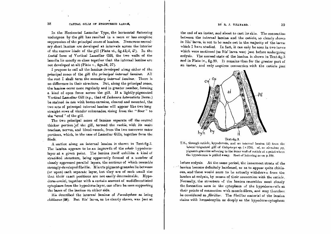
blade. A section along an internal lamina is shown in Text-fig.3.
The lamina appears to be an ingrowth of the whole hypoderm-
layer at a given point. The lamina itself exhibits a kind of
stratified strueture, being apparently formed of a number of closely appressed parallel layers, the sections of which resemble strongly-developed fibrillae. Minute pigment-granules lie between (or upon) each separate layer, but they are of such small size that their exact positions are not easily determinable. Hypo-
derm-nuclei, together with a certain amount of undifferentiated cytoplasm from the hypoderm-layer, can often be seen supporting
the bases of the lamina on either side.
His described the internal lamina of Pseudophcea as being
chitinous (28). But Ris' larva, as he clearly shows, was just at
the end of an instar, and about to cast its skin. The connection
between the internal lamina and the cuticle, so clearly shown in Ris' larva, is not to be made out in the majority of the larva! ,
which I have studied. In fact, it can only be seen in two larvae
which were sectioned (as His' larva was) just before undergoing
ecdysis. The normal state of the lamina is shown in Text-fig.3 and in Plate iv., fig.29. It remains thus for the greater part of an instar, and only acquires connection with the cuticle just
Text-fig.3. T.8., through cuticle, hypodcrmis, and an internal lamina (d) from the
lateral triquetral gill of Calopteryx sp. ( x 370). al, an alveoluat, pt, pigment-granules adhering to the inner wall of cuticle at a point where the hypodermis is pulled away. Rest of lettering as on p.109.
before ecdysis. At the same period, the innermost strata of the
lamina become definitely hardened, so as to appear quite chitin-ous, and these would seem to be actually withdrawn from the lamina at ecdysis, by means of their connection with the cuticle. Normally, the structure of the lamina resembles most closely
the formation seen in the cytoplasm of the hypoderm-cells at their points of connection with muscle-fibres, and may therefore be considered as fibrillar. Thefibritlar. material of the lamina stains with Inernatoxylin as deeply as the hypoderm-cytoplasm
so dAUDAL GILLS OP gYGOPTICRIb !ARV*,
itself, and remains unstained by eosin. Also, it is bordered, in the hypodermis itself, by a fine basement-membrane, seen as a dark line under high magnifications. Further, in all larval
stages except just before ecdysis, a definite pigment-layer inter-venes between the cuticle and the base of the internal lamina,
as shown in Text-fig.3. As to the formation of these internal lamina, I must. express
my agreement with Bis' opinion that this lies partly upon the mechanical side, viz., that they act as additional supports for the alveolar meshwork in the interior of the gill. But they do
not exist for this purpose only. They carry the basement-mem-
branc of the hypodermis along their entire length; and they show, at frequent intervals, connections with the alveolar walls themselves. It seems almost certain, though it cannot be proved by actual examination, owing to the excessive fineness of the
meshwork, that a thin layer of hypoderm-cytoplasm must con-nect all the nuclei of the alveolar meshwork, and that the laminae arc the principal bearers of this cytoplasm into the gill-interior. In ogler words, where single or few hypoderm-cells have passed
inwards and helped to form the meshwork, their points of con-
nection with the hypoderm-layer are indicated simply by the passage of the basement-membrane into the alveolar wall. But
at those points where masses of hypoderm-cells have been thrown
inwards, a lamina is formed, and much of the surrounding mesh-
work is probably due to the further ingrowth of the cells carried in along the lamina. Whether the cytoplasmic connections between the scattered nuclei of the meshwork persist throughout larval life or not, we know that they must have existed during
the formation of the meshwork, and that they must continue to
exist so far as additions to the membrane are made with the growth of the larva at each ecdysis. 1 see, therefore, in the internal laminae, not only a. mechanical device for the support of the meshwork, but also the principal carriers of the hypodermis
inwards for the formation of that meshwork.
The Btood-Canals.
In the abdomen of the larva, the blood travels forwards
through the heart and aorta, backwards in the general body-cavity
BY R. J. TILLYARD.
or inemoccele. This backward movement occurs both dorsally and ventrally. The main stream runs backwards ventrally,
above the ventral nerve-cord, but below and latero-ventrally to the alimentary canal. A smaller stream runs backwards more
dorsally, just below and on either side of the heart itself. The change from backward to forward current takes place in
the last three segments of the abdomen, and is entirely due to the suetional pull through the ostia of the heart. These Ostia are situated in the eighth and ninth segments. Thus some of the blood will not travel ventrally backwards beyond the eighth segment, being sucked into the ostia of that segment. Similarly, some more blood will not get beyond the ninth segment. A portion of this circulates only around the confines of the anal area, before it returns to the ostia of the ninth segment. The
rest, comprised in three distinct streams, passes into the caudal gills, and circulates along almost their entire length (i.e., to the confluence of the two blood-canals, and back again).
This circulation, and the vessels in which it takes place, can best be seen from Text-fig.4. In the basal piece of each gill, the invinoccele is large, and continuous with the general body-cavity. As, however, it approaches the level of the breaking-joint (bj), it narrows rapidly, and finally passes through the extensive hypo-derm-layer at the breaking-joint as two minute canals, one situ-ated dorsally, the other ventrally. . On entering the gills, these
two canals widen out considerably, and become the closed-in btood-canals of the gill. Of these two, that which carries the blood into the gill maybe termed afferent and primary, the latter term indicating (as we shall discover when we study the onto-geny and phylogeny of the gill) that it was in existence long
before the second blood channel was formed, at a time when the efferent part of the circulation was effected (like the backward circulation in the abdomen), merely along the undifferentiated
htemoccele of the gill. The other blood-canal, which carries the blood away from the gill; may be termed efferent and secondary.
In order to examine the circulation of blood in the gills, it is necessary to select a transparent, lightly pigmented larva For this purpose, amongst the Australian-species available, the larva

4.
7
62 CAUDAL GILLS OF ZYGOPTERID LARY4E,
of Ischnura heterosticta is easily the most suitable. If placed in
a small dish or watch-glass, this larva will persist in resting with its gills in a vertical plane (their natural position). To overcome this difficulty, I found it best to remove the larva
altogether from water, and place it sideways upon a glass-slide.
A cover-slip was then let down upon the gills, and water from a pipette was run into the wedge-shaped space between the slide and the cover-slip. Thus the gills were enclosed in water, while the rest of the larva was in air. In such a position, the larva
will live for some hours, and seldom makes any attempt to move.
10
1 10
111:;'..:
''''
_....
"I 5)
Text-fig.4. Diagram of the blood-circulation in the caudal gills and last three abdo-
minal segments of Ischnura heterosticta Burm. (Middle portions of
gills cut away). The seven currents shown are as follows :-1, dorsal circulation in seg. 8; 2, ditto in seg. 9; 3, circulation in median gill
(mg); 4, ventral circulation in seg. 8; 5, ditto in seg. 9; 6, ditto in
seg. 10, with extension to left cercoid (c); 7, circulation in left lateral
gill (ly). a, anus; bj, breaking-joint; c, cercoid; cf, confluence . of
blood-canals in gill; dh, dorsal blood-canal; h, heart; ng, ganglion of
seg. 8; ast, ostia; oh, ventral blood-canal; 8-10, abdominal segments.
The circulation of blood in the gills is very remarkable. I had naturally expected to find the blood entering the gills by the ventral canal, and passing out of them dorsally. But I
BY R. .1. TILLYARD. 63
found that this is only true for the lateral gills. In the median gill, the circulation is reversed, the blood passing in dorsally, and passing out again ventrally. As will be seen in Text-fig.4, the blood that enters the median gill (the line of arrows marked
3 in the figure) is part of the dorsal backward blood-stream. On
reaching the base of the gill, a single pulsation of the heart causes it to shoot sharply along the narrow dorsal blood-canal for some considerable distance. It then travels by a series of shorter jerks (each corresponding with one heart-beat) along the
dorsal canal, and round the distal confluence (cf) into the ventral eanal. Along the ventral canal it passes forwards again by short jerks, until at last it passes out of the gill in the same sharply shooting manner as that by which it entered. The distance covered by a given corpuscle in the jerks of entry and exit appeared to me to be between two and three times as long as
the distance covered in a single ordinary jerk. I think the increase must be wholly due to increased pressure forcing the
blood along at the breaking-joint, where the canals become sud-
denly narrowed-- just in the same way that a river flows fastest
in the narrowest part of its bed.
Thus we see that, in the median gill, the dorsal blood-canal is
afferent and primary, the venventratoad-canal efferent and
secondary.
Turning now to the lateral gills, the course of the blood in the
left lateral gill is marked by the line of arrows marked 7 in Text-fig.4. This is part of the main ventral backward stream of the blood along the left side of the abdominal hwmoccele. A .
similar stream along the right side supplies the right lateral gill.
Each of these streams enters the lateral gill on its own side by - the ventral canal, in the same sudden shooting manner as that
already noticed for the median gill. Passing, by a series of jerks, distally to the confluence (cf ), the blood returns along the dorsal canal. On passing out from the gill, the blood passes
obliquely upwards and forwards towards the heart, and meets with one portion of the blood-stream emerging ventrally from the median gill, since this latter stream is constrained, on approach-ing the heart, to divide into two in order to avoid the blind end
lg ...

64
CAUDAL GILLS OF ZYGOPTRIIID LA RWE,
of the heart itself, and the ligaments connecting it posteriorly to the wall of the tenth segment. Thus, finally. the blood from all three gills enters the ostia of the ninth segment in two streams, one from the right, the other from the left of the heart.
In the lateral gills, therefore, the ventral blood-canal is aPrent and primary, the dorsal blood-canal e.fferent and secondary.
In Text-fig.4, the seven lines of arrows numbered 1-7 are
intended to show all possible courses for any blood-corpuscle entering the eighth segment from in front. Of these, only Nos.
3 and 7 concern the gills. It should be noticed that portion of
No.6 (the ventral circulation of seg. 10) passes into and out of the larval cercoids (c), in which no blood-canals are developed.
The difference in the circulation of blood in the' median and
lateral gills puzzled me greatly, and I could find no explanation
to account for it, until I thought of studying the caudal pro-
cesses of the May-flies (Plectoptera). Then, as I hope to explain more fully in Part iv., I discovered that, in this Order, each
process possesses only one blood-canal, which is dorsal in the medikn process and ventral in the lateral process. Also, in these caudal processes of the May-fly, the blood passes iwio the process along the closed canal, the return journey being made along the open haemoccele of the process-cavity. Thus it became evident that the afferent canal, whether dorsally or ventrally
placed, must always be the primary canal, originally present in
the organ before it took on the form now seen in the Zygoptera; whereas the efferent canal is only a secondary formation, being in fact nothing more nor less than what is left of the original open haemoccele of the gill, after the elaboration of the alveolar
meshwork, which occupies by far the greater volume of the
haemoccele. Further interesting evidence along the same lines
may he expected from the study of the Ontogeny of the Caudal Gills themselves.
The actual circulation of the blood-corpuscles in the gill seems
to be entirely confined to the two blood-canals. That is to say, although blood-plasma is to be found in the nerve-canals, some of the alveoli, and occasionally in the small spaces of the haerno-=le around or near the tracheae (Text-fig.9, Up), and although
. BY R. J. TILLYARD. 65
occasional large amcebocytes (Text-fig.9, ac) may also be found in these positions, being probably caught and imprisoned during an ecdysis, yet the travelling oat-shaped corpuscles or nziocytes (Inc) are never found, in sections, anywhere in the gills except in the blood-canals. A typical group of blood-corpuscles, including
both miocytes and amcebocytes, is shown, greatly enlarged, collected near the wall of a blood-canal, at a point where it impinges upon the hypodermis, in Text-fig.5.
Text-fig.5. Blood-corpuscles in blood-canal near base of gill of A ustroagrion cyane
(Selys). From a transverse section ( x 1000). Lettering as on p.109.
The Nervous System. The Nerve-Supply of the Caudal Gills is, as might be ex-
pected, of very uniform structure throughout the whole Sub-order, and appears to be but little affected by changes in the
form of the gill itself, or in the number of principal tracheae supplying it. The typical distribution of nerves to the gills, here described, was found in all forms examined by me in the families Calopterygidce and Agrionidce. In the Lestidce, the distribution is altered by the suppression of one pair of principal
nerves, in the median gill only. The nerve-supply of the lateral
gills remains the same in all three families.
The last abdominal ganglion of the ventral nervous system of the Dragonfly Larva is the large ganglion lying in the eighth
segment. This ganglion is formed by the fusion of three original
pairs of ganglia, viz., (hose of the eighth, ninth, and tenth
abdominal segments. It gives off four pairs of principal nerves, viz., the genital nerves, the eighth segment nerves, the ninth segment nerves, and the tenth segment nerves, the last being the
6

mcg
Icg'
66 CAUDAL GILLS OF ZYGOPTERID LAnV/E,
most posterior pair. The whole nerve-supply of the caudal gills
is supplied from these last. On entering the tenth segment, the tenth segment nerves lie
latero-ventrally on either side of the rectum. At about one-
third of the distance through the segment, each nerve divides into two strong branches, one passing slightly more dorsad, the other slightly more ventrad, and the former bending slightly outwards away from the level of the original nerve. Of these two branches, the ventral soon gives off a strong nerve to the
wall of the rectum, the posterior rectal nerve (Text-fig.6, rn), and
nvo
B. Text•fig.6.
Diagrams to show positions of principal nerve of the gills, A dorsal, B
lateral view. a, anus; Alt, abdomen; c, cercoid; dl, dorsal nerve of
left lateral gill; dr , ditto of right lateral gill; dnt, left, and dm' right
dorsal nerve of median gill; tcg, left, and teg right caudal gill; nicg, median caudal gill; asi ,,, left, and right tenth segment nerve; R, rectum; rn, posterior rectal nerves; vi , ventral nerve of left lateral
gill; yr, ditto of right lateral gill; vm, left, and ytn', right ventral
nerve of median gill. Reconstructed from series of transverse sections.
then, running straight on, it enters the base of the lateral caudal
gill of its own side, forming the ventral nerve of the lateral gilt
(vt, v1'). This is a large nerve, lying very conspicuously upon
BY R. J. TILLYARD. 67
the wall of the ventral blood-canal, on the side farthest from the plane of symmetry of the gill-system. The other, or dorsal, branch divides into two a little further on, one branch continu-ing a fairly straight course, a little outside and dorsad of the level of the nerve vl , to enter the base of the lateral gill of its own side, as the dorsal nerve qf ihe lateral gill (dl, dl')—a nerve considerably smaller than vi, but placed in a similar position to it upon the wall of the dorsal blood-canal.
The second branch passes. rapidly upwards and inwards over
the anal opening, and branches into two just before entering the base of the median gill. Its two branches form the dorsat and ventral nerves of the median gill (dm, vm) respectively, of its own side. A corresponding pair of nerves enters the median gill from the other side. Thus, the nerve-supply of the median gill, like that of the median ocellus, is a double one, symmetrical about the plane of symmetry of the larva.
In sections of the gills of Synlestes and Austrolestes, 1 can find no trace of the two ventral nerves of the median gill. It would
appear, therefore, that these nerves are suppressed, in the family Lestidre. On the other hand, it is easy to trace several strong
nerves arising from the dorsal nerves at various levels along the gill-length, and passing closely around one or other of the two main longitudinal trachea, to supply the ventral portion of the
The structure of one of the main dorsal or ventral gill-nerves may be seen from Plate vi., figs.44, 45. A cross-section passes through a considerable number of parallel nerve-fibres, the nuclei of which are found at fairly frequent intervals along the course of the nerve. Frequently the same section will intercept two nuclei, as in Plate vi., fig.45, and occasionally three. The nuclei
occur in all positions, either near the centre, or upon the circum-ference, but seldom project beyond the regular periphery of the
ner ye. The main gill-nerves retain their position either in, or very
close" to, the walls of the main blood-canals, throughout the whole length of the gill. Only in the Saccoid Gill-type are they removed any distance from these canals. In this type, also, their
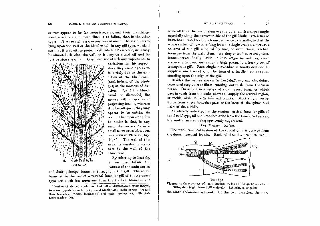
1111.1i it1"41151164011110k■0111011(il olounionun •
111111111111111 Itaite r .0005,clomm111111111111111011111101 I (5 ((NM (pp” ilIll' A01111111■Immt0
,Iurfanititurailliuminlimmomni -qt ill
met, ftwilligfill111111111111111111111(1111111
68 CAUDAL GILLS OF ZYGOPTERID LARV1E,
courses appear to be far more irregular, and their branchings more numerous and more difficult to follow, than in the other types. If we examine a cross-section of one of the main nerves lying upon the wall of the blood-canal, in any gill-type, we shall see that it may either project well into the Inemoccele, or it may
lie almost flush with the wall, or it may be closed off and lie
just outside the canal. One need not attach any importance to 0 a u 0 0 variations in this respect,
° o since they would appear to
be mainly due to the con- dition of the blood-canal
,„ (and, indeed, of the whole
00° gill) at the moment of fix- 0 0 0 (I
0
0 case, the nerve runs in a 0 0 0
0 as shown in Plate vi., figs.
44, 45. The wall of this
canal is similar in struc- ture to the wall of the
o „ - 0 blood-canal. By referring to Text-fig.
7, we may follow the
courses of the main nerves
and their principal branches throughout the gill. The nerve- branches, in the case of a vertical lamellar gill of the A yrionid
type. are much less numerous than the tracheal branches, and
* Portion of stained whole mount of gill of Austroagrion vane (Selys),
to show hypoderm-nuclei (nu), blood-canals (hm), main nerves (ay) and
their branches, internal laminae (it) and main trachea, (tr), with their
branehes:T( 1 00 ).
BY R. J. TILLYARD.
come off from the main stem usually at a much sharper angle, especially along the narrower side of the gill-blade. Such nerve-branches themselves branch once or twice externally, so that the whole system of nerves, arising from the single branch, innervates an area of the gill supplied by two, or even three, tracheal branches from the main stem. As they extend outwards, these branch-nerves finally divide up into single nerve-fibres, which are easily followed out under a high power, in a freshly cut-off transparent gill. Each single nerve-fibre is finally destined to
supply a small scnsilla, in the form of a tactile hair or spine, standing upon the edge of the gill.
Besides the nerves shown in Text-fig.7, one can also detect occasional single nerve-fibres running outwards from the main nerve. There is also a series of stout, short branches, which pass inwards from the main , nerves to supply the central region, or rachis, with its large tracheal trunks. Short single nerve-fibres from these branches pass to the bases of the spines and hairs of the midrib.
As already indicated, in the median vertical lamellar gills of the Lestid type, all the branches arise from the two dorsal nerves, the ventral nerves being apparently suppressed.
The Tracheal System.
The whole tracheal system of the caudal gills is derived from the dorsal tracheal trunks. Each of these divides into two in
ation. For if the blood- canal be distended, the nerves will appear as if projecting into it, whereas if it be collapsed, they may appear to be outside its wall. The important point
to notice is that, in any
small nerve-canalof itsown,
Text -fig.8. Diagram to show courses of main tracheae at base of Triquetro-quadrate
Gill-system (right lateral gill omitted). Lettering as on p.109.
the ninth abdominal segment. Of the two branches, the more
30
000 0°0
0 0 0 0 0 0 00 ;4:° 00 c, po 06,
tin nv hin 6 .11 fiVbm Text-fig.7.*
0 0

70 CAUDAL GILLS OP ZYGOPTERID LARVX, RP R. 3. TILLYARD. 71
ventral one passes slantingly downwards to enter the base of the lateral gill of its own side. On entering the gill, this brauell again divides into two, in all except the Lestid sub-type of gill-
The more dorsal branch runs straight back through the tenth segment to enter the base of the median gill, alongside its fellow from the other dorsal trunk. Thus the median gill receives two
main tracheal stems at its base. In all Lamellar Types of gill, these two trunks run alongside one another through the gill, giving off numerous branches. In the Saccoid and Triquetro-quadrate Types, however, each divides, on entering the gill
proper, into two large tracheae, of which one is placed dorsally above the other. Thus, in these types, the median gill has four
parallel main tracheae running through it. This arrangement is
shown diagrammatically in Text-fig.8. The courses of the main tra-
cheae, their manner and frequency of branching, and the form of their smaller branches or "twigs," vary so much that it seems advisable to
leave the description of these parts
under the sections dealing with the separate types of gills.
The structure of the tracheae in the caudal gills does not differ in
any respect from that of tracheae in any other part of the larval body. Being derived from the dorsal trunks, they are, like these latter, always deeply pigmented,
the pigment-granules lying in the
ectotrachea. This pigmentation is found throughout the tracheal
system, from the main stems down to the final branches from
* T.8., through a main trachea in gill of Calopteryx sp. ( x 370), to show
reduced portion of Inemociele (hc), [containing blood-plasma (hip) and iumebocytes (ac)], in which the trachea lies. at, alveolus; ect, ectotrachea. end, endotrachea; il, internal lamina; nu„ nucleus of ect; nu„ nucleus of end.
which the capillaries arise. Only in the capillaries themselves is there no pigmentation. The capillaries also differ from the larger tracheae in lacking the spiral thread of the endotrachea, which is quite conspicuously visible in sections of the larger
tracheae. The nuclei of the ectotrachea closely resemble those of the hypodermis, and their structure is practically the same A transverse section through one of the main tracheae of Calopterya•
is shown in Text-fig.9, while a comparison of the ectotrachea with the hypodermis in Argiolestes can be made by referring to Plate vi., fig.44.
Tex Wig. 10. -Capillary loops of tracheal system at extreme tip of gill of A it,groagrion
cyan (Selys); ( x 370). Drawn from the freshly cut-off gill.
Each trachea lies in a narrow portion of the lmmoceele, closed off, usually, by the alveolar meshwork, sometimes in part by the internal lamina or the hypodermis. This narrow channel is not
easily discovered in most sections, but when it encloses some blood•plasma, as it sometimes does, it can then be clearly deter-mined, as in Text-fig.9.
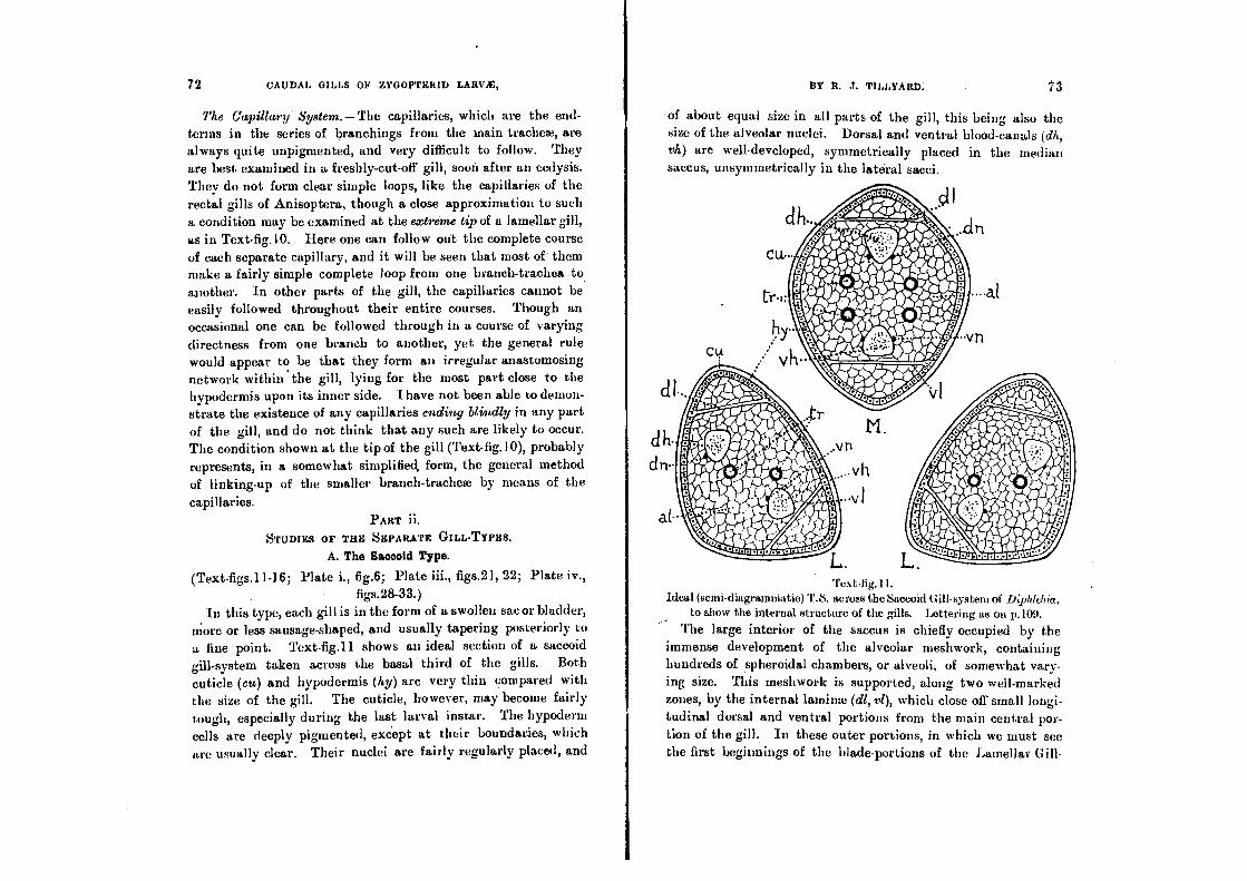
72 CAUDAL GILLS OF ZYGOPTERID LARVX,
The Capillary System. —The capillaries, which are the end-terms in the series of branchings from the inain trachea, are always quite unpigmented, and very difficult to follow. They
are best examined in a freshly-cut-off gill, soon after an ecdysis.
They do not form clear simple loops, like the capillaries of the
rectal gills of Anisoptera, though a close approximation to such
a condition may be examined at the extreme tip of a lamellar gill,
as in Text-fig.10. Here one can follow out the complete course
of each separate capillary, and it will be seen that most of them make a fairly simple complete loop from one branch-trachea to another. In other parts of the gill, the capillaries cannot be easily followed throughout their entire courses. Though an occasional one can be followed through in a course of varying directness from one branch to another, yet the general rule would appear to be that they form an irregular anastomosing network within the gill, lying for the most part close to the
hypodermis upon its inner side. nave not been able to demon-strate the existence of any capillaries ending btindly in any part
of the gill, and do not think that any such are likely to occur.
The condition shown at the tip of the gill (Text-fig.I0), probably
represents, in a somewhat simplified, form, the general method of linking-up of the smaller branch-trachea by means of the
capillaries.
PART ii. STUDIES OF THE SEPARATE GILL-TYPES.
A. The Saccold Type.
(Text-figs.11 - 16; Plate i., fig.6; Plate iii., figs.21, 22; Plate iv.,
figs.28-33.) In this type, each gill is in the form of a swollen sac or bladder,
more or less sausage-shaped, and usually tapering posteriorly to a fine point. Text-fig.11 shows au ideal section of a saccoid
gill-system taken across the basal third of the gills. Both
cuticle (cu) and hypodermis (hy) arc very thin compared with
the size of the gill. The cuticle, however, may become fairly
tough, especially during the last larval instal.. The hypoderm cells are deeply pigmented, except at their boundaries, which are usually clear. Their nuclei are fairly regularly placed, and
DY R. J. TILLYARD. 73
of about equal size in all parts of the gill, this being also the size of the alveolar nuclei. Dorsal and ventral blood-canals (dh, vh) are well-developed, symmetrically placed in the median saccus, unsymmetrically in the lateral sacci.
Text-fig.1 I. Ideal (semi-diagrammatic) T.S. across the Saccoid Gill-system of hiphtebia,
to show the internal structure of the gills. Lettering as on p.109.
The large interior of the saccus is chiefly occupied by the immense development of the alveolar meshwork, containing
hundreds of spheroidal chambers, or alveoli, of somewhat vary-
ing size. This meshwork is supported, along two well-marked zones, by the internal lamina (dl, vl), which close of small longi-tudinal dorsal and ventral portions from the main central por-
tion of the gill. In these outer portions, in which we must see the first beginnings of the blade-portions of the Lamellar

Text-fig.12.*
74 CAUDAL GILLS 01P ZYGOPTERID LARVt,
types, there is nothing but a weakly-formed meshwork, sup-ported by occasional secondary laminae, and a few branch tracheae
of very small size. The tracheal plan of a saccoid gill is always somewhat
irregular, the main longitudinal tracheae being free to take a
tortuous course in the alveolar meshwork. As in all caudal
gills, two main tracheae penetrate the base of the median gill, while one only passes through the base of each lateral gill. But,
in the median gills belonging to the Epallagince, it is usual to
find each main trachea giving off a strong ventral longitudinal
branch, so that, in nearly all cross-sections of the gill, there appear to be four longitudinal tracheae in the gill. In the lateral gills, likewise, the single main trachea divides into two soon after
entering the gill, and, fairly frequently, one of these branches
may again divide, so as to give three longitudinal tracheae in this
gill. In the median saccoid gills belonging to the Protoneurince
(IV costicta), one sometimes finds only two main tracheae in all
three gills, yet I have frequently noted four in the median, and sometimes three in the laterals. These gills are so frequently cast off and regenerated, that it is not easy to determine whether
any of this variation is due to such a cause, or whether the
number of main tracheae is essentially variable. In all cases examined by me, there are four longitudinal nerves
in the median gill, and two in the laterals, as shown in Text-fig.
11 (dn, vn). Thc two following sub-types have been studied :-
(i.) The Simple Saccus (Text-figs. 12, 13). This is found in
the genera Pseuclophaa and Diphlebia, both belonging to the
Epallagiace, and in the genus Cora (.71torince). I have only
studied the gills of Diphlebia. Ris' work on Pseudophau (28) shows that the gills in this genus are closely similar to those of
Diphlebia, differing principally in. being even more tensely
swollen, and much less pointed at the tips. The gills of Cora,
as studied by Calvert (3), differ from those of the other two
genera in being sharply truncated posteriorly.
Thc gills of Diphlebia (Text-figs.12, 1 3) are large, cumbersome
sacs, which clearly impede the movements of the larva. These
13Y D. J. TILLYARD. 75
larvae are rock-dwellers in fast mountain-streams, being only found in the shallow rapids, and never in the deep still pools. They ding to the under-surface of flat rocks lying in the main current, in company with large (undescribed) Perlid lame, which they some-what resemble, except for their gills. They are, however, unlike the Perlid larvae, very sluggish, and are only capable of rapid movements when in the water. When a rock is lifted up with a Diphtebia-larva beneath it, the dragging of the huge gills upon the wet rock-surface effectually prevents any speedy movement.
The gills can be fixed and kept in 70
per tent. alcohol, without collapsing; but,
when dehydrated and cleared, they almost invariably collapse. They are, therefore, exceedingly difficult to section satisfac-
torily. They are very opaque, dull grey,
brownish, or blackish, according to the locality and the colour of the rocks in-habited. In sandstone or ironstone creeks, a brownish colour prevails; in granitic or basaltic creeks, grey or black-ish. Their shape varies somewhat in different individuals. Sometimes the gills are so tense as to be definitely
sausage-shaped; in other cases, the gills are not so tense, and there is just a suspicion of the formation of longitudinal edges or midribs. In the latter case, the lateral gills may be described as triquetral, but with extremely convex faces.
The whole cuticle of the gill is covered by irregularly placed hairs, most of which are long and slender. They are especially long and numerous upon the posterior pointed portion of the gill, giving it a somewhat shaggy appearance.
The cuticle itself is quite clear and unpigmented. It is com-
posed of numerous exceedingly thin parallel strata, the outermost of which differs from the rest in staining deeply and easily with
eosin. The total thickness of the cuticle only averages about 8p,. The hypo dermis is a very thin epithelial layer, only about
Gills of Diphtebia tedoides Says, in natural position, dorsal view; ( x 4).

0 ,,Text-tig.13.*
half the thickness of the cuticle, i.e., averaging only 4p. in thick-
ness. It is everywhere so deeply pigmented that it is almost impossible to distinguish either cell-boundaries or nuclei. This pigment consists of minute granules of a blackish substance de-
posited in all parts of the cytoplasm of the hypodermis. It is only when the hypodermis is cut tangentially, so as to expose
the polygonal outline of its cells, that it can be seen definitely that the pigment is less dense along the cell-boundaries, and densest towards the middle
of each cell, especially around the nuclei. The latter are of the usual oval shape, and vary from 8 to 10/A in length.
The distribution of tracheae can be seen from Text-fig.13. The main trunks pursue a somewhat tortuous course, often coiling round one another. They give off large irregular branches, which run obliquely outwards to-wards the margin of the gill. These large
branches again break up very irregularly into
numerous smaller branches, which approach
the hypodermis at all points of the gill, and
finally turn to run parallel with it, giving off
numerous capillaries. The courses of these latter cannot be followed owing to the density
of the hypoderm pigment-layer.
The differences between the gills in the three known larvae are only small, but appear
to be of sufficient value to separate the species.
D. lestoides Selys, has the longest gills (10.5-
llmm.), with the tips more slenderly pointed, and less hairy, than in the other two species.
The lateral gills of D. lestoides are also usually
distinctly triquetral, the inner faces being definitely flattened
against the median gill. D. nymphoides Tillyard, has hairier
gills, 9 . 5-10 mm. long, somewhat less pointed, and generally much
Left lateral gill of Diphlebia leeloide,s Selys, cleared to •show tracheal
system; ( x 8).
more swollen or sausage-shaped. In D. euphwoides Tillyard, the gills are shorter still, 8.5-9 mm. long, shaggy, shortly pointed, and tensely swollen.
(ii.) The Constricted Saccus (Text-6g.14). This very remark-able form of gill is found in the genera Neosticta and Nososticta among the Protoneurince. Both larva; are rock-dwellers, with habits closely similar to those of Diphlebia, except that they are often found on the sides of larger rocks in deep, still pools.
These larvae can move fairly quickly under water, and their gills are certainly not as cumbrous as those of Diphlebia.
The shape of the gill may be roughly described as that of a rather short candle surmounted by a flame, the basal joint represent-ing the candle, the distal joint the flame. The constriction between the two joints is very strongly
marked, and is only just sufficiently wide to allow of the passage of the
main tracheae and the blood-canals. The cuticle is covered everywhere with small, scattered hairs, which become longer and more abundant
upon the distal end of the basal
joint, and also over the whole of the distal joint. As in the gills of Diphlebia, the cuticle is quite clear and unpigmented. The hypodermal pigment, unlike that of Diphlebia, is not dis-tributed equally all over the gill, but is chiefly confined to three transverse zones, viz., around the base of the gill, around the distal third of the basal joint, and upon all except the extreme
base and tip of the distal joint. This is well shown in Plate i., fig.6. The hypodermis is of about the same thickness, 4p, as in Diphlebia, and its nuclei are also of the same size as in that genus. The cuticle, however, is only 5n in thickness, though otherwise closely resembling that of Diphlebia in structure.
Internally, the structure of the basal joint of
Diphtebia,resembles very closely the structure of the gill of Diphlebia, except that
76 CAUDAL GILLS Oil' 2YGOI.TERID LARVA, BY R. J. TILLYARD. 77
*Gills of Neaqicta canescens Tillyard, in natural position, dorsal view;( x 5).

Text-fig.16.•
78 CAUDAL GILLS OF ZYGOPTERID LABY/E, BY R. J. TILLYARD. 79
the individual alveoli is smaller. The positions of the blood-canals, nerves, and internal laminae, and
the distribution of the
to larger tracheal, are very
similar in both forms. The branch tracheae, how-
ever, are more numerous,
and come off less oblique-
ly from the main stems. Their branchings towards the hypodermis are also of a more dendritic char-
Text-fig.15. aeter, as can be seen in
T.S. through the constriction of the median P1. i., fig.6. In the less
gill of Neostieta CalteReen8 Tillyard; pigmented parts of the
( x233). Lettering as on p.109. gill, it is possible to see
bundles of capillaries starting out from the somewhat " knobby"
and deeply pigmented ends of
the dendritic branches. These
capillaries seem to run in all directions upon the inner border of the hypoderrnis, and their
general distribution closely re-sembles that seen in the Nodate
Lamellar type of gill. At the constriction, the
alveolar meshwork is reduced
to a minimum. The two blood-
canals pass through it with their inner walls almost in contact, separating the main tracheae
to left and right. A trans_ verse section through the con-
striction in the median gill of Neostieta is shown in Text-fig.l 5.
* T.S. through distal joint of the median gill of Neosticta canescens Till-
yard; ( x 130). hm, confluence of the two blood-canals; ar, nerve; other
lettering as on p.109,
The structure of the distal joint, examined in sections, is seen to be rather peculiar. It is, in fact, intermediate between the true saccoid type and the triquetro-quadrate type, being slightly
flattened laterally, as seen in Text-fig.16. The alveolar mesh-work is present, and is supported by numerous horizontally-placed internal laminae (i1). The trachem in this joint are all of small size, the main stems qHickly breaking up into numerous small branches. The two blood-canals unite soon after entering the basal part of the joint.
The length of the gills in Neostieta is 5-6 mm., of which the distal joint occupies at the most one-third. Their greatest breadth is about 1.2 mm. The gills of Xososticta closely resemble those of Neostieta in size and shape, but may be distinguished, in common with the whole larva, by being of a more definite black-and-white banded pattern, the pattern in Neostieta being banded dark brown and pale yellowish-brown,
B. The Triquetro -Quadrate Type. (Text-figs,17-18; Plate iii., figs.23-24; Plate v., figs.34-35.)
This type is not represented in Australia, being confined to the Calopterygince, which are absent from our continent (though the genus Neurobasis occurs in Papua). Through the kindness of Dr. P. P. Calvert, of Philadelphia, I have been able to section
and study a number of gills of this type taken from larva of un-determined species of Calopteryx and Iletcerina. The import-ance of this type of gill is that it forms a 'connecting-link or intermediate stage between the Saccoid and Lamellar Types. I shall not, therefore, attempt to elucidate specific or even generic differences, which are of no importance for the purposes of this
paper. An ideal transverse section of this type of gill, taken at
about one-third of the total length of the gill from the base, is shown in Text-fig.17.
The term triquetro-quadrate, which I have suggested as suit-able to denote the type of gill-system found in the Calopterygince, is intended to suggest the form of the transverse section across the three gills. The lateral gills, which are always considerably longer than the median, might be best described as triangular
the size of
c11-1 d

Text-fig.18."
BY R. J. TILLYAUD, 8 I 80 CAUDAL GILLS OF ZYGOPTRKID LARVA,
prisms except that they become gradually enlarged towards the tip, and finally taper to a rather blunt point. Their transverse
sections are everywhere triangular. The term "triquetral" is
frequently used to describe these gills, and seems to be the most
suitable term available.
Text-4.17. Ideal (semi-diagrammatic) T.S. across the Triquetro-quadrate Gill-system
of Calopteryx, to show the internal structure of the gills. Lettering
as on p.109.
The shorter median gill has all the appearances of a vertical
lamella, except that it is somewhat thickened. This gill also increases in width towards the tip, and then tapers to a point.
The distal part of the gill appears to be curved over downwards, giving it a somewhat "nodding" appearance. This curvature is shown also in the blood-canals, which arc also slightly down-
curved towards the tip (Text-fig.18). Now if a transverse sec-
tion be made anywhere along the gill, it will be seen to be definitely diamond-shaped, with the two upper sides of the sec-tion equal and shorter that the two lower ones, which are also equal. Thus it is quadrangutar in section. The complete gill-sys-tem, therefore, might be described as "triquetro- quadrangular," a
cumbersome term which I have shortened to triquetro-quadrate. Owing to their long immersion in alcohol, I can
find little or no traces of the tracheal system in whole mounts of my gills, though the collapsed trachea are easily seen in sections. As far as I am able to make it out, the general arrangement of the tracheal system appears to be very similar to that described in Synlestes (p. 87). The following account of the Triquetro-quadrate Type is made
almost entirely from a study of transverse and longitudinal sections.
i. The Median or Quadrate Gill (Plate iii., fig.23; Plate v., fig.34). The cuticle is unpigmented, and considerably thicker than in the saccoid type. This thickening is especially noticeable at the two lateral angles of the cross section (mr in Text-fig. 17). Here the total thickness is 151z or more, of which the hard, outer structure, staining deeply with eosin, may occupy 3p, or more. The cuticle throughout the portions of the gill lying close
above the lateral angles is somewhat thicker than that below them. Seen from outside, in the whole mount, the thickened cuticle
of the lateral angles forms a kind of mid-rib for the gill, on either side of it. In the distal portion of the gill, the central area
between the two midribs stands out clearly thickened, in contrast with the outer portions, which become thin and blade-like. This closely approximates to the condition seen in the median gills of the Vertical Lamellar type, in which I have termed the thickened central portion, with the two midribs, the rachis, the thin outer portions the blade.
• Gills of adapteryx sp., lateral view, from a specimen in alcohol; ( x 6).
7

82 CAUDAL GILLS OF ZYGOPTERID LARVAE,
Hairs are absent from the cuticle. Stiff spines or bristles are developed along each mid-rib, also along ate dorsal and ventral edges of the blade.
The hypodermis is, on the whole, a very thin, somewhat irregular layer of cells, usually only 3-414 in thickness. Around the four angles of the cross-section, it is thicker, especially beneath the two mid-ribs. Pigmentation is only of moderate density. There appear to be two transverse zones in the distal half of the gill (Calopteryx) whcre the pigmentation is much less dense than elsewhere.
The central portion or rachis of the gill may be considered to be bounded by the two zones of principal internal laminae (dl, v/), which bound the two blood-canals externally. Within the cavity of the rachis, all the more important structures of the gill are collected. There are well-defined, oval, dorsal and ventral blood-canals (dh, vh). In the walls of each canal, there runs a pair of longitudinal nerves (nv), of which the dorsal pair are con-siderably larger than the ventral pair. A little nearer to thc centre of the gill, and almost touching the blood-canals, lie the two pairs of main longitudinal tracheae (tr). The rest of the rachis is occupied with the alveolar meshwork, which is much less abundant than in the more capacious saccoid gills.
The interior of the blade of the gill, both dorsally and ven-trally, is occupied by a small amount of alveolar meshwork, sup-ported at irregular intervals by short, transverse, secondary internal laminae (Plate v,, fig,34, dl'). Branch tracheae of con-
. siderable size are to be seen traversing this part of the gill obliquely.
ii. The Lateral or Triquetral Gills (Plate iii., fig.24; Plate v,, fig.35). The cuticle and hypodermis resemble those of the median gill, but are considerably thicker, the cuticle averaging 1314 or more in thickness, the hypodermis 614 or more. The cuticle is thickened along all three edges; and carries spines or bristles there. The thickening is, however, especially noticeable along the our edge (Text-fig.17, mr), which forms the truc mid-rib of the gill, corresponding with the mid-rib seen in the lateral gills of Lestidce,
BY R. J. TILLYARD. 83
Internally, the structure of the triquetral gill closely resembles that of a lateral saccoid gill, except for the shape. There is a very great development of the alveolar meshwork. The zones of principal internal laminae are arranged in such a way as to cut off, in cross-section, the upper and the lower inner corners of the triangle, the dorsal laminae lying almost horizontally, and the ventral ones not far from vertically. This arrangement, again, is paralleled in the obliquity of these laminae in the lateral gills of the saccoid type.
In the central part of the gill, the two blood-canals, the two main longitudinal tracheae, and the two main nerves, run un-symmetrically, the ventral set being placed at a lower and inner level compared with the dorsal. A very peculiar development, confined, as far as I can ascertain, to gills of this type, is the splitting-up of the blood-canals, each of which gives off an im-portant longitudinal branch to run in the outer portion of the gill, on the other side of the principal internal laminae (Text-fig. 17, dh, vh). All four branches become confluent again ncar the distal end of the gill.
Secondary internal laminae are developed at frequent intervals, within the two -portions of the gill cut off by the principal laminae. These laminae lie parallel with the corresponding principal laminae, but not all at the same level, so that sometimes two or more of them may be cut in the same section.
C. The Lamellar Type. (Text-figs.19-29; Plate i., figs.1-5, 7-11; Plate ii., flgs.12-20;
Plate iii., figs.25-27; Plates v. and vi., figs.36-39.
In this type, all three gills are reduced to flattened lamellar organs, with consequently a great reduction in the volume of the interior of the gill. The alveolar meshwork becomes much reduced in amount, and is principally confined to the thickened portion of the gill, lying beneath the mid-rib. This thickened central portion, I propose to term the rachis, using the term
blade for the outer and flatter portions of the lamella. The rachis is always bounded, either on one or both sides, by
a thickened ridge of cuticle, forming a strong mid-rib (mr). In the interior of thc rachis run the longitudinal tracheal trunks,
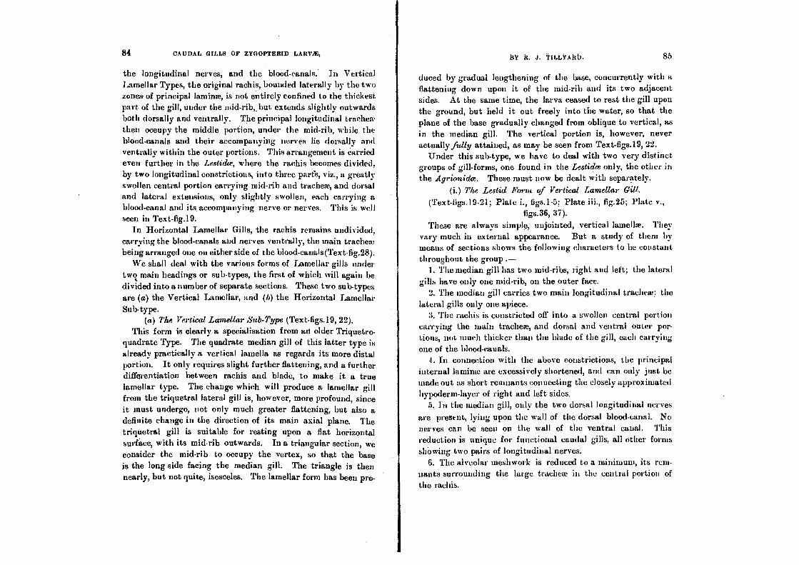
84 CAUDAL GILLS OF ZYGOPTERID LARVAE,
the longitudinal nerves, and the blood-canals. In Vertical Lamellar Types, the original rachis, bounded laterally by the two zones of principal laminae, is not entirely confined to the thickest part of the gill, under the raid-rib,. but. extends slightly outwards both dorsally and ventrally. The principal longitudinal tracheae then occupy the middle portion, under the mid-rib, while the blood•eanals and their accompanying nerves lie dorsally and ventrally within the outer portions. This arrangement is carried even further in the Lestidce, where the rachis becomes divided, by two longitudinal constrictions, into three parts, viz., a greatly swollen central portion carrying mid-rib and tracheae, and dorsal and lateral extensions, only slightly swollen, each carrying a blood-canal and its. accompanying nerve or nerves. This is well seen in Text-fig.19.
In Horizontal Lamellar Gills, the rachis remains undivided, carrying the blood-canals and nerves ventrally, the main trachea being arranged one on either side of the blood-canals(Text-fig.28).
We shall deal with the various forms of Lamellar gills under two main headings or sub-types, the first of which will again be divided into a number of separate sections. These two sub-types are (a) the Vertical Lamellar, and (b) the Horizontal Lamellar Sub-type.
(a) The Vertical Lamellar Sub-Type (Text-figs.19, 22). This form is clearly a specialisation from an older Triquetro-
quadrate Type. The quadrate median gill of this latter type is already practically a vertical lamella as regards its more distal portion. It only requires slight further flattening, and a further differentiation between rachis and blade, to make it a true lamellar type. The change which will produce a lamellar gill from the triquetral lateral gill is, however, more profound, since it must undergo, not only much greater flattening, but also a definite change in the direction of its main axial plane. The triquetral gill is suitable for resting upon a flat horizontal surface, with its midrib outwards. In a triangular section, we consider the mid-rib to occupy the vertex, so that the base is the long side facing the median gill. The triangle is then nearly, but not quite, isosceles. The lamellar form has been pro-
h% J. TatLA.Altb. 85
duced by gradual lengthening of the base, concurrently with a flattening down upon it of the mid-rib and its two adjacent sides. At the same time, the larva ceased to rest the gill upon the ground, but held it out freely into the water, so that the plane of the base gradually changed from oblique to vertical, as in the median gill. The vertical portion is, however, never actually fully attained, as may be seen from Text-figs.19, 22.
Under this sub-type, we have to deal with two very distinct groups of gill-forms, one found in the Lestidce only, the other in the .Agrionidce. These must now be dealt with separately.
(i.) The Lestid Form of Vertical Lamellar Gill.
(Text-figs.19-21; Plate i., figs.1-5; Plate iii., fig.25; Plate v., figs.36, 37).
These are always simple, unjointe,e1, vertical lamellae. They vary much in external appearance. But a study of them by means of sections shows the following characters to be constant throughout the group .—.
1. The median gill has two mid-ribs, right and left; the lateral gills have only one mid-rib, on the outer face.
2. The median gill carries two main longitudinal tracheae; the lateral gills only one apiece.
3. The rachis is constricted off into a swollen central portion carrying the main tracheae, and dorsal and ventral outer por-tions, not much thicker than the blade of the gill, each carrying one of the blood-canals.
4. In connection with the above constrictions, the principal internal laminae are excessively shortened, and can only just be made out. as short remnants connecting the closely approximated hypoderm-layer of right and left sides.
5. In the median gill, only the two dorsal longitudinal nerves
are present, lying upon the wall of the dorsal blood-canal. No nerves can be seen on the wall of the ventral canal. This reduction is unique for functional caudal gills, all.other forms showing two pairs of longitudinal nerves.
6. The alveolar meshwork is reduced to a minimum, its rem-nants surrounding the large tracheae in the central portion of the rachis.
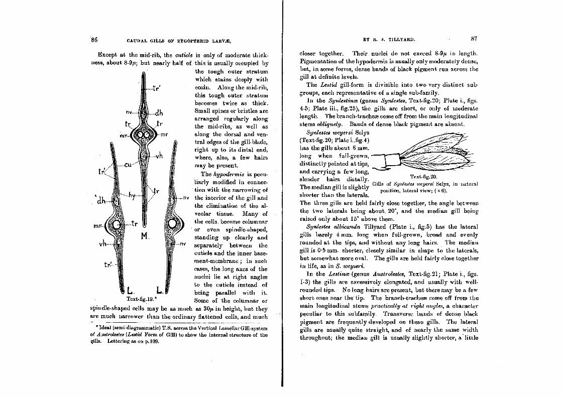
Text•fig.20.
Gills of Syniestes weyersi Selys, in natural position, lateral view; ( x 6).
86 CAUDAL GILLS OP ZYGOPYILICID LAItYit,
Except at the mid-rib, the cuticle is only of moderate thick-ness, about 8-9p; but nearly half of this is usually occupied by
the tough outer stratum which stains deeply with eosin. Along the mid-rib, this tough outer stratum becomes twice as thick. Small spines or bristles are arranged regularly along the mid-ribs, as well as along the dorsal and ven-tral edges of the gill-blade, right up to its distal end, where, also, a few hairs may be present.
The hypodermis is pecu- liarly modified in connec- tion with the narrowing of
nv the interior of the gill and the elimination of the al- veolar tissue. Many of the cells. become columnar or even spindle-shaped, standing up clearly and separately between the cuticle and the inner base- ment-membrane ; in such cases, the long axes of the nuclei lie at right angles to the cuticle instead of being parallel with it. Some of the columnar or
spindle-shaped cells may be as much as 30p in height, but they are much narrower than the ordinary flattened cells, and much
*Ideal (semi-diagranimatic) T.S. across the Vertical Lamellar Gill-system of Austrolestes (Lestid Form of Gill) to show the internal structure of the gills. Lettering as on p.109.
13Y R. J. TaLvAttn. 87
closer together. Their nuclei do not exceed 8-9p in length. Pigmentation of the hypodermis is usually only moderately dense, but, in some forms, dense bands of black pigment run across the gill at definite levels.
The Lestid gill-form is divisible into two very distinct sub-groups, each representative of a single sub-family.
In the Synlestince (genus Synlestes, Text-fig.20; Plate i., figs. 4-5; Plate iii., fig.25), the gills are short, or only of moderate length. The branch-tracheae come off from the main longitudinal stems obliquely. Bands of dense black pigment are absent.
Synlestes weyersi Selys (Text-fig.20; Plate i..fig.4) has the gills about 6 mm. long when full-grown, distinctly pointed at tips, and carrying a few long, slender hairs distally. The median gill is slightly shorter than the laterals. The three gills are held fairly close together, the angle between the two laterals being about, 20°, and the median gill being raised only about 15° above them.
Synlestes albicauda Tillyard (Plate i., fig.5) has the lateral gills barely 4 mm. long when full-grown, broad and evenly rounded at the tips, and without any long hairs. The median gill is 0.5 mm. shorter, closely similar in shape to the laterals, but somewhat more oval. The gills are held fairly close together in life, as in S. weyseri.
In the Lestince (genus Austrolestes, Text-fig.21; Plate i., figs. 1-3) the gills are excessively elongated, and usually with well-rounded tips. No long hairs are present, but there may be a few short ones near the tip. The branch-tracheae come off from the main longitudinal stems practically at right angles, a character peculiar to this subfamily. Transverse bands of dense black pigment are frequently developed on these gills. The lateral gills are usually quite straight, and of nearly the same width throughout; the median gill is usually slightly shorter, a little
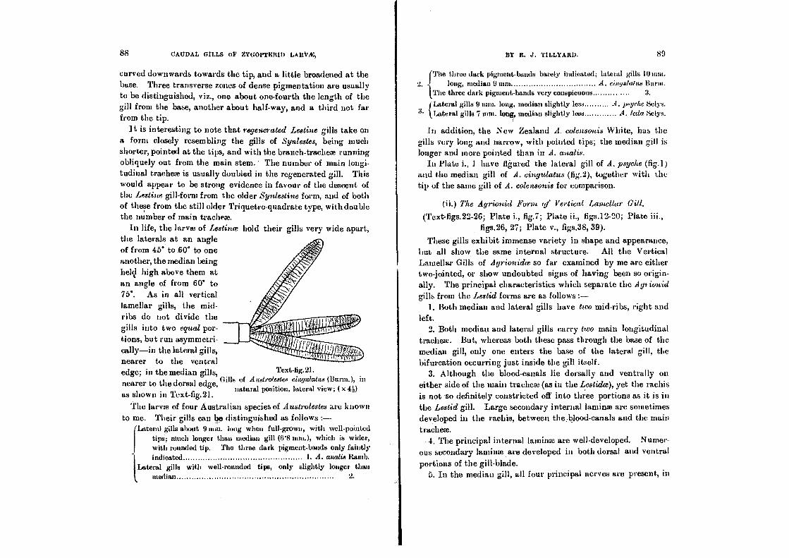
88 CAI/DAL GILLS of ZYGOPTEIIIll ltV/E, 13t R. J. TILLYARD. 80
curved downwards towards the tip, and a little broadened at the base. Three transverse zones of dense pigmentation are usually to be distinguished, viz., one about one-fourth the length of the gill from the base, another about half-way, and a third not far from the tip.
It is interesting to note that regenerated Lestine gills take on a form closely resembling the gills of Synlestes, being much shorter, pointed at the tips, and with the branch-tracheae running obliquely out from the main stem. ' The number of main longi-tudinal trachea is usually doubled in the regenerated gill. This would appear to be strong evidence in favour of the descent of the Lestine gill-form from the older Synlestine form, and of both of these from the still older Triquetro-quadrate type, with double the number of main trachea.
In life, the larvae of Lestince hold their gills very wide apart, the laterals at an angle of from 45° to 60° to one another, the median being hell high above them at an angle of from 60' to 75°. As in all vertical lamellar gills, the mid-ribs do not divide the gills into two equal por-tions, but run asymmetri-cally—in the lateral gills, nearer to the ventral edge; in the median gills, Text -lig.21.
nearer to the dorsal edge, Gills of A uatrotestee eingtdatua (Bunn.), in
natural position, lateral view; ( x 4D as shown M Text-fig.21.
The larva of four Australian species of Austrolestes are known to me. Their gills can be distinguished as follows :—
'Attend gills about 9 mm. long when full-grown, with well-pointed {tips; much longer than median gill (6'8 rnm.), which is wider,
rounded unded tip. The three dark pigment-bands only faintly
indicated I. A. aualia Ram/.
Lateral gills with well-rounded tips, only slightly longer than
median 2.
IThe three dark pigment-bands barely indicated; lateral gills 10 inni,
2. I
long, median 9 mm • A. eiugulatue The three dark pigment-bands very conspicuous 3.
(Lateral gills 9 mm. long, median slightly less 4. peyehe 8elys.
3• ,Lateral gills 7 mm. long, median slightly less A. Leda Selys.
In addition, the New Zealand A. colensonis White, has the gills very long and narrow, with pointed tips; the median gill is longer and more pointed than in A. anatis.
In Plate i., I have figured the lateral gill of A. psyche (fig.1) and the median gill of A. cingulatus (fig.2), together with the tip of the same gill of A. colensonis for comparison.
(ii.) The Agrionid Form qf Vertical Lameltar Gitl.
(Text-figs.22-26; Plate i., fig.7; Plate ii., figs.12-20; Plate figs.26, 27; Plate v., figs.38, 39).
These gills exhibit immense variety in shape and appearance, but all show the same internal structure. All the Vertical Lamellar Gills of Agrionidre so far examined by me are either two-jointed, or show undoubted signs of baying been so origin-ally. The principal characteristics which separate the A 91 iunid
gills from the Lestid forms are as follows 1. Both median and lateral gills have two mid-ribs, right and
left. 2. Both median and lateral gills carry two main longitudinal
tracheae. But, whereas both these pass through the base of the median gill, only one enters the base of the lateral gill, the bifurcation occurring just inside the gill itself.
3. Although the blood-canals lie dorsally and ventrally on either side of the main trachea (as in the Lestidce), yet the rachis is not so definitely constricted off into three portions as it is in the Lestid gill. Large secondary internal lamina arc sometimes developed in the rachis, between the Mood-canals and the main
trachea. • 4. The principal internal lamina are well-developed. Numer-
ous secondary lamina are developed in both dorsal and ventral portions of the gill-blade.
5. In the median gill, all four principal nerves are present, in

BY R. J. TILLYARD. 91 90 CAUDAL GILLS OP ZYGOPTERID LARY2P,
their usual positions (as in all functional gills except those of
Lestidce).
Text-fig.22. Ideal (send-diagrammatic) T.S. across the Vertical Lamellar Gill•system
of Aurgroagrion (Agrionid Form of Gill), to show the internal struc-ture of the gill. Lettering as on p.109.
6. The alveolar meshwork is reduced, but less so than in the
Lesticke; traces of it still existing in the gill-blades as well as in
the rachis.
The•cutick averages about 9p in thickness, but the tough outer stratum is usually not more than 2p in thickness. Along the mid-rib, this outer stratum becomes thickened to 4p or 5p; it is also somewhat thickened along the dorsal and ventral edges of the gill. Spines are present upon the mid-ribs and edges of the gill, but usually only from the base to some definite point, mark-ing the end of the original basal joint of the gill.
The hypodermis is not modified into columnar or spindle-shaped cells as in the Lestidce, but remains everywhere a flat epithelium, with the nuclei lying parallel to the cuticle. There is, however, a very distinct difference in size between the nuclei found in the basal joint of the gill, and most of those found in its more distal portion. The former average about fip in length, and are even larger (up to 10p.) in those cells from which the principal internal laminae are developed; the latter only average 6p in length, and do not stain with the same intensity. In many places, the hypoderm-layer is exceedingly thin, only 3-4p
in thickness, so that the nuclei, even though lying parallel with the cuticle, cause periodical bulgings in the hypodermis. In spite of the narrowness of the gill-blade, many of the cells of the hypodermis seem to take part in forming alveolar tissue; if the gill be stained in Coto, this meshwork can be seen lying everywhere within the basal portion of the gill (except at the extreme edges), and even in the more distal part, the meshwork can be seen running along both sides of the rachis, nearly to the tip.
In some gills, hypodermal pigment is almost absent; in others, the pigment may occur either in transverse bands, or in irregular patches. A few are deeply pigmented all over.
The tracheal system varies much in details, but the branches never come off from the main sterns perpendicularly (as in Lestinee), but always obliquely.
In mining and describing the sub-groups into which I propose to divide these gills, I have had in mind their descent from an original two-jointed gill. Four phylogenetic stages can be seen in this process, and I have classified the gills accordingly, as follows :—
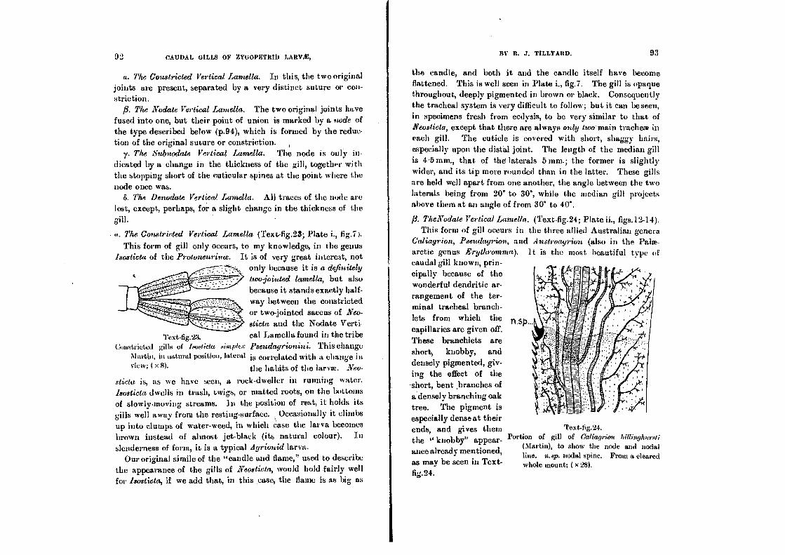
'17ext-fig.23.
Constricted gills of Isosticta simptcx
Ala•in, in natural position, lateral view; ( x 8).
ia). It is the most beautiful type of
Text-tig.24. Portion of gill of Caliagrion
(Martin), to show the node and nodal line. n.sp. nodal spine. From a cleared whole mount; ( x 28).
93 CAUbAL GILLS OF ZYGOPBTBIO LARVA'.',
a. The Constricted Vertical Lamella. In this, the two original joints are present, separated by a very distinct suture or con-striction.
13. The Nodate Vertical Lamella. The two original joints have fused into one, but their point of union is marked by a node of the type described below (p.94), which is formed by the reduc-tion of the original suture or constriction. ,
y. The Subnodate Vertical Lamella. The node is only in-dicated by a change in the thickness of the gill, together with the stopping short of the cuticular spines at the point where the node once was.
8. The Denodate Vertical Lamella. All traces of the node are lost, except, perhaps, for a slight change in the thickness of the gill.
a. The Constricted Vertical Lamella (Text-fig.23; Plate i., fig.7).
This form of gill only occurs, to my knowledge, in the genus Isosticta of the Protoneurince. It is of very great interest, not
only because it is a definitely
two jointed lamella, but also because it stands exactly half-way .between the constricted or two-jointed saccus of Neo-
sticta and the Nodate Verti-cal Lamella found in the tribe Pseudayrionini. This change is correlated with a change in the habits of the larvae. Neu-
stiettt is, as we have seen, a rock-dweller in running water.
Isosticta dwells in trash, twigs, or matted roots, on the buttons of slowly-moving streams. In the position of rest, it holds its gills well away from the resting-surface. Occasionally it climbs up into dumps of water-weed, in which case the larva becomes brown instead of almost jet-black (its natural colour). In slenderness of form, it is a typical Agrionid larva.
Our original simile of the "candle and flame," used to describe the appearance of the gills of Neosticta, would hold fairly well
for Isosticta, if we add that, in this ,ease, the flame is as big as
BY R. 3. TILLYARD. 93
the candle, and both it and the candle itself have become flattened. This is well seen in Plate i., fig.7. The gill is opaque throughout, deeply pigmented in brown or black. Consequently the tracheal system is very difficult to follow; but it can be seen, in specimens fresh from ecdysis, to be very similar to that of Neosticta, except that there are always only two main tracheae in
each gill. The cuticle is covered with short, shaggy hairs, especially upon the distal joint. The length of the median gill is 4'5 mm., that of the laterals 5 mm.; the former is slightly wider, and its tip more rounded than in the latter. These gills are held well apart from one another, the angle between the two laterals being from 20° to 30°, while the median gill projects above them at an angle of from 30° to 40°.
lg. TheNadate Vertical Lamella. (Text- ►g.24; Plate ii., figs.12-14). This form of gill occurs in the three allied Australian genera
Caliagrion, Pseudagrion, and Austroagrion (also in the Palae-arctic genus Erythromn caudal gill known, prin-cipally because of the wonderful dendritic ar-rangement of the ter-minal tracheal branch-lets from which the capillaries are given off. These branchlets are short, knobby, and densely pigmented, giv-ing the effect of the •short, bent ,branches of a densely branching oak tree. The pigment is especially densest their ends, and gives them the " knobby" appear-ance already mentioned, as may be seen in Text-fig.24.
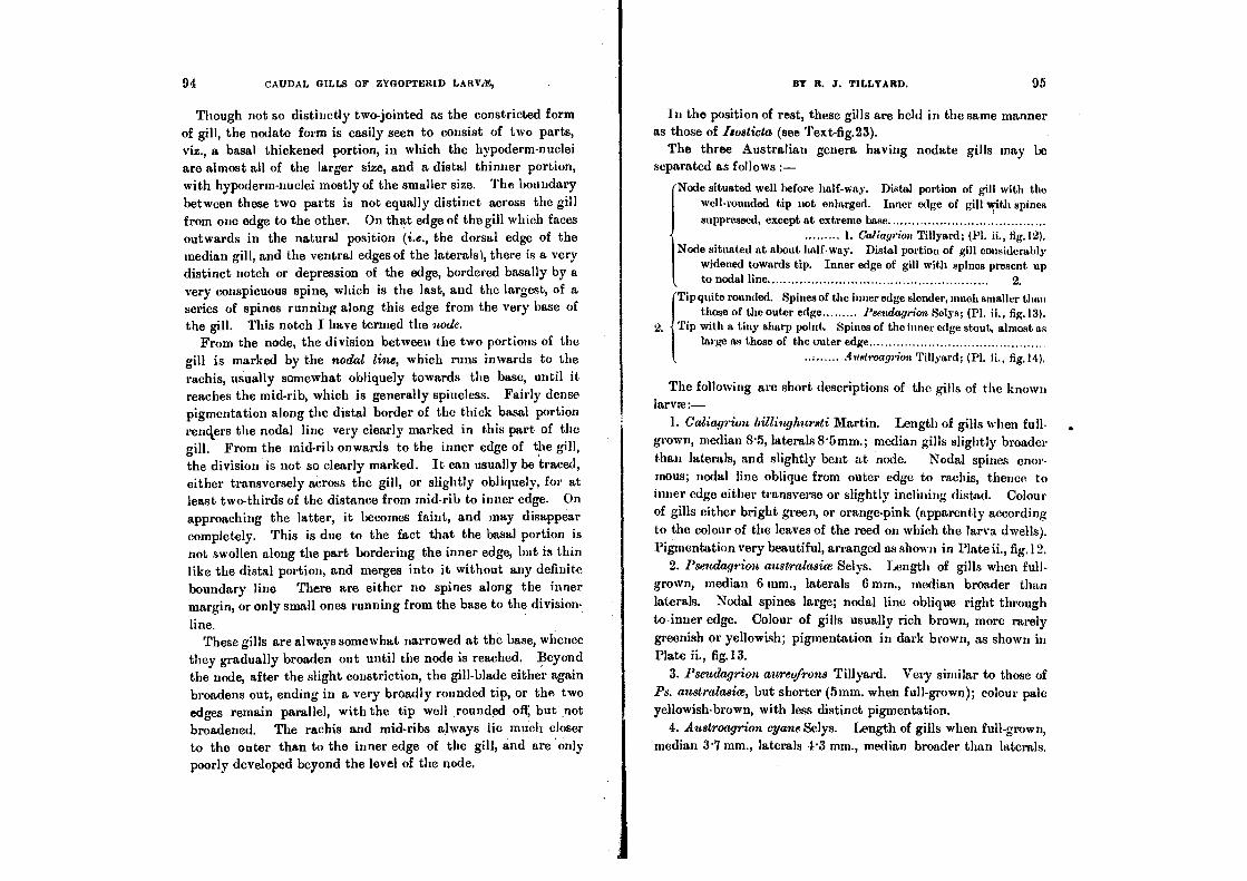
Though not so distinctly two-jointed as the constricted form of gill, the nodate form is easily seen to consist of two parts, viz., a basal thickened portion, in which the hypoderm-nuclei are almost all of the larger size, and a distal thinner portion, with hypoderm-nuclei mostly of the smaller size. The boundary between these two parts is not equally distinct across the gill from one edge to the other. On that edge of the gill which faces outwards in the natural position (i.e., the dorsal edge of the median gill, and the ventral edges of the laterals), there is a very distinct notch or depression of the edge, bordered basally by a very conspicuous spine, which is the last, and the largest, of a series of spines running along this edge from the very base of the gill. This notch I have termed the node.
From the node, the division between the two portions of the
gill is marked by the nodal line, which runs inwards to the rachis, usually somewhat obliquely towards the base, until it reaches the mid-rib, which is generally spineless. Fairly dense pigmentation along the distal border of the thick basal portion renders the nodal line very clearly marked in this part of the gill. From the mid-rib onwards to the inner edge of the gill, the division is not so clearly marked. It can usually be traced, either transversely across the gill, or slightly obliquely, for at least two-thirds of the distance from mid-rib to inner edge. On approaching the latter, it becomes faint, and may disappear completely. This is due to the fact that the basal portion is not swollen along the part bordering the inner edge, but is thin like the distal portion, and merges into it without any definite boundary line There are either no spines along the inner margin, or only small ones running from the base to the division-
line. These gills are always somewhat narrowed at the base, whence
they gradually broaden out until the node is reached. Beyond the node, after the slight constriction, the gill-blade either again broadens out, ending in a very broadly rounded tip, or the two edges remain parallel, with the tip well rounded ofi but not broadened. The rachis and mid-ribs always lie much closer to the outer than to the inner edge of the gill, and are only poorly developed beyond the level of the node.
In the position of rest, these gills are held in the same manner as those of Isosticta (see Text-fig.23).
The three Australian genera having nodate gills may be separated as follows :-
Node situated well before half-way. Distal portion of gill with the well-rounded tip not enlarged. Inner edge of gill with spines
suppressed, except at extreme base 1. Caliagrion Tillyard; (Pl. ii., fig. 12).
Node situated at about, halfway. Distal portion of gill considerably widened towards tip. Inner edge of gill with spines present up to nodal line 2.
Tip quite rounded. Spines of the inner edge slender, much smaller than those of the outer edge Pseudagrion Selys; (Pl. ii., fig.13).
2. Tip with a tiny sharp point. Spines of the inner edge stout, almost as large as those of the outer edge 4ustroagn'on. Tillyard; (Pl. IL, fig.14).
The following are short descriptions of the gills of the known larvae:—
1. Caliagrion billinghursti Martin. Length of gills when full-grown, median 8.5, laterals 8.5mm.; median gills slightly broader than laterals, and slightly bent at node. Nodal spines enor- mous; nodal line oblique from outer edge to rachis, thence to inner edge either transverse or slightly inclining distal. Colour of gills either bright green, or orange-pink (apparently according to the colour of the leaves of the reed on which the larva dwells). Pigmentation very beautiful, arranged as shown in Plate ii., fig.12.
2. Pseuclagrion australasim Selys. Length of gills when full-grown, median 6 mm., laterals 6 mm., median broader than laterals. Nodal spines large; nodal line oblique right through to-inner edge. Colour of gills usually rich brown, more rarely greenish or yellowish; pigmentation in dark brown, as shown in Plate ii., fig.13.
3. Pseuclagrion aureofrons Tillyard. Very similar to those of Ps. australasice, but shorter (5mm. when full-grown); colour pale yellowish-brown, with less distinct pigmentation.
4. Austroagrion cyan Selys. Length of gills when full-grown, median 3.7 mm., laterals 4.3 mm., median broader than laterals.
94 CAUDAL GILLS OF ZYGOPTERID LARVA; BY R. J. TILLYARD. 95

96 CAUDAL GILLS OF ZYGOPTERID LARVA!, BY R. J. TILLYARD. 97
Nodal spines only moderately large, those of inner border almost as large; nodal lino oblique, especially so from mid-rib to inner edge. Colour of gills dull brownish or yellowish, crossed with two bands of brown or grey pigment, one at node, and the other half-way between node and tip (Plate ii., fig.14).
The gills of the Paloearctic Erythromma naias Hansemann,
appear to resemble most closely those of Caliagrion. Under the
present classification, Erythromma falls into the Tribe Agrionini,
on account of its wing-venation, whereas the three Australian genera belong to the Tribe Pseudayrionini. It is interesting,
therefore, to note that, in its wing-venation, Caliagr;on ap-
proaches most closely of the three to the venational stage which
separates the Ayrionini from the Psendayrioniui,
y. The Subuodate Vertical Lamella
(Tex t-fig.25 ; Plate ii., figs. 15, 16).
A stage further on in the process of the elimination of the suture of the originally two-jointed lamella may be seen . in the
sodnutdate form of gill, which occurs iii the genera Ischnura and
Text-fig.25.
Subnodate gills of hichuura heterosticta Burin., in natural position, lateral view; ( x 6).
Ceriagrion. Here the original nodal constriction is lost, but the position of the node is still clearly marked by the sharp ending of the row of spines along the outer edge of the gill. Con-currently with this, the gill becomes definitely pointed, with the branch traehete coming off from the main stems very obliquely, and more regularly than in the nodate form of gill; while the dendritic form of the end-branches, though still present, is not by any means so noticeable. Small spines are present on the
inner edge of the gill, but only for a short distance from the base. In the resting position, the gills are held much as in the constricted and nodate forms.
The two genera with subnodate gills may be separated as followsi-
{Gills short and broad, not much pointed; nodal spines large; no spines developed along the mid ribs Ceriayrion. Selys; (Pl. IL, fig.15).
Gills rather long and slender, moderately well pointed; nodal spines rather small; spines developed along the mid-ribs as far as the level of the node Ischnura Charp.; (Pl. ii., fig. l6).
The gills of the following species are known:- 1.
Ceriayrion erubeseens Selys. Length of both median and
lateral gills 2.5 mm.* Median gill wider and less pointed than laterals. Base of gill rather narrow, stalk-like, distal half broadened out. Node situated about half-way along gill. Colour dull greyish, a touch of yellowish-brown near bases; no pigment-bands. Branch trachete very closely crowded together (Plate ii., fig.15).
2.
Ischnura heterosticta Burin. Length of gills when full-grown, median 5.7 mm., laterals 6 mm.; median gill slightly wider and less pointed than laterals, and a little bent. Gills
• lanceolate, the base only slightly narrowed; the width of the gill increasing up to about three-fourths of the total length, when the width quickly narrows as the edges approach the pointed tip. Node situated at about two-fifths of the gill-length. Colour pale yellow, cream, or pale green; no pigment-bands, but some-times slight patches of brownish pigment may be seen at inter-vals along the rachis. Branch traehete very regularly arranged, not closely crowded together (Text-fig. 25; Plate ii., fig.16).
3. Ischnura aurora Br. Closely resembling those of I. &tem-
stieta in structure and shape, but much smaller when full-grown (only 4 mm.). Colour greyish-black. (The larva is black, while that of I. h,eterosticta is green or yellow).
" Both the larwe taken and preserved by me were at the end of the Penultimate instar; a third larva was bred out, but the gills of the exuviw shrivelled up.
8
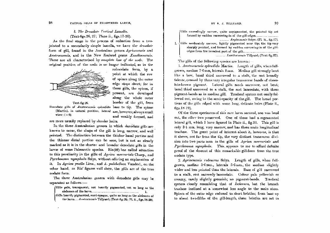
98 CAUDAL GILLS OF ZYGOPTERID LARVAE, BY it. J. TILLYA RD. 99
S. rrlw Denudate Vertical Lamella.
(Text-figs.26, 27; Plate ii., figs.17-20).
As the final stage in the process of reduction from a two-
jointed to a secondarily simple lamella, we have the denodale
form of gill, found in the Australian genera Agrioeiteinis; and
Anstrocnemis, and in the New Zealand genus Xantkomemis. These arc all characterised by complete loss of the node. The original position of the node is no longer indicated, as in the
subnodate form, by a
point at which the row
of spines along the outer edge stops short; for in
these gills, the spines, if
present, are developed
along the whole outer
Text -fig.26. border of the gill, from Denodate gills of Austroesemis splendida base to tip. The spines
(Martin), in natural position, lateral are, however, always small view; ( x 6).
and weakly formed, and are more usually replaced by slender hairs.
In the three Australasian genera in which denOdate gills are
knoWn to occur, the shape of the gill is long, narrow, and well
pointed. The distinction between the thicker basal portion and
the thinner distal portion can be seen, but is not as clearly
marked as it is in the shorter and broader denodate gills in the
larvae of some Pahearctic species. Ris(27) has called attention to this peculiarity in the gills of Agrion mercuriale Charp., and Pyrrh,osoma nymphnla Selys, without.offering an explanation of it. In Agrion- puella Linn., and A. pulchellum Vanderl., on the other hand, as Ris' figures well show, the gills are of the true
nodate form.
The three Australasian genera with denodate gills may be separated as follows :-
(Gills pale, transparent, not heavily pigmented, not as long as the abdomen of the larva 1.
Gills heavily pigmented, semi-opaque, quite as long as the abdomen of the larva... A aRtrocaem iR Tillyard; (Text-tig.26; Pl. IL, figs.18-20).
Gills exceedingly narrow, quite unpigmented, the pointed tip not formed by sudden narrowing-in of the gill-edges.
Argeorncatig Selys; (Pl. ii., tig.17). 1. Gills moderately narrow, lightly pigmented near tip; the tip very
sharply pointed, and formed by sudden narrowing-in of the gill- edges from the broadest part of the gill Varthoesenas Tillyard; (Text-fig.27).
The gills of the following species are known:- 1. Avstroenemis splendida Martin. Length of gills, when full-
grown, median 7.6 min, laterals 8mm. Median gill strongly bent
like a bow, basal third narrowed to a stalk, the rest broadly falcate, crossed by three very irregular transverse bands of choco- late-brown pigment. Lateral gills much narrower, not bent;
basal third narrowed to a stalk, the rest lanceolate, with three
pigment-bands as in median gill. Tracheal system not easily fol-
lowed out, owing to the semi-opacity of the gill. The broad por-tions of the gills edged with some long, delicate hairs (Plate ii., figs. 1 8- 1 9).
Of the three specimens of this rare larva secured, one was bred
out, the other two preserved. One of these had a regenerated
lateral gill, which I have figured in Plate ii., fig.20. This gill is
only 3.4 min. long, very narrow, and has three main longitudinal
tracheae. The great point of interest about it, however, is that it shows, not far from the tip, the very distinct transverse divi-
sion into two parts seen in the gills of Agrion mercuriale and Pyrrhosoma nymph/Ida. This. appears to me to afford definite proof of the descent of this remarkable gill-form from the true nodate type.
2. Agriocnernis rubescens Selys. Length of gills, when full-
grown, median 5-6min., laterals 5-6mm., the median slightly
wider and less pointed than the laterals. Base of gill narrowed
to a stalk, rest narrowly lanceolate. Colour pale yellowish or
creamy, rarely slightly greenish; BO pigment-bands. Tracheal
system closely resembling that of Ischnura, but the branch
tracheie inclined at a 'somewhat less angle to the main stem.
Spines of the outer edge reduced to short bristles; from base up to about two-fifths of the gill-length, these bristles are set in

strong chitinous bases, indicating the original set of spines end-
ing at the node; on the inner edge, there is a set of strong chitinous bases carrying small► bristles along the basal fourth of the gill, corresponding with the series of spines in the same posi-
tion in Ischnura. (Cf.
PredHction figs.16, 17).
This gill-form is obviously a simple reduction
from the subnodate form seen in Isclenatra, and
is very closely allied to it.
3. Xanthocnemis zelandica Selys. Length of
gill, when full-grown, about 4 mm. Gills lan-
ceolate, broadest near tips, then suddenly nar-rowing to form the sharply-pointed tips. Colour
pale brownish, yellowish or greenish, a band of
medium brownish pigment across the broad
part near the tip. Tracheal system much as in Ischnura, but with fewer branches, more regu-
larly placed. Outer edge of gill furnished from
base to near tip with short bristles, set in a
regular series of strongly-projecting chitinous
bases. A few irregularly placed bristles along
basal portion of inner edge. Towards tip, both edges carry some long, delicate hairs (Text-fig.27).
(b) The Horizontal Lamellar Sub-type.
(Text-figs.28-29; Plate i., figs.8-11; Plate vi., figs.40-45).
This extraordinary form of gill is only found, as far as I am
aware, in the Australian genus Argiolestes, belonging to the-sub-family Jleyapodayrioninm. I do not think that it will prove to
be typical of that ancient subfamily, in which we might reason-
ably expect to find a more generalised form of gill dominant,
when the larvae of many genera become known.
The gills are broad, leaf-like structures spread out in the hori-
zontal plane, so as to resemble closely in form the "tail-fan" of
Decapod Crustacea. In life, they are held out extended at an
angle of from 10° to 20° to the plane of rest. When walking,
Denrwlate median gill of Xanthoenemis zelandiect (Selys). From a cleared whole mount; ( x 151.
the larva often raises these gills considerably,
together with the terminal part of the abdo-
men. When resting on a rock-surface, the
larva, if alarmed, at once depresses the gills
closely against the rock. A curious point
about these gills is that they are slightly concave ()it their upper surfaces. This is well seen in . the cross-section in Tex t-1ig.28. It is due to the almost complete suppression of
the dorsal mid-rib in all three gills, the ventral mid-ribs being strongly developed.
The median gill is symmetrical, and lies
quite horizontally. The laterals are asym-inetrical,—the outer portion of the blade
being wider than the inner,—and are held
at a slight angle to the horizontal, slanting upwards and outwards.
The transverse section of one of these
gills may be described as an extremely flat-
tened triangle, except near the base of the
gill, where the cavity is wider, as shown in
Plate vi., figs.40-41. The vertex of the
triangle is represented by the ventral ► id-rib, the base by the wide upper surface of
the blade. The raehis is small, subtrian-
gular, and carries along its middle line the blood-canals, which lie practically in contact
with one another. The dorsal blood-canal
is smaller and more regularly formed than
the ventral. On either side of the blood-
canals, towards time lateral limits of the
raehis, run the two main longitudinal tra-
theme. The nerve-supply is as usual, the
* Ideal (semi-diagrammatic) T.S. across the Hori-zontal Lamellar Gill-system of A•gioteRte.4, to show the internal struetnre of the gills. Lettering 11.9 on p.109.
n. J. TiLLYARb. 101 100 CAUDAL GILLS OF ZYOOPTERID LA IIV/E,

102 CAUDAL GILLS OV ZYGOPTEBID LARVit, BY R. J. TLLLYABD. 103
median gill possessing a pair of dorsal longitudinal nerves and a
pair of ventral ones, while the lateral gills have only one of each,
placed on the outer wall of the corresponding blood-canal.
The radius of the median gill is bounded internally on either side by a series of secondarily developed internal, lamina; (Plate vi., fig.43,i1) passing from the dorsal to the ventral side of the
gill. Similar but less strongly developed laminae occur at inter-vals at different levels along the blade of the gill (i/'). These lamina are not, of course, the original laminae of the gill before
it became horizontally flattened. The remnants of the original
series of principal laminae, running horizontally across the gill,
are to be seen only in a set of slender laminae crossing the raehis below the ventral blood-canal (Plate vi., fig.42 , v/). These lamina! mark off ventrally what was the original raehis of the gill from
the remnant of its ventral blade, which has now become the
portion just above the mid-rib. The present blade of the gill, to
right and left of the new radius, is purely an extension of the
old raehis itself, due to intense flattening and widening in the
horizbntal plane. As will be seen from Text-fig.,28, the original
right and left mid-ribs of the gill, before it became horizontal,
are still preserved as the swollen, somewhat knobbed, edges of . the new blade.
'The cuticle is rather thin, averaging about fitt in thickness, of.
which the tough outer stratum occupies from 2 to 3tt. The
hypodermis is slightly thinner than the cuticle, averaging about
5i,t, in width, but somewhat irregular. The nuclei mostly lie with their long axes parallel with the cuticle. They are of
-mall size, only 5-6tt in length, except near the base of the gill,
where many of the nuclei are considerably larger. The hypo-
dermis is almost everywhere pigmented, but in certain transverse
zones the pigment-granules are exceedingly abHndant, ;utmost
hiding the nuclei from view.
Considering how narrow the space in the interior of these gills
is, it is rather surprising to find the alveolar meshwork present,
not only in the raehis, but also throughout, the blade. The
meshwork is most extensi vely developed in the space between
the two main tracheae and the mid-rib. The nuclei of the mesh-
work closely resemble those of the hypodermis in size and shape.
The tracheal system is moderately well developed, but the
branch tracheae are neither so abundant nor so regularly placed
as in Vertical Lamellar Gills.
These gills are frequently lost or east off by the larva!, the breaking-joint being very perfeCtly formed. Consequently lame are very commonly found with one or more regenerated gills. In these there will always be found four main longitudinal trachea!, in the case of the median gill, and
usually three (rarely four) in the laterals. The regenerated gill is
stouter and less expanded than the
normal gill, and more resembles a
rather flattened saceus (Plate i., fig.
11). Usually a transverse band of dark pigment separates a thicker
basal portion from a thinner distal
portion, and suggests the origin of
this type from an originally two- Text-fig.29.*
jointed form.
The larvae of only two species are known, and their gills may
be distinguished as follows:— Length of gills, when full-grown, 6-7 mm., the median gill I '.1-2
wide, the laterals slightly less. Colour pale brownish, semi-opaque, the distal half crossed by a very wide band of chocolate-brown pigment; slight irregular pigmentation of the same colour is present also at about two-fifths from the base (PL i., figs.8-9).
A. icteromehm Selys. Length of gills, when full-growu, the median gill very wide,
1 '8-2nun., the laterals nearly as wide. Colour pale brownish, send•opaque, with slight irregular darker pigmentation towards tip (Pl. i., fig. 10) A. p iaercx Says.
U. The Reduced (Non-functional) Type (Text - figs.:30 - V).
The Saecoid, Triquetro-quadrate, and Lamellar Types of
Candid Gills are all characterised, morphologically, by the pos-
* Horizontal lamellar gills of A rgioteste. ,4 iderontelas Selys, in natural osition, dorsal view; ( x 6).
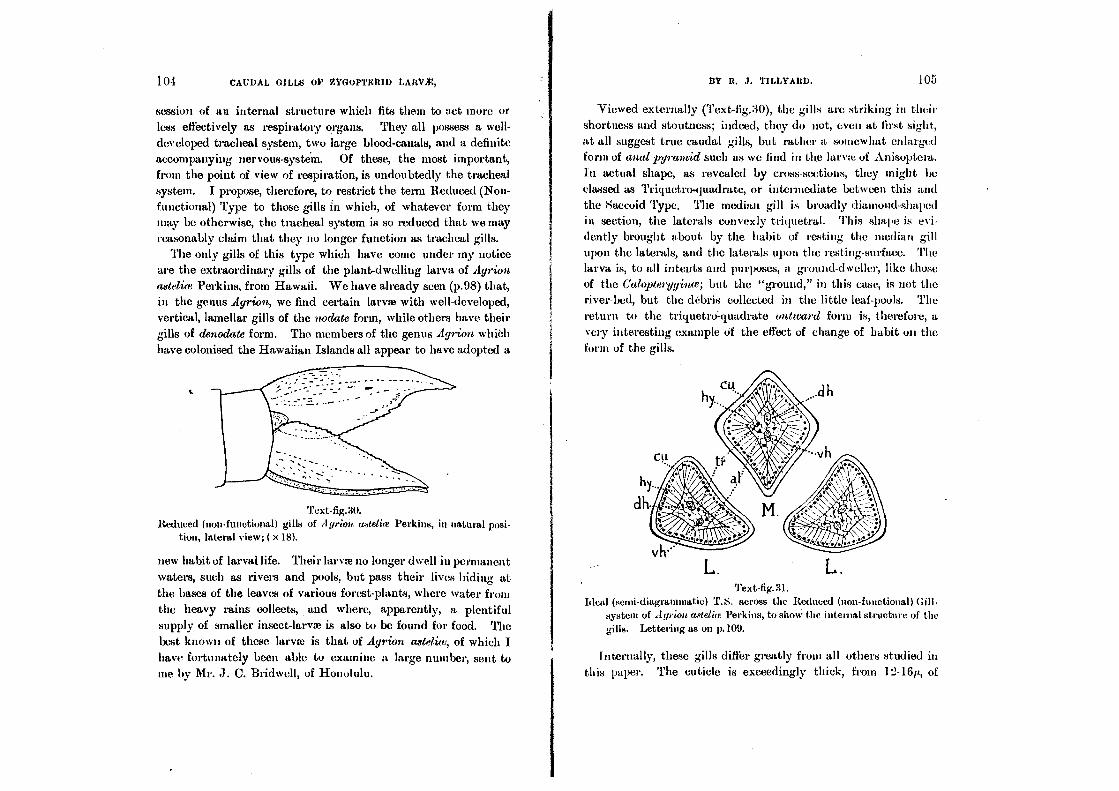
104 CAUDAL GILLS O1' ZYGOPTERID LARVA:,
session of an internal structure which fits them to act more or
less effectively as respiratory organs. They all possess a well-
developed tracheal system, two large blood-canals, and a definite
accompanying nervous-system. Of these, the most important,
from the point of view of respiration, is undoubtedly the tracheal
system. I propose, therefore, to restrict the term Reduced (Non-
functional) Type to those gills in which, of whatever form they
may be otherwise, the tracheal system is so reduced that we may reasonably claim that they no longer function as tracheal gills.
The only gills of this type which have come under my notice
are the extraordinary gills of the plant-dwelling larva of Ayrion astelim Perkins, from Hawaii. We have already seen (p.98) that, in the genus Agrion, we find certain larvae with well-developed,
vertical, lamellar gills of the nodate form, while others have their
gills of denodate form. The members of the genus Agrion which
have colonised the Hawaiian Islands all appear to have adopted a
Text-fig.30. Reduced (non-functional) gills of ilgrioir (Wetke Perkins, in natural posi-
tion, lateral view; ( x H).
new habit of larval life. Their lame no longer dwell in permanent
waters, such as rivers and pools, but pass their lives hiding at
the bases of the leaves of various forest-plants, where water from the heavy rains collects, and where, apparently, a plentiful
- supply of smaller insect-larvae is also to be found for food. The
best known of these larvae is that of Agrion astelice, of which I have fortunately been able to examine a large number, sent to
me by Mr. J. C. Bridwell, of Honolulu.
13Y R. J. TILLYARD. 105
Viewed externally (Text-fig.30), the gills arc striking in their
shortness and stoutness; indeed, they do not, even at first sight,
at all suggest true caudal gills, but rather a somewhat enlarged form of anal pyramid such as we find in the lame of Anisoptera.
In actual shape, as revealed by cross-sections, they might be
classed as Triquetroviadrate, or intermediate between this and
the 8accoid Type. The median gill is broadly diamond-shaped in section, the laterals convexly triquetral. This shape is evi-
dently brought about by the habit of resting the median gill
upon the laterals, and the laterals upon the resting-surface. The larva is, to all intents and purposes, a ground-dweller, like those of the Caloptergyince; but the "ground," in this case, is not the
river-bed, but the debris collected in the little leaf-pools. The
return to the triquetro .-quadrate outward form is, therefore, a
very interesting example of the efleet of change of habit on the
form of the gills.
Text-fig.31. Ideal (semi-diagraminatic) T.S. across the Reduced (non-functional) Gill.
system of At/rim, mg/cum Perkins, to show the internal structure of the gills. Lettering as on p.109.
Internally, these gills differ greatly from all others studied in
this paper. The cuticle is exceedingly thick, from 1:1-16p, of
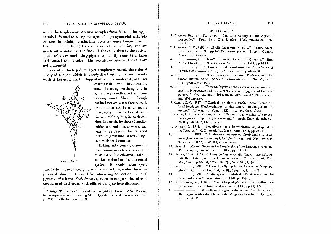
Ey R. J. TILLYARD. 107 106 CAUDAL GILLS OP ZYGOPTERID LARY,E,
which the tough outer stratum occupies from 2-4/L. The hypo-
(tennis is formed of a regular layer of high pyramidal cells, 24p.
or more in height, terminating upon an inner basement-mem-
brane. The nuclei of these cells are of normal size, and are
nearly all situated at the base of the cells, close to the cuticle.
These cells are moderately pigmented, chiefly along their bases
and around their nuclei. The boundaries between the cells are
not pigmented. Internally, the hypoderm-layer completely bounds the reduced
cavity of the gill, which is chiefly filled with an alveolar mesh-
work of the usual kind. Supported in this meshwork, one can distinguish two blood-canals,
small in many sections, but in
some places swollen out and con-
taining much blood. Longi-
tudinal nerves are either absent,
or so fine as not to be traceable in sections. No tracheae of large
size are visible, but, in each sec-
tion, five or six trachea of smaller
calibre are cut; these would ap-
pear to represent the reduced main longitudinal tracheal sys-
tem with its branches.
Taking into consideration the
great increase in thickness in the
cuticle and hypodermis, and the
marked reduction of the tracheal
system, it would seem quite
justifiable to class these gills as a separate type, under the name
proposed above. it would be interesting to section the anal
pyramid of a large .Eschnid larva, so as to compare the internal
structure of that organ with gills of the type here discussed.
• Aet ual T. 8. across interior of median gill of A grim/ (k`f td Perkins, for comparison with Text-fig.31. Hymxlermis and cuticle omitted. ( x'2.181. Lettering as on p.109.
BIBLIOGRAPHY.
1. BALFOUR-BROWN E, F., 1909.—" The Life-History of the Agrionid Dragonfly." Proc. Zool. Soc. London, 1909, pp.253-285, Pls. xxxiii.-iv.
2. CALVERT, P. P., 1893.—" North American Odonata." Trans. Amer. Ent. Soc., xx., 1893, pp.153-268, three plates. [Part i. General Account of Odonata].
3. s , 1911-15.—" Studies on Costa Rican Odonata." Ent. News, Philad. i. " The Larva of Cora." xxii., 1911, pp.49-64.
4. , iii. "Structure and Transformation of the Larva of illecistogaater mode8tus." Op. cit., xxii., 1911, pp-449-460.
5. , vi. " Transformation, External Features and At- tached Diatoms of the Larva of Thauntatoncara. Op. cit., xxvi., 1915, pp.295-305, Pl. xi.
6. , vii. " Internal Organs of the Larva of Thauntataneura, and the Respiration and Rectal Tracheation of Zygopterid Larva in general." Op. cit., xxvi., 1915, pp.385-395, 435.447, Pls.xv.-xvii., and bibliography.
7. CARUS, C. G., 1827.—" Entdeckung eines einfachett vom Herzen aus beschleuuigten 131utkreislaufes in den Laiven netzfliiglicher In-seeteti." Leipzig. L. Voss. 1827. pp.1-40, three plates.
8. CitiLn, C. AL, and Youbm, A. N., 1903.—" Regeneration of the .Ap-pendages in nymphs of the Agrionithc." Arch. Entwiektnech. xv., 1903, pp.543-602, Pls. xx.-xxii.
9. Dt•ouR, L., 1849.—" Des divers modes de respiration aquatique clans les Insectes." C. R. Acad. Sei. Paris, xxix., 1849, pp.763.770.
10. 1852.—" Etudes anatomiques et physiologiques, et ob- servations sur les loaves des Libellules." Ann. Sci. Nat., 3''° SCr., Tome xvii., 1852, pp.65-111, three plates.
IL EAST, A., 1900.—` 'Notes on the Respiration of the Dragonfly Nymph." Entomologist, London, xxxiii., 1900, pp.211-12.
12. BAIREN , H. A.. 185:3.—" &ton Defour fiber (lie Larven der Libellee wit Berucksichtigung der friiheren Arbeiten." Stett. cut. Zeit. xiv., 1853, pp.98-106, 237-8, 260-270, 311-325, 334-346.
13. , 1880.—" Essai (Fun Synopsis des .Larves (le CaloptC•y- gines." C. R. Soc. Ent. Belg. xxiii., 1880, pp. lxv.-lxvii.
14. , 1880.—"Beitrag zur Kenatnis des Tracheensystenis der Libellee-Larven." Zool. Anz. iii., 1880, pp.1:37-161.
15. ILI N DIARSCH, A., 1903.—" Zur Morphologie des Hinterleibes der Odonaten." A1111. Hofinus. Wien, xviii., 1903, pp.117-122.
111. , 1904.—Beitierkungen zit der Arbeit des Herrn Prof. Dr. Heyinons fiber (lie Abdominalaidilinge der Libellen." /.r., xix., 1904, 0p.5140:3.

108 CAUDAL mLLS oP ZYGorrtnty LAtrViE, ItY R. J. TILLYARD. 109
17. HEYNIONS, R., 1904.—" Die Hinterleibsaillifinge der Libelien and ihrer Larven." Ann. Hofinits. Wien, xix., 1904, pp.21-58, Pl. i.
18. HUBNER, 0., 1902.—" Neue Versuche acts dent Gebiet der Regenera-tion and litre Beziehungen zu Anpasstingserscheintingen." Zoo!. Jahrh. Syst. xv., 1902, pp.461-498, Pls.28-29. (Summary in Zoo!.
Ceara1bl. 1902, pp.427-8).
19. NIArert , J., I892.—" Sur la respiration des la,ry es (le Libtillttles." Bull. See. Philomath. Paris, (8) Tonle 4, 189'2, pp. P22-4.
M LILL, L. C., 1895.—" The Natural History of Aquatic Insects." London, Macmillan and Co., 1895, (Dragonflies, Chap. ix., pp.328-
345). 21, INEEniLANI, J. B., 1911.—" Descriptions of dragonfly nymphs of the
subfamily Caloptergyidte." Ent. News, Philad., xxii.. 1911, pp.145-
154, two plates. , 191 I . —" Notes on a few Nymphs of Agrioniisi of the
Hagen Collection." t.c., xxii., 1911, pp.341-5, one plate.
23. PERKINS, R. C. T.., l897.—" Notes on some Hawaiian Insects.'' Paw. Cambridge Soc., Mass., ix., 1897, pp.373. -380. (Terrestrial Nymphs).
24. PonTrEn, P., 1911.—"Recherches physiologiques sur les Insectes Aquatiques." Arch. Zool. Exp. Gen. (5) viii., 1911, pp.89-379,
Plates i.-iv. (Calopteryx, pp.216-7).
25. PutsFat, G. L., 1915.—" Preliminary Notes on some problems con-nected with respiration in Insects generally, and in aquatic forms in particular." Proc. Cambridge Phil. Soc., xviii., Pt. ii., 1914,
pp.63.70. 26. RE-tumuli, R. A., 1734-4'2.—" Memoires pour scrvir it l'histoire natal-
relic ct is l'auatomie des Insectes." 6 vols., Paris, '267 plates. (See Vol. 6, inentoire xi., "Des watches it quatre ailes itommes Demoi-
selles," pp.387-456, Pls.35-41). RIS, F., 1909.—" Die Stisswasserfamat Deutschlands." Jena, G.
Fischer, 1909, iv., pp.1-67. 28., 191'2.—" Ueber Odonaten von Java mid lirakatan." Gra-
venhage, Tijdschr. Ent., Iv., 191'2, pp.157-183, Pls. 6-8. (Morphology
of larva of Enplueal. 29. , 1913.—" Die Atraungs-Organe der anisopteren Libellen-
larven." Mitt. schweiz. ent, Ges. xii., 1913, pp.9'2-99. (Refer-
ence to rectal respiration in Calopteryx, p.95).
30. ROESEL VON ROSENII0F, I749.—" Der Wasser-Insecten zweytc Rlasse. Der nionatlich-lierausgegebenen Insecten-Belustigung." Part 2. Nurnberg.
31. RosTErt, D. A., 1886. —" Cenn° Monographic° degli Oclonati (lei Gruppo behnora." 1301. Soc. Ent, xviii., 1886, pp.2311-258,
ii.-vi.
32. POSTER, D. A., 1888.—"Contributio allo studio delle forme larvali degli Odonati." Centro Iconographic() della Larve-Ninfe (lei Cando. branchiati." Lc., xx., 1888, pp.159-176, Pls.
33. ROUSSEAU, E., 1909.—" Etude monographique des larves des Odonates d'Europe." Ann. Biol. lacustre, Bruxelles. iii., 1909, pp.300.366.
34. SonmIDT•SCHWEDT, E., 1891.—" Kerfe u. kerflarven d. siissen 1Vassers, besonders der stehenden Gewiisser." In " Die Tier u. Pflanzenwelt d. Siisswhssers," von Dr. 0. Zacharias, Bd. ii., 1891, pp.51-122. Leipzig, J. J. Weber.
35. Slum', D., 1901.—"Cambridge Natural History. Insects, Part i." Chap. xviii.; dragonflies, pp.409-428.
36. 'lrr.LYARn, R. J., 1909.—" Studies in the Life-Histories of Australian Odonata. No.2: Dipldelda tettloide8 Selys." Proc. Linn. Soc. N. S. Wales, Sydney, xxxiv., 1999, pp.370-383, Pl. xxxiii.
37. , 1912.—"On the genus Diphtebia, with descriptions of new species, and life-histories." I.e., xxxvi., 1911 (1912), pp.584- 604, Pls. xix.-xx.
38. , 1916.—"Further Observations on the Emergence of Dragonfly-Lary' from the Egg. With special Reference to the Problem of Respiration." - c., xli., 1016, pp.388-416. (Zygoptera, pp.410-414).
39. 't'trtirEL, R., 1898-9, 1908.—" Die Geradthigler Mitteleuropas." lst, Ed., Eisenach, N. Wilekens. 2nd Ed., Gotha, F. E. Pertbes (p.66).
EXPLANATION OF ABBREVIATIONS USED IN TEXT-FIGURES AND PLATES.
anus—ac, anicebocyte—at, alveolus—alit, alveolar nucleus—blp, blood-plasma—c or c', cercoid—cu, soft inner layer of cuticle—car', hard outer layer of cuticle--dh, dorsal blood-canal—dt, principal dorsal internal lamina—chr, dorsal longitudinal nerve of gill—DT, Dr, dorsal tracheal trunks—ect, ectotrachea—ent, or end, endotrachea—h, hair (oil cuticle)—hc, hiemoc(ele —11y, hypodermic—it, internal lamina—it', secondary internal lamina-
tg, or L, lateral caudal gill—nrc, miocyte (oat-shaped corpuscle)-mcy, mg, or AI, median caudal gill—op, spiral thread of endotrachea--tr, main longitudinal trachea of gill—fir', branch trachea of gill—oh, ventral blood-canal—r principal ventral internal lamina—rn, ventral longitudinal nerve of gill—S-10, abdominal segments,
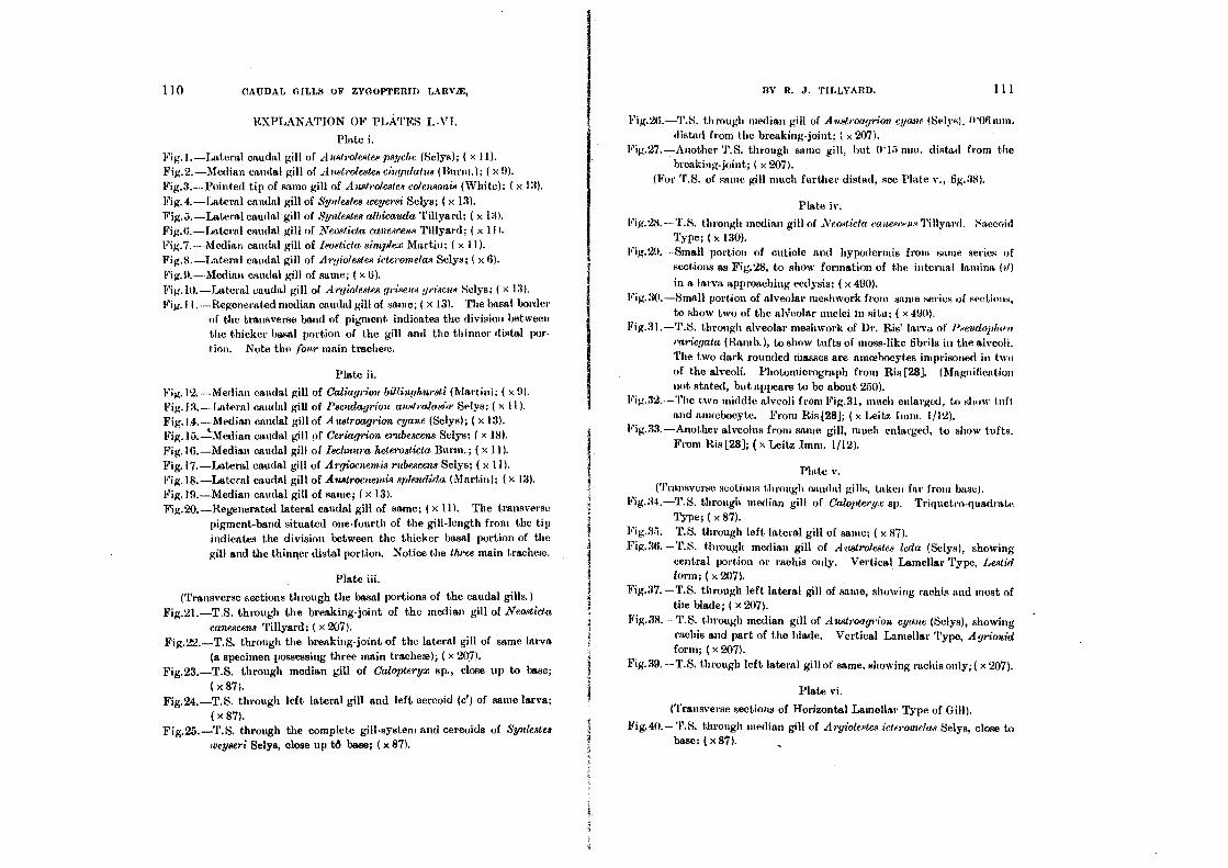
110 CAUDAL GILLS OF ZYGOPTERID LARVA:,
EXPLANATION OF PLATES I.-VI.
Plate i.
Fig. I.-Lateral caudal gill of Austrolestes psyche (Selys); ( x Fig.2.-Median caudal gill of Austrolestes cingulatus (Burro.); ( x 9).
Fig.3.-Pointed tip of same gill of Anstrolestes colensonis (White): ( x 13).
Fig.4.-Lateral caudal gill of Synlestes .weyersi Selys; ( x 13).
Fig.5.-Lateral caudal gill of Synlestes albicauda Tillyard; ( x 1:1). Fig.6.-Lateral caudal gill of Neosticta canexcens Tillyard; ( x 11). Fig. 7.-Median caudal gill of Isosticta simplex Martin; ( x 11).
Fig.8.-Lateral caudal gill of Argiolestes ideromelas Selys; ( x 6).
Fig.9.-Median caudal gill of same; ( x 6). Fig.10.-Lateral caudal gill of Argiolestes griseus grisems Selys; ( x 13).
Fig. I I .-Regenerated median caudal gill of same; ( x 13). The basal border of the transverse band of pigment indicates the division between the thicker basal portion of the gill and the thinner distal por-
tion. Note the four main tracheae.
Plate ii.
Fig.12.-Median caudal gill of Caliagrion billinghursti (Martin); ( x 9).
Fig.13.-Lateral candid gill of Reencktgrion auRtrctlasife Selys: ( x 11). Fig. I 4.-Median caudal gill of A ustroagrion cyane (Selys); ( x 1:3).
Fig.15.-`̀'Median caudal gill of Ceriagrion ernbescens Selys; ( x 18). Fig.16.-Median caudal gill of Ischnura heterosticta Burin.; ( x 11). Fig.17.-Lateral caudal gill of Argiocnemis rubescens Selys; ( x L 1). Fig.18.-Lateral caudal gill of Anstrocumnis splendida (Martin); ( x 13). Fig.19.-Median caudal gill of same; ( x 13). Fig.20.-Regenerated lateral caudal gill of same; ( x 11). The transverse
pigment-band situated one-fourth of the gill-length from the tip indicates the division between the thicker basal portion of the gill and the thinner distal portion. Notice the three main tracheae.
Plate iii.
(Transverse sections through the basal portions of the caudal gills.) Fig.21.-T.S. through the breaking-joint of the median gill of Neostieta
canescens Tillyard; ( x 207). Fig.22.-T.S. through the breaking-joint of the lateral gill of same larva
(a specimen possessing three main tracheae); ( x 207).
Fig.23.-T.S. through median gill of Calopteryx sp., close up to base;
( x 87). Fig.24.-T.S. through left lateral gill and left cercoid (c') of same larva;
( x 87). Fig.25.-T.S. through the complete gill-system and cereoids of Synlestes
tveyseri Selys, close up td base; ( x 87).
BY R. J. TILLYARD. 111
Fig.26.-T.S. through median gill of A ustroagrion cyane (Selys), 0 .06 tom. distad from the breaking-joint; ( x 207).
Fig.27.-Another T.S. through same gill, but 0'15 mm. distad from the breaking-joint; ( x 207).
(For T.S. of same gill much further distad, see Plate v., fig.38).
Plate iv.
Fig.28.-T.S. through median gill of Neostida ranexcemq saacoid
Type; ( x 130). Fig.29. -Small portion of cuticle and hypodermis from same series of
sections as Fig.28, to show formation of the internal lamina (d)
in a larva approaching ecdysis; ( x 490). Fig.30.-Small portion of alveolar meshwork from same series of sections,
to show two of the aNeolar nuclei in situ; ( x 490). Fig.31.-T.S. through alveolar meshwork of Dr. Ris' larva of Pseudophma
rariegata (Ramb.), to show tufts of moss-like fibrils in the alveoli. The two dark rounded masses are amcebocytes imprisoned in two of the alveoli. Photomicrograph from Ris [28]. (Magnification not stated, but appears to be about 250).
Fig.32.---The two middle alveoli from Fig.31, much enlarged, to show tuft and anuchocyte. From Ris [28]; ( x Leitz Imm. I/12).
Fig.33.-Another alveolus from same gill, much enlarged, to show tufts. From Ris [28]; ( x Leitz Imm. 1/12).
Plate v.
(Transverse sections through candid gills, taken far front base). Fig.:34.-T.S. through median gill of Calopteryx sp. Triquetro-quadrate
Type; ( x 87). Fig.:35. -T.S. through left lateral gill of same; ( x 87). Fig.:36. -T.S. through median gill of Austrolestes Zola (Selys), showing
central portion or rachis only. Vertical Lamellar Type, Lestid form; ( x207).
Fig.37.-T.S. through left lateral gill of same, showing rachis and most of the blade; ( x 207).
Fig.38. T.S. through median gill of Austroagrion cyane (Selys), showing rachis and part of the blade. Vertical Lamellar Type, A grionid form; ( x 207).
Fig. 39. --T.S. through left lateral gill of same, showing racing only; ( x 207).
Plate vi.
(Transverse sections of Horizontal Lamellar Type of Gill).
Fig.40.- T.s. through median gill of Argiolestes icteromelas Selys, close to base; ( x 87).

PL. I,
R ./ cid.
112 CAUDAL GILLS OF ZYGOPTERID LAMM.
Fig.41. -T.S. through right lateral gill of same, at anne level as fig.40; ( x 87).
Fig.42. — T.S. through median gill of same, at about one-third of the gill-length from the base; rachis and small portion of blade only;
( x 207). Fig 43.—T.S. through same gill as fig.43 taken far distad, to show con-
fluence of dorsal and ventral blood-canals; much of the blade omitted; ( x 207).
Fig.44.—A detail from same series of sections as figs.40-43, much enlarged, to show portion of hypodermis and cuticle, ecto- and endotrachea of the left main trachea, two nuclei in the alveolar meshwork and the right dorsal longitudinal nerve. The figure is shown inverted, as drawn under the microscope. It corresponds with the part of the gill marked x in fig.40; ( x 833).
Fig.43. —T.S. through a longitudinal nerve (nn) and neighbouring alveolar nucleus (aln,L The section cuts the nerve so as to pass through two nuclei; ( x 1250).
N.B. —With the exceptions of Figs.31-33 (from Ris), all the figures in Plates iii.-vi., are wash-drawings made from camera-lucida drawings of double-stained, lOtt sections.
Caudal Gills of Zygopterous Larvie: 1-5, Lestid:s; 0-11, Agrionillw.
P.L.1.N.S.W. 1017. PL.
Caudal Gills of Zygopterolis Larvae : 12-20, Agrion
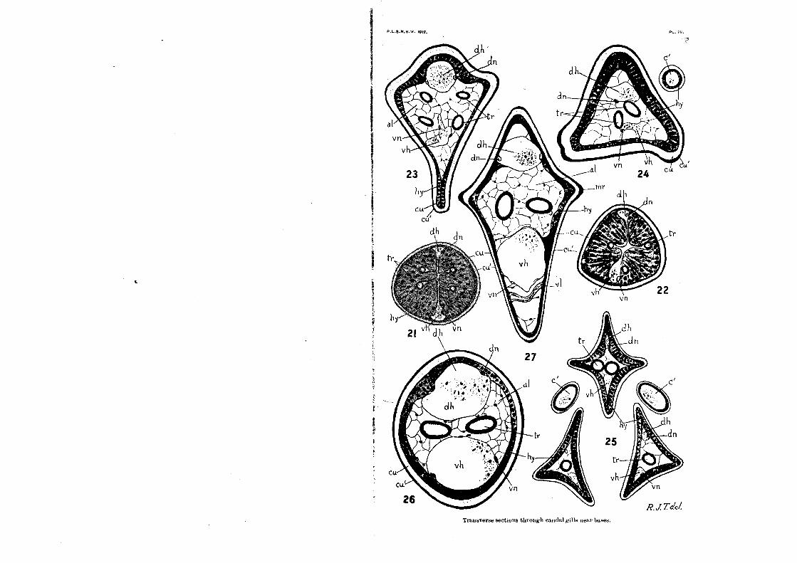
Transverse sections through caudal gills near buses.
P.L.S.N.S.W. 1017. Pl. Pl.
vn v h
23
by
Cu Cu
dh
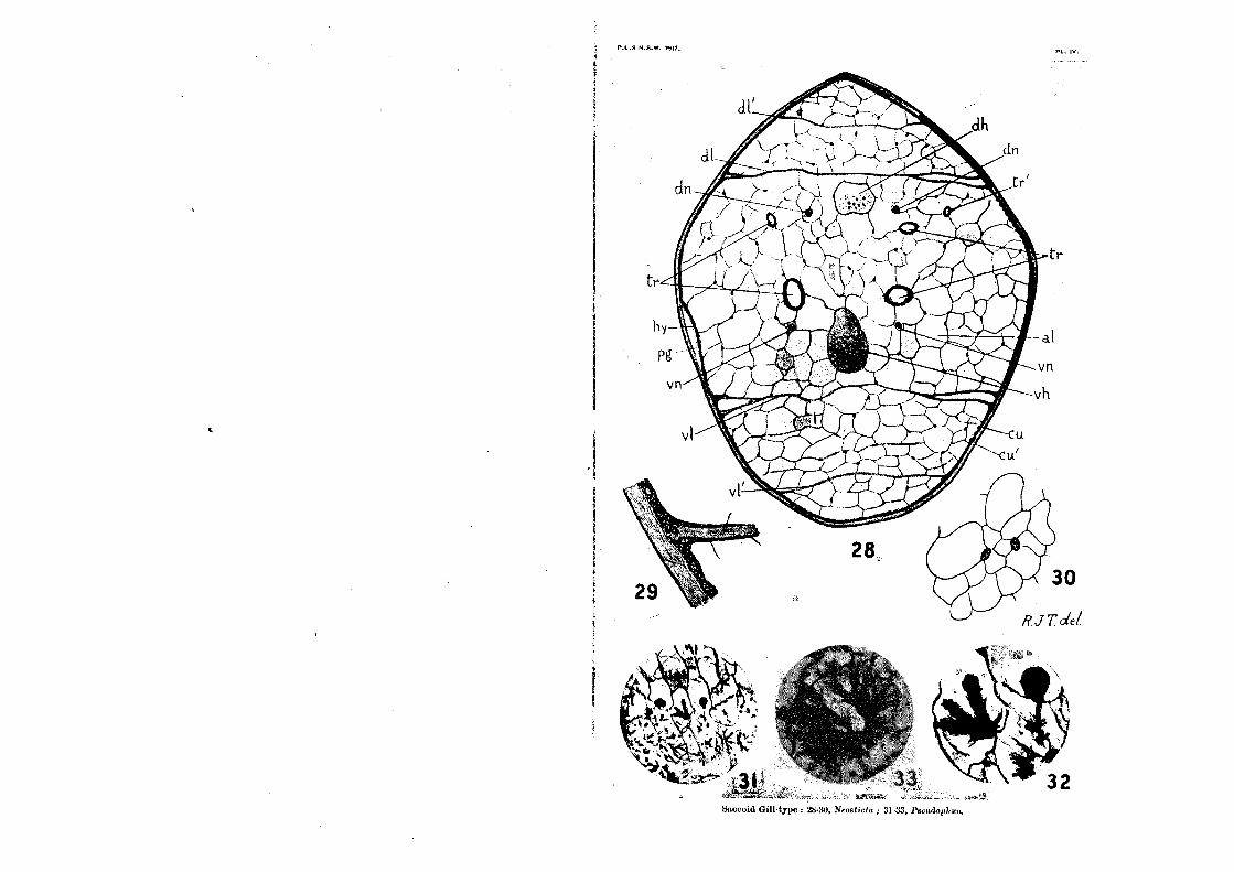
P.L.S.N.S.W. 1017. In. W.

P.I.S.N.S.W. 1917.
Figs. TH.-metro-quadrate Type.

vi,
111r
RJ Td.- t Jrue input:11 I atiwohlur 111.1k 13