on the skeleton, and affinities of the paired fins of ceratodus, with observations upon those of the...
TRANSCRIPT

1887.1 ON T H E PAIRED FINS O F CERATODUS. 3
Mr. Tegetemier exhibited and made remarks on some heads of the Sumatran Rhinoceros (Bh. sumatrensis), male, female, and young, forwarded from Sarawak, Borneo, by hIr. Brooke-Lowe.
Prof. Bell exhibited a specimen of Nereis pelagica which he had received from his excellent correspondent hlr. It. L. Spencer of Guernsey, and which was remarkable for the bifid arrangement of the posterior portion of the body. IIe remarked that although Mr. Robertson, of Oxford, Dr. IIorst, and himself had put on record Lumbrici with trifid ends, which probably were not really uucommon, he had not been able to find any record of a similar condition in a Polychsete.
A communication was read from Messrs. 13. B. Brady, F.R.S., W . K. Parker, P.R.S., and 1’. Rupert Jones, F.R.S., containing an account of the Foraminifera procured on the Abrohlos Bank dariug the cruise of H.M.S. ‘ Plumper ’ in 1857.
This memoir will be printed in the Society’s ‘ Transactions.’
The following papers were read :- --
1. On the Skeletou and Affinities of the Paired Fins of Ceratodus, with Observations upon those of the Elasmo- braizchii. By G. B. HOWES, F.Z.S., F.L.S.,Assist. Prof. of Zoology, Normal School of Science and Royal School of Mines, S. Kensington.
[Received December 14, 1886.1
(Plates I.-111.)
CONTENTS. Page I. Introduction ............................................................ ,.. ...... 11. On the Structure of the Ceratodus Fins in general and of the Pelvic
III. On the Pectoral member of Ceratodtks compared with the Pelvic one
3
Fins in particular ......................................................... 4
of the same and the Pectoral one of the Plagiostomes ............ 11 16 18
IV. On the proxinml Postaxial Elements of the Ceratodics Pelvic Fin ... V. On the Morphology of tbe Axis of the Cemtodfts Fin ..................
with that of Ceratodzcs ................................................... 28 VII. Conclusions.. ...................................................................... 24
VII I . List of Authorities referred to ................................................ 24 IX. Description of the Plates (1.-111.) 26
VI. On tlie Eomologies of the Chiniieroid Fin-ekeleton, as couipared
.......................................... I. Introduction.
I have lately described (17, p. 277) the vertebral column of a Frog in connexion with which there had taken place, under disloca- tion of tlie urostyle, a process tantamount to that of reproduction of a lost part. While searching for literature bearing upon this subject,
1*

4 PROF. G. B. HOWES ON THE SKELETON AND [Jan. 18,
there came under my notice a short paper by Traquair (26, p. 143), in which he describes the restoration of parts of the tail of Proto- pterus. Finding that he had discovered certain irregularities in the skeletal elements of the said restored tails, and knowing lhat Haswell had recorded (15) some irregularities of the Ceratodus paired fins, it occurred to me that the same determining cause might have been at work in the two cases-i. e., that Haswell’s “branching” fins might perchance be “restored ” ones, like Traquair’s. I was soou undeceived ; for, apart from Hasmell’s paper, I have had the good fortune to examine one such fin, sent by him to Prof. Huxley. The deductions arrived at in the sequel have arisen out of a study of it and of the fins of five other individuals. Three of them mere kindly lent me by my master, Prof. Huxley; of the two which remain, one forms part of our teaching-collection at South Een- sington ; for the loan of the other I am indebted to my Demon- strator, Mr. M. F. Woodward.
I t is remarkable that Gunther, in his Monograph on Ceratodus (14), does not mention Traquair’s discovery already alluded to. It is clear that that author’s paper must have escaped him, as I fail to find note of i t under either “Pisces ” or “ Ganoidei,” as reported by him for the ‘ Zoological Record ’ during both its year of publication and the succeeding one.
The structural plan of the fin of Ceratodzis is too familiar to merit detailed description here. Husley has described (19,.p. 46 et sep.) its general features with exceeding care, and I shall, in accordance with his system, speak of the segments of the axis as “mesomeres.”
The lateral rays will be described, under the same nomenclature, as parameres; those which look dorsally when the fin is placed against the side wall of the body (anteriorly when it is held out at right angles thereto) I shall speak of as preaxia l ; those which look ventrally under the first-named condition (posteriorly under the last-named) I shall describe as postaxiaz. Preaxial and postaxid correspond to the “ dorsal ’’ and “ventral ” of the Germans. As the basal segment of‘ the axis differs in its essential characters from those which follow upon it, I shall refer to i t as the proximal meso- mere (the ‘‘ zwischen-Stuck ” of Davidoff (7), the “ erste Glied” of Solineider (23)).
11. On the Xtructure of the Ceratodus Paired Fins in general and of the Pelvic Fins in particular.
The majority of observations made thus far upon the fins of Ceratodus bear especially upon the pectoral member ; its pelvic representative has received less attention. Davidoff (7) and Haswell (15) have dealt most fully with it, the last-named author espe- cially as to certain “ irregularities ” mentioned in the Introduction. Fig. 1 is a faithful representation of the pelvic fin presented by him to Prof. Huxley; and as it does not appear to correspond with any one figured in his own paper, I proceed to describe it in detail.
The fin reached me cleaned and prepared, as represented in the

1887.1 PAIRED FINS OF CERATOD~S. 5
figure, and it had first to be ascertained from which side of the body it mas derired. Its proximal mesomere (mp., fig. 1) carries a large tubercle (ta.), which, as Schneider has lately pointed out, '' bei der Brnstflosse ventral, bei der Bauchflosse dorsal steht "-when the limb is in apposition with the body-wall. This process is, in all pelvic fins examined by me, somewhat crescent-shaped and out- wardly directed, its inner face being excavated. In the fin under discussion its outer surface was flattened ; but as its inner one sloped obliquely outwards, I conclude that that fin was a right-sided one. It is represented in the figure as seen from the dorsal aspect. I ts axis is for the most part unequally segmented and irregular, the proximal mesomere being the least modified portion thereof, as com- pared with the more normal fin. The second mesomere is greatly elongated, and it bears upon its postaxial border (left hand of the figure) a notched lobe, with mhich are connected five parameres. The two distal of these break up peripherally, and, on examining the individual specimen, it is hard to conjecture how far the lines of de- marcation between the parameres and the lobe, and between it and the main piece of the axis, map represent the last traces of original lines of separation, or the lines of cleavage of a primarily continuous sheet. Preaxially, the second mesomere carries five parameres ; these are fairly uniformly set upon it, and the distal one of the series branches in a true dichotomy. Interposed between the free ends of the two proxi- mal of these rays there is a smaller one (marked * in the figure), which I take t o resemble those found by Daridoff (7, p. 127), occa- sionally lying free at the distal end of the fin. The rest of the skeleton is chiefly remarkable as concerns the axis ; this appears to be longitudinally cleft, and made up of a longer preaxial and a shorter postaxial piece, both of which are very irregularly segmented. All the parameres borne upon it, however, are simple unbranched rods, which differ fro% those more generally present only as regards their feeble segmentation.
On examining the above-named fin with care, my attention became arrested by the cartilage marked r in the figure, the characters and relations of mhich are altogether exceptional. Wiedersheim has oalled attention (30) to the fact that in Protopterus the basal seg- ment of the axis may bear a lateral piece. To the consideration o f this I shall return. I n no regular cerntodus fin (i. e. that bearing an equally segmented axis) yet described has there been found, post- axially, a cartilage like the above named, attached directly to the basal mesomere. That element is generally held to be destitute of rays. Giinther has figured (14,.pl. 36. fig. 3) a pelvic fin of the right side, which bears lateral cartilages in the above-named region ; but I find no mention of the fact in his text. I t is to me inex- plicable for what reason he should have failed to describe so remarknble a feature. I shall return, in the sequel, to the discussion of this fin. Haswell has figured and described (15, figs. 5, 6, 7 ')
1 His fig. 2 is said to be a representation in outline of the pectoral fin, after Huxley. It IS untortuna;e that the boundary-line between the two basal mesomeres, incheated m the original, should hare been omitted.

6 PROF. G. B. HOWES ON THE SKELETON AND [Jan. 18,
what I imagine may represent the cartilage in question, and that, as is here the case, in irregular fins. Be it, as it there exists, what it may, its characters in the fin figured by me are still further noteworthy. The entire fin-skeleton (fig. l), with the exception of this bar and the proximal mesomere, is very slender and leaf-like ; the two elements just named (which, be it remembered, are in direct connexion) are relatively massive and much thicker and more powerful than the rest. The bar r, instead of being ellipsoidal in transverse section, as is invariably the case with even the most powerful parameres, is expanded along its free border in a manner strikingly suggestive of the metapterygium as it exists in many Elasmobranchs. I t is segmented into a main piece and two small terminal ones, and appears, at first sight, to represent an element‘of greater importance than an ordinary ray.
The fact that this new element appears in ‘‘ irregular ” fins, taken in conjunction with the fact that no such structure has hitherto been recorded for a ‘‘ regular ” f in , appears at first sight to detract from its novelty. Before proceeding further, therefore, three questions must Le met :-I. How far is the fin under discussion abnormal? 2 . Can the existence of the new element be demonstrated for a more normal fin 1, m d 3. If so, under what structural conditions does it exist 1 Giinther, in his original description of the Cercitodus fin, described (11, p. 532) certain “ slivht irregularities” in the distribution of the rays. I-Tusley (19, p. 4h7), cornrnrnting upon these, reniarks that they are “in respect of the median pieces . . . . cniistant peculiarities of 110 emnll importance.“ Davidoff (7, p. 1 2(i) describes the stern of the pc.lvic fin as consisting of a row of pieces “ deren Zahl bei den verschiedenen Individuen betrachtlich variirt ; ” he adds-‘‘ nirgends faid ich eiii so nnregelmiissiges Verhaltniss derselben zu einander, wie es Giinther auf seiner Figur abbildet.” Other writers have observed this irregularity, and the last of them (Schneider) has formulated the distribution of tlie parameres of bot,h fins. He states (23, pp. 521-22), “ bei der Brnsttlosse sitzt dorsalaarts am zweiten bis elften Gliede des Hanptstratils, und zwar ail der distaleu Gelenk- fliiche, j e ein Seitenstrahl. Veutralwarts sitzen am zweiten Gliede des Hauptstrahls hinter eiiiander fiinf Seitenstrahlen, am dritten und vierten Gliede je zwei, an den folgenden einer. Bei der Bauchflosse tragen die Glietler des Hauptstrahls ventralwarts je einen Reitenstrahl, dorsalwarts je zwei Seitenstrahlen.” I have taken some pains to test the reliability of this very definite statement, and am in a position to assert with equal assurance that the only constant character as yet recognized is the attachment of one ray to the pre- axial border of each pectoral mesomere (cf. figs. 5 S: 6). Even in so modified a fin as that of fig. 5 , where several of the parameres are branched and two are directly confluent, this rille holds ; and in no regular pectoral fin yet examined has an exception to it been found, I give below a table of average distribution of the parameres of those segmeuts dealt with by Schneider, calculated out from observations made upon eight pectoral and ten pelvic fins.

1887.1 PAIRED FINS OF CERATODUS. I ,
Pectoral $n. Postaxial. Preaxial.
Seg. ii. iii. iv. v. 11.
Schneider . . . . 5 2 2 1 1 Observed . . . . 3.5 1-9 1.6 1.3 1
Pelvic&. Postaxial. Preaxial. Seg. ii. 11.
Schneider. . . . 1 2 Observed . . . . 2.1 2’5
Further comment upon the pectoral member may be deferred until later. Concerning the ten pelvic fins examined by me, I may add that in eight the second mesomere bore, preaxially two parameres (figs. 3 & 7); in a ninth three; in a tenth four. In most cases two postaxial rays were present (fig. 7). One fin, interesting beyond this, bore (fig. 2, right hand, as drawn) preaxially two rays, post- axially four, that being a precise reversal in duplicate of the condi- tion observed by Schneider. I n no case have I observed the distri- bution recorded by him.
The parameres of all the fins alluded to were, for the most part, rod-like and segmented; but in not a few instances they were branched or otherwise modified (cf . figs. 1, 5 , 7). Reflection upon the facts recorded concerning them, to say the least, shakes our trust iu the supposed regularity of their distribution. That, however, can no longer be asserted, in view of the truly remarkable condition of one pair of fins, which belotiged to a fish in all respects normal and healthy (fig. 2 ) . Guntter first directed attention to the sickle- shaped contour of the Ceratodus fin, and all subsequent observers are agreed as to the asymmetry of its two lobes. Schneider states (23, p. 521) :-‘‘ das zweite Glied des Hauptstrahls zertiillt durch eine Laugsgruhe in zwei Stiicke. Das eiiie Sriick behiilt die Richtung des Hauptstrahls, das andere Stiick divergirt niit demselben und zwar bei der Brustflosse diirsalwlrts, bei der Bauchfosse ventral- wiirts.” And further, ‘‘ Die Seitenstrahlen der dorsalen Hiilfte der einen Flosse entsprechen derjrnigen der ventralen Hiilfte deranderen.” h cnrsory glance at the pair of fins now under consideration (fig. 2 ’) is sufficient to show how erroneous is this deduction. That Schneider has accurately represented the facts for the animals a t his disposal, I have no doubt ; but that his conclusions are incapable of a wider application is here proven.
I was at one time under the impression, from a n examination of Davidoff‘s figures (7, pl. 9. figs. G i? 7), that he had beon dealing with a similar pair of fins ; but 1 am no longer in doubt. His drawing of the fin-skeleton of fig. 7 is
4 not in accord with the description given, as regards the pelvis and basal mesomere. He, inoreoTer, states emphatically (p. 127), ‘: die Zahl der ventralen resp. meclialcn Reihe [referring to the p:ir:imeres] entsprieht genau der Zahl der Gliedstiicko cles Stainines, wilhreiicl diqjenige der dorazleu resp. lateralen Roilie hst goiiau uni d.is Doppelte grcisser ist ? ”

8 PROF. G. B. HOWES ON THE SKELETON AND [Jan. 18,
Fig. 2 represents the ventral aspect of the pair of fins afore named, as they lay in life. They were attached to the pelvic cartilage ( p l . ) by a fibrous buffer, identical with that described by Davidoff (7, p.. 124). The free end of the hip-girdle terminated in front in a pointed extremity (processus impar of Davidoff), which, as already observed by Gunther (14, p. 535) and that author (7, p. 1241, was bent towards the left side. I figure this (fig. 2 a), as its distortion is here much more marked than in any specimen yet drawn.
According to Schneider (23, p. 521) “Die Curve, welche der dorsale Rand jeder Flosse beschreibt, ist verschieden von der Curve des ventralen Randes. Nun ist der dorsale Rand der einen Flosse congruent mit dem ventralen Rande der anderen.” In the specimen here figured, the two fins were sickle-shaped ; the inner half of the preaxial border of the left one was straight, as repre- sented in the figure. It will be observed that as they lay flattened out, their free ends were both directed towards the animal’s right side ; the excavated border, which imparts to the fin-lobe its sickle- shape, was preaxial for the right fin, postaxial for the left. When applied to the sides of the body, the apex of’ the former looked dorsally, that of the latter ventrally. Tlit contour of the Ceratodus fin is variable ; occasionally its opposite margins are symmetrical with respect to the axis ; but the differences in symmetry hetweeii these two fins more than cover those which I have observed between any two meiiibers a t niy disposal. Turiiiiig n o w to the supporting skeleton, it will be seen that the second mesomere bears, as Schneider has pointed out, ail accessory lobe ( I l k “autleres Stuck ” referred to above). That, however, iiistead of bcing syrnriietric;il, as he claims it to be, is, in this specinieii, uiisyiiiinetrical to the utinost-for the right fin it is postaxial, for the left one preasia!. Further comment is needless, as the drawing which I give speaks for itself. Thns far the characters 0 1 the pelvic f i i i , as detined by Schneider, are seen to be inconstant and untenable : more than that, however ; for, iii thHt the preaxial lobe of the one fin corresponds almost to a degree (with the exception of one feature, to which I shall return) with the postaxial lobe of its fellow and vice versd, there are embodied in the two the more important differences held by him to exist between the pectoral and pelvic members.
Schneider goes on to say (p. 523), “ wenn man die symmetrische Stellung der vorderen und hinteren Flosse in Betractit zieht, SO leuchtet die Aehrilichkeit des ersteri Gliedes des I-lauptstrahls .mit Humerus uiid Femur des zaeiten Gliedes des Hauptstrahls mit Ulna-Radius und Tibia-Fibula ein.” I have shown above that the characters of this “zweites Glied ” are inconstant for the pelvic fin. Its accessory lobe is present 011 that side on which the parameres are stoutest, be it preaxial or postaxial; and examination of the specimen under my hand suggests unmistakably that it has arisen as the result of coalescence between the second mesomere and the confluent basrs of the two proximal parameres. The well-ltrlown lobe of the pectoral nieiiiber (cf. figs. 5 and G, mt.), firat accurately

1887.1 PAIRED FINS O F CERATODUS. 9
described by Huxley (19, p. 49), to which Schneider likens that of the pelvic fin, is constaut in its relationships and invariably post- axial. I emphatically deny that structural similarity of the second mesomere o f t h e fore and hind fins suggested by him, while I desire to lodge a protest against the unqualified assertion that (23, p. 523) " das Problem der Entwickeiung von Arm und Bein, welches gegen- wiirtig so vielfach behandelt worden ist, wird dadurch . . . . seiner Losung einen Schritt naher gefuhrt."
The great variation here demonstrated in the relative number and calibre of the parameres of opposite sides of the normal pelvic fin at least shows that the numerical differences existing between them and those of the so-called irregular fin described at the outset are insignificant. What now of the " branching," to which attention was origiiially directed by Iiaswrll(15, p. 7) ? I n the fin furnished by him all the rays not indicated in thr drawing (fig. 1) are simple and unbranched, though somewhat unusually elongated. Many of them are transversely segmented. The question resolves itself into this- Can the irregularities represented in fig. 1 as it stands be shown to exist in a more normal fin ? Bifurcation of the terminal portion of one or more parairirres is no exceptional feature. Giinther (1.1) and Davidoff (7) have both described it for the pelvic f iu , and I fignre 2n example (tig. f ) in which it had attained a marked develop- ment. Fig. 5 shows that it is no new peculiarity for the pectoral fin also '. I have seen a dichotoiiiy of the pectoral paramere in one other case, and that in a f i i i in all other respects normal. The trans- verse segmeiitatioii of the axis of Haswell's fin (fig. 1) is not a whit more remarkable tlinii that of fig. 7 ; while in the tin there reprr- sented, as in the pectoral one of fig. 5, irregulariiics of the preaxial parameres existed which far excred in abnorinality (if such it may be terined) aiiything forthcoiiiing in the first-named specimen. Briefly stated, Haswell's f i i i differs most conspicuously from that of the more constant type in respect to the loiigititdinal cleavage of the axis. This phenomenon has already beeir recorded by Haswell, and that in a fin which recalls the one here described (1 5, pl. 1. fig. 6). Allmecht has figured and described (Sitzungsb. d. koiiig. preuss. Akad. Berlin, vol. xxxii. p. 545, 1886) a specirnen 01' Protopterm (P. uiznectens) in which the distal half of the axis of the left pectoral f in had siinilarly bifurcated '.
Haswell (15, p. 8), cornnientiilg upon the " branching" process which he first described, asserts the belief that " it is reasonable to
I found, on esaiuiniiig this specimen iuiuutely, that m:my of the parameres terminated in suiall nodules siich iis are represeihxl a t *, 011 comparison with the other specimens dissected by me, 1 mu convinced that siinilnr terminal seg- ments existed in two cases, but that, owing to their delicate nature, they had been for the most part torn away in lhe process of dissection. The free ends of the rays from which they had been tlius railloved presented a oliaract,wirtic: trunwted appearance, ideirtical with tliat, rapreseiitecl in some of the ray3 so carefully drawn by Do,viiloff (7) . Putting all together, I iiicline to the belief tliat the terminal nodules i i i quc:stion w e of hir ly general occurrence.
The deductions whicli he has drawn f'roni the study of tliisfin appear t,n m e no less unwarraiitable t l ~ i ~ n l l iosc of SclmPider alludccl to nhoTo.

10 PROF. G. B. HOWES ON THE SKELETON AND [Jan. 18,
regard it as an instance of atavism, and so pointing back to a pre- existing condition in which the fin-skeleton consisted of branching jointed cartilaginous elements supporting a cutaneous exparision considerably broader than that of the fin of the living Cerntodus forsteri.” If, as therein suggested, the typical paramere has arisen from a confluence of branching-elements, such as exist to-day among some Elasmobraiiclis, and if it be that the meso- meres have been formed by the fusion of the basal ends of the parameres as they now stand, each mesomere would be morpho- logically double, and the longitudinal cleavage of this axis would thereby receive au intelligible interpretation. I am doubtful as to the probability of such a process having been involved, but, in the absence of any data upon the development of the fin, I put forward the suggestion as a possible means of accounting for the apparent irregularity. I n support of this conception of the origin of the parameres, it may be stated that their reduction in number is proportionate to the thickening of the fin border. Schneider says (23, p. 521) that “ die Seitenstrahlen der clorsalen I-Iiilfte der einen Flosse entsprechen derjenigen der ventralcn Hiilfte der anderen.” I find, however, that when (as in the left-hand fin of fig. 2-right-hand one of the drawing) that lobe which is generally thickened remains thin, its supporting rays are more numerous and of smaller calibre than usual. When, on the other hand, as was also the case in the f in represeutcd, thc usually thin lobe becotlies thickcned, its support- ing rays get less numerous: in proportion as they become more powerful. Stated otlierwise, these facts go far to prove that the thickciiing of one or other of the fin-borders is mainly due to confluence and subsequent increase in calibre of the parameres. Suggestive, indeed, in view of all this is tlie occasional bifurcation of a linear series of postaxial parameres, such as is represented in
Perusal of the foregoing pages will show conclusively that Haswell’s “ branching ” f in is, when compared with those of a number of other individuals, little, if at all abilormal. Tliere yet remailis for corrsiderution that cartilage ( r , fig. 1) which, as I have stated, is connected with the proximal mesoniere ; and it has now to be inquired if a representative thereof is forthcoming in a more typical fin. After long searching I found an unmistakable represeiitative of it, and that in none other than the left fin of the remarkable pair represented ili fig. 2. Fig. 3 is a drawing of the upper third of the same. The postaxiiil lobe was snpported, as has been already stated, by a series of delicate parameres, of which there were two to each of the ray-bearing mesomeres figured, with the exception of the first and third (cf. fig. 2 ) . The prcaxial lobe was, contrnry to the gerieral’rulc, supported by :I wries of largcr and more powerful parameres; of these there was one to each of the above-named segiiients, with the exception of the first. None o f t h e parameres showed the slig!itest trace of branching.
Proximiilly to the postaxial rays there lay the cartilage, r , of fig. 3. This element was relatively far smaller than was that of the
fig. 7.
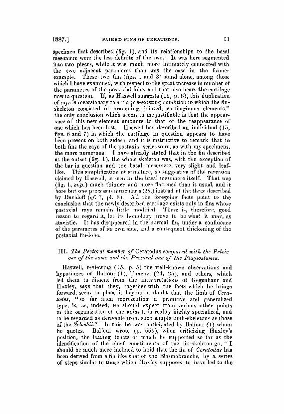
1887.1 PAIRED FINS OF CERATODUI. 11
specimen first described (fig. I), and its relationships to the basal mesomere were the less definite of the two. I t was here segmented into two pieces, while it was much more intimately connected with the two adjacent parameres than was the case in the former example. These two fins (figs. 1 and 3) stand alone, among those which I have examined, with respect to the great increase in number of the parameres of the postaxial lobe, and that also bears the cartilage now in question. If, as IJaswell suggests (1 5 , p. S), this duplication of rays is reversionary to a “ a pre-existing condition in which the fin- skeleton consisted of branching, jointed, cartilaginous elements,” the only conclusion which seems to mejustifiable is that the appear- ance of this new element amounts to that of the reappearance of one which has been lost. Haswell has described an individual (15, figs. 6 and 7) in which the cartilage in question appears to have been present on both sides ; and it is instructive to remark that in both fins ttic rays of the postaxial series were, as with my specimens, the more numerous. I have already stated that in the fin described at the outset (fig. l), tlie whole skeleton was, with the exception of the bar in question and the basd rnesomere, very slight and leaf- like. This simplification of structure, so suggestive of the reversion claimed by IIaswell, is seen in the basal mesomere itself. That was (fig. 1, n ~ p . ) inucli thinner a i d more flattened than is usual, and it bore but one proccssus niusculiiris ( t b . ) instead of the three described by Davicloff (cf. 7, 1’1. 8). All the foregoing facts point t o the conclusion tliat the newly described cartilage exists only in fins whose postaxial rays remain little modified. There is, therefore, good reason to regard it, let its homology prove to be what it may, as atavistic. I t has disappeared in the normal fin, under a conflueiice of the parameres of its onn side, axid a consequent thickening of the postaxial fin-lobe.
111. The Pectoral member of Ceratodus compured with the Pelvic one of the same /md the Pectorul otie of the Pkugtostoir‘es.
Haswell, reviewing (15, p. 5 ) the well-known observations and hypotheses of Unltour ( l ) , Thaclier (23, 2 5 ) , and others, which led them to dissent from the interpretations of Gegenbaur and Huxley, says tliat they, together with the facts which he brings forward, seem to place it beyond a doubt that the limb of Cera- todus, “so far from representing a primitive and generalized type, is, RS, indeed, we sliould expect t’rom viirious other points in the organization of the anirnd, in reality highly specialized, and to be regarded as derivable trom such simple limb-skeletons as those of the Seluchii.” I n this he was anticipated by Balfour (1) whom he quotes. Ualfour wrote (p. GF9), when criticizing Iluxley’s position, tlie leading tenets ot which he suppoited so far as the identification of tlie chief constituents of the fin-skeleton go, ‘( I should be much mcre inclined to hold that the fin of Ceratodus has been derived from a f in like that of the Elasmobranchs, by a series of steps similar to thobe which H a d e y supposes to have led to the

12 PROF. G. B. HOWES ON THE SKELETON AND [Jan. 18,
establishment of the Elasmobranch fin, but in exactly the reverse order ’”.
The researches of Huxley and Balfour have proved that the propterygium of Gegenbaur (figs. r 9 and 10, p t . ) represents, throughout the Elasmobranch series, but one or more preaxial rays. I t is the most variable of the three basal elements of the Shark‘s fin, and most observers are now agreed as to its morphological un- importance. The above-nained writers are further at one in their estimate of the morphological value of the Elasmobranch meso- and metapterygia (ms., mt., figs. 9 and 10). That they disagree, however, upon a t least one vital issue is well known, and the balance of opinion holds to-day that the solution of the ‘archi- pterygium ’ question is to be sought in a reconciliation between their views.
Huxley has described and figured (19, p. 48) the maximum development yet observed for the so-called propterygium of the Cerutodus pectoral fin. That structure cannot be definitely re- cognized in the pelvic fill. The determination of Huxley (19), Balfour (l), and v. Rautenfeld (22), which regards the axis of the Ceratodus fin as the mesopterygium, is too familiar to call for com- ment here. I t must suffice to state that I accept it in the main, if‘ not wholly, and assume for the present that the entire axis has the value which Iluxley first assigned to it.
I t is a t this point necessary to discuss, more fully than heretofore, the nature of the differences between the pectoral arid pelvic fin- skeletons of Cemtodus. Schneider has asserted (23, p. 521) that ‘‘ die Seitenstrahlen der dorsalen urid ventralen I-ldfte der Flossen sind nngleich,” also that the (‘ Seitenstrahlen der dorsalen Halfte der einen Plosse entsprechen derjenigen der ventralen Halfte der anderen.” There is an undoubted tendency towards the assumption of the condition which he thus formulates for Ceratodus, and it seems to me probable that a common determining cause may have led up to it and the condition realized in Protopteme (cf. Schneider, p. 524) ; but the defiiiition no longer holds invariable for the former animal, in view of the facts thus far adduced. I have already stated that the presence of one preaxial paramere in connexion with each mesomere is a constant character of the Ceratodus pectoral fin, and I turn now to the distribution of the postaxial rays. I have given on p. 7 the average distribution for eight pectoral fins exarnined. The minimum observed was, taking the mesomeres in order of succession from within outwards, 3 . 1 . 2 . 1, the maximum 4 . 2 . 2 . 2 . In 110 case have I observed five rays in attachment with the second niesomere, as stated by Schoeider. Of the eight specimens examined, the second mesomeres of five bore each three rays; the third and fourth ot’ sewn each two ; and the fittli of six each one. I t is thus certain that variation in the clistri- bution of the postaxial parnmeres (cf. fig. 6 ) is, beyond doubt, far ’ Gunther originally advanced a somewhat similar opinion (14, p. 634),
but lie conceived of the process ss liaviiig gone 011 along lilies as j e t incapable of support.

1887.1 PAIRED FINS O F CERATODUS. 13
less marked than with the pelvic member; but the fact which stands out most clearly is that the second mesomere invariably bears the greatest number of these rays. They are carried (figs. 5 and 6, mt. ) upon a special lobe of the axial cartilage (“ das divergirende Stiick ” of Schneider) already alluded to. The free border of this lobe slopes, in every case examined by me, gradually towards the proximal mesomere ( m . ~ . ) , and it is, moreover, in all, marked off from the body of the second mesomere by a deep furrow (indicated in the figs. by a dotted line).
I now proceed to discuss its homology, and having arrived at the conclusions to be formulated in the sequel through a comparison with the pectoral fin of Cestracion, I pass a t once to the considera- tion of that.
Gegenbaur and I-Iuxley are both agreed that the base of the Cestracion fin is supported by two cartilages (fig. 10) held by them to represent the mesopterygium (rns.) and the metapterygium (mt.) of other Selachians. Most recent writers have adopted their views (cf. Hubrecht and Sagemalil in Bronn’s Klassen und Ordnungen des Thier-Reichs,’ vol. vi. part 4, Pisces). I-Iuxley, instituting a comparison (1 9, p. 50) between the corresponding fins of Cestracion and Notidanus, regards them as representative of the transition stages in the shortening of the Cercctodus-like (‘ archipterygium,” by which he concludes the typical fish-fin has arisen. Brgenbairr (9, p. 148) likens the Cestracion fin to that of Acanthias, and sajs “ das Propterygium fehlt ginzlich.”
Huxley, holding further that the propterygium (preaxial ray) of Cestracion is removed from the shoulder-girdle, as in Ceratodus, asserts that in Scyllium (pp. 50-5’2) “ t h e f‘urther shortening of the axis gives rise to still greater clianges. The axial cartilage (meso- pterygium) is relatively small ; but the enlarged postaxial cartilage (metapterygiurn) has extended upwards along the postaxid face of the first, until it has not only reached the articular surface of the pectoral arch, but furnishes a large part of the articular cavity. In like manner the proximal preaxial ray (propterygium) has asceded aloiig the preaxial face of‘the axial cartilage, until it also is able to furnish a facet which completes the anterior part of the cup for the condyle of the pectoral arch.” He holds therefore that the pec- toral fins of Notidanus, Cestracion, and Scyllium represent, in the order enumerated, the successive steps in the modification alluded to above, and he, in accordance with the statements quoted coii- cerning the propterygium and metapterygium, relegates the two former fills to his category of the (‘ unibasal ” type, as distinguished from that of the latter animal, which he holds to typify the “ tribasal” one predominant among the Plagiostomes (cf. table which accom- panies his essay).
From an exaniination of the fins of two young Cestracions, I can state without further hesitation that the mesopterygium of the adult is (as Mivart has suspected, 21, p. 477) a compound of the pro- and mesopterygia. Fig. 9 represents one of the fins referred to. The animal died at the period at which the two (ms. and p t . )

14 [Jan. 18,
were beginning to unite. The propterygium (pt .) is seen to com- pose fully the aiiterior third of the whole mass, its base being r i d e up of two srgments, the proximal one of which contributes nearly half the articular cup, entering at least as fully into the formation of the same as does its representative in Scyllium (cf. Hoxley, 19, p. 48, fig. 10). In the adult fin (fig. 10) the original boundary line betwerii the pro- and mesoptrrygia is represrnted by a groove indicated in I-Iusley’s figure (liere reproduced) by a dotted line.
The pectoral fin of Cestmcion is thus shown to conform to tlie Selachian type, being identical most nearly with that. as represented by Acanthias (c$ Gegenbaur, 9, pl. 9. fig. 4, and Mivart, 21, 1’1. 77. fig. 2). Cesti-acion must, on tlie evidence now forthcoming, relinquish its position in the series established by I-Iuxley ; it must, to say the least, change places with Notidunus. The main articu- lation of the fin of the last-named fish is established, ns is well known, through the agency of the mesopterygium. Gegenbaur originally de- scribed a basal preaxial bar in Notidanus, and he homologizes it (9, p. 140) with his propterygium. This enters, if anything, more fully into the articulation with the shoulder-girdle than does its representative in Cestrucion. There is connected with its distal end, in Hexachzcs, a smaller piece ( c j . Gegenbaur, op. c i t . pl. 9. fig. 1) which appears to represeiit the second segnieut of Cestracion, reduced, as an outcome of the great expansioii of the front border of the meso- pterjgiuni. Mivart has suggested ( 2 I , p. 444) that the mesoptery- giuni represents a co;ileaceiice of the pro- and mrsopterygia ; but I am inclined, upoii careful examiliation of the specimen under my hand, to dissent from that view.
The metapterygium of Notidaizus enters iiito a feeble but definite coniiesion with tlie pectoral arch, such as is not the case in Cestra- cion. That the fins of these two genera differ from those of some of the Sharks is indisputable, but they do so to an insignificant degree, incapable in itself of supporting the “ unibasal ” type ; that, in face of the facts here adduced I , rests upon an insufficient basis.
Returning iiow to Ceratodus ; the lobe which, in the.pectora1 fin, carries the 3-5 proximal postasial parameres (figs. 5 arid 6 mt. ) is, as has been stated previously, marked off from the adjoining meso- mere by a deep furrow.
Gunther observes (14, p. 532) that the conjoined mass shows, in “liorizorital srction,” lilies of ‘‘ the former divisions ” into what lie holds to correspond to the “ three carpals” (pterygia) of most Plagiostorries. Set- ting aside this difference for the moment, I desire to call attention to the similarity of the furrow described above to that which
PROF. G. B. HOWES ON THE SKELETON A N D
This has been denied by IIuxlry (19, p. 47).
When, moreover, it is considered that in the pect.ora1 fin of Polyptmu, which IIuxley relegates to the “ hibasal ” category, tho mrsoptei-ygiuiii is (as Gegenbaur pointed out, 10, p. 130) excluded from articulation with the limb- girdlc, the statement that (p. 55) “ the mesopterygiuin is the prosirnal piece of the axial skeleton, which co1istant)ly retains its primary articulation with the pectoral arch,” must needs be modified.

1887.1 PAIRED FINS OF CERATODUS. 15
marks ofl‘ the propterygium from the mesopterygium in the adult Cestracion. On a comparison of the two, I subinit, with sotne degree of coiifidence, the opinioii that the postaxial lobe of the second mesomere of Ceratodus (fiqs. 5 and 6, ~ t . ) is the homologue of the Elasmobranch mrtapterygiurn. Comparisou of that lobe and its attached rays. (fig. G ) with the rnetapterygium of Cestracion and its rays (fig. 9) reveals a striking similarity, eveii in dcitnil, between the two. Did the metapterygium of the Shark nnite, as does its propterygiuoi, with the axial plate ms., it would be difficult iudeed to find a distiiiction between the first uaiiied mid that which, in Ceratodus, I claim as its homologue.
The homology which I liere seek to establish bears out, with certain modifications, Balfour’s view cited that “ the f in of Ceratodus has been derive‘d from a fin like that of the Elasmobranchs.” That observer first recognized (1,. p. 668) that the metapterygium (his basipterygium) is morphologically the most important and, phyloge- netically, the most primitive of the basal elements ; while he suspected (ibid.), but did not demonstrate the fact, that that structure is formed by the coalescence of rays. Huxley had already asserted this belief, in dealing with the metapterygium of Notidanus (19, p. SO), which he regarded as being “ formed by the coalescence of the axial ends of the postaxial rays” (presumably 011 the shortening of the fin axis). Dohrn lias recently substantiated the deductions of Tliacher, Mivart, and Ualfour under this head, in having found that the meta- pterygium is (8, p. l74), in both pectoral a i d pelvic fins of the Sharlr, like the basal bar of the mediau fins, niade up of “ unpaare Knorpelstrahlen, die anflinglich oder jede Verbindung mit andereri Skelettelementen bleiben.” H e reiterates the statement on p. 182 in the words “ was als Basipterygium beschriebeii ist, stellt nur die verschmolzeuen, wei sehr uah au einander leigenden, Basen der Flosseostralilen dau und esistirt niclit unabhiiugig von diesen.”
In face of the above facts, my view demands that a primary dis- tinction shall be demonstrated between the second pectoral mesomere in Cerutodus and that lobe which I hold to represent the meta- pterygium. I n Giinther’s original specimen (fig. 8) the said lobe was not represented in that which is now kuowii to beits typical form, while the rays ( r ) which are usually attached thereto were for the most part independent. The proximal two of these appear to have been somewhat smaller than usual, but it is highly iuterestirig to note that the two distal ones were unitiug at their bases to form a plate-like structure (mi.) which showed no sigus of confluence with the adjacent mesomere. This plate corresponds in its mode of origin with the metapterygium (basipterygium of Balfour), as defined by the above-named authors, and, in its relationships to the rest of the fin-skeleton, with the lobe now under consideration. I regard its coudition as there represented to be indicative of the primary independence which my interpretation necessitates.
Gunther goes on to say (p. 532) h a t lie found “lines of the former divisions ” of the second iiiesornere of this specirneii pre- served, in the shape of tracts of wliitc couiieetive tissue. Huxley

16 [Jan. 18,
denies the existence of these, as has previously been stated ; but let them be present or not, it is certain, should Giinther’s observation hold good, that they cannot indicate the original lines of separation between pro-, meso-, and inetapterygia, as now understood.
PROF. G. H. HOWES ON THE SKELETON AND
IV. On the proximal Postaxial Elements of the Ceratodus Pelvic Fin. The cartilage which I have already described (figs. 1 and 3 ) as
directly connected with the postaxial border of the basal mesomere of the Ceratodue pelvic fin is ray-like, but relatively powerful, in one of the two specimens (fig. 1). I n the other (fig. 3) it is alto- gether smaller and segmented into but two pieces, instead of into three, as in the former specimen. While it here meets the distal end of the proximal mesomere, it is much more intimately con- nected with the second piece of the axis than iu the former speci- men; but on the supposition that the cartilage is homologous in both fins, its condition in fig. 3 is precisely that which would result from a further reduction of that of fig. 1, such as there is good ground to believe, for reasons previously alleged, has actually gone on. In the second specimen the cartilage in question is further interesting, in that it bears one and is in close connexion with a second of the proximal parameres.
In the specimen figured by Giinther (14, pl. 36. fig. 4) already referred to (p. 5 ) , the proximal piece of the axis bears two carti- lages. The distal carti- lage is ray-like, and stands related to the base of the proximal mesomere as does an ordinary postaxial paramere to the corre- sponding border of a typical mesomere. The proximal cartilage appears to have been free of the basal piece altogether. I t is, as shown in the fignre, plate-like, and I have little doubt but that it was formed by the confluence of the basal ends of a t least the two rays which it carries. These skeletal elements, as they standin Giinther’s specimen, combine the characters of those of the two described by me (figs. 1 & 3). The postaxial parameres are, as in my specimens, much simpler than usnal, arid the whole series of lateral rays are in his fin more uniformly distributed than in general. The basal plate is (fig. 4, rnt.), like the corresponding bar of fig. 1, in near relationship with the proximal mesomere, although but loosely connected therewith ; while it agrees with the corresponding element of fig. 3 in giving attachment to a couple of rays. Comparing the proximal postaxial elements of my two specimens and Gunther’s figure with the corresponding region of the pectoral fin-skeleton, and reflecting that the typical metapterygium is formed by a confluence of the basal ends of the rays of that region, I incline to the belief that the vestiges in. yuesti9n represent that lobe of the fore limb which I claim as the metapterygium, together with its associated rays.
Should the cartilages now under discussion have the morpholo- gical value which I am seeking to establidi for them AS probable, the well-known viewsjof Gegenbaur (10, ll), and Huxley (19) will
Fig. 4 is a reproduction of his drawing.

1887.1 ‘PAIRED FINS OF CERATODUS. 17
recei* refutation, proportionate to the support furnished for those more especially of Galfour, Haswell, and Dohrn already cited.
9 s stated previously, the cartilage r of fig. 1 is ray-likp, but stonter and more powerful than that of any ordinary paramere.
In seeking light on this question, one naturally turns to Poly- pterus, the affinities between which and the Dipizoi, originally pointed out by Huxley (18), have nowhere been denied. The Polypterus pectoral fin is, as is well known, supported upon three basal elements. The inesopterygiuni (fig. 11, ms.) is held by all to represent that of the Plagiostomes, and no one has yet challenged Gegenbaur’s determination (9, p. 148) of the homology between the elongated postaxial bar (mt . ) of this fish aiid the metapterygium of the Plagiostomes and Chimaroids. Huxley says of this fin (19, p. 53) that ‘‘ the Scyllium type is essentially preserved.” Comparison of the Polypterus pectoral fin (fig. 11) with the pelvic fin of Ceratodus represented in fig. 1 would appear at first sight to suggest a homology between the basal postaxial bar (r) of the latter and the metapterygial bar (mt.) of the former. If this be justified, it would further appear, accepting the homology of the metapterygium of Polypterus with that of the Elasmo- h n c h i i , that the two fins might have been derived along a line of modification characterized by the assumption on the part of the metapterygium of a ray-like character, and by the subsequent elongation of the mesopterygial plate (m.). The probable truth of the latter assertioii seems to me very great indeed. The meso- pterggium is, in Polypterus (fig. 11, ms.), already elongated beyond the limits met with elsewhere, displacing in the process the mar- ginal rays. Continue that elongation, and there could only result a Ceratodus-like product. As concerns the former supposition, how- ever, comparison of the fin-skeletons represented in figs. 1,3, & 4 is sufficient in itself to show that the proxirrial postaxial ray offig. 1 most probably represents the distal one of those related to the proximal niesomere of fig. 4. Comparison of the latter (fig. 4) with the proximnl end of the pectoral f in of the same side of the same animal (fig. 8 ) shows unmistakably that in the plate-like structure result- ing from the fusion of the basal ends of the two proximal para- meres we have t,o deal with the last trace of the metapterygium, defining that, as must now be done, as a product of the confluence o f the inner ends of the proximal postaxial rays, the distal ray being, from the nature of its relations therenith, one of the same series,
Consideration of the above facts renders the homology of the sup- posed metapterygium of Polypterus somewhat doubtful. Gegen- baur, when pointing to the same, realized the similarity between both pro- and metapterygia so-called by him (fig. 11,pt. and mt.) and the marginal rays1. H e at first suggested (10, p. 139) the possibility that the exclusion of the niesopterygium from connexion with the
The difficulty of interpretation of the supposed propterygium is greatly increased by the presence of the cartilage markecl * in fig. 11,-by no means the least puzzling element in this fin. As will be seen, it is grafted upon the ante- rior border of the propterygial rod ; from it, however, it is perfectly distinct in
PROC. ZOOL. Soc.-l887, NO. 11. 2

18 [Jan. 18,
shoulder-girdle may have beef due to a displacement of the same by two rays. Should this be so, the metapterygium must there have disappeared, as from the Cercltodus pelvic fin, under the correspoiidillg’eillargenieiit of the mesopterygial plate. The o111y alternative view possible is that the nietapterygium does repre- sent th:it of the Elasmobranchs. ‘ I f this he so, comparison of the pectoral f in of PoZypterus (fig. 1 1 ) with that of the Plagiostomes, as represented in Scyllium, where the mesopterygium is relatively small, would seem to +ow that the loss of connexion between the ineta- pterygium and its rays has been to no small extent due to a displacement of the latter by the elongation of the expanding mesopterypium, no less than by the simplification in structure of the metapterygium itself. The last step in the t’ormer process would appear, indeed, to be retained i n the living Polypterus (** fig. 11).
In the absence of embryological data further discussion of this difficulty would be fruitless. I t is much more pertinent to observe that in both Ceratodus and Polypterus the, initial step in the modi- fication has been, in any case, one of elongation of the mesoptery- gium, and evidence has been adduced to show that in the Dipnoi (if not in Polypterus also) the metapterygiurn has been thereupon reduced and finally suppressed. The only traces of either it or its rays yet recorded in the Cerntodus pelvic member are forthcoming in fins whose postaxial parameres are more numerous and less specialized than is generally’ the case. If this simplification in structure of the most specialized portion of the pelvic fin-skeleton is, as I lmve attempted to show, reversionary to a condition which has been lost, the characters of those. elements which reappear under the simplification, when compared with those to which they most nearly correspond in the pectoral fin, go far towards bearing out the presumed origin of the Ceratoclzrs fin from a primarily expaiidcd predecessor.
PROF. G . B. ROWES ON THE SKELETON A N D
V. On the Jorphology of the At& of the Ceratodns Fin. The eiitire axis of the Ceratodus fin is held by Husley (19)
Bnlfour’ ( I ) , and Y. Rautenfeld ( 2 2 ) to represent an elongated
the Lwo specimens examined by me. I t has been figured hy Wie(lrrbheiu1 (29, p. log), but I hare been unable to f i i~ t l n dcficription of it. I tliink it ]lot improbable that it may hare beeii derived froin the mcsopterygiuin, the closely related lower anterior r i d of uliich mag (as Wiederslieim has shown) incert itstall‘ between t,lie suppoeecl prnptrrygiuin and the marginal rays. In the irbsenre of embryological data further disrussion of i t would bc useless.
Banr has recenrly called attention (3, p. G ) to the fact that Gervais has priority over Humphrey in t,he miinriation of the hypotlirsis t.hnt the paired fins are disnieniberecl portions of a lateral fold. Gerrais writes (12) :-“ Si l’on eonsidhrc que les rayons des nageoircs iuipnires des poissons ont une annlogie incontest.able nTec ceux dont la rbiuiion formrx lea n:igeoires paires des m6mes nniniaux, c’ebt i dire leurs membrev vtritubles, on est nnturellemeiit conduit b se demander s’ils ne seraient leu homologues de oes derniers, et si l’btat d’isole- nient dans lequel ils rrstent les unes par rapport aux aut.res, ne resulterait pas de re quo chacun d’eux ne conserve pas compldtenient ses rapports avec celui des segiuenta osteodesniiqnes dont il est tributuire. Alors on pourrait les

1887.1 PAIRED FINS OF CERATODUS. 19
mesopterygium, and by Gegenbaur (11) to represent the mrta- pterygium. The points of difference between the two sets of obser- vers are so well known that recapitulation of them would be super- fluous here. I incline most nearly to Buxley’s view, and hold thqt the axis represents mainly, if not wholly, the mesopterygium of the Sharks ; but I regard its condition in the latter as typical of its earlier and more primitive state. I moreover think it not unlikely that the short-lobed fin of the Crossopterygida will prove to be of an earlier type of structure than that of the elongated one of the living Dipnoi. Extended observations along the lines already laid down by Traquair, in his hfonograph 011 Tristichopterus (27), are greatly to be desired. That the elements described by him are probably, and that those described by Goldfuss (13) and Kner (20) in Xenacnnthus are certainly, homologous with those of the axis of the Ceratodus fin, I fully believe, and we have here the founda- tion of a line of study which must soon yield fruitful results’. Wiedersheim is the only worker who, to my knowledge, has offered an opinion upon the last named. He says (29 p. 195), speaking of the Ceratodus fin, “ dass dieser Organisationsplan der Brustflosse auch bei untergegangenen Fischgeschlechtern eine Rolle gespielt haben muss, steht unzweifelhaft fest und ich mochte dahei nur an den aus der Permformation stammenden Xenacanthus decheni erinnern.”
There can no longer be much doubt that the confluence so frequently seen between one or more rays and the mesopterygium of the Elasmobranchs represents the last trace of the process by which that structure is formed (cf. Dohm, cited on p. 15). This granted, it becomes a question as to how far the axis of the Ceratodus fin, as here defined, represents a further eltension of this fusion of pri- marily parallel rays or an elongation of the mesopterygial plate, as it exists in the Sliarks. The fact that irregularity in distribution of the parameres is generally accompanied by that of the segmentatioii of the axis in Ceratodus (cf. especially figs. 1, 5, & 7 ) , shows that there is an intimate connesion between the two; and this is the more obvious on reflection that Davidoff has shown (7, p. 145) that the segmentation of the axis does not stand in constant relationship to the muscular attachments ’. I t is moreover inconceivable, if
regarder comine autant cle rayons membraux rest& libres, et ils seraient les homologoes de ceux qui, par leur asservat,ion, donnent naissance aux membres proprewent dits sur d’autres poiuts du corps.”
I look with great satisfaction upon the work now being done in this direc- tion by Smith Woodward (cf. P. 2. S. 1886). I t is time that some such check should be kept upon the deductions of the embryologist (cf. Baur, RIorph. Jahrb. vol. viii. p. 453). ’ He writes, “ n a wir aucli nn cler Rlusknlatur eine den einzelnen Scgmenten der Starnmreihe entsprechende Gliederung fsnden, zo gewinnt dieae Ansiclit an Wahrsheinliclikeit, obwolil inimerliin noch einzuwenden ist, dass die physiologische Bedeutung dieser Oliederung eine nur ausserst miniinale seiu kann, dass ferner auch die Bwischensehnen der Stainmuskulstur in gar keiner &heren Beziehung zu den Segrnenten der Stammenreihe stdien.” I could find no difference between the muscles of the fin represented in fig. 6 and those described by him.
2*
20 [Jan. 18,
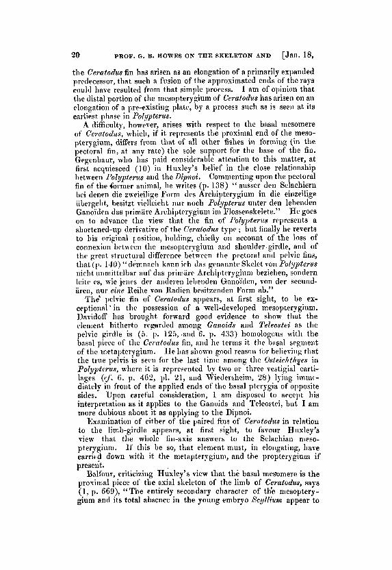
the Ceratodus fin has arisen as an elongation of a primarily expanded prcdecessor, t,hat such a fusion of the approximated ends of the rays could have resulted from that simple process. 1 am of opinion that the distal portion of the mesopterygium of Ceratodus has arisen on an elongation of a pre-existing plate, by a process such as is seen at its earliest phase in Polypterus.
A difficulty, however, arises with respect to the basal mesomere of Cercctodzis, whiclr, if it represents the proxinial end of the meso- pterygium, differs from that of all other fishes in forming (in the pectoral fin, a t any rate) the sole support for the base of the fin. Gegenbaur, who has paid considerable attention to this matter, a t first acquiesced ( 1 0 ) in Husley’s belief in the close relationship between PoZypterus and the Dipnoi. Commenting upon the pectoral fin of the (brmer animal, he writes (p. 138) “ ausser den Selachiern bei rlenen die zxeieilige Form des Archipterygium in die einzeilige iibergelit, besitzt vielleicht nur noch Polypterus uuter den lebenden Ganoi’den das primiire Archipterygium ini Flossenskelete.” I-le goes on to advance the view that the fin of Polypterus represents a shortened-up derivative of the Ceratodus type ; but finally he reverts to his original position, holding, chiefly on accoont of the loss of connexion between the mesopterygium and shoulder-girdle, and of the p e n t structural difference betwern the pectoral and pelvic fins, that (1). 140) “demnach kann ieh das genannte Skelet von Polypterus nirht uiimittelbar auf das primiire Archiptrrygium beziehen, sondern leite es, wie j e w s der anderen 1el)euden Gnnoi’den, von der secund- aren, nur eine Reihe von Radien besitzenden Form ab.”
The‘ pelvic fin of Ceratodus appears, at first sight, to be ex- ceptional * in the possession of a well-developed mesopterygium. Davidoff has brought forward good evidence to show that the element hitherto regarded among Ganoids and Te7eostei as the pelvic girdle is (5. 11. 125, mid 6. p. 433) homologous with the basal piece of the Ceratodus fin, and he terms it the basal segnieiit of the metapterygium. H e has shomn good reason fbr helieviitg that the true pelvis is seen for the last time among the Osteichthyes in Polypterus, where it is represented by two or three vestigial carti- lages (cf, G . p. 4G2, 111. 21, and Wiedersheini, 28) lying imnic- diately in front of the applied ends of the basal pterygia of opposite sides. Upon careful consideration, I am disposed to accept his interpretation as it applies to the Ganoids and Teleoetci, but I am more dubious about it as applying to the Dipnoi.
Examination of either of the paired fins of Cer’erntodus in relation to the limb-girdle appears, a t first sight, to favour Huxley’s view that the whole fin-axis answers to the Selachian nieso- pterygium. If this be so, that element must, in elongating, have carried down with it the metapterygium, and the propterygium if present.
Balfour, criticizing Huxley’s view that the basal mesomere is the proxinial piece of the axial skeleton of the limb of Ceratodus, says (1, p. SSS), “ T h e entirely secondary character of tlie mesoptery- gium and its total absence in the young embryo Xcylliurn appear to
PROF. G . B. ROWES ON THE SKELETON AND

1887.1 PAIRED F I N m F CERATODUS. 21
me as conclusive against Huxley’s view, as is the character of the embryonic f in against that of Gegenbaur.”
Examination of the pectoral fin of Ceratodus shows that the e1e:rients which I hold to represent the metapterygium and its rays (figs. 5 , 6, & 8, mt. & r.) are related to the postaxial border of the second mesomere. There is a t most a bare suggestion of a relation- ship to the proximal mesomere (m.p.). In the hind limb this is otherwise, for those parts of the skeleton which most nearly repeat the characters cf the presumed metapterygium of the fore limb are unmistakably connected (figs. 1 & 4) with the proximal mesomere. If they really represent the metapterygium and its rays as they occur among the lower fishes, it is, I think, not unlikely, p tting together these facts and those recorded by Davidoff, that while the metaptery- gium has for the most part disappeared, the proximal mesomere may, after all, turn out to represent the proximal end of that structure as defined by Huxley, early differentiated and segmented off.
The above suggestion, should it be substantiated, wonld explain the fact that the proximal mesomere of Ceratodus is the only con- stituent of the fin-axis whose characters are constant. I t would simplify onr conceptions of the fins of the Ganoids and Dipnoi, and bring into harmony the supposed divergent modifications of the fins of opposite extremities ; while it would show the pelvic member to be, on the whole, less modified than is usually thought. I am disposed to think, moreover, that it receives support from the absence of preaxial rays in connexion with the basal Fiesomere of Ceratodzis; from the complete exclusion of the mesopterggium from connexion with the shoulder-girdle in Polypterus ; acid from the condition of the pelvic fin of that animal, already alluded to, 110 less than from the marked tendency towards an increased development of the proximal end of the pectoral metapterygium among the living Ganoids.
Still more suggestive is the condition of the basal elements of a Protopterus pectoral fin represented in fig. 8 a. Wiedeisheim has (as I have already mentioned, p. 5) showu that the proximal piece of the pectoral fin-skeleton of this animal bears ray-like elements. He describes a smaller distinct ventral (postaxial) one and a larger dorsal (preaxial) one, which is confluent with the main piece (proxfmal mesomere as compared with Ceratodus). I have examined two specimens ; in one of them the latter is much smaller than in his example, while in the other (fig. 8 a) there is no trace of it. I can only conclude therefore that it is a lobe of the basal mesomere, variable in character. Not so with the former ; that is in both perfectly distinct, being separated froni the basal mesomere by a fibrous tract, such as subdivides any two segments from each other. In that specimen which was destitute of the preaxial process (fig. 8 a) its characters are still further noteworthy. It is elongated and shows traces of subdivision into two pieces, the basal one of‘ which is swollen and enlarged in common with tlie proximal mesomere (m.p.), and from that it appears most clearly to have been derived. Tlie secoiid segment of thc axis is in relation with both tlie proximal

1887.) PAIRED FINS OF CERATODUS. 23
condition. I ts postaxial border is supported by a cartilage, admitted by all to represent the metapterygium (fig. 12, mt.). This appears to be produced out into a preaxial lobe, which is regarded by Davidoff (4. p. 470, pl. 28. fig. 3, and pl. 29. fig. IS), who last described it, as consisting of a single piece answering to the propterygium.
It also recalls most closely that lobe f’rom which Balfour held ( 1 , 11. 667) that both pro- and mesopterygia are derived. I n a young Chimaroid pelvic fin examined by me (fig. 12), the lobe in question is seen to be formed by the fusion of three preaxial rays, and careful .examination has shown that the last traces of an original separation between it and the metapterygium (indicated in the drawing by it dotted h ie ) exist. Did that persist, the fin would correspond in all essential respects with thezpectoral member, as I have defined it ; and I hold that this preaxial lobe is neither more nor less than the pro- pterygium ’. Mivart cominerits (21, p. 465) upon the “close resemblance ’’ between the pectoral and pelvic tins of the Chimaroids. Comparing the pelvic fin of these animals (Callorhynchus) with the pectoral ones ot’ Acanthias and Xcymnus, he concludes (p. 456) that the basal cartilage represents, in tlie former, all three pterygia fused into one. The considerations put forward above, taken together with the fact that tlie mesopterygium never appears in the Plagio- stome’s pelvic fin, beyond the insignificant degree observed by Haswell, appear to me to negative this view.
The facts now under notice suggest, but do not prove, that the mesopterygium is never represented at all in the Cliimaxoids ; and that with respect to that feature those fishes stand on a lower platform than do the living I’lagiostonles. Moreover, if the preaxial cartilages of their pectoral member represent the propterygiurn, as I believe, an absolute structural identity is proven between the pectoral and pelvic fins of the group. Both would appear to have been derived from the fins of an ancestor in which the mesopteryg,‘ rium was not differentiated ; and if so, that element must have been of compara- tively late origin.
Davidoff has pointed to the existence of structural similarities between the hip-girdles of Chimaera and C‘eeratodus (7, pp. 142-3) ; and if the magnificent array of structural affinities between the two, so successfully demonstrated by Huxley (19), have the weight which he assigns to them, I think it more than probable, if, as I have suggested, the basal mesomere of Ceratodzis is a derivative of the metapterygium, that the paired fins of the Dipnoi may have arisen, side by side with those of the Plngiostomes, from some such form as is today represented by Chirncera-the fusion of the rays to form the mesopterygium having gone on independently, the intercalation of that structure between the applied bases of the pro- and meta- pterygia, so characteristic of the PlagiostomeE, having been a coin-
paratively late process.
1 The free ray represented at * in fig.,lO has been described by Davidoff (op. cit. p. 451). The spur-like outgrowth of tlie sane, al i ich I think niag not ini- prolmbly repre~eiit tho coalesced vestige of a second similar one, wits not present in liis spcuimcn.

24 PROF. G. B. HOWES ON THE SKELETON AND [Jan. 18,
If the above suggestioii should prove to have weight, the conditioii of tlie basal parts of the Polypterus fin, in which the mesopterygiuin is in no way in connexion with the shoulder-girdle, can only be a lowly one.
I t may not be inappropriate here to call attention to the coiiceptioii lately put forward by Baur (2, p. 663) conceriiiiig tlie morphology of the clieiropterygiuin. H e returns to Gegcnbaur's first position, and maiiitairis that the limb of the land-animal has been derived directly from the ichthyoptcrygium. l i i that case the Ceratodus fin, as it stands, caii only represent the initial phase in a line of modifi- cation of the iclitliyopterygium, culminating in Protopterus (to include Lepidosiren. Cf. Aycrs, Jenaisclie Zeitschr. vol. xviii. p. 479, 1685, and Schneidcr, op. c i t . ) .
Ditvidoff claims that the Ceratodus pelvic fin (7: p. 127) " trotz der Einfnclilieit des Ganzeii, sich bedeuterd komphcirter gestaltet, als bei den fruher bcarbeiteten Fisclien." 1-lc uses the words in a physiological seiise, it is true, but that in face of' his concluding statement that (p. 160) '' clas Eudergel~nis aber bestelit dariii, dass von der Ceratodus-Estremiiit sicli cliejenige dcr Baie oline Schwier- igkeiten ableiten liisst." This is, in my opiiiion, far from proven.
VII . Conclusions.
1. That the characters of the skeleton of the Ceratodzcs paired fins are iiiconstmt, except for those of tlie preasinl parwilcres of tlie pectoml fin a i d tlic: I ~ ~ s a l meso~tiere of both pector:ll a i d pelvic fins.
2. 'l'iint a mcta~iterygiuin is :ilw;iys preseiit i n tlic fore Ii i i ib, in a reduced coiitlition niitl usually coiifluetlt with the secoiid inesornere.
3. Tilat traces of what appears to represent :L metapterygium are occasioiially to be niet wi th in the hind limb, under conditions which point to atavism.
4. That tlie basal mesomere of the Cemtodus fin may conceivably have been clerivetl from tlie metapterygiutn.
5. That the strrrctnral features of both paired fins of the Chirnse- roids arc ideutical, a i d cliaracterized by tlie absence of a riieso- pterypiuin.
6. That the paired f i n s of the P1;iLtiostomes ant1 Dipnoi have, in all pro1i:rbility,' arisen iiidepeirdently Goiii a type o t f i t ; most ne:trly represented t)y that of the liriiig CIii i i~m(~ids.
7. Proven iraciclenfull~.-'l'liat tile bas;il cartilage u f tlie Ceesti*ucio~z pectoral tin, usually regarded as the nicsopterygiuin, is a corripouiid of the pro- and niesopterygia of other l'lab' riostoiiies.
VIII. List of Authorities referred to.
1. BALFOUR, F. Rf.-On the Developinent of the Skeleton of the Paired Fins of Elasriiobranchii. l'roc. %ool. Soc. 1881, p. 656.
2. BAUR, (3.-Ueber das Archipter!gium uiid die Eiitwicklung des Clieiropterqgiums ilus dem Ichthyopter~ giurn. Zool.hrizeiger, vol. viii. p. 663 (1885).

1887.1 PAIRED FINS OF CERATODUS. 25
3. BAUR, G.-Historische Bemerkung. Month. Internat. Journ. of Anat. & Hist. vol. iii. no. 1 (1886).
4. DAVIDOFF, M. v.-Beitrage zur vergleichd. Anat. der hinteren Gliedmasse der Fische. Morph. Jahrb. vol. v. p. 450 (1879).
5. DAVIDOFF, &I. v.-Coiitinuatiou of the above. Morph. Jahrb. vol. Ti. p. 433 (1880).
6. DAVIDOFF, M. v.-Ueber das Skelett der hinteren Gliedmasse der Gaiioidei holostei und der physostomen Kuochenfische. Morph. Jahrb. vol. vi. p. 125 (1880).
7. UAVIDOFF, M. v.-Beitr. zur vergleichd. Anat. der hinteren Gliedmasse der Fische. Morph. Jahrb. vol. ix. p. 116 (1 883).
8. DOHRN, h.-Studien zur Urgeschichte des Wirbelthierkorpers. IV. Mittheilung. aus der Zoolog. Stat. zu Neapel, vol. v. p. 102 (1884).
9. GEGENBAUR, C. --Untersuchg. zur vergleichd. Anat. der Wirbelth. Heft 2. Brustfosse der Fische. Leipzig, 1865.
10. GEGENBAUR, C. - Ueber das Archipterygium. Jenaisch. Zeitschr. vol.vii. p. 131 (1873).
11. GEGENBAUR, C.-Zur Morphologie der Gliedmsassen der Wirbelth.
12. GERVAIS, P.--hIBmoire sur la cornparaison des membres chez les mimaux vertdbris. Ann. d. Sci. Nat. sdr. 3, Zoologie, vol. xx. p. 21 ( 1 853) ; also De la comp. des Metnbres chez les Ariirnaus vertCbr6s. Paris, 1853.
13. GomFuss.-Beitr. zur vorweltlichen Fauna des Steinkohleu Gebirges. Bonn, 1847.
14. GENTHER, A.-Description of Ceratodus. Phil. Trans. vol. clxi. p. 51 1 (1871).
15. HASWELL, W. A.-On the Structure of the Paired Fins of Cera- todus, with Remarks on the general Theory of the Vertebrate Limb.
16. HASWELL, W. -4.-Studies of the Elasmobranch Skeleton. Pr. Liun. SOC. New S. Wales, vol. ix. p. 71 (1884).
17. HOWES, G. k-On some Abnormalities of the Frog’s Verte- bral Column. Bnat. Anzeiger, vol. i. p. 277 (1886).
18. HUXLEY, T. H.-Preliminary Essay upon the Systematic Arrangerimit of the Fishes of the Devonian Epoch. Mem. Geol. Survey, dec. x. p. 1 (1861).
19. HUXLEY, T. l-I.--On Ceratodus forstmi, with observations on the classification of Fishes.
20. KNER, R.-Ueb. Orthacunthus Bechenii, oder Xenacanthus dechenii. Sizb. d. Kais. Akad. d. Wiss. Wien, vol. lv. Abth. 1, p. 540 (1867).
21. MIVART, ST. GEo.-Notes on the Fins of Elasmobranchs.
22. RAUTENFELD, E. v.-Morph. Untersuchg. iiber das Skelet Uorpat,
23. SCHNEIDER, A.--l!eber die Flossen tier Dipnoi und die Sys-
Morph. Jahrb. vol. ii. p. 396 ( lS76) .
Pr. Linn. SOC. New S. Wales, vol. vii. p. 2 (1882).
Proe. Zool. SOC. 1876, p. 21.
Trans. Zool. SOC. vol. X. p. 439 (1879).
der hinteren Gliedmassen von Ganoid. uiid Teleost. 1884.

26 ON THE SKELETON ETC. OF CERATODUS. [Jan. 18,
tematik von Lepidosiren und Protopterue. Zool. Anzeiger, vol. ix. p. 521 (1886).
24. THACHER, J. K.-Median and paired Fins, a contribution to the history of vertebrate limbs. Trans. Connectic. Acad. vol. iii. p. 251 (1877).
25. THACHER, J. E. Ventral fins of Ganoids. Ibid. vol. iv. p. 233 (1878).
26. TRAQUAIR, R. H.-On the Restoration of the Tail in Proto- pterus annectena. British Assoc. Reports, 1871, pt. 2, p. 143.
27. TRAQUAIR, R. H.-On the Structure and Affinities of' Tris- tichopterus alatus. Trans. R. SOC. Edinb. vol. xxvii. p. 383 (1876).
28. WIEDERSHEIM, R.- Ueber das Beckeo der Fische. Morph. Jahrb. vol. vii. p. 326 (1881).
29. WIEDERSHEIM, R.-Lehrbuch der vergleichd. Anat. der Wirbelthiere. Zweite Auflage. Jena, 1886.
30. WIEDERSHEIM, R.-Das Skelet nnd Nervensy stem von Lepidosiren (Protopterua). Jenaische Zeitschr. vol. xiv. p. 155 (1860).
IX. DESURIPTION OF THE PLATES. PLATES I.-111.
With the exception of fig. 2, all are drawn in the same relative position.
Right side, dorsal aspect. Nat. The postaxial (internal) border looks towards the left hand. Fig. 1. A '' branching " pelvic fin of Ceratodics.
size. 2. The two pelvic fins of a second specimen. 20. The anterior portion of the pelvic girdle of the same. 3. The proximal third of the left fin of fig. 2. 4. Proximal third of a right pelvic fin ; indicated as seen from the ventral
(After Giinther.)
Ventral aspect. XI+. Nat. size.
Ventral aspect. Nat. size.
aspect, for sake of comparison with figs. 1 and 3. Nat. size.
.i. The left pectoral fin of fig. 2. 0. Proxirun1 portion of the corresponding fin of a fourth specimen.
Ventral aspect. Nat. size. Ven-
tral asp&t. Not. size. 7. Proximal portion of the left pelvic fin of the same specimen. Ventral
aspect. Nat. size.
to mine scale as figs. 5 and 6. 8. The proximal third of the left pectoral fin of a fifth specimen.
8a. Basal portion of a left pectoral fin of Protopfei*.zcs. 9. The left pectoral fin of Cestracioia philippi.
Drawn (After Giinther.)
x 4. Young 2, dorsal aspect.
10. The basal portion of a similar fin; same aspect. Adult. (After Reduced to the same scale as fig. 9 tor sake of comparison.
11. The left pectoral fin of a young Polypferics. 12. The left pelvic fin of a young Chinicera (C. moizstrosa), 6. Ventral
In'firs. 5 and 7 the parameres not represented in full were all rod-like and
Half nat. size.
Huxley.) Ventral aspect. x2.
aspect. x1-i.
- unbranched. - The dotted lines in fim 9 and 12 represent lines of fusion observed : those in
figs. 5, 6, and 10 are inh-ed .
Refereme Letters. cp. Base o f clasper. f: Iiitcr-articular ligament. m.p. Proximal mesomere.
nis. Mesopterygiuin. nit. Metapterygium. /A. Pelvic girdle. yr . Preavial fin.bolder. ps. Postaxid fin-border. pf. Propterygium. r. Metapteryginl rays.

F Z.S.1887.Pl.IV


Gaschar o don ron deleh

P.Z.S.1887.pI VII.
Werlt.Newnmn CCo q.
C ascharo don ronde leu I

P Z.S .1887.P1 I J I H