ontario association of pathologists annual … · ontario association of pathologists annual...
TRANSCRIPT

ONTARIO ASSOCIATION OF PATHOLOGISTS ANNUAL GENERAL MEETING
September 14-17, 2017 Deerhurst Skyline Resort,
Huntsville, Ontario
Royal College Accreditation
This conference is an Accredited Group Learning Activity (Section 1) as defined by the Maintenance of Certification program of the Royal College of Physicians and Surgeons of Canada.
This activity was approved by the Canadian Association of Pathologists.
Through an agreement between the Royal College of Physicians and Surgeons of Canada and the American Medical Association, physicians may convert Royal College MOC credits to AMA PRA Category 1 Credits™. Information on the process
to convert Royal College MOC credit to AMA credit can be found at www.ama-assn.org/go/internationalcme.

The OAP is the oldest pathology organization in the country and has been holding Annual General Meetings since 1937. As a not-for-profit medical society organized for educational and scientific purposes, the purpose of the meeting is to foster excellence in the practice of pathology and laboratory medicine in Ontario. OAP’s scientific AGM is Ontario’s largest gathering of pathologists, residents and decision makers with a focus on pathology. Featuring high-profile keynote speakers cutting-edge panels on current issues in pathology and a wealth of networking activities, the conference is the province’s premier event for evidence-informed discussion and debate on health care. The
Annual General Meeting will feature a multi-disciplinary scientific program, renowned guest speakers, robust trade show, and a great social program. The Annual Meeting is designed as an Accredited Group Learning Activity (Section 1) as defined by the Maintenance of Certification program of the Royal College of Physicians and Surgeons of Canada and includes: • Excellent topical scientific program • Cancer Care Ontario/OAP Symposium • Political and economic updates • Engaging social events and activities • Scientific Exhibitors & Vendors
Members also have the opportunity to vote on resolutions presented during the AGM formal business session. Feedback from members and exhibitors has indicated that the size of the meeting allows for excellent opportunities throughout the meeting to engage with speakers and network with other attendees. We look forward to seeing you in Deerhurst! Russell Price OAP President
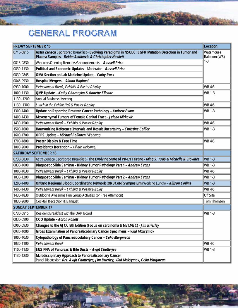
FRIDAY SEPTEMBER 15 Location 0715-0815 Astra Zeneca Sponsored Breakfast - Evolving Paradigms in NSCLC: EGFR Mutation Detection in Tumor and
Plasma Samples - Bekim Sadikovic & Christopher Howlett Waterhouse Ballroom (WB) 1-3 0815-0830 Welcome/Opening Remarks/Announcements – Russell Price
0830-1130 Political and Economic Updates - Moderator – Russell Price 0830-0845 OMA Section on Lab Medicine Update – Cathy Ross 0845-0930 Hospital Mergers – Simon Raphael 0930-1000 Refreshment Break, Exhibits & Poster Display WB 4/5 1000-1130 QMP Update – Kathy Chorneyko & Annette Ellenor WB 1-3 1130 -1200 Annual Business Meeting 1130- 1300 Lunch in the Exhibit Hall & Poster Display WB 4/5 1300-1400 Update on Reporting Prostate Cancer Pathology – Andrew Evans WB 1-3 1400-1430 Mesenchymal Tumors of Female Genital Tract – Jelena Mirkovic 1430-1500 Refreshment Break – Exhibits & Poster Display WB 4/5 1500-1600 Harmonizing Reference Intervals and Result Uncertainty – Christine Collier WB 1-3 1600-1700 OFPS Update – Michael Pollanen (Webinar) 1700-1800 Poster Display & Free Time WB 4/5 1800-2000 President’s Reception – All are welcome! SATURDAY SEPTEMBER 16 0730-0830 Astra Zeneca Sponsored Breakfast - The Evolving State of PD-L1 Testing - Ming S. Tsao & Michelle R. Downes WB 1-3 0830-1000 Diagnostic Slide Seminar - Kidney Tumor Pathology Part 1 – Andrew Evans WB 1-3 1000-1030 Refreshment Break – Exhibits & Poster Display WB 4/5 1030-1200 Diagnostic Slide Seminar - Kidney Tumor Pathology Part 2 – Andrew Evans WB 1-3 1200-1400 Ontario Regional Blood Coordinating Network (ORBCoN) Symposium (Working Lunch) – Allison Collins WB 1-3 1400-1430 Refreshment Break – Exhibits & Poster Display WB 4/5 1430-1830 Outdoor & Awesome Fun Group Activities (or Free Afternoon) Off Site 1830-2000 Cocktail Reception & Banquet Tom Thomson SUNDAY SEPTEMBER 17 0730-0815 Resident Breakfast with the OAP Board WB 1-3 0830-0900 CCO Update – Aaron Pollett 0900-0930 Changes to the AJCC 8th Edition (Focus on carcinoma & NET/NEC) - Jim Brierley 0930-1000 Gross Examination of Pancreaticobiliary Cancer Specimens – Vlad Maksymov 1000-1030 Cytopathology of Pancreaticobiliary Cancer – Celia Marginean 1030-1100 Refreshment Break WB 4/5 1100-1130 EUS FNA of Pancreas & Bile Ducts – Avijit Chatterjee WB 1-3 1130-1230 Multidisciplinary Approach to Pancreaticobiliary Cancer
Panel Discussion: Drs. Avijit Chatterjee, Jim Brierley, Vlad Maksymov, Celia Marginean

SATURDAY SEPTEMBER 16 Location 0730-0830 Astra Zeneca Sponsored Breakfast - The Evolving State of PD-L1 Testing - Ming S. Tsao & Michelle R. Downes Waterhouse
Ballroom (WB) 1-3
0800-0900 The Pathologist’s Assistant and the Dermatopathologist: We’re a Team – James J Limacher Arthur Lismer 0900-1000 Recognizing the Unrecognizable : At the Gross Bench – Jasmin Radhi 1000-1030 Refreshment Break – Exhibits & Poster Display WB 4/5 1030-1130 Biobanking Melanoma: opportunities for high impact research – Diane Chadwick Arthur Lismer 1130-1200 IHC – a PAs peek inside – Alan Wolff 1200-1300 Lunch in the Exhibit Hall WB 4/5 1300-1400 Case Studies
Case 1: Under Pressure –Terry Irvine Case 2: Dermatofibrosarcoma Protuberans of the Scalp in a 2-Year-Old Male – Susan Cromwell Case 3: Melanoma of the Eye – Martin Grealish
Arthur Lismer
1400-1430 Refreshment Break – Exhibits & Poster Display WB 4/5 1430-1545 Case Studies
Case 4: Purely Placental – Corinne Fletcher Case 5: Gallbladder Pathology (A Pathologists’ Assistant Perspective) – Adrian Bahneanu Case 6: It is Not a Tumour – Solange Malhotra
Arthur Lismer
1830-2000 Cocktail Reception & Banquet Tom Thomson
Don’t miss out on these organized group activities!
Explore the lake with Yamaha pontoon boat cruises! $25+tx per person - Max. 12ppl per boat (Min. 4ppl) Departs 16:00 (2 hrs maximum)
Treetop Trekking Huntsville The Huntsville park has aerial courses ranging in difficulty from beginner to advanced so there is fun to be had for just about everyone aged 9+ and at least 4'7" tall. Their Zip Line & Aerial Game Treks last about 3-hours and are guided by their friendly staff. $51.99+tx Adult (16+), $41.99+tx Youth (12-15), $37.99+tx Child (9-11) – Max 49 ppl Departs 1:30 (3 hrs maximum)
Required: Close-toed shoes, long hair tied at the back (ponytail), drinking water Recommended: Active wear clothing, snacks, gloves (gardening gloves work) Restrictions: weight maximum is 250 lbs, height minimum is 4’7″ or Advanced course is 5′ and 12 years old or older. See bottom of page for more terms and conditions.
Horseback riding - Woodland Trail Rides Saddle up for a guided ride through scenic trails with stunning views. $65 + tx pp – Max 6 ppl per trip Departs 15:00 (1 hr) & 16:30 (1 hr)

OAP101
FOLLICULAR DENDRITIC CELL SARCOMA ARISING IN BACKGROUND OF CASTLEMAN DISEASE: A CASE REPORT
Hina Chaudry, 1 Aleksandra Paliga,1, 2 Philip Berardi1, 2
1Ottawa Hospital Research Institute, Ottawa, ON, Canada, 2Department of Pathology, University of Ottawa, Ottawa, ON, Canada Objective: Follicular Dendritic Cell Sarcoma (FDCS) is a rare neoplasm of dendritic-histiocytic origin, generally occurring in lymph nodes (LN) and sometimes found in extranodal sites. It is occasionally associated with Castleman’s disease, a rare lymphoproliferative disorder. We sought to present a case of FDCS arising in the background of hyaline-vascular Castleman’s disease; an association that has been reported by few authors.1 Case Presentation: A 22-year-old male presented with a mass in the right supraclavicular area. The patient was completely asymptomatic with no history of pain, weight loss or fever and had been feeling well otherwise. Core needle biopsy of right supraclavicular LN raised suspicion of FDCS on morphological and immunohistochemical findings showing features of follicular dendritic cells. A definitive diagnosis was not possible because of an inadequate specimen andadditional characterization was necessary to exclude the alternative diagnosis. Spindle cell neoplasm is a good differential diagnosis that should be considered to rule out whenever FDCS is suspected. An excisional biopsy was hence performed on the mass. Microscopic and immunohistochemical findings confirmed the diagnosis of FDCS with one enlarged LN showing regressed germinal centers with mantle zones and multiple hyalinized vessels, consistent with the hyaline-vascular type of Castleman’s disease. Conclusion: FDCS is an uncommon intermediate grade malignant tumor and its association with Castleman‘s disease is an extremely rare entity. Our case supports the hypothesis of the transformation of Castleman’s disease into FDCS.
Disclosure Statement: Clinical case not been presented before.
References: 1Tiffany Chen, MD;, and Purva Gopal (2017) Follicular Dendritic Cell Sarcoma. Archives of Pathology & Laboratory Medicine: April 2017, Vol. 141, No. 4, pp. 596-599.
OAP102
HIGH VANGL2 EXPRESSION IS ASSOCIATED WITH BASAL-LIKE PHENOTYPE IN AXILLARY NODE-NEGATIVE BREAST CANCER
Catherine L. Forse1, Dushanthi Pinnaduwage2, Anna Marie Mulligan1,3, Shelley B. Bull2,4, Gordana Kuruzar2,5 Benjamin Pakuts2, Helen McNeill2,6, Irene L. Andrulis1,2,5,6 1Laboratory Medicine and Pathobiology, University of Toronto, Toronto, ON, CA, 2Lunenfeld-Tanenbaum Research Institute, Sinai Health System, Toronto, ON, CA, 3Laboratory Medicine Program, University Health Network, Toronto, ON, CA, 4Dalla Lana School of Public Health, University of Toronto, Toronto, ON, CA, 5Pathology & Laboratory Medicine, Mount Sinai Hospital, Toronto, ON, CA, 6Molecular Genetics, University of Toronto, Toronto, ON, CA
Background: Vang-like 2 (VANGL2) is a component of the non-canonical Wnt/planar cell polarity pathway that is important for collective cell migration during embryonic development. VANGL2 overexpression is associated with increased breast tumor cell proliferation, increased migration, and decreased metastasis-free survival in breast cancer patients.
Objective: The aim of this study was to determine if VANGL2 gene expression was associated with poor prognostic characteristics and decreased disease-free survival (DFS) in patients with axillary node-negative (ANN) breast cancer.
Methodology: Immunohistochemistry was performed for VANGL2, estrogen receptor (ER), progesterone receptor (PR), human epidermal growth factor receptor 2 (HER2), Ki67, and p53 on tissue microarrays generated from 668 prospectively
ascertained ANN breast tumors. Tumors were subdivided into the major molecular subgroups (Luminal A, Luminal B, HER2+, basal-like). Luminal subtypes were defined using Ki67, p53 and PR expression. VANGL2 expression was divided into three groups (low, moderate, high) based on Allred score. Descriptive baseline analyses compared frequency distributions of clinicopathologic factors among the three groups. DFS analyses were conducted for association of VANGL2 expression with risk of distant recurrence within the whole group and molecular subgroups by the log-rank test with Kaplan-Meier survival curves.
Results: High VANGL2 expression was significantly associated with poor prognostic features including high histological grade (P=0.0368), ER negativity (P=0.0195), and with the basal-like breast cancer phenotype (P<0.0001). Although the association between high VANGL2 expression and decreased DFS did not reach statistical significance, a trend was observed in both whole group and basal-like subgroup analyses.
Conclusions: The observed association between VANGL2 expression and aggressive tumor characteristics may indicate a functional role for the protein in breast cancer development and progression.
OAP103
COMPARISON OF CLINICOPATHOLOGICAL OUTCOMES FOR INSUFFICIENT AND SCANT ENDOMETRIAL SAMPLES
Emily A. Goebel1, C. Meg McLachlin1, Helen Ettler, Michele M. Weir1 1Department of Pathology and Laboratory Medicine, Western University, London ON, Canada
Objective: There are no defined pathologic criteria for reporting endometrial samples (ems) with limited tissue as either scant or insufficient. There is also no consensus on the clinical follow up of patients with these samples and whether terminology reflects outcome. The purpose of this study was to compare clinicopathological outcomes for scant and insufficient ems.
Design: A retrospective chart review was conducted on all patients with insufficient or scant ems from 2010-13, at our center. Clinicopathological features of the patients were recorded including age, repeat sampling, hysterectomy, and final pathology diagnosis. Five gynecologists were surveyed using an anonymous questionnaire about their practice for managing patients with these samples.
Results: In the survey of gynecologists, 4/5 reported that they managed patients with scant or insufficient biopsies/curettings similarly.
Table 1: Comparison of Outcomes of Patients with Insufficient and Scant Endometrial Samples
Endometrial sample types* (n=1149)
Insufficient (49%)
Scant (51%)
P value
Repeat ems 33% 31% 0.33 Hyperplasia on repeat ems
9.6% 8% 0.70
Malignancy on repeat ems
7% 2.8% 0.07
Subsequent Surgery 16% 11% 0.02 Repeat ems prior to surgery
45% 37.5% 0.36
Malignancy on hysterectomy
19% 9% 0.10
*ems: 89% biopsies, 11% curettings
Conclusion: The similar repeat ems rate between scant and insufficient samples suggests that our clinicians are choosing similar management for both of these terminologies. This finding is in keeping with the results of the questionnaire. Given the malignancy outcomes, both insufficient and scant ems merit repeat patient sampling in the appropriate context. As follow-up to this study, our center has conducted a review of scant and insufficient ems to determine pathologic criteria for reporting.
Disclosure Statement: This abstract was presented as a poster at the USCAP Annual Meeting in San Antonio, Texas, Mar 4-10, 2017.

OAP104
HISTOLOGIC CHARACTERISTICS OF RADICAL CYSTECTOMY SPECIMENS FROM PATIENTS WHO RECEIVED NEOADJUVANT CHEMOTHERAPY: A SINGLE-INSTITUTIONAL RETROSPECTIVE REVIEW.
Elan Hahn1,2, Bin Xu1,2, Michelle R. Downes1,2
1 Department of Laboratory Medicine and Pathobiology, University of Toronto, Toronto, Ontario, Canada; 2 Sunnybrook Health Sciences Centre, Toronto, Ontario, Canada
Background: Urothelial carcinoma of bladder is the most common malignancy affecting the urinary system. Surgical resection is the mainstay of treatment, but neoadjuvant chemotherapy (NAC) has been shown to confer survival benefits in patients with muscle-invasive bladder cancer, with a 5-year survival benefit of ~5%.
Objectives: The objectives of this study were to evaluate the histologic characteristics of NAC cystectomies and compare them to cystectomies which only had preoperative transurethral resection of the bladder tumour (TUR) to ascertain whether the response could be attributed to NAC or TUR alone. The second objective was to identify any difference in patient outcome between cohorts.
Design: This was a single-institution retrospective review of 48 radical cystectomy specimens from patients who had received NAC. We divided this group into NAC responders (<pT2, pN0) and non-responders (≥pT2, pN1). We used a group of TUR only cystectomies (n=16) as our control. We recorded the following histologic features in each case: fibrosis, hyalinization, inflammation, calcification, giant cells, necrosis, histiocytes, cholesterol, hemorrhage, and lymph node changes.
Results and Conclusions: There was significant overlap in the histologic features between the NAC responders (n=5), NAC non-responders (n=43), and TUR control group (n=16). Outcomes were similar between the NAC responders and NAC non-responders. In specimens that show response to
neoadjuvant therapy, it is hard to differentiate whether the observed response is due to TUR or NAC.
Disclosure Statement: This project has not been presented previously.
OAP105
DIAGNOSTIC RATES BEFORE AND AFTER IMPLEMENTATION OF THE PARIS SYSTEM FOR REPORTING URINARY CYTOLOGY
Matthew Kubica1, Wen Xie2, Alison Nell1, Stephen Pautler2, C. Meg McLachlin1, Mariamma G. Joseph1 Departments of 1Pathology and Laboratory Medicine and 2Urology, London Health Sciences Centre and Western University, London, ON Background: Urine cytology is an efficient method of diagnosing clinically occult urothelial carcinoma; however, until recently there were no standardized criteria for reporting urine cytology and over-reporting of “atypical” cases posed challenges for clinical management. In 2016, The Paris System for Reporting Urinary Cytology (TPS) was developed as a means of standardizing urine cytology reporting through the use of set morphological criteria and diagnostic categories. TPS was implemented by the cytopathology division at London Health Sciences Centre (LHSC) in September of 2016. Methods: We compared urine cytology results from two time periods: before (September-December 2015) and after (September-December 2016) the implementation of TPS at LHSC. Statistical comparisons in rates of diagnostic categories were performed using chi-square analyses (P < 0.05 significant).
Results: 2576 urine cytology specimens were submitted during the study periods and the results are summarized as follows:

Sept – Dec 2015
Sept – Dec 2016
p-value
Total Cases 1317 1259 - Negative for HGUC*
978 (74%)
1123 (89%)
0.002
Atypical Urothelial Cells (AUC)
288 (22%)
115 (9%) < 0.001
Suspicious for HGUC
29 (2.2%) 4 (0.3%) < 0.001
Positive for HGUC
22 (1.7%) 17 (1.4%) 0.51
*HGUC, high grade urothelial carcinoma
Discussion: Implementation of TPS at our institution has resulted in an increase in the proportion of negative cases reported, with a corresponding significant reduction in the number of atypical and suspicious cases. The proportion of positive cases did not differ between periods. It is likely that cases previously called atypical are now being reported as negative. Our results suggest that implementing TPS may provide clearer diagnostic direction for clinicians.
Keywords: urine cytology, Paris system, urothelial carcinoma, atypical urothelial cells
OAP106
IMPLEMENTATION OF NOVEL EDUCATIONAL TECHNIQUES IN PATHOLOGISTS’ ASSISTANT (PA) STUDENT/RESIDENT TRAINING
Emily G. M. Kyle, MSc1, Jose A. Gomez, MD1,2, Nancy G. Chan, MD1,2
1Department of Pathology and Laboratory Medicine, Western University, London, ON, 2London Health Sciences Centre, London, ON
Objective: To assess the effectiveness of new teaching activities in preparing PA students and residents for describing surgical specimens.
Design and Methods: On day one, trainees were each given a food item (i.e., “specimen”) and asked to write a description of it. Each “specimen” was visible only to the person writing the description. Each trainee then read their descriptions out loud while the others tried to identify the “specimen”. Participants were also encouraged to compare the “specimens” with
potential specimens in the gross room. Trainees then filled out a Likert scale-style survey assessing their own abilities at the “gross description”. Students subsequently completed 8 teaching sessions, including introductions to pathology terminology and gross description organization. The activity and survey were then repeated at the conclusion of these teaching sessions.
Results: Trainees felt more competent describing items during the hands-on activity after the implementation of the 8 teaching sessions.
Conclusions: Our novel educational techniques are effective in preparing PA students and residents for grossing surgical specimens.
Disclosure Statement: This work has not been presented previously.
OAP107
SMALL CELL CARCINOMA OF THE ESOPHAGUS. A CASE REPORT AND REVIEW OF LITERATURE
Jeremy Kwok1, Khalida Nasim2, 1Medical student at Schulich school of Medicine, 2Pathologist at St. Thomas Elgin General hospital
Introduction: Primary small cell carcinoma of the esophagus is a relatively rare malignancy. It is an aggressive form of cancer with poor prognosis. Most of the cases are in extensive stage at the time of diagnosis. Clear treatment guidelines are not available. Surgery is the treatment of choice in limited stage disease, but multimodal treatment is recommended in advance stage cases.
Case Report: A 50 year old male presented to the emergency department in acute renal failure with fever and diarrhea. The endoscopic examination revealed an endoluminal mass at the lower end of the esophagus. CT scan revealed metastasis to the liver. The case was reported as small cell carcinoma of the esophagus on histopathological and immuno-histochemical examination. Upon further investigation, no signs of pulmonary involvement were found. The patient’s disease stage was assumed inoperable and was offered chemotherapy.

Conclusions: Primary small cell carcinoma of the esophagus is a rare entity. The prognosis is poor and a multimodal treatment approach is required in advance cases.
OAP108
PANFOLLICULOMA, REPORT OF A RARE CUTANEOUS NEOPLASM AND LITERATURE REVIEW
Nikoo Parvinnejad1, Christine Orr1, Ami Wang1, David Hurlbut1
1Department of Pathology and Molecular Medicine, Queen’s University, Kingston, ON
Objectives: Panfolliculoma is a rare benign follicular neoplasm first described by Ackerman et al in 1993 and with less than 40 cases reported to date. It shows differentiation toward all elements of a hair follicle. The aim of our report is to discuss the classic clinicopathological features and potential diagnostic pitfalls associated with this rare entity.
Methods: We report a case of panfolliculoma that presented at an unusual site (abdomen), with review of the current literature on this topic.
Data and results: Our patient is a 74 year old man who underwent right hemicolectomy for colonic adenocarcinoma. At the time of surgery, he was found to have an abdominal skin lesion, which was resected as a presumed basal cell carcinoma. Histopathology revealed a cystic, well circumscribed lesion in the dermis with connection to the overlying epidermis. There is evidence of differentiation toward all components of a normal hair follicle, including: basaloid follicular germinative cells, cells with clear cytoplasm resembling the outer root sheath, and trichilemmal-type compact keratinization. Focally, bright red trichohyalin granules, yellowish cornified cells and ghost cells are identified.
Conclusion: Differential diagnosis includes other benign and malignant adnexal tumour, such as trichoblastoma, trichoepithelioma, trichofolliculoma, and basal cell carcinoma. It is important to recognize the diagnostic features of panfolliculoma to avoid overcalling malignancy.
Keywords: Panfolliculoma, Cutaneous, Neoplasm
OAP109
POTENTIAL ROLE OF DESORPTION ELECTROSPRAY IONIZATION (DESI) MASS SPECTROMETRY IMAGING (MSI) AS AN ADJUNCT TO HISTOLOGY FOR MARGIN ASSESSMENT IN LUMPECTOMIES FOR BREAST CANCER
Kevin Yi Mi Ren MD1, Martin Kaufmann PhD2, Nicole Morse1, Amanda Shuo Xu MD1, John Rudan MD2, David Berman MD PhD1, Sonal Varma MD1. 1Queen's University, Pathology and Molecular Medicine, Kingston, Ontario, CA, 1Queen's University, Surgery, Kingston, Ontario, CA
Objective: Obtaining negative margins in breast cancer surgeries is critical in preventing local recurrence. Re-operation rate due to positive margin is around 15% for lumpectomies, and could be improved by ancillary intraoperative tissue identification techniques. DESI MSI is an emerging technique capable of tissue characterization based on metabolite profiling. It can be applied to unstained frozen section slides to create tissue images based on the relative abundance of metabolites that topographically match the histology sections. We present a pilot study assessing DESI's ability to perform a simulated margin analysis in lumpectomies.
Design and methods: Fresh tumor samples with adjacent non-neoplastic tissue were collected from 10 lumpectomies with biopsy-proven invasive ductal carcinomas. Each tissue edge of the specimen was defined as a simulated margin. Unstained frozen section slides were subjected to DESI MSI, and subsequently stained by hematoxylin and eosin (H&E) for histologic correlation.
Results: Metabolites with distinct mass-to-charge ratios (m/z) were found to be characteristically abundant in tumor (m/z=863) versus non-neoplastic tissue (m/z=215), respectively. DESI MSI based on individual metabolite was able to accurately highlight tumor at tissue margins in samples with large tumor nests.
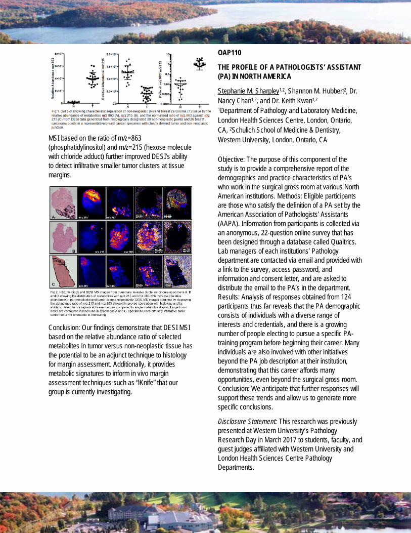
MSI based on the ratio of m/z=863 (phosphatidylinositol) and m/z=215 (hexose molecule with chloride adduct) further improved DESI’s ability to detect infiltrative smaller tumor clusters at tissue margins.
Conclusion: Our findings demonstrate that DESI MSI based on the relative abundance ratio of selected metabolites in tumor versus non-neoplastic tissue has the potential to be an adjunct technique to histology for margin assessment. Additionally, it provides metabolic signatures to inform in vivo margin assessment techniques such as “iKnife” that our group is currently investigating.
OAP110
THE PROFILE OF A PATHOLOGISTS’ ASSISTANT (PA) IN NORTH AMERICA
Stephanie M. Sharpley1,2, Shannon M. Hubbert2, Dr. Nancy Chan1,2, and Dr. Keith Kwan1,2
1Department of Pathology and Laboratory Medicine, London Health Sciences Centre, London, Ontario, CA, 2Schulich School of Medicine & Dentistry, Western University, London, Ontario, CA Objective: The purpose of this component of the study is to provide a comprehensive report of the demographics and practice characteristics of PA’s who work in the surgical gross room at various North American institutions. Methods: Eligible participants are those who satisfy the definition of a PA set by the American Association of Pathologists’ Assistants (AAPA). Information from participants is collected via an anonymous, 22-question online survey that has been designed through a database called Qualtrics. Lab managers of each institutions’ Pathology department are contacted via email and provided with a link to the survey, access password, and information and consent letter, and are asked to distribute the email to the PA’s in the department. Results: Analysis of responses obtained from 124 participants thus far reveals that the PA demographic consists of individuals with a diverse range of interests and credentials, and there is a growing number of people electing to pursue a specific PA-training program before beginning their career. Many individuals are also involved with other initiatives beyond the PA job description at their institution, demonstrating that this career affords many opportunities, even beyond the surgical gross room. Conclusion: We anticipate that further responses will support these trends and allow us to generate more specific conclusions.
Disclosure Statement: This research was previously presented at Western University’s Pathology Research Day in March 2017 to students, faculty, and guest judges affiliated with Western University and London Health Sciences Centre Pathology Departments.
OAP111
SURGICAL PATHOLOGY EXPERIENCE WITH ENDOSCOPIC SUBMUCOSAL DISSECTION (ESD) AND PERORAL ENDOSCOPIC TUMOUR RESECTION (POET): CASE EXAMPLES FROM KINGSTON HEALTH SCIENCES CENTRE
Kevin K. Song1, Robert Bechara2, David J. Hurlbut1 1 Department of Pathology and Molecular Medicine, Queen’s University, Kingston, Ontario, 2 Department of Medicine (Division of Gastroenterology), Queen’s University, Kingston, Ontario
Introduction: Endoscopic submucosal dissection (ESD) is an advanced endoscopic technique used to treat neoplastic lesions of the GI tract that are superficial and endoscopically accessible. The intact resected specimen allows for accurate pathological assessment of depth of tumour invasion (if carcinoma is present) and resection margin status. Another novel endoscopic procedure is peroral endoscopic tumour resection (POET) used for resection of submucosal tumours. To illustrate examples of the surgical pathology encountered with these newer techniques, we present two cases from our institution where these endoscopic procedures were used.
Method: Retrospective case review.
Results: Case 1: A 68 year old man was referred for treatment of a 5 cm rectal polyp. Clinical and endoscopic evaluation determined the lesion to be superficial. It was resected en bloc by ESD producing an intact specimen that was pinned out. Histopathology showed a completely excised tubulovillous adenoma with focal high grade dysplasia and no evidence for carcinoma.
Case 2: A 58 year old man underwent POET resection of a 1.8 cm well-circumscribed mural esophageal tumour at the gastroesophageal junction. Pathology showed a leiomyoma. The patient was discharged the next day without complications.
Conclusion: ESD and POET are advanced endoscopic therapeutic procedures that are relatively new for the Canadian healthcare system. ESD offers advantages over endoscopic piecemeal resection by
allowing resection of large superficial lesions within the GI tract en bloc, facilitating accurate pathological assessment of resection margins. Knowledge of these minimally invasive procedures will assist the surgical pathologist in the assessment of specimens resulting from these newer techniques.
Disclosure Statement: This abstract has not been previously presented
OAP112
QUALITY IMPROVEMENT IN CALCIFIED SPECIMEN PROCESSING: A COMPARISON OF DECALCIFICATION SOLUTIONS AND DURATIONS
Duc-Vinh Thai1, Lara Richer1, Jason Karamchandani1, Miriam Blumenkrantz1
1Department of Pathology, McGill University Health Center, Montreal, QC, CA
Objectives: Tissue decalcification is an essential step in the processing of calcified specimens. This study compares the effects of different decalcification methods on hematoxylin and eosin (H&E) staining and on immunohistochemistry and chromogenic in situ hybridization (CISH) studies routinely used in the assessment of decalcified specimens.
Design and Methods: Eight pediatric tonsils were selected as proxies for bone marrow. They were routinely processed and decalcified with RDO (Apex), Surgipath Decalcifier I (Leica) or 10% formic acid (ACP Chemicals), for one to 24 hours. Tissue slides were assessed for H&E, with 22 immunohistochemical antibodies (including CD3, CD20, CD45, and Ki-67) and with kappa and lambda light chain CISH. Quality was semi-quantitatively scored on a three-point scale.
Results: Optimal H&E staining quality was maintained in tonsils treated in SurgiPath Decalcifier I or formic acid, but there was progressive loss of basophilia in tissue treated for two hours or more in RDO. In the immunohistochemical and CISH studies, the cumulative scores of RDO-treated tonsils were significantly lower than the Surgipath Decalcifier I or formic acid groups. There was a significant decline in antigenicity by 24 hours of decalcification in both

Surgipath Decalcifier I and RDO groups while it was maintained in the formic acid group.
Conclusions: In as little as two hours, decalcifying agents can harm the staining quality and antigenicity of well-fixed tissue. Our study highlights the importance of validating the effect of specific decalcification procedures on routine stains and on immunohistochemistry and CISH studies for accurate diagnostic interpretation.
Disclosure Statement: The abstract has been presented at the Canadian Association of Pathologists - Association canadienne des pathologistes Annual Meeting in Price-Edward Island on June 12 2017.
OAP113
IMPACT OF THE PARIS SYSTEM FOR REPORTING URINARY CYTOLOGY ON THE PREVALENCE OF THE DIAGNOSTIC CATEGORIES: A COMPARATIVE STUDY AT A LARGE ACADEMIC INSTITUTION
Yujing Wang MDCM1, Manon Auger MD, FRCPC1, Fadi Brimo MD, FRCPC1
1Department of Pathology, McGill University and McGill University Health Center, Montreal, PQ, CA
Background: The 2016 Paris System for Reporting Urine Cytology (PSRUC) introduced specific cytological criteria for the diagnosis of "atypical" (AUC), "suspicious for high grade urothelial carcinoma" (SHGUC) and "high grade urothelial carcinoma" (HGUC) categories. Prior to this, the diagnosis of "atypical" was not standardized. We implemented the PSRUC at our institution in January, 2016.
Objectives: To evaluate the impact of the PSRUC on the prevalence of different cytological diagnostic categories.
Design: A comparative study was conducted over a 6 month period in 2013 (pre-PSRUC), including 1653 patients and 2,371 specimens versus a 6 month period in 2016 (post-PSRUC), including 1478 patients and 2,392 specimens.
Results and Conclusions: Whereas 18.6% of specimens were diagnosed as "atypical" in 2013, 14.4% were diagnosed as AUC in 2016, showing a decrease in 4.2% (p<0.0001). Fewer diagnoses of AUC in the voided category accounts for most of this change (14.5% versus 20%, p<0.0001). Concurrently, the prevalence of the "benign" category increased from 2013 to 2016 (75.4% versus 80%, p<0.0001). There was no significant change in the percentage of SHGUC and HGUC between 2013 and 2016 (3% vs. 2.4%, p=0.243; 3% vs. 3.2%, p=0.933, respectively).
The PSRUC significantly affected the distribution of benign and AUC cases, resulting in a decrease in the percentage of the AUC category. The comparable rates of SHGUC and HGUC pre- and post-PSRUC likely reflect the fact that the pre-PSRUC diagnostic criteria used at our institution for SHGUC and HGUC were very similar to those of the PSRUC.
OAP114
UTILITY OF PAX5/PD-1 DOUBLE STAINING IN THE PATHOLOGICAL DIAGNOSIS OF ANGIOIMMUNOBLASTIC T-CELL LYMPHOMA
Amanda Shuo Xu1, Kevin Yi Mi Ren1, Jilliann Jaynes, David Pierre Lebrun1
1Department of Pathology and Molecular Medicine, Queen’s University, Kingston, On, CA
Objective: Angioimmunoblastic T-cell lymphoma (AITL) is one of the most common subtypes of peripheral T-cell lymphomas with an aggressive clinical course. It can resemble a number of reactive and neoplastic conditions and lacks a unifying diagnostic hallmark. AITL cells have the immunophenotype of follicular T-helper (TFH) cells; however, immunostains are difficult to interpret due to the intimate association with B-cells. We developed a double stain for a cytoplasmic TFH marker, PD-1, and a nuclear B-cell marker, PAX5. We hypothesize that distinguishing AITL cells from B-cells using this stain will aid the distinction between AITL and its mimics.
Methodology: The PAX5/PD-1 double stain was established using the Leica PAX5(1EW) Bond Ready-To-Use mouse monoclonal antibody and Cell Marque PD-1(NAT105) mouse monoclonal antibody following

optimal dilution, antigen retrieval and incubation. PAX5 and PD-1 were detected as nuclear DAB and cytoplasmic Fast Red signals, respectively. Tonsils and appendices were used as control tissues. The double stain was applied to 16 retrospectively-identified AITL samples. Two follicular hyperplasia cases, one nodular lymphocyte predominant Hodgkin lymphoma (NLPHL) and one Castleman disease were also double stained.
Results: In control tissues, PD-1-positive cells were largely restricted to the follicle centers; 10-20% of paracortical PAX5-negative lymphocytes expressed PD-1 weakly. In AITL, 10-95% of the paracortical PAX5-negative atypical cells were strongly positive for PD-1. No double-positive cells were identified. The staining pattern observed in follicular hyperplasia was identical to that seen in control tissues. In NLPHL, the PD-1 positive T-cells were observed rimming the PAX5-positive LP cells. In Castleman disease, a reduced number of PD-1-positive cells was noted in the regressed follicle centers and few were seen in the paracortex.
Conclusion: By permitting the confident determination of PD-1 expression status among non-B lymphocytes, the PAX5/PD-1 double stain has potential utility in the pathological diagnosis of AITL. Currently, we are collecting more cases to compare the staining pattern between AITL and its diagnostic mimics, including: Hodgkin lymphoma, peripheral T-cell lymphoma NOS, T-cell/histiocyte-rich B-cell lymphoma, Castleman disease and follicular hyperplasia.
DISTINGUISHED PATHOLOGIST AWARD - The OAP Distinguished Pathologist Award is intended to recognize individuals who have made a positive and lasting contribution to our profession. The intent of this award is to recognize individuals throughout the history of the organization and not just from the recent past.
• KATHERINE CHORNEYKO • MICHAEL SHKRUM
PRESIDENT SERVICE AWARD - The Service Award is intended to honour OAP members who have shown exemplary dedication and service to the Ontario Association of Pathologists. In giving this award the board will consider service as a board member or other activities done for or on behalf of the organization which exemplify dedication and service to the Ontario pathology community.
• CHRISTINA MACMILLAN

The OAP’s Annual General Meeting will be held on Friday, September 15, 1130-1200. Presented to the OAP membership for approval will be the 2017-18 Slate of Officers, budget and the year-end financial statements. Documents are available in the members’ area of the OAP website: https://ontariopathologists.org/2017-annual-meeting/agm-documents/
Reminder, you must be a member in good standing to vote. If you are unsure if you have paid your dues, please see the registration desk at the conference centre to confirm. You can only vote if your 2017 membership dues have been remitted.
The Nominating Committee, in consultation with the Board and Executive, has completed their work and proposes the below Slate of Officers commencing at the completion of the AGM on Friday, September 15. Note the changes highlighted in bold black and listed below:
• President • Vice President • Past President
• Member-at-Large x 3 • Past President
President Dr. Celia Marginean Ottawa 2017 – 2019 - (2-Year Term)
Vice President Dr. Anna Plotkin Mississauga 2017 – 2019 - (2-Year Term)
Past President Dr. Russell Price Barrie 2017 – 2019 - (2-Year Term)
Secretary Treasurer Dr. Satish Chawla Niagara Falls 2016-2019 - (3-Year Term)
Member-at-Large Dr. Christopher Davidson Kingston 2015-2018 - (3-Year Term)
Member-at-Large Dr. Sharyn Smith Stratford 2015 – 2018 - (3-Year Term)
Member-at-Large Dr. Corwyn Rowsell Toronto 2016 – 2019 - (3-Year Term)
Member-at-Large Vacant
2017 – 2019 - (2-Year Term)
Member-at-Large Dr. Asghar Naqvi Hamilton 2017 – 2020 - (3-Year Term)
Member-at-Large Vacant
2017 – 2020 - (3-Year Term)
The Nominating Committee consists of the President, the two immediate Past-Presidents and two members who have been elected from the floor by the previous Annual General Meeting. The consent of the proposed nominees has been obtained, and their eligibility has been verified, for inclusion on the above slate.
In Accordance with the OAP Bylaws, we are accepting nominations for the two (2) Member-at-Large vacant positions. One is a 2-year term and the other is a 3-year term*. The Board of Directors is comprised of six (6) directors, preferably representing the six areas of the Province. Nominations are open to Active Members from all of Ontario though preference would be to receive nominations from those located in Windsor, Ottawa or Northern Ontario.
The Nominating Committee or Active Members (dues paid for 2017) may submit a nomination, in writing or via email, and with the consent of the nominee, to be presented from the floor at the AGM.
The elections, if necessary, will be by secret ballot. Scrutineers will be appointed by the President. The nominee receiving the majority of favorable votes will be considered elected to the office for which they were nominated.
*A vacancy occurred during the year when a Board Member resigned. The Executive Committee is empowered to name a successor to fill the office and act until the next Annual General Business Meeting, at which time the office will be filled in accordance with the procedure outlined in the By-Laws. Dr. Asghar Naqvi kindly agreed to assumed active duty on the Board prior to the AGM and will continue there after as a Member-at-Large.
The Executive would like to thank Drs. David Shum, Brian Cummings, David Hwang and Hector Li-Chang for their service to the Board of Directors. We appreciate your time invested in the OAP as we strike for success in our vision, mission and goals of the Association.
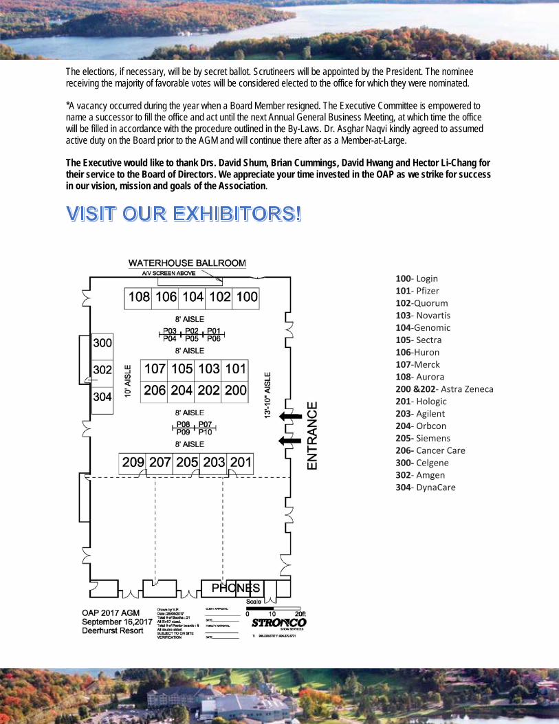
100- Login 101- Pfizer 102-Quorum 103- Novartis 104-Genomic 105- Sectra 106-Huron 107-Merck 108- Aurora 200 &202- Astra Zeneca 201- Hologic 203- Agilent 204- Orbcon 205- Siemens 206- Cancer Care 300- Celgene 302- Amgen 304- DynaCare

Double Platinum & Breakfast Sponsor Gold Lunch Sponsor &
Silver
Silver
Bronze Exhibitors