ontogeny of innate t lymphocytes – some innate lymphocytes
TRANSCRIPT
REVIEW ARTICLEpublished: 10 October 2014
doi: 10.3389/fimmu.2014.00486
Ontogeny of innateT lymphocytes – some innatelymphocytes are more innate than othersDavid Vermijlen1* and Immo Prinz 2
1 Faculty of Pharmacy, Université Libre de Bruxelles (ULB), Bruxelles, Belgium2 Institute of Immunology, Hannover Medical School, Hannover, Germany
Edited by:Tobias R. Kollmann, University ofBritish Columbia, Canada
Reviewed by:Gilles Chiocchia, Institut National de laSanté et de la Recherche Médicale,FranceJohn J. Priatel, University of BritishColumbia, Canada
*Correspondence:David Vermijlen, Faculty of Pharmacy,Université Libre de Bruxelles (ULB),Boulevard du Triomphe, Accès 2, 1050Bruxelles, Belgiume-mail: [email protected]
Innate lymphocytes have recently received a lot of attention. However, there are differentideas about the definition of what is “innate” in lymphocytes. Lymphocytes without V(D)J-rearranged antigen receptors are now termed innate lymphoid cells (ILCs) and includecells formerly known as natural killer (NK) cells. Also, lymphocytes that are innate shouldbe able to recognize microbial or stress-induced patterns and react rapidly without priorsensitization, as opposed to adaptive immune responses. Formally, genuine innate lym-phocytes would be present before or at birth. Here, we review the ontogeny of humanand mouse innate T lymphocyte populations. We focus on γδ T cells, which are prototypelymphocytes that often use their V(D)J rearrangement machinery to generate geneticallyencoded predetermined recombinations of antigen receptors. We make parallels betweenthe development of γδ T cells with that of innate αβ T cells [invariant (i)NKT and mucosa-associated invariantT cells] and compare this with the ontogeny of innate B cells and ILCs(including NK cells). We conclude that some subsets are more innate than others, i.e.,innate lymphocytes that are made primarily early in utero during gestation while others aremade after birth. In practice, a ranking of innateness by ontogeny has implications for thereconstitution of innate lymphocyte subsets after hematopoietic stem cell transplantation.
Keywords:T cell, gammadeltaT cells, ILC, fetal, neonatal, HSCT, BTN3A1, Skint1
INTRODUCTIONImmune responses are traditionally classified into two types:innate and adaptive. In textbooks, immune cells are usuallyassigned to one of these arms of the immune system, for exam-ple, neutrophils and macrophages in the innate arm and con-ventional T cells, restricted by MHC molecules, in the adaptivearm. Innate lymphocytes, however, can rearrange clonal antigenreceptor loci [T cell antigen receptor (TCR) or B cell antigenreceptor (BCR)] but at the same time show characteristics of theinnate immune system: recognition of molecular patterns, rapidresponse, and no need for clonal expansion. In addition, there arelymphocytes not expressing a TCR or BCR, including the naturalkiller (NK) cells with “NK activity” that were discovered 40 yearsago (1, 2). More recently, the family of non-T/non-B cells hasbeen extended to include a series of other innate lymphocytescollectively called “innate lymphoid cells (ILCs)” (3). However,the boundaries between lymphocytes of the innate and adap-tive immune system are blurred. On the one hand, non-T/non-Binnate lymphocytes such as NK cells can adapt to their envi-ronment (4) and even possess memory characteristics after viralinfection (5–7), reviewed in Ref. (8–10). On the other hand, T andB lymphocytes that undergo V(D)J recombination to rearrangetheir antigen receptor loci can be innate lymphocytes as well:within T cells one has αβ TCR-expressing T cells of which theTCR does not recognize MHC/peptide complexes [invariant NKT(iNKT) cells and mucosa-associated invariant T cells (MAIT) (11,12) and T cells expressing γδ TCRs (13, 14)], within B cells onehas B1 and marginal zone (MZ) B lymphocytes (15, 16).
Literally, “innate lymphocytes” would implicate that these cellsare generated before birth and that their presence would notdepend on environmental cues. In this review, emphasis will begiven to the ontogeny of the different types of innate lymphocytesin humans and mice. In both organisms, some subsets of innatelymphocytes are generated mainly during fetal life, while othersare made later in life. The timing of innate lymphocyte gener-ation could depend on the stem cells of which they are derivedfrom and/or the specific fetal environment. This knowledge can beimportant in clinical settings, for example, to have insight into thereconstitution of innate-like lymphocytes during hematopoieticstem cell transplantation (HSCT).
INNATE-LIKE T LYMPHOCYTESγδ T CELLSLike conventional αβ T cells and B cells, γδ T cells use V(D)Jgene rearrangement with the potential to generate a set of highlydiverse receptors to recognize antigens. This diversity is mainlygenerated in the complementary-determining region 3 (CDR3)encoded within the TCR or BCR loci (14, 17). The tripartite sub-division of lymphocytes possessing rearranged receptors into Bcells, αβ T cells, and γδ T cells has been conserved since the emer-gence of jawed vertebrates, more than 450 million years ago (18),and may even originate from earlier evolutionary events (19). Amajor difference between αβ T cells and γδ T cells is the way theyrecognize antigens. In contrast to conventional αβ T cells, γδ Tcells are not dependent on classical MHC molecules presentingpeptides. Based on the ligands that have been identified, it appears
www.frontiersin.org October 2014 | Volume 5 | Article 486 | 1
brought to you by COREView metadata, citation and similar papers at core.ac.uk
provided by Frontiers - Publisher Connector
Vermijlen and Prinz Ontogeny of innate lymphocytes
that γδ TCRs can recognize antigens in an antibody-like fashion,while the TCR of other γδ T cell subsets can bind to the MHC-like protein CD1d loaded with lipids (13, 20–25). Although thereare common characteristics among γδ T cells, it is clear that γδ
T cells, analogous to αβ T cells, do not represent a homogenouspopulation of cells with a single physiological role (26). γδ T cellsare typically grouped according to the type of Vγ chain (in mice)or Vδ chain (in humans) they express. Of note, their TCR recom-bination machinery is often used to generate always the same γδ
TCRs of rather limited diversity and the expression of the respec-tive invariant γδ TCR recombinations is often associated with theiranatomical location and/or function (14, 27, 28).
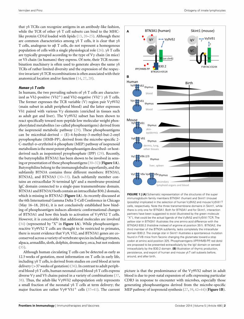
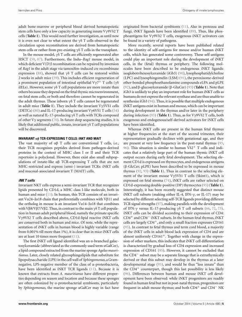
Human γδ T cellsIn humans, the two prevailing subsets of γδ T cells are character-ized as Vδ2-positive (Vδ2+) and Vδ2-negative (Vδ2–) γδ T cells.The former expresses the TCR variable (V) region pair Vγ9Vδ2(main subset in adult peripheral blood) and the latter expressesVδ1 paired with various Vγ elements (enriched in tissues suchas adult gut and liver). The Vγ9Vδ2 subset has been shown toreact specifically toward non-peptide low molecular weight phos-phorylated metabolites (so-called phosphoantigens) derived fromthe isoprenoid metabolic pathway (29). These phosphoantigenscan be microbial-derived – (E)-4-hydroxy-3-methyl-but-2-enylpyrophosphate (HMB-PP), derived from the microbe-specific 2-C-methyl-d-erythritol 4-phosphate (MEP) pathway of isoprenoidmetabolism is the most potent phosphoantigen described- or host-derived such as isopentenyl pyrophosphate (IPP) (29). Recently,the butyrophilin BTN3A1 has been shown to be involved in sens-ing or presentation of these phosphoantigens (30–32) (Figure 1A).Butyrophilins belong to the immunoglobulin superfamily, and thesubfamily BTN3A contains three different members: BTN3A1,BTN3A2, and BTN3A3 (30–33). Each subfamily member con-tains an extracellular N-terminal IgV and a membrane-proximalIgC domain connected to a single-pass transmembrane domain.BTN3A1 and BTN3A3 both contain an intracellular B30.2 domain,which is missing in BTN3A2 (Figure 1A). As recently discussed atthe 6th International Gamma Delta T-Cell Conference in Chicago(May 16–18, 2014), it is not conclusively established how bind-ing of phosphoantigen induces allosteric conformational changesof BTN3A1 and how this leads to activation of Vγ9Vδ2 T cells.However, it is conceivable that additional molecules are involved(34) (represented by “X” in Figure 1A). While phosphoantigen-reactive Vγ9Vδ2 T cells are thought to be restricted to primates,there is recent evidence that Vγ9, Vδ2, and BTN3A1 genes are co-conserved across a variety of vertebrate species including primates,alpaca, armadillo, sloth, dolphin, dromedary, orca, but not rodents(35).
Although human circulating T cells can be detected as early as12.5 weeks of gestation, most information on T cells in early life,including γδ T cells, is derived from studies on cord blood at termdelivery (>37 weeks of gestation) (36). In contrast to adult periph-eral blood γδ T cells, human neonatal cord blood γδ T cells expressdiverse Vγ and Vδ chains paired in a variety of combinations (37,38). Thus, the adult-like Vγ9Vδ2 subpopulation only representsa small fraction of the neonatal γδ T cells at term delivery; themajor fraction are rather Vγ9–Vδ1+ cells (37–41). The current
B30.2 FVBTAC:E324X
IgC
IPP/HMB-PP
IgV
BTN3A3:H351R
A BTN3A1 (human) Skint1 (mouse)
‘X’(on chr6) ‘X’
Vγ9Vδ2
IgC
IgV Vγ5Vδ1
B
Human
Mouse
Birth
thymus
skin-
epidermis
thymus
dermis, lung,
other #ssues
Vγ9 Vδ2thymus
blood
blood
thymusVγx Vδ1
?
?
thymus
liver, spleen
Vγ5 Vδ1
Vγ6 Vδ1
Vγ1 Vδ6.2/3
Vγ1 Vδx Vγ4 Vδx thymus
secondary
lymphoid organs and blood
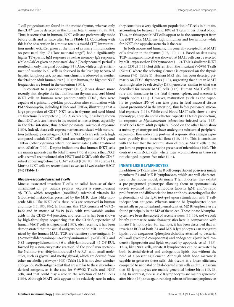
FIGURE 1 | (A) Schematic representation of the structures of the superimmunoglobulin family members BTN3A1 (human) and Skint1 (mouse)(possibly) implicated in the selection of human Vγ9Vδ2 and mouse Vγ5Vδ1 Tcells, respectively. Note the three transmembrane domains in Skint1, whilethere is only one for BTN3A1. Both for BTN3A1 and for Skint1, interactionpartners have been suggested to exist (illustrated by the green molecule“X”), that could be the actual ligands of the Vγ9Vδ2 and Vγ5Vδ1 TCR. Theyellow star in BTN3A1 illustrates the one amino acid difference with theBTN3A3 B30.2 (histidine instead of arginine at position 351). BTN3A2, thethird member of the BTN3A subfamily, lacks completely the intracellulardomain B30.2. The orange star in Skint1 illustrates a spontaneous mutationfound in FVB mice from Taconic changing the glutamate toward a stopcodon at amino acid position 325. Phosphoantigens (IPP/HMB-PP, red dots)are proposed to be presented extracellularly by the IgV domain or sensedintracellularly by the B30.2 domain. (B) Illustration of thymic production,persistence, and export of human and mouse γδ T cell subsets before,around, and after birth.
picture is that the predominance of the Vγ9Vδ2 subset in adultblood is due to post-natal expansion of cells expressing particularCDR3 in response to encounter with microbes, especially thosegenerating phosphoantigens derived from the microbe-specificMEP pathway of isoprenoid synthesis (27, 39, 42–44) (Figure 1B).
Frontiers in Immunology | Immunotherapies and Vaccines October 2014 | Volume 5 | Article 486 | 2
Vermijlen and Prinz Ontogeny of innate lymphocytes
Furthermore, this subset has been shown to differentiate rapidlyafter birth within the first year of life (45). This view is consistentwith recent deep sequencing studies that found a limited diversityin the adult TRG (T cell receptor gamma) repertoire. There, theTRG repertoire of peripheral blood γδ T cells from three indepen-dent donors was dominated by canonicalVγ9JγP sequences,whichmade up to 45% of all amplified TRG sequences (46). Cord bloodγδ T cells can produce already significant amounts of IFN-γ after abrief polyclonal stimulation (38, 45, 47). Emphasizing the acquisi-tion of functional competence in utero, IFN-γ was produced by γδ
T cells sampled from premature births, and, although 1 month’spost-partum environmental exposure invariably increased theirTNF-α production, it had no consistent effect on IFN-γ (47).To investigate whether the human fetus could produce particu-lar “fetal-type” of γδ T cells, we recently investigated γδ subsets inhuman fetal blood before birth. Surprisingly, rather than requir-ing post-natal microbial exposure, Vγ9Vδ2 T cells turned out tobe the predominant blood subset in the second trimester fetus andexpressed a semi-invariant TCR (48). This population is later onreplaced by other γδ T cell subsets so thatVδ1+ T cells predominateat birth (Figure 1B). Combined with a low level or absence of theVδ2 chain in post-natal thymi (39, 49, 50), our observations pointtoward a fetal wave of blood Vγ9Vδ2 production before 30 weeksof gestation (Figure 1B). Thus, it appears that human Vδ2+ T cells,and in particular Vγ9Vδ2 T cells with canonical Vγ9JγP sequences(51), are more innate than Vδ2− (Vδ1+ and Vδ3+) T cells.
Mouse γδ T cellsγδ T cells are the first T cells that leave the thymus in mice.Those primary T cells are mainly innate γδ T cells that usetheir TCR recombination machinery to generate always the sameγδ TCRs with no or little junctional diversity (14, 27). Specif-ically, the first specialized γδ T cell population, called den-dritic epidermal T cells (DETCs), is exclusively generated beforebirth (Figure 1B). It migrates to and populates the mouse skinepidermis already in utero. It has a fixed TCR composed ofinvariant Vγ5Jγ1Cγ1 and invariant Vδ1Dδ2Jδ2 (Vγ5Vδ1 TCR inshort) without P- or N-nucleotides (52, 53). The same canonicalVδ1Dδ2Jδ2 chain is employed in combination with an invariantVγ6Jγ1Cγ1 TCR chain in IL-17-producing Vγ6Vδ1 T cells. Thedevelopment of Vγ6Vδ1 cells is also confined to the embryonicthymus (Figure 1B). Vγ6Vδ1 cells were initially thought to berestricted to uterus and tongue (54) but subsequently also foundin many other tissues such as lung (55), liver (56), dermis (57, 58),secondary lymphoid organs (59), and intestinal lamina propria(60). Next, IL-4 producing Vγ1+ γδ NKT cells with restrictedVδ6Dδ2Jδ1 junctions and semi-invariant Vγ1Jγ4Cγ4 junctionsdevelop perinatally and preferentially localize in liver and spleen(61–63) (Figure 1B). In contrast, γδ T cells circulating in bloodand secondary lymphoid organs mostly contain either Vγ1 orVγ4 rearrangements. These are also produced after birth and arethought to have highly diverse TCR repertoires (27, 28, 54, 64,65). In particular, the pool of peripheral Vγ4+ T cells is het-erogeneous and contains both innate IL-17-producing cells andcells that are rather biased to IFN-γ production. These can besegregated according to a CCR6+CD27− and CCR6–CD27+ sur-face marker phenotype, respectively (66–68). In addition, Vγ4+ T
cells also comprise CD27+CD45RBhigh cells, a subset that read-ily produces IFN-γ upon stimulation with IL-18 and IL-12 (69).Furthermore, the requirements for final differentiation of Vγ4+
and other T cells into effector γδ T cells may vary between γδ
T cell types depending on their ontogeny (70). For example, itwas recently proposed that Vγ4+ T cells but not Vγ6+ T cellsrequire extrathymic environment for imprinting of skin-homingproperties and acquisition of an IL-17-producing phenotype (71).
Selection and peripheral activation of γδ T cellsThe above described characteristics of mouse and human γδ
T cells [fetal wave of production, (semi-) invariant TCR, pre-programing for production of IFN-γ or IL-17] have been linkedin the mouse to the action of selecting elements, most notablySkint1, an immunoglobulin superfamily member, that selects themurine intraepidermal Vγ5Vδ1 T cell repertoire (13, 66, 72, 73)(Figure 1A). The possibility exists that the butyrophilin BTN3A1may act as a selecting element for Vγ9Vδ2 T cells (Table 1), givenits role in mediating stimulation by phosphoantigens, and its strik-ing homology to Skint1, so far the only known natural selectingelement for γδ T cells (13, 30–32, 73–75) (Figure 1A). This wouldestablish a much stronger parallel between human and murineγδ T cells than is usually articulated, as phosphoantigen-reactiveVγ9Vδ2 T cells are described to be restricted to primates. Otherparallels between human Vγ9Vδ2 and mouse Vγ5Vδ1 cells are thatboth the V γ5 (TRGV5) and the V γ9 (TRGV9) gene segments arethe most downstream located (and thus closest to the Cγ1 genesegment) within the TRG gene cluster (76). In the mouse, thislocalization has been shown to contribute to early production ofVγ5Vδ1 cells (77). Of all human V δ gene segments, V δ2 (TRVD2)shows the highest similarity to mouse V δ1 (TRVD1): they are theonly two members of a separate cluster in a dendogram com-paring all V δ and Vα gene segments of human and mouse (78).Thus, different species may produce different variations of early γδ
T cells according to their specific needs (such as phosphoantigen-reactive cells in humans and skin-homing cells in mice),but similarmechanisms may be used to achieve this (Figure 1).
Responses of human γδ T cells in early life infections have beeninvestigated recently. Placental malaria, which can produce phos-phoantigens via the MEP pathway, has been shown to alter theVγ9 repertoire and to slightly increase the percentage of central-memory Vγ9Vδ2 T cells (85). Other γδ T cells subsets, such as apublic/invariant Vγ8Vδ1 TCR-expressing subset, have been shownto be involved in responses to congenital CMV infection (38) [see“Immune response to Cytomegalovirus in Early Life” within thisResearch Topic for further details (Huygens et al. under review)].Thus, it appears that depending on the type of pathogen infectingthe fetus, different types of γδ T cell subsets react.
Regeneration of γδ T cells in adultsOf note, recent studies have shown that adult HSCT and umbilicalcord blood transplantation (UCBT) result in the appearance ofγδ T cells in the blood of these patients (79, 86). These γδ T cellswere mainly from a different type than the Vγ9Vδ2 T cell subset,and a major influence of CMV on these non-Vγ9Vδ2 T cells wasobserved. Vγ9Vδ2 T cells failed to reach the median of the normalrange during a 2-year follow-up (79), which might indicate that
www.frontiersin.org October 2014 | Volume 5 | Article 486 | 3
Vermijlen and Prinz Ontogeny of innate lymphocytes
T ab
le1
|Ove
rvie
wo
fin
nat
eT
cells
wit
hth
eir
can
did
ate
sele
ctin
g/e
du
cati
on
elem
ents
and
asso
ciat
edac
tiva
tors
,pre
fere
nti
alti
min
go
fp
rod
uct
ion
,an
dth
eir
reco
nst
itu
tio
naf
ter
stem
cell
tran
spla
nta
tio
n.
γδ
TiN
KT
MA
IT
Hu
man
Mo
use
Hu
man
Mo
use
Hu
man
Mo
use
(Sem
i-)in
varia
ntTC
RVγ9J
γP
Cγ1
Vγx
Vγ5J
γ1C
γ1
Vγ6J
γ1C
γ1
Vγ1J
γ4C
γ4
Vγ1+
and
Vγ4+
cells
a
Vα24
–JαQ
Vα14
–Jα14
Vα7.
2–Jα
22Vα19
–Jα33
Vδ2
Vδ1
aVδ1
Dδ2
Jδ2
Vδ1
Dδ2
Jδ2
Vδ6
Dδ2
Jδ1
Vγ4+
cells
aVβ1 1
Vβ8/
Vβ7/
Vβ2
Olig
oclo
nalC
DR
3β
Sel
ectin
g/ed
ucat
ion
elem
ent
BTN
3A1?
?S
kint
1?
??
CD
1dC
D1d
MR
1M
R1
But
yrop
hilin
But
yrop
hilin
-
like
onfe
tal
thym
icst
rom
a
MH
Ccl
ass
I-lik
eon
DP
thym
ocyt
es
MH
Ccl
ass
I-lik
eon
DP
thym
ocyt
es
MH
Ccl
ass
I-lik
eon
DP
thym
ocyt
es
MH
Ccl
ass
I-lik
eon
DP
thym
ocyt
es
Sm
allm
olec
ule
activ
ator
/sel
fan
tigen
deriv
edfr
omce
llula
r
met
abol
ism
(inca
seof
MA
IT:(
com
men
sal)
mic
robi
alm
etab
olis
m)
Pren
yl-
pyro
phos
phat
e/
isop
reno
id
met
abol
ite(e
.g.,
IPP
)
??
??
?Li
pids
(e.g
.,
β-G
lcC
er;
pLP
E)
Lipi
ds(e
.g.,
β-G
lcC
er;
pLP
E)
Tran
sito
ry
neo-
antig
ens:
5-O
E-R
U;5
-OP-
RU
;
mic
robi
alvi
tam
inB
2
prec
urso
r+gl
yoxa
lor
met
hylg
lyox
al
Als
otr
ansi
tory
neo-
antig
ens?
:
5-O
E-R
U;5
-OP-
RU
;
mic
robi
alvi
tam
inB
2
prec
urso
r+gl
yoxa
lor
met
hylg
lyox
al
Pref
eren
tially
mad
e
befo
rebi
rth
+−
++
±(p
erin
atal
)−
+−
±(?
)−
Rec
onst
itutio
naf
ter
HS
CT
(with
refe
renc
e)
±Po
or(7
9)+
(79)−
(80)
−(5
4)±
Poor
(63)
+(5
9)+
(82,
83)
+(8
4)?
?
a The
seTC
Rar
eno
tre
gard
edas
(sem
i-)in
varia
ntTC
R.H
owev
er,t
his
does
not
excl
ude
that
thes
epo
pula
tions
incl
ude
subs
ets
cont
aini
ng(s
emi-)
inva
riant
TCR
s.
5-O
E-R
U,5
-(2-o
xoet
hylid
enea
min
o)-6
-D-r
ibity
lam
inou
raci
l;5-
OP-
RU
,5-(2
-oxo
prop
ylid
enea
min
o)-6
-D-r
ibity
lam
inou
raci
l;B
TN3A
1,bu
tyro
phili
n3A
1;H
SC
T,he
mat
opoi
etic
stem
cell
tran
spla
ntat
ion.
Frontiers in Immunology | Immunotherapies and Vaccines October 2014 | Volume 5 | Article 486 | 4
Vermijlen and Prinz Ontogeny of innate lymphocytes
adult bone-marrow or peripheral blood derived hematopoieticstem cells have only a low capacity in generating innate Vγ9Vδ2 Tcells (Table 1). This would need further investigation, as until nowit is even not clear to what extent the γδ T cells observed in thecirculation upon reconstitution are derived from hematopoieticstem cells or rather from pre-existing γδ T cells in the transplant.
In the mouse model, γδ T cells are efficiently regenerated afterHSCT (59, 87). Furthermore, the Indu–Rag1 mouse model, inwhich deficient V(D)J recombination can be repaired by inversionof Rag1 in the adult stage via tamoxifen-induced cre recombinaseexpression (88), showed that γδ T cells can be restored within2 weeks in adult mice (59). This includes efficient regeneration ofa prominent population of intestinal epithelial Vγ7+ T cells (γδ
iIELs). However, some γδ T cell populations are more innate thanothers because they depend on the fetal thymic microenvironment,on fetal stem cells, or both and therefore cannot be regenerated inthe adult thymus. These inborn γδ T cells cannot be regeneratedin adult mice (Table 1). They include the invariant Vγ5Vδ1 cells(DETCs) (80) and IL-17-producing invariant Vγ6Vδ1 T cells (54)as well as natural IL-17-producing γδ T cells with TCRs composedof other Vγ segments (70). In future deep sequencing studies, it islikely that additional public, innate, invariant γδ T cell populationswill be discovered.
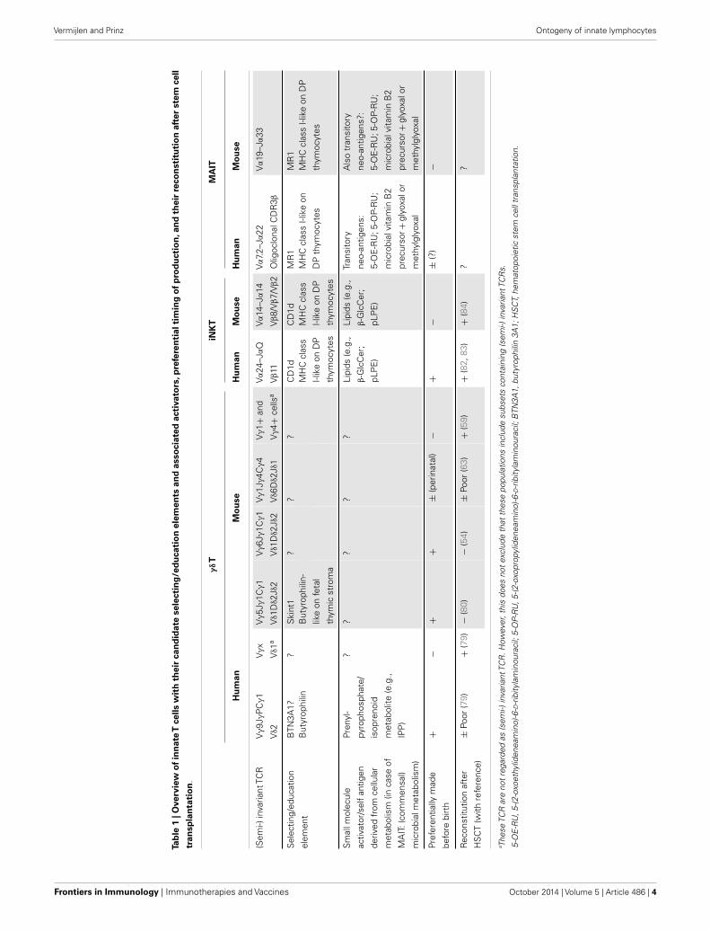
INVARIANT αβ TCR-EXPRESSING T CELLS: iNKT AND MAITThe vast majority of αβ T cells are conventional T cells, i.e.,their TCR recognizes peptides derived from pathogen-derivedproteins in the context of MHC class I or II and their TCRrepertoire is polyclonal. However, there exist also small subpop-ulations of innate-like αβ TCR-expressing T cells that are notMHC-restricted and express (semi-) invariant TCRs: iNKT cellsand mucosal-associated invariant T (MAIT) cells.
iNK T cellsInvariant NKT cells express a semi-invariant TCR that recognizeslipids presented by CD1d, a MHC class I-like molecule, both inhuman and mice (11). In human, this TCR consists of an invari-ant Vα24–Jα18 chain that preferentially combines with Vβ11 andthe ortholog in mouse is an invariant Vα14–Jα18 that combineswith Vβ8/Vβ7/Vβ2. Thus, in contrast to the main γδ T cell popula-tion in human adult peripheral blood, namely the primate specificVγ9Vδ2 T cells described above, CD1d-lipid reactive iNKT cellsare conserved both in human and mice. Of note, while the repre-sentation of iNKT cells in human blood is highly variable (rangefrom 0.001% till more than 1%), it is clear that in mice iNKT cellsare at least 10 times more frequent (11).
The first iNKT cell ligand identified was an α-branched galac-tosylceramide (abbreviated as the commonly used term αGalCer),a lipid compound extracted from the marine sponge Agelas mauri-tianus. Later, closely related glycosphingolipids that substitute forlipopolysaccharide (LPS) in the cell wall of Sphingomonas, a Gram-negative, LPS-negative member of the class of α-proteobacteria,have been identified as iNKT TCR ligands (11). Because it isknown that extracts from A. mauritianus have different proper-ties depending on season and location and because these spongesare often colonized by α-proteobacterial symbionts, particularlyby Sphingomonas, the marine sponge αGalCer may in fact have
originated from bacterial symbionts (11). Also in protozoa andfungi, iNKT ligands have been identified (89). Thus, like phos-phoantigens for Vγ9Vδ2 T cells, exogenous iNKT activators canbe found in a variety of pathogens.
More recently, several reports have been published relatedto the identity of self-antigens for mouse and/or human iNKTcells, which has generated some controversy. These self-antigenscould play an important role during the development of iNKTcells, in the (fetal) thymus or periphery. The following mol-ecules have been described to be endogenous iNKT ligands:isoglobotrihexosylceramide (iGb3) (90), lysophosphatidylcholine(LPC) and lysosphingomelin (LSM) (91), the peroxisome-derivedether-bonded phosphoethanolamine compounds eLPA and pLPE(92), and β-glucosylceramide (β-GluCer) (93) (Table 1). Note thatiGb3 is unlikely to play an important role for human iNKT cells ashumans do not express the relevant synthase and are thus unable tosynthesize iGb3 (94). Thus, it is possible that multiple endogenousiNKT antigens exist in human and mouse, which can be importantduring development in the thymus and/or later in the peripheryduring infection (93) (Table 1). Thus, as for Vγ9Vδ2 T cells, bothexogenous and endogenous/self-derived activators for iNKT cellshave been identified.
Whereas iNKT cells are present in the human fetal thymusat higher frequencies at the start of the second trimester, theirrepresentation gradually declines with gestational age, and theyare present at very low frequency in the post-natal thymus (95,96). This situation is similar to human Vδ2+ T cells and indi-cates that a relatively large part of the human thymic iNKT celloutput occurs during early fetal development. The selecting ele-ment CD1d is expressed on thymocytes, and endogenous antigens(β-GlcCer; pLPE) have been described to be expressed in mousethymus (92, 93) (Table 1). Thus in contrast to the selecting ele-ment of the invariant mouse Vγ5Vδ1 T cells (Skint1), which isexpressed on fetal stroma (74), iNKT cells are rather selected onCD1d-expressing double-positive (DP) thymocytes (95) (Table 1).Interestingly, it has been recently suggested that distinct mouseiNKT cell subsets (making either IL-4, IFN-γ, or IL-17) may beselected by different selecting self-TCR ligands providing differentTCR signal strengths (97), making parallels with the developmentof IFN-γ versus IL-17-producing γδ T cell subsets (66, 72, 73).iNKT cells can be divided according to their expression of CD4:CD4+ and CD4− iNKT subsets. In the human fetal thymus, iNKTcells are largely CD4+, and only a small fraction expresses CD161(95). In contrast to fetal thymus and term cord blood, a majorityof the iNKT cells in adult blood lack expression of CD4 and arealmost uniformly CD161+. Together with change in the expres-sion of other markers, this indicates that iNKT cell differentiationis characterized by gradual loss of CD4 expression and increasedexpression of CD161 (95). However, it cannot be excluded thatthe CD4− subset may be a separate lineage that is extrathymicallyderived or that this subset may develop in the thymus at a laterdevelopmental stage (95), and would be thus “less innate” thanthe CD4+ counterpart, though this last possibility is less likely(96). Differences between human and mouse iNKT cell devel-opment have been observed: while iNKT progenitors are readilyfound in human fetal but not in post-natal thymus, progenitors arefrequent in adult mouse thymus; and both CD4+ and CD4− NK
www.frontiersin.org October 2014 | Volume 5 | Article 486 | 5
Vermijlen and Prinz Ontogeny of innate lymphocytes
T cell progenitors are found in the mouse thymus, whereas onlythe CD4+ can be detected in the human fetal thymus (95, 98, 99).Thus, it seems that in human, iNKT cells are preferentially madebefore birth and in mice after birth (Table 1). Coinciding withthis is the observation in a mouse tetanus toxoid (TT) immuniza-tion model: αGalCer given at the time of primary immunizationon post-natal day 17 (“late neonatal stage”) had a significantlyhigher TT-specific IgM response as well as memory IgG response,while αGalCer given on post-natal day 7 (“early neonatal period”)resulted in only marginal boosting (81). Also, while a high enrich-ment of murine iNKT cells is observed in the liver (up to 50% ofhepatic lymphocytes), no such enrichment is observed in neitherthe fetal nor adult human liver (100); in human, the highest iNKTfrequencies are found in the omentum (101).
In contrast to a previous report (102), it was shown morerecently that, despite the fact that human thymus and cord bloodiNKT cells in humans were predominantly CD4+, they werecapable of significant cytokine production after stimulation withPMA/ionomycin, including IFN-γ and TNF-α, illustrating that alarge proportion of CD4+ NKT cells in thymus and cord bloodare functionally competent (103). Also recently, it has been shownthat iNKT cells can mature in the second trimester fetus, especiallyin the fetal intestine, thus before post-natal microbial exposure(100). Indeed, these cells express markers associated with matura-tion (although percentages of CD4+ iNKT cells are relatively highcompared to adult iNKT cells) and can readily produce IFN-γ andTNF-α (other cytokines where not investigated) after treatmentwith αGalCer (100). Despite indications that human iNKT cellsare mainly generated in the fetal thymus (95), it appears that iNKTcells are well reconstituted after HSCT and UCBT, with the CD4+
subset appearing before the CD4− subset β (82, 83, 104) (Table 1).Murine iNKT cells are reconstituted as well at 12 weeks after HSCT(84) (Table 1).
Mucosa-associated invariant T cellsMucosa-associated invariant T cells, so-called because of theirenrichment in gut lamina propria, express a semi-invariantαβ TCR, which recognizes (modified) microbial vitamin B2(riboflavin) metabolites presented by the MHC class I-like mol-ecule MR1. Like iNKT cells, these cells are conserved in humanand mice (12, 105, 106). In humans, this TCR consists of Vα7.2–Jα22 and in mouse of Vα19–Jα33, with two variable aminoacids in the CDR3 V–J junction, and recently is has been shownby high-throughput sequencing that the CDR3β repertoire ofhuman MAIT cells is oligoclonal (107). Also recently, it has beendemonstrated that the actual antigens bound to MR1 and recog-nized by the human MAIT TCR are transitory neo-antigens, 5-(2-oxoethylideneamino)-6-d-ribitylaminouracil (5-OE-RU) and5-(2-oxopropylideneamino)-6-d-ribitylaminouracil (5-OP-RU),formed by a non-enzymatic reaction of the riboflavin metabo-lite 5-amino-6-d-ribitylaminouracil (5-A-RU) with small mole-cules, such as glyoxal and methylglyoxal, which are derived fromother metabolic pathways (108) (Table 1). It is not clear whetherendogenous/self-antigens exist that are similar to these microbial-derived antigens, as is the case for Vγ9Vδ2 T cells and iNKTcells, and that could play a role in the selection of MAIT cells(109). Although MAIT cells appear to be relatively rare in mice,
they constitute a very significant population of T cells in humans,accounting for between 1 and 10% of T cells in peripheral blood.Thus, on this aspect MAIT cells appear to be the counterpart fromthe iNKT cells: MAIT are high in human and low in mice, whilefor iNKT, the opposite scenario is the case.
In both mouse and humans, it is generally accepted that MAITcells develop in the thymus (105, 110, 111). Based on data usingTCR-transgenic mice, it was shown that MAIT cells can be selectedby MR1 expressed on DP thymocytes (112). This is similar to iNKTcells (CD1d) (112), but different from the invariant Vγ5Vδ1 T cells(Skint1) where the selecting element is expressed on the thymicstroma (74) (Table 1). Human MR1 also has been detected pri-marily on CD3+ thymocytes (113), suggesting that human MAITcells might also be selected by DP thymocytes, similar to what wasdescribed for mouse MAIT cells (112). Human MAIT cells arerare and immature in the fetal thymus, spleen, and mesentericlymph nodes (111). However, maturation (such as the capac-ity to produce IFN-γ) can take place in fetal mucosal tissues(most pronounced in the intestine), thus before post-natal micro-bial exposure (111). While cord blood MAIT cells show a naïvephenotype, they do show effector capacity (TNF-α production)in response to Mycobacterium tuberculosis-infected cells (113).MAIT cells from adult peripheral blood on the other hand showa memory phenotype and have undergone substantial peripheralexpansion, thus indicating post-natal response after antigen expo-sure, possibly from bacterial flora (110, 113). This is consistentwith the fact that the accumulation of mouse MAIT cells in thegut lamina propria requires the presence of microbiota (106). Thiscontrasts with iNKT cells, where their accumulation in tissues isnot changed in germ-free mice (114).
INNATE-LIKE B LYMPHOCYTESIn addition to T cells, also the B cell compartment possesses innatemembers: B1 and MZ B lymphocytes, which are well character-ized in the mouse model. As innate T lymphocytes, they exhibita pre-programed phenotype allowing them to spontaneouslysecrete so-called natural antibodies (mostly IgM) and/or rapidproliferation and differentiation into antibody-secreting cells (alsopreferentially of the IgM isotype) upon stimulation with T cell-independent antigens. Whereas murine B1 lymphocytes locateessentially in peritoneal and pleural cavities,MZ B lymphocytes arefound principally in the MZ of the spleen. These innate B lympho-cytes have been the subject of recent reviews (15, 16), and we onlybriefly summarize some characteristics here in comparison withinnate T lymphocytes. For example, it has been described that aninvariant BCR of both B1 and MZ B lymphocytes can recognizelipids, both exogenous (phosphorylcholine attached to bacterialcell-wall glycolipid components) and endogenous (oxidized low-density lipoprotein and lipids exposed by apoptotic cells) (115).Thus, like iNKT cells, innate B lymphocytes can be activated byboth bacterial-derived and endogenous lipids, but without theneed of a presenting element. Although adult bone marrow iscapable to generate these cells, this occurs at a lower efficiencycompared to neonatal or fetal-derived stem cells and thus it seemsthat B1 lymphocytes are mainly generated before birth (15, 88,116). In contrast, mouse MZ B lymphocytes are mainly generatedafter birth (16), thus again ranking subsets of innate lymphocytes
Frontiers in Immunology | Immunotherapies and Vaccines October 2014 | Volume 5 | Article 486 | 6
Vermijlen and Prinz Ontogeny of innate lymphocytes
according to their “innateness”: B1 lymphocytes are more innatethan MZ B lymphocytes.
In the human system, it would be interesting to know whetherthese innate B lymphocytes, especially the most innate B1 cells, arereconstituted after HSCT. However, these investigations are com-plicated because of the lack on a consensus on the clear identifica-tion of B1 lymphocytes in human. Indeed, recently a population ofcells have been described that show functional similar characteris-tics as murine B1 lymphocytes (117). However, the identificationof these cells has generated controversy, including the finding that(part of) these cells might rather be pre-plasmablasts (118, 119).The candidate human B1 lymphocytes described by Griffin et al.(117) were present in cord blood and declined with age, indi-cating their preferential generation before birth as for mouse B1lymphocytes, but this needs further investigation. In contrast tomouse MZ lymphocytes, it appears that human MZ B lympho-cytes are mainly generated in the fetus (16, 120). A similar speciesdifference exists for iNKT cells, which are also mainly generatedafter birth in mice, but before birth in human (Table 1). However,direct comparison between the mouse and human immune sys-tem in early life should be done with the notion that in general thehuman immune system develops relatively early during gestationcompared to the development in mice. In experiments to investi-gate immune responses, one therefore usually uses day 7 old miceto compare immune responses with human neonates (121, 122).
NK CELLS AND OTHER INNATE LYMPHOID CELLSTo segregate them from T cells and B cells, lymphocytes withoutantigen receptor rearrangement are collectively termed ILCs (3).These ILCs include the well-established NK cells, lymphoid tissueinducer (LTi) cells (123), and a family of developmentally relatedILCs that are effectors and regulators in immunity and in tissuedevelopment and remodeling (124).
Natural killer cells were originally described as a cell subsetcapable of rapidly eliminating target cells without the need forprior sensitization (125). NK cells are important in conferringprotection from viral infections, such as CMV [see also “ImmuneResponse to Cytomegalovirus in Early Life” within this ResearchTopic (Huygens et al. under review)], and in the immunosur-veillance of transformed cells. NK cell activation is the result ofthe integration of signals provided by activation receptors suchas NKG2D, recognizing stress-induced ligands such as MICA inhuman and Rae-1 in mice, and inhibitory receptors such as mem-bers of the killer-cell immunoglobulin-like receptor (KIR) familyin humans and C-type lectin Ly49 family in mice, recognizing thepresence of “self” by binding MHC class I molecules. Besides theirrole in balancing NK activity in the periphery, KIR and Ly49 mem-bers play also an important role in the“education”or“licensing”ofNK cells during their development (126, 127). It has been shownrecently that human fetal NK cells differentiate early in utero andare highly responsive to cytokines and antibody-mediated stim-ulation but respond poorly to MHC class I-negative target cells.Indeed, in contrast to adult NK cells, it appears that expression ofKIRs did not educate fetal NK cells but rendered them hypore-sponsive to target cells lacking MHC class I (128). This selectivehyporesponsiveness could contribute to fetal–maternal tolerancein utero. Thus, human NK cells can be made early during gestation,
but they appear to be functionally different than adult NK cells.It remains to be established whether this reflects different wavesof NK cell development: an early wave of fetal NK cells and post-natal wave of NK cells. Upon HSCT in human, NK cells appearto reconstitute well and rapidly, with influence of CMV on theirreconstitution [see also “Immune Response to Cytomegalovirusin Early Life” within this Research Topic (Huygens et al. underreview)] (129), while other ILC populations appear to reconstitutemore slowly (130).
In contrast to the KIR repertoire on human fetal NK cells, theLy49 repertoire on mouse fetal and neonatal NK cells is veryrestricted: among the entire murine Ly49 family, Ly49E is theonly member expressed before birth (131). Conversely, whereasthe population of NK cells bearing one or more of the otherLy49 members progressively increases after birth, the percentageof Ly49E-expressing cells gradually decreases with almost no adultNK cells displaying this receptor (131). Fetal liver progenitor cellswere shown to be much more efficient in the generation of Ly49E-expressing NK cells than adult bone-marrow progenitors (132).Interestingly, Ly49E is also the only Ly49 member expressed byVγ5Vδ1 cells (DETC) (133). Despite this particular fetal expres-sion pattern, it appears that Ly49E does not play a crucial role indifferentiation and/or function of (fetal) NK cells (134).
Natural killer cells and other ILCs share common precursorsduring ontogeny as demonstrated in mouse models. Thereby,development of all sorts of ILCs depends on common gamma-chain cytokine signaling (135), in particular via IL-7R and IL-15R(3, 136), and the expression of the transcription factor helix–loop–helix inhibitor Id2 (137, 138). In addition, while the basic leucinezipper transcription factor E4BP4/Nfil3 was initially described asspecifically required for the development and maturation of NKcells (139, 140), it is emerging very recently that E4BP4/Nfil3 isalso crucial for the development of all other ILC subsets (141,142). Also recently, two independent studies identified precursorsthat were specific for the ILC lineages. A common, IL-7R- andId2-expressing progenitor named “CHILP” gives rise to all IL-7R-expressing, “helper-like” ILC lineages including LTi cells but lackspotential to differentiate into B and T cells or NK cells (143). Inaddition, a precursor termed ILCP, defined by transient expressionof high amounts of the transcription factor PLZF, gives rise to allILC lineages except LTi cells but lacks potential to differentiate intoB and T cells or NK cells (144). Importantly, both CHILP and ILCPwere found in fetal liver and adult bone marrow. Also, both precur-sors reconstituted ILC1, ILC2, and ILC3 lineages upon transfer intoRag- and IL-7R- deficient recipients (into Rag2-/-Il2rg -/- mice).Collectively, these transfer experiments advocated that ILC1, ILC2,and ILC3 populations can be regenerated from ILC precursors inan adult organism. However, it is currently unclear to what extentILC precursors and ILCs are de novo regenerated in the steadystate (Andreas Diefenbach, personal communication). Also, it ispresently not clear how efficiently ILCs as well as CHILP andILCP precursors are regenerated after HSCT. In human HSCT,current protocols to monitor immune recovery do not includeILCs (145). Moreover, recipient CD45+CD3-RORγt+ ILCs per-sisted long-term post-HSCT in the intestinal lamina propria oftransplanted mice showing that ILCs are relatively radio-resistant(146). At the same time, more than 1/3 of the ILCs in recipient
www.frontiersin.org October 2014 | Volume 5 | Article 486 | 7
Vermijlen and Prinz Ontogeny of innate lymphocytes
lamina propria were donor-derived 2 months post-HSCT, suggest-ing that it is likely that development of ILCs is not restricted toearly ontogeny (146).
CONCLUDING REMARKS AND PERSPECTIVESAfter reviewing the ontogeny of innate lymphocytes, it becomesclear that some subsets (such as mouse Vγ5Vδ1 T cells and B1 lym-phocytes) are more innate than others (such as ILCs, MAIT, andMZ B lymphocytes). Overall, the preferential generation of innatelymphocyte subsets before birth appears to have implications fortheir reconstitution after HSCT, but this does not need necessarilyto be the case, as indicated for human iNKT cells (Table 1). Manyinnate lymphocytes, in particular the very innate γδ T cell ones,expand in the periphery and persist as long-lived self-renewingcells. At the same time, these cells show above-average resistanceto ionizing radiation (57–59, 147). This observation can be aconfounding factor for determining the regeneration potential ofinnate lymphocyte populations.
A further common attribute of Vγ9Vδ2 T cells, iNKT cells, andMAIT cells is that all recognize small metabolites derived fromhost cellular or (commensal) microbial metabolism using semi-invariant TCRs and monomorphic presenting/selecting elements(Table 1). This contrasts with conventional or mainstream αβ Tcells that use TCRs with highly variable CDR3 regions recognizingprocessed peptides from pathogen-derived proteins presented bypolymorphic MHC molecules. These metabolites, together withtheir selecting/education element, can play an important role dur-ing the development of these innate cells. Recently, it has beenshown that the TCR signaling of murine innate-like T lympho-cytes, such as the IL-17-producing Vγ6Vδ1 subset, is critical fortheir development but that this signaling is attenuated in theperiphery where these cells become more responsive to innate-likesignals such as derived from cytokine receptors (69). However,since this did not apply to iNKT cells, it appears not to be ageneral phenomenon for all innate-like T lymphocytes. As dis-cussed by Wencker et al. (69), by altering their TCR responsemode, subsets of innate-like T cells may resemble ILC (that bydefinition lack constraint by a TCR). However, these innate-likeT cells are still different from ILCs as they are not fully aner-gic to TCR signals and the prerequisite for developmental TCRsignaling may provide a means of “quality control” for lym-phocytes entering the innate compartment that is not availablefor ILCs.
The production of different types of innate lymphocytes beforebirth compared to later in life (such as expressing different TCRs asfor γδ T cells, or showing a different “KIR educational response”for human NK cells), may be the result of a “layered” produc-tion of immune cells by fetal HSC versus adult HSCs. Whileevidence for this exists for some subsets, such as the fetal wavesof murine Vγ5Vδ1 T lymphocytes (80) and B1 lymphocytes (15)with life-long self-renewal, this still needs to be established for oth-ers (including human Vγ9Vδ2 T cells and NK cells). Also for othercells of the immune system, a similar mechanism of fetal wavesappears to be operational: in steady state, most tissue macrophagepopulations in mice are derived from embryonic precursors, areseeded before birth, and can maintain themselves in adults by self-renewal (148). Also, compared to adult HSC, human fetal HSC
gives rise to a distinct type of conventional CD4 T cells with a biastoward immune tolerance (149).
Finally, it will be interesting to further investigate whether the“age” of human stem cells in stem cell transplantation (such as ageof donors, or adult bone marrow/peripheral blood HSCT versusUCBT) can influence the type of innate lymphocytes that is recon-stituted. This can have important clinical consequences such asinfluence on graft-versus-leukemia effect or post-transplantationCMV infection [e.g., Ref. (79, 86)].
REFERENCES1. Kiessling R, Klein E, Wigzell H. “Natural” killer cells in the mouse. I. cyto-
toxic cells with specificity for mouse Moloney leukemia cells. specificityand distribution according to genotype. Eur J Immunol (1975) 5(2):112–7.doi:10.1002/eji.1830050208
2. Vivier E, Tomasello E, Baratin M, Walzer T, Ugolini S. Functions of naturalkiller cells. Nat Immunol (2008) 9(5):503–10. doi:10.1038/ni1582
3. Spits H, Artis D, Colonna M, Diefenbach A, Di Santo JP, Eberl G, et al. Innatelymphoid cells – a proposal for uniform nomenclature. Nat Rev Immunol(2013) 13(2):145–9. doi:10.1038/nri3365
4. Vivier E. What is natural in natural killer cells? Immunol Lett (2006) 107(1):1–7.doi:10.1016/j.imlet.2006.07.004
5. Min-Oo G, Bezman NA, Madera S, Sun JC, Lanier LL. Proapoptotic Bim regu-lates antigen-specific NK cell contraction and the generation of the memory NKcell pool after cytomegalovirus infection. J Exp Med (2014) 211(7):1289–96.doi:10.1084/jem.20132459
6. Sun JC, Beilke JN, Lanier LL. Adaptive immune features of natural killer cells.Nature (2009) 457(7229):557–61. doi:10.1038/nature07665
7. O’Leary JG, Goodarzi M, Drayton DL, von Andrian UH. T cell- and B cell-independent adaptive immunity mediated by natural killer cells. Nat Immunol(2006) 7(5):507–16. doi:10.1038/ni1332
8. Sun JC, Lanier LL. NK cell development, homeostasis and function: parallelswith CD8(+) T cells. Nat Rev Immunol (2011) 11(10):645–57. doi:10.1038/nri3044
9. Lanier LL, Sun JC. Do the terms innate and adaptive immunity create concep-tual barriers? Nat Rev Immunol (2009) 9(5):302–3. doi:10.1038/nri2547
10. Vivier E, Raulet DH, Moretta A, Caligiuri MA, Zitvogel L, Lanier LL, et al.Innate or adaptive immunity? The example of natural killer cells. Science (2011)331(6013):44–9. doi:10.1126/science.1198687
11. Bendelac A, Savage PB, Teyton L. The biology of NKT cells. Annu Rev Immunol(2007) 25:297–336. doi:10.1146/annurev.immunol.25.022106.141711
12. Gapin L. Check MAIT. J Immunol (2014) 192(10):4475–80. doi:10.4049/jimmunol.1400119
13. Vantourout P, Hayday A. Six-of-the-best: unique contributions of gammadeltaT cells to immunology. Nat Rev Immunol (2013) 13(2):88–100. doi:10.1038/nri3384
14. Bonneville M, O’Brien RL, Born WK. Gammadelta T cell effector functions: ablend of innate programming and acquired plasticity. Nat Rev Immunol (2010)10(7):467–78. doi:10.1038/nri2781
15. Montecino-Rodriguez E, Dorshkind K. B-1 B cell development in the fetus andadult. Immunity (2012) 36(1):13–21. doi:10.1016/j.immuni.2011.11.017
16. Cerutti A, Cols M, Puga I. Marginal zone B cells: virtues of innate-likeantibody-producing lymphocytes. Nat Rev Immunol (2013) 13(2):118–32.doi:10.1038/nri3383
17. Chien YH, Konigshofer Y. Antigen recognition by gammadelta T cells. ImmunolRev (2007) 215:46–58. doi:10.1111/j.1600-065X.2006.00470.x
18. Hayday AC. [gamma][delta] cells: a right time and a right place for a con-served third way of protection. Annu Rev Immunol (2000) 18:975–1026.doi:10.1146/annurev.immunol.18.1.975
19. Hirano M, Guo P, McCurley N, Schorpp M, Das S, Boehm T, et al. Evolu-tionary implications of a third lymphocyte lineage in lampreys. Nature (2013)501(7467):435–8. doi:10.1038/nature12467
20. Willcox CR, Pitard V, Netzer S, Couzi L, Salim M, Silberzahn T, et al.Cytomegalovirus and tumor stress surveillance by binding of a human gam-madelta T cell antigen receptor to endothelial protein C receptor. Nat Immunol(2012) 13(9):872–9. doi:10.1038/ni.2394
Frontiers in Immunology | Immunotherapies and Vaccines October 2014 | Volume 5 | Article 486 | 8
Vermijlen and Prinz Ontogeny of innate lymphocytes
21. Adams EJ, Chien YH, Garcia KC. Structure of a gammadelta T cell receptor incomplex with the nonclassical MHC T22. Science (2005) 308(5719):227–31.doi:10.1126/science.1106885
22. Zeng X, Wei YL, Huang J, Newell EW, Yu H, Kidd BA, et al. Gammadelta Tcells recognize a microbial encoded B cell antigen to initiate a rapid antigen-specific interleukin-17 response. Immunity (2012) 37(3):524–34. doi:10.1016/j.immuni.2012.06.011
23. Hayday A, Vantourout P. A long-playing CD about the gammadelta TCR reper-toire. Immunity (2013) 39(6):994–6. doi:10.1016/j.immuni.2013.11.016
24. Luoma AM, Castro CD, Mayassi T, Bembinster LA, Bai L, Picard D, et al. Crys-tal structure of Vdelta1 T cell receptor in complex with CD1d-sulfatide showsMHC-like recognition of a self-lipid by human gammadelta T cells. Immunity(2013) 39(6):1032–42. doi:10.1016/j.immuni.2013.11.001
25. Uldrich AP, Le Nours J, Pellicci DG, Gherardin NA, McPherson KG, Lim RT,et al. CD1d-lipid antigen recognition by the gammadelta TCR. Nat Immunol(2013) 14(11):1137–45. doi:10.1038/ni.2713
26. Pang DJ, Neves JF, Sumaria N, Pennington DJ. Understanding the complex-ity of gammadelta T-cell subsets in mouse and human. Immunology (2012)136(3):283–90. doi:10.1111/j.1365-2567.2012.03582.x
27. Carding SR, Egan PJ. Gammadelta T cells: functional plasticity and hetero-geneity. Nat Rev Immunol (2002) 2(5):336–45. doi:10.1038/nri797
28. Prinz I, Silva-Santos B, Pennington DJ. Functional development of gammadeltaT cells. Eur J Immunol (2013) 43(8):1988–94. doi:10.1002/eji.201343759
29. Eberl M, Hintz M, Reichenberg A, Kollas AK, Wiesner J, Jomaa H. Microbialisoprenoid biosynthesis and human gammadelta T cell activation. FEBS Lett(2003) 544(1–3):4–10. doi:10.1016/S0014-5793(03)00483-6
30. Wang H, Henry O, Distefano MD, Wang YC, Raikkonen J, Monkkonen J, et al.Butyrophilin 3A1 plays an essential role in prenyl pyrophosphate stimula-tion of human Vgamma2Vdelta2 T Cells. J Immunol (2013) 191(3):1029–42.doi:10.4049/jimmunol.1300658
31. Vavassori S, Kumar A, Wan GS, Ramanjaneyulu GS, Cavallari M, El Daker S,et al. Butyrophilin 3A1 binds phosphorylated antigens and stimulates humangammadelta T cells. Nat Immunol (2013) 14(9):908–16. doi:10.1038/ni.2665
32. Sandstrom A, Peigne CM, Leger A, Crooks JE, Konczak F, Gesnel MC, et al.The intracellular B30.2 domain of butyrophilin 3A1 binds phosphoantigensto mediate activation of human Vgamma9Vdelta2 T cells. Immunity (2014)40(4):490–500. doi:10.1016/j.immuni.2014.03.003
33. Rhodes DA, Stammers M, Malcherek G, Beck S, Trowsdale J. The cluster ofBTN genes in the extended major histocompatibility complex. Genomics (2001)71(3):351–62. doi:10.1006/geno.2000.6406
34. Riano F, Karunakaran MM, Starick L, Li J, Scholz CJ, Kunzmann V, et al.Vgamma9Vdelta2 TCR-activation by phosphorylated antigens requires buty-rophilin 3 A1 (BTN3A1) and additional genes on human chromosome 6. EurJ Immunol (2014). doi:10.1002/eji.201444712
35. Karunakaran MM, Gobel TW, Starick L, Walter L, Herrmann T. Vgamma9 andVdelta2 T cell antigen receptor genes and butyrophilin 3 (BTN3) emerged withplacental mammals and are concomitantly preserved in selected species likeAlpaca (Vicugna pacos). Immunogenetics (2014) 66(4):243–54. doi:10.1007/s00251-014-0763-8
36. Lewis DB, Wilson CB. Developmental immunology and role of host defensesin fetal and neonatal susceptibility to infection. In: Remington JS, Klein JO,editors. Infectious Disease of the Fetus and Newborn Infant. Philadelphia, PA:Elsevier Saunders (2006). p. 87–210.
37. Morita CT, Parker CM, Brenner MB, Band H. TCR usage and functionalcapabilities of human gamma delta T cells at birth. J Immunol (1994)153(9):3979–88.
38. Vermijlen D, Brouwer M, Donner C, Liesnard C, Tackoen M, Van RysselbergeM, et al. Human cytomegalovirus elicits fetal gammadelta T cell responsesin utero. J Exp Med (2010) 207(4):807–21. doi:10.1084/jem.20090348
39. Parker CM, Groh V, Band H, Porcelli SA, Morita C, Fabbi M, et al. Evidence forextrathymic changes in the T cell receptor gamma/delta repertoire. J Exp Med(1990) 171(5):1597–612. doi:10.1084/jem.171.5.1597
40. Cairo C, Mancino G, Cappelli G, Pauza CD, Galli E, Brunetti E, et al. V delta2 T-lymphocyte responses in cord blood samples from Italy and Cote d’Ivoire.Immunology (2008) 124(3):380–7. doi:10.1111/j.1365-2567.2007.02784.x
41. Moens E, Brouwer M, Dimova T, Goldman M, Willems F, Vermijlen D. IL-23Rand TCR signaling drives the generation of neonatal Vgamma9Vdelta2 T cellsexpressing high levels of cytotoxic mediators and producing IFN-gamma andIL-17. J Leukoc Biol (2011) 89(5):743–52. doi:10.1189/jlb.0910501
42. De Libero G, Casorati G, Giachino C, Carbonara C, Migone N, Matzinger P,et al. Selection by two powerful antigens may account for the presence of themajor population of human peripheral gamma/delta T cells. J Exp Med (1991)173(6):1311–22. doi:10.1084/jem.173.6.1311
43. Davodeau F, Peyrat MA, Hallet MM, Houde I, Vie H, Bonneville M. Peripheralselection of antigen receptor junctional features in a major human gammadelta subset. Eur J Immunol (1993) 23(4):804–8. doi:10.1002/eji.1830230405
44. Kalyan S, Kabelitz D. Defining the nature of human gammadelta T cells: a bio-graphical sketch of the highly empathetic. Cell Mol Immunol (2013) 10(1):21–9.doi:10.1038/cmi.2012.44
45. De Rosa SC, Andrus JP, Perfetto SP, Mantovani JJ, Herzenberg LA, Herzen-berg LA, et al. Ontogeny of gamma delta T cells in humans. J Immunol (2004)172(3):1637–45. doi:10.4049/jimmunol.172.3.1637
46. Sherwood AM, Desmarais C, Livingston RJ, Andriesen J, Haussler M, CarlsonCS, et al. Deep sequencing of the human TCRgamma and TCRbeta reper-toires suggests that TCRbeta rearranges after alphabeta and gammadelta T cellcommitment. Sci Transl Med (2011) 3(90):90ra61. doi:10.1126/scitranslmed.3002536
47. Gibbons DL, Haque SF, Silberzahn T, Hamilton K, Langford C, Ellis P, et al.Neonates harbour highly active gammadelta T cells with selective impairmentsin preterm infants. Eur J Immunol (2009) 39(7):1794–806. doi:10.1002/eji.200939222
48. Silva-Santos B, Schamel WW, Fisch P, Eberl M. Gammadelta T-cell conference2012: close encounters for the fifth time. Eur J Immunol (2012) 42(12):3101–5.doi:10.1002/eji.201270101
49. McVay LD, Carding SR, Bottomly K, Hayday AC. Regulated expressionand structure of T cell receptor gamma/delta transcripts in human thymicontogeny. EMBO J (1991) 10(1):83–91.
50. McVay LD, Jaswal SS, Kennedy C, Hayday A, Carding SR. The generation ofhuman gammadelta T cell repertoires during fetal development. J Immunol(1998) 160(12):5851–60.
51. McVay LD, Carding SR. Extrathymic origin of human gamma delta T cellsduring fetal development. J Immunol (1996) 157(7):2873–82.
52. Asarnow DM, Kuziel WA, Bonyhadi M, Tigelaar RE, Tucker PW, Allison JP.Limited diversity of gamma delta antigen receptor genes of Thy-1+ den-dritic epidermal cells. Cell (1988) 55(5):837–47. doi:10.1016/0092-8674(88)90139-0
53. Havran WL, Grell S, Duwe G, Kimura J, Wilson A, Kruisbeek AM, et al. Limiteddiversity of T-cell receptor gamma-chain expression of murine Thy-1+ den-dritic epidermal cells revealed by V gamma 3-specific monoclonal antibody.Proc Natl Acad Sci U S A (1989) 86(11):4185–9. doi:10.1073/pnas.86.11.4185
54. Itohara S, Farr AG, Lafaille JJ, Bonneville M, Takagaki Y, Haas W, et al. Hom-ing of a gamma delta thymocyte subset with homogeneous T-cell receptors tomucosal epithelia. Nature (1990) 343(6260):754–7. doi:10.1038/343754a0
55. Sim GK, Rajaserkar R, Dessing M, Augustin A. Homing and in situ differenti-ation of resident pulmonary lymphocytes. Int Immunol (1994) 6(9):1287–95.doi:10.1093/intimm/6.9.1287
56. Hamada S, Umemura M, Shiono T, Tanaka K, Yahagi A, Begum MD, et al.IL-17A produced by gammadelta T cells plays a critical role in innate immu-nity against listeria monocytogenes infection in the liver. J Immunol (2008)181(5):3456–63. doi:10.4049/jimmunol.181.5.3456
57. Sumaria N, Roediger B, Ng LG, Qin J, Pinto R, Cavanagh LL, et al. Cutaneousimmunosurveillance by self-renewing dermal gammadelta T cells. J Exp Med(2011) 208(3):505–18. doi:10.1084/jem.20101824
58. Gray EE, Suzuki K, Cyster JG. Cutting edge: identification of a motile IL-17-producing gammadelta T cell population in the dermis. J Immunol (2011)186(11):6091–5. doi:10.4049/jimmunol.1100427
59. Haas JD, Ravens S, Duber S, Sandrock I, Oberdorfer L, Kashani E, et al. Devel-opment of interleukin-17-producing gammadelta T cells is restricted to a func-tional embryonic wave. Immunity (2012) 37(1):48–59. doi:10.1016/j.immuni.2012.06.003
60. Sheridan BS, Romagnoli PA, Pham QM, Fu HH, Alonzo F III, Schubert WD,et al. gammadelta T cells exhibit multifunctional and protective memory inintestinal tissues. Immunity (2013) 39(1):184–95. doi:10.1016/j.immuni.2013.06.015
61. Azuara V, Levraud JP, Lembezat MP, Pereira P. A novel subset of adult gammadelta thymocytes that secretes a distinct pattern of cytokines and expresses avery restricted T cell receptor repertoire. Eur J Immunol (1997) 27(2):544–53.doi:10.1002/eji.1830270228
www.frontiersin.org October 2014 | Volume 5 | Article 486 | 9
Vermijlen and Prinz Ontogeny of innate lymphocytes
62. Gerber DJ, Azuara V, Levraud JP, Huang SY, Lembezat MP, Pereira P. IL-4-producing gamma delta T cells that express a very restricted TCR repertoire arepreferentially localized in liver and spleen. J Immunol (1999) 163(6):3076–82.
63. Grigoriadou K, Boucontet L, Pereira P. Most IL-4-producing gamma deltathymocytes of adult mice originate from fetal precursors. J Immunol (2003)171(5):2413–20. doi:10.4049/jimmunol.171.5.2413
64. Takagaki Y, Nakanishi N, Ishida I, Kanagawa O, Tonegawa S. T cell receptor-gamma and -delta genes preferentially utilized by adult thymocytes for thesurface expression. J Immunol (1989) 142(6):2112–21.
65. Bonneville M, Itohara S, Krecko EG, Mombaerts P, Ishida I, Katsuki M, et al.Transgenic mice demonstrate that epithelial homing of gamma/delta T cellsis determined by cell lineages independent of T cell receptor specificity. J ExpMed (1990) 171(4):1015–26. doi:10.1084/jem.171.4.1015
66. Ribot JC, Debarros A, Pang DJ, Neves JF, Peperzak V, Roberts SJ, et al. CD27 is athymic determinant of the balance between interferon-gamma- and interleukin17-producing gammadelta T cell subsets. Nat Immunol (2009) 10(4):427–36.doi:10.1038/ni.1717
67. Haas JD, Gonzalez FH, Schmitz S, Chennupati V, Fohse L, Kremmer E, et al.CCR6 and NK1.1 distinguish between IL-17A and IFN-gamma-producinggammadelta effector T cells. Eur J Immunol (2009) 39(12):3488–97. doi:10.1002/eji.200939922
68. Martin B, Hirota K, Cua DJ, Stockinger B, Veldhoen M. Interleukin-17-producing gammadelta T cells selectively expand in response to pathogen prod-ucts and environmental signals. Immunity (2009) 31(2):321–30. doi:10.1016/j.immuni.2009.06.020
69. Wencker M, Turchinovich G, Di Marco BR, Deban L, Jandke A, Cope A, et al.Innate-like T cells straddle innate and adaptive immunity by altering antigen-receptor responsiveness. Nat Immunol (2014) 15(1):80–7. doi:10.1038/ni.2773
70. Chien YH, Zeng X, Prinz I. The natural and the inducible: interleukin(IL)-17-producing gammadelta T cells. Trends Immunol (2013) 34(4):151–4.doi:10.1016/j.it.2012.11.004
71. Cai Y, Xue F, Fleming C, Yang J, Ding C, Ma Y, et al. Differential devel-opmental requirement and peripheral regulation for dermal Vgamma4 andVgamma6T17 cells in health and inflammation. Nat Commun (2014) 5:3986.doi:10.1038/ncomms4986
72. Jensen KD, Su X, Shin S, Li L, Youssef S, Yamasaki S, et al. Thymic selec-tion determines gammadelta T cell effector fate: antigen-naive cells makeinterleukin-17 and antigen-experienced cells make interferon gamma. Immu-nity (2008) 29(1):90–100. doi:10.1016/j.immuni.2008.04.022
73. Turchinovich G, Hayday AC. Skint-1 identifies a common molecular mecha-nism for the development of interferon-gamma-secreting versus interleukin-17-secreting gammadelta T cells. Immunity (2011) 35(1):59–68. doi:10.1016/j.immuni.2011.04.018
74. Lewis JM, Girardi M, Roberts SJ, Barbee SD, Hayday AC, Tigelaar RE. Selectionof the cutaneous intraepithelial gammadelta+ T cell repertoire by a thymicstromal determinant. Nat Immunol (2006) 7(8):843–50. doi:10.1038/ni1363
75. Boyden LM, Lewis JM, Barbee SD, Bas A, Girardi M, Hayday AC, et al.Skint1, the prototype of a newly identified immunoglobulin superfamily genecluster, positively selects epidermal gammadelta T cells. Nat Genet (2008)40(5):656–62. doi:10.1038/ng.108
76. Lefranc MP, Lefranc G. The T Cell Receptor Facts Book. London: Academic Press(2001).
77. Xiong N, Zhang L, Kang C, Raulet DH. Gene placement and competition con-trol T cell receptor gamma variable region gene rearrangement. J Exp Med(2008) 205(4):929–38. doi:10.1084/jem.20071275
78. Clark SP, Arden B, Kabelitz D, Mak TW. Comparison of human and mouseT-cell receptor variable gene segment subfamilies. Immunogenetics (1995)42(6):531–40. doi:10.1007/BF00172178
79. Knight A, Madrigal AJ, Grace S, Sivakumaran J, Kottaridis P, Mackinnon S,et al. The role of Vdelta2-negative gammadelta T cells during cytomegalovirusreactivation in recipients of allogeneic stem cell transplantation. Blood (2010)116(12):2164–72. doi:10.1182/blood-2010-01-255166
80. Ikuta K, Kina T, MacNeil I, Uchida N, Peault B, Chien YH, et al. A devel-opmental switch in thymic lymphocyte maturation potential occurs at thelevel of hematopoietic stem cells. Cell (1990) 62(5):863–74. doi:10.1016/0092-8674(90)90262-D
81. Chen Q, Ross AC. α Galactosylceramide stimulates splenic lymphocyte prolif-eration in vitro and increases antibody production in vivo in late neonatal-agemice. Clin Exp Immunol (2014). doi:10.1111/cei.12447
82. Rubio MT, Moreira-Teixeira L, Bachy E, Bouillie M, Milpied P, Coman T, et al.Early posttransplantation donor-derived invariant natural killer T-cell recoverypredicts the occurrence of acute graft-versus-host disease and overall survival.Blood (2012) 120(10):2144–54. doi:10.1182/blood-2012-01-404673
83. de Lalla C, Rinaldi A, Montagna D, Azzimonti L, Bernardo ME, SangalliLM, et al. Invariant NKT cell reconstitution in pediatric leukemia patientsgiven HLA-haploidentical stem cell transplantation defines distinct CD4+ andCD4− subset dynamics and correlates with remission state. J Immunol (2011)186(7):4490–9. doi:10.4049/jimmunol.1003748
84. Zietara N, Lyszkiewicz M, Witzlau K, Naumann R, Hurwitz R, LangemeierJ, et al. Critical role for miR-181a/b-1 in agonist selection of invariant nat-ural killer T cells. Proc Natl Acad Sci U S A (2013) 110(18):7407–12.doi:10.1073/pnas.1221984110
85. Cairo C, Longinaker N, Cappelli G, Leke RG, Ondo MM, Djokam R, et al. Cordblood Vgamma2Vdelta2 T cells provide a molecular marker for the influenceof pregnancy-associated malaria on neonatal immunity. J Infect Dis (2014)209(10):1653–62. doi:10.1093/infdis/jit802
86. Scheper W, van Dorp S, Kersting S, Pietersma F, Lindemans C, Hol S,et al. GammadeltaT cells elicited by CMV reactivation after allo-SCT cross-recognize CMV and leukemia. Leukemia (2013) 27(6):1328–38. doi:10.1038/leu.2012.374
87. Chennupati V, Worbs T, Liu X, Malinarich FH, Schmitz S, Haas JD, et al. Intra-and inter-compartmental movement of gammadelta T cells: intestinal intraep-ithelial and peripheral gammadelta T cells represent exclusive nonoverlap-ping populations with distinct migration characteristics. J Immunol (2010)185(9):5160–8. doi:10.4049/jimmunol.1001652
88. Duber S, Hafner M, Krey M, Lienenklaus S, Roy B, Hobeika E, et al. Induc-tion of B-cell development in adult mice reveals the ability of bone marrow toproduce B-1a cells. Blood (2009) 114(24):4960–7. doi:10.1182/blood-2009-04-218156
89. Anderson BL, Teyton L, Bendelac A, Savage PB. Stimulation of naturalkiller T cells by glycolipids. Molecules (2013) 18(12):15662–88. doi:10.3390/molecules181215662
90. Zhou D, Mattner J, Cantu C III, Schrantz N, Yin N, Gao Y, et al. Lysosomalglycosphingolipid recognition by NKT cells. Science (2004) 306(5702):1786–9.doi:10.1126/science.1103440
91. Fox LM, Cox DG, Lockridge JL, Wang X, Chen X, Scharf L, et al. Recognition oflyso-phospholipids by human natural killer T lymphocytes. PLoS Biol (2009)7(10):e1000228. doi:10.1371/journal.pbio.1000228
92. Facciotti F, Ramanjaneyulu GS, Lepore M, Sansano S, Cavallari M, KistowskaM, et al. Peroxisome-derived lipids are self antigens that stimulate invari-ant natural killer T cells in the thymus. Nat Immunol (2012) 13(5):474–80.doi:10.1038/ni.2245
93. Brennan PJ, Tatituri RV, Brigl M, Kim EY, Tuli A, Sanderson JP, et al. Invariantnatural killer T cells recognize lipid self antigen induced by microbial dangersignals. Nat Immunol (2011) 12(12):1202–11. doi:10.1038/ni.2143
94. Christiansen D, Milland J, Mouhtouris E, Vaughan H, Pellicci DG, McConvilleMJ, et al. Humans lack iGb3 due to the absence of functional iGb3-synthase:implications for NKT cell development and transplantation. PLoS Biol (2008)6(7):e172. doi:10.1371/journal.pbio.0060172
95. Sandberg JK, Stoddart CA, Brilot F, Jordan KA, Nixon DF. Development ofinnate CD4+ alpha-chain variable gene segment 24 (Valpha24) natural killerT cells in the early human fetal thymus is regulated by IL-7. Proc Natl Acad SciU S A (2004) 101(18):7058–63. doi:10.1073/pnas.0305986101
96. Berzins SP, Cochrane AD, Pellicci DG, Smyth MJ, Godfrey DI. Limited cor-relation between human thymus and blood NKT cell content revealed by anontogeny study of paired tissue samples. Eur J Immunol (2005) 35(5):1399–407.doi:10.1002/eji.200425958
97. Lee YJ, Holzapfel KL, Zhu J, Jameson SC, Hogquist KA. Steady-state pro-duction of IL-4 modulates immunity in mouse strains and is determinedby lineage diversity of iNKT cells. Nat Immunol (2013) 14(11):1146–54.doi:10.1038/ni.2731
98. Pellicci DG, Hammond KJ, Uldrich AP, Baxter AG, Smyth MJ, Godfrey DI.A natural killer T (NKT) cell developmental pathway involving a thymus-dependent NK1.1(-)CD4(+) CD1d-dependent precursor stage. J Exp Med(2002) 195(7):835–44. doi:10.1084/jem.20011544
99. Gapin L, Matsuda JL, Surh CD, Kronenberg M. NKT cells derive from double-positive thymocytes that are positively selected by CD1d. Nat Immunol (2001)2(10):971–8. doi:10.1038/ni710
Frontiers in Immunology | Immunotherapies and Vaccines October 2014 | Volume 5 | Article 486 | 10
Vermijlen and Prinz Ontogeny of innate lymphocytes
100. Loh L, Ivarsson MA, Michaelsson J, Sandberg JK, Nixon DF. Invariant naturalkiller T cells developing in the human fetus accumulate and mature in the smallintestine. Mucosal Immunol (2014). doi:10.1038/mi.2014.13
101. Lynch L, O’Shea D, Winter DC, Geoghegan J, Doherty DG, O’Farrelly C. Invari-ant NKT cells and CD1d(+) cells amass in human omentum and are depletedin patients with cancer and obesity. Eur J Immunol (2009) 39(7):1893–901.doi:10.1002/eji.200939349
102. Baev DV, Peng XH, Song L, Barnhart JR, Crooks GM, Weinberg KI, et al. Dis-tinct homeostatic requirements of CD4+ and CD4− subsets of Valpha24-invariant natural killer T cells in humans. Blood (2004) 104(13):4150–6.doi:10.1182/blood-2004-04-1629
103. Chan AC, Leeansyah E, Cochrane A, d’Udekem DY, Mittag D, Harrison LC,et al. Ex-vivo analysis of human natural killer T cells demonstrates heterogene-ity between tissues and within established CD4(+) and CD4(-) subsets. ClinExp Immunol (2013) 172(1):129–37. doi:10.1111/cei.12045
104. Beziat V, Nguyen S, Exley M, Achour A, Simon T, Chevallier P, et al. Shaping ofiNKT cell repertoire after unrelated cord blood transplantation. Clin Immunol(2010) 135(3):364–73. doi:10.1016/j.clim.2010.01.010
105. Tilloy F, Treiner E, Park SH, Garcia C, Lemonnier F, de la SH, et al. An invari-ant T cell receptor alpha chain defines a novel TAP-independent major histo-compatibility complex class Ib-restricted alpha/beta T cell subpopulation inmammals. J Exp Med (1999) 189(12):1907–21. doi:10.1084/jem.189.12.1907
106. Treiner E, Duban L, Bahram S, Radosavljevic M, Wanner V, Tilloy F, et al. Selec-tion of evolutionarily conserved mucosal-associated invariant T cells by MR1.Nature (2003) 422(6928):164–9. doi:10.1038/nature01433
107. Lepore M, Kalinicenko A, Colone A, Paleja B, Singhal A, Tschumi A, et al.Parallel T-cell cloning and deep sequencing of human MAIT cells reveal sta-ble oligoclonal TCRbeta repertoire. Nat Commun (2014) 5:3866. doi:10.1038/ncomms4866
108. Corbett AJ, Eckle SB, Birkinshaw RW, Liu L, Patel O, Mahony J, et al. T-cell acti-vation by transitory neo-antigens derived from distinct microbial pathways.Nature (2014) 509(7500):361–5. doi:10.1038/nature13160
109. Margulies DH. The in-betweeners: MAIT cells join the innate-like lymphocytesgang. J Exp Med (2014) 211(8):1501–2. doi:10.1084/jem.2118insight3
110. Martin E, Treiner E, Duban L, Guerri L, Laude H, Toly C, et al. Stepwisedevelopment of MAIT cells in mouse and human. PLoS Biol (2009) 7(3):e54.doi:10.1371/journal.pbio.1000054
111. Leeansyah E, Loh L, Nixon DF, Sandberg JK. Acquisition of innate-like micro-bial reactivity in mucosal tissues during human fetal MAIT-cell development.Nat Commun (2014) 5:3143. doi:10.1038/ncomms4143
112. Seach N, Guerri L, Le Bourhis L, Mburu Y, Cui Y, Bessoles S, et al. Double-positive thymocytes select mucosal-associated invariant T cells. J Immunol(2013) 191(12):6002–9. doi:10.4049/jimmunol.1301212
113. Gold MC, Eid T, Smyk-Pearson S, Eberling Y, Swarbrick GM, Langley SM, et al.Human thymic MR1-restricted MAIT cells are innate pathogen-reactive effec-tors that adapt following thymic egress. Mucosal Immunol (2013) 6(1):35–44.doi:10.1038/mi.2012.45
114. Park SH, Benlagha K, Lee D, Balish E, Bendelac A. Unaltered phenotype, tis-sue distribution and function of Valpha14(+) NKT cells in germ-free mice.Eur J Immunol (2000) 30(2):620–5. doi:10.1002/1521-4141(200002)30:2<620::AID-IMMU620>3.3.CO;2-W
115. Bendelac A, Bonneville M, Kearney JF. Autoreactivity by design: innate B andT lymphocytes. Nat Rev Immunol (2001) 1(3):177–86. doi:10.1038/35105052
116. Barber CL, Montecino-Rodriguez E, Dorshkind K. Reduced production ofB-1-specified common lymphoid progenitors results in diminished poten-tial of adult marrow to generate B-1 cells. Proc Natl Acad Sci U S A (2011)108(33):13700–4. doi:10.1073/pnas.1107172108
117. Griffin DO, Holodick NE, Rothstein TL. Human B1 cells in umbilical cord andadult peripheral blood express the novel phenotype CD20+ CD27+ CD43+CD70-. J Exp Med (2011) 208(1):67–80. doi:10.1084/jem.20101499
118. Covens K, Verbinnen B, Geukens N, Meyts I, Schuit F, Van Lommel L, et al.Characterization of proposed human B-1 cells reveals pre-plasmablast pheno-type. Blood (2013) 121(26):5176–83. doi:10.1182/blood-2012-12-471953
119. Tangye SG. To B1 or not to B1: that really is still the question! Blood (2013)121(26):5109–10. doi:10.1182/blood-2013-05-500074
120. Scheeren FA, Nagasawa M, Weijer K, Cupedo T, Kirberg J, Legrand N, et al.T cell-independent development and induction of somatic hypermutationin human IgM+ IgD+ CD27+ B cells. J Exp Med (2008) 205(9):2033–42.doi:10.1084/jem.20070447
121. Siegrist CA. The challenges of vaccine responses in early life: selected examples.J Comp Pathol (2007) 137(Suppl 1):S4–9. doi:10.1016/j.jcpa.2007.04.004
122. Willems F, Vollstedt S, Suter M. Phenotype and function of neonatal DC. Eur JImmunol (2009) 39(1):26–35. doi:10.1002/eji.200838391
123. Mebius RE, Rennert P, Weissman IL. Developing lymph nodes collectCD4+CD3− LTbeta+ cells that can differentiate to APC, NK cells, and fol-licular cells but not T or B cells. Immunity (1997) 7(4):493–504. doi:10.1016/S1074-7613(00)80371-4
124. Spits H, Di Santo JP. The expanding family of innate lymphoid cells: regula-tors and effectors of immunity and tissue remodeling. Nat Immunol (2011)12(1):21–7. doi:10.1038/ni.1962
125. Kiessling R, Klein E, Pross H, Wigzell H. “Natural” killer cells in the mouse.II. Cytotoxic cells with specificity for mouse Moloney leukemia cells. Charac-teristics of the killer cell. Eur J Immunol (1975) 5(2):117–21. doi:10.1002/eji.1830050208
126. Kim S, Poursine-Laurent J, Truscott SM, Lybarger L, Song YJ, Yang L, et al.Licensing of natural killer cells by host major histocompatibility complex classI molecules. Nature (2005) 436(7051):709–13. doi:10.1038/nature03847
127. Anfossi N, Andre P, Guia S, Falk CS, Roetynck S, Stewart CA, et al. HumanNK cell education by inhibitory receptors for MHC class I. Immunity (2006)25(2):331–42. doi:10.1016/j.immuni.2006.06.013
128. Ivarsson MA, Loh L, Marquardt N, Kekalainen E, Berglin L, Bjorkstrom NK,et al. Differentiation and functional regulation of human fetal NK cells. J ClinInvest (2013) 123(9):3889–901. doi:10.1172/JCI68989
129. Della CM, Falco M, Muccio L, Bertaina A, Locatelli F, Moretta A. Impact ofHCMV Infection on NK cell development and function after HSCT. FrontImmunol (2013) 4:458. doi:10.3389/fimmu.2013.00458
130. Munneke JM, Bjorklund AT, Mjosberg JM, Garming-Legert K, Bernink JH,Blom B, et al. Activated innate lymphoid cells are associated with a reducedsusceptibility to graft versus host disease. Blood (2014). doi:10.1182/blood-2013-11-536888
131. Van Beneden K, Stevenaert F, De Creus A, Debacker V, De Boever J, Plum J, et al.Expression of Ly49E and CD94/NKG2 on fetal and adult NK cells. J Immunol(2001) 166(7):4302–11. doi:10.4049/jimmunol.166.7.4302
132. Stevenaert F,Van Beneden K, De Creus A, Debacker V, Plum J, Leclercq G. Ly49Eexpression points toward overlapping, but distinct, natural killer (NK) cell dif-ferentiation kinetics and potential of fetal versus adult lymphoid progenitors.J Leukoc Biol (2003) 73(6):731–8. doi:10.1189/jlb.0902443
133. Van Beneden K, De Creus A, Stevenaert F, Debacker V, Plum J, Leclercq G.Expression of inhibitory receptors Ly49E and CD94/NKG2 on fetal thymicand adult epidermal TCR V gamma 3 lymphocytes. J Immunol (2002)168(7):3295–302. doi:10.4049/jimmunol.168.7.3295
134. Filtjens J, Taveirne S, Van Acker A, Van Ammel E, Vanhees M, Kerre T, et al.Abundant stage-dependent Ly49E expression by liver NK cells is not essen-tial for their differentiation and function. J Leukoc Biol (2013) 93(5):699–711.doi:10.1189/jlb.0812378
135. Cao X, Shores EW, Hu-Li J, Anver MR, Kelsall BL, Russell SM, et al. Defectivelymphoid development in mice lacking expression of the common cytokinereceptor gamma chain. Immunity (1995) 2(3):223–38. doi:10.1016/1074-7613(95)90047-0
136. Schmutz S, Bosco N, Chappaz S, Boyman O, Acha-Orbea H, Ceredig R, et al.Cutting edge: IL-7 regulates the peripheral pool of adult ROR gamma+ lym-phoid tissue inducer cells. J Immunol (2009) 183(4):2217–21. doi:10.4049/jimmunol.0802911
137. Yokota Y, Mansouri A, Mori S, Sugawara S, Adachi S, Nishikawa S, et al. Devel-opment of peripheral lymphoid organs and natural killer cells depends on thehelix-loop-helix inhibitor Id2. Nature (1999) 397(6721):702–6. doi:10.1038/17812
138. Boos MD, Yokota Y, Eberl G, Kee BL. Mature natural killer cell andlymphoid tissue-inducing cell development requires Id2-mediated suppres-sion of E protein activity. J Exp Med (2007) 204(5):1119–30. doi:10.1084/jem.20061959
139. Kamizono S, Duncan GS, Seidel MG, Morimoto A, Hamada K, Grosveld G,et al. Nfil3/E4bp4 is required for the development and maturation of NK cellsin vivo. J Exp Med (2009) 206(13):2977–86. doi:10.1084/jem.20092176
140. Gascoyne DM, Long E, Veiga-Fernandes H, de Boer J, Williams O, Sed-don B, et al. The basic leucine zipper transcription factor E4BP4 is essen-tial for natural killer cell development. Nat Immunol (2009) 10(10):1118–24.doi:10.1038/ni.1787
www.frontiersin.org October 2014 | Volume 5 | Article 486 | 11
Vermijlen and Prinz Ontogeny of innate lymphocytes
141. Geiger TL, Abt MC, Gasteiger G, Firth MA, O’Connor MH, Geary CD,et al. Nfil3 is crucial for development of innate lymphoid cells and hostprotection against intestinal pathogens. J Exp Med (2014) 211(9):1723–31.doi:10.1084/jem.20140212
142. Seillet C, Rankin LC, Groom JR, Mielke LA, Tellier J, Chopin M, et al. Nfil3 isrequired for the development of all innate lymphoid cell subsets. J Exp Med(2014) 211(9):1733–40. doi:10.1084/jem.20140145
143. Klose CS, Flach M, Mohle L, Rogell L, Hoyler T, Ebert K, et al. Differ-entiation of type 1 ILCs from a common progenitor to all helper-likeinnate lymphoid cell lineages. Cell (2014) 157(2):340–56. doi:10.1016/j.cell.2014.03.030
144. Constantinides MG, McDonald BD, Verhoef PA, Bendelac A. A commit-ted precursor to innate lymphoid cells. Nature (2014) 508(7496):397–401.doi:10.1038/nature13047
145. Perales MA, van den Brink MR. Immune recovery after allogeneic hematopoi-etic stem cell transplantation: is it time to revisit how patients are monitored?Biol Blood Marrow Transplant (2012) 18(11):1617–9. doi:10.1016/j.bbmt.2012.09.007
146. Hanash AM, Dudakov JA, Hua G, O’Connor MH, Young LF, Singer NV, et al.Interleukin-22 protects intestinal stem cells from immune-mediated tissuedamage and regulates sensitivity to graft versus host disease. Immunity (2012)37(2):339–50. doi:10.1016/j.immuni.2012.05.028
147. Heylmann D, Rodel F, Kindler T, Kaina B. Radiation sensitivity of human andmurine peripheral blood lymphocytes, stem and progenitor cells. Biochim Bio-phys Acta (2014) 1846(1):121–9. doi:10.1016/j.bbcan.2014.04.009
148. Ginhoux F, Jung S. Monocytes and macrophages: developmental pathwaysand tissue homeostasis. Nat Rev Immunol (2014) 14(6):392–404. doi:10.1038/nri3671
149. Mold JE, Venkatasubrahmanyam S, Burt TD, Michaelsson J, Rivera JM, Galk-ina SA, et al. Fetal and adult hematopoietic stem cells give rise to distinct Tcell lineages in humans. Science (2010) 330(6011):1695–9. doi:10.1126/science.1196509
Conflict of Interest Statement: The authors declare that the research was conductedin the absence of any commercial or financial relationships that could be construedas a potential conflict of interest.
Received: 15 July 2014; accepted: 22 September 2014; published online: 10 October2014.Citation: Vermijlen D and Prinz I (2014) Ontogeny of innate T lymphocytes –some innate lymphocytes are more innate than others. Front. Immunol. 5:486. doi:10.3389/fimmu.2014.00486This article was submitted to Immunotherapies and Vaccines, a section of the journalFrontiers in Immunology.Copyright © 2014 Vermijlen and Prinz. This is an open-access article distributed underthe terms of the Creative Commons Attribution License (CC BY). The use, distributionor reproduction in other forums is permitted, provided the original author(s) or licensorare credited and that the original publication in this journal is cited, in accordance withaccepted academic practice. No use, distribution or reproduction is permitted whichdoes not comply with these terms.
Frontiers in Immunology | Immunotherapies and Vaccines October 2014 | Volume 5 | Article 486 | 12