oocyte-specific expression of mouse zp-2: developmental
TRANSCRIPT

Vol. 10, No. 4MOLECULAR AND CELLULAR BIOLOGY, Apr. 1990, p. 1507-15150270-7306/90/041507-09$02.00/0Copyright ©) 1990, American Society for Microbiology
Oocyte-Specific Expression of Mouse Zp-2: DevelopmentalRegulation of the Zona Pellucida GenesLI-FANG LIANG,* STEVEN M. CHAMOW, AND JURRIEN DEAN
Laboratory of Cellular and Developmental Biology, National Institute of Diabetes and Digestive and Kidney Diseases,Bethesda, Maryland 20892
Received 6 September 1989/Accepted 14 December 1989
The zona pellucida surrounds all mammalian oocytes and plays a vital role at fertilization and in earlydevelopment. The genes that code for two of the mouse zona proteins (ZP2 and ZP3) represent a
developmentally regulated set of genes whose expression serves as markers of mouse oocyte growth anddifferentiation. We previously characterized the single-copy Zp-3 gene and showed that its expression is oocytespecific and restricted to a narrow window of oocyte development. We now define the Zp-2 gene transcript andshow that it is coordinately expressed with Zp-3 only during the 2-week growth phase of oogenesis that occurs
prior to ovulation. Like Zp-3, the expression of Zp-2 is restricted to oocytes, and, although not detectable inresting oocytes, both ZP2 and ZP3 transcripts accumulate to become very abundant messengers in50-,um-diameter oocytes. Ovulated eggs contain ZP2 and ZP3 transcripts which are 200 nucleotides shorterthan those found in growing oocytes and have an abundance of less than 5% of the peak levels. In an attemptto understand the molecular details associated with the developmentally regulated, tissue-specific gene
expression of the zona genes, the Zp-2 genetic locus has been characterized and its 5' flanking sequences havebeen compared with those of Zp-3. Both genes contain three short (8- to 12-base-pair) DNA sequences of 80 to88% identity located within 250 base pairs of their transcription start sites.
At birth the mouse ovary contains 10,000 to 15,000 pri-mordial oocytes (29), the vast majority of which are in theprophase of the first meiotic division. During the reproduc-tive life of the female, cohorts of these resting oocytes (10 to15 p.m in diameter) enter into a 2-week growth phase whichculminates in meiotic maturation and subsequent ovulation(3, 11). Virtually nothing is known about the signals thatinduce this growth and differentiation, but one potentialmarker of these phenomena is the expression of the zonapellucida genes.The zona pellucida surrounds all mammalian oocytes; in
the mouse it is composed of three sulfated glycoproteins,ZP1, ZP2, and ZP3 (7, 45). The mouse zona proteins arecoordinately synthesized in the growing oocyte (8, 45) andare secreted to form a filamentous zona matrix (39) in whichZP2 and ZP3 complex into copolymers cross-linked by ZP1(24). At fertilization, sperm initially bind to ZP3 via 0-linkedoligosaccharide side chains (9, 10, 21). Following the induc-tion of the sperm acrosome reaction on the surface of thezona, ZP2 acts as a secondary sperm receptor that isnecessary for the maintenance of sperm binding to the egg(6). ZP2 is proteolytically cleaved after fertilization (5, 36),and this modification, along with presumed changes in ZP3,are postulated to play an important role in the postfertiliza-tion block to polyspermy.The genes that code for the mouse zona pellucida repre-
sent a remarkable set of developmentally regulated, oocyte-specific genes which specifiy products vital to fertilizationand early mammalian development. We have previouslycharacterized Zp-3 (14, 41, 42), a single-copy mouse genethat is expressed uniquely during the growth and differenti-ation of oocytes (40). We now report the isolation andcharacterization of Zp-2, a second mouse zona pellucidagene, and characterize its tissue-specific expression.
* Corresponding author.
MATERIALS AND METHODS
Screening and isolation of mouse ZP2 cDNA and genomicDNA clones. A Xgtll ovarian cDNA library was screened (6x 10⁶ recombinant bacteriophages) initially with anti-zonapolyclonal antisera (42), and positive clones were rescreenedwith IE-3, a monoclonal antibody specific for ZP2 (20). Asingle positive 927-base-pair (bp) cDNA insert was sub-cloned into the EcoRI cloning site of the Bluescript KS (+)vector, pZP2.1 (Fig. 1, nucleotides [nt] 22 to 948), and theinsert was used to rescreen the library. Two overlappingclones were identified and subcloned: pZP2.2 contained1,723 bp (Fig. 1, nt 460 to 2182) and pZP2.3 contained 1,179bp plus 39 adenosines (Fig. 1, nt 1023 to 2201). A near-full-length ZP2 cDNA (Fig. 1, nt 22 to 2201) was constructedand designated pZP2.4.A recombinant AJ1 mouse genomic library (15) was
screened (1.25 x 10⁶ recombinant bacteriophages) with theEcoRI insert of pZP2.4. The presence of the entire Zp-2locus was confirmed by rescreening positive clones with a 5'-(EcoRI-StyI fragment of pZP2.1; nt 22 to 258) and a 3'(HincII-EcoRI fragment of pZP2.2; nt 2017 to 2182)-specificprobe.
Isolation of RNA. Total RNA was isolated from tissuedissected from 3-week-old NIH Swiss mice, and poly(A)+RNA was obtained by fractionation of total RNA byoligo(dT) column chromatography (42). Total RNA wasisolated from 15-, 40-, 50-, and 65-p.m oocytes obtained fromthe ovaries of 3-day-old, 1-week-old, 2-week-old, and 3-week-old female mice, respectively (26). Ovulated eggs wereisolated from the oviducts of 6-week-old female mice 16 hafter human chorionic gonadotropin administration (25), andRNA was prepared as above. Granulosa cells were isolatedfree of oocytes from ovarian follicles (3-week-old mice), andtotal RNA was prepared.
Northern (RNA) blot analysis. RNAs were electrophoresedin 3% formaldehyde-1.2% agarose gels (18), transferred toGeneScreen Plus membrane (Du Pont, NEN Research Prod-
1507
on Decem
ber 6, 2018 by guesthttp://m
cb.asm.org/
Dow
nloaded from
1508 LIANG ET AL. MOL. CELL. BIOL.
30 60 90CAC CTC GOC GCT TTG GTG GTA CCT TCC MC GA7CCG AGG TOG CAG AGG AAA GCA TCT GTA AGC TCT CCG TOC GGC AGG AGC ATC TAC AGG
Net Ala Arg Trp Gln Arg Lye Ala Ser Val Ser Ser Pro Cys Gly Arg Ser Ile Tyr Ar4
120 4 150 isoTTT CTT TCC CTC TTA TTC ACC CTT OTG ACT TCA GTG AAC TCA OTA AGC CTT CCT CAG TCC GAG AAT CCT GCC TTC CCA GGC ACT CTC ATTPhe Lou Ser Lou Lou Ph. Thr Leu Va1 Thr Ser Val Asn Ser Val Ser LAu Pro Gln Ser Glu Asn Pro Ala Phe Pro Gly Thr Leu Ile
210 240 270TGT GAC AM GAC GM CTG AGA ATT GM mTT TCA AGC AGA mTT GAC ATG GM AM TOG MT CCT TCT GTG GTO GAT ACC CTT GOT AGT GMCys Asp Lys Aep Glu Va1 Arg Ile Glu Phe Ser Ser Arg Phe Asp Net Glu Lys Trp Aen Pro Ser Val Val Asp Thr Leu Gly Ser Glu
300 330 360ATT TTG MC TGC ACT TAT GCT CTG GAC TTG GM AGG TTC OTC CTG MG TTC CCT TAC GAG ACC TOC ACT ATA MA OTG GTT GGT GGA TACIle Leu Asn Cys Thr Tyr Ala Leu Asp Leu Glu Arg Phe Val Leu Lys Phe Pro Tyr Glu Thr Cys Thr Ile Lys Val Val Gly Gly Tyr
390 420 450CAG GTG MC ATC AGA GTG OGG GAC ACC ACC ACT GAT GTG AGA TAT AM GAT GAC ATG TAT CAT TTC TTC TOT CCA GCT ATT CM GCA GAGGln Val Asn Ile Arg Val Gly Asp Thr Thr Thr Asp Val Arg Tyr Lys Asp Asp Net Tyr His Phe Phe Cys Pro Ala Ile Gln Aia Glu
480 510 540ACC CAT GAG ATT TCA GM ATT GTI OTC TOC AGG AGA GAT CTA ATA TCT TTT TCT TTC CCA CM CTT TTC TCT AGO CTT GCT GAT GM MCThr His Glu Ile Ser Clu Ile Vai Val Cys Arg Arg Asp LAu Ile Ser Phe Ser Phe Pro Gln L.u Phe Ser Arg Leu Ala Asp Glu Asn
570 600 630CAG AAT GTA TCT GAG ATG GGA TOG ATr CTT MG ATT GGC MT GGT ACA AGA GCC CAC ATT CTG CCC TTG MG GAT 0CC ATA OTA CM GGAGln Asn Vai Ser Glu Het Gly Trp Ile Val Lys Ile Gly Asn Gly Thr Arg Aia His Ile Leu Pro Leu Lys Asp Ala Ile Val Gln Gly
660 690 720mTT MT CTT CTG ATT GAC AGC CAG MA GTG ACT CTC CAC GTG CCA GCC MT GCT ACT GGA ATA GTr CAC TAT OTG CM GAG AGC AGC TATPhe Asn LAu Lou Ile Asp Ser Gln Lys Vai Thr Leu His Vel Pro Aia Asn Ai. Thr Gly Ile Vai His Tyr Val Gln Glu Ser Ser Tyr
750 780 810CTC TAT ACT OTG CAG CTO GAG CTC TTG TTC TCA ACC ACT COG CAG MG ATC OTC TTC TCA TCA CAC GCT ATC TGC GCA CCA OAT CTT TCTLou Tyr Thr Vai Gln Leu Glu Lou Lou The Ser Thr Thr Gly Gln Lys Ile Vai Phe Ser Ser His Aia Ile Cys Ala Pro Asp Lou Ser
840 870 900OTG GCT TCT MT OCT ACA CAC ATO ACT CTC ACT ATA CCA GMA Tm CCT GOG MG CTA GAG TCT OTG GAC mT! GGA CM TOG AGC ATC CCTVai Ala Cys Aen Ai. Thr His Net Thr LAu Thr Ile Pro Glu The Pro Gly Lys Lou Glu Ser Vai Asp Phe Gly Gln Trp Ser Ile Pro
930 960 990GAG GAC CM TOG CAT GCC MT GGA ATT GAC MA GMA GCA ACA MT GGC TTC AGA TTC MT TTC AGA MA TCT CTC CTG MA ACT MA CCCGlu Asp Gln Trp His Aia Asn Gly Ile Asp Lys Clu Aia Thr Asn Gly Leu Arg Lou Asn Phe Arg Lys Ser Leu Leu Lys Thr Lys Pro
1020 1050 1080TCT GM AA TOT CCA TTC TAC CAG TIC TAC CTC TCT TCA CTC MO CTO ACC TTC TAC TTC CM GM MC ATO CTA TCC ACA OTG ATA GATSer Glu Lys Cys Pro The Tyr Gln Phe Tyr Lou Ser Ser Lou Lys LAu Thr Phe Tyr Phe Gln Gly Asn Net Leu Ser Thr Val Ile Asp
1110 1140 1170CCT GAG TCC CAC TGT GAG TCA CCA OTC TCT ATA GAT GM CTC TGT GCA CAG GAT GOG mTT ATO GAC mTI GAG OTC TAC AGC CAC CM ACAPro Glu Cys His Cys Glu Ser Pro Va1 Ser Ile Asp Clu Leu Cys Ala Gln Asp Gly Phe Net Asp Phe Clu Val Tyr Ser His Gln Thr
1200 1230 1260AA CCC GCA CTO MC CTG GAC ACC CTC CTO OTG GCA MT TCC TCT TGC CAG CCT ATT TTC MG GTG CAG TCT OTC GGO CTT GWA AGG TTLys Pro Aia LAu Asn Lou Asp Thr Leu LAu Val Gly Asn Ser Ser Cys Gln Pro Ile Phe Lys V.1 Gln Ser Val Gly Leu Aia Arg Phe
1290 1320 1350CAC ATA CCT CTG MT OGA TOT GGA ACA AGG CAG MA ITT GM GOT GAT MA OTC ATC TAT GAG MT GMA ATA CAT GCT CTC TOG GM MCHis Ile Pro Lou Asn Gly Cys Gly Thr Arg Gln Lys Phe Glu Gly Asp Lys ValIle Tyr Clu Asn Glu le fil Al. LAu Trp Clu Asn
1380 1410 1440CCA CCC TCC MC ATT GTA TTC AGA MC AGC GAG TTC AGG ATO ACA OTA AGA TOC TAT TAC ATC AGA GAC AOT ATO CTA CTA MT GCC CATPro Pro Ser Asn Ile V.1 Phe Arg Asn Ser Clu PhT Arg Net Thr Vai Arg Cys Tyr Tyr Ile Arg Asp Ser Net Lou Leu Asn Ala His
1470 1500 1530OTC MA OGA CAT CCT TCT CCA GAG CCC TmT OTA MG CCA GGC CCA CTC OTC TTC OTC CTA CM ACA TAC CCA GAC CM TCC TAC C COGOVa1 Lys Gly His Pro Ser Pro Clu Ala Phe Va1 Lys Pro Gly Pro Leu Va1 Leu Va1 Leu Gln Thr Tyr Pro Asp Gln Ser Tyr Gln Arg
1560 1590 1620CCT TAC AGG MG GAT GAG TAC CCT CTA GTC AGG TAC CTC COC CAG CCA ATC TAC ATO GM CTO MG OTC TTC AGC AGG MC GAT CCC MCPro Tyr Arg Lys Asp Glu Tyr Pro Lou Va1 Arg Tyr LAu Arg Gln Pro Ile Tyr Net Glu Va1 Lys V-1 LAu Ser Arg Asn Asp Pro Asn
1650 1680 1710ATC MG CTO OTC TTA GAT GAC TGC TOG GCA ACT TCT TCT GAG GAC CCC GCC TCT CCC CCT CAG TOG CAG ATT OTC ATG GAT GGC TOT GMIle Lys LAu V.1 LAu Asp Asp Cye Trp Ala Thr Ser Ser Clu Asp Pro Ala Ser Ala Pro Gln Trp Gln Ile Vel Net Asp Gly Cys Glu
1740 1770 1800TAT GMA CTO GAC MC TAC COC ACT ACT TTC CAC CCA GCT GCC TOO TCT GCA GCC CAT TCC OOT CAC TAC CAG AGG Tm1 GAT GTG MG ACTTyr Glu LAu Asp Aen Tyr Arg Thr Thr Phe His Pro Ala Gly Ser Ser Ale Al. His Ser Gly His Tyr Gln Arg Phe Asp Val Lys Thr
1830 1860 1890ITI 0CC Tm1 CTA TCA GAG GCA COG GGOG CTC TCC AGC CTG ATC TAC TTC CAC TGC AGT GCC TTG ATC TOT MC CM GTC TCT CTT GAC TCCPhe Ala The Va1 Ser Glu Al. Arg Gly Leu Ser Ser Leu Ile Tyr Phe His Cys Ser Ale Leu Ile Cys Asn Gln Val Ser LAu Asp Ser
1920 1950 1980CCT CTC TGC TCT OTG ACT TOC CCT GCA TCA CTG AGG AGC MA CGA GAG GCC MOC A GM GAC ACA ATO ACG GTT AGC CTT CCA OGA CCTPro Leu Cys Ser Va1 Thr Cys Pro Al. Ser Leu Arg Ser Lys Arg Glu Ala Asn Lys Olu Asp Thr Net Thr Val Ser Leu Pro Gly Pro
2010 2040 2070ATT CTC TTI CTC TCA GAT OTC TCT TCA TCC MA GOT CTT GAC CCC AGC AGC TCT GAG ATT ACC MO GAT ATT ATT CCO MG GAT ATT GCTIle Leu Lou Lou Ser Asp Val Ser Ser Ser Lys Gly Va1 Asp Pro Ser Ser Ser Glu Ile Thr Lys Asp Ile Ile Ala Lys Asp Ile Ala
2110 2130 2160TCT AA AGA CTG GGT GCT OTG GCT GCA CTA OTG GGC TCA GOT OTC ATT CTA 0CC TTC ATC TOT TAC CTG TAT MG MA AGA ACT ATA AGGSer Lys Thr Leu Gly Al. Val Ala Ala Leu Val Gly Ser Ala Val Ile L.u Gly Phe Ile Cys Tyr Leu Tyr Lys Lys Arg Thr Ile Arg
2190TIC MT GAO[TCAGAO TTGM ATA AAG AGA CTG CAG TCPhe Asn His
on Decem
ber 6, 2018 by guesthttp://m
cb.asm.org/
Dow
nloaded from

OOCYTE-SPECIFIC EXPRESSION OF Zp-2 1509
ucts) and probed with a nick-translated 32P-labeled cDNAinsert from pZP2.1, pZP2.4, or pZP3.2 (42). As a positivecontrol the blots were reprobed with a 3'P-labeled actinprobe (42).ZP2 and ZP3 RNA transcripts used as standards were
synthesized from pZP2.4 and pGEM3.1, respectively.pZP2.4 was linearized with BamHI, and RNA transcripts(2,320 nt) were transcribed from the T3 promoter by using anRNA transcription kit (Stratagene Inc.). ZP3 transcripts (800nt) from pGEM3.1 were synthesized as previously described(40). The numbers of ZP2 and ZP3 transcripts per oocytewere calculated by comparing the band intensities (as deter-mined by densitometry of autoradiographs) of the ZP2 andZP3 transcripts in oocytes with the band intensities of theZP2 and ZP3 standards, correcting for the difference inlength between the mRNAs and the standards.DNA sequencing. DNA sequence was obtained with
Gemseq (Promega Biotec) and Sequenase (U.S. BiochemicalCorp.) sequencing kits by using synthetic oligonucleotidesbased on ZP2 cDNA sequences as primers. ZP2 cDNAinserts from pZP2.1, pZP2.2, and pZP2.3 were sequenced,and the identity of these clones was confirmed by comparingthe amino acid translation of the cDNA sequences withamino acid sequences determined from an isolated trypsin-digested ZP2 peptide and the N terminus of ZP2 isolatedfrom the zona (Fig. 1).A 2.1-kbp DNA fragment from the Zp-2 genomic insert
containing the 5'-flanking regions and the first two exons ofZp-2 and a 3.3-kbp DNA fragment from the insert containingthe 3' flanking region and the last four exons of Zp-2 weresubcloned into pBluescript and designated pGZP2.1 andpGZP2.2, respectively. Both genomic subclones were se-quenced, and the sequence of the remaining exons wasdetermined by using the Zp-2 recombinant phage DNA as atemplate.Amino acid sequencing of ZP2 protein. ZP2 (20 pug) was
isolated by electroelution from sodium dodecyl sulfate-gelsand carboxyamidomethylated (42). After removal of sodiumdodecyl sulfate by filtration through Extractigel-D (PierceChemical Co.), the extracted protein was lyophilized, sus-pended (0.25 mg/ml in 0.2 M ammonium acetate [pH 8.51),and digested with trypsin (1:25, wt/wt) for 2 h at 37°C. Thesample was acidified to a final concentration of 1% trifluo-roacetic acid and the tryptic peptides separated by reverse-phase high-pressure liquid chromatography (46). The aminoacid sequence of a single peptide was determined by D.Atherton, Rockefeller University, by using gas phase micro-sequencing. N-terminal amino acid sequences were initiallydetermined by Applied Biosystems, Inc., and subsequentlyconfirmed by M. Moos, National Institutes of Health.
SI nuclease analysis. The single-stranded 5' genomic probefor the Si nuclease analysis was prepared by using asynthetic oligonucleotide primer complementary to mappositions 62 to 78 of ZP2 cDNA. A 350-bp BspMI fragmentof pGZP2.1 was used as a template along with Taq polymer-ase in a DNA thermocycler by using the above primer. A0.2-ng portion of the resulting 108-nt single-stranded frag-ment (end labeled and gel purified) was hybridized to 0.5 ,ug
A B1 2 3 4 5 6 7 1 2
Kb
2.37- so
1.35
0.24 -
FIG. 2. Oocyte-specific expression of Zp-2. (A) Autoradiographof a Northern blot analysis of mouse tissues probed with 32P-labeledinsert from pZP2.1. Lanes: 1, 10 ,ug of total ovarian RNA; 2, 1 ,ug ofpoly(A)+ ovarian RNA: 3. 10 ,ug of ovarian poly(A)- RNA; 4 to 7,10 ,ug of total RNA from brain, heart, liver, and testes, respectively.(B) Autoradiograph of Northern blot analysis of total RNA from 50growing oocytes (lane 1) and granulosa cells from 25 follicles (lane 2)isolated from 3-week-old mice and probed as in panel A. Numbers tothe left are molecular size markers in kilobases.
of mouse ovarian poly(A)+ RNA or 10 ,ug of mouse liverRNA to 5 x the COt11, and digested with 800 U of Si nuclease(13). The digestion products were analyzed on a 6% poly-acrylamide sequencing gel. Primer extension assays wereperformed as previously described (41).
Determination of intron size. The sizes of the introns weredetermined by sequencing with synthetic oligonucleotideprimers used in cDNA sequencing or by polymerase chainreactions. Zp-2 recombinant phage genomic DNA was usedas the template for polymerase chain reactions primed withsynthetic oligonucleotides that mapped at positions at eitherside of each intron. We performed 25 cycles of polymerasechain reaction and determined the sizes of products onagarose and polyacrylamide gels.
RESULTS
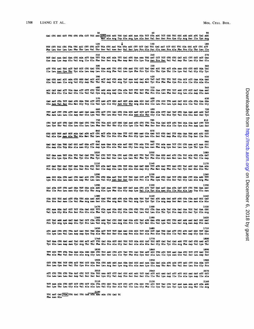
Characterization of the ZP2 mRNA and the ZP2 protein.The structure of the cytoplasmic ZP2 mRNA (Fig. 1) wasdeduced from the nucleic acid sequence of near-full-lengthcDNAs and a genomic clone containing exon 1. The 5' end ofthe mRNA was defined by the protection of a 78-nt fragmentafter Si nuclease digestion of a 108-nt single-stranded probederived from genomic DNA (map positions -30 to 78) whichhad been hybridized to ovarian poly(A)+ mRNA (data notshown). The 2,201-nt ZP2 mRNA has a very short 5'untranslated region of 30 nt and a similarly short (32-nt) 3'
FIG. 1. Structure of the ZP2 mRNA and protein. The nucleic acid sequence of near-full-length cDNAs and exon 1 of Zp-2 were used todeduce the structure of the ZP2 mRNA and resultant protein. The initiation and termination codons are boxed, and the polyadenylation signalis overlined. The single 2139-nucleotide open reading frame is translated into protein in line 2. The 34-amino-acid signal peptide is indicatedby the wavy line, and the arrow points to the signal peptidase cut site. Amino acid sequences which were experimentally determined byisolation and direct sequencing of an N-terminal and an internal ZP2 peptide are underlined with a dashed line. The seven potential N-linkedglycosylation sites [Asn-X-(Thr/Ser)] are underlined with a solid line.
VOL. 10, 1990
on Decem
ber 6, 2018 by guesthttp://m
cb.asm.org/
Dow
nloaded from

1510 LIANG ET AL.
AVSLPQ TLGSE
I I
NH. II
-1.5
SSYLI-
I I I I
VSLP
I COOH
I s1rll lw l 1 's'WT11'rI r I *Hydropathidty
H1H 1. H H H- H N "1-4- a-Helix...-... - ...- -..-.- nCharge
200 300 400 500 600
SSYL VSLPQ
] COOH
Hydropathicdty
-2.5
I_- a-Helix... ........-.... ...... - Charge
1 100 200 300 400 Amino Acid ResidueFIG. 3. Secondary structure of the deduced ZP2 and ZP3 proteins. (A) Schematic representation of the ZP2 mRNA with a single open
reading frame containing a signal peptide (0) and seven potential N-linked glycosylation sites ( ). The hydropathicity of the protein wasdetermined by the Kyte and Doolittle algorithm (33), and the a-helical structure was determined by the method of Garnier et al. (22). (B) Sameas panel A, but for ZP3. The three amino acid identities in the ZP2 and ZP3 polypeptides are indicated by the single-letter amino acid codeabove each protein: VSLPQ, Val-Ser-Leu-Pro-Gln beginning at residues 35 and 645 (contains only the first four residues) in ZP2 and 420 inZP3; TLGSE, Thr-Leu-Gly-Ser-Glu beginning at residue 76 in ZP2 and 72 in ZP3; SSYL, Ser-Ser-Tyr-Leu, beginning at residue 228 in ZP2and 416 in ZP3.
untranslated region (Fig. 1). Although the nucleic acid se-quence is distinctly different from that of ZP3, this samemotif of short 5' and 3' untranslated regions is preserved inboth zona transcripts (41). Oligo(dT)-purified ZP2 transcriptsmigrate on Northern blots with a molecular size of 2.4kilobases (Fig. 2), which suggests that ZP2 mRNA (like ZP3)isolated from growing oocytes contains an approximately200-nt poly(A) tail. The comigration of rRNA with ZP3 mayaccount for the slight difference in mobility of ZP3 tran-scripts detected in total and poly(A)+ ovarian RNAs (Fig. 2).The ZP2 mRNA has a single open reading frame of 2,139
nt which codes for a polypeptide of 80,217 daltons represent-ing 713 amino acids (10.8% acidic, 9.5% basic, 10.2% aro-matic, and 34.8% hydrophobic). The amino acid sequencecontains seven Asn-X-(Ser/Thr) sequences which are poten-
tial N-linked glycosylation sites (Fig. 1 and 3A). The first 34amino acids of the open reading frame are absent from theN-terminal amino acid sequence obtained from sodium do-decyl sulfate-polyacrylamide gel electrophoresis-purified se-creted ZP2 protein and presumably represent a signal pep-tide. The amino acids at the -1 and -3 positions from thepresumptive signal peptidase cleavage site are Ser and Asn,respectively; these are in accordance with the (-3, -1) ruleproposed by von Heijne (48, 49). The resultant secretedprotein would have a molecular mass of 76,373 daltons.No overall sequence similarity was detected between ZP2
and ZP3 at either the nucleic acid or the amino acid level.However, there are two 5-amino-acid regions that are iden-tical in both polypeptides (Fig. 3). The most striking one(Val-Ser-Leu-Pro-Gln) is located at the exact N terminus of
1
B100
TLGSE
NH2
700 Amino Acid Residue
H "m '-4
MOL. CELL. BIOL.
-I
on Decem
ber 6, 2018 by guesthttp://m
cb.asm.org/
Dow
nloaded from

OOCYTE-SPECIFIC EXPRESSION OF Zp-2 1511
JK\ 7.1000
024-
R 40 50 60 Ov
Oocytes Diameter (Wm)FIG. 4. Developmental expression of Zp-2 and Zp-3 during oo-
genesis. (A) Autoradiograph of Northern blot analysis of RNAisolated from mouse oocytes or eggs probed with 32P-labeled insertfrom pZP2.4 and from pZP3.2 (42). Lanes indicate stage of oocytedevelopment: 1, 800 resting oocytes; 2, 200 oocytes of diameter 40,um; 3, 200 oocytes of diameter 50 ,um; 4, 200 oocytes of diameter 65,um; 5, 200 ovulated eggs; 6, 800 ovulated eggs. Blots were exposedat -70°C for 4 days (lanes 1 to 5) or 10 days (lane 6). Numbers to theleft represent molecular size markers. Arrows to the right of lane 6indicate ZP2 and ZP3 transcripts. (B) Quantitation of the abundanceof ZP2 (LI) and ZP3 (A) transcripts during oogenesis based ondensitometry of hybridization signals in panel A and the hybridiza-tion signal obtained by using increasing amounts of synthetic ZP2and ZP3 transcripts. Abbreviations: R, resting oocytes; OV, ovu-lated eggs.
the secreted ZP2 protein and at the exact carboxyl terminusof the ZP3 protein. Four of these five amino acids (Yal-Ser-Leu-Pro) are also present a second time in ZP2 atposition 645. Another 5-amino-acid identity (Thr-Leu-Gly-Ser-Glu) is present at a comparable distance from the aminoterminus of ZP2 (amino acid residue 76) and ZP3 (aminoresidue 72). Based on a comparison with the frequencies ofamino acids in 500 proteins (16), the probabilty that these5-amino-acid identities arose by chance is 1 x 10' and 2.6x 10⁻⁵, respectively. There is an additional 4-amino-acididentity between the two proteins (Ser-Ser-Tyr-Leu) posi-tioned at amino acid residue 228 in ZP2 and 416 in ZP3.Otherwise, although there is an increase in the number ofglycine (85%) and alanine (55%) residues, the amino acidcomposition of ZP2 appears comparable to that of otherproteins (16), and there does not appear to be a pronouncedclustering of a particular amino acid along the polypeptidebackbone. Neither ZP2 nor ZP3 have a high degree of
predicted a-helical structure (22), and hydropathicity plotsof the two polypeptides (33) indicate that ZP2 has a veryhydrophobic region near its carboxy terminus (Fig. 3), aspreviously described for ZP3 (41). These hydrophobic re-gions, along with the short identical amino acid regions, maybe important for the interaction of zona polypeptides in theextracellular zona matrix.
Oocyte-specific expression of ZP2 transcripts. To examinethe tissue-specific expression of Zp-2, we performed North-ern blot analysis on poly(A)+ RNA from ovary and totalRNA from ovary, brain, heart, liver, and testes. A singlepoly(A)+ transcript with a molecular size of 2.4 kilobaseswas detected in ovarian RNA (Fig. 2A). To determinewhether this expression could be localized further, weisolated total RNA from oocytes and granulosa cells derivedfrom the ovaries of 3-week-old female mice. Using Northernblots probed with a ZP2 cDNA, we detected a singletranscript in oocyte RNA, but not in granulosa cell RNA(Fig. 2B). As controls, the above Northern blots were alsoreprobed with a labeled P-actin cDNA and actin transcriptswere detected (data not shown). Thus, Zp-2 appears to beexpressed only in oocytes, making it, along with Zp-3, amember of a set of oocyte-specific genes.The accumulation of ZP2 transcripts during oocyte growth
was investigated by isolating total RNA from resting (15-,um), 40-, 50-, and 65-,um-diameter oocytes as well as fromovulated eggs. After electrophoresis in formaldehyde-aga-rose gels and membrane transfer, the RNA was probed withZP2- and ZP3-specific cDNA inserts (Fig. 4A). Knownamounts of synthetic ZP2 and ZP3 RNA transcripts electro-phoresed on the same gels were used as standards toquantitate the levels of ZP2 and ZP3 transcripts (data notshown). Neither zona transcript was detected in restingoocytes (Fig. 4A), even when the RNA from four times asmany oocytes was examined. However, substantial amountsof ZP2 and ZP3 transcripts were detected in 40-,um-diameteroocytes (Fig. 4A), and the amount of ZP2 transcripts in-creased to a maximum of 1,000 fg per oocyte while theamount of ZP3 transcripts increased to a maximum level of400 fg per oocyte in 50-p.m oocytes (Fig. 4B). Based onearlier estimations (17, 27), the peak values of ZP2 and ZP3represent 1% and 0.4%, respectively, of the total poly(A)+content of growing oocytes. The ZP2 and ZP3 transcriptsaccumulated in parallel, and ZP2 was present in approxi-mately twofold molar excess throughout oocyte growth.As oocyte growth continued, the number of both zona
transcripts decreased, and in ovulated eggs there was lessthan 5% of their peak levels. To better visualize the ZP2 andZP3 transcripts in ovulated eggs, RNA was isolated from 4times as many eggs and after hybridization the Northern blotwas exposed for 2.5 times as long (Fig. 4A, lane 6). The sizesof both ZP2 and ZP3 transcripts were distinctly decreased inovulated eggs from their sizes in growing oocytes. The
l1 / 3 4
1 11 15 Il 1
56789 10 11121314 1516I 6 7 89 10 \ 1718
1 Kb
FIG. 5. Schematic representation of the intron-exon map of Zp-2. Dark bars represent each of the 18 exons, which range in size from 45to 190 bp and are separated by 17 introns ranging in size from 81 to 1490 nt. The entire transcription unit is 12.1 kbp long.
A B
Kb
2.34-
1.35 --
1 2 3 4 5 6
a -
VOL. 10, 1990
3'
on Decem
ber 6, 2018 by guesthttp://m
cb.asm.org/
Dow
nloaded from

1512 LIANG ET AL.
TABLE 1. Exon and intron sizes of mouse Zp-2 gene
Exon Position Length (nt) Intron Length (nt)
1 1-80 80 1 90"2 81-169 89 2 9603 170-253 84 3 356"'4 254-348 95 4 14905 349-498 150 5 5806 499-543 45 6 5307 544-705 162 7 12308 706-802 97 8 95a9 803-987 185 9 80010 988-1114 127 10 108011 1115-1296 182 11 88"12 1297-1388 92 12 97"13 1389-1513 125 13 139"14 1514-1703 190 14 103015 1704-1839 136 15 81"16 1840-1936 97 16 314"17 1937-2014 78 17 85018 2015-2201 187
a Exact length determined by sequencing.
magnitude of the decrease is roughly 200 nt, which corre-sponds to the estimated length of their poly(A) tails.Genomic locus and exon-intron mapping of Zp-2. A ge-
nomic clone containing a 14.4-kbp insert was isolated from aAJ1 mouse library and included the entire mouse Zp-2 locusas well as 1.5 kbp of 5'-flanking regions and 0.8 kbp of3'-flanking regions. Oligonucleotide primers, used to se-quence the ZP2 cDNA, were used to sequence the Zp-2gene, and 18 exons ranging from 45 to 190 bp were identified(Fig. 5; Table 1). The nucleotide sequence of the Zp-2 exonswas identical to that determined for the ZP2 cDNA. Thesizes of the introns, which ranged from 81 to 1,490 bp, weredetermined either by direct sequencing or by analyzingpolymerase chain reaction products primed with syntheticoligonculeotides that mapped to regions flanking the introns(Table 1). Each exon (except exon 1) is preceded by a spliceacceptor consensus sequence (12), and each (except exon18) is followed by a splice donor consensus sequence (Table2).
TABLE 2. Immediate flanking sequences of Zp-2 exons
5'-Flanking sequence" Exon no. 3'-Flanking sequence"
1 GTGAGGCATTCTTCTCCTATCCAG 2 GTATGTCTCTTGTCTGTTGTCCAG 3 GTATGTAGTACGTGTCTTCTGTAG 4 GTAAGCAAGTGTGTTTGTACACAG S GTAAGTGATGTGTTTTTTTTCCAG 6 GTAAGAATACAATGAATTTTGAAG 7 GTAGGTTTGAAATCTGACCCCCAG 8 GTGAGGCCTGAGCGTTTTGTACAG 9 GTATGTTTGTTACTCTCGTTCCAG 10 GTAAGCATGTTTGTCCACTTACAG 11 GTGAGTAGCAACCCAACATTGAAG 12 GTGTGATGCAAAACATTTTTCTAG 13 GTGAGGTGTCGCTGTTAATTGCAG 14 GTATGTACTCCCCCTGTTTCCTAG 15 GTATGTGATCAATTCCTCTTCCAG 16 GTAAAAATTCAACTCCCACCACAG 17 GTAAACATTTAGCTTTCTTTGTAG 18
a The splice acceptor and splice donor consensus sequences are in boldletters.
The start of transcription of Zp-2 was defined by the Sinuclease protection assay as described above and confirmedby the identification of an 81-nt primer extension product(after hybridization to ovarian RNA but not liver RNA) byusing a synthetic oligonucleotide primer complementary tomap positions 62 to 81 on the ZP2 cDNA (data not shown).These data document that exon 1 of Zp-2 contains 80 nt and,as described above, the resultant ZP2 transcript has an 5'untranslated region of 30 nt. The 3' end of the transcriptionunit was deduced by comparing the cDNA sequence of twoindependently isolated clones with the sequence of thegenomic clone (data not shown). Thus, exon 18 is 187 bp inlength, and the entire transcription unit encompasses 12.1kbp.The copy number of Zp-2 in the mouse genome was
estimated by digesting genomic equivalents of the Zp-2 XJ1genomic locus clone and mouse genomic DNA with restric-tion endonucleases and probing them with the ZP2 insertfrom pZP2.4. Comparison of the band intensity between theZp-2 XJ1 genomic locus clone DNA and mouse genomicDNA suggested that Zp-2 is present in the mouse genome inlow copy number (data not shown). Mouse genomic DNAand the Zp-2 genomic recombinant phage DNA were di-gested with four different restriction enzymes and examinedby Southern blot analysis. Examination of the bandingpatterns of the restriction enzyme-digested genomic DNAindicated that all bands observed could be accounted for inthe restriction enzyme-digested XJ1 DNA, thus providingevidence that there is only one copy of Zp-2 in the mousegenome (data not shown).
5'-Flanking region of Zp-2. The Zp-2 5'-flanking regioncontains a TATAA box at -31 and a CCAAT box at -69 bpfrom the transcription start site. Several novel tandemrepeats have been identified in the Zp-3 locus (14, 31), theonly other oocyte-specific gene that has been found in mice.However, similar repeats were not detected in the first 1.5kbp of the 5'-flanking region or in the 0.8-kbp 3'-flankingregion of Zp-2. A comparison of the 5'-flanking sequences ofZp-2 and Zp-3 detected three homologous regions, ranging insize from 8 to 12 bp, within the first 250 bp of the transcrip-tion start site. The three elements, GTGAAAGGGTGG (atbp -63), ATTCTGGT (at bp -194), and ACTCACCTGG (atbp -219), are 80 to 88% conserved between the two oocyte-specific genes. Although present in other genes, they arerarely located in the 5'-flanking regions and are not related toother cis elements that have been reported in the literature.Provocatively, similar sequences are also found at approxi-mately the same distance upstream of the transcription startsite of the human Zp-3 gene (M. E. Chamberlin, Ph.D.thesis, Johns Hopkins University, Baltimore, Md., 1989).Their role in the oocyte-specific expression of the zona genesremains to be determined.
DISCUSSION
Zp-2 and Zp-3 encode two proteins that are secreted andparticipate in the formation of the mouse zona pellucida. Theexpression of the two zona genes is oocyte specific anddevelopmentally regulated. Although unrelated to one an-other by sequence and located on separate chromosomes(34), the transcripts of these two genes accumulate coordi-nately during the growth phase of oogenesis prior to ovula-tion. At present, the regulation of their expression is poorlyunderstood. From our investigations we hope to learn moreabout the mechanisms of oocyte-specific gene expression
MOL. CELL. BIOL.
on Decem
ber 6, 2018 by guesthttp://m
cb.asm.org/
Dow
nloaded from

OOCYTE-SPECIFIC EXPRESSION OF Zp-2 1513
and to make use of these findings to begin to determine thesignals that induce resting oocytes to enter into growth anddifferentiation.
During mouse oogenesis there is a rapid accumulation ofpoly(A)+ RNA, and full-grown oocytes contain 85 to 90 pg ofpoly(A)+, which represents 20% of the total RNA (1. 2).Approximately one-fourth of the poly(A)+ RNA is found onpolysomes, while the rest accumulates as stable maternalRNA for use later in oogenesis (1, 17). Although the expres-sion of a number of genes has been examined during mouseoogenesis, to date only Zp-2 and Zp-3 appear to be ex-pressed exclusively in oocytes (40, 41, 43) (see above).Neither ZP2 nor ZP3 transcripts are detected in resting,primordial oocytes but quickly accumulate as abundantmRNAs when oocytes enter into their growth phase. At theirpeak levels, ZP2 and ZP3 mRNAs represent approximately1% and 0.4%, respectively, of the total poly(A)+ RNA.Although turnover studies have not been reported, given thegreat abundance of the zona transcripts we speculate thatboth ZP2 and ZP3 are quite stable during oogenesis.Throughout oocyte growth the molar ratio of ZP2 to ZP3transcripts is 2:1, although the reported protein ratio isapproximately 1:1 (50). This suggests that posttranscrip-tional regulation may plav an important role in Zp-2 and Zp-3gene expression.
It is noteworthy that both ZP2 and ZP3 mRNA have short5' (30 and 29 nt, respectively) and 3' (32 and 16 nt, respec-tively) untranslated regions. This motif appears to be evolu-tionarily conserved in that the size of ZP3 mRNA is the samein at least three mammalian species (41). Short 5' and 3'untranslated regions, although not unknown (4, 28, 30, 38)are unusual, and they may be important for the processing ofZP2 and ZP3 transcripts. The stability of bulk oocytepoly(A)+ RNA decreases during meiotic maturation andsubsequent ovulation such that more than 50% of thepoly(A)+ RNA is either deadenylated or degraded (37). Thiscomposite observation reflects widely divergent processingof individual mRNAs: some are deadenylated, some aredegraded (37), and some are polyadenylated (26, 37). Thedecline of the ZP2 and ZP3 transcripts during this stage ofoogenesis is dramatic and places them in the class of mRNAswhich are degraded during meiotic maturation and ovula-tion.The poly(A)+ ZP2 and ZP3 transcripts are actively trans-
lated into protein in growing oocytes (8, 45) and thus appearto associate with the polysome fraction of RNA. The subse-quent decrease in zona protein synthesis observed in thelatter stages of oocyte growth (45) corresponds to a declinein the abundance of the two zona transcripts. The low levelor absence of zona protein synthesis in fully grown oocytes(8) and ovulated eggs (45) correlates temporally with aprecipitous decline in the abundance of ZP2 and ZP3 tran-scripts to less than 5% of the peak levels. Furthermore, thezona transcripts detected in ovulated eggs are 200 nt shorter[the approximate length of their poly(A) tail] than thatobserved in growing oocytes, suggesting that deadenylationmay have occurred during meiotic maturation and ovulation.The presence of a poly(A) tail has been associated with thetranslatability of a number of mouse oocyte mRNAs includ-ing tissue plasminogen activator, hypoxanthine phosphori-bosyltransferase, actin, and ox-tubulin (2, 24, 35), and similarobservations have been made with Spisiula (44) and Xen2opius(19, 35) oocytes. Thus, it appears that the absence of ZP2and ZP3 zona protein synthesis after ovulation may be dueboth to the low level of zona transcripts and to an inability to
translate them in their shortened, presumably deadenylated,form.
Both ZP2 and ZP3 are heavily glycosylated proteins (7,45). The 713-amino-acid peptide of ZP2 contains sevenpotential N-linked glycosylation sites, six of which havebeen reported to be derivatized (23), and more than 100potential 0-linked glycosylation sites. Incubation of growingoocytes in the presence of tunicamycin inhibits N-linkedglycosylation and results in the detection of an 81,000-daltonprotein (23), which is larger than the 76,373-dalton polypep-tide chain deduced from DNA sequence, a discrepancy thatmay result from 0-linked glycosylation. Both the ZP2 andZP3 proteins have a signal peptide which directs theirsecretion to the extracellular matrix of the zona pellucida,and there may be additional signals to direct the processingof the intracellular proteins. Although neither polypeptidechain contains previously described intracellular traffickingsignals (32, 47), the three regions of amino acid identity inZP2 and ZP3 may provide such functions.
Electron microscopy and biochemical analysis of the zonapellucida suggest that ZP2 and ZP3 are arranged into longfilaments which are cross-linked by ZP1 (24). Little is knownabout the protein domains that modulate these interactions,but the aforementioned amino acid identities may be impor-tant, as may the very hydrophobic region at the C terminusof each polypeptide. After the initial binding of sperm to thezona pellucida at fertilization, ZP2 acts as a secondary spermreceptor (6). Following fertilization, ZP2 is proteolyticallycleaved, releasing a 23,000-dalton glycopeptide which isbound via a disulfide bond(s) to the larger, 90,000-daltonmoiety (5, 36). Candidate proteases responsible for thiscleavage have recently been identified (36), and this bio-chemical modification of ZP2 is associated with the postfer-tilization block to polyspermy.
Both Zp-2 and Zp-3 are present in single copy in the mousegenome, and although they are located on separate chromo-somes (34), their expression is coordinately regulated andtissue specific. If the transcription of zona genes is governedby a common factor or factors, the identical regulatoryregions may be identified in the control regions of the twogenes. Both genes have a TATAA box in their 5'-flankingregion, and Zp-2, but not Zp-3, has a CCAAT box at -69from the transcription start site. Novel tandem repetitivesequences have been identified at the 5'-flanking regions ofZp-3 (14, 31), but there are no similar sequences upstream ofthe Zp-2 transcription start site. However, both zona genescontain three short sequence similarities (8 to 12 bp) ar-ranged at comparable distances from the transcription startsite in the first 250 bp of the 5'-flanking region. Studies areunder way to determine whether these DNA sequences havea functional role in the expression of Zp-2 and Zp-3. Theseinvestigations may lead to the identification of commonfactors which regulate the coordinate expression of theoocyte-specific zona pellucida genes.
ACKNOWLEDGMENTSWe thank M. J. Ringuette and K. Kenward for help in rescreening
the Agtll library, and we appreciate the critical reading of themanuscript by R. Bachvarova and J. Piatigorsky.
LITERATURE CITED1. Bachvarova, R. 1985. Gene expression during oogenesis and
oocyte development in the mammal, p. 453-524. In L. W.Browder (ed.). Developmental biology: a comprehensive syn-thesis. vol. 1. Plenum Publishing Corp.. New York.
2. Bachvarova, R., V. De Leon, A. Johnson, G. Kaplan, and B. V.Paynton. 1985. Changes in total RNA. polyadenylated RNA,
VOL. 10. 1990
on Decem
ber 6, 2018 by guesthttp://m
cb.asm.org/
Dow
nloaded from

1514 LIANG ET AL.
and actin mRNA during meiotic maturation of mouse oocytes.Dev. Biol. 108:325-331.
3. Baker, T. G. 1972. Oogenesis and ovulation, p. 17-45. In C. R.Austin and R. V. Short (ed.), Reproduction in mammals: germcells and fertilization, vol. 1. Cambridge University Press,Cambridge.
4. Bishop, D. F., D. H. Calhoun, H. S. Bernstein, P. Hantzopoulos,M. Quinn, and R. J. Desnick. 1986. Human a-galactosidase A:nucleotide sequence of a cDNA clone encoding the matureenzyme. Proc. Natl. Acad. Sci. USA 83:4859-4863.
5. Bleil, J. D., C. F. Beall, and P. M. Wassarman. 1981. Mamma-lian sperm-egg interaction: fertilization of mouse eggs triggersmodification of the major zona pellucida glycoprotein, ZP2.Dev. Biol. 86:189-197.
6. Bleil, J. D., J. M. Greve, and P. M. Wassarman. 1988. Identifi-cation of a secondary sperm receptor in the mouse egg zona
pellucida: role in maintenance of binding of acrosome-reactedsperm to eggs. Dev. Biol. 128:376-385.
7. Bleil, J. D., and P. M. Wassarman. 1980. Structure and functionof the zona pellucida: identification and characterization of theproteins of the mouse oocyte's zona pellucida. Dev. Biol.76:185-202.
8. Bleil, J. D., and P. M. Wassarman. 1980. Synthesis of zona
pellucida proteins by denuded and follicle-enclosed mouse
oocytes during culture in vitro. Proc. Natl. Acad. Sci. USA77:1029-1033.
9. Bleil, J. D., and P. M. Wassarman. 1980. Mammalian sperm-egginteraction: identification of a glycoprotein in mouse egg zonaepellucidae possessing receptor activity for sperm. Cell 20:873-882.
10. Bleil, J. D., and P. M. Wassarman. 1988. Galactose at thenonreducing terminus of 0-linked oligosaccharides of mouse
egg zona pellucida glycoprotein ZP3 is essential for the glyco-protein's sperm receptor activity. Proc. Natl. Acad. Sci. USA85:6778-6782.
11. Brambell, F. W. R. 1928. The development and morphology ofthe gonads of the mouse. III. The growth of the follicle. Proc. R.Soc. London Ser. B 103:258-272.
12. Breathnach, R., and P. Chambon. 1981. Organization andexpression of eukaryotic split genes coding for proteins. Annu.Rev. Biochem. 50:349-383.
13. Calzone, F. J., R. J. Britten, and E. H. Davidson. 1987. Mappingof gene transcripts by nuclease protection assays and cDNAprimer extension, p. 611-632. In S. L. Berger and A. R. Kimmel(ed.), Guide to molecular cloning techniques. Academic Press,Inc., Orlando, Fla.
14. Chamberlin, M. E., and J. Dean. 1989. Genomic organization ofa sex specific gene: the primary sperm receptor of the mousezona pellucida. Dev. Biol. 131:207-214.
15. Chien, Y.-H., J. K. Gascoigne, J. Kavaler, N. E. Lee, and M. M.Davis. 1984. Somatic recombination in a murine T-cell receptorgene. Nature (London) 309:322-326.
16. Dayhoff, M. O., and B. C. Orcutt. 1979. Methods for identifyingproteins by using partial sequences. Proc. Natl. Acad. Sci. USA76:2170-2174.
17. De Leon, V., A. Johnson, and R. Bachvarova. 1983. Half-livesand relative amounts of stored and polysomal ribosomes andpoly(A)+ RNA in mouse oocytes. Dev. Biol. 98:400-408.
18. Derman, E., K. Krauter, L. Walling, C. Weinberger, M. Ray,and J. E. Darnell, Jr. 1981. Transcriptional control in theproduction of liver-specific mRNAs. Cell 23:731-739.
19. Drummond, D. R., J. Armstrong, and A. Colman. 1985. Theeffect of capping and polyadenylation on the stability, move-ment and translation of synthetic messenger RNAs in Xenopusoocytes. Nucleic Acids Res. 13:7375-7394.
20. East, I. J., and J. Dean. 1984. Monoclonal antibodies as probesof the distribution of ZP2, the major sulfated glycoprotein of themurine zona pellucida. J. Cell Biol. 98:795-800.
21. Florman, H. M., and P. M. Wassarman. 1985. 0-linked oligosac-charides of mouse egg ZP3 account for its sperm receptoractivity. Cell 41:313-324.
22. Garnier, J., D. J. Osguthorpe, and B. Robson. 1978. Analysis ofthe accuracy and implications of simple methods for predicting
the secondary structure of globular proteins. J. Mol. Biol.120:97-120.
23. Greve, J. M., G. S. Salzmann, R. J. Roller, and P. M. Wassar-man. 1982. Biosynthesis of the major zona pellucida glycopro-tein secreted by oocytes during mammalian oogenesis. Cell31:749-759.
24. Greve, J. M., and P. M. Wassarman. 1985. Mouse egg extracel-lular coat is a matrix of interconnected filaments possessing astructural repeat. J. Mol. Biol. 181:253-264.
25. Hogan, B., F. Constantini, and E. Lacy. 1986. Manipulating themouse embryo: a laboratory manual. Cold Spring Harbor Lab-oratory, Cold Spring Harbor, N.Y.
26. Huarte, J., D. Belin, A. Vassalli, S. Strickland, and J.-D.Vassalli. 1987. Meiotic maturation of mouse oocytes triggers thetranslation and polyadenylation of dormant tissue-type plasmi-nogen activator mRNA. Genes Dev. 1:1201-1211.
27. Jahn, C. L., M. M. Baran, and R. Bachvarova. 1976. Stability ofRNA synthesized by the mouse oocyte during its major growthphase. J. Exp. Zool. 197:161-172.
28. Jehn, C.-H., T. Deng, D. Li, J. DeWille, and L. F. Johnson. 1986.Mouse thymidylate synthase messenger RNA lacks a 3' un-translated region. Proc. Natl. Acad. Sci. USA 83:8482-8486.
29. Jones, E. C., and P. L. Krohn. 1961. The relationship betweenage, numbers of oocytes and fertility in virgin and multiparousmice. J. Endocrinol. 21:469-495.
30. Kelley, D. E., C. Coleclough, and R. P. Perry. 1982. Functionalsignificance and evolutionary development of the 5'-terminalregions of immunoglobulin variable-region genes. Cell 29:681-689.
31. Kinloch, R. A., R. J. Roller, C. M. Fimiani, D. A. Wassarman,and P. M. Wassarman. 1988. Primary structure of the mousesperm receptor polypeptide determined by genomic cloning.Proc. Natl. Acad. Sci. USA 85:6409-6413.
32. Klausner, R. D. 1989. Sorting and traffic in the central vacuolarsystem. Cell 57:703-706.
33. Kyte, J., and R. F. Doolittle. 1982. A simple method fordisplaying the hydropathic character of a protein. J. Mol. Biol.157:105-132.
34. Lunsford, R. D., N. A. Jenkins, C. A. Kozak, L.-F. Liang, C. M.Silan, N. G. Copeland, and J. Dean. 1990. Genomic mapping ofmurine Zp-2 and Zp-3: two oocyte-specific loci encoding zonapellucida proteins. Genomics 6:184-187.
35. McGrew, L. L., E. Dworkin-Rastl, M. B. Dworkin, and J. D.Richter. 1989. Poly (A) elongation during Xenopus oocytematuration is required for translational recruitment and is me-diated by a short sequence element. Genes Dev. 3:803-815.
36. Moller, C. C., and P. M. Wassarman. 1989. Characterization ofa proteinase that cleaves zona pellucida glycoprotein ZP2 fol-lowing activation of mouse eggs. Dev. Biol. 132:103-112.
37. Paynton, B. V., R. Rempel, and R. Bachvarova. 1988. Changesin state of adenylation and time course of degradation ofmaternal mRNAs during oocyte maturation and early embry-onic development in the mouse. Dev. Biol. 129:304-314.
38. Peterson, C. A., and J. Piatigorsky. 1986. Preferential conserva-tion of the globular domains of the ,A3.A1-crystallin polypep-tide of the chicken eye lens. Gene 45:139-147.
39. Phillips, D. M., and R. Shalgi. 1980. Surface architecture of themouse and hamster zona pellucida and oocyte. J. Ultrastruct.Res. 72:1-12.
40. Philpott, C. C., M. J. Ringuette, and J. Dean. 1987. Oocyte-specific expression and developmental regulation of ZP3, thesperm receptor of the mouse zona pellucida. Dev. Biol. 121:568-575.
41. Ringuette, M. J., M. E. Chamberlin, A. W. Baur, D. A. Sobieski,and J. Dean. 1988. Molecular analysis of cDNA coding for ZP3,a sperm binding protein of the mouse zona pellucida. Dev. Biol.127:287-295.
42. Ringuette, M. J., D. A. Sobieski, S. M. Chamow, and J. Dean.1986. Oocyte-specific gene expression: molecular characteriza-tion of a cDNA coding for ZP3, the sperm receptor of the mousezona pellucida. Proc. Natl. Acad. Sci. USA 83:4341-4345.
43. Roller, R. J., R. A. Kinloch, B. Y. Hiraoka, S. S.-L. Li, andP. M. Wassarman. 1989. Gene expression during mammalian
MOL. CELL. BIOL.
on Decem
ber 6, 2018 by guesthttp://m
cb.asm.org/
Dow
nloaded from

OOCYTE-SPECIFIC EXPRESSION OF Zp-2 1515
oogenesis and early embryogenesis: quantification of threemessenger RNAs abundant in fully grown mouse oocytes.Development 106:251-261.
44. Rosenthal, E. T., and J. V. Ruderman. 1987. Widespreadchanges in the translation and adenylation of maternal messen-ger RNAs following fertilization of Spisula oocytes. Dev. Biol.121:237-246.
45. Shimizu, S., M. Tsuji, and J. Dean. 1983. In vitro biosynthesis ofthree sulfated glycoproteins of murine zonae pellucidae byoocytes grown in follicle culture. J. Biol. Chem. 258:5858-5863.
46. Tempst, P., M. W. Hunkapiller, and L. E. Hood. 1984. Separa-
tion of peptides by reverse-phase high-performance liquid chro-matography using propyl- and cyanopropylsilyl supports. Anal.Biochem. 137:188-195.
47. Verner, K., and G. Schatz. 1988. Protein translocation acrossmembranes. Science 241:1307-1313.
48. Von Heijne, G. 1985. Signal sequences: the limits of variation. J.Mol. Biol. 184:99-105.
49. Von Heijne, G. 1986. A new method for predicting signalsequence cleavage sites. Nucleic Acids Res. 14:4683-4690.
50. Wassarman, P. M. 1988. Zona pellucida glycoproteins. Annu.Rev. Biochem. 57:415-442.
VOL. 10, 1990
on Decem
ber 6, 2018 by guesthttp://m
cb.asm.org/
Dow
nloaded from