open access research development and external validation
TRANSCRIPT

1Hong Jyoung, et al. BMJ Open 2019;9:e024007. doi:10.1136/bmjopen-2018-024007
Open access
Development and external validation of new nomograms by adding ECG changes (ST depression or tall T wave) and age to conventional scoring systems to improve the predictive capacity in patients with subarachnoid haemorrhage: a retrospective, observational study in Korea
Ju young Hong,1 Je Sung You,1 Min Joung Kim,1 Hye Sun Lee,2 Yoo Seok Park,1 Sung Phil Chung,1 Incheol Park1
To cite: Hong Jyoung, You JS, Kim MJ, et al. Development and external validation of new nomograms by adding ECG changes (ST depression or tall T wave) and age to conventional scoring systems to improve the predictive capacity in patients with subarachnoid haemorrhage: a retrospective, observational study in Korea. BMJ Open 2019;9:e024007. doi:10.1136/bmjopen-2018-024007
► Prepublication history for this paper is available online. To view these files please visit the journal online (http:// dx. doi. org/ 10. 1136/ bmjopen- 2018- 024007).
Received 4 May 2018Revised 21 December 2018Accepted 8 January 2019
For numbered affiliations see end of article.
Correspondence toDr Yoo Seok Park; pys0905@ yuhs. ac
Research
© Author(s) (or their employer(s)) 2019. Re-use permitted under CC BY-NC. No commercial re-use. See rights and permissions. Published by BMJ.
AbstrACtObjectives To develop new nomograms by adding ECG changes (ST depression or tall T wave) and age to three conventional scoring systems, namely, World Federation of Neurosurgical Societies (WFNS) scale, Hunt and Hess (HH) system and Fisher scale, that can predict prognosis in patients with subarachnoid haemorrhage (SAH) using our preliminary research results and to perform external validation of the three new nomograms.Design Retrospective, observational studysetting Emergency departments (ED) of two university-affiliated tertiary hospital between January 2009 and March 2015.Participants Adult patients with SAH were enrolled. Exclusion criteria were age <19 years, no baseline ECG, cardiac arrest on arrival, traumatic SAH, referral from other hospital and referral to other hospitals from the ED.Primary outcome measures The 6 month prognosis was assessed using the Glasgow Outcome Scale (GOS). We defined a poor outcome as a GOS score of 1, 2 or 3.results A total of 202 patients were included for analysis. From the preliminary study, age, ECG changes (ST depression or tall T wave), and three conventional scoring systems were selected to predict prognosis in patients with SAH using multi-variable logistic regression. We developed simplified nomograms using these variables. Discrimination of the developed nomograms including WFNS scale, HH system and Fisher scale was superior to those of WFNS scale, HH system and Fisher scale (0.912 vs 0.813; p<0.001, 0.913 vs 0.826; p<0.001, and 0.885 vs 0.746; p<0.001, respectively). The calibration plots showed excellent agreement. In the external validation, the discrimination of the newly developed nomograms incorporating the three scoring systems was also good, with an area under the receiver-operating characteristic curve value of 0.809, 0.812 and 0.772, respectively.
Conclusions We developed and externally validated new nomograms using only three independent variables. Our new nomograms were superior to the WFNS scale, HH systems, and Fisher scale in predicting prognosis and are readily available.
IntrODuCtIOn Subarachnoid haemorrhage (SAH) is defined as haemorrhage in the subarach-noid space, and the reported incidence is 9–11 per 100 000 person years worldwide.1 SAH accounts for 4% of all cerebrovascular diseases and develops at a relatively young age, with approximately 50% of patients experiencing disability or death.2 Because of the high mortality and low recovery rate, predicting prognosis in patients with SAH is crucial.1–3 Botterell et al presented the first grading system for SAH in 1956,4 and different systems have since been developed.
strengths and limitations of this study
► Our nomograms are the first to combine ECG chang-es and other prognostic factors in patients with sub-arachnoid haemorrhage.
► Our nomogramsare solely based on ECG changes, age and conventional scoring systems, which are easily and readily obtainable during the patient’s course in the ED.
► Because this study is a retrospective, observational study, the predictive probability could be overesti-mated more than in a prospective study.
on Novem
ber 22, 2021 by guest. Protected by copyright.
http://bmjopen.bm
j.com/
BM
J Open: first published as 10.1136/bm
jopen-2018-024007 on 20 February 2019. D
ownloaded from

2 Hong Jyoung, et al. BMJ Open 2019;9:e024007. doi:10.1136/bmjopen-2018-024007
Open access
The Hunt and Hess (HH) system, the World Federation of Neurosurgical Societies (WFNS) scale and the Fisher scale have been widely used globally.5–7 These three systems are simple but do not include clinically important factors, such as comorbidity, age and vital signs on admis-sion, and differences between the grades for each system are ambiguous. A range of scoring scales has been devel-oped to overcome the limitations of the existing scoring scales.5–10
Factors such as age, past medical history (hyperten-sion, diabetes mellitus, history of brain disease), blood pressure and initial level of consciousness on admis-sion and cerebral aneurysm size have been reported to be independently associated with prognosis of patients with SAH.8 11ECG abnormalities have also been studied as a prognostic factor. ECG abnormalities are observed in 40%–90% of patients with SAH12–14 and have been associated with hyperactivity of the sympathetic nervous system due to autonomic nervous system dysregulation.15 Since the hyperactivity of the sympathetic nerve reflects the degree of cerebral haemorrhage, ECG changes may be associated with patient prognosis. The most common reported abnormalities are QT prolongation, ST segment changes and T wave abnormalities.15 16 A number of studies have investigated the prognostic value of ECG abnormali-ties.17 18 However, the role of an abnormal ECG as a prog-nostic predictor remains debatable. QTc changes and ST segment changes have been reported as the param-eters most significantly associated with prognosis,17 19 although further studies reported no significant associa-tion between ECG change and outcome.12 14 19 Because of this uncertainty, the existing system for predicting the prognosis of patients with SAH does not include ECG changes as a prognostic variable. However, ECG is an inexpensive test that is readily available in an emergency setting. Because accelerated sympathetic activity reaches its climax in the first 24 hours after the onset of SAH, ECG abnormalities develop during the early stage of disease, disappearing over time.13 15 16 Therefore, it is important to consider ECG changes as an initial prognostic factor. We conducted preliminary research to investigate the asso-ciation between the above-mentioned prognostic factors,
including ECG changes, and prognosis of patients with SAH and found that age, ECG changes and the three scoring systems were associated with patient prognosis.20
Among the computational models for predicting prognosis, the nomogram is very useful because it is a pictorial representation of a statistical predictive model that generates a numerical probability of a clin-ical event. It is more accurate than the conventional method using OR.21 Therefore, the objective of this study was to develop new nomograms that can predict prognosis of patients with SAH using the results of our preliminary research and to perform external validation of the new nomograms.
MAterIAls AnD MethODsstudy design and populationsWe performed a retrospective, observational study to develop new nomograms that can predict prognosis of patients with SAH and to perform external validation of the new nomograms. This study was conducted on two independent cohorts of patients with SAH from two hospitals. We enrolled patients with SAH diag-nosed by brain CT or xanthochromia of the cerebro-spinal fluid in the emergency department (ED) of Severance Hospital between January 2009 and March 2015 as primary cohort. Exclusion criteria were as follows: (1) age less than 19 years; (2) administration of a cardiovascular drug, such as calcium-channel blockers, with no baseline ECG; (3) cardiac arrest on arrival; and (4) traumatic SAH. Additionally, patients were excluded if they were referred from other hospi-tals because initial ECG could not be obtained and patients were often treated with medication affecting the cardiovascular system. Patients referred to other hospitals from the ED were also excluded as their prognosis could not be evaluated. For model external validation, 141 patients with SAH were enrolled in another ED of Gangnam Severance Hospital between January 2011 and December 2014 in accordance with the inclusion and exclusion criteria. We collected
Figure 1 Flow diagram of the study subjects. ED, emergency department.
on Novem
ber 22, 2021 by guest. Protected by copyright.
http://bmjopen.bm
j.com/
BM
J Open: first published as 10.1136/bm
jopen-2018-024007 on 20 February 2019. D
ownloaded from

3Hong Jyoung, et al. BMJ Open 2019;9:e024007. doi:10.1136/bmjopen-2018-024007
Open access
sufficient data to score all variables in the established nomogram.
Patient and public involvementPatients and public were not involved in the development of the research questions or in the design of the study because of its retrospective nature.
Data collectionOne investigator (J.YH) retrospectively collected the data through a review of the medical records. We collected demographic data, vital signs on admission and past medical history, including a history of hypertension, diabetes mellitus and cardiovascular and cerebrovas-cular diseases. The Glasgow Coma Scale (GCS), HH and WFNS scores were obtained from assessing the neurolog-ical abnormality and the level of consciousness. Patient
outcome was assessed using the Glasgow Outcome Scale (GOS) by one emergency physician blinded to the ECG findings. The amount of blood, the location and size of cerebral aneurysm and the presence or absence of intra-ventricular haemorrhage and intracerebral haemor-rhage were determined based on CT scans reported by board-certified radiologist. ECG findings were assessed by another emergency physician blinded to patient prog-nosis using the Marquette Universal System of Electro-cardiography (MUSE, GE Healthcare, Milwaukee, USA). Abnormal ECG findings were classified into four groups: ST elevation, ST depression, T wave inversion and tall T wave. ST elevation was defined as ≥0.1 mV elevation in at least two contiguous leads other than leads V2–V3. For leads V2–V3, the following cut-off points were applied: ≥0.2 mV in men ≥40 years, ≥0.25 mV in men <40
Table 1 Clinical characteristics of study populations
Good outcome Poor outcome Total
P value(n=111) (n=91) (n=202)
Age (year) 53.7 (12.3) 61.1 (13.7) 57.0 (13.4) <0.001
Sex, male 36 (32.4%) 36 (39.6%) 72 (35.6%) 0.305
Vital signs
Systolic blood pressure (mm Hg) 154.5 (29.3) 169.6 (41.0) 154.5 (35.7) 0.003
Heart rate (/min) 80.5 (17.5) 84.7 (22.1) 82.4 (19.8) 0.137
Respiratory rate (/min) 15.5 (2.2) 16.7 (6.0) 16 (4.3) 0.072
Body temperature (℃) 36.4 (0.5) 36.3 (0.7) 36.4 (0.6) 0.123
Chronic comorbidities
Hypertension 32 (28.8%) 43 (47.3%) 75 (37.1%) 0.007
Diabetes mellitus 7 (6.3%) 9 (9.9%) 16 (7.9%) 0.435
Cerebrovascular accident 5 (4.5%) 3 (3.3%) 8 (4.0%) 0.516
CT findings
Aneurysmal SAH 87 (88.4%) 61 (67.0%) 148 (73.3%) 0.227
Posterior circulation aneurysm 23 (20.7%) 18 (19.8%) 41 (20.3%) 0.178
Aneurysm sizes (mm) 4.0 (3.2) 4.6 (4.9) 4.3 (4.0) 0.258
Intracerebral haemorrhage 7 (6.3%) 26 (28.6%) 33 (16.3%) <0.001
Intraventricular haemorrhage 17 (15.3%) 41 (45.1%) 58 (28.7%) <0.001
ECG changes
ST elevation 6 (5.4%) 11 (12.1%) 17 (8.4%) 0.125
ST depression 2 (1.8%) 29 (31.9%) 31 (15.3%) <0.001
T wave inversion 11 (9.9%) 10 (11%) 21 (10.4%) 0.821
Tall T wave 1 (1%) 9 (9.9%) 10 (5.0%) 0.006
QTc (msec) 458.1 (39.1) 479.1 (53.0) 467.6 (46.9) 0.002
Scoring systems*
WFNS scales 1 (1~1) 4 (2~5) 2 (1~4) <0.001
HH system 2 (2~2) 4 (3~5) 3 (2~4) <0.001
Fisher grade 3 (3~3) 4 (3~4) 3 (3~4) <0.001
Data are presented as frequencies (%) or mean (SD), unless otherwise indicated.*median (IQR).HH, Hunt and Hess; SAH, subarachnoid haemorrhage; WFNS, World Federation of Neurosurgical Societies.
on Novem
ber 22, 2021 by guest. Protected by copyright.
http://bmjopen.bm
j.com/
BM
J Open: first published as 10.1136/bm
jopen-2018-024007 on 20 February 2019. D
ownloaded from
4 Hong Jyoung, et al. BMJ Open 2019;9:e024007. doi:10.1136/bmjopen-2018-024007
Open access
years or ≥0.15 mV in women. ST depression was defined as ≥0.05 mV depression in all leads. An inverted T wave was defined as less than 0.1 mV from the baseline, and a tall T wave was defined as greater than 1 mV in depth.19 QTc was redefined and measured based on heart rate and was adjusted using Bazett’s formula. There were no missing data except initial ECG because all variables used were collected in usual practice. The primary outcome of this study was assessed at 6 months after disease onset using GOS. We defined a poor outcome as a GOS score of 1, 2 or 3.
Data analysisStatistical analysis was performed using SAS (V.9.2) and R package (V.3.1.3, http://www. R- project. org). The sample size was calculated on the basis of the area under the receiver-operating characteristic curve (AUC) of the nomogram. A difference of 0.1 between the conventional scoring system with an AUC of 0.78 and new nomogram with an AUC of 0.88 was selected as the minimum clin-ically significant value. We assumed that the allocation ratio of good and poor outcome group was 4.5 to 5.5. The correlation between the two predictive models was assumed to be 0.5. We estimated that a sample size of 183 patients would be sufficient to evaluate the primary outcome at a significance level of 0.05 (two-sided) with 80% power. The sample size of the external validation cohort was determined to be about 70% of the sample size of the primary cohort.
Categorical variables were described as frequencies (%). Parametric data are presented as mean (SD) and non-para-metric data are presented as the median and IQR. We used the independent t-test for comparison of two groups distributed normally, whereas the Mann–Whitney U test was used for the comparison of two groups that were not distributed normally. Fisher’s exact test was used for cate-gorical variables. From the preliminary study, age, ECG
changes (ST depression or tall T wave) and three conven-tional scoring systems such as WFNS scale, HH system and Fisher scale were selected to predict prognosis in patients with SAH on the basis of the results of multi-vari-able logistic regression using backward Wald selection. As ST depression and tall T wave formation do not occur simultaneously, they were considered one variable. The scoring systems were entered individually as continuous variables into the model with clinical variables, CT find-ings and ECG abnormalities. In this study, we developed three simplified nomograms including age, ECG changes and each scoring system and compared the predictive performance of new nomograms with those of the WFNS scale, HH system and Fisher scale. The performance of the nomograms in predicting outcomes was validated with respect to discrimination and calibration. Nomogram predictive accuracy (discrimination) is measured via a concordance index (c-index), analogous to AUC, which quantifies the level of concordance between predicted probabilities and the actual chance of the event of interest occurring. A value of 0.5 indicates no predictive discrim-ination, and a value of 1.0 indicates perfect separation of patients with different outcomes. The Delong method was used to compare the C-indexes of each model. Calibration of the nomogram determines how far the predicted prob-abilities are from the observed outcome frequencies using graphic representations (calibration curve). A plot along the 45-degree line would indicate a perfect calibration model in which the predicted probabilities are identical to the actual outcomes. The calibration curves are presented as apparent and bias-corrected calibration plots using the bootstrapping methods with 200 re-samples. The predic-tive accuracy and calibration of the new nomograms were then externally validated with data derived from another ED (validation set; n=141). A value of p<0.05 was consid-ered statistically significant.
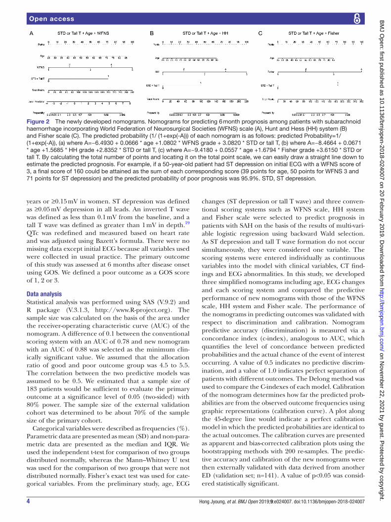
Figure 2 The newly developed nomograms. Nomograms for predicting 6 month prognosis among patients with subarachnoid haemorrhage incorporating World Federation of Neurosurgical Societies (WFNS) scale (A), Hunt and Hess (HH) system (B) and Fisher scale (C). The predicted probability (1/ (1+exp(-A))) of each nomogram is as follows: predicted Probability=1/(1+exp(-A)), (a) where A=−6.4930 + 0.0666 * age +1.0802 * WFNS grade + 3.0820 * STD or tall T, (b) where A=−8.4664 + 0.0671 * age +1.5685 * HH grade +2.8352 * STD or tall T, (c) where A=−9.4180 + 0.0557 * age +1.6794 * Fisher grade +3.6150 * STD or tall T. By calculating the total number of points and locating it on the total point scale, we can easily draw a straight line down to estimate the predicted prognosis. For example, if a 50-year-old patient had ST depression on initial ECG with a WFNS score of 3, a final score of 160 could be attained as the sum of each corresponding score (39 points for age, 50 points for WFNS 3 and 71 points for ST depression) and the predicted probability of poor prognosis was 95.9%. STD, ST depression.
on Novem
ber 22, 2021 by guest. Protected by copyright.
http://bmjopen.bm
j.com/
BM
J Open: first published as 10.1136/bm
jopen-2018-024007 on 20 February 2019. D
ownloaded from

5Hong Jyoung, et al. BMJ Open 2019;9:e024007. doi:10.1136/bmjopen-2018-024007
Open access
resultsstudy populationA total of 665 patients with SAH were initially enrolled in the present study during the study period. Most patients (n=411; 61.8%) were excluded because they were referred from or to other hospitals. We also excluded 52 patients for the following reasons: age <19 years (n=12), no initial ECG (n=3), cardiac arrest on arrival (n=6) and SAH due to trauma (n=31). Finally, 202 patients were included in the primary analysis (figure 1). The baseline characteristics and ECG changes related to prognosis are summarised in table 1. Of the 202 patients, 111 (54.9%) had a good outcome with a GOS of 4 or 5.
eCG changes of patients with sAhOf the 202 patients, 79 (39.1%) had ECG changes, which included ST elevation in 17 (8.4%), ST depression in 31 (15.3%), T wave inversion in 21 (10.4%) and tall T wave in 10 (5.0%) patients. ST depression and tall T waves were detected more often in patients with a poor outcome than in those with a good outcome (29 patients (31.9%) vs two patients (1.8%); p<0.001 and 9 patients (9.9%) vs. one patient (0.9%); p=0.006, respectively).The QTc of patients with a poor outcome was significantly longer
than that of patients with a good outcome (479.1 (53.0) s vs 458.1 (39.1) s; p=0.002).
nomograms for the prediction of prognosisIn this study, we established three nomograms incor-porating each scoring system (figure 2).The Harrell’s C-index for the new nomogram including the WFNS scale to predict prognosis of patients with SAH (0.912; 95% CI, 0.871 to 0.954) was significantly higher than that of the WFNS scale (0.813; 95% CI, 0.758 to 0.868; p<0.001). In addition, the C-indexes of the two nomograms including the HH system and Fisher scale were greater than those of the HH system and Fisher scale (0.913 (0.872 to 0.955) vs 0.826 (0.772 to 0.879); p<0.001 and 0.885 (0.839 to 0.931) vs 0.746 (0.687 to 0.805); p<0.001, respectively). There was no significant difference in the predictive accuracy of the three newly established models (p=0.350). The cali-bration plots presented an excellent agreement between predicted and observed probabilities of the 6 month prognosis and exhibited a close approximation between the probabilities (figure 3).
external validation of the nomogramsThe new nomograms were externally validated using the independent dataset (figure 1) listed in table 2. The
Figure 3 Performance of the nomograms incorporating the World Federation of Neurosurgical Societies scale (A), Hunt and Hess system (B) and Fisher scale (C). Receiver-operating characteristic curves of the nomograms are on the top line. The discrimination ability of the newly developed nomograms was good, with an area under the receiver-operating characteristic curve (AUC) value of 0.912 (95% CI, 0.871 to 0.954), 0.913 (95% CI, 0.872 to 0.955) and 0.885 (95% CI, 0.839 to 0.931), respectively. The calibration curves are on the bottom line. All calibration plots (dotted lines) show close approximation to the logistic calibration (solid lines), indicating excellent agreement between the predicted and observed probabilities of the 6 month prognosis. HH, Hunt and Hess; STD, ST depression; WFNS, World Federation of Neurosurgical Societies.
on Novem
ber 22, 2021 by guest. Protected by copyright.
http://bmjopen.bm
j.com/
BM
J Open: first published as 10.1136/bm
jopen-2018-024007 on 20 February 2019. D
ownloaded from

6 Hong Jyoung, et al. BMJ Open 2019;9:e024007. doi:10.1136/bmjopen-2018-024007
Open access
C-indexes of nomograms including the WFNS scale, HH systems and Fisher scale were 0.809 (0.735–0.884), 0.812 (0.737–0.886) and 0.772 (0.691–0.852), respectively. The calibration plots presented an acceptable agreement in the external validation cohort between the nomogram prediction and actual observation (figure 4).
DIsCussIOnIn this study, we developed and externally validated new nomograms using three independent variables to determine the 6 month prognosis in patients with SAH admitted to the ED. The three independent variables were age, ECG changes, including ST depression or tall T wave formation, and conventional scoring systems, such as the WFNS scale, HH systems and Fisher scale. The new nomograms demonstrated a significantly higher predic-tive accuracy than conventional scoring systems.
The association between ECG changes and prog-nosis has been previously studied; however, the associa-tion between abnormal ECG and prognosis prediction remains controversial. We previously investigated whether the ECG could be used to evaluate patient prognosis. The most frequent ECG change associated with SAH is QTc prolongation.12 17 20 22 In 2012, Huang et al investi-gated whether early ECG abnormalities recorded in the ED were associated with in-hospital mortality among patients with SAH. QTc prolongation was found to be independently associated with in-hospital mortality.17 Another study also demonstrated the association between in-hospital mortality and QTc prolongation.22 However, the underlying mechanism for this finding remains unclear. In our previous study, QTc prolongation was associated with poor prognosis and mortality (survivor vs.
Table 2 Clinical characteristics of the external validation group
Good outcome
Poor outcome Total
P value(n=77) (n=64) (n=141)
Age (year) 53.2 (11.1) 59.6 (14.5) 56.2 (13.2) 0.04
ECG changes
STD or tall T 13 (16.9%) 33 (51.6%) 46 (32.6%) <0.001
Scoring systems*
WFNS scales 1 (1~1) 3 (1~4) 1 (1~4) <0.001
HH system 2 (2~2) 3 (2~4) 2 (1~4) <0.001
Fisher grade 3 (3~3) 4 (3~4) 3 (3~4) <0.001
Data are presented as frequencies (%) or mean (SD), unless otherwise indicated.*Median (IQR).HH, Hunt and Hess; STD, ST depression; WFNS, World Federation of Neurosurgical Societies.
Figure 4 External validation of the nomograms incorporating the World Federation of Neurosurgical Societies (WFNS) scale (A), Hunt and Hess system (HH) (B) and Fisher scale (C). Receiver operating characteristic curves of the nomograms are on the top line. The discrimination ability of newly developed nomograms was good, with an area under receiver-operating characteristic curve (AUC) value of 0.809 (95% CI, 0.735 to 0.884), 0.812 (95% CI, 0.737 to 0.886), and 0.772 (95% CI. 0.691 to 0.852), respectively. The calibration curves are on the bottom line. The calibration plots presented good agreement between the nomogram prediction and actual observation. STD, ST depression.
on Novem
ber 22, 2021 by guest. Protected by copyright.
http://bmjopen.bm
j.com/
BM
J Open: first published as 10.1136/bm
jopen-2018-024007 on 20 February 2019. D
ownloaded from

7Hong Jyoung, et al. BMJ Open 2019;9:e024007. doi:10.1136/bmjopen-2018-024007
Open access
non-survivor; 464.9±46.4 ms vs. 486.0±47.5 ms, p=0.04). However, multi-variable logistic analysis revealed that QTc prolongation was not independently associated with poor prognosis in patients with SAH.20 Other ECG changes associated with poor prognosis in patients with SAH are ST depression and non-specific ST segment changes (NSSTC).17 A sudden increase in intracranial pressure compresses the brain, triggering a sympathetic discharge. This creates a relative ischaemic state of the myocardium and causes ischaemic changes on the ECG. ST depression and NSSTC were found to be significantly associated with mortality or poor neurologic outcome.17 22 Furthermore, the combination of tall T waves, tall P waves, large U waves and prolonged QT has been associated with increased mortality.23 In our study, ST depression or tall T waves were also independently associated with poor prognosis.
In addition to ECG changes, reports have suggested that other factors may be associated with prognosis among patients with SAH.8 11 24–26 In our study, patient age was significantly associated with prognosis. Age was found to be a major independent prognostic factor in many studies.24–26 This could be explained by the fact that the aged brain may have less ability to recover from initial bleeding. Increased initial bleeding among aged patients also explains the poor outcomes.24
Classification and scoring systems to predict the prognosis of patients with SAH have been developed since 1956. The most broadly used systems are the HH system and WFNS scale.7 The HH system divides disease status into five grades based on patient symptoms and level of consciousness.10 Although this system is a good reflection of patient prog-nosis, the demarcation between each grade is ambiguous because of less clearly defined scales of consciousness.7 To overcome this shortcoming, the WFNS scale uses the GCS as the prognostic predictor and the neurologic deficit was added to the differentiation between WFNS grades 2 and 3. However, there was occasional overlap between grades 2 and 3 and the predicted outcomes may not differ substan-tially.27 28 To reduce the ambiguity between WFNS grades 2 and 3, a modified WFNS scale was developed.9 However, the modified WFNS scale failed to show any significant prognostic differences between grades 3 and 4.23
The association between radiological findings and patient outcome has been previously reported.7 In 1980, Fisher et al developed a scale assigning a grade according to the pattern of blood demonstrated on the initial CT scan for the prediction of cerebral vasospasm after SAH.6 Although it was designed to predict vasospasm, the predic-tive value of patient outcome has also been reported. However, the Fisher scale was designed when radiolog-ical technology resolution was only 10% of the current resolution. In the clinical context, it is uncommon for blood clots less than 1 mm in true thickness to occur in the subarachnoid space and to have no blood visible on the initial CT scan. Therefore, Fisher grades 1 and 2 were uncommon.7
Several advances, including the refinement of neurosur-gical techniques, have taken place in SAH management
since these scales were developed. Furthermore, reports found that the demarcation of the grades using these scales was ambiguous. To overcome these issues, we devel-oped simple models by adding age and ECG changes to the existing models. The nomogram can generate an individual probability of a clinical event, such as mortality, by integrating prognostic variables.21 With this advantage, a nomogram is being utilised to predict disease prognosis in different fields. Our nomograms used only three prog-nostic factors, such as existing scoring systems, age and ECG changes. The existing scoring systems are widely used. Age and ECG changes are easily and readily obtain-able during the patient’s admission at the ED. Our nomo-grams were the first approach to combine ECG changes and other prognostic factors in patients with SAH. This combination approach had more accurate predictive power than those of conventional scoring system alone, and there was excellent agreement between predicted and observed probabilities of 6 month prognosis.
Our study has several limitations. First, this study was a retrospective analysis; therefore, the results could differ from those of other centres, and the predictive probability could be overestimated more than in a prospective study. Second, we excluded patients referred from or to other hospitals, and this raises the possibility of selection bias. However, the patients were excluded because initial ECG could not be obtained or patients’ prognosis could not be evaluated. Finally, interval from the onset of symptoms to arrival at the ED differs for each patient. ECG changes may not have been visible when patients presented late to the ED. We need to assess the applicability of our new nomograms in future prospective studies and validate them in a multi-centre study.
COnClusIOnsWe developed new nomograms using only three indepen-dent variables: ECG changes including ST depression or tall T wave formation, age and widely used conventional scoring systems, such as the WFNS scale, HH systems and Fisher scale. Our new nomograms are valuable in predicting the 6 month prognosis of patients with SAH at an early stage after ED admission. Our new models are superior to the WFNS scale, HH systems and Fisher scale in predicting prognosis and are readily available.
Author affiliations1Emergency Medicine, Yonsei University College of Medicine, Seoul, Republic of Korea2Biostatistics, Yonsei University College of Medicine, Seoul, Republic of Korea
Contributors YSP designed this study with JYH, ICP and SPC. JYH, JSY and MJK contributed to the data acquisition. HSL and YSP performed the data analysis. JYH and YSP drafted the manuscript, and all other authors read and approved the final manuscript.
Competing interests None declared.
Patient consent for publication Not required.
ethics approval This study was approved by the Institutional Review Board of our hospital (no. 4-2015-0345).
on Novem
ber 22, 2021 by guest. Protected by copyright.
http://bmjopen.bm
j.com/
BM
J Open: first published as 10.1136/bm
jopen-2018-024007 on 20 February 2019. D
ownloaded from

8 Hong Jyoung, et al. BMJ Open 2019;9:e024007. doi:10.1136/bmjopen-2018-024007
Open access
Provenance and peer review Not commissioned; externally peer reviewed.
Data sharing statement The datasets used and/or analysed during the current study are available from the corresponding author on reasonable request.
Open access This is an open access article distributed in accordance with the Creative Commons Attribution Non Commercial (CC BY-NC 4.0) license, which permits others to distribute, remix, adapt, build upon this work non-commercially, and license their derivative works on different terms, provided the original work is properly cited, appropriate credit is given, any changes made indicated, and the use is non-commercial. See: http:// creativecommons. org/ licenses/ by- nc/ 4. 0/.
referenCes 1. de Rooij NK, Linn FH, van der Plas JA, et al. Incidence of
subarachnoid haemorrhage: a systematic review with emphasis on region, age, gender and time trends. J Neurol Neurosurg Psychiatry 2007;78:1365–72.
2. Okon M, Adebobola NI, Julius S, et al. Stroke incidence and case fatality rate in an urban population. J Stroke Cerebrovasc Dis 2015;24:771–7.
3. Zacharia BE, Hickman ZL, Grobelny BT, et al. Epidemiology of aneurysmal subarachnoid hemorrhage. Neurosurg Clin N Am 2010;21:221–33.
4. Botterell EH, Lougheed WM, Scott JW, et al. Hypothermia, and interruption of carotid, or carotid and vertebral circulation, in the surgical management of intracranial aneurysms. J Neurosurg 1956;13:1–42.
5. Report of World Federation of Neurological Surgeons Committee on a Universal Subarachnoid Hemorrhage Grading Scale. J Neurosurg 1988;68:985–6.
6. Fisher CM, Kistler JP, Davis JM. Relation of cerebral vasospasm to subarachnoid hemorrhage visualized by computerized tomographic scanning. Neurosurgery 1980;6:1–9.
7. Rosen DS, Macdonald RL. Subarachnoid hemorrhage grading scales: a systematic review. Neurocrit Care 2005;2:110–8.
8. Germanson TP, Lanzino G, Kongable GL, et al. Risk classification after aneurysmal subarachnoid hemorrhage. Surg Neurol 1998;49:155–61.
9. Sano H, Satoh A, Murayama Y, et al. Modified World Federation of Neurosurgical Societies subarachnoid hemorrhage grading system. World Neurosurg 2015;83:801–7.
10. Hunt WE, Hess RM. Surgical risk as related to time of intervention in the repair of intracranial aneurysms. J Neurosurg 1968;28:14–20.
11. Wan A, Jaja BNR, Schweizer TA, et al. Clinical characteristics and outcome of aneurysmal subarachnoid hemorrhage with intracerebral hematoma. J Neurosurg 2016:1344–51.
12. Davis TP, Alexander J, Lesch M. Electrocardiographic changes associated with acute cerebrovascular disease: a clinical review. Prog Cardiovasc Dis 1993;36:245–60.
13. Katsanos AH, Korantzopoulos P, Tsivgoulis G, et al. Electrocardiographic abnormalities and cardiac arrhythmias in structural brain lesions. Int J Cardiol 2013;167:328–34.
14. Sommargren CE. Electrocardiographic abnormalities in patients with subarachnoid hemorrhage. Am J Crit Care 2002;11:48–56.
15. Fodstad H, Kelly PJ, Buchfelder M. History of the cushing reflex. Neurosurgery 2006;59:1132–7. discussion 37.
16. Kawahara E, Ikeda S, Miyahara Y, et al. Role of autonomic nervous dysfunction in electrocardio-graphic abnormalities and cardiac injury in patients with acute subarachnoid hemorrhage. Circ J 2003;67:753–6.
17. Huang CC, Huang CH, Kuo HY, et al. The 12-lead electrocardiogram in patients with subarachnoid hemorrhage: early risk prognostication. Am J Emerg Med 2012;30:732–6.
18. Kawasaki T, Azuma A, Sawada T, et al. Electrocardiographic score as a predictor of mortality after subarachnoid hemorrhage. Circ J 2002;66:567–70.
19. Schuiling WJ, Algra A, de Weerd AW, et al. ECG abnormalities in predicting secondary cerebral ischemia after subarachnoid haemorrhage. Acta Neurochir 2006;148:853–8.
20. Hong JY, You JS, Kim MJ, et al. Prognostic factor analysis including electrocardiogram change in patients with subarachnoid hemorrhage. Journal of The Korean Society of Emergency Medicine 2017;28:62–70.
21. Balachandran VP, Gonen M, Smith JJ, et al. Nomograms in oncology: more than meets the eye. Lancet Oncol 2015;16:e173–e180.
22. Zhang L, Qi S. Electrocardiographic abnormalities predict adverse clinical outcomes in patients with subarachnoid hemorrhage. J Stroke Cerebrovasc Dis 2016;25:2653–9.
23. Tjahjadi M, Hernesniemi J. Aneurysmal subarachnoid hemorrhage grading scales: something old, something borrowed, something new, and silver sixpence in our shoes!. World Neurosurg 2015;83:1037–8.
24. Lagares A, Gómez PA, Lobato RD, et al. Prognostic factors on hospital admission after spontaneous subarachnoid haemorrhage. Acta Neurochir 2001;143:665–72.
25. Naval NS, Kowalski RG, Chang TR, et al. The SAH Score: a comprehensive communication tool. J Stroke Cerebrovasc Dis 2014;23:902–9.
26. Rosengart AJ, Schultheiss KE, Tolentino J, et al. Prognostic factors for outcome in patients with aneurysmal subarachnoid hemorrhage. Stroke 2007;38:2315–21.
27. Hirai S, Ono J, Yamaura A. Clinical grading and outcome after early surgery in aneurysmal subarachnoid hemorrhage. Neurosurgery 1996;39:441–6.
28. Gotoh O, Tamura A, Yasui N, et al. Glasgow Coma Scale in the prediction of outcome after early aneurysm surgery. Neurosurgery 1996;39:19–25.
on Novem
ber 22, 2021 by guest. Protected by copyright.
http://bmjopen.bm
j.com/
BM
J Open: first published as 10.1136/bm
jopen-2018-024007 on 20 February 2019. D
ownloaded from