optic nerve crush: axonal responses in wild … nerve crush: axonal responses in wild-type and bcl-2...
TRANSCRIPT

Optic Nerve Crush: Axonal Responses in Wild-Type and bcl-2Transgenic Mice
Sabrina Chierzi,1,2 Enrica Strettoi,1 Maria Cristina Cenni,1 and Lamberto Maffei1,2
Istituto di Neurofisiologia del Consiglio Nazionale delle Ricerche, and 2Scuola Normale Superiore, 56127 Pisa, Italy
Retinal ganglion cells of transgenic mice overexpressing theanti-apoptotic protein Bcl-2 in neurons show a dramatic in-crease of survival rate after axotomy. We used this experimentalsystem to test the regenerative potentials of central neuronsafter reduction of nonpermissive environmental factors. Sur-vival of retinal ganglion cells 1 month after intracranial crush ofthe optic nerve was found to be 100% in adult bcl-2 mice and44% in matched wild-type (wt) mice. In the optic nerve, andparticularly at the crush site, fibers regrowing spontaneously orsimply sprouting were absent in both wt and bcl-2 mice. Weattempted to stimulate regeneration implanting in the crushednerves hybridoma cells secreting antibodies that neutralizecentral myelin proteins, shown to inhibit regeneration (IN-1
antibodies) (Caroni and Schwab, 1988). Again, we found thatregeneration of fibers beyond the site of crush was virtuallyabsent in the optic nerves of both wt and bcl-2 mice. However,in bcl-2 animals treated with IN-1 antibodies, fibers showedsprouting in the proximity of the hybridoma implant. Theseresults suggest that neurons overexpressing bcl-2 are capableof surviving axotomy and sprout when faced with an environ-ment in which inhibition of regeneration has been reduced.Nevertheless, extensive regeneration does not occur, possiblybecause other factors act by preventing it.
Key words: survival; axotomy; regeneration; sprouting; bcl-2;myelin
In the adult CNS of mammals, regeneration of transected axonsis usually absent. The main reasons are usually ascribed to themassive death of damaged neurons and to the inhibitory roleexerted by CNS.
The optic nerve is a suitable CNS model to approach theproblem of regeneration. Its transection leads to the fast andmassive death of axotomized retinal ganglion cells (RGCs)(Villegas-Perez et al., 1988; Berkelaar et al., 1994). Several strat-egies have been followed to increase their survival (Carmignotoet al., 1989; Thanos et al., 1989; Maffei et al., 1990; Mansour-Robaey et al., 1994); recently, we showed the protecting effects ofthe anti-apoptotic gene bcl-2 on axotomized RGCs (Bonfanti etal., 1996; Cenni et al., 1996).
The upregulated expression of bcl-2 prevents neuronal death inseveral in vitro systems (Allsopp et al., 1993; Zhong et al., 1993).The efficacy of bcl-2 in promoting neuronal survival in vivo hasbeen evaluated by means of a transgenic mouse overexpressingthe bcl-2 gene in neurons (Martinou et al., 1994); 2 months afterthe transection of the optic nerve, 63% of retinal ganglion cellssurvive in the bcl-2 mouse, whereas survival in the wild type (wt)is 5%. The proximal segments of injured axons, at least near tothe optic nerve head, is also preserved (Cenni et al., 1996); lightresponses in RGCs are maintained as well (Porciatti et al., 1996).Among the numerous attempts to counteract the process of
ganglion cell death after optic nerve lesion, bcl-2 overexpressionis by far the most successful strategy.
We asked whether axotomized RGCs of bcl-2 transgenic miceare able to regrow their severed fibers in vivo across the lesionsite. This question seemed particularly relevant in view of recentfindings supporting the notion that the bcl-2 gene is implicated inneuronal differentiation (Middleton et al., 1998), rate of axonalelongation (Hilton et al., 1997), and duration of cell cycle steps(Adams and Cory, 1998). In addition, it has been reported thatcultured RGCs from adult bcl-2 mice retain the ability of growingtheir processes on embryonic tectal slices (Chen et al., 1997).
To study regeneration in the optic nerve, the strong hostility ofmature CNS to axonal elongation has to be counteracted. Exper-iments in vivo show that mammalian RGCs are indeed capable ofregenerating their axons through transplants of peripheral nerves(Vidal-Sanz et al., 1987). Inhibitory molecules have been identi-fied that arrest growth cone progression; in particular, a 250 kDaprotein fraction, purified from CNS myelin, exerts a powerfulantagonistic effect on axonal growth; the monoclonal antibodyIN-1, raised against this fraction (Caroni and Schwab, 1988), iscapable of effectively counteracting the inhibitory action of CNSmyelin, allowing neuronal regeneration in many different CNSareas (for review, see Brosamle and Schwab, 1996).
In this study, we made use of the IN-1 antibody to create a lesshostile environment for fiber regrowth; we crushed the opticnerve of wt and bcl-2 mice and implanted hybridoma cells secret-ing the IN-1 antibody in the proximity of the damaged nerve. Wereport the effects of such treatment 1 month after crush.
MATERIALS AND METHODSAnimals. Transgenic mice from the NSE73a line, developed by J-CMartinou (Serono, Geneva, Switzerland), were used in the present studyas in previous studies (Cenni et al., 1996; Chierzi et al., 1998). Thesemice overexpress the human protein Bcl-2 in neurons under the controlof neuron-specific enolase. Characteristics of the transgenics are re-
Received Dec. 21, 1998; revised July 14, 1999; accepted July 16, 1999.This work was partially supported by the European Economic Community (Bio-
tech Grant BIO4-CT96–0774) and by the TeleThon Foundation Project 934. We areindebted to Dr. Martin E. Schwab for the IN-1 and HRP hybridoma cells. We thankDr. GianMichele Ratto and Dr. Tommaso Pizzorusso for helpful comments on thismanuscript; Dr. Elena Putignano for performing PCR; and the staff of the Istitutodi Neurofisiologia for technical support.
Drs. Chierzi and Strettoi contributed equally to this work.Correspondence should be addressed to Enrica Strettoi, Istituto di Neurofisiologia
del Consiglio Nazionale delle Ricerche, Via San Zeno 51, 56127 Pisa, Italy.Copyright © 1999 Society for Neuroscience 0270-6474/99/198367-10$05.00/0
The Journal of Neuroscience, October 1, 1999, 19(19):8367–8376

ported in Martinou et al. (1994). Briefly, our colony started in 1994 froman heterozygous bcl-2 male founder of the NSE73a line crossed with afemale of the C57BL6/J strain (The Jackson Laboratory, Nossan, Milan,Italy); the colony was developed by selecting transgenic males fromoffspring and crossing them over with nontransgenic C57BL6/J females;bcl-2 females have a closed vagina and cannot be used for reproduction.The presence of the transgene was detected by PCR on DNA extractedfrom tail tissue of animals. The presence of the human Bcl-2 protein wasoccasionally revealed by immunohistochemistry on retinal tissue usingthe anti-human bcl-2 antibody from Dako (Milan, Italy) (clone 124); highdensity of retinal ganglion cells and large size of the optic nervesconfirmed a typical bcl-2 phenotype. Mice used as controls were C57BL6/J of the same litters but bcl-2-negative. A total number of 36 mice wereused in this study; they ranged in age between 3 and 6 months.
Hybridoma cells. Mouse hybridoma cells producing IN-1 antibody(clone IN-1 C4) and control hybridoma cells producing antibodiesagainst HRP (clone HRP 4HI) were used. Details about clone isolationand culturing conditions are given by Caroni and Schwab (1988). Beforesurgery, cells were thawed and resuspended at a concentration of 50,000cells/ml and grown in culture for 4–5 d in Iscove’s medium supplementedwith 6% fetal bovine serum, 2 mM L-glutamine, 100 U/ml penicillin–streptomycin, and 50 mM b-mercaptoethanol. Before implantation, thesupernatants of all the hybridoma cells used were tested for the produc-tion of mouse IgM (in the case of IN-1 C4) or IgG (in the case of HRP4HI), by using the assay Boehringer Mannheim (Milan, Italy) IsoStrip.IN-1 supernatant was also used to immunostain the NI 250 fraction ofmyelin in the intact mouse optic nerve and tract. The staining, revealedwith anti-mouse FITC secondary antibodies (dilution of 1:200; Sigma,Milan, Italy), was very intense throughout the optic pathway.
Surgery. The animals were deeply anesthetized with Avertin (1.2%tribromoethanol and 2.4% amylene hydrate in distilled water, 2 ml/100gm body weight), and the left optic nerve was crushed mechanically withthin surgical forceps. The crush was performed intracranially, ;3 mmfrom the posterior pole of the eye; special care was taken to avoidmechanical damage to the retinal artery. We used three different exper-imental protocols: simple crush of the optic nerve (six wt and seven bcl-2animals used); crush plus injection of IN-1 hybridoma cells (six wt and sixbcl-2 used); crush of the optic nerve plus injection of HRP antibodies(four wt and three bcl-2 used).
Hybridoma injection. Six wt and six bcl-2 mice received an injection ofIN-1, producing hybridoma cells directly into the lesioned nerve at thesite of the crush operation. Approximately 3–5 3 10 5 cells in 1 ml ofHBSS were injected using a glass pipette pulled to a final tip of 20–30 mmdriven by an oil microinjector. Hybridoma cells were prestained with DiI(2 mg/ml in DMSO per milliliter of HBSS) to allow further recognition.Control experiments were performed injecting in the crushed nerveshybridoma cells producing antibody against HRP.
Histology. Four weeks after surgery, the left eyes of the operatedanimals were injected with 2 ml of 20% neurobiotin tracer (VectorLaboratories, Burlingame, CA) in saline solution. One day after neuro-biotin injection, the animals were perfused transcardially with 4% para-formaldehyde in 0.1 M phosphate buffer; both optic nerves were carefullydissected, rinsed, cryoprotected with 30% sucrose, and flat frozen on acryostat stage. Consecutive frozen sections were cut at 12 mm thicknessand reacted with 1:400 avidin-FITC to visualize neurobiotin. Adjacentsections were immunostained with antibodies against the 200 kDa sub-unit of neurofilaments (clone N52; dilution of 1:200; Sigma) to allowvisualization of the whole fiber population (Berry et al., 1996; Burne etal., 1996). Selected sections were immunostained using the F4/80 ratmonoclonal antibody (dilution of 1:10; Serotec, Oxford, UK) specific formouse microglia and macrophages (Austyn and Gordon, 1981). Second-ary antibodies were tetramethylrhodamine isothiocyanate–anti-mouseIgG (Sigma, dilution 1:50) and Alexa 488-anti rat IgG (dilution of 1:400;Molecular Probes, Eugene, OR), respectively.
The whole series of sections obtained from each nerve was examinedsystematically with an Zeiss (Milan, Italy) Axioplan microscopeequipped with epifluorescence. Representative sections were analyzed indetail with a Leica (Milan, Italy) TCS-NT confocal microscope. Confocalseries were obtained through the whole section thickness (10–12 mm);each focal series was visualized as a projection on single plane.
The optic nerves of two wt and two bcl-2 mice that had received asimple crush were post-fixed in 4% glutaraldehyde in 0.1 M phosphatebuffer for 12 hr. Subsequently, each nerve was dissected transversally infour blocks, from the optic nerve head to the optic chiasm, which wereprocessed separately; one block contained the optic nerve head and
another the crush site, which was readily recognizable under the dissect-ing scope for its yellowish, transparent appearance. Nerve blocks weretreated with 2% osmium tetroxide, stained with 1% uranyl acetate inmaleate buffer, dehydrated in ethanol, and embedded in Epon–Araldite.Semithin sections, 1- to 2-mm-thick, were cut with a diamond knife fromthe blocks containing the optic nerve heads and from those containingthe lesion site, sectioned from its proximal side. Sections were stainedwith Epoxy Tissue Stain (Electron Microscopy Sciences, Fort Washing-ton, PA) and examined with a Zeiss Axioplan photomicroscope.
Survival. The left retinas of three wt and three bcl-2 animals that hadreceived crush of the left optic nerves 4 weeks before, plus the retinas ofthree wt that had received an injection of IN-1 hybridoma cells, werestained as whole mounts with 2 mM ethidium homodimer II (MolecularProbes) and examined at the confocal microscope. The total number ofcells in the ganglion cell layer (GCL) was estimated from counting 16–20regularly spaced 125 3 250 mm fields located in the four retinal quad-rants and multiplying the average cellular densities to the retinal areasmeasured with an image analyzer (Imaging Inc., Ontario, Canada). Thenumber of displaced amacrines and ganglion cells in the intact retinas ofwt and bcl-2 mice were obtained from separate studies (Cenni et al.,1996; Jeon et al., 1998). Survival was estimated by subtracting thenumber of displaced amacrines from the total number of cells found inthe GCL 1 month after surgery; the obtained values were expressed as afraction of the number of GCs of the intact retinas.
Quantification of sprouting. A total of 20 nerves in which fibers werestained with neurobiotin were used to obtain a semiquantitative evalua-tion of axonal sprouting. Animals were distributed in the followinggroups: three wt mice that had received a simple crush (wt-crush); threebcl-2 mice with simple crush (bcl-2 crush); three wt mice with crush plusinjection of HRP-hybridoma cells (wt HRP); three bcl-2 mice with crushplus HRP-hybridoma cells (bcl-2 HRP); three wt mice with crush plusIN-1-hybridoma cells (wt IN-1); and three bcl-2 mice with crush plusIN-1 hybridoma cells (bcl-2 IN-1). For each crushed nerve, representa-tive sections were analyzed at the confocal microscope as describedabove. Images representing projections of nerve sections near the crushsite were transferred on a microcomputer imaging device (Imaging, Inc.)M4 image analyzer. The intensity of fluorescence staining of fibers wasdetermined by means of densitometric analysis at 10 different spots,spaced regularly along two selected lines perpendicular to the major axisof the nerve section. The first line corresponds to the border of thetransected fibers (the virtual edge at which the majority of fibers stops);the second line is taken at a more proximal position (within a distance of300 mm from the first line, toward the optic nerve head). At each location,in each section, we calculated the ratio between the optical density (OD)measured at the first line and the OD at the second line, respectively.This value is referred to as ODR (optical density ratio).
RESULTSEffects of crush lesion in wt and bcl-2 mice 1 monthafter surgeryFigure 1 reports the fractions of surviving retinal ganglion cells inadult wt and bcl-2 mice (three retinas for each strain) 1 monthafter intracranial crush of the optic nerve. In the wt, optic nervecrush results in the survival of approximately half retinal ganglioncells (44%). On the other hand, in the bcl-2 mouse, virtually100% of RGCs are still alive. This confirms the powerfulsurvival-promoting effect of the bcl-2 gene.
Figure 2 shows the effects of crush lesion onto optic fiberpreservation in wt and bcl-2 optic nerves in transversal semithinsections (two nerves examined for each strain). Both optic nervesare illustrated at the emergence from the posterior pole of the eyeand at the crush site, respectively. At the optic nerve head,individual degenerating fibers are visible: however, the overallnerve structure is still well preserved, especially in the bcl-2mouse (Fig. 2A–D). Toward the crush site, fibers with disorga-nized morphology become more evident, in both the wt and thebcl-2 nerve (Fig. 2E,F). Axons have retracted and a central coreof vacuolization, and tissue reorganization has appeared. Highermagnification of the lesion site shows individual dying fibers allover the whole nerve surface; in the bcl-2, normal fibers are still
8368 J. Neurosci., October 1, 1999, 19(19):8367–8376 Chierzi et al. • Optic Nerve Crush in WT and bcl-2 Transgenic Mice

visible, mostly in the superficial ring of the nerve (Fig. 2G,H).When crushed optic nerves are analyzed in longitudinal sectionsafter neurobiotin injection in the corresponding eyes, individualfibers are visualized along their course to the lesion point (Fig. 3).After crush, fibers end at different positions along the longitudinalaxis of the nerve, depending on the variable extent of retraction.We define as “border of the fibers” the line (perpendicular to thelongitudinal axis of the nerve) at which the majority of axonsstops. This line represents one of the boundaries we used fordensitometric analysis (see below). It is important to point outthat, 1 month after crush, there is no spontaneous sprouting of thefibers or elongation beyond the border, in both the wt and bcl-2nerves (four nerves examined in longitudinal serial sections foreach strain of mice).
Effects of IN-1 hybridoma cell injectionsTo create a favorable environment to optic fiber regeneration, weinjected in the optic nerves hybridoma cells producing an anti-body (IN-1) known to bind and neutralize a powerful inhibitor offiber elongation associated with CNS myelin (NI-250).
Injection of 1 ml of suspension containing 3–5 3 105 cells wasperformed in the crushed optic nerves of mice. The supernatantof IN-1 hybridoma cells was tested for the production of mouseIgM before each injection. The actual number of cells injectedcan be expected to vary to some extent, because some of thesuspension was observed to flow out from the nerve after pipettewithdrawal. The successful outcome of the injection was tested atthe time of nerve dissection; hybridoma cells, labeled with DiIcould be visualized with a fluorescence microscope in the intactnerves and are visible in Figures 4 and 5 in longitudinal nervesections. One month after crush, hybridoma cells appeared mostlyconcentrated near the injection site without remarkable migrationinside the nerve. To check whether IN-1 hybridoma injectioncould exert some effect in preventing neuronal cell death, wecomputed the number of resilient ganglion cell in the retinas ofthree wt mice treated with IN-1 antibody. In these animals, RGCsurvival was in the same range (54%) observed in the case of wtmice with a simple crush lesion (Fig. 1). Consequently, in our
system, we can exclude a role of IN-1-producing hybridoma cellsin affecting neuronal survival.
The effects of IN-1 on crushed fibers were evaluated aftersystematic examination of longitudinal sections of optic nervesafter neurobiotin injection in the corresponding eyes. In both wtand bcl-2 (six animals for each strain), we observed that themajority of fibers that reached the crush site interrupted abruptly(Fig. 4A,B). Only in the case of one bcl-2, fibers were seen toregrow for ;500 mm beyond the major border of fibers. Elonga-tion occurred in axons running at the surface of the optic nerve.In all the remaining cases, fibers did not succeed in growing morethan 10–20 mm. We can conclude that, despite of the administra-tion of IN-1 hybridoma cells in the lesion area, we did not observeany significant regeneration.
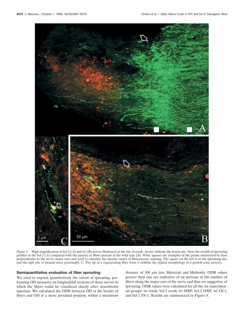
On the other hand, we observed a sprouting promoting effectafter injection of IN-1 hybridoma cells; in all bcl-2 mice, we foundfibers that sprouted at the lesion site. Figure 5A shows the longi-tudinal section of one of these nerves in which sprouting isparticularly profuse. Fibers appear green, and DiI-stained hybrid-oma cells are red. Many axon terminals can be seen at the site ofcrush (indicated by arrows); some of them show large size andintense staining, as is typical of degenerating fibers, although themajority are thin and sprout profusely. Sprouting processes haveoften a tortuous course with ramifications. Similar sproutingprofiles, in variable number and degree of complexity, could beobserved in all the IN-1-treated bcl-2 mice. Immunocytochemis-try with an antibody against the heavy subunit of neurofilamentsshowed that large-sized degenerating fibers were intenselystained, whereas the thinner ones, exhibiting a tortuous courserevealed by neurobiotin injection, were usually not labeled. Theabsence of neurofilament staining in processes with morphologytypical of newly formed fibers confirms that they are sproutingaxonal terminals. It has been shown that the heavy subunit ofneurofilaments is absent from the tips of actively growing axons(Foster et al., 1987).
Figure 4B also shows the result of the injection of IN-1-producing hybridoma cells in crushed nerves of wt mice. As canbe observed at high magnification (Fig. 5B), most of the axons inthe wt end freely at the border of fibers, without obvious ramifi-cations. Retraction is evident. A quantitative analysis of fiberbehavior in various experimental groups is illustrated later andincludes an evaluation of the sprouting effect.
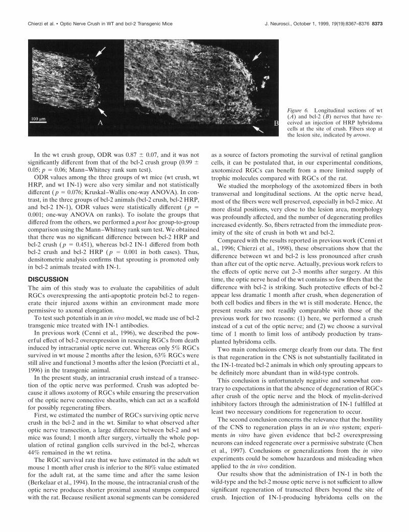
Control experimentsControl experiments were performed injecting in the crushednerves of four bcl-2 and three wild-type mice hybridoma cellsproducing antibodies against HRP; cell injections followed thesame protocol used for IN-1 hybridoma cells. Longitudinal sec-tions of the nerves were examined systematically after neurobi-otin injections in the eyes. Results are shown in Figure 6. Axonsbehaved mostly as in the case of a simple crush; namely, theystopped at the lesion site without sprouting or elongating, in bothwt and bcl-2 animals. Retraction at the lesion site was againvisible. We concluded that the injection of hybridoma cells thatdo not release antibodies interfering with axonal elongation is notsufficient per se to promote sprouting in bcl-2 mice.
In addition, we looked for major differences between wt andbcl-2 in the macrophage–microglia response to crush after hy-bridoma cell injection. Figure 7 shows F4/80 staining in crushednerves of wt and bcl-2 that had received an injection of HRP cells.
Figure 1. Survival rate of retinal ganglion cells 1 month after intracranialcrush of the left optic nerve. Columns are percentage average survivalswith error bars. The first column refers to wild-type retinas (n 5 3) andthe second column to bcl-2 retinas (n 5 3). Notice that survival is 100%in bcl-2 animals. The last column shows the survival rate of ganglion cellsin wt retinas (n 5 3) after injection of IN-1 hybridoma cells at the site ofcrush.
Chierzi et al. • Optic Nerve Crush in WT and bcl-2 Transgenic Mice J. Neurosci., October 1, 1999, 19(19):8367–8376 8369
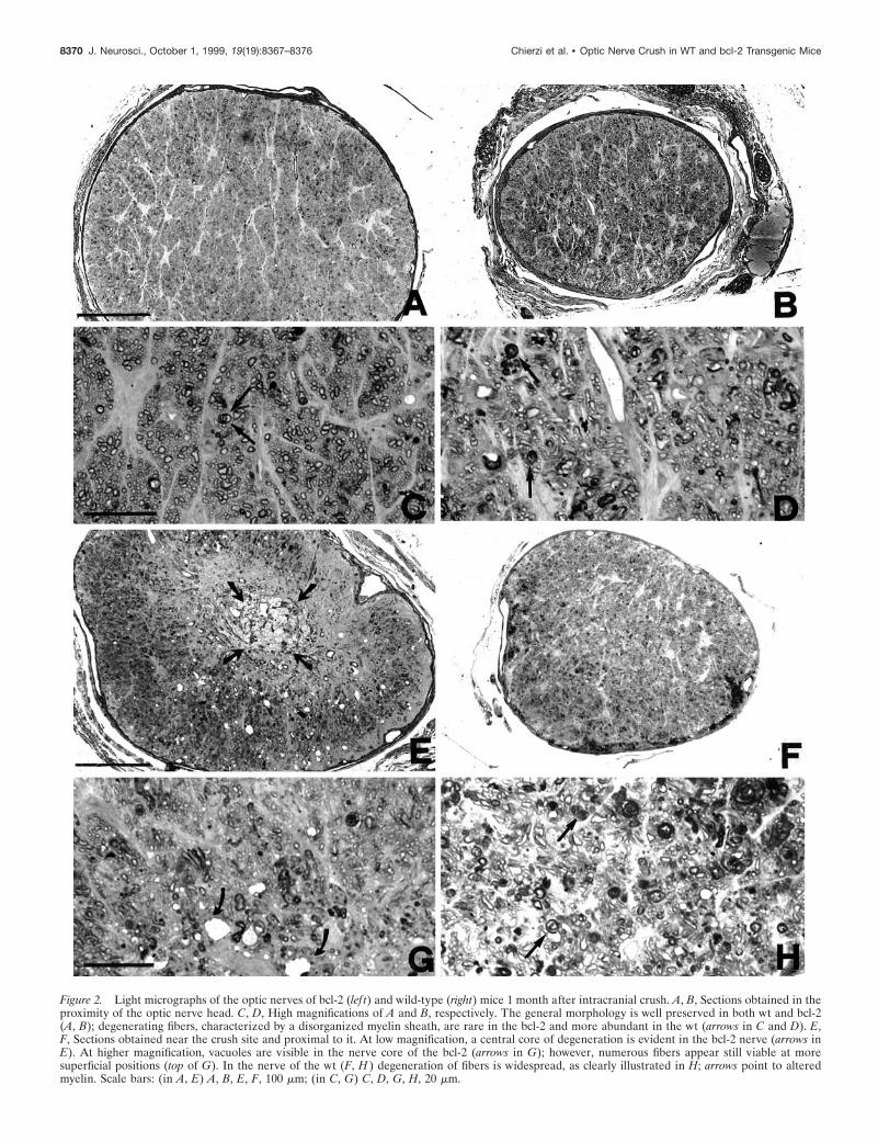
Figure 2. Light micrographs of the optic nerves of bcl-2 (lef t) and wild-type (right) mice 1 month after intracranial crush. A, B, Sections obtained in theproximity of the optic nerve head. C, D, High magnifications of A and B, respectively. The general morphology is well preserved in both wt and bcl-2(A, B); degenerating fibers, characterized by a disorganized myelin sheath, are rare in the bcl-2 and more abundant in the wt (arrows in C and D). E,F, Sections obtained near the crush site and proximal to it. At low magnification, a central core of degeneration is evident in the bcl-2 nerve (arrows inE). At higher magnification, vacuoles are visible in the nerve core of the bcl-2 (arrows in G); however, numerous fibers appear still viable at moresuperficial positions (top of G). In the nerve of the wt (F, H ) degeneration of fibers is widespread, as clearly illustrated in H; arrows point to alteredmyelin. Scale bars: (in A, E) A, B, E, F, 100 mm; (in C, G) C, D, G, H, 20 mm.
8370 J. Neurosci., October 1, 1999, 19(19):8367–8376 Chierzi et al. • Optic Nerve Crush in WT and bcl-2 Transgenic Mice

The staining revealed numerous cells, identifiable as macro-phages because of their large size, scattered all over the nerves,although mostly concentrated at the lesion site. They stained sointensely that small-sized microglial cells were primarily ob-scured. F4/80-positive cells of similar morphology were absentfrom the contralateral, intact nerves in which only microglia waslabeled. There were no evident differences between wt and bcl-2
mice in the F4/80 pattern of immunoreactivity. Thus, we canexpect that macrophage–microglia recruitment follows similarrules in the two cases. However, this issue can be clarified onlyafter a systematic analysis of the immune response after opticnerve crush in the two strains of mice, similar to the study ofLawson et al. (1994). Because this was beyond the scope of thisstudy, we did not make any attempt to go into further details.
Figure 4. Longitudinal sections of crushed optic nerves of bcl-2 (A) and wt (B) mice 1 month after ON crush and implantation of IN-1 hybridoma cells.Fibers are labeled green by neurobiotin injected into the eye and revealed with FITC-avidin. Hybridoma cells, stained with DiI, appear red. Arrows pointto the crush site. C, Staining with F4/80 antibody, specific for mouse microglia and macrophages. Double-labeled cells (short arrow) are immunopositivecells that have engulfed DiI, and the red ones are hybridoma cells (long arrow).
Figure 3. Longitudinal sections of wt(A) and bcl-2 (B) nerves shown near thecrush site (arrows) after neurobiotin in-jection in the corresponding eyes. Thisand the following images have been ob-tained at the confocal microscope as ex-plained in Materials and Methods. Theoptic nerve head is located to the lef t.Note that, in both cases, resilient fibersfail to pass beyond the crush site and donot sprout.
Chierzi et al. • Optic Nerve Crush in WT and bcl-2 Transgenic Mice J. Neurosci., October 1, 1999, 19(19):8367–8376 8371
Semiquantitative evaluation of fiber sproutingWe tried to express quantitatively the extent of sprouting, per-forming OD measures on longitudinal sections of those nerves inwhich the fibers could be visualized clearly after neurobiotininjection. We calculated the ODR between OD at the border offibers and OD at a more proximal position, within a maximum
distance of 300 mm (see Materials and Methods). ODR valuesgreater than one are indicative of an increase in the number offibers along the major axis of the nerve and thus are suggestive ofsprouting. ODR values were calculated for all the six experimen-tal groups: wt crush, bcl-2 crush, wt HRP, bcl-2 HRP, wt IN-1,and bcl-2 IN-1. Results are summarized in Figure 8.
Figure 5. High magnification of bcl-2 (A) and wt (B) nerves illustrated at the site of crush. Arrows indicate the lesion site. Note the wealth of sproutingprofiles in the bcl-2 (A) compared with the paucity of fibers present in the wild type (B). White squares are examples of the points intersected by linesperpendicular to the nerve major axes and used to calculate the density values of fluorescence staining. The square on the lef t is at the sprouting site,and the right one is located more proximally. C, The tip of a regenerating fiber from A exhibits the typical morphology of a growth cone (arrow).
8372 J. Neurosci., October 1, 1999, 19(19):8367–8376 Chierzi et al. • Optic Nerve Crush in WT and bcl-2 Transgenic Mice
In the wt crush group, ODR was 0.87 6 0.07, and it was notsignificantly different from that of the bcl-2 crush group (0.99 60.05; p 5 0.06; Mann–Whitney rank sum test).
ODR values among the three groups of wt mice (wt crush, wtHRP, and wt IN-1) were also very similar and not statisticallydifferent ( p 5 0.076; Kruskal–Wallis one-way ANOVA). In con-trast, in the three groups of bcl-2 animals (bcl-2 crush, bcl-2 HRP,and bcl-2 IN-1), ODR values were statistically different ( p 50.001; one-way ANOVA on ranks). To isolate the groups thatdiffered from the others, we performed a post hoc group-to-groupcomparison using the Mann–Whitney rank sum test. We obtainedthat there was no significant difference between bcl-2 HRP andbcl-2 crush ( p 5 0.451), whereas bcl-2 IN-1 differed from bothbcl-2 crush and bcl-2 HRP ( p 5 0.001 in both cases). Thus,densitometric analysis confirms that sprouting is promoted onlyin bcl-2 animals treated with IN-1.
DISCUSSIONThe aim of this study was to evaluate the capabilities of adultRGCs overexpressing the anti-apoptotic protein bcl-2 to regen-erate their injured axons within an environment made morepermissive to axonal elongation.
To test such potentials in an in vivo model, we made use of bcl-2transgenic mice treated with IN-1 antibodies.
In previous work (Cenni et al., 1996), we described the pow-erful effect of bcl-2 overexpression in rescuing RGCs from deathinduced by intracranial optic nerve cut. Whereas only 5% RGCssurvived in wt mouse 2 months after the lesion, 63% RGCs werestill alive and functional 3 months after the lesion (Porciatti et al.,1996) in the transgenic animal.
In the present study, an intracranial crush instead of a transec-tion of the optic nerve was performed. Crush was adopted be-cause it allows axotomy of RGCs while ensuring the preservationof the optic nerve connective sheaths, which can act as a scaffoldfor possibly regenerating fibers.
First, we estimated the number of RGCs surviving optic nervecrush in the bcl-2 and in the wt. Similar to what observed afteroptic nerve transection, a large difference between bcl-2 and wtmice was found; 1 month after surgery, virtually the whole pop-ulation of retinal ganglion cells survived in the bcl-2, whereas44% remained in the wt retina.
The RGC survival rate that we have estimated in the adult wtmouse 1 month after crush is inferior to the 80% value estimatedfor the adult rat, at the same time and after the same lesion(Berkelaar et al., 1994). In the mouse, the intracranial crush of theoptic nerve produces shorter proximal axonal stumps comparedwith the rat. Because resilient axonal segments can be considered
as a source of factors promoting the survival of retinal ganglioncells, it can be postulated that, in our experimental conditions,axotomized RGCs can benefit from a more limited supply oftrophic molecules compared with RGCs of the rat.
We studied the morphology of the axotomized fibers in bothtransversal and longitudinal sections. At the optic nerve head,most of the fibers were well preserved, especially in bcl-2 mice. Atmore distal positions, very close to the lesion area, morphologywas profoundly affected, and the number of degenerating profilesincreased evidently. So, fibers retracted from the immediate prox-imity of the site of crush in both wt and bcl-2.
Compared with the results reported in previous work (Cenni etal., 1996; Chierzi et al., 1998), these observations show that thedifference between wt and bcl-2 is less pronounced after crushthan after cut of the optic nerve. Actually, previous work refers tothe effects of optic nerve cut 2–3 months after surgery. At thistime, the optic nerve head of the wt contains so few fibers that thedifference with bcl-2 is striking. Such protective effects of bcl-2appear less dramatic 1 month after crush, when degeneration ofboth cell bodies and fibers in the wt is still moderate. Hence, thepresent results are not readily comparable with those of theprevious work for two reasons: (1) here, we performed a crushinstead of a cut of the optic nerve; and (2) we choose a survivaltime of 1 month to limit loss of antibody production by trans-planted hybridoma cells.
Two main conclusions emerge clearly from our data. The firstis that regeneration in the CNS is not substantially facilitated inthe IN-1-treated bcl-2 animals in which only sprouting appears tobe definitely more abundant than in wild-type controls.
This conclusion is unfortunately negative and somewhat con-trary to expectations in that the absence of degeneration of RGCsafter crush of the optic nerve and the block of myelin-derivedinhibitory factors through the administration of IN-1 fulfilled atleast two necessary conditions for regeneration to occur.
The second conclusion concerns the relevance that the hostilityof the CNS to regeneration plays in an in vivo system; experi-ments in vitro have given evidence that bcl-2 overexpressingneurons can indeed regenerate over a permissive substrate (Chenet al., 1997). Conclusions or generalizations from the in vitroexperiments could be somehow hazardous and misleading whenapplied to the in vivo condition.
Our results show that the administration of IN-1 in both thewild-type and the bcl-2 mouse optic nerve is not sufficient to allowsignificant regeneration of transected fibers beyond the site ofcrush. Injection of IN-1-producing hybridoma cells on the
Figure 6. Longitudinal sections of wt(A) and bcl-2 (B) nerves that have re-ceived an injection of HRP hybridomacells at the site of crush. Fibers stop atthe lesion site, indicated by arrows.
Chierzi et al. • Optic Nerve Crush in WT and bcl-2 Transgenic Mice J. Neurosci., October 1, 1999, 19(19):8367–8376 8373

crushed nerve has been used by Weibel et al. (1994) to induceregeneration of transected RGC axons. They implanted IN-1-producing hybridoma cells over the crushed optic nerves of ratsthat had received FGF into the vitreous and observed long-distance regeneration of few axotomized fibers. However, theexperimental design of Weibel et al. differs from the one used inthe present study in some relevant aspects; they injected fibroblastgrowth factor to prevent RGC death and axonal degeneration,whereas in our experimental protocol, both these effects wereprevented by overexpression of bcl-2. Furthermore, the animalsused were relatively immature (16- to 18-d-old), whereas we usedadult mice. This difference might be relevant in the amount ofregeneration occurring afterward, because it is well known that
lesioned fibers of the immature CNS show a more pronouncedattitude to sprouting and regeneration compared with fibers ofthe mature CNS (Bates and Stelzner, 1993).
The possibility exists that the concentration reached by theIN-1 antibody was not high enough to counteract the inhibitoryaction of myelin, although we used relatively large amounts ofhybridoma cells directly at the crush site. It is well known thateven very low concentrations of myelin can arrest growth coneprogression in vitro. Because IN-1 hybridoma cells did not mi-grate in the distal stump of the optic nerves in which degeneratingmyelin persists for a long time, we can be confident that the effectsof myelin inhibitors have been counteracted within the immediateproximity of the crush site. Indeed, this is confirmed by the
Figure 7. F4/80 staining in crushed nerves of wt (A) and bcl-2 (B) that had received an injection of HRP cells. The crush site is indicated by arrows.Stained cells are dense at the lesion site; C and D (enlargements of the fields shown as rectangles in A and B) illustrate large size cells having a roundmorphology, typical of macrophages. Notice that there are not obvious differences in the staining between A and B.
8374 J. Neurosci., October 1, 1999, 19(19):8367–8376 Chierzi et al. • Optic Nerve Crush in WT and bcl-2 Transgenic Mice

observation that sprouting was promoted by IN-1 hybridoma cellsin bcl-2.
In addition, the failure of the majority of axons to regenerate inthe IN-1-treated bcl-2 mice can be explained by postulating thatother inhibitory factors in addition to myelin proteins are presentat the lesion site.
In particular, the formation of a scar, a process involvingdifferent cell types of the nervous and immune systems (activatedastrocytes, activated microglia, meningeal cells, and inflamma-tory macrophages and leukocytes) is believed to play an impor-tant role in creating a hostile environment to regeneration. In-deed, we observed macrophage concentration at the crush siteafter hybridoma injection, in both wt and bcl-2. In the crushedoptic nerve, the scar is extending across the whole nerve thick-ness; regenerating fibers have to grow through a region of greatlymodified and rearranged tissue, because no bridges of intact opticnerve remain for their passage. Different lines of evidence havepointed to reactive astrocytes of the scar as powerful elementsimpeding axonal regrowth (McKeon et al., 1995; Davies et al.,1997). Reaction of astrocytes to injury results in morphological
hypertrophy and in the synthesis and secretion in the extracellu-lar matrix of inhibitory molecules, such as the proteoglycan chon-droitin sulfate (McKeon et al., 1995). Reactive astrocytes takenfrom the crushed optic nerves of rats represent a nonpermissivesubstrate for the elongation of CNS axons (Bahr et al., 1995).
Even if in our experimental model the axons failed to elongatebeyond the crush site, in bcl-2 mice injected with IN-1-producinghybridoma cells, we observed axonal sprouting at the border oftransected fibers. We tried to express quantitatively the extent ofsprouting observed in the different experimental cases calculatingoptical density ratios at the lesion site for different experimentalprotocols. We find that, in wt animals, no treatment is able toincrease significantly ODRs, whereas in bcl-2 mice, the injectionof IN-1-producing hybridoma cells induces a significant increasein ODRs, indicating an effect in promoting sprouting.
It appears that the administration of IN-1 antibodies to thecrushed optic nerve can unmask an effect of bcl-2 in eliciting thesprouting of transected fibers.
These results suggest a correlation between bcl-2 overexpres-sion and axonal growth in an adult animal. Such effect of bcl-2,distinct from the well known survival effect, is in agreement withsome recent findings: embryonic retinal ganglion cells (embryonicday 18) from bcl-2 mice have been shown to retain elongationcapabilities typical of earlier stages of development, which areabsent in RGCs of wt mice of the same age (Chen et al., 1997).In addition, neurons transfected with the bcl-2 gene show anincrease in the outgrowth of neuritic processes (Zhang et al.,1996; Middleton et al., 1998). Our results also correlate withrecent studies on the expression of the endogenous bcl-2 gene inprimate brain: the Bcl-2 protein, expressed diffusely during braindevelopment, becomes restricted in adult life to distinct structuresof the CNS, characterized by persistent plasticity (Bernier andParent, 1998).
Although the mechanisms of action of bcl-2 are not well clar-ified, some hypothesis can be formulated to explain a possiblepermissive action of the transgene overexpression on axonalsprouting, in the presence of IN-1. It is known that the effect ofthe CNS myelin protein NI 250 (inhibited by IN-1 antibodies) inmediating growth cone collapse is associated with a large increasein cytosolic calcium released from intracellular stores. The ad-ministration of IN-1 antibodies inhibits growth cone collapse andprevents the increase in cytosolic calcium (Bandtlow et al., 1993).The Bcl-2 protein is involved in the control of the calciumhomeostasis as well; the protein, localized on the membrane ofintracellular organelles, such as endoplasmic reticulum, nucleus,and mitochondria, is capable of preventing Ca efflux from suchstores to the cytoplasm in response to apoptotic stimuli (Lam,1994; Guo et al., 1997; Miller, 1998). Thus, it is conceivable thatIN-1 administration and bcl-2 overexpression can act synergisti-cally, both limiting the increase of calcium levels associated withgrowth cone arrest. When IN-1 antibodies alleviate the stronginhibition exerted by myelin, bcl-2 fibers are able to sprout.
REFERENCESAdams JM, Cory S (1998) The bcl-2 protein family: arbiters of cell
survival. Science 281:1322–1326.Allsopp TE, Wyatt S, Patterson HF, Davies AM (1993) The proto-
oncogene bcl-2 can selectively rescue neurotrophic factor-dependentneurons from apoptosis. Cell 73:295–307.
Austyn JM, Gordon S (1981) F4/80, a monoclonal antibody directedspecifically against the mouse macrophage. Eur J Immunol 11:805–815.
Bahr M, Przyrembel C, Bastmeyer M (1995) Astrocytes from adult ratoptic nerves are nonpermissive for regenerating retinal ganglion cellaxons. Exp Neurol 131:211–220.
Figure 8. Semiquantitative analysis of fiber responses in different exper-imental conditions. Top shows the wt groups; bottom shows the bcl-2.Columns represent average OD ratios with standard errors. The asteriskpoints out the significant difference among the bcl-2 IN-1 group and theremaining two.
Chierzi et al. • Optic Nerve Crush in WT and bcl-2 Transgenic Mice J. Neurosci., October 1, 1999, 19(19):8367–8376 8375

Bandtlow CE, Schmidt MF, Hassinger TD, Schwab ME, Kater SB (1993)Role of intracellular calcium in NI-35-evoked collapse of neuronalgrowth cones. Science 259:80–83.
Bates CA, Stelzner DJ (1993) Extension and regeneration of corticospi-nal axons after early spinal injury and the maintenance of corticospinaltopography. Exp Neurol 123:106–117.
Berkelaar M, Clarke DB, Wang Y-C, Bray GM, Aguayo AJ (1994)Axotomy results in delayed death and apoptosis of retinal ganglion cellsin adult rats. J Neurosci 14:4368–4374.
Bernier PJ, Parent A (1998) Bcl-2 protein as a marker of neuronalimmaturity in postnatal primate brain. J Neurosci 18: 2486–2497.
Berry M, Carlile J, Hunter H (1996) Peripheral nerve explants graftedinto the vitreous body of the eye promote the regeneration of retinalganglion cell axons severed in the optic nerve. J Neurocytol 25:147–170.
Bonfanti L, Strettoi E, Chierzi S, Cenni MC, Liu X-H, Martinou J-C,Maffei L, Rabacchi SA (1996) Protection of retinal ganglion cells fromnatural and axotomy-induced cell death in neonatal transgenic miceoverexpressing bcl-2. J Neurosci 16:4186–4194.
Brosamle C, Schwab ME (1996) Axonal regeneration in the mammalianCNS. Semin Neurosci 8:107–113.
Burne JF, Staple JK, Raff M (1996) Glial cells are increased proportion-ally in transgenic optic nerves with increased numbers of axons. J Neu-rosci 16:2064–2073.
Carmignoto G, Maffei L, Candeo P, Canella R, Comelli C (1989) Ef-fects of NGF on the survival of rat retinal ganglion cells following opticnerve section. J Neurosci 9:1263–1272.
Caroni P, Schwab ME (1988) Antibody against myelin-associated inhib-itor of neurite growth neutralizes nonpermissive substrate properties ofCNS white matter. Neuron 1:85–96.
Cenni MC, Bonfanti L, Martinou J-C, Ratto GM, Strettoi E, Maffei L(1996) Long-term survival of retinal ganglion cells following opticnerve section in adult bcl-2 transgenic mice. Eur J Neurosci8:1735–1745.
Chen DF, Schneider GE, Martinou J-C, Tonegawa S (1997) bcl-2 pro-motes regeneration of severed axons in mammalian CNS. Nature385:434–439.
Chierzi S, Cenni MC, Maffei L, Pizzorusso T, Porciatti V, Ratto GM,Strettoi E (1998) Protection of retinal ganglion cells and preservationof function after optic nerve lesion in bcl-2 transgenic mice. Vision Res38:1537–1543.
Davies SJA, Fitch MT, Memberg SP, Hall AK, Raisman G, Silver J(1997) Regeneration of adult axons in white matter tracts of the centralnervous system. Nature 390:680–683.
Foster GA, Dahl D, Lee VMY (1987) Temporal and topographic rela-tionship between the phosphorylated and nonphosphorylated epitopesof the 200 kDa neurofilament protein during development in vitro.J Neurosci 7:2651–2663.
Guo Q, Sopher BL, Furukawa K, Pham DG, Robinson N, Martin GM,Mattson MP (1997) Alzheimer’s presenilin mutation sensitizes neuralcells to apoptosis induced by trophic factor withdrawal and amyloid pep-tide: involvement of calcium and oxyradicals. J Neurosci 17:4212–4222.
Hilton M, Middleton G, Davies AM (1997) Bcl-2 influences axonalgrowth rate in embryonic sensory neurons. Curr Biol 7:798–800.
Jeon CJ, Strettoi E, Masland RH (1998) the major cell populations ofthe mouse retina. J Neurosci 18:8936–8946.
Lam M, Dubyak G, Chen L, Nunez G, Miesfeld RL, Distelhorst CW(1994) Evidence that Bcl-2 represses apoptosis by regulating endoplas-mic reticulum-associated Ca 21 fluxes. Proc Natl Acad Sci USA91:6569–6573.
Lawson LJ, Frost L, Risbridger J, Fearn S, Perry VH (1994) Quantifi-cation of the mononuclear phagocyte response to Wallerian degener-ation of the optic nerve. J Neurocytol 23:729–744.
Maffei L, Carmignoto G, Perry VH, Candeo P, Ferrari G (1990)Schwann cells promote the survival of rat retinal ganglion cells afteroptic nerve section. Proc Natl Acad Sci USA 87:1855–1859.
Mansour-Robaey S, Clarke DB, Wang YC, Bray GM, Aguayo AJ (1994)Effects of ocular injury and administration of brain-derived neurotro-phic factor on survival and regrowth of axotomized retinal ganglioncells. Proc Natl Acad Sci USA 91:1632–1636.
Martinou J-C, Dubois-Dauphin M, Staple J, Rodriguez I, Frankowski H,Missoten M, Albertini P, Talabot D, Catsicas S, Pietra C, Huarte J(1994) Overexpression of bcl-2 in transgenic mice protects neuronsfrom naturally occurring cell death and experimental ischemia. Neuron13:1017–1030.
McKeon RJ, Hoke A, Silver J (1995) Injury-induced proteoglycans in-hibit the potential for laminin-mediated axon growth on astrocyticscars. Exp Neurol 136:32–43.
Middleton G, Pin LGP, Wyatt S, Davies AM (1998) Bcl-2 accelerates thematuration of early sensory neurons. J Neurosci 18:3344–3350.
Miller RJ (1998) Mitochondria—the Kraken wakes! Trends Neurosci21:95–97.
Porciatti V, Pizzorusso T, Cenni MC, Maffei L (1996) The visual re-sponse of retinal ganglion cells is not altered by optic nerve transectionin transgenic mice overexpressing bcl-2. Proc Natl Acad Sci USA93:14955–14959.
Thanos S, Bahr M, Barde Y-A, Vanselow J (1989) Survival and axonalelongation of adult retinal ganglion cells. In vitro effects of lesionedsciatic nerve and brain-derived neurotrophic factor. Eur J Neurosci1:19–26.
Vidal-Sanz M, Bray GM, Villegas-Perez MP, Thanos S, Aguayo AT(1987) Axonal regeneration and synapse formation in the superiorcolliculus by retinal ganglion cells in the adult rat. J Neurosci7:2894–2909.
Villegas-Perez MP, Vidal-Sanz M, Bray GM, Aguayo AJ (1988) Influ-ences of peripheral nerve grafts on the survival and regrowth ofaxotomized retinal ganglion cells in adult rats. J Neurosci 8:265–280.
Weibel D, Cadelli D, Schwab ME (1994) Regeneration of lesioned ratoptic nerve fibers is improved after neutralization of myelin-associatedneurite growth inhibitors. Brain Res 642:259–266.
Zhang KZ, Westberg JA, Holtta E, Andersson LC (1996) Bcl-2 regu-lates neuronal differentiation. Proc Natl Acad Sci USA 93:4504–4508.
Zhong LT, Sarafian T, Kane D, Charles A, Mah S, Edwards R, BredsenD (1993) Bcl-2 inhibits death of central neural cells induced by mul-tiple agents. Proc Natl Acad Sci USA 90:4533–4537.
8376 J. Neurosci., October 1, 1999, 19(19):8367–8376 Chierzi et al. • Optic Nerve Crush in WT and bcl-2 Transgenic Mice