optically active, amphiphilic poly(meta-phenylene ... jacs... · optically active, amphiphilic...
TRANSCRIPT
Optically Active, Amphiphilic Poly(meta-phenylene ethynylene)s:Synthesis, Hydrogen-Bonding Enforced Helix Stability, and DirectAFM Observation of Their Helical StructuresMotonori Banno,† Tomoko Yamaguchi,† Kanji Nagai,†,§ Christian Kaiser,‡ Stefan Hecht,*,‡
and Eiji Yashima*,†
†Department of Molecular Design and Engineering, Graduate School of Engineering, Nagoya University, Chikusa-ku, Nagoya,464-8603, Japan‡Department of Chemistry, Humboldt-Universitat zu Berlin, Brook-Taylor-Strasse 2, 12489 Berlin, Germany
*S Supporting Information
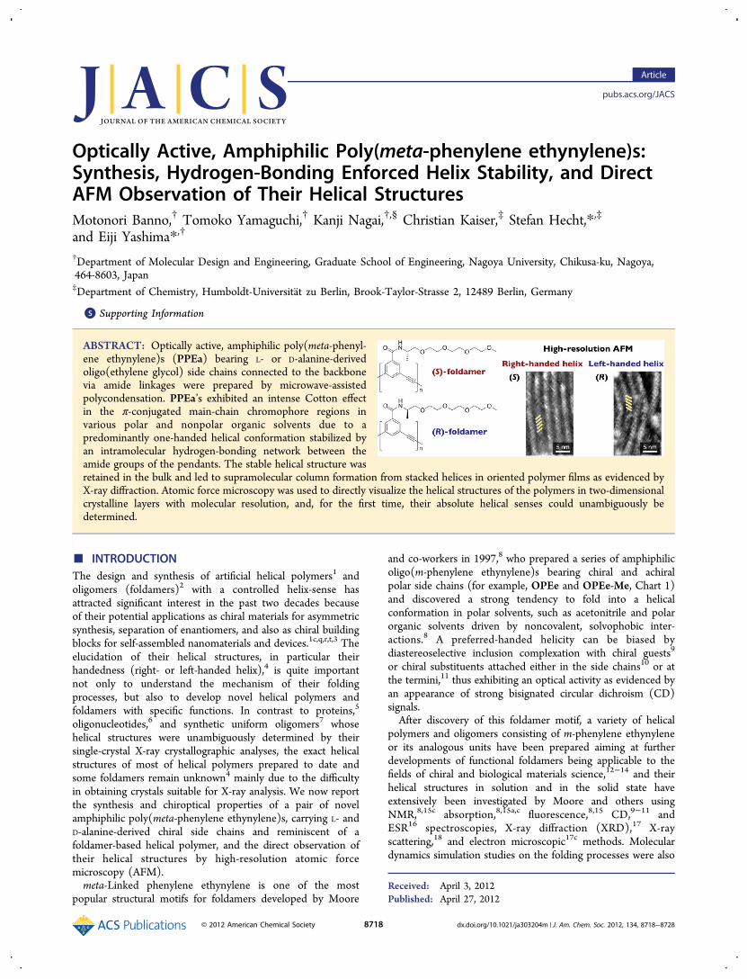
ABSTRACT: Optically active, amphiphilic poly(meta-phenyl-ene ethynylene)s (PPEa) bearing L- or D-alanine-derivedoligo(ethylene glycol) side chains connected to the backbonevia amide linkages were prepared by microwave-assistedpolycondensation. PPEa’s exhibited an intense Cotton effectin the π-conjugated main-chain chromophore regions invarious polar and nonpolar organic solvents due to apredominantly one-handed helical conformation stabilized byan intramolecular hydrogen-bonding network between theamide groups of the pendants. The stable helical structure wasretained in the bulk and led to supramolecular column formation from stacked helices in oriented polymer films as evidenced byX-ray diffraction. Atomic force microscopy was used to directly visualize the helical structures of the polymers in two-dimensionalcrystalline layers with molecular resolution, and, for the first time, their absolute helical senses could unambiguously bedetermined.
■ INTRODUCTIONThe design and synthesis of artificial helical polymers1 andoligomers (foldamers)2 with a controlled helix-sense hasattracted significant interest in the past two decades becauseof their potential applications as chiral materials for asymmetricsynthesis, separation of enantiomers, and also as chiral buildingblocks for self-assembled nanomaterials and devices.1c,q,r,t,3 Theelucidation of their helical structures, in particular theirhandedness (right- or left-handed helix),4 is quite importantnot only to understand the mechanism of their foldingprocesses, but also to develop novel helical polymers andfoldamers with specific functions. In contrast to proteins,5
oligonucleotides,6 and synthetic uniform oligomers7 whosehelical structures were unambiguously determined by theirsingle-crystal X-ray crystallographic analyses, the exact helicalstructures of most of helical polymers prepared to date andsome foldamers remain unknown4 mainly due to the difficultyin obtaining crystals suitable for X-ray analysis. We now reportthe synthesis and chiroptical properties of a pair of novelamphiphilic poly(meta-phenylene ethynylene)s, carrying L- andD-alanine-derived chiral side chains and reminiscent of afoldamer-based helical polymer, and the direct observation oftheir helical structures by high-resolution atomic forcemicroscopy (AFM).meta-Linked phenylene ethynylene is one of the most
popular structural motifs for foldamers developed by Moore
and co-workers in 1997,8 who prepared a series of amphiphilicoligo(m-phenylene ethynylene)s bearing chiral and achiralpolar side chains (for example, OPEe and OPEe-Me, Chart 1)and discovered a strong tendency to fold into a helicalconformation in polar solvents, such as acetonitrile and polarorganic solvents driven by noncovalent, solvophobic inter-actions.8 A preferred-handed helicity can be biased bydiastereoselective inclusion complexation with chiral guests9
or chiral substituents attached either in the side chains10 or atthe termini,11 thus exhibiting an optical activity as evidenced byan appearance of strong bisignated circular dichroism (CD)signals.After discovery of this foldamer motif, a variety of helical
polymers and oligomers consisting of m-phenylene ethynyleneor its analogous units have been prepared aiming at furtherdevelopments of functional foldamers being applicable to thefields of chiral and biological materials science,12−14 and theirhelical structures in solution and in the solid state haveextensively been investigated by Moore and others usingNMR,8,15c absorption,8,15a,c fluorescence,8,15 CD,9−11 andESR16 spectroscopies, X-ray diffraction (XRD),17 X-rayscattering,18 and electron microscopic17c methods. Moleculardynamics simulation studies on the folding processes were also
Received: April 3, 2012Published: April 27, 2012
Article
pubs.acs.org/JACS
© 2012 American Chemical Society 8718 dx.doi.org/10.1021/ja303204m | J. Am. Chem. Soc. 2012, 134, 8718−8728
reported.19 A helix formation with a preferable helical sense ofoligo(m-phenylene ethynylene) foldamers was evidenced by theappearance of CD signals,9−11 and the helical structuresincluding helical pitch were postulated by XRD measure-ments.17b Nevertheless, the absolute helical sense (right- or left-handed helix) of the oligo- and poly(m-phenylene ethynylene)foldamers with optical activity still remains unknown because ofdifficulty in obtaining optically active crystalline samples.The direct observation of helical polymers by scanning probe
microscopy is one of the most promising methods for providingconvincing evidence of their helical structures including thehelical pitch, handedness, and excess of helical handedness, butthe visualization of helical structures is still challenging, andtheir real images of right- and left-handed helical structures bymicroscopy remain difficult to observe.20
Recently, we found that the rigid rodlike helical poly-(phenylacetylene)s21 and poly(phenyl isocyanide)s22 bearing L-or D-alanine pendants with a long alkyl chain self-assembled toform two-dimensional (2D) crystals with regular helix-bundlestructures on highly oriented pyrolytic graphite (HOPG) uponexposure to organic solvent vapors such as benzene. High-resolution AFM revealed their helical conformations in the 2Dcrystals and enabled us to determine the molecular length andpacking, helical pitch, and handedness as well. This method has
been proved to be versatile to observe helical structures ofother helical polymers that involve a dynamically racemichelical poly(phenylacetylene) carrying achiral pendants,21c asupramolecular helical stereocomplex composed of isotactic-and syndiotactic poly(methyl methacrylate)s,23 and a double-stranded helical polymer.24
In this study, we synthesized novel optically active,amphiphilic poly(m-phenylene ethynylene)s and investigatedtheir preferred-handed helical structures in solution and in thesolid state by absorption, CD, and IR spectroscopies as well asXRD and AFM observations. The polar chiral oligo(ethyleneglycol) side chains are readily derived from either L- or D-alanine and are linked to the aromatic backbone via an amidelinkage ((S)- or (R)-PPEa), thereby replacing the ester linkageof Moore’s foldamers with an amide one (Chart 1). Because ofthese amide residues, the helical structures of (S)- and (R)-PPEa were anticipated to be stabilized by intramolecularhydrogen bonds, and the polymer backbones should thereforebe more rigid than those of Moore’s ester counterparts. Thishydrogen-bonding intrastrand stabilization should facilitatesupramolecular organization in the bulk and in particularformation of 2D crystals on a substrate while maintaining thepolymers’ helical conformations and indeed, for the first time,enabled us to observe hollow helical structures in oriented
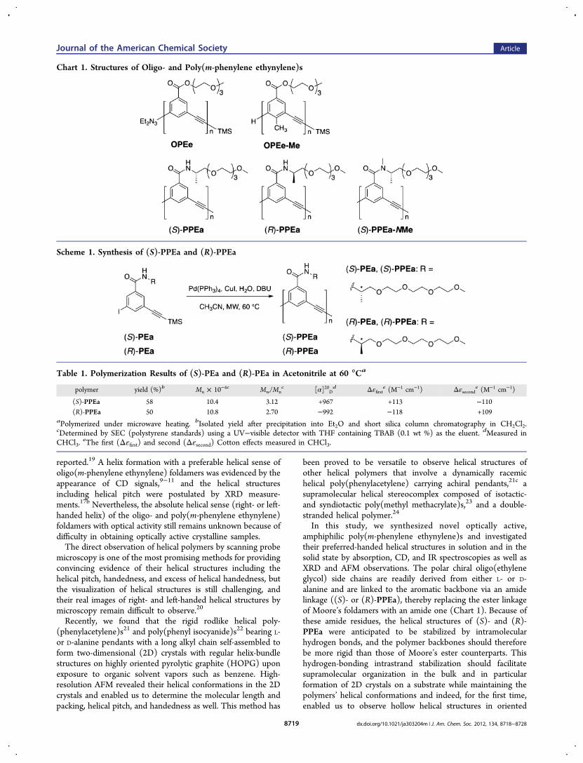
Chart 1. Structures of Oligo- and Poly(m-phenylene ethynylene)s
Scheme 1. Synthesis of (S)-PPEa and (R)-PPEa
Table 1. Polymerization Results of (S)-PEa and (R)-PEa in Acetonitrile at 60 °Ca
polymer yield (%)b Mn × 10−4c Mw/Mnc [α]20D
d Δεfirste (M−1 cm−1) Δεseconde (M−1 cm−1)
(S)-PPEa 58 10.4 3.12 +967 +113 −110(R)-PPEa 50 10.8 2.70 −992 −118 +109
aPolymerized under microwave heating. bIsolated yield after precipitation into Et2O and short silica column chromatography in CH2Cl2.cDetermined by SEC (polystyrene standards) using a UV−visible detector with THF containing TBAB (0.1 wt %) as the eluent. dMeasured inCHCl3.
eThe first (Δεfirst) and second (Δεsecond) Cotton effects measured in CHCl3.
Journal of the American Chemical Society Article
dx.doi.org/10.1021/ja303204m | J. Am. Chem. Soc. 2012, 134, 8718−87288719
polymer films by XRD and to determine the absolute helicalhandedness of the poly(m-phenylene ethynylene)s by high-resolution AFM.
■ RESULTS AND DISCUSSION
Synthesis and Polymerization of Optically Active m-Phenylene Ethynylenes. The optically active monomers, m-trimethylsilylethynyl iodobenzenes bearing an L- or D-alanineresidue with a tri(ethylene glycol) as the pendant through anamide linkage ((S)- or (R)-PEa, respectively), were synthesizedas outlined in Schemes S1−S4, and these were polymerizedaccording to the previously reported method using themicrowave-assisted polycondensation25 in acetonitrile at 60°C (Scheme 1). The polymerization results are summarized inTable 1. The polymerization of (S)-PEa and (R)-PEahomogeneously proceeded, yielding high molecular weightpolymers as an orange-brown solid in moderate yield (58% and50%, respectively). The number-average molecular weight (Mn)and its distribution (Mw/Mn) were estimated to be 10.4 × 10⁴
and 3.12 for (S)-PPEa as well as 10.8 × 10⁴ and 2.70 for (R)-PPEa, as determined by size exclusion chromatography (SEC)with polystyrene standards using tetrahydrofuran (THF)containing 0.1 wt % tetra-n-butylammonium bromide(TBAB) as the eluent.Chiroptical Properties in Dilute Solution. Moore and
co-workers previously reported that oligo(m-phenyleneethynylene)s bearing electron-withdrawing ester residues withpolar tri(ethylene glycol) chains as the pendants (OPEe, Chart1) solvophobically fold into helical conformations in polarsolvents, such as acetonitrile and aqueous organic solutions.8
This strong tendency of helical folding is attributed to theintramolecular π−π stacking between nonadjacent electron-poor phenylene ethynylene residues in conjunction with thefavorable interactions between the polar side chains and thepolar solvent, while the nonpolar folded backbone is hiddenfrom the polar solvent. On the contrary, in less polar “good”solvents for both backbone and side chains, such as CHCl3, theoligomers adopt a random-coil conformation. These conforma-tional states are also dependent on the chain-length8c,15a andtemperature8a,15b as well as solvent polarities.11c
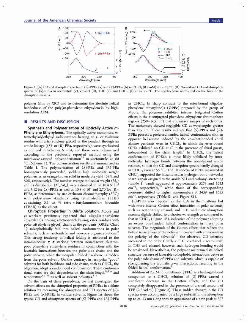
On the basis of these precedents, we first investigated thesolvent effects on the chiroptical properties of PPEas in a dilutesolution by measuring the absorption and CD spectra of (S)-PPEa and (R)-PPEa in various solvents. Figure 1A shows thetypical CD and absorption spectra of (S)-PPEa and (R)-PPEa
in CHCl3. In sharp contrast to the ester-bound oligo(m-phenylene ethynylene)s (OPEe) prepared by the group ofMoore, the polymers exhibited intense, bisignated Cottoneffects in the π-conjugated phenylene ethynylene chromophoreregions (250−365 nm) that are mirror images of each other.The monomers showed negligible CD at wavelengths greaterthan 275 nm. These results indicate that (S)-PPEa and (R)-PPEa possess a preferred-handed helical conformation with anopposite helix-sense induced by the covalent-bonded chiralalanine pendants even in CHCl3, in which the ester-boundOPEe exhibited no CD at all in the presence of chiral guests,independent of the chain length.9 In CHCl3, the helicalconformation of PPEa’s is most likely stabilized by intra-molecular hydrogen bonds between the nonadjacent amideresidues, so that the CD and absorption spectra hardly changedin CHCl3 even at 55 °C. The IR spectra of PPEa measured inCHCl3 supported the intramolecular hydrogen-bond networks;sharp signals assigned to the amide NH and carbonyl stretching(amide I) bands appeared at approximately 3274 and 1633cm−1, respectively,26 while those of the correspondingmonomer shifted to higher wavenumbers at 3439 and 1655cm−1, respectively (Table S1 and Figure S1).(S)-PPEa also displayed similar CDs in their patterns but
with more intense Cotton effect intensities in polar solvents,such as acetonitrile, ethanol, and THF and their absorptionmaxima slightly shifted to a shorter wavelength as compared tothat in CHCl3 (Figure 1B), indicative of the polymer adoptingan excess one-handed helical conformation in these polarsolvents. The magnitude of the Cotton effects that reflects thehelical sense excess of the polymer increased with an increase inthe polarity of the solvents;11c the observed CD intensityincreased in the order CHCl3 < THF < ethanol < acetonitrile.In THF and ethanol, however, such hydrogen bonding wouldbe weakened. Nevertheless, the polymer maintained its helicalstructure because of favorable solvophobic interactions betweenthe polar side chains of PPEa and solvents, which is capable ofstrengthening the aromatic π−π interactions, resulting in thefolded helical conformation.8
Addition of 2,2,2-trifluoroethanol (TFE) as a hydrogen-bondcompetitor to a CHCl3 solution of (S)-PPEa caused asignificant decrease in the Cotton effects, and the CDcompletely disappeared in the presence of a small amount ofTFE (2.5 vol %) (Figure 2). These sudden changes in the CDspectra were accompanied by a large red-shift in the absorptionup to ca. 13 nm along with an appearance of a new peak at 307
Figure 1. (A) CD and absorption spectra of (S)-PPEa (a) and (R)-PPEa (b) in CHCl3 (0.5 mM) at ca. 25 °C. (B) Normalized CD and absorptionspectra of (S)-PPEa in acetonitrile (c), ethanol (d), THF (e), and CHCl3 (f) at ca. 25 °C. The spectra were normalized on the basis of theabsorption maxima.
Journal of the American Chemical Society Article
dx.doi.org/10.1021/ja303204m | J. Am. Chem. Soc. 2012, 134, 8718−87288720
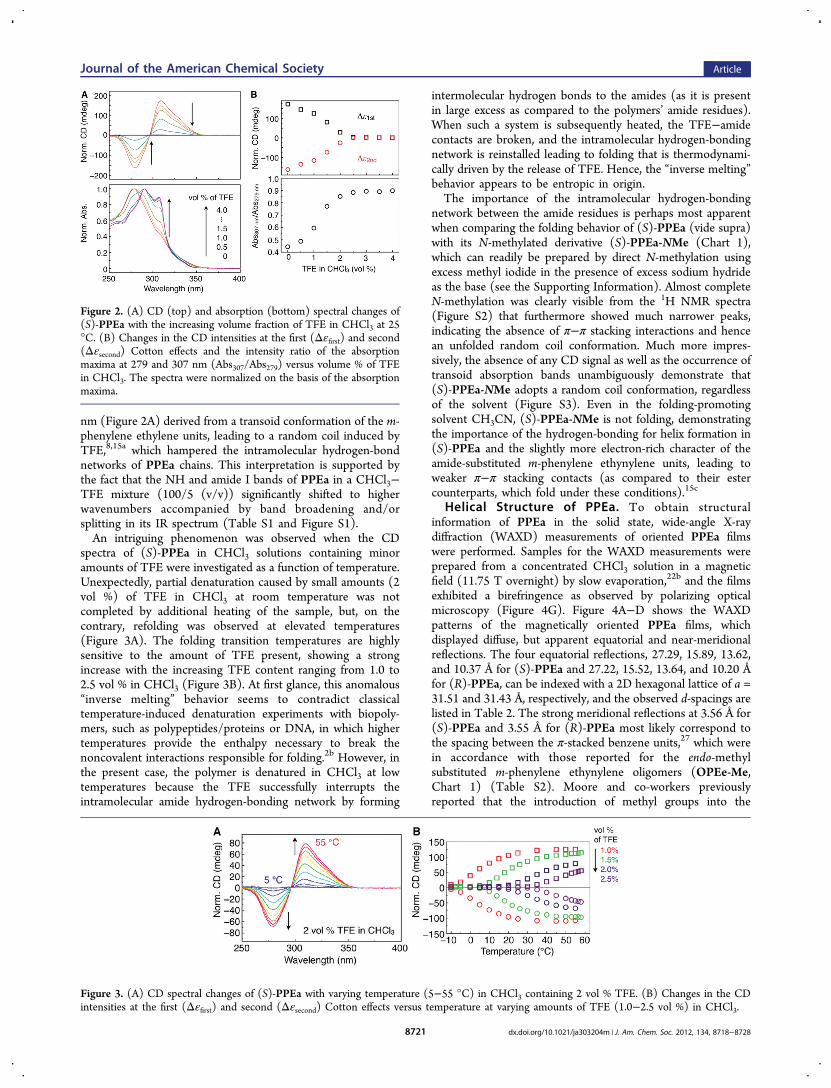
nm (Figure 2A) derived from a transoid conformation of the m-phenylene ethylene units, leading to a random coil induced byTFE,8,15a which hampered the intramolecular hydrogen-bondnetworks of PPEa chains. This interpretation is supported bythe fact that the NH and amide I bands of PPEa in a CHCl3−TFE mixture (100/5 (v/v)) significantly shifted to higherwavenumbers accompanied by band broadening and/orsplitting in its IR spectrum (Table S1 and Figure S1).An intriguing phenomenon was observed when the CD
spectra of (S)-PPEa in CHCl3 solutions containing minoramounts of TFE were investigated as a function of temperature.Unexpectedly, partial denaturation caused by small amounts (2vol %) of TFE in CHCl3 at room temperature was notcompleted by additional heating of the sample, but, on thecontrary, refolding was observed at elevated temperatures(Figure 3A). The folding transition temperatures are highlysensitive to the amount of TFE present, showing a strongincrease with the increasing TFE content ranging from 1.0 to2.5 vol % in CHCl3 (Figure 3B). At first glance, this anomalous“inverse melting” behavior seems to contradict classicaltemperature-induced denaturation experiments with biopoly-mers, such as polypeptides/proteins or DNA, in which highertemperatures provide the enthalpy necessary to break thenoncovalent interactions responsible for folding.2b However, inthe present case, the polymer is denatured in CHCl3 at lowtemperatures because the TFE successfully interrupts theintramolecular amide hydrogen-bonding network by forming
intermolecular hydrogen bonds to the amides (as it is presentin large excess as compared to the polymers’ amide residues).When such a system is subsequently heated, the TFE−amidecontacts are broken, and the intramolecular hydrogen-bondingnetwork is reinstalled leading to folding that is thermodynami-cally driven by the release of TFE. Hence, the “inverse melting”behavior appears to be entropic in origin.The importance of the intramolecular hydrogen-bonding
network between the amide residues is perhaps most apparentwhen comparing the folding behavior of (S)-PPEa (vide supra)with its N-methylated derivative (S)-PPEa-NMe (Chart 1),which can readily be prepared by direct N-methylation usingexcess methyl iodide in the presence of excess sodium hydrideas the base (see the Supporting Information). Almost completeN-methylation was clearly visible from the 1H NMR spectra(Figure S2) that furthermore showed much narrower peaks,indicating the absence of π−π stacking interactions and hencean unfolded random coil conformation. Much more impres-sively, the absence of any CD signal as well as the occurrence oftransoid absorption bands unambiguously demonstrate that(S)-PPEa-NMe adopts a random coil conformation, regardlessof the solvent (Figure S3). Even in the folding-promotingsolvent CH3CN, (S)-PPEa-NMe is not folding, demonstratingthe importance of the hydrogen-bonding for helix formation in(S)-PPEa and the slightly more electron-rich character of theamide-substituted m-phenylene ethynylene units, leading toweaker π−π stacking contacts (as compared to their estercounterparts, which fold under these conditions).15c
Helical Structure of PPEa. To obtain structuralinformation of PPEa in the solid state, wide-angle X-raydiffraction (WAXD) measurements of oriented PPEa filmswere performed. Samples for the WAXD measurements wereprepared from a concentrated CHCl3 solution in a magneticfield (11.75 T overnight) by slow evaporation,22b and the filmsexhibited a birefringence as observed by polarizing opticalmicroscopy (Figure 4G). Figure 4A−D shows the WAXDpatterns of the magnetically oriented PPEa films, whichdisplayed diffuse, but apparent equatorial and near-meridionalreflections. The four equatorial reflections, 27.29, 15.89, 13.62,and 10.37 Å for (S)-PPEa and 27.22, 15.52, 13.64, and 10.20 Åfor (R)-PPEa, can be indexed with a 2D hexagonal lattice of a =31.51 and 31.43 Å, respectively, and the observed d-spacings arelisted in Table 2. The strong meridional reflections at 3.56 Å for(S)-PPEa and 3.55 Å for (R)-PPEa most likely correspond tothe spacing between the π-stacked benzene units,27 which werein accordance with those reported for the endo-methylsubstituted m-phenylene ethynylene oligomers (OPEe-Me,Chart 1) (Table S2). Moore and co-workers previouslyreported that the introduction of methyl groups into the
Figure 2. (A) CD (top) and absorption (bottom) spectral changes of(S)-PPEa with the increasing volume fraction of TFE in CHCl3 at 25°C. (B) Changes in the CD intensities at the first (Δεfirst) and second(Δεsecond) Cotton effects and the intensity ratio of the absorptionmaxima at 279 and 307 nm (Abs307/Abs279) versus volume % of TFEin CHCl3. The spectra were normalized on the basis of the absorptionmaxima.
Figure 3. (A) CD spectral changes of (S)-PPEa with varying temperature (5−55 °C) in CHCl3 containing 2 vol % TFE. (B) Changes in the CDintensities at the first (Δεfirst) and second (Δεsecond) Cotton effects versus temperature at varying amounts of TFE (1.0−2.5 vol %) in CHCl3.
Journal of the American Chemical Society Article
dx.doi.org/10.1021/ja303204m | J. Am. Chem. Soc. 2012, 134, 8718−87288721
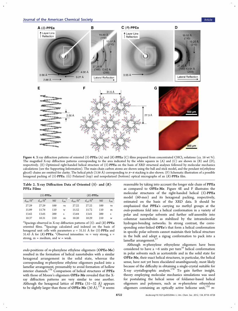
endo-positions of m-phenylene ethylene oligomers (OPEe-Me)resulted in the formation of helical nanotubules with a similarhexagonal arrangement in the solid state, whereas thecorresponding m-phenylene ethylene oligomers packed into alamellar arrangement, presumably to avoid formation of hollowinterior channels.17b Comparison of helical structures of PPEawith those of Moore’s oligomers OPEe-Me revealed that the X-ray diffraction patterns are very similar to one another.Although the hexagonal lattice of PPEa (31−32 Å) appearsto be slightly larger than those of OPEe-Me (30 Å),17b it seems
reasonable by taking into account the longer side chain of PPEaas compared to OPEe-Me. Figure 4E and F illustrates themolecular structures of the right-handed helical (S)-PPEamodel (60-mer) and its hexagonal packing, respectively,estimated on the basis of the XRD data. It should beemphasized that PPEa’s carrying no methyl groups at theendo-positions fold into a helical conformation in a variety ofpolar and nonpolar solvents and further self-assemble intocolumnar nanotubules as stabilized by the intramolecularhydrogen-bonding networks. In strong contrast, the corre-sponding ester-linked OPEe’s that form a helical conformationin specific polar solvents cannot maintain their helical structurein the bulk and adopt a zigzag conformation to pack into alamellar arrangement.17
Although m-phenylene ethynylene oligomers have beenconsidered to have a ∼6 units per turn16 helical conformationin polar solvents such as acetonitrile and in the solid state forOPEe-Me, their exact helical structures, in particular, the helicalsense, have not yet been elucidated unambiguously, most likelybecause of the difficulty in obtaining a single-crystal suitable forX-ray crystallographic analysis.17b To gain further insight,theory employing molecular mechanics simulations was usedfor postulating the helical sense of foldamer-based helicaloligomers and polymers, such as m-phenylene ethynyleneoligomers containing an optically active helicene unit,11d m-
Figure 4. X-ray diffraction patterns of oriented (S)-PPEa (A) and (R)-PPEa (C) films prepared from concentrated CHCl3 solutions (ca. 16 wt %).The magnified X-ray diffraction patterns corresponding to the area indicated by the white squares in (A) and (C) are shown in (B) and (D),respectively. (E) Optimized right-handed helical structure of (S)-PPEa on the basis of XRD structural analyses followed by molecular mechanicscalculations (see the Supporting Information). The main-chain carbon atoms are shown using the ball and stick model, and the pendant tri(ethyleneglycol) chains are omitted for clarity. The helical pitch (3.56 Å) corresponding to π−π stacking is also shown. (F) Schematic illustration of a possiblehexagonal packing of (S)-PPEa. (G) Polarized (top) and nonpolarized (bottom) optical micrographs of an (R)-PPEa film.
Table 2. X-ray Diffraction Data of Oriented (S)- and (R)-PPEa Films
(S)-PPEa (R)-PPEa
dobs/Åa dcal/Å
b hkl Iobsc dobs/Å
a dcal/Åb hkl Iobs
c
27.29 27.29 100 vs 27.22 27.22 100 vs15.89 15.76 110 w 15.52 15.72 110 m13.62 13.65 200 s 13.64 13.61 200 s10.37 10.31 210 m 10.20 10.29 210 w
aSpacings observed in X-ray diffraction patterns of (S)- and (R)-PPEaoriented films. bSpacings calculated and indexed on the basis ofhexagonal unit cells with parameters a = 31.51 Å for (S)-PPEa and31.43 Å for (R)-PPEa. cObserved intensities: vs = very strong, s =strong, m = medium, and w = weak.
Journal of the American Chemical Society Article
dx.doi.org/10.1021/ja303204m | J. Am. Chem. Soc. 2012, 134, 8718−87288722
ethynylpyridine oligomers bearing saccharide moieties as theterminal,13d and azobenzene-bound poly(m-phenylene ethyny-lene) containing D-amino acid residues.12j Optically activepoly(m-phenylene ethynylene-alt-p-phenylene ethynylene)sbearing chiral pendants were also suggested to adopt aleft14b- or right14c,d-handed helix in solution judging from theCotton effect patterns in their CD spectra. However, thesecomputational methods certainly involve a significant un-certainty, and hence an experimental approach for reliablestructure elucidation is highly desirable.Recently, we developed a facile method to directly observe
the helical structures of several synthetic helical polymersincluding poly(phenylacetylene)s bearing L- or D-alanineresidues with a long alkyl chain as the pendants.21 Flatpoly(phenylacetylene) monolayers were first formed epitaxiallyon the basal plane of the graphite, on which helicalpoly(phenylacetylene) further self-assembled into chiral 2Dhelix-bundled crystals upon exposure to organic solventvapors.21a The surface of the 2D crystals was extremely smoothand flat, which enabled us to observe high-resolution AFMimages and to determine the molecular packing, helical pitch,and handedness when combined with XRD analysis. Hence, we
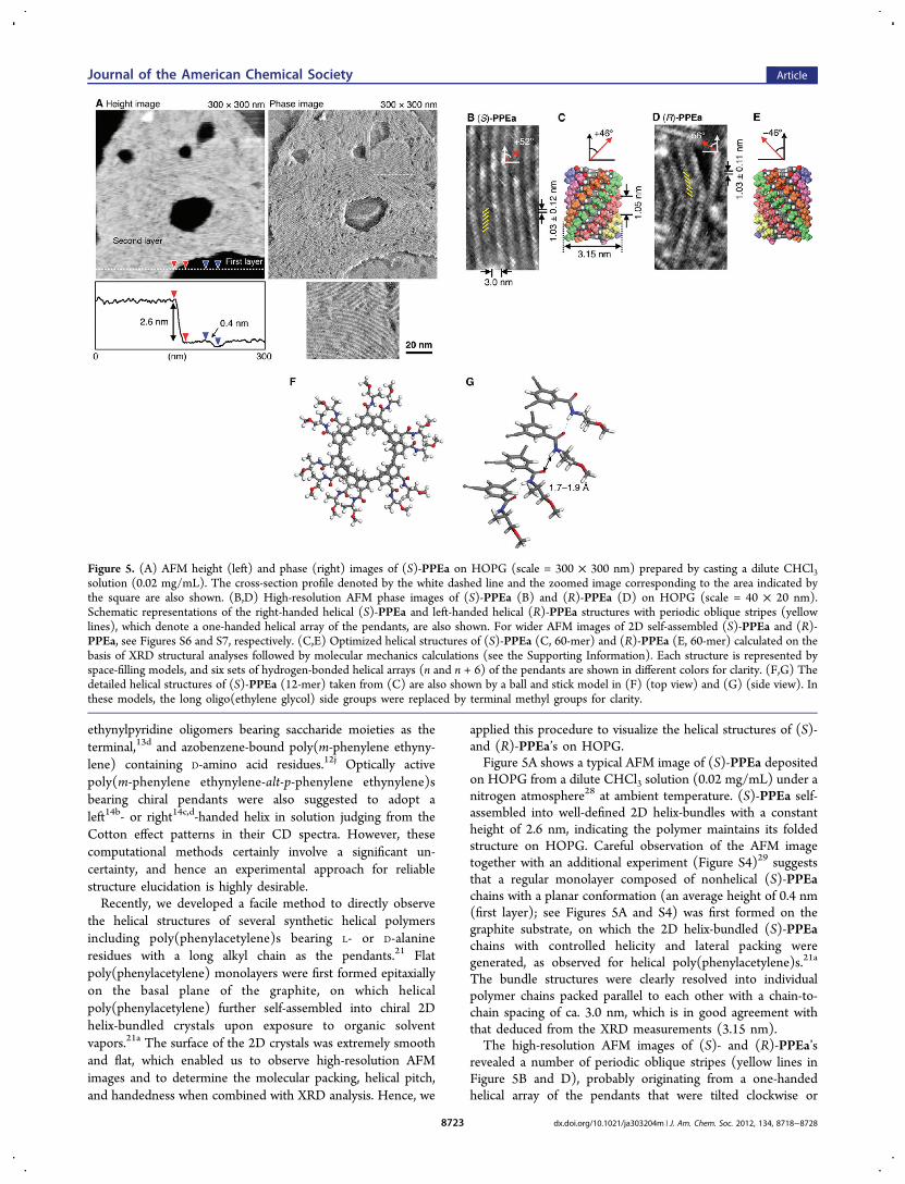
applied this procedure to visualize the helical structures of (S)-and (R)-PPEa’s on HOPG.Figure 5A shows a typical AFM image of (S)-PPEa deposited
on HOPG from a dilute CHCl3 solution (0.02 mg/mL) under anitrogen atmosphere28 at ambient temperature. (S)-PPEa self-assembled into well-defined 2D helix-bundles with a constantheight of 2.6 nm, indicating the polymer maintains its foldedstructure on HOPG. Careful observation of the AFM imagetogether with an additional experiment (Figure S4)29 suggeststhat a regular monolayer composed of nonhelical (S)-PPEachains with a planar conformation (an average height of 0.4 nm(first layer); see Figures 5A and S4) was first formed on thegraphite substrate, on which the 2D helix-bundled (S)-PPEachains with controlled helicity and lateral packing weregenerated, as observed for helical poly(phenylacetylene)s.21a
The bundle structures were clearly resolved into individualpolymer chains packed parallel to each other with a chain-to-chain spacing of ca. 3.0 nm, which is in good agreement withthat deduced from the XRD measurements (3.15 nm).The high-resolution AFM images of (S)- and (R)-PPEa’s
revealed a number of periodic oblique stripes (yellow lines inFigure 5B and D), probably originating from a one-handedhelical array of the pendants that were tilted clockwise or
Figure 5. (A) AFM height (left) and phase (right) images of (S)-PPEa on HOPG (scale = 300 × 300 nm) prepared by casting a dilute CHCl3solution (0.02 mg/mL). The cross-section profile denoted by the white dashed line and the zoomed image corresponding to the area indicated bythe square are also shown. (B,D) High-resolution AFM phase images of (S)-PPEa (B) and (R)-PPEa (D) on HOPG (scale = 40 × 20 nm).Schematic representations of the right-handed helical (S)-PPEa and left-handed helical (R)-PPEa structures with periodic oblique stripes (yellowlines), which denote a one-handed helical array of the pendants, are also shown. For wider AFM images of 2D self-assembled (S)-PPEa and (R)-PPEa, see Figures S6 and S7, respectively. (C,E) Optimized helical structures of (S)-PPEa (C, 60-mer) and (R)-PPEa (E, 60-mer) calculated on thebasis of XRD structural analyses followed by molecular mechanics calculations (see the Supporting Information). Each structure is represented byspace-filling models, and six sets of hydrogen-bonded helical arrays (n and n + 6) of the pendants are shown in different colors for clarity. (F,G) Thedetailed helical structures of (S)-PPEa (12-mer) taken from (C) are also shown by a ball and stick model in (F) (top view) and (G) (side view). Inthese models, the long oligo(ethylene glycol) side groups were replaced by terminal methyl groups for clarity.
Journal of the American Chemical Society Article
dx.doi.org/10.1021/ja303204m | J. Am. Chem. Soc. 2012, 134, 8718−87288723
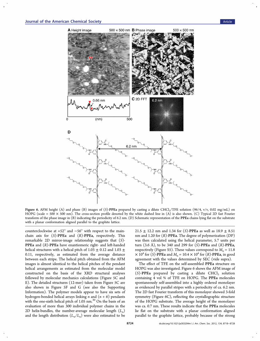
counterclockwise at +52° and −56° with respect to the main-chain axis for (S)-PPEa and (R)-PPEa, respectively. Thisremarkable 2D mirror-image relationship suggests that (S)-PPEa and (R)-PPEa have enantiomeric right- and left-handedhelical structures with a helical pitch of 1.03 ± 0.12 and 1.03 ±0.11, respectively, as estimated from the average distancebetween each stripe. The helical pitch obtained from the AFMimages is almost identical to the helical pitches of the pendanthelical arrangements as estimated from the molecular modelconstructed on the basis of the XRD structural analysesfollowed by molecular mechanics calculations (Figure 5C andE). The detailed structures (12-mer) taken from Figure 5C arealso shown in Figure 5F and G (see also the SupportingInformation). The polymer models appear to have six sets ofhydrogen-bonded helical arrays linking n and (n + 6) pendantswith the one-sixth helical pitch of 1.05 nm.30 On the basis of anevaluation of more than 300 individual polymer chains in the2D helix-bundles, the number-average molecular length (Ln)and the length distribution (Lw/Ln) were also estimated to be
21.5 ± 12.2 nm and 1.34 for (S)-PPEa as well as 18.9 ± 8.51nm and 1.20 for (R)-PPEa. The degree of polymerization (DP)was then calculated using the helical parameter, 5.7 units perturn (3.6 Å), to be 340 and 299 for (S)-PPEa and (R)-PPEa,respectively (Figure S5). These values correspond to Mn = 11.8× 10⁴ for (S)-PPEa andMn = 10.4 × 10⁴ for (R)-PPEa, in goodagreement with the values determined by SEC (vide supra).The effect of TFE on the self-assembled PPEa structure on
HOPG was also investigated. Figure 6 shows the AFM image of(S)-PPEa prepared by casting a dilute CHCl3 solutioncontaining 4 vol % of TFE on HOPG. The PPEa moleculesspontaneously self-assembled into a highly ordered monolayeras evidenced by parallel stripes with a periodicity of ca. 6.2 nm.The 2D fast Fourier transform of this monolayer showed 3-foldsymmetry (Figure 6C), reflecting the crystallographic structureof the HOPG substrate. The average height of the monolayerwas ca. 0.7 nm. These results indicate that the PPEa moleculeslie flat on the substrate with a planar conformation alignedparallel to the graphite lattice, probably because of the strong
Figure 6. AFM height (A) and phase (B) images of (S)-PPEa prepared by casting a dilute CHCl3/TFE solution (96/4, v/v, 0.02 mg/mL) onHOPG (scale = 500 × 500 nm). The cross-section profile denoted by the white dashed line in (A) is also shown. (C) Typical 2D fast Fouriertransform of the phase image in (B) indicating the periodicity of 6.2 nm. (D) Schematic representation of the PPEa chains lying flat on the substratewith a planar conformation aligned parallel to the graphite lattice.
Journal of the American Chemical Society Article
dx.doi.org/10.1021/ja303204m | J. Am. Chem. Soc. 2012, 134, 8718−87288724
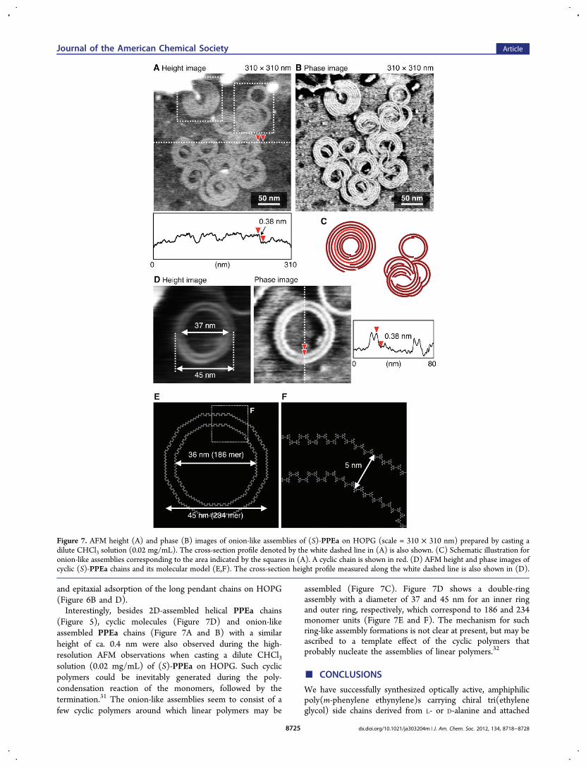
and epitaxial adsorption of the long pendant chains on HOPG(Figure 6B and D).Interestingly, besides 2D-assembled helical PPEa chains
(Figure 5), cyclic molecules (Figure 7D) and onion-likeassembled PPEa chains (Figure 7A and B) with a similarheight of ca. 0.4 nm were also observed during the high-resolution AFM observations when casting a dilute CHCl3solution (0.02 mg/mL) of (S)-PPEa on HOPG. Such cyclicpolymers could be inevitably generated during the poly-condensation reaction of the monomers, followed by thetermination.31 The onion-like assemblies seem to consist of afew cyclic polymers around which linear polymers may be
assembled (Figure 7C). Figure 7D shows a double-ringassembly with a diameter of 37 and 45 nm for an inner ringand outer ring, respectively, which correspond to 186 and 234monomer units (Figure 7E and F). The mechanism for suchring-like assembly formations is not clear at present, but may beascribed to a template effect of the cyclic polymers thatprobably nucleate the assemblies of linear polymers.32
■ CONCLUSIONS
We have successfully synthesized optically active, amphiphilicpoly(m-phenylene ethynylene)s carrying chiral tri(ethyleneglycol) side chains derived from L- or D-alanine and attached
Figure 7. AFM height (A) and phase (B) images of onion-like assemblies of (S)-PPEa on HOPG (scale = 310 × 310 nm) prepared by casting adilute CHCl3 solution (0.02 mg/mL). The cross-section profile denoted by the white dashed line in (A) is also shown. (C) Schematic illustration foronion-like assemblies corresponding to the area indicated by the squares in (A). A cyclic chain is shown in red. (D) AFM height and phase images ofcyclic (S)-PPEa chains and its molecular model (E,F). The cross-section height profile measured along the white dashed line is also shown in (D).
Journal of the American Chemical Society Article
dx.doi.org/10.1021/ja303204m | J. Am. Chem. Soc. 2012, 134, 8718−87288725
to the aromatic backbone via an amide linkage. In sharpcontrast to the analogous ester-linked oligo(m-phenyleneethynylene)s, the amide-linked polymers adopt remarkablystable, excess one-handed helical conformations in a variety ofpolar and nonpolar solvents. The helical structure is stabilizedby six sets of intramolecular hydrogen-bonding helical arraysbetween the pendants as characterized by absorption, CD, andFT-IR spectroscopies, as well as comparison with thecorresponding N-methylated derivative. Because of its excep-tional stability, the helix was retained even in the solid state,allowing its unprecedented structural characterization. There-fore, for the first time, the helical structures of m-phenyleneethynylene foldamers including the helical pitch and helicalsense could unambiguously be determined by high-resolutionAFM observations combined with X-ray diffraction measure-ments. The present study implies that previously unsolvedhelical structures of other helical polymers and oligomers,1r,4
and π-stacked supramolecular helical assemblies derived fromsmall molecules,1j may be determined by high-resolution AFMif 2D self-assembled helix-bundled crystals are available onsubstrates. From a more applied perspective, the finding thatduring the hierarchical supramolecular organization of ourpolymers their hollow helical structure is retained in the solidstate offers the prospect of engineering chiral nanoporousmaterials, suitable for separation and sensing of (chiral)compounds and analytes, respectively.
■ ASSOCIATED CONTENT*S Supporting InformationExperimental details. This material is available free of charge viathe Internet at http://pubs.acs.org.
■ AUTHOR INFORMATIONCorresponding [email protected]; [email protected] Address§Department of Applied Chemistry, Graduate School ofEngineering, Nagoya University, Chikusa-ku, Nagoya,464−8603, Japan.NotesThe authors declare no competing financial interest.
■ ACKNOWLEDGMENTSThis work was supported in part by a Grant-in-Aid for ScientificResearch (S) from the Japan Society for the Promotion ofScience (JSPS) and the Global COE Program “Elucidation andDesign of Materials and Molecular Functions” of the Ministryof Education, Culture, Sports, Science, and Technology, Japan.M.B. expresses his thanks for the JSPS Research Fellowship forYoung Scientists (No. 9164).
■ REFERENCES(1) (a) Green, M. M.; Park, J.-W.; Sato, T.; Teramoto, A.; Lifson, S.;Selinger, R. L. B.; Selinger, J. V. Angew. Chem., Int. Ed. 1999, 38, 3138−3154. (b) Mayer, S.; Zentel, R. Prog. Polym. Sci. 2001, 26, 1973−2013.(c) Nakano, T.; Okamoto, Y. Chem. Rev. 2001, 101, 4013−4038.(d) Cornelissen, J. J. L. M.; Rowan, A. E.; Nolte, R. J. M.; Sommerdijk,N. A. J. M. Chem. Rev. 2001, 101, 4039−4070. (e) Fujiki, M.; Koe, J.R.; Terao, K.; Sato, T.; Teramoto, A.; Watanabe, J. Polym. J. 2003, 35,297−344. (f) Yashima, E.; Maeda, K.; Nishimura, T. Chem.-Eur. J.2004, 10, 42−51. (g) Lockman, J. W.; Paul, N. M.; Parquette, J. R.Prog. Polym. Sci. 2005, 30, 423−452. (h) Lam, J. W. Y.; Tang, B. Z. Acc.Chem. Res. 2005, 38, 745−754. (i) Aoki, T.; Kaneko, T.; Teraguchi, M.
Polymer 2006, 47, 4867−4892. (j) Maeda, K.; Yashima, E. Top. Curr.Chem. 2006, 265, 47−88. (k) Rudick, J. G.; Percec, V. New J. Chem.2007, 31, 1083−1096. (l) Masuda, T. J. Polym. Sci., Part A: Polym.Chem. 2007, 45, 165−180. (m) Yashima, E.; Maeda, K. Macromolecules2008, 41, 3−12. (n) Yashima, E.; Maeda, K.; Furusho, Y. Acc. Chem.Res. 2008, 1166−1180. (o) Pijper, D.; Feringa, B. L. Soft Matter 2008,4, 1349−1372. (p) Kim, H.-J.; Lim, Y.-B.; Lee, M. J. Polym. Sci., Part A:Polym. Chem. 2008, 46, 1925−1935. (q) Liu, J.; Lam, J. W. Y.; Tang, B.Z. Chem. Rev. 2009, 109, 5799−5867. (r) Yashima, E.; Maeda, K.; Iida,H.; Furusho, Y.; Nagai, K. Chem. Rev. 2009, 109, 6102−6211.(s) Furusho, Y.; Yashima, E. J. Polym. Sci., Part A: Polym. Chem. 2009,47, 5195−5207. (t) Schwartz, E.; Koepf, M.; Kitto, H. J.; Nolte, R. J.M.; Rowan, A. E. Polym. Chem. 2011, 2, 33−47. (u) Kennemur, J. G.;Novak, B. M. Polymer 2011, 52, 1693−1710.(2) (a) Gellman, S. H. Acc. Chem. Res. 1998, 31, 173−180. (b) Hill,D. J.; Mio, M. J.; Prince, R. B.; Hughes, T. S.; Moore, J. S. Chem. Rev.2001, 101, 3893−4011. (c) Seebach, D.; Beck, A. K.; Bierbaum, D. J.Chem. Biodiversity 2004, 1, 1111−1239. (d) Huc, I. Eur. J. Org. Chem.2004, 17−29. (e) Foldamers: Structure, Properties, and Applications;Hecht, S., Huc, I., Eds.; Wiley-VCH: Weinheim, 2007. (f) Goodman,C. M.; Choi, S.; Shandler, S.; DeGrado, W. F. Nat. Chem. Biol. 2007, 3,252−262. (g) Li, Z.-T.; Hou, J.-L.; Li, C. Acc. Chem. Res. 2008, 41,1343−1353. (h) Gong, B. Acc. Chem. Res. 2008, 41, 1376−1386.(i) Saraogi, I.; Hamilton, A. D. Chem. Soc. Rev. 2009, 38, 1726−1743.(j) Ni, B.-B.; Yan, Q.; Ma, Y.; Zhao, D. Coord. Chem. Rev. 2010, 254,954−971. (k) Guichard, G.; Huc, I. Chem. Commun. 2011, 47, 5933−5941. (l) Roy, A.; Prabhakaran, P.; Baruah, P. K.; Sanjayan, G. J. Chem.Commun. 2011, 47, 11593−11611.(3) (a) Goh, M.; Matsushita, S.; Akagi, K. Chem. Soc. Rev. 2010, 39,2466−2476. (b) Kane-Maguire, L. A. P.; Wallace, G. G. Chem. Soc. Rev.2010, 39, 2545−2576. (c) Ho, R.-M.; Chiang, Y.-W.; Lin, S.-C.; Chen,C.-K. Prog. Polym. Sci. 2011, 36, 376−453.(4) Yashima, E. Polym. J. 2010, 42, 3−16.(5) Schulz, G. E.; Schirmer, R. H. Principles of Protein Structure;Springer-Verlag: New York, 1979.(6) (a) Saenger, W. Principles of Nucleic Acid Structure; Springer-Verlag: New York, 1984. (b) Kennard, O.; Hunter, W. N. Angew.Chem., Int. Ed. Engl. 1991, 30, 1254−1277.(7) (a) Ute, K.; Hirose, K.; Kashimoto, H.; Nakayama, H.; Hatada,K.; Vogl, O. Polym. J. 1993, 25, 1175−1186. (b) Ito, Y.; Ohara, T.;Shima, R.; Suginome, M. J. Am. Chem. Soc. 1996, 118, 9188−9189.(c) Tanatani, A.; Yokoyama, A.; Azumaya, I.; Takakura, Y.; Mitsui, C.;Shiro, M.; Uchiyama, M.; Muranaka, A.; Kobayashi, N.; Yokozawa, T.J. Am. Chem. Soc. 2005, 127, 8553−8561. (d) Dolain, C.; Jiang, H.;Leger, J.-M.; Guionneau, P.; Huc, I. J. Am. Chem. Soc. 2005, 127,12943−12951. (e) Suginome, M.; Noguchi, H.; Murakami, M. Chem.Lett. 2007, 36, 1036−1037. (f) Kendhale, A. M.; Poniman, L.; Dong,Z.; Laxmi-Reddy, K.; Kauffmann, B.; Ferrand, Y.; Huc, I. J. Org. Chem.2011, 76, 195−200.(8) (a) Nelson, J. C.; Saven, J. G.; Moore, J. S.; Wolynes, P. G. Science1997, 277, 1793−1796. For reviews, see: (b) Ray, C. R.; Moore, J. S.Adv. Polym. Sci. 2005, 177, 91−149. (c) Stone, M. T.; Heemstra, J. M.;Moore, J. S. Acc. Chem. Res. 2006, 39, 11−20. (d) Zhao, Y.; Moore, J.S. In Foldamers: Structure, Properties, and Applications; Hecht, S., Huc,I., Eds.; Wiley-VCH: Weinheim, 2007; Chapter 3, pp 75−108.(9) (a) Prince, R. B.; Barnes, S. A.; Moore, J. S. J. Am. Chem. Soc.2000, 122, 2758−2762. (b) Tanatani, A.; Mio, M. J.; Moore, J. S. J.Am. Chem. Soc. 2001, 123, 1792−1793. (c) Tanatani, A.; Hughes, T.S.; Moore, J. S. Angew. Chem., Int. Ed. 2002, 41, 325−328. (d) Stone,M. T.; Moore, J. S. Org. Lett. 2004, 6, 469−472. (e) Goto, K.; Moore,J. S. Org. Lett. 2005, 7, 1683−1686.(10) (a) Prince, R. B.; Brunsveld, L.; Meijer, E. W.; Moore, J. S.Angew. Chem., Int. Ed. 2000, 39, 228−230. (b) Brunsveld, L.; Prince, R.B.; Meijer, E. W.; Moore, J. S. Org. Lett. 2000, 2, 1525−1528.(c) Prince, R. B.; Moore, J. S.; Brunsveld, L.; Meijer, E. W. Chem.-Eur.J. 2001, 7, 4150−4154. (d) Brunsveld, L.; Meijer, E. W.; Prince, R. B.;Moore, J. S. J. Am. Chem. Soc. 2001, 123, 7978−7984. (e) Goto, H.;Heemstra, J. M.; Hill, D. J.; Moore, J. S. Org. Lett. 2004, 6, 889−892.
Journal of the American Chemical Society Article
dx.doi.org/10.1021/ja303204m | J. Am. Chem. Soc. 2012, 134, 8718−87288726
(11) (a) Gin, M. S.; Yokozawa, T.; Prince, R. B.; Moore, J. S. J. Am.Chem. Soc. 1999, 121, 2643−2644. (b) Gin, M. S.; Moore, J. S. Org.Lett. 2000, 2, 135−138. (c) Hill, D. J.; Moore, J. S. Proc. Natl. Acad. Sci.U.S.A. 2002, 99, 5053−5057. (d) Stone, M. T.; Fox, J. M.; Moore, J. S.Org. Lett. 2004, 6, 3317−3320.(12) (a) Shinohara, K.-i.; Aoki, T.; Kaneko, T.; Oikawa, E. Polymer2001, 42, 351−355. (b) Hecht, S.; Khan, A. Angew. Chem., Int. Ed.2003, 42, 6021−6024. (c) Ramos, A. M.; Meskers, S. C. J.; Beckers, E.H. A.; Prince, R. B.; Brunsveld, L.; Janssen, R. A. J. J. Am. Chem. Soc.2004, 126, 9630−9644. (d) Arnt, L.; Tew, G. N. Macromolecules 2004,37, 1283−1288. (e) Khan, A.; Kaiser, C.; Hecht, S. Angew. Chem., Int.Ed. 2006, 45, 1878−1881. (f) Khan, A.; Hecht, S. Chem.-Eur. J. 2006,12, 4764−4774. (g) Li, C.; Guo, Y.; Lv, J.; Xu, J.; Li, Y.; Wang, S.; Liu,H.; Zhu, D. J. Polym. Sci., Part A: Polym. Chem. 2007, 45, 1403−1412.(h) Kaneko, T.; Yoshimoto, S.; Hadano, S.; Teraguchi, M.; Aoki, T.Polyhedron 2007, 26, 1825−1829. (i) Inoue, M.; Teraguchi, M.; Aoki,T.; Hadano, S.; Namikoshi, T.; Marwanta, E.; Kaneko, T. Synth. Met.2009, 159, 854−858. (j) Sogawa, H.; Shiotsuki, M.; Matsuoka, H.;Sanda, F. Macromolecules 2011, 44, 3338−3345.(13) (a) Inouye, M.; Waki, M.; Abe, H. J. Am. Chem. Soc. 2004, 126,2022−2027. (b) Abe, H.; Masuda, N.; Waki, M.; Inouye, M. J. Am.Chem. Soc. 2005, 127, 16189−16196. (c) Waki, M.; Abe, H.; Inouye,M. Chem.-Eur. J. 2006, 12, 7839−7847. (d) Abe, H.; Murayama, D.;Kayamori, F.; Inouye, M. Macromolecules 2008, 41, 6903−6909.(e) Abe, H.; Machiguchi, H.; Matsumoto, S.; Inouye, M. J. Org. Chem.2008, 73, 4650−4661.(14) (a) Tan, C.; Pinto, M. R.; Kose, M. E.; Ghiviriga, I.; Schanze, K.S. Adv. Mater. 2004, 16, 1208−1212. (b) Zhao, X.; Schanze, K. S.Langmuir 2006, 22, 4856−4862. (c) Liu, R.; Shiotsuki, M.; Masuda,T.; Sanda, F. Macromolecules 2009, 42, 6115−6122. (d) Liu, R.;Sogawa, H.; Shiotsuki, M.; Masuda, T.; Sanda, F. Polymer 2010, 51,2255−2263. (e) Ji, E.; Wu, D.; Schanze, K. S. Langmuir 2010, 26,14427−14429. (f) Huang, Y.-Q.; Fan, Q.-L.; Liu, X.-F.; Fu, N.-N.;Huang, W. Langmuir 2010, 26, 19120−19128.(15) (a) Prince, R. B.; Saven, J. G.; Wolynes, P. G.; Moore, J. S. J. Am.Chem. Soc. 1999, 121, 3114−3121. (b) Yang, W. Y.; Prince, R. B.;Sabelko, J.; Moore, J. S.; Gruebele, M. J. Am. Chem. Soc. 2000, 122,3248−3249. (c) Lahiri, S.; Thompson, J. L.; Moore, J. S. J. Am. Chem.Soc. 2000, 122, 11315−11319.(16) Matsuda, K.; Stone, M. T.; Moore, J. S. J. Am. Chem. Soc. 2002,124, 11836−11837.(17) (a) Prest, P.-J.; Prince, R. B.; Moore, J. S. J. Am. Chem. Soc.1999, 121, 5933−5939. (b) Mio, M. J.; Prince, R. B.; Moore, J. S.;Kubel, C.; Martin, D. C. J. Am. Chem. Soc. 2000, 122, 6134−6135.(c) Kubel, C.; Mio, M. J.; Moore, J. S.; Martin, D. C. J. Am. Chem. Soc.2002, 124, 8605−8610.(18) Kelley, R. F.; Rybtchinski, B.; Stone, M. T.; Moore, J. S.;Wasielewski, M. R. J. Am. Chem. Soc. 2007, 129, 4114−4115.(19) (a) Pickholz, M.; Stafstrom, S. Chem. Phys. 2001, 270, 245−251.(b) Elmer, S.; Pande, V. S. J. Phys. Chem. B 2001, 105, 482−485.(c) Sen, S. J. Phys. Chem. B 2002, 106, 11343−11350. (d) Lee, O.-S.;Saven, J. G. J. Phys. Chem. B 2004, 108, 11988−11994. (e) Elmer, S.P.; Pande, V. S. J. Chem. Phys. 2004, 121, 12760−12771. (f) Adisa, B.;Bruce, D. A. J. Phys. Chem. B 2005, 109, 7548−7556. (g) Adisa, B.;Bruce, D. A. J. Phys. Chem. B 2005, 109, 19952−19959. (h) Elmer, S.P.; Park, S.; Pande, V. S. J. Chem. Phys. 2005, 123, 114902. (i) Elmer,S. P.; Park, S.; Pande, V. S. J. Chem. Phys. 2005, 123, 114903.(j) Nguyen, H. H.; McAliley, J. H.; Batson, W. R.; Bruce, D. A.Macromolecules 2010, 43, 5932−5942. (k) Nguyen, H. H.; McAliley, J.H.; Bruce, D. A. Macromolecules 2011, 44, 60−67.(20) Tanaka, H.; Kawai, T. Nat. Nanotechnol. 2009, 4, 518−522.(21) (a) Sakurai, S.-i.; Okoshi, K.; Kumaki, J.; Yashima, E. Angew.Chem., Int. Ed. 2006, 45, 1245−1248. (b) Sakurai, S.-i.; Okoshi, K.;Kumaki, J.; Yashima, E. J. Am. Chem. Soc. 2006, 128, 5650−5651.(c) Sakurai, S.-i.; Ohsawa, S.; Nagai, K.; Okoshi, K.; Kumaki, J.;Yashima, E. Angew. Chem., Int. Ed. 2007, 46, 7605−7608. (d) Louzao,I.; Seco, J. M.; Quinoa, E.; Riguera, R. Angew. Chem., Int. Ed. 2010, 49,1430−1433. (e) Freire, F.; Seco, J. M.; Quinoa, E.; Riguera, R. Angew.Chem., Int. Ed. 2011, 50, 11692−11696. (f) Ohsawa, S.; Sakurai, S.-i.;
Nagai, K.; Banno, M.; Maeda, K.; Kumaki, J.; Yashima, E. J. Am. Chem.Soc. 2011, 133, 108−114. (g) Ohsawa, S.; Sakurai, S.-i.; Nagai, K.;Maeda, K.; Kumaki, J.; Yashima, E. Polym. J. 2011, 1−9. For reviews,see refs 1m, 1n, 1r, and 4, and (h) Kumaki, J.; Sakurai, S.-i.; Yashima,E. Chem. Soc. Rev. 2009, 38, 737−746.(22) (a) Kajitani, T.; Okoshi, K.; Sakurai, S.-i.; Kumaki, J.; Yashima,E. J. Am. Chem. Soc. 2006, 128, 708−709. (b) Onouchi, H.; Okoshi, K.;Kajitani, T.; Sakurai, S.-i.; Nagai, K.; Kumaki, J.; Onitsuka, K.; Yashima,E. J. Am. Chem. Soc. 2008, 130, 229−236. (c) Wu, Z.-Q.; Nagai, K.;Banno, M.; Okoshi, K.; Onitsuka, K.; Yashima, E. J. Am. Chem. Soc.2009, 131, 6708−6718. (d) Banno, M.; Wu, Z.-Q.; Nagai, K.; Sakurai,S.-i.; Okoshi, K.; Yashima, E. Macromolecules 2010, 43, 6553−6561.(e) Kumaki, J.; Kajitani, T.; Nagai, K.; Okoshi, K.; Yashima, E. J. Am.Chem. Soc. 2010, 132, 5604−5606. (f) Miyabe, T.; Iida, H.; Banno, M.;Yamaguchi, T.; Yashima, E. Macromolecules 2011, 44, 8687−8692.(23) Kumaki, J.; Kawauchi, T.; Okoshi, K.; Kusanagi, H.; Yashima, E.Angew. Chem., Int. Ed. 2007, 46, 5348−5351.(24) Maeda, T.; Furusho, Y.; Sakurai, S.-i.; Kumaki, J.; Okoshi, K.;Yashima, E. J. Am. Chem. Soc. 2008, 130, 7938−7945.(25) Khan, A.; Hecht, S. Chem. Commun. 2004, 300−301.(26) For oligo(m-phenylene ethynylene) derivatives whose helicalconformations are stabilized by intramolecular hydrogen bonds, see:(a) Cary, J. M.; Moore, J. S. Org. Lett. 2002, 4, 4663−4666. (b) Yang,X.; Brown, A. L.; Furukawa, M.; Li, S.; Gardinier, W. E.; Bukowski, E.J.; Bright, F. V.; Zheng, C.; Zeng, X. C.; Gong, B. Chem. Commun.2003, 56−57. The molecular dynamics simulation studies alsosupported such intramolecular hydrogen bonds formed between theamide groups, resulting in a stable helical conformation of an oligo(m-phenylene ethynylene) bearing amide pendants in chloroform. See refs19j and 19k. For other helical polymers and foldamers whose helicalstructures are stabilized by intramolecular hydrogen bonds, see refs 2d,2f−i, 12j, 14c, and 14d. For polyisocyanides: (c) Cornelissen, J. J. L.M.; Donners, J. J. J. M.; de Gelder, R.; Graswinckel, W. S.; Metselaar,G. A.; Rowan, A. E.; Sommerdijk, N. A. J. M.; Nolte, R. J. M. Science2001, 293, 676−680. (d) de Witte, P. A. J.; Hernando, J.; Neuteboom,E. E.; van Dijk, E. M. H. P.; Meskers, S. C. J.; Janssen, R. A. J.; vanHulst, N. F.; Nolte, R. J. M.; García-Parajo, M. F.; Rowan, A. E. J. Phys.Chem. B 2006, 110, 7803−7812. (e) Hase, Y.; Mitsutsuji, Y.; Ishikawa,M.; Maeda, K.; Okoshi, K.; Yashima, E. Chem.-Asian J. 2007, 2, 755−763. (f) Okoshi, K.; Nagai, K.; Kajitani, T.; Sakurai, S.-i.; Yashima, E.Macromolecules 2008, 41, 7752−7754. For poly(N-propargylamide)s:(g) Nomura, R.; Tabei, J.; Masuda, T. J. Am. Chem. Soc. 2001, 123,8430−8431. (h) Nomura, R.; Tabei, J.; Masuda, T. Macromolecules2002, 35, 2955−2961. (i) Gao, G.; Sanda, F.; Masuda, T.Macromolecules 2003, 36, 3932−3937. (j) Tabei, J.; Shiotsuki, M.;Sanda, F.; Masuda, T. Macromolecules 2005, 38, 5860−5867. Forpoly(N-propargylcarbamate)s: (k) Nomura, R.; Nishiura, S.; Tabei, J.;Sanda, F.; Masuda, T. Macromolecules 2003, 36, 5076−5080. Forpoly(N-propargylsulfamide)s: (l) Deng, J.; Tabei, J.; Shiotsuki, M.;Sanda, F.; Masuda, T. Macromolecules 2004, 37, 5538−5543. Forpoly(propargyl ester)s: (m) Sanda, F.; Araki, H.; Masuda, T.Macromolecules 2005, 38, 10605−10608. For poly(N-propargylphos-phonamidate)s: (n) Yue, D.; Fujii, T.; Terada, K.; Tabei, J.; Shiotsuki,M.; Sanda, F.; Masuda, T. Macromol. Rapid Commun. 2006, 27, 1460−1464. For poly(N-propargylurea)s: (o) Deng, J.; Luo, X.; Zhao, W.;Yang, W. J. Polym. Sci., Part A: Polym. Chem. 2008, 46, 4112−4121.For poly(N-butynylamide)s: (p) Suzuki, Y.; Tabei, J.; Shiotsuki, M.;Inai, Y.; Sanda, F.; Masuda, T. Macromolecules 2008, 41, 1086−1093.For poly(1-methylpropargyl-N-alkylcarbamate)s: (q) Shirakawa, Y.;Suzuki, Y.; Terada, K.; Shiotsuki, M.; Masuda, T.; Sanda, F.Macromolecules 2010, 43, 5575−5581. For poly(phenylacetylene)s:(r) Li, B. S.; Cheuk, K. K. L.; Salhi, F.; Lam, J. W. Y.; Cha, J. A. K.;Xiao, X.; Bai, C.; Tang, B. Z. Nano Lett. 2001, 1, 323−328. (s) Aoki,T.; Kaneko, T.; Maruyama, N.; Sumi, A.; Takahashi, M.; Sato, T.;Teraguchi, M. J. Am. Chem. Soc. 2003, 125, 6346−6347. (t) Okoshi,K.; Sakajiri, K.; Kumaki, J.; Yashima, E. Macromolecules 2005, 38,4061−4064. (u) Terada, K.; Masuda, T.; Sanda, F. Macromolecules2009, 42, 913−920.
Journal of the American Chemical Society Article
dx.doi.org/10.1021/ja303204m | J. Am. Chem. Soc. 2012, 134, 8718−87288727
(27) Although weak meridional reflections at 4.00, 3.27, and 2.86 Åwere also observed for (S)-PPEa, we could not characterize thesereflections, which may be related to the electron densities along withthe main-chain axis.(28) When the sample was exposed to organic solvent vaporsaccording to a previously reported method,21,22,24 almost similar AFMimages were obtained, indicating that organic solvent vapors may notbe essential for obtaining 2D assembled helix-bundles at least forPPEa.(29) Casting a more dilute solution (0.01 mg/mL) of (S)-PPEa onHOPG produced the isolated monolayers with an average height of 0.4nm (first layer, Figure S4), which was much less than the moleculardiameter of a helically folded PPEa model (3.2 nm). Furtherdeposition of the same dilute solution on the monolayers on HOPGresulted in the formation of the 2D helix-bundled (S)-PPEa assemblies(second layer) whose AFM images were almost identical to thoseshown in Figure 5A.(30) When (S)-PPEa has such a right-handed helical array of thependants, the main-chain has a 5.7 units per turn right-handed helicalstructure.(31) For review on the synthesis of cyclic polymers, see: (a) Laurent,B. A.; Grayson, S. M. Chem. Soc. Rev. 2009, 38, 2202−2213. Forrecent examples of AFM observations of cyclic polymers, see:(b) Kawaguchi, D.; Nishu, T.; Takano, A.; Matsushita, Y. Polym. J.2007, 39, 271−275. (c) Schappacher, M.; Deffieux, A. Science 2008,319, 1512−1515. (d) Schappacher, M.; Deffieux, A. J. Am. Chem. Soc.2008, 130, 14684−14689. (e) Schappacher, M.; Deffieux, A. Angew.Chem., Int. Ed. 2009, 48, 5930−5933. (f) Boydston, A. J.; Holcombe,T. W.; Unruh, D. A.; Frechet, J. M. J.; Grubbs, R. H. J. Am. Chem. Soc.2009, 131, 5388−5389. (g) Xia, Y.; Boydston, A. J.; Grubbs, R. H.Angew. Chem., Int. Ed. 2011, 50, 5882−5885. (h) Lahasky, S. H.;Serem, W. K.; Guo, L.; Garno, J. C.; Zhang, D. Macromolecules 2011,44, 9063−9074. (i) Zhang, K.; Lackey, M. A.; Wu, Y.; Tew, G. N. J.Am. Chem. Soc. 2011, 133, 6906−6909.(32) For related Vernier-templating, see: O’Sullivan, M. C.; Sprafke,J. K.; Kondratuk, D. V.; Rinfray, C.; Claridge, T. D. W.; Saywell, A.;Blunt, M. O.; O’Shea, J. N.; Beton, P. H.; Malfois, M.; Anderson, H. L.Nature 2011, 469, 72−75.
Journal of the American Chemical Society Article
dx.doi.org/10.1021/ja303204m | J. Am. Chem. Soc. 2012, 134, 8718−87288728