optimal programming management of ventricular tachycardia ... · optimal programming management of...
TRANSCRIPT
Available online at www.jbr-pub.org
Open Access at PubMed Central
The Journal of Biomedical Research, 2015, 29(1):35-43
Review Article
Optimal programming management of ventricular tachycardia storm
in ICD patients
Zhiyong Qian, Jianghong Guo, Zhiyong Zhang, Yao Wang, Xiaofeng Hou, Jiangang Zou*
Department of Cardiology, First Affiliated Hospital, Nanjing Medical University, Nanjing, Jiangsu 210029, China.
Abstract
Ventricular tachycardia storm (VTS) is defined as a life-threatening syndrome of three or more separate episodes
of ventricular tachycardia (VT) leading to implantable cardioverter defibrillator (ICD) therapy within 24 hours.
Patients with VTS have poor outcomes and require immediate medical attention. ICD shocks have been shown
to be associated with increased mortality in several studies. Optimal programming in minimization of ICD shocks
may decrease mortality. Large controlled trials showed that long detection time and high heart rate detection thresh-
old reduced ICD shock burden without an increase in syncope or death. As a fundamental therapy of ICD, anti-
tachycardia pacing (ATP) can terminate most slow VT with a low risk of acceleration. For fast VT, burst pacing
is more effective and less likely to result in acceleration than ramp pacing. One algorithm of optimal programming
management during a VTS is presented in the review.
Keywords: implantable cardioverter defibrillator, optimal programming, ventricular tachycardia storm
Introduction
Implantable cardioverter defibrillator (ICD) has
revolutionized the preventive treatment of patients at
risk for sudden cardiac death and has been widely used
for these high-risk individuals[1-3]
. However, ICD does
not target the pathological substrate responsible for
ventricular arrhythmias. Consequently, a certain per-
centage of ICD recipients experience multiple episodes
of ventricular tachycardia (VT) and/or ventricular fibril-
lation (VF) in a syndrome called ‘‘electrical storm’’
(ES). ES is a devastating event and associated with an
adverse prognosis and reduced quality of life[4]. The
review focuses on VT storm (VTS) and optimal pro-
gramming strategies for VTS in ICD patients.
Although there is still no consensus on formal defi-
nition of VTS, it is generally accepted that three or
more separate episodes of VT leading to ICD therapies
[antitachycardia pacing (ATP) or shock] over a
24 hour period constitute VTS[4-6]
. The end of a VTS
is defined as termination of ES without VT recurrence
in two weeks[7]. No study to date has examined the
threshold burden of ventricular arrhythmias which
would result in an adverse outcome. Whether ventricu-
lar arrhythmias should rely on ICD therapies and how
to define the time interval of separate episodes of VT
are still unsettled. The definition of VTS remains
empiric and is subject to amendment pending the
results of large outcome studies[6].
Occurrence of VTS and its implication
In patients who have ICDs for secondary prevention
of sudden cardiac death, 10%-20% will experience a
*Corresponding author: Jiangang Zou, MD, PhD, Department of
Cardiology, First Affiliated Hospital, Nanjing Medical University, 300
Guangzhou Road, Nanjing, Jiangsu 210029, China. E-mail: jgzou@
njmu.edu.cn.
Received 06 November 2014, Accepted 12 December 2014, Epub 05
January 2015
The authors reported no conflict of interests.
’ 2015 by the Journal of Biomedical Research. All rights reserved. doi: 10.7555/JBR.29.20140146
VTS within two years of ICD implant[8]. In contrast,
the incidence is lower for primary prevention patients
at 4% over an average of 20.6 months[6]. Possible trig-
gering factors are acute heart failure, acute myocardial
ischemia, electrolyte disturbance, acute infection, and
abnormal sympathetic activity. However, there are still
other undetermined causes which were reported to
account for 64% of VTS events[9].
A meta-analysis showed that ICD for secondary
prevention, monomorphic VT as triggering arrhythmia,
lower ejection fraction and class I anti-arrhythmic
drugs were associated with ES, which could be used
to define high risk populations for ES[10]
. Advanced
age and male gender in ES patients had a trend towards
increased prevalence, but with no statistical signifi-
cance. One study observed that the combination of left
ventricular ejection fraction , 25% and QRS duration
. 120 ms was a powerful predictor of the occurrence
of ES[11].
Many studies have consistently found that VTS is
associated with higher mortality in both secondary and
primary prophylaxis patients. A recent meta-analysis
enrolling 5,912 cases showed that ES was a strong
mortality risk factor, which accounted for a 3.2-fold
increased risk of death and was associated with a 3.4-
fold increased risk for the composite endpoint of death,
heart transplantation, and hospitalization for heart
failure[10]. However, whether VTS directly contributes
to increased mortality or is merely a marker of progres-
sion of the underlying disease remains elusive[5-6]
. In
fact, it has also been shown that repeated ICD shocks
delivered for treatment of refractory VTmight contribute
to the progression of heart failure and cardiac injury,
which in turn increases mortality[12].
The occurrence of VTS seriously impairs the quality
of life and psychological status of the patients[4-6]
. As a
clinical emergency, physicians should intervene early
to prevent the recurrence of ventricular arrhythmias.
The comprehensive management of VTS includes
seeking and eliminating the triggering factors using
sedation, anti-arrhythmic drugs, ICD programming,
catheter ablation and/or cardiac sympathetic denervation.
Optimization of programming to prevent unnecessary
shocks is paramount to these patients.
Optimal programming strategies of VTS
in ICD patients
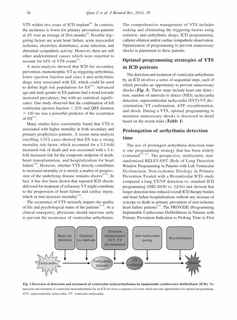
The detection and treatment of ventricular arrhythmias
by an ICD involves a series of sequential steps, each of
which provides an opportunity to prevent unnecessary
shocks (Fig. 1). These steps include heart rate detec-
tion, number of intervals to detect (NID), tachycardia
detection, supraventricular tachycardia (SVT)-VT dis-
crimination, VT confirmation, ATP, reconfirmation,
and shock. During a VTS, optimal programming to
minimize unnecessary shocks is discussed in detail
based on the recent trials (Table 1).
Prolongation of arrhythmia detection
time
The use of prolonged arrhythmia detection time
is one programming strategy that has been widely
evaluated[13-16]
. The prospective, multicentre, non-
randomized RELEVANT (Role of Long Detection
Window Programming in Patients with Left Ventricular
Dysfunction, Non-ischemic Etiology in Primary
Prevention Treated with a Biventricular ICD) study
compared a long VT/VF detection vs. standard ICD
programming (NID 30/40 vs. 12/16) and showed that
longer detection time reduced overall ICD therapy burden
and heart failure hospitalizations without any increase of
syncope or death in primary prevention of non-ischemic
heart failure patients[13]. The PROVIDE (Programming
Implantable Cardioverter Defibrillators in Patients with
Primary Prevention Indication to Prolong Time to First
Fig. 1 Overview of detection and treatment of ventricular tachyarrhythmias by implantable cardioverter defibrillator (ICD). The
detection and treatment of ventricular tachyarrhythmias by an ICD involves a sequence of events which provide opportunities for optimal programming.
SVT: supraventricular tachycardia; VT: ventricular tachycardia.
36 Qian Z et al. J Biomed Res, 2015, 29
Shock) study reported that in a large cohort of patients, a
combination of programmed parameters including higher
detection rate, longer detection intervals, empiric ATP,
and optimized SVT discriminators were associated with
a significant reduction of ICD shock therapy with reduc-
tion in all-cause mortality and without increasing
arrhythmic syncope[14]. The MADIT-RIT (Multicenter
Automatic Defibrillator Implantation Trial-Reduce
Inappropriate Therapy) study was a large randomized
controlled trial and included 1,500 patients[15]. In this
study, patients were randomly assigned to the delayed
(with a 60-second delay at 170 to 199 bpm, a 12-second
delay at 200 to 249 bpm, and a 2.5-second delay at
> 250 bpm) and the conventional groups. The results
showed that programming with a prolonged delay was
associated with reductions in inappropriate therapy
and all-cause mortality. The recent ADVANCE III
(Avoid Delivering Therapies for Non-sustained
Arrhythmias in ICD Patients III) trial has demonstrated
that the use of a long detection interval (30 out of 40
intervals), combined with ATP during charging, signif-
icantly reduces the rate of appropriate therapies (ATP
and shocks) and inappropriate shocks in comparison
with the standard detection interval (18 out of 24) in
single, dual and triple chamber ICDs, regardless of
indication[16]. The ADVANCE III trial confirmed and
Table 1. Clinical trials of shock reduction programming
Study Year Cases
Follow-up
time Design CAD (%)
Secondary
prevention (%) Detection group Control group
PainFREE Rx II[22] 2004 634 11 months RCT 85 52 FVT: 188-250 bpm;
NID 18/24; ATP; Shock
FVT: shock
PREPARE[20] 2008 1391 1 year OBS 64 0 VF: 250 bpm; NID 30/40
FVT: 182 bpm; NID 30/40;
ATP61
VT: 167 bpm;
NID 32; monitor only
Physician tailored
RELEVANT[13] 2009 324 14 months OBS 0 0 VF: 182-500 bpm; NID 30/40
FVT: 182-250 bpm; NID 30/40
ATP61
VT: 167-182 bpm; NID 32
VF: NID 12/16
VT: NID 16
MADIT-RIT[15] 2012 1500 1.4 years RCT 53 0 High-rate therapy group:
VF: 200 bpm; 2.5s; ATP61
VT: 170 bpm; monitor only
Delayed group:
VF: 250 bpm; 2.5s; ATP61
FVT: 200 bpm; 12s; ATP61
VT: 170 bpm; 60s; ATP61
VF: 200 bpm; 1s;
ATP61
VT: 170 bpm; 2.5s;
ATP61
ADVANCE III[16] 2013 1902 12 months RCT 60 25 VF: 188 bpm; NID 30/40;
ATP61
VT: 150 bpm; NID 32;
monitor only
VF: 188 bpm; NID
18/24; ATP61
VT: 150 bpm;
NID 32; monitor only
PROVIDE[14] 2014 1670 530 days RCT 62 0 VF: 250 bpm; NID 12
VT2: 214 bpm; NID 18;
ATP61
VT1: 181 bpm; NID 25;
ATP62
VF: 214 bpm; NID 12
VT2: 181 bpm;
NID 12; ATP62
VT1: 150 bpm;
NID 12; monitor only
PainFree SST[18] 2014 1308 10.6
months
OBS 43 34 VF: 188 bpm (or faster if VT enabled)
Primary prevention:
NID 30/40;
Secondary prevention:
NID randomized to 18/24
vs. 30/40
SVT Limit: 230 bpm
SST algorithms: ON
OBS: observational, nonrandomized study; RCT: randomized clinical trial; NID: number of intervals to detect; VT: ventricular tachycardia; VF:
ventricular fibrillation; FVT: fast ventricular tachycardia; SVT: supraventricular tachycardia; ATP: antitachycardia pacing; CAD: coronary artery
disease.
Optimal programming for ES 37
reinforced the results of MADIT-RIT in a larger and
broader primary prevention ICD population. A meta-
analysis including the above four trials concluded that
the use of long detection time could significantly
decrease the burden of inappropriate shock therapy
(RR 0.50) and all-cause mortality (RR 0.77) without
significant increase in the risk of syncope[17].
The PainFree SST trial was designed to evaluate the
inappropriate shock free rate at one-year post implant
in primary and secondary prevention patients with sin-
gle, dual and triple chamber ICDs by SmartShock
Technology (SST)[18]. The primary results showed that
over 98% of the patients were free of inappropriately
shocked episodes during their first year after implanta-
tion and no difference was detected between primary
(1.6% with inappropriate shocks) and secondary (2.3%)
prevention patients. A cohort of secondary prevention
patients were randomized in a 1:1 fashion to either a
standard interval (NID 5 18/24) or prolonged interval
(NID 5 30/40) detection of VT/VF > 188 bpm (VF
zone). In this large randomized trial involving high-risk
secondary prevention patients, longer detection intervals
did not increase the risk of syncope, ensuring the safety
of this programming strategy. Prolonged interval detec-
tion programming did not impact the rates of inap-
propriate shocks, which were low using advanced
discrimination algorithms in both groups at one year.
In conclusion, the increase of detection time could
prevent both inappropriate shocks (shocks for rhythms
other than VT or VF) and unnecessary shocks (shocks
for self-terminating episodes) in patients with VTS.
However, we should closely evaluate the status of car-
diac function to determine if each individual could
tolerate possible risk resulted from the delayed therapy
of malignant ventricular arrhythmias. Additionally, VT
may recur immediately after successful ATP, which
may lead to misclassification of unsuccessful ATP,
and subsequent unnecessary shocks may be delivered[5].
In some ICDs, we can decrease the number of intervals
for declaring an end of episode to obtain an early defini-
tion of return to sinus rhythm.
Increasing heart rate detection
threshold
According to the heart rate, programmable zones are
strictly defined. ICDs usually provide three zones, VT,
fast VT (FVT), and VF. The description of arrhythmia
characteristics in ICD patients with primary and sec-
ondary prevention indications could guide the settings
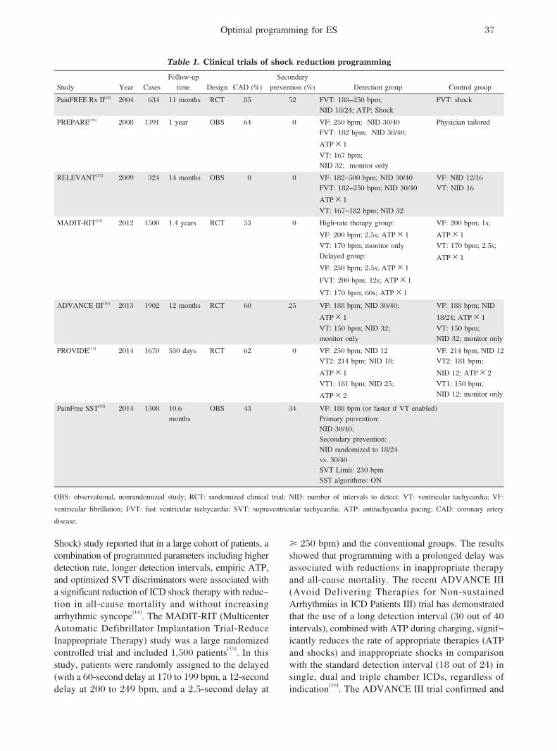
of programmable zones[19]. A shorter VT cycle length
(CL, 303¡54 ms vs. 366 ¡ 71 ms) and a longer
SVT CL (363 ¡ 70 ms vs. 323 ¡ 75 ms) are found
in patients with primary preventive ICDs compared
with those with secondary prevention indications
(Fig. 2). These data indicate that ICD patients of pri-
mary prevention have faster VT and smaller overlap of
SVT and VT; as a result, these patients may benefit
from higher rate detection zones. On the contrary,
patients with a secondary prevention indication will
benefit from slower detection zones and SVT-VT dis-
crimination algorithms because of the greater overlap
between SVT and VT. Several trials have investigated
the strategy combined with longer detection time in
primary prevention patients.
In the PREPARE (Primary Prevention Parameters
Evaluation) study, detection rate was set to 182 bpm
(FVT) and VF 250 bpm[20]. In the MADIT-RIT trial,
detection rate for ventricular arrhythmias was set to
200 bpm with a 2.5-second delay in the high-rate ther-
apy group[15]. Similarly, the PROVIDE study defined
VT, FVT and VF zones as 181 bpm, 214 bpm, and
250 bpm, respectively[14]
. Consistently, these high
Fig. 2 Rates of ventricular arrhythmias detected in 978 patients in whom an ICD was implanted for primary and secondary
prevention. Patients with primary preventive ICDs have a shorter cycle length of ventricular tachycardia compared with secondary preventive patients
and may benefit from higher detection zones. (Cited from: Wilkoff BL, et al. J Cardiovasc Electrophysiol. 2004;15(9):1002-1009[19]
with permission)
38 Qian Z et al. J Biomed Res, 2015, 29

heart rate detection groups had significant reductions in
ICD shocks without an increase in syncope or death. In
Mayo Clinic, the recommended detection rate of pri-
mary prevention patients is set to 200 bpm with a 5-
to 9-second delay for ICD therapies[5].
Increasing the efficacy of ATP
ATP is rapid pacing at a CL shorter than VT that ter-
minates reentrant VT by penetrating the circuit and
blocking the reentry. ATP can reduce unnecessary
shocks, improve quality of life, and lengthen pulse gen-
erator life. Previous studies have demonstrated that ATP
terminated 85%-90% of slow VT (, 188-200 bpm)
with a low risk of acceleration (1%-5%)[21]
. In
PainFREE Rx II (Pacing Fast Ventricular Tachycardia
Reduces Shock Therapies) trial, patients were rando-
mized to ATP followed by shock or shock alone for the
treatment of FVT (188-250 bpm)[22]. The results showed
that a single 8-pulse burst of ATP was successful in
terminating FVT in 72% of episodes and resulted in a
significant reduction in shocks with improved quality of
life and low rates of VT acceleration and syncope
(Fig. 3). However, recent studies showed that efficacy
of ATP with longer NID was not as high as previously
reported. In ADVANCE III trial, ATP efficacy for FVT
(CL 240-320 ms) was 54% [generalized estimating
equation (GEE) adjusted]. One latest report showed that
a new automatic ATP algorithm (an initial ATP train
based on heart rate history, subsequent trains based
on post-ATP interval and shocks applied at timer expiry)
improved ATP efficacy for FVT to 59% (GEE
adjusted)[23].
ATP mode selection
ATP was usually classified as burst if all paced beats
were delivered at the same CL, and ramp if the beat-to-
beat pacing CL was shortened within each pacing train.
Burst and ramp pacing sequences have similar efficacy
Fig. 3 Results of the PainFree Rx II (Pacing Fast Ventricular Tachycardia Reduces Shock Therapies) trial. The upper graph shows
distribution of ventricular arrhythmias by detection zone and median cycle length. The pie charts show percentages of terminating therapy for FVT
episodes in each arm. ATP could terminate FVT in 72% of episodes. FVT: fast ventricular tachycardia; ATP: antitachycardia pacing. (Cited from:
Wathen MS, et al. Circulation. 2004;110(17):2591-2596[22]
with permission)
Optimal programming for ES 39
in slow VTs[21]. For FVT with CL , 300 ms, burst is
more effective and less likely to result in acceleration
than ramp. One study has investigated the efficacy of
four ATP modes, including burst, ramp, scan (if the
pacing CL was shortened between each pacing train),
and ramp/scan (if the pacing CL was shortened both
between and within each pacing train). As a result, when
the VT rate was . 200 bpm, ATP was less successful,
Table 2. Suggested ICD programming for specific ICD indications
Condition Arrhythmia Programming
Primary prevention VF: > 200 bpm Longer detection time or 30 of 40 NID.
Use 1-2 sequences of burst.
FVT: 170-199 bpm Monitor only.
Secondary prevention VF: > 200 bpm 30 of 40 NID.
Use 1-2 sequences of burst.
FVT: 170-199 bpm Use multiple sequences of ATP.
VT: , 170 bpm Monitor only.
ICD: Implantable cardioverter defibrillator; NID: number of intervals to detect; VT: ventricular tachycardia; VF: ventricular fibrillation; FVT: fast
ventricular tachycardia.
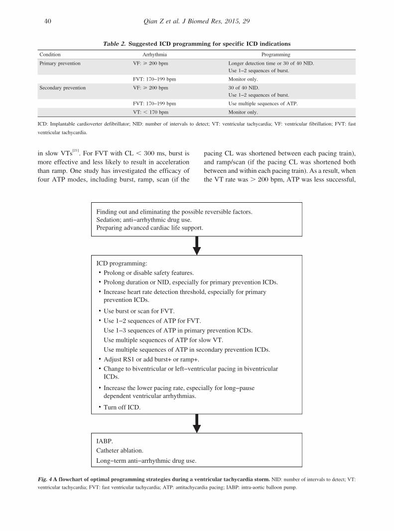
Fig. 4 A flowchart of optimal programming strategies during a ventricular tachycardia storm. NID: number of intervals to detect; VT:
ventricular tachycardia; FVT: fast ventricular tachycardia; ATP: antitachycardia pacing; IABP: intra-aortic balloon pump.
40 Qian Z et al. J Biomed Res, 2015, 29

however, burst and scan exhibited greater efficacy
(81.2% and 87.1% respectively) compared with ramp
(57.1%)[24].
Multi-site ATP
Several studies have reported greater success with
biventricular ATP. One retrospective and observational
study showed that in heart failure patients with biven-
tricular (BiV) ICD, ATP efficacy of left ventricular
(LV) and BiV pacing was higher than right ventricular
(RV) ATP[25]. BiV-ATP and LV-ATP were safer than
RV-ATP for slow VT (150-188 bpm). The ADVANCE
CRT-D trial was a randomized and controlled multicen-
ter trial aimed at comparing the efficacy and safety of
BiV- versus RV-ATP in heart failure patients treated
with cardiac resynchronization therapy-defibrillator[26].
The results showed that BiV-ATP seemed to be more
effective and safer in ischemic patients than RV-ATP.
Multi-sequence ATP
The majority of FVTs were successfully treated by one
or two ATP attempts. Only a small minority of patients
were responsive to. 3 ATPs. Programming a high num-
ber of ATP attempts in the FVT zone is both safe and
efficient and could prevent shocks in ICD recipients[27].
One latest study demonstrated that a second burst pacing
increased the effectiveness of ATP (GEE-adjusted, 65%
vs. 75%) for FVT (CL 250-320 ms) and therefore,
reduced the need for high-energy shocks[28].
Adjusting R-S1 interval (%RR)
Generally, burst CL should be 85%-90% of the VT
CL for FVTs and 70%-80% for slow VTs[21]. When an
unsuccessful ATP occurred, analysis of the return CL
is helpful to optimize ICD programming[5]. The CL of
the drive train could be shortened or an extrastimulus
added at the end of the drive train (for example, ‘‘burst
+’’ mode) in order to penetrate the circuit. If the CL of
VT is unaffected, increasing the number of paced beats
could facilitate penetrating the circuit by peeling away
refractoriness.
Anti-arrhythmic drug therapy
Intensive therapy of the anti-arrhythmic drugs, espe-
cially b-blockers and amiodarone, could help decrease
the VT rate and increase the ATP efficacy[29].
Increasing the lower pacing rate
temporally
In some patients, overdrive pacing by increasing the
lower pacing rate of the ICD may avoid the long pause
after a ventricular ectopic beat, shorten the QT interval
and suppress recurrent VT/VF, particularly if dual
chamber pacing is available[5,30]
.
Overall, during a VT storm, ICD programming
should focus on minimizing shocks. Table 2 shows
the suggested optimal programming for shock reduction
based on ICD indications. Importantly, safety features
that a shock is applied after a programmable time win-
dow independently from ATP should be prolonged or
disabled[5]. Additionally, ICD therapies are often turned
off in hospitalized patients. Catheter ablation might be
the only option when a VTS is refractory to programming
and drug therapies. A flowchart of optimal programming
management during a VTS is exhibited in Fig. 4.
Conclusion
VTS, a life-threatening emergency, is associated with
poor prognosis in ICD patients and requires immediate
medical attention. Optimal programming aiming at
reducing inappropriate or unnecessary shocks may
provide up to a 30% relative decrease in mortality with
no apparent increase in the risk of syncope[31]. During a
VTS, ICD programming focuses on minimizing the
frequency of shocks. Long detection time and high heart
rate detection threshold are key methods, especially for
primary prevention patients. ATP therapy should be
intensified in many ways including ATP mode, number
of sequences, and pacing sites. However, the comprehensive
management of VTS including optimal pharmacological
therapy, sedation, trigger or substrate ablation and dener-
vation is comparably essential.
The optimal programming strategies from the present
clinical trials are mostly aimed at ICD patients for primary
prevention of sudden cardiac death. Future studies are
needed to clarify the roles of different programming
strategies on secondary prevention patients.
Acknowledgment
We thank Dr. Jian Cao (Senior principal scientist,
Medtronic) for his diligent review of this manuscript.
References
[1] Moss AJ, Zareba W, Hall WJ, et al. Multicenter Automatic
Defibrillator Implantation Trial II Investigators. Prophylactic
implantation of a defibrillator in patients with myocardial
infarction and reduced ejection fraction. N Engl J Med
2002;346(12):877-883.
[2] Bardy GH, Lee KL, Mark DB, et al. Sudden Cardiac
Death in Heart Failure Trial (SCD-HeFT) Investigators.
Amiodarone or an implantable cardioverter-defibrillator
for congestive heart failure. N Engl J Med 2005;352(3):
225-237.
[3] Zipes DP, Camm AJ, Borggrefe M, et al. ACC/AHA/ESC
2006 Guidelines for Management of Patients With
Ventricular Arrhythmias and the Prevention of Sudden
Optimal programming for ES 41

Cardiac Death: a report of the American College of
Cardiology/American Heart Association Task Force and
the European Society of Cardiology Committee for
Practice Guidelines (writing committee to develop
Guidelines for Management of Patients With Ventricular
Arrhythmias and the Prevention of Sudden Cardiac
Death): developed in collaboration with the European
Heart Rhythm Association and the Heart Rhythm
Society. Circulation 2006;114(10):e385-484.
[4] Borne RT, Varosy PD, Masoudi FA. Implantable cardio-
verter-defibrillator shocks: epidemiology, outcomes, and
therapeutic approaches. JAMA Intern Med 2013;173(10):
859-865.
[5] Madhavan M, Friedman PA. Optimal programming of
implantable cardiac-defibrillators. Circulation 2013;128(6):
659-672.
[6] Gao D, Sapp JL. Electrical storm: definitions, clinical
importance, and treatment. Curr Opin Cardiol 2013;
28(1):72-79.
[7] Greene M, Newman D, Geist M, et al. Is electrical storm
in ICD patients the sign of a dying heart? Outcome of
patients with clusters of ventricular tachyarrhythmias.
Europace 2000;2(3):263-269.
[8] Exner DV, Pinski SL, Wyse DG, et al. Electrical storm
presages nonsudden death: the antiarrhythmics versus
implantable defibrillators (AVID) trial. Circulation
2001;103(16):2066-2071.
[9] Brigadeau F, Kouakam C, Klug D, et al. Clinical predic-
tors and prognostic significance of electrical storm in
patients with implantable cardioverter defibrillators. Eur
Heart J 2006;27(6):700-707.
[10] Guerra F, Shkoza M, Scappini L, et al. Role of electrical
storm as a mortality and morbidity risk factor and its clin-
ical predictors: a meta-analysis. Europace 2014;16(3):
347-353.
[11] Arya A, Haghjoo M, Dehghani MR, et al. Prevalence and
predictors of electrical storm in patients with implantable
cardioverter-defibrillator. Am J Cardiol 2006;97(3):389-
392.
[12] Joglar JA, Kessler DJ, Welch PJ, et al. Effects of repeated
electrical defibrillations on cardiac troponin I levels. Am J
Cardiol 1999;83(2):270-272.
[13] Gasparini M, Menozzi C, Proclemer A, et al. A simplified
biventricular defibrillator with fixed long detection inter-
vals reduces implantable cardioverter defibrillator (ICD)
interventions and heart failure hospitalizations in patients
with non-ischemic cardiomyopathy implanted for primary
prevention: The RELEVANT study. Eur Heart J 2009;
30(22):2758-2767.
[14] Saeed M, Hanna I, Robotis D, et al. Programming
implantable cardioverter-defibrillators in patients with
primary prevention indication to prolong time to first
shock: results from the PROVIDE study. J Cardiovasc
Electrophysiol 2014;25(1):52-59.
[15] Moss AJ, Schuger C, Beck CA, et al. MADIT-RIT Trial
Investigators. Reduction in inappropriate therapy and
mortality through ICD programming. N Engl J Med
2012;367(24):2275-2283.
[16] Gasparini M, Proclemer A, Klersy C, et al. Effect of long-
detection interval vs. standard-detection interval for
implantable cardioverter-defibrillators on antitachycardia
pacing and shock delivery: the ADVANCE III rando-
mized clinical trial. JAMA 2013;309(18):1903-1911.
[17] Scott PA, Silberbauer J, McDonagh TA, et al. Impact of
prolonged implantable cardioverter-defibrillator arrhyth-
mia detection times on outcomes: A meta-analysis.
Heart Rhythm 2014;11(5):828-835.
[18] Schloss EJ, Auricchio A, Kurita T, et al. PainFree SST
trial primary results: low shock rates in patients with dual
and triple chanmber ICDs using novel detection algo-
rithms. Heart Rhythm 2013;10(5)Supplement: AB28-4.
[19] Wilkoff BL, Hess M, Young J, et al. Differences in
tachyarrhythmia detection and implantable cardioverter
defibrillator therapy by primary or secondary prevention
indication in cardiac resynchronization therapy patients.
J Cardiovasc Electrophysiol 2004;15(9):1002-1009.
[20] Wilkoff BL, Williamson BD, Stern RS, et al. PREPARE
Study Investigators. Strategic programming of detection
and therapy parameters in implantable cardioverter-
defibrillators reduces shocks in primary prevention
patients: results from the PREPARE (Primary Prevention
Parameters Evaluation) study. J Am Coll Cardiol 2008;
52(7):541-550.
[21] Sweeney MO. Antitachycardia pacing for ventricular
tachycardia using implantable cardioverter defibrillators:
Substrates, methods and clinical experience. Pacing Clin
Electrophysiol 2004;27(9):1292-1305.
[22] Wathen MS, DeGroot PJ, Sweeney MO, et al. Prospective
randomized multicenter trial of empirical antitachycardia
pacing versus shocks for spontaneous rapid ventricular
tachycardia in patients with implantable cardioverter-
defibrillators: Pacing fast Ventricular Tachycardia
Reduces Shock Therapies (PainFREE Rx II) Trial results.
Circulation 2004;110(17):2591-2596.
[23] Yee R, Fisher JD, Birgersdotter-Green U, et al. Performance
and safety of a new automatic antitachycardia pacing algo-
rithm based upon electrophysiologic first principles. Heart
Rhythm 2014;11(5)Supplement:AB27-4.
[24] Dewland TA, Carter N, Jones P, et al. Influence of ven-
tricular arrhythmia rate and device programming on anti-
tachycardia pacing efficacy: results from the ALTITUDE
study. Heart Rhythm 2014;11(5)Supplement: AB27-3.
[25] Haghjoo M, Hajahmadi M, Fazelifar AF, et al. Efficacy
and safety of different antitachycardia pacing sites in the
termination of ventricular tachycardia in patients with
biventricular implantable cardioverter-defibrillator.
Europace 2011;13(4):509-513.
[26] Gasparini M, Anselme F, Clementy J, et al. ADVANCE
CRT-D Investigators. BIVentricular versus right ventri-
cular antitachycardia pacing to terminate ventricular
tachyarrhythmias in patients receiving cardiac resynchro-
nization therapy: the ADVANCE CRT-D Trial. Am Heart
J 2010;159(6):1116-1123.e2.
[27] Martins RP, Blangy H, Muresan L, et al. Safety and effi-
cacy of programming a high number of antitachycardia
pacing attempts for fast ventricular tachycardia: a pro-
spective study. Europace 2012;14(10):1457-1464.
[28] Anguera I, Dallaglio P, Sabate X, et al. The benefit of a
second burst antitachycardia sequence for fast ventricular
tachycardia in patients with implantable cardioverter defi-
brillators. Pacing Clin Electrophysiol 2014;37(4):486-
494.
42 Qian Z et al. J Biomed Res, 2015, 29

[29] Connolly SJ, Dorian P, Roberts RS, et al. Comparison of
beta-blockers, amiodarone plus beta-blockers, or sotalol
for prevention of shocks from implantable cardioverter
defibrillators: the OPTIC Study: a randomized trial.
JAMA 2006;295(2):165-171.
[30] Kurisu S, Inoue I, Kawagoe T, et al. Temporary overdriv-
ing pacing as an adjunct to antiarrhythmic drug therapy
for electrical storm in acute myocardial infarction. Circ
J 2005;69(5):613-616.
[31] Tan VH, Wilton SB, Kuriachan V, et al. Impact of pro-
gramming strategies aimed at reducing non-essential
implantable cardioverter defibrillator therapies on mortal-
ity-a systematic review and meta-analysis. Circ Arrhythm
Electrophysiol 2014;7(1):164-170.
9$<$'B$ '33$!' ;$ ?=;'C'< ;'=? C=9 $ 90/
9$0$ �$ 9;'<0$� :D80'��$! =?0'?$
;� 5� ������� 5� ����� >�� ������� ���� ���� � ������ ���
4�5� ��� ���� �" �4 �� ����������
Optimal programming for ES 43