or lect
TRANSCRIPT


OBJECTIVES

On completion of this concept,
the student will be able to:
1. Define the three phases of the peri-
operative period.
2. Identify the causes of preoperative anxiety
and describe the nursing measures to
alleviate it.
3. Identify legal and ethical considerations
related to informed consent.
4. Develop a preoperative teaching plan
designed to promote the patient’s recovery
from anesthesia and surgery, thus preventing
postoperative complications.
5. Describe the interdisciplinary approach to
the care of the patient during surgery.

6. Describe the principles of surgical asepsis.
7. Identify adverse effects of surgery and
anesthesia.
8. Identify the use of the nursing process for
optimizing patient outcomes during the
intra-operative period.
9. Identify assessment parameters appropriate
for the early detection of postoperative
complications.

COURSE
OUTLINE

Preoperative Nursing Management
a. Peri-operative and Peri-anesthesia Nursing
b. Surgical Classifications
1. Emergency/Emergent surgery
2. Urgent
3. Required
4. Elective
5. Optional
c. Informed Consent

d. Psychosocial Nursing Assessment and
Interventions
1. Alleviating Fear
2. Respecting Spiritual and Cultural
Beliefs
e. General Physical Assessment
1. Nutritional Status – Nutrients
important for wound healing and
recovery

2. Drug and Alcohol use
3. Respiratory status
4. Cardiovascular status
5. Hepatic and Renal Function
6. Endocrine Function
7. Immunologic Function
8. Previous Medication Therapy

PATIENT EDUCATION
1. Deep Breathing and Coughing
Exercises
2. Mobility and Active Body Movement
3. Pain Management
4. Cognitive Coping Strategies
a. Imagery
b. Distraction
c. Optimistic Self-recitation

g. Preoperative Nursing Management
1. Managing Nutrition and Fluids
2. Preparing Bowel for Surgery
3. Preparing the skin
h. Immediate Preoperative Nursing
Interventions
1. Administering pre-anesthetic
medications

2. Maintaining the Preoperative Record
3. Transporting the patient to the
pre-surgical Suite
4. Attending to Family Needs
II Intraoperative Nursing Management
a. The Surgical Environment
1. Physical Layout of the O.R. suite

1. Location
2. Principles in Design
3. Exchange Areas
4. Peripheral Support Areas
5. Operating Room
b. Asepsis, Infection Control and
Principles of Sterile Technique
1. Surgical Conscience

2. Infection
a. Process of Infection
b. Classification of Infection
c. Factors Affecting Infection Rates
d. Classification of Operative Wounds
e. Sources of Contamination
f. Infection Control
g. Environmental Control

3. Principles of Sterile Technique
4. Methods of sterilization and
Disinfection
c. Surgical Scrub, Gowning and Gloving
1. The Surgical Scrub
2. Gowning and Gloving Techniques
The Surgical Team
a. Patient

b. Intraoperative Nurses
1. Circulating Nurse
2. Instrument Nurse
3. Division of Duties: Setup,
Procedure, Cleanup
c. Anesthesiologist and Anesthetist
d. Surgeon / Assistant Surgeon
The Surgical Experience

a. Anesthesia: an overview
b. Patients Positions on the Operating
table
c. Preparation of the Operative Site and
Draping
Potential Intra-operative Complications
a. Nausea and Vomiting
b. Hypoxia and other Respiratory
complications

c. Hypothermia
d. Malignant Hypothermia
Postoperative Nursing Management
The Postanesthesia Care Unit
a. Admitting Patient to the PACU
b. Nursing Mgt. in PACU

1. Assessing the patient
2. Maintaining patent airway
3. Maintaining Cardiovascular
stability
4. Relieving Pain and Anxiety
5. Determining readiness for
discharge from the post-anesthesia
care unit

b. The Hospitalized Postoperative Patient
a. Receiving the patient in the clinical unit
b. Nursing Management during the first
hours after surgery
1. Assessing and managing
ventilation
2. Assessing and managing
hemodynamics

3. Assessing and Managing Surgical Sites
4. Assessing and Managing Pain
5. Maintaining Normal Body Temperature
6. Assessing Mental Status
7. Assessing Neurovascular status
8. Assessing and Managing
Gastrointestinal Status
9. Assessing and Managing Voluntary
Voiding

10. Encouraging Mobility
11. Maintaining Safe Environment
12. Providing Emotional Support to
Patient and Family
c. The First Postoperative day to the day of
discharge
1. Relieving Pain
2. Preventing Respiratory Complications
3. Preventing Deep Vein Thrombosis

4. Encouraging Activity and Promoting
Self-care
5. Preventing wound infection and
providing wound care
a. Phases of wound healing
b. Factors affecting wound healing
c. Sterile dressing techniques

6. Managing wound complications
a. Hematoma
b. Wound Sepsis
c. Wound Dehiscence and
Evisceration
7. Resuming Oral intake and Promoting
Bowel Function

Peri-Operative
Nursing

Perioperative Nursing
Surgery – a branch of medicine that deals with
disease and trauma through surgical /
operative procedures.
History of Surgery:
- the earliest recorded sign of surgery was
found in ancient Egyptian papyri.
- they were for treatments of the back, chest ,
and shoulders.

Perioperative Nursing
- earliest known surgeon used a large amount
of tools like: knives, awls, drills, scissors,
saws, forceps, clamps, syringes, mirrors,
needles, cast, splints, & bandages
- surgery has been around since 3,000 B.C.
through today.
- among the first surgeons were battlefield
doctors in the Napoleonic Wars who were
primarily concerned with amputations.

Perioperative Nursing
- naval surgeons were often barber-surgeons,
who combined surgery with their main jobs
as barbers.
- history of surgery can be divided into three
eras: ancient, middle & modern

SUSHRUTA
The “Father of
surgery” and
inventor of Plastic
Surgery.

JOSEPH LISTER
The discoverer of
surgical sepsis and
Listerine named in
his honor.

ALFRED BLALOCK
The first modern
day successful
open heart
surgery in 1944.

CHRISTIAN BARNARD
The cardiac
surgeon who
first performed
the heart
transplantation
in 1967.

HARVEY CUSHING
Pioneer of brain
surgery.

GAVRIL ILIZAROV
Russian
orthopedic
surgeon who
invented the
procedure to
lengthen or
reshape limb
bones.

Perioperative Nursing
Perioperative Nursing – refers to the activities
performed by the professional nurse during
the client’s total surgical experience.
Perioperative period – encompasses a client’s
total surgical experience , including the
preoperative, intraoperative and
postoperative phases.

Phases of Peri-operative
Nursing
1. Preoperative Phase – begins with the
decision to perform surgery and ends with
the client’s transfer to the operating room
(O.R.).
2. Intraoperative Phase – begins when the
client is received in the O.R. and ends with
his admission to the post-anesthesia care
unit (PACU) / Recovery Room.

Phases of Peri-operative
Nursing
3. Postoperative Phase – begins when the
client is admitted to postanethesia care unit
and extends through follow-up home or clinic
evaluation.
Categories of Surgery based on Urgency:
1. Emergent / Emergency - patient requires
immediate attention, disorder may be life
threatening.

indication: without delay
examples: severe bleeding (gunshot or stab
wounds), bladder or intestinal obstruction,
fractured skull and extensive burns
2. Urgent – patient requires prompt attention.
indication: within 24-30 hours
examples: acute gallbladder infection, kidney
or ureteral stones, appendicitis

3. Required – patient needs to have surgery.
indication: plan within a few weeks or months
examples: prostatic hyperplasia (without
obstruction), thyroid disorders and cataracts
4. Elective – procedure performed by choice
indication: failure to have surgery not
essential
examples: repair of scars, hernia and vaginal
repair
5. Optional – decision rest with the patient.
indication: personal preference
example: cosmetic surgery
Classification of Surgery:
1. Diagnostic – e.g. biopsy or exploratory
laparotomy (Ex-Lap)
2. Curative – e.g. tumor excision & inflamed
vermiform appendix

Classification of Surgery
3. Reparative / Reconstructive– bringing back
to its normal functioning. Repair of damaged
organ.
4. Palliative – reduce intensity of
uncomfortable symptoms but not to produce
a cure.

Classification of
Surgery
a. Ablative – Involves removal of an organ.
(suffix used: “ectomy”) appendectomy
b. Constructive – Involves repair of
congenitally defective organ.
(suffixes used are “plasty”, “orrhapy”,
“pexy”) cheiloplasty & orchidopexy
c. Reconstructive – Involves repair of
damaged organ. (plastic surgery)

Classification of
Surgery
Categories of Surgery based on Magnitude/Extent:
A. Major Surgery
Criteria:
1. High risk 4. Large amount of blood loss
2. Extensive 5. Vital organs may be handled
3. Prolonged or be removed
B. Minor Surgery
Criteria:
1. Generally not prolonged
2. Leads to few serious complications
3. Involves less risk

Informed
Consent

Informed Consent – permission obtained from
a patient to perform a specific test or
procedure.
Criteria for a Valid Informed Consent:
1. Voluntary consent – valid consent must be
freely given without coercion.
2. Competent patient – individual who is
autonomous and can give or withhold
consent.
3. Patient able to comprehend – information
must be written and delivered in language
understandable to the patient.

4. Informed subject - consent must be in
writing & must contain the following:
a. Explanation of the procedure and its risk.
b. Description of benefits and alternatives.
c. Instructions that the patient may withdraw
consent.
d. A statement informing the patient if the
protocol differs from customary
procedure.

Informed Consent is necessary in the
following procedures:
1. Invasive procedures – surgical incision, a
biopsy, cystoscopy or paracentesis.
2. Procedures requiring sedation or anesthesia
3. Non-surgical procedure – arteriography,
lumbar puncture
4. Procedures involving radiation

Nursing Responsibilities:
1. The surgeon must provide a clear and
simple explanation of the surgical
procedure.
2. The nurse may ask the patient to sign the
consent form.
3. The nurse may witness the patient’s
signature.

4. If the patient needs additional information
about the procedure, nurse notifies the
surgeon.
5. The nurse ascertains that the consent form
has been signed before administering
psychoactive drugs.
6. If the patient is a minor, unconscious or
incompetent, permission must be obtained
from a responsible family member or legal
guardian.

7. An emancipated minor (married or
independently living or earning on his own)
may sign his own consent form.
8. No patient should be urged or coerced to
sign an operative permit.
9. In an emergency, a surgeon can operate
without the patient’s informed consent.
10. Refusing to undergo a surgical procedure
is a person’s legal right and privilege.


Preoperative
Phase

Preoperative Nursing Problems:
1. Anxiety related to the surgical experience
(anesthesia & pain) & outcome of surgery.
2. Fear related to perceived threat of the
surgical procedure and separation from
support system.

3. Knowledge deficit of preoperative
procedures and protocols and
postoperative expectations
Preoperative Nursing Management:
1. Teach deep-Breathing, Coughing and
Incentive Spirometer
2. Encourage mobility and active body
movement

3. Pain management – patient-controlled
analgesia (PCA), epidural catheter bolus or
infusion & patient controlled epidural
analgesia (PCEA)
4. Teach cognitive coping strategies
a. Imagery – patient concentrates on a
pleasant experience or restful scene.
b. Distraction – patient thinks of an enjoyable
story or recites a favorite poem or song.

PATIENT
CONTROLLED
ANALGESIA


PRE-OP NURSING
MANAGEMENT
Deep Breathing and Coughing Exercises


LEG and FOOT
EXERCISES


c. Optimistic self-recitation – patient recites
optimistic thoughts
(“I know all will go well”)
Instruction for Ambulatory Surgical patients:
a. Inform the patient the scheduled date and
time of the surgery and where to report.
b. Instruct what to bring
(insurance card, list of meds)
c. Instruct what to leave at home
(jewelry, watch )

d. Instruct what to wear (loose-fitting,
comfortable clothes & flat shoes)
e. Remind the patient not to eat or drink as
directed (fasting period of 8 hours or more
is recommended)
Preoperative Psychosocial Management:
1. Reducing Preoperative Anxiety
– music therapy

2. Decreasing Fear
3. Respecting Cultural, Spiritual and Religious
Beliefs
General Preoperative Nursing Management:
1. Managing Nutrition and Fluids
2. Preparing the Bowel for Surgery
3. Preparing the Skin

Immediate Preoperative Nursing Management:
1. Administering Pre-anesthetic Medication
2. Maintaining the Preoperative Record
3. Transporting the Patient to the Pre-surgical
Area
4. Attending to Family Needs

Nursing Evaluation:
1. Reports relief of anxiety
2. Reports that fear is decreased
3. Voices understanding of surgical
intervention
4. Shows no evidence of preoperative
complications


Intra-
operative
Phase

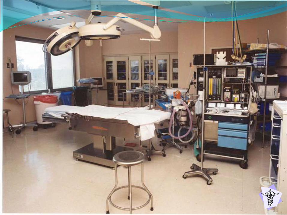
Surgical Environment
Physical Layout of the O.R. Suite:
1. Location – operating room is situated that is
central to all supporting services
(laboratory, radiology, pathology & central
supply room)
2. Principles in Design –
a. Exclusion of contamination from outside
the suite with sensible traffic patterns
within the suite.

b. Separation of clean areas from
contaminated areas within the suite.
3. Exchange Areas -
Surgical Area:
a. Unrestricted zone – street clothes are
allowed
b. Semi-restricted zone – Attire consist of
scrub clothes and caps
c. Restricted zone – scrub clothes, shoe
covers, caps and masks are worn

4. Peripheral Support Areas –
a. Central Administrative Control
b. Offices
c. Conference Room/Classroom
d. Laboratory / Radiology Services
e. Anesthesia Work & Storage Areas
f. Housekeeping Storage Areas
g. Utility Room

h. General Workroom
i. Storage Room
j. Sterile Supply Room
k. Instrument Room
l. Scrub Room
5. Operating Room – surgical suite is behind
double doors ( sliding doors, swing doors )

- Access is limited to authorized
personnel.
- External precautions include adhering
to principles of surgical asepsis.
- Strict control of the operating room
environment is required.
- OR has special air filtration devices to
screen out contaminating particles,
dust, and pollutants.
- Temperature, humidity & airflow
patterns are controlled.


Infection
Infection – is the product of the entrance,
growth, metabolic activities & patho-
physiologic effects of microorganism in
living tissues.
Three Stages of infection:
1. Invasion
2. Localization
3. Resolution leading to recovery

Infection
Process

Acute Bacterial Infection
(most common sepsis in surgical patients)
Wound infection begins 4th to 8th postoperative
Cellulitis pain, redness & swelling
(diffuse inflammatory process)
RBC’s, Leukocytes & Macrophages infiltrate the cells
(localization & containment of infecting microorganism)
Abscess / Pus formation
(suppuration)
If localization is inadequate
Spreading & extension occur causing regional infection
Microorganism & metabolic products carried into the lymphatic system

Lymphangitis
Failure of lymph nodes to hold infection
Uncontrolled cellulitis
Systemic infection occurs chills, fever, signs of toxicity
Septic Thrombophlebitis
Septic emboli in circulation
causing more infection and abscess in remote tissues
Elevates the patient’s metabolic rate 30% to 40% above average
(imposing stress on the body’s vital systems)
Body’s defenses still not able to overcome the infectious process
Septic shock
(Fever, restlessness, hypotension, hypoxia, cloudy sensorium, tachycadia)
rapid breathing, DIC, metabolic acidosis and oliguria
Death

Classification of
Infection
Classification of Infection:
1. Community-Acquired Infections – are
natural disease processes that developed
or were incubating before a patient’s
admission to the hospital or ambulatory
care facility.
2. Communicable Disease – Systemic
bacterial, viral or fungal infections may
be transmitted from one person to
another (HIV, hepatitis & Tuberculosis)

3. Spontaneous Infections – Localized
infections requiring surgical diagnosis and
or treatment for management or that occur
as adjuvants to medical therapy
(acute appendicitis, cholecystitis & bowel
perforation with peritonitis)
4. Nosocomial Infections – are hospital-
associated or acquired during the course of
health care of the patient.

Types of Nosocomial Infections:
1. Exogenous – infection is acquired from
sources outside the body
( personnel & environment )
2. Endogenous – infection develops from
sources within the body.
( abdominal sepsis caused from enteric flora
due to perforation )

Classification of Surgical Wounds:
1. Clean Wound
- No inflammation present
- Procedure under ideal O.R. conditions
- No break in sterile technique
- GIT, respiratory, genitourinary &
oropharyngeal cavity not entered
Infection rate: 1% to 5%

2. Clean-Contaminated Wound
- No inflammation or infection present
- Minor break in technique occurred
- Primary closure, wound drained
- GIT, respiratory, genitourinary tracts &
oropharyngeal cavity entered under
controlled conditions & no spillage &
contamination
Infection rate: 8% to 11%

3. Contaminated Wound
- Major break in technique occurred
- Open fresh traumatic of less than 4 hours
- Acute non purulent inflammation present
- Gross spillage/contamination from GIT
- Entrance to genitourinary or biliary tracts
with infected urine or bile present
Infection rate: 15% to 20%

4. Dirty and Infected Wound
- Organism present in surgical field before
procedure
- Perforated viscus
- Old traumatic wound of more than 4 hours
- Existing clinical infection: acute bacterial
inflammation encountered, with or without
purulence
Infection rate: 27% to 40%

Sources of
Contamination
1. Skin
2. Hair
3. Nasopharynx
4. Fomites
5. Air
6. Human Error
7. Cross Infection


STERILE / AUTOCLAVE TAPES

PREPARING A
STERILE FIELD


O.R. SCRUB SUIT

Surgical Team
The Surgical Team / Perioperative Team:
1. Circulating Nurse – also known as the
“circulator”
Responsibilities:
a. Manages the operating room
b. Protects patient’s safety and health by
monitoring the activities of the surgical
team.


Checks and verifies the consent form.
Ensure fire safety precautions,
cleanliness, proper temperature, humidity
and lighting of the O.R.
Monitors safe functioning of the
equipments.
Coordinates with the surgical / peri-
operative team and monitors aseptic
practices.
Documents O.R. surgical activities

3. Scrub Nurse – responsible for scrubbing for
the surgery.
Responsibilities:
a. Setting up sterile tables
b. Preparing sterile sutures, ligatures &
special equipments
c. Assisting the surgeon & assistant
surgeon, taking care tissue specimens
d. Count all needles, sponges & instruments
together with the circulating nurse


4. Surgeon – head of the surgical team
Responsibilities
a. Performs the surgical procedure
5. RN/INTERN/Co-Surgeon First Assistant –
practices under the supervision of the
surgeon
Responsibilities:
a. Suturing and handling of tissues
b. Providing exposure at the operative field
c. Providing homeostasis


6. Anesthesiologist – is a physician
specifically trained in the art and science of
anesthesiology.
- Anesthetist is a qualified health care
professional who administer anesthetics.
Responsibilities:
a. Interviews and assesses the patient
b. Select & administer appropriate
anesthesia
c. Monitors V/S, ECG, ABG & anesthesia
levels


7. Post Anesthesia Care Unit (PACU) Nurse –
responsible for caring for the patient until
the patient has recovered from the effects
of anesthesia.
Responsibilities:
a. Monitors V/S and post-operative
complications
(bleeding, respiratory distress etc)
b. Carry out postoperative orders
c. Refer any abnormalities to the physician

Anesthesia
Anesthesia – a state of narcosis, analgesia,
relaxation and loss of reflexes.
Levels of Sedation and Anesthesia:
1. Minimal sedation – is a drug-induced state
wherein patient can respond normally to
verbal command. Cognitive & coordination
is impaired but respiratory &
cardiovascular is not affected.

2. Moderate Sedation – a depressed level
of consciousness that does not impair
the patient’s ability to maintain patent
airway & respond to physical
stimulation and verbal commands, often
called
“ monitored anesthesia care”
( intravenous drugs: midazolam &
diazepam )

3. Deep sedation – is a drug induced
state which a patient cannot be
easily aroused but can respond
purposely after repeated
stimulation.
• Difference of deep sedation and
anesthesia is that the anesthetized
patient is not arousable.

Types of
Anesthesia
Types of Anesthesia:
1. General anesthesia – (inhaled or
intravenously) refers to drug-induced
depression of the central nervous system
that produces analgesia, amnesia and
unconsciousness.
volatile liquids – Halothane, Isofluorane,
methoxyflurane, enflurane

Tranquilizers and Sedative-
Hypnotics - Midazolam ( Midazolam )
Diazepam ( Valium )
Opioids - Morphine, Meperidine Hcl
( Demerol )
Neurolept Analgesics – Fentanyl

Dissociative Agent – Ketamine
( Ketalar )
Barbiturates – Thiopental Sodium
( Pentothal )
Methohexital Na ( Brevital )
Nonbarbiturates Hypnotics –
Propofol (Diprivan)

2. Regional Anesthesia
– is a form of local anesthesia that
suspends sensation and motion in a body
region or part, the patient is awake and
continuous monitoring is required.
3. Spinal Anesthesia
– is a local anesthetic injected into the
subarachnoid space at the lumbar level to
block nerves and suspend sensation and
motion to the lower extremities, perineum
and lower abdomen.

4. Conduction Blocks – suspend sensation
and motion on various groups of nerves.
Types of conduction blocks:
a. Epidural block – anesthetic into space the
dura mater
b. Brachial plexus – produces anesthesia on
the arm
c. Paravertebral block – produces
anesthesia of the chest, abdominal wall &
ext.
d. Transacral (caudal) – anesthesia of the
perineum

General Anesthesia
Spinal Anesthesia

Local Anesthetics Agents
1. Lidocaine (xylocaine) – topical or injection
Advantages: Rapid, longer duration of action
compared with procaine & free from local
irritation effect
2. Bupivacaine (sensorcaine) – infiltration,
peripheral nerve block & epidural
Advantages: Duration is 2-3 times longer
than lidocaine

Local Anesthetics Agents
3. Procaine (Novocaine) – subcutaneously,
intramuscular, intravenously & spinal
Advantages: low toxicity & inexpensive
4. Tetracaine (Pontocaine) – topical,
infiltration & nerve block
Advantages: low toxicity & inexpensive

Stages of General Anesthesia:
Stage I Beginning anesthesia
– feeling of warmth, dizziness &
detachment may be experienced, unable to
move extremities easily, experiences
roaring, ringing & buzzing in the ears.
Stage II Excitement
– characterized by struggling, shouting,
laughing, crying, increased pulse and
irregular respirations. Pupils dilate but
contract to light.

Stages of Anesthesia
Stage III Surgical Anesthesia
– patient is unconscious and lies quietly
on the table, surgical procedure
begins. Pupils are small but contract
when exposed to light. Respirations
are regular, pulse rate normal, skin is
pink and slightly flushed.

Stages of Anesthesia
Stage IV Medullary Depression/Danger
– this stage is reached when too much
anesthesia has been administered.
Respiration is shallow, pulse is weak &
thready, pupils dilated & non-reactive,
cyanosis develops & without prompt
intervention death rapidly follows.

Types of Anesthesia
2. Regional Anesthesia – is a form of local
anesthesia that suspends sensation and
motion in a body region or part, the
patient is awake and continuous
monitoring is required.
3. Spinal Anesthesia – is a local anesthetic
injected into the subarachnoid space at
the lumbar level to block nerves and
suspend sensation and motion to the
lower extremities, perineum and lower
abdomen.

Intraoperative Complications
Potential Intraoperative Complications:
Nausea and Vomiting
– if it occurs, turn patient to side, the
head of the table is lowered and a basin
is provided to collect vomitus.
- Suction saliva and vomited gastric
contents.
- Administration of anti-emetics.

Anaphylaxis
– is a life threatening acute allergic
reaction that causes vasodilation,
hypotension and bronchial constriction.
- carefully observe the patient for
changes in V/S and symptoms of
anaphylaxis.

Hypoxia & other Respiratory Complications
– inadequate ventilation, occlusion of the
airway, inadvertent intubation of the
esophagus and hypoxia are potential problems
of general anesthesia
- Peripheral perfusion & pulse oximetry are
monitored continuously.
- Vigilant assessment of the patient’s
oxygenation status is a primary function of the
anesthesiologist or anesthetist or circulating
nurse.

Hypothermia – body temperature below 36.6
- caused by low temperature in OR, infusion of
cold fluids, inhalation of cold gases, open
body wounds, decreased muscle activity and
advanced age.
Malignant Hyperthermia
– is an inherited muscle disorder chemically
induced by anesthetic agent.
- Susceptible people include those with
strong and bulky muscles, a history of
muscle cramps or muscle weakness and
unexplained temperature elevation.

Pathophysiology of Malignant Hyperthermia
Halothane, Enflurane (GA gases), Succinylcholine (muscle relaxant),
Stress
Muscle cell activity
Muscles cells composed of inner fluid (sarcoplasm) and
Outer surrounding membrane
Calcium (essential factor in muscle contraction) is normally stored in
sarcoplasm
Nerve impulses stimulate the muscle
Calcium is released, allowing contraction to occur
Pumping action mechanism return calcium to the sac
so that muscle can relax

Pathophysiology of Malignant Hyperthermia
Malignant Hyperthermia, this mechanism is disrupted
Calcium ions accumulate causing clinical symptoms of
hypermetabolism
Increases muscle contraction (rigidity), hyperthermia
Damage to the Central Nervous system

Clinical Manifestation:
1. Tachycardia >150 beats/min. (earliest
sign)
2. Hypotension
3. Decreased cardiac output
4. Oliguria
5. Body temperature >40 Celsius (late sign)
6. Cardiac arrest

Medical Management:
1. Discontinuing the anesthesia and
surgery
2. Administration of a muscle relaxant
and Sodium Bicarbonate
3. Decrease body temperature
4. Correct electrolyte imbalance
Nursing Management:
- Identify patient’s at risk, recognize the
signs & symptoms, have appropriate
medications and equipment available.

Disseminated Intravascular
Coagulopathy
( DIC )
- is a life-threatening condition
characterized by thrombus
formation and depletion of
select coagulation proteins.

Patient Position on the Operating
Table:
1. Dorsal recumbent
– flat on the back, used for most
abdominal surgeries.
2. Trendelenberg position
- the head & body are lowered,
used for surgery on the lower
abdomen and pelvis.

3. Lithotomy position
– patient positioned at the back with
the legs and thighs flexed used for
perineal, rectal and vaginal
surgical procedures.
4. Sims or lateral position
– patient positioned on the non-
operative side, used for renal
surgery.

SUPINE

SITTING

LITHOTOMY

REVERSE
TRENDELENBERG

LATERAL

PRONE

Preparation of the
Operative Site
-Skin preparation (skin prep) begins
before the patient arrive in the OR.
Purpose:
- is to render the surgical site as free as
possible from transient and resident
microorganisms, dirt, and skin oil so the
incision can be made through the skin
with minimal danger of infection from this
source.


DRAPING
Draping - is the procedure of covering the
patient and surrounding areas with a
sterile barrier to create and maintain an
adequate sterile field.

SURGICAL
INSTRUMENTS

Surgical Instruments
Important Nursing Consideration:
Surgical instruments are designed to
provide the tools the surgeon needs for its
maneuver, they are classified by their
functions whether small, short, long,
straight, curve, sharp or blunt. All surgical
instruments should be used for their
intended purposes only and should not be
abused.

Parts of the Surgical
Instrument
Finger Ring
Ratchets
Shank Boxlock/Hinge Joint
Tip
Jaws

Classification of Instruments:
1. Cutting and Dissecting – instruments that
have sharp edges, used to dissect,
incise, separate, cut and excise tissues.
Nursing Responsibilities:
1. These instruments should be kept
separate from other instruments.
2. Demand careful handling at all times.
Examples:
Scalpels, Blades, Scissors, Knives, Bone
cutters, Curettes and Biopsy forceps

2. Grasping and Holding
– instruments used to grasp or hold
tissues (soft or hard) during the
surgical operation.
Examples:
Thumb forceps, Tissue forceps, Allis
forceps, Babcock forceps, Tenaculum,
Bone holders

3. Clamping or Occluding – instruments
used to apply pressure or occluding blood
vessels to prevent bleeding.
Examples: Kelly/Clamps, Pean, Ochsner,
Vascular mixter

Mosquito Clamp Kelly / Clamp Vascular Mixter

4. Retracting or Exposing – instruments used
to pull aside tissues, muscles & other
structures for exposure of the surgical site.
Types:
a.) Handheld retractor
b.) Self-retaining retractor
Examples: Balfour, Army/Navy, Richardson,
Malleable, Hooks and Deaver

Army-Navy Balfour / Self-retaining

Deaver Richardson Double-ended Richardson

5. Suturing and stapling
– instruments used to close/suture
the tissues and other structures of
the operative site.
Examples: Needle holder, free
needles (round or cutting),
Atraumatic needle and staplers

Needle Holder Free Needles
Skin Stapler Atraumatic Suture Needle

6. Viewing Instruments
– used to view the operative site.
Examples: Speculum and
Endoscopes

Endoscopes
Vaginal Speculum

7. Suctioning and Aspirating
– instruments used to suction
blood and other body fluids on the
operative site.
Examples: Poole Suction, Cannula,
Trocar, Yankeur suction, Frazier
Suction

Yankeur Suction
Poole Suction
Frazier

8. Dilating and Probing
– dilating instruments are used to
enlarge orifice and ducts while a
probe is used to explore a
structure or to locate an
obstruction
Examples: Common bile duct
dilators, esophageal dilators,
Probes
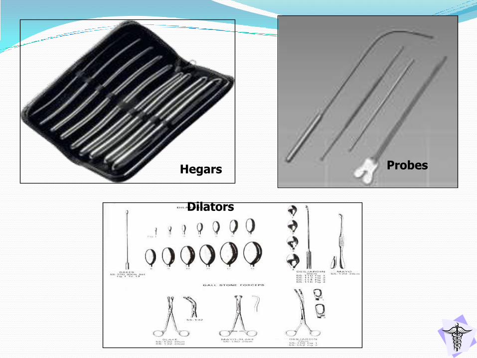
Hegars Probes
Dilators

9. Accessory instruments
– used in addition to basic
instruments.
Examples: Towel clips, Bovie
pencil, Ruler

Towel Clips
Surgical Ruler
Cautery Pad
Cautery Cord
Bipolar Cautery TipKidney Basin

Key Points in handling the instrument:
1. Scrub person counts all instruments &
sharps with circulating nurse (before and
after) in the procedure.
2. Never pile the instruments on top of
each other.
3. Know the name & use of the
instrument.
4. Handle the instrument individually.
5. Hand the surgeon/asst. surgeon the
correct instrument.
6. Pass the instrument firmly & decisively.
7. Careful handling of sharp instruments
at all times.

Postoperative
Phase

Objective of Postoperative Period:
1. Maintain adequate body system
functions.
2. Restore homeostasis
3. Alleviate pain and discomfort
4. Prevent postoperative
complications
5. Ensure adequate discharge
planning and teaching

Post-Anesthesia Care Unit
Postanesthesia Care Unit (PACU)
– is located adjacent to the operating rooms,
patients under anesthesia are placed in this
unit for easy access to experienced, highly
skilled nurse, anesthesiologists, nurse
anesthetist, surgeons and special
equipments & medications.
- PACU is kept quiet, clean & free of
unnecessary equipments & well ventilated.


Phases of PACU:
1. Phase I PACU – used during the
immediate recovery phase and intensive
nursing care is provided
2. Phase II PACU – is reserved for patients
who requires less frequent observation
and less nursing care
- the patient is prepared for discharge.

Admitting Patient to PACU:
1. Anesthesiologist or anesthetist is
responsible in transferring the patient from
the O.R. to the PACU
2. Avoid unnecessary body exposure.
3. Avoid rough handling
4. Avoid hurried movement & rapid changes in
position

5. Nurse who admits patient to the PACU
reviews the following information:
a. Medical diagnosis and type of surgery
performed
b. Pertinent past medical history &
allergies
c. Patient’s age and general condition,
airway patency & vital signs
d. Anesthetics & other medication used in
the procedure
Nursing Management in the PACU:
Assessing the Patient
a. Appraise air exchanges status & note
skin color.
b. Verify & identify operative status &
surgeon.
c. Assess neurologic status (LOC)
d. Examine operative site & check
dressings

e. Perform safety checks
– good body alignment, side rails &
restraints for IVF & blood transfusion
f. Require briefing on problems
encountered in OR
Maintaining a Patent Airway
a. Lateral position with neck extended
b. Keep airway in place until fully awake
c. Suction secretions

d. encourage deep breathing
e. administer humidified oxygen as ordered
Maintaining Cardiovascular Stability
a. Monitor VS and report abnormalities
b. Observe signs & symptoms of shock and
hemorrhage

Classic signs/symptoms of shock:
1. Pallor
2. Cool & moist skin
3. Rapid Breathing
4. Cyanosis of the lips, gums & tongue
5. Rapid, weak, thready pulse
6. Decreasing pulse pressure
7. Hypotension & concentrated urine
c. Promote comfort & maintain safety
d. Continuous monitoring until patient is
completely out of anesthesia

e. Recognize & minimize factors that
may affect the patient in PACU.
Relieving Pain & Anxiety
a. Opioid analgesics administration
b. Allow family member to visit PACU
Controlling Nausea & Vomiting
a. Administration of anti-emetics
( metoclopramide (plasil), promethazine )

Determining Readiness for Discharge from
the PACU:
1. Stable vital signs
2. Orientation to person, place, events and time
3. Uncompromised pulmonary function
4. Pulse oximetry readings indicating adequate
blood oxygen saturation
5. Urine output at least 30 cc/hr
6. Nausea & vomiting absent or under control
7. Minimal pain

Modified Aldrete Scoring System –
determine the patient’s general
condition and readiness for transfer from
PACU, it allows more objective
assessment at regular interval.

Shock – response of the body to a
decrease in the circulating blood volume
which results to poor tissue perfusion &
inadequate tissue oxygenation (tissue
hypoxia)
1. Hemorrhage – copious escape of blood
from the blood vessel
Capillary: slow, generalized oozing
Venous: dark in color and bubble out
Arterial: spurts & is bright red in color

Clinical Manifestations:
1. Apprehension, restlessness, thirst,
cold, moist, pale skin
2. Deep & rapid RR, low body
temperature
3. Low cardiac output
Medical Management:
1. Vitamin K, Hemostan
2. Ligation bleeders, pressure dressing,
BT & IV fluids

2. Femoral Phlebitis / Deep
Thrombophlebitis – often occurs after
operation on the lower abdomen or during
the course of septic conditions as
ruptured ulcer or peritonitis.
Etiologic factors:
1. Injury: damage to vein
2. Hemorrhage
3. Prolonged immobility
4. Obesity / Debilitation

Clinical Manifestations:
1. Pain
2. Redness
3. Swelling
4. Heat / warmth
5. Homan’s sign (cardinal sign)

Nursing Management:
(Active Intervention)
1. Bed rest, elevate affected leg with
pillow support
2. Wear anti-embolic support hose from
the toes to the groin
3. Avoid massage on the calf of the leg
4. Initiate anticoagulant therapy as
ordered

Preventions:
1. Hydrate adequately (to prevent
hemoconcentration)
2. Leg exercises and ambulate early
3. Avoid any restricting devices
4. Preventing use of bed rolls, knee
gatches, dangling over the side of the
bed with pressure on popliteal area

3. Wound Infections
Etiologic Factors:
a. Staphylococcus aureus
b. Escherichia coli
c. Proteus vulgaris
d. Pseudomonas aerogenosa
e. Anaerobic bacteria

Clinical Manifestations:
1. Redness, swelling, pain, warmth
2. Pus or other discharges on the
wound
3. Foul smell from the wound
4. Elevated temperature, chills
5. Tender lymph nodes on the axilla or
groin

Rule of thumb
1. Fever 1st
24 hours – Pulmonary infection
2. Within 48 hours – Urinary Tract Infection
3. Within 72 hours – Wound Infection
Preventive Interventions:
1. Housekeeping cleanliness in the OR
2. Strict Aseptic Technique
3. Antibiotic therapy

4. Wound Complications
Kinds
1. Hemorrhage / Hematoma
2. Wound dehiscence – disruption in the
coaptation of wound edges
3. Wound Evisceration – dehiscence with
outpouching of abdominal organs


Nursing Management:
1. Apply abdominal binder
2. Encourage proper nutrition
3. Keep in Bed
4. Stay with client, have someone call M.D.
5. Cover exposed intestine with sterile, moist
saline dressing
6. Supine or semi-fowlers, bend knees to
relieve tension on abdominal muscle

5. Pulmonary Complications
– atelectasis, Brochitis, Bronchopneumonia,
Lobar pneumonia, Hypostatic pulmonary
congestion & pleurisy
Nursing Management:
1. Reinforce deep breathing, coughing,
turning exercises
2. Encourage early ambulation
3. Incentive spirometer

6. Intestinal Obstruction (3rd
– 5th
Postop day)
– Loop of intestine may kink due to
inflammatory adhesion
Clinical Manifestation:
1. Intermittent sharp, colicky abdominal
pains
2. Nausea and vomiting (fecaloid)
3. Abdominal distention, hiccups
4. Diarrhea, shock & death

Nursing Management:
1. NGT insertion
2. Administer electrolyte / IV as ordered
3. Prepare for possible surgical
intervention
7. Hiccups – intermittent spasms of the
diaphragm causing a sound “hic” that
result from the vibration of closed vocal
cords as air suddenly into the lungs

Etiologic Factor:
1. irritation of phrenic nerve between the
spinal cord and terminal ramifications on
undersurface of the diaphragm.
Nursing Management:
1. Remove the cause
2. NGT for abdominal distention
3. Hold breath while taking a large
swallow of water / Metoclopramide
administration
4. Breath in and out paper bag (CO2)

Promoting Home and Community-Based Care:
1. Teaching Patient’s self care
a. Give written instructions on medications,
medical check-ups, wound care, activity
& diet.
b. Provide the nurse and surgeon’s number
2. Continuing Care
a. Assess patient’s physical status (surgical
incision, respiratory, cardiovascular &
pain management)

3. Previous teachings is reinforced as
needed
4. Change the wound dressings,
monitor the drainage system &
administer medications
5. Patient reminded of the importance
of follow-up appointments.
