oral controlled release liquid dosage forms
TRANSCRIPT

ORAL CONTROLLED RELEASE LIQUID DOSAGE
FORMS (RECONSTITUTABLE POWDER) BY ION
EXCHANGE RESINS
Dissertation zur Erlangung des akademischen Grades des
Doktors der Naturwissenschaften (Dr. rer. nat.)
eingereicht im Fachbereich Biologie, Chemie, Pharmazie
der Freien Universität Berlin
vorgelegt von
NADEEM ISHAQUE KHAN
aus Pakistan
Feburary, 2019

1. Gutachter: Prof. Dr. Roland Bodmeier
2. Gutachter: Prof. Dr. Philippe Maincent
Tag der mündlichen Prüfung: 17/05/2019

To my parents and family,
with love and gratitude

Acknowledgements
I extend my heartiest thanks to Almighty Allah, who provided me strength to complete this work.
First of all, I want to say a million thanks to my supervisor Prof. Dr. Roland Bodmeier for
providing me the opportunity to work in his world-renowned research group. I am extremely
grateful to him for all his support throughout my Ph.D. studies. It is just because of his continued
guidance and encouragement that made me able to complete this tiny piece of work.
I am grateful to Prof. Dr. Philippe Maincent for co-evaluating this thesis. I cannot forget to thank
Dr. Abid Riaz Ahmed, Dr. Andrei Dashevsky, Dr. Muhammad Irfan and Mr. Naveed Zafar for
their never-ending support and fruitful discussions throughout my Ph.D. studies. Their scientific
input helped me a lot to complete my doctoral studies.
It is also my honor, to thank Higher Education Commission (HEC) of Pakistan for providing
financial support. In addition, I would like to thank German Academic Exchange Service (DAAD)
for its collaboration with HEC to maintain all records.
Thanks to all my ex- and present colleagues; Dr. Mesut Ciper, Dr. Martin Körber, Dr. Gaith
Zoubari, Dr. Rebaz H Ali, Dr. Anis, Dr. Kathrin Burki, Dr. Agnieszka Solik, Dr. Jelena Teodosic,
Dr. Rahul Sonawane, Marco Bellini, Miriam Colombo, Reza Goldoozian, Marina Kolbina,
Chengcheng Zhao, Luisa Duque, Jia Deng, Prutha Gaitonde, Gabriela Karsubke, Stefan Walter,
Eva Ewest and Andreas Krause for their support and providing a friendly atmosphere during my
stay at the institute.
Last but not least, I would like to express my deepest gratitude to my spiritual master (Jalal-ud-
Din Rumi and Dr. Allama Iqbal) and my parents and the rest of family for their love and my wife
and daughters, their patience and their everlasting support throughout my whole life.

I
TABLE OF CONTENTS
CHAPTER PAGE
1 CHAPTER 1. INTRODUCTION ........................................................................................ 1
1.1 BACKGROUND ................................................................................................................. 2
1.2 STRUCTURE OF ION EXCHANGE RESINS ........................................................................ 2
1.3 TYPES OF ION EXCHANGE RESINS ................................................................................. 4
1.3.1 Strong acid cation exchange resin............................................................................... 5
1.3.2 Weak acid cation exchange resin ................................................................................ 5
1.3.3 Strong base anion exchange resin ............................................................................... 5
1.3.4 Weak base anion exchange resin ................................................................................ 5
1.4 PHYSICOCHEMICAL PROPERTIES OF ION EXCHANGE RESINS...................................... 6
1.4.1 Cross linkage ............................................................................................................... 6
1.4.2 Ion Exchange Capacity ............................................................................................... 7
1.4.3 Swelling/expansion of ion exchange Resins ............................................................... 8
1.4.4 Moisture contents of ion exchange resins ................................................................... 8
1.4.5 Particle size of ion exchange resin .............................................................................. 8
1.4.6 Equilibration rate ........................................................................................................ 9
1.4.7 Flow rate ..................................................................................................................... 9
1.5 USES OF ION EXCHANGE RESINS ................................................................................... 9
1.5.1 Pharmaceutical use of ion exchange resins ................................................................. 9
1.5.2 Taste Masking ........................................................................................................... 10
1.5.3 Sustained/extended release ....................................................................................... 10
1.5.4 Site specific drug delivery systems ........................................................................... 10
1.5.5 Gastric drug delivery systems ................................................................................... 11
1.5.6 Nasal/Ophthalmic drug delivery system ................................................................... 11
1.5.7 Stability improvement ............................................................................................... 11
1.5.8 Treatment of cancer .................................................................................................. 12
1.6 COATING OF DRUG-RESIN COMPLEX (DRC) ............................................................... 12

II
1.6.1 Coating Equipment and different types of coating ................................................... 12
1.6.2 Fluidized bed coater .................................................................................................. 12
1.6.3 Aqueous dispersion coating ...................................................................................... 13
1.6.4 Organic coating ......................................................................................................... 14
1.6.5 Polymers for coating ................................................................................................. 15
1.6.5.1 Ethyl cellulose .............................................................................................................. 15
1.6.5.2 Polyvinyl acetate .......................................................................................................... 16
1.6.5.3 Acrylates ....................................................................................................................... 16
1.6.5.4 Eudragit® NE 30D ........................................................................................................ 16
1.6.5.5 Eudragit® RS/RL 30D .................................................................................................. 17
1.6.5.6 Eudragit® L 30D-55 ...................................................................................................... 18
1.7 MECHANISM OF FILM FORMATION .............................................................................. 19
1.8 SOLUTION/SUSPENSION LAYERING .............................................................................. 21
1.9 ADDITIVES .................................................................................................................... 22
1.9.1 Pore formers .............................................................................................................. 22
1.9.2 Plasticizers ................................................................................................................ 23
1.9.3 Anti-taking agents ..................................................................................................... 23
1.10 STORAGE STABILITY .................................................................................................... 24
1.11 ORAL LIQUID-CONTROLLED RELEASE FORMULATIONS .............................................. 26
1.12 COMMERCIAL PRODUCTS OF ION EXCHANGE RESINS ................................................. 26
1.13 CHALLENGES ................................................................................................................ 29
1.13.1 Tackiness............................................................................................................... 29
1.13.2 Incomplete release of drug from drug-resin complex (resinates) ......................... 31
1.13.3 Drug leaching/ drug retention by IERs ................................................................. 32
1.14 RESEARCH OBJECTIVES ............................................................................................... 33
2 CHAPTER 2. MATERIALS AND METHODS ............................................................... 34
2.1 MATERIALS ................................................................................................................... 35
2.2 METHODS ...................................................................................................................... 36
2.2.1 Purification of ion exchange resins ........................................................................... 36
2.2.2 Moisture content of ion exchange resins .................................................................. 36
2.2.3 Swelling of ion exchange resins ............................................................................... 36

III
2.2.4 Particle size measurement ......................................................................................... 37
2.2.4.1 Powder laser diffraction ............................................................................................... 37
2.2.4.2 Liquid laser diffraction ................................................................................................. 37
2.2.4.3 Light Microscopy ......................................................................................................... 37
2.2.5 Reduction of particle size of resinates ...................................................................... 37
2.2.6 Drug loading on resin/preparation of drug-resin complex (DRC) ............................ 38
2.2.6.1 Batch method ................................................................................................................ 38
2.2.6.2 Column method ............................................................................................................ 39
2.2.7 Effect of temperature, pH and particle size on drug loading .................................... 39
2.2.8 Conductivity studies and change in pH .................................................................... 40
2.2.9 Vacuum filtration and drying of resinates ................................................................ 40
2.2.10 Characterization of drug-resin complex (resinates) .............................................. 40
2.2.10.1 Powder x-ray diffraction (PXRD) ................................................................................ 40
2.2.10.2 Fourier transforms infra-red spectroscopy (FTIR) ....................................................... 41
2.2.11 Wet granulation of resinates ................................................................................. 41
2.2.12 Coating of drug-resin complex (resinates) ............................................................ 41
2.2.12.1 Eudragit® NE 30D ........................................................................................................ 42
2.2.12.2 Aquacoat® 30D and Surelease® .................................................................................... 43
2.2.12.3 Kollicoat® SR 30D........................................................................................................ 43
2.2.12.4 Eudragit® RS 30D and Eudragit® RL 30D ................................................................... 43
2.2.12.5 Eudragit® L 30D-55 ...................................................................................................... 44
2.2.13 Diluent granules for reconstitution ....................................................................... 44
2.2.14 Stability studies ..................................................................................................... 45
2.2.15 Leaching of drug in suspending vehicle ............................................................... 45
2.2.16 Medium uptake and weight loss ........................................................................... 45
2.2.17 Determination of pore size inside ion exchange resin particles ............................ 46
2.2.18 Preparation of pellets from drug-resin complex ................................................... 46
2.2.18.1 Layering of DRC (drug-resin complex) on NP core .................................................... 46
2.2.19 Dissolution studies ................................................................................................ 46
2.2.20 Video monitoring during drug release .................................................................. 47
3 CHAPTER 3. RESULTS AND DISCUSSION ................................................................. 48

IV
3.1 DEVELOPMENT OF CONTROLLED RELEASE DRUG DELIVERY SYSTEM USING PUROLITE®
C100MRNS (CATION EXCHANGE RESIN) ................................................................................ 49
3.1.1 Drug loading ............................................................................................................. 49
3.1.2 Characterization of resinates ..................................................................................... 53
3.1.3 Drug release from uncoated and coated resinates ..................................................... 56
3.1.4 Stability Studies ........................................................................................................ 67
3.1.5 Drug leaching and stability studies over one week after reconstitution ................... 71
3.1.6 Conclusion ................................................................................................................ 74
3.2 DEVELOPMENT OF ENTERIC DRUG DELIVERY SYSTEM USING AMBERLITE® IR69F
(CATION EXCHANGE RESIN) ...................................................................................................... 75
3.2.1 Drug loading ............................................................................................................. 75
3.2.2 Characterization of resinates ..................................................................................... 79
3.2.3 Drug release and influence of ionic strength ............................................................ 85
3.2.4 Drug leaching and stability over one week after reconstitution ............................... 87
3.2.5 Conclusion ................................................................................................................ 88
3.3 EVALUATION OF PUROLITE® C100CAMRNS (CATION EXCHANGE RESIN) AS A DRUG
COMBINATION CARRIER ........................................................................................................... 89
3.3.1 Drug loading ............................................................................................................. 89
3.3.2 Characterization of resinates with X-ray powder diffraction and FTIR ................... 90
3.3.3 Drug release from uncoated and coated resinates ..................................................... 96
3.3.4 Conclusion .............................................................................................................. 100
3.4 TO ENHANCE THE ROBUSTNESS OF RESERVOIR SYSTEM (PELLETS) BY PREVENTING
DIFFUSION OF DRUG INTO COATED POLYMER BY LAYERING OF DRUG-RESIN COMPLEX ON NP
CORE…… ............................................................................................................................... 101
3.4.1 Drug loading on anion exchange resins .................................................................. 101
3.4.2 Increase in solubility of ibuprofen from drug-resin complex ................................. 107
3.4.3 Layering of drug-resin complex on NP core and drug release ............................... 109
3.4.4 Conclusion .............................................................................................................. 115
4 CHAPTER 4. SUMMARY ............................................................................................... 116
5 CHAPTER 5. ZUSAMMENFASSUNG.......................................................................... 120

V
6 CHAPTER 6. REFERENCES ......................................................................................... 124
7 CHAPTER 7. PUBLICATIONS...................................................................................... 139
8 CHAPTER 8. CURRICULUM VITAE .......................................................................... 141

VI
List of abbreviations
API Active pharmaceutical ingredient
IER Ion exchange resin
c.l. Coating level
DBS Dibutyl sebacate
d.l. Drug loading
DSC Differential scanning calorimetry
DRC Drug-resin complex
DVB Divinyl benzene
EC Ethyl cellulose
GIT Gastro intestinal tract
HPC Hydroxypropyl cellulose
HPMC Hydroxypropyl methylcellulose
IPA Isopropanol
K-SR Kollicoat® SR 30 D
MCC Microcrystalline cellulose
MFT Minimum film forming temperature
NP Nonpareils (sucrose starter cores)
PBS Phosphate buffer saline
PEG Polyethylene glycol
PPL Propranolol HCl
PVA Polyvinylalcohol
PVP Polyvinylpyrrolidone
PXRD Powder X-ray diffraction
SD Standard deviation
SLS Sodium lauryl sulfate
TBC Tributyl citrate
TEC Triethyl citrate
AERs Anion exchange resins

Chapter 1. Introduction
1
1 Chapter 1. Introduction

Chapter 1. Introduction
2
1. Introduction
1.1 Background
Ion exchange resins are cross-linked synthetic high molecular weight solid polymers. They are
insoluble not only in water but also in organic solvents. They were discovered in the mid-20th
century as a part of drug delivery system, when they were first time used in oral suspensions
(Chaudhry et al., 1956). Their size varies from 0.05 - 1 mm in diameter, they usually exist as beads,
white or yellowish, fabricated from an organic polymer substrate. The exchange of ions takes place
with simultaneous releasing of other ions, thus the process is called ion exchange (Bilandi et al.,
2014). In the ion exchange process, ions of similar charges are exchanged between liquid (solution)
and solid (resin). It is a reversible process and undergoes an equilibration between ionic sites of
the exchange resin and the ions of the solution (Martin, 1955). The research over the last few years
has revealed that IERs are equally suitable for drug delivery technologies, including controlled
release, transdermal, nasal, topical and taste masking. An ion exchange resin is exhibited like small
beads with a diameter between 0.3 - 2 mm. It is usually white or yellowish and fabricated from an
organic polymer substrate backbone (Srikanth et al., 2010).
1.2 Structure of Ion Exchange Resins
Notwithstanding their insolubility, the resins have ionizable ionic sites (hydrophilic functional
groups) in their structures. These ionic groups have specific pKa values which can carry acidic
and basic drugs (Jenke, 1989). The polymer backbone is usually made of Polystyrene, which is
cross-linked to a moiety of divinyl benzene (DVB) with side chains of ionic functional groups
generating the pores between cross-linked chains (Figure 1, 2). The pores are of very small size,
only few angstrom (A°). The hydrated ion exchange resins have the pore size of 1-2 nm (10-20A°)
(Bilandi et al., 2014). The ion exchange resins having macro porous nature, in addition to their
small gel pores, they also have macrospores with a size of about 20 to 100 nm (200 to 1000 A°).
The interlinked chains of the resins give the polymer more stability and a tri-dimensional structure.
The higher the amount of cross linking, the more rigid is the polymer structure. The cross links are
evenly distributed along the matrix of the ion exchange resins (Bilandi et al., 2014). The Ion
exchange process is always a reversible process of ionic change between a liquid phase and a solid
phase, without change in conformation or properties of the latter (Anand et al., 2001).

Chapter 1. Introduction
3
Figure 1. A small fraction of a polystyrene chain.
Figure 2. Schematic presentation of the general structure of an ion exchange resin cross linked with
divinyl benzene (DVB) adapted from (Bilandi et al., 2014).
A. B.
Figure 3. Schematic presentation of ion exchange resin (A) gel structure with pores; (B) macro porous
structure (François., 2015).

Chapter 1. Introduction
4
Figure 4. Schematic presentation of the general structure of an ion exchange resin adapted from
(Srikanth et al., 2010).
Several steps are involved in the manufacturing of ion exchange resins, out of which the following
two steps are most critical (Sawaya et al., 1987).
▪ Polymerization of the ion exchange resin matrix
▪ Functionalization (attachment of functional groups to the resins matrix)
The process of polymerization is normally carried out in a suspension medium. It is done with
stirred reactors (batch polymerization) or special "jetting" equipment. The manufactured polymers
are small spherical beads 200 to 500 µm in diameter (Helfferich, 1962). The uniform bead sizes
are produced by jetting process, whereas Gaussian distributed particle size beads are manufactured
by the batch polymerization process. These beads swell to a size of 300 to 1200 µm on subsequent
hydration steps. The drug resin complexation converts the drug to amorphous form leading to
improved drug dissolution (Becker, 1959).
1.3 Types of Ion Exchange Resins
There are various kinds of ion exchange resins. Mostly resins that are used for commercial purpose
are usually made of polystyrene sulfonate backbone (François de Dardel et al., 2008). They are
classified as follows (Figure 5).

Chapter 1. Introduction
5
1.3.1 Strong acid cation exchange resin
These types of resins have a chemical behavior like a strong acid. They are highly ionized in their
acid form as well as in their salt form. Usually, this type of resin contains a sulfonic acid group
(R-SO3-H+) which exchanges a H+ and a sulfonic salt (R-SO3
-Na+) which exchanges a Na+. Such
type of resins can convert to a metal salt to its corresponding acid. The most important fact about
these resins, their exchange capacity is independent of the pH of the medium. The pKa values of
these resins is below 1. They are ionizable at all physiological pH of the body (Srikanth et al.,
2010).
1.3.2 Weak acid cation exchange resin
Weak acid cation exchange resins, like weak acids, show a pH-dependent ionization and thus
possess limited activity below a pH of 5. Their functional group is usually a COOH, exchanging
H+ in acidic environments. They are usually used for taste masking in the pharmaceutical field.
The pKa value of these ion exchange resins is around 4 – 5, which makes them more ionizable in
pH higher than their pKa (Srikanth et al., 2010). The degree of dissociation of a weak acid resin is
strongly influenced by the solution pH. Consequently, capacity of ion exchange resin depends in
part on the solution pH. A typical weak acid resin has limited capacity below a pH of 6.0, making
it unsuitable for deionizing acidic metal finishing waste water.
1.3.3 Strong base anion exchange resin
These resins have usually a quaternary ammonium group (NH4 +) in their structure and exchange
mainly OH- and Cl- ions. Like the strong acid cation exchange resins, they are ionizable at all pH
ranges of GIT and thus suitable for sustained release drug delivery system. The strong base anion
exchange resins (AERs) have a pKa value of 14 which makes them ionizable at all physiological
pH conditions (Helfferich, 1962).
1.3.4 Weak base anion exchange resin
The dissociation of weak base anion exchange resins is highly influenced by pH of the medium.
These resins have a pKa value of 8 - 10. The common functional groups include polyalkyl amine
chains, which may be primary or secondary amines.

Chapter 1. Introduction
6
Weak base resins are like weak acid resins. Hence, weak base resins exhibit minimum exchange
capacity above a pH of 7.0.
Figure 5. Different types of ion exchange resins.
1.4 Physicochemical Properties of Ion Exchange Resins
IERs have specific properties like ion exchange capacity, acid base strength, particle size, porosity
and swelling. The release characteristics of drug from resinates usually depend on these properties.
Drug resinates are generally prepared with purified resins and appropriate drugs.
1.4.1 Cross linkage
The amount of crosslinking depends on the proportions of different monomers used in the
polymerization step. The practical ranges are between 4 % to 16 % (Boyd et al., 1947). Resins
with very low crosslinking tend to be wet and change dimensions markedly depending on the type
of ion they possess. Mostly, all properties of ion exchange resins based on their cross-linking of
poly styrene chains in their matrix (Akerman et al., 1999). Low amount of cross-linking is
characterized by large amount of moisture, high degree of permeability, high equilibrium rate and
less incidence of physical stability.
Ion exchange resins
Cation exchange
resin
Strong cation exchange resin
R-SO3H group
weak cation exchange resin
R-COOH group
Anion exchange
resin
Strong anion exchange resin
R-(CH3)4Cl group
weak anion exchange resin
R-(CH3)3 Cl group

Chapter 1. Introduction
7
Figure 6. Schematic presentation of different properties, co-related with DVB cross linking of ion exchange
resin.
1.4.2 Ion Exchange Capacity
The capacity of an ion exchange resin is the total number of equivalents available for exchange
per unit weight or unit volume of resin. The capacity may be expressed in terms of milliequivalents
(meq) per dry/wet gram of resin or milliequivalents per milliliter. The situation is reversed, when
a wet volume basis is used to measure the capacity on a resin. Although fewer functional groups
are introduced into a highly crosslinked resin, these groups are spaced closer together on a volume
basis because the volume of water is reduced by the additional crosslinking. Thus, the capacity on
a wet volume basis increases as cross-linking increased (Akerman et al., 1998).
Resistance to oxidation
Pore size & swelling
Mobility of ions (kinetics)
Moisture holding capacity
Rigidity and internal strength
High
Low High
PR
OP
ER
TIE
S
DVB CROSS LINKING
Ion exchange
capacity

Chapter 1. Introduction
8
Figure 7. A co-relation shown between DVB cross linking and total ion exchange capacity of ion exchange
resins.
1.4.3 Swelling/expansion of ion exchange Resins
When water comes in contact with the hydrophilic ionic sites of the resin, it forms solvation shells
leading to uncoiling of resin matrix which leads to swelling of ion exchange resins. The swelling
of the ion exchange resin depends on % DVB cross-linking. The degree of swelling is inversely
proportional to the cross-linking (Boyd et al., 1947).
1.4.4 Moisture contents of ion exchange resins
The moisture holding property of ion exchange resins changes with the extent of cross-linking of
the resin. For instance, sulfonic acid groups attract water, and this water is tenaciously held inside
each resin particle. The quaternary ammonium groups of strong anion exchange resins also act in
the similar way (Boyd et al., 1947).
1.4.5 Particle size of ion exchange resin
The size of the resin is of key importance, it is controlled during the polymerization step of their
manufacturing of ion exchange resins. The ASTM sieves are used to achieve size uniformity.

Chapter 1. Introduction
9
Particle size also decides, which method is appropriate for drug loading on them. It is important to
describe that for fine particle size of irregularly shape (powder) resin. The batch process is always
preferred, the column process is used for bigger spherical form resins (Borodkin et al., 1979).
1.4.6 Equilibration rate
Reactions of ion exchange resins are always equilibrium based. The bigger ion takes more time to
diffuse into the resin with the same extent cross-linking of their polymer chains. The DVB cross
linking has a definite influence on the time required for an ion to reach equilibrium. An ion
exchange resin with a high degree of cross-linking offers, more hindrance to the diffusion of
various ions through it. Hence, the time required to reach equilibrium is much longer. In general,
the larger the ion or molecule diffusing into an ion exchange resin, or the more highly cross-linked
the polymer, the longer will be the time required to reach equilibrium conditions (Jenke et al.,
1989).
1.4.7 Flow rate
Ion exchange processes are usually carried out in columns with the resin resting on a suitable
support. Liquids may be processed either up-flow or down-flow through such columns. The
spherical particles of ion exchange resin resist the flowing of a liquid through or around them. The
smaller the particle size, the greater will be its resistance against which a liquid flow. This
resistance goes up very rapidly when particles smaller than 100 mesh are employed (Yuan et al.,
2014).
1.5 Uses of Ion Exchange Resins
1.5.1 Pharmaceutical use of ion exchange resins
The drug delivery from the IERs depends on the ionic surroundings but are less susceptible to
other conditions such as the enzymatic degradation, temperature or the site of absorption.
Considering this, IERs have been shown as important delivery systems especially for the
gastrointestinal tract (GIT) but also for the topical pathway, nasal, iontophoretic and for taste
masking (Guo et al, 2009). There are many advantages of using IER as drug delivery systems,

Chapter 1. Introduction
10
such as their low running cost, their inert nature, uniform size, spherical shape, equilibrium driven
process of loading and also reproducible dissolution profiles (Jeonget al., 2008).
1.5.2 Taste Masking
A great number of drugs have an unpleasant taste, making the formulation of such drugs is a big
challenge. This represents a crucial parameter regarding patient compliance and marketing
acceptability of these formulation. At the salivary pH of 6.5, the resinates are not soluble and thus,
maintaining the original complex and not allowing the drug to be dissociated and bitter tasted
(Lumy et al., 1991). Moreover, lower concentration of ions in the saliva compared to the stomach,
ensures minimum drug release in the mouth. There are already some formulations with IERs as
taste masking agents. For example, pseudoephedrine can be successfully masked using the
polymethacrylic acid IER (Amberlite® CG-50). Nicorette is a chewing gum for smoking cessation,
contains nicotine ionically complexed to an IER, providing a slow elution/release from the resin
particles for an extended activity of nicotine. It is important to select the appropriate resin with
low cross-linking in their matrix structures for taste masking (Yoshida et al., 2013 ).
1.5.3 Sustained/extended release
IERs play one of the major roles in sustained/controlled release of drugs due to their drug retention
properties and prevent dose dumping. The physical properties of ion exchange resins also make
this system advantageous for sustained release, as the drug always liberated in a uniform and
continuous way with help of counter ions (Anand 2001). The biphetamine is used as an anti-obesity
agent and for behavioral control in children. It contains amphetamine and dextroamphetamine
ionically complexed with a strong acid CER containing sulfonic groups (Guo et al., 2009). Another
example of sustained release formulation, where IERs are used is the complex of pseudoephedrine
with Amberlite® IRP 69 (Kelleher et al., 1991).
1.5.4 Site specific drug delivery systems
Delivering the drug to site specific locations can have many of advantages in therapeutics. The
accumulation of drug in the desired place, assuring a minimum effective concentration, reducing
systemic toxicity. It is an important advantage in cytotoxic drugs and some antibiotics and reducing

Chapter 1. Introduction
11
the first pass effect of some drugs. This area has a great potential for ion exchange resins, being
rarely investigated in this regard (Whitehead, et al., 1998, Anand 2001).
1.5.5 Gastric drug delivery systems
Some drugs, such as metformin, furosemide, cyclosporine, allopurinol and ciprofloxacin are
mainly absorbed in the upper gastrointestinal tract. To increase the time, floating gastric
formulations have been developed. In these formulations, the drug is complexed with the resin,
with a bicarbonate matrix. As it reaches stomach, the resin exchanges bicarbonate with hydrogen
ions from the gastric acid, thus releasing carbon dioxide. Since the gas is trapped inside the outer
membrane, and the particle floats (Sharma et al., 2014). Some anion exchange resins also possess
mucoadhesive properties due to electrostatic interaction with the mucosal epithelium. The use of
bioadhesive capacity makes it possible to design new gastroretentive formulations. Microparticles
with mucoadhesive properties of coated resinates containing cholestyramine increases the drug
permanence in the stomach and the rate of absorption (Umamaheshwari et al., 2003). This property
can also enhance the delivery of tetracyclines to the gastric sites with Helicobacter Pylori and
enhanced the therapeutic effect in the gastric ulcer treatment (Irwin et al., 1990).
1.5.6 Nasal/Ophthalmic drug delivery system
There are some studies describing the nasal drug delivery, especially of proteins. In one study, a
formulation of nicotine was delivered through the nasal pathway (Illum, 1999). The critical
condition for nasal drug delivery with IER, its ion exchange capacity, which needs to be higher
than 0.2 meq/L (Kelleher et al., 1991). Currently, formulations for ophthalmic drug release, e.g.
Betoptic S, (sterile ophthalmic suspension containing 0.25% Betoxalol HCl) bound to a strong
acid cation resin. In the eye, the resin exchanges the drug with the sodium ions present in tears.
The formulation is used for the treatment of glaucoma (Guo et al., 2009).
1.5.7 Stability improvement
The ionic drugs form the complexes with ion exchange resins, the resinates have a better stability
than the drug alone as used in formulations of vitamin B12 (Srikanth et al., 2010). The vitamin
plays a major role in the cellular division process and used for the treatment of patients with

Chapter 1. Introduction
12
intrinsic factor deficiency. The conventional formulations of vitamin B12 have a shelf-life of only
3 months. The formulations with IERs not only increased the stability of this drug but also
extending the shelf-life up to 2 years ( Fazal et al., 2012; B.E et al., 1958 ). This formulation is
widely used today. Another good example of improved stability with IER formulations is nicotine,
which discolors in the presence of oxygen and light. New formulations containing nicotine loaded
resinates, to protect the drug from chemical and physical degradation process and extending its
stability ( Hite et al., 2013 ).
1.5.8 Treatment of cancer
The formulation of cytotoxic drugs complexed with IER is an innovative and interesting approach
in cancer specific targeted delivery. The tumor tissues are usually more acidic pH 6 or 6.5 than
normal healthy tissues pH 7.4 (Yoshida et al., 2013). This pH difference is due to the disrupted
cellular metabolism of tumor tissues, since tumor cells grow faster, being consumed high levels of
glucose and producing lactic acid ( Zhou et al., 2011). The drugs, such as doxorubicin have an
ionic nature to make complex with resins and form resinates (Joneset al., 1989).
1.6 Coating of drug-resin complex (DRC)
In order to protect the incorporated API, against light, oxygen, moisture, and degradation by gastric
juice, pharmaceutical dosage forms are coated. Additionally, coatings are applied onto the dosage
forms to enhance appearance, promote identification or to mask the taste and odor of bitter/
obnoxious drugs. For different release profiles, if the release mechanisms has to be adapted, a
suitable coating is selected, for instance, enteric coating, colon targeting, pulsatile release,
extended release or fast dissolving coatings. To achieve the desired release profile, different
polymers which are characterized by different solubility and swelling properties in water, gastric
and intestinal fluids, are screened (Grunnerson and Bruno, 1990).
1.6.1 Coating Equipment and different types of coating
1.6.2 Fluidized bed coater
For coating of smaller cores, such as pellets, granules and powders, fluidized bed equipment is
used. The three widely used methods for fluidized bed coating are top, bottom (Wurster) and

Chapter 1. Introduction
13
tangential (rotary granulator) spray methods (Jones, 1994; Deasy, 1991). A typical description of
a fluidized bed coater with top and bottom spray nozzles (Figure 8). As compared to the Wurster
and tangential process, the top spray process is less efficient and productive (Mehta et al,1986). It
is also less effective for drug layering than rotary equipment. In all these methods water or organic
liquids are used as solvent or dispersion medium, which is removed by drying during the coating
process.
Figure 8. A fluidized bed coater with top and Wurster bottom spray nozzles.
1.6.3 Aqueous dispersion coating
Polymeric coating materials for extended release are insoluble in water and commercially available
as aqueous colloidal dispersion. They are classified as true latexes or pseudo latexes depending on
manufacturing method employed. In aqueous dispersion systems, water acts as carrier for the
polymer particles. A complete film formation is due to the deformation of polymer molecules into
each other on removal of water from the formulation (Osterwald, 1985; Sun et al., 1999).

Chapter 1. Introduction
14
1.6.4 Organic coating
For water insoluble polymers, organic solvents are preferred over aqueous polymer dispersion
coatings. Most commonly used solvents are alcohols like ethanol and isopropanol. For polymers
having structural units can form hydrogen bonds. The water is the best co-solvent 3-5% water is
added to the mixture of ethanol-acetone to dissolve polymethacrylate copolymers (Lehmann et al.,
1989). The high viscosity of polymeric solutions is an important aspect for the organic coatings.
Which in turn depends on the affinity of the solvent to the polymer and the molecular weight of
polymers. The spreading of polymer chains occurs when the solvent has a high affinity for the
polymer chain of highly viscous polymer solutions. On the contrary, when the affinity of the
solvent for polymer is low, some polymer chains aggregate and shrink which leads to low
viscosity. That is why mixtures of solvents can provide better dissolution properties for the
polymer along with an appropriate solution viscosity. In organic coatings, solvent is evaporated,
and a continuous film is formed throughout the surface of the substrate. During the coating of
polymers from organic solution the film is formed at room temperature irrespective of the Tg of
the polymer. The most important phase in the film formation is the gel formation. Solvents which
do not produce gel lead to the formation of poor films characterized by poor transparency in the
dry state (Spitael and Kinget, 1980). It has been noted that sprayed films show more porosity
compared to casted films. The droplet-like structure created during spraying, remains apparent in
the final film structure (Spitael and Kinget, 1977).
A mixed solvent system, contains a good solvent of high evaporation rate under the said coating
conditions. It is sometimes very useful in the mechanism of film formation. The film forming
processes are as follows. Firstly, a droplet of polymer solution reaches the core surface and spread
when the polymer solution has a sufficiently low viscosity. Secondly, the more volatile solvent
quickly evaporates. The polymer solution in the poor solvent becomes less sticky and gelation
takes place at higher polymer concentration. However, there is also higher tendency of the polymer
to retain the solvent of higher affinity in the film (Lehmann, 1994).

Chapter 1. Introduction
15
Table 1. Comparison between aqueous dispersion coating and organic coating
aqueous coating organic coating
▪ Economical, as water is used as solvent
▪ No risk of environmental hazards
▪ High solid contents with low viscosity
could be applicable
▪ Expensive due to use of organic solvents
▪ High risk of environmental hazards
▪ High solid contents difficult to coat
1.6.5 Polymers for coating
1.6.5.1 Ethyl cellulose
Ethyl cellulose (EC), a cellulose derivative substituted with ethoxy groups, is an insoluble, non-
swellable polymer and by itself is impermeable to water. Its monographs exist in the European,
Japanese and United States Pharmacopoeia (Rekhi and Jambhekar, 1995).
It is used for moisture protection, taste masking and controlled drug release formulations. It is
hydrophobic in nature. It is semi-synthetic polymer manufactured from cellulose and transferred
with sodium hydroxide to alkali cellulose. Owing to its insoluble nature in gastro-intestinal tract
and neutral side chains, it provides pH independent drug release (Siepmann et al., 2007). As it is
non-toxic, non-irritant and non-allergic, it is widely used in oral drug delivery system as film
former. Its permeability is very low approximately one tenth of cellulose acetate. (Bindschaedler
et al.,1983).
Figure 9. Chemical structure of ethyl cellulose.

Chapter 1. Introduction
16
1.6.5.2 Polyvinyl acetate
An aqueous polymer dispersion produced by an emulsion polymerization process was developed
and available with 30% solid contents. It is abbreviated as Kollicoat® SR 30D. It consists of 27%
polyvinyl acetate, 2.7% polyvinyl pyrrolidone (PVP) as a pore former, and 0.3% sodium
laurylsulfate (Kollicoat® SR 30 D). The dispersion is used for taste masking purpose of pH
independent extended release formulations (Dashvesky et al., 1999; Kolter and Ruchatz, 1999).
With mixing of other polymers, it is used for target specific films, for instance, colon targeting
(Rock et al., 2000). It has MFT 18 ºC in plasticized state and give brittle film in dry state. Different
plasticizers are added to improve mechanical properties of the coating and the final MFT is based
on the concentration and type of plasticizer.
Figure 10. Chemical structure of Polyvinyl acetate.
1.6.5.3 Acrylates
A well-known acrylate, Poly (methyl methacrylate) is used in different industries and known as
Plexiglas. It has important properties such as good long-term stability, low specific gravity and
hardness. For pharmaceutical purposes, acrylates of different chemical compositions and solubility
are available under the trademark Eudragit® (Eudragit® technical information, 2010). The acrylate
copolymers, which are insoluble over the entire physiological pH range, are especially used for
extended release dosage forms.
1.6.5.4 Eudragit® NE 30D
The neutral poly (ethylacrylate-methylmethacrylate) [poly(-EA-MMA)] with ratios of 2:1
(Eudragit® NE 30 D/NE 40 D) is present in aqueous latex dispersions, produced by emulsion
polymerization. It is commonly used for wet granulation, transdermal formulation or buccal
Chapter 1. Introduction
17
patches and extended release film coating. Soft and flexible films are formed at room temperature
without the need of plasticizer. Anti-tacking agents are used to reduce the stickiness of the
formulation.
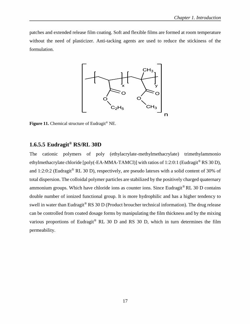
Figure 11. Chemical structure of Eudragit® NE.
1.6.5.5 Eudragit® RS/RL 30D
The cationic polymers of poly (ethylacrylate-methylmethacrylate) trimethylammonio
ethylmethacrylate chloride [poly(-EA-MMA-TAMCl)] with ratios of 1:2:0:1 (Eudragit® RS 30 D),
and 1:2:0:2 (Eudragit® RL 30 D), respectively, are pseudo latexes with a solid content of 30% of
total dispersion. The colloidal polymer particles are stabilized by the positively charged quaternary
ammonium groups. Which have chloride ions as counter ions. Since Eudragit® RL 30 D contains
double number of ionized functional group. It is more hydrophilic and has a higher tendency to
swell in water than Eudragit® RS 30 D (Product broucher technical information). The drug release
can be controlled from coated dosage forms by manipulating the film thickness and by the mixing
various proportions of Eudragit® RL 30 D and RS 30 D, which in turn determines the film
permeability.

Chapter 1. Introduction
18
1.6.5.6 Eudragit® L 30D-55
The Eudragit® L 30 D-55 is the aqueous dispersion of an anionic copolymer based on methacrylic
acid and ethyl acrylate. The ratio of the free carboxyl groups to the ester groups is approx. 1:1. The
monomers are randomly distributed along the copolymer chain. Based on SEC method, average
molar mass (Mw) of Eudragit® L 30 D-55 is approx. 320,000 g/mol. It is milky-white liquid of
low viscosity with a faint characteristic odour. The dispersion is miscible with water in any
proportion, the milky-white appearance being retained. A clear or slightly cloudy, viscous solution
is obtained by mixing 1 part of Eudragit® L 30 D-55 with 5 parts of acetone. The same results are
obtained by mixing with ethanol or isopropyl alcohol. Initially, the polymer is precipitated, then
dissolves again in the excess of organic solvent. A clear or slightly cloudy liquid is obtained by
mixing 1 part of Eudragit® L 30 D-55 with 2 parts of 1 N sodium hydroxide (Product broucher
technical information).
Figure 13. Chemical structure of Eudragit® L30D-55.
Figure 12. Chemical structure of Eudragit® RS/RL.

Chapter 1. Introduction
19
1.7 Mechanism of film formation
Film formation from aqueous polymeric dispersions is completely different from the conventional
organic solution coatings, where the polymer solution undergoes a gel transition upon solvent
evaporation to form the final film. For aqueous dispersions, this process is more complex. Film-
forming polymer latex is deposited from an aqueous colloidal dispersion of discrete polymer
spheres and the formation of a continuous film is then entirely dependent on the minimum film
forming temperature (MFT) of the polymer (Lehmann, 1994; Keshikawa and Nakagami, 1994).
At temperature above MFT, coalescence of latex particles takes place. The addition of plasticizers
is required to achieve MFT above the coating temperature for the aqueous polymer dispersions
(Bodmeier et al., 1997). The flexibility and toughness of the resulting films is improved by addition
of plasticizer. The plasticizer also softens the dispersed polymer particles and facilitate their
deformation and final coalescence (Harris and Ghebre-Sellassie, 1997). The mechanism of latex
film formation, compaction, deformation, cohesion and polymer chain inter-diffusion. Each stage
is characterized by the corresponding phase of the latex layer on the substrate and the associated
changes in the evaporation rate of the aqueous dispersion medium. The film formation from
aqueous dispersions can be explained in three main phases. A description of film formation
mechanism from aqueous polymer dispersion is elaborated below (Figure 14).
Phase1: Water evaporation and particle concentration
It is longest of all three phases. It persists until polymer reaches approximately 60-70% volume
fraction. At first, polymer particles freely move with Brownian motion, elastically rebounding and
colliding with one another. The latex surface and solid contents become concentrated by the
evaporation of water from the latex surface. The tendency of polymer particles to move around
ceases when they come into close contact with one another.

Chapter 1. Introduction
20
Figure 14. Mechanism of film formation from aqueous colloidal polymer dispersion.
Phase II. Deformation of latex particles
This phase begins when the polymer particles come into contact, and iridescence may be seen on
the latex surface. The evaporation rate per unit of open wet latex remains constant. The overall
rate of evaporation decreases significantly during the second phase. Since the drying progresses
further, the polymer particles are no longer mobile in the bulk latex and pack in an ordered array.
This packing is as a hexagonal close packed lattice. It has been suggested that hexagonal close
packing is theoretically possible, as well as cubic close packing. However, both shapes are not
easily distinguishable, since they share many geometrical features.
Phase III polymer chain inter-diffusion across particle boundaries
This phase starts with the initial formation of a continuous film. The rate of evaporation finally
slows down the water leaves the film initially via inter-particle channels and then by diffusion
through the fused polymer skin. During this phase, soft latex gains mechanical properties on
getting more homogenous. As polymer chain interfusion takes place, a process variously termed
as maturation, autohesion, or further gradual coalescence and particle interface tend to become
less distinct. The film finally gets its drastic properties during this phase due to polymer chain

Chapter 1. Introduction
21
entanglements. In 1958, Voyutskii proposed a theory stating that the surface tension forces (Dillon-
Matheson-Bradford) and capillary forces (Brwon) were inadequate to account for the physical
properties indicated by the latex films, instead, these resulted from conglomeration or “autohesion”
such as mutual inter-diffusion of free polymer chain ends across the particle-particle interface in
the coalesced film. As a result, the mutual inter-diffusion of polymer chain ends makes the latex
film homogenous and thus improves its physical-mechanical properties (Harris and Ghebre-
Sellassie, 1997).
1.8 Solution/suspension layering
Its spraying of different layers of drug or drug resin complex, or on starter core with help of binders
in form of solution/suspension. The composition of core usually of some inert material like
granules of drug itself or mixture of starch and sucrose or microcrystalline cellulose. In this process
the drug-resin complex is dispersed in a binder solution and then layered on MCC/NP core in a
fluidized bed coater. As the droplets of drug-resin complex touches the surface of the core, they
solidify and forms solid bridges between core and initial drug-resin layer. By this technique,
successive layers of drug-resin complex are achieved with additional layer of ions (Christensen
and Bertelsen, 1997).
Various technologies are used for solution/suspension layering. Fluidized bed technology is most
commonly used technology for layering. In this technology, the drug-resin complex or simple drug
is sprayed from the top, from the bottom (using Wurster insert) or tangentially (Jones, 1994).
Because of the unorganized fluidization patterns and the unavoidable spray drying, top spray mode
is considered as less effective in film coating and drug layering, than bottom or tangential modes
(Mehta et al., 1986; Iyer et al., 1993).
The Wurster process is preferable for suspension and solution layering, because of its ability to
apply high quality films/layers after the palletization operation. This technique enables layering of
100-150% w/w solids based on the starting/initial core weight (Ghebre-Sellassie, 1989).

Chapter 1. Introduction
22
1.9 Additives
For several reasons, different types of excipients are used in aqueous polymeric dispersions, most
common being plasticizers, pore formers and anti-taking agents. These excipients strongly
influence the film properties, coating process and release rate of the coated dosage form. The
coating formulations need to be optimized in order to have optimized coating conditions.
1.9.1 Pore formers
To adjust the drug release of extended release coatings, pore formers are added into formulations.
The pore formers which are most commonly used are sugars (sucrose, lactose, sorbitol), salts
(sodium chloride, calcium phosphate) and hydrophilic polymers (polyethylene glycol,
polyvinylpyrolidone and HPMC) or surfactants (sodium lauryl sulfate) (Muhammad et al., 1992;
Li et al., 1990; Erdmann et al., 2000). These pore formers during dissolution leach out from coating
polymer and enhance permeability of the membrane. The concentration of the pore former can
control the release kinetics of ethyl cellulose coatings and different polysorbates used as additives
(Samani, 1999). The drug release from aqua coat coating was increased by using urea as pore-
former which made the film more porous (Appel and Zentre, 1991). Also, the drug release from
the aqua coat coatings was stabilized under stress conditions by adding pore formers (Siepmann et
al., 2007; Siepmann et al., 2008; Muschert et al., 2009).
Figure 15. Schematic presentation of suspension/solution layering technique (Glatt GmbH, 2013).

Chapter 1. Introduction
23
1.9.2 Plasticizers
Plasticizers give flexibility to the hard and brittle polymers. These are organic solvents having high
boiling points. They act by reducing cohesive intermolecular forces within the polymer chain, thus
changing polymer properties. For example, plasticizers cause increase in elongation, flexibility
and reduction in the glass transition temperature and tensile strength of the polymer. The purpose
of plasticizer to reduce the minimum film forming temperature (MFT) below the coating
temperature (Bindschaedler et al., 1983). The plasticizer diffuses into polymer particles and
promotes the particle deformation and coalescence to form a smooth film. The plasticizer should
be compatible with the polymeric particles for good plasticization effect. Incompatible plasticizers
cause the coagulation of aqueous polymer dispersions. Mostly, these phenomena occur with ethyl
acrylate/methacrylic acid copolymer formulations (Kollicoat® MAE 30 D) (Flöβer et al., 2000;
Dangel et al., 2000). Thus, selection of appropriate plasticizer must be made carefully. The
plasticizer could have an anti-plasticizer effect if taken in lower concentration than the appropriate
concentration required for optimum plasticization in the coating solution (Guo,1994; Guo et
al.,1999).
1.9.3 Anti-taking agents
Anti-tacking/separating agents and pigments are commonly added to aqueous polymer
formulations to reduce agglomeration or sticking of coated particles during the coating process.
Glyceryl monostearate (GMS), magnesium stearate, titanium dioxide and talc are most commonly
used anti-tacking agents in aqueous film coatings. Talc as an anti-tacking agent known to have a
sedimentation tendency and causing the blockade of the nozzles during coating process. Therefore,
during coating process dispersion must be kept under continuous stirring. The amount of pigments
in the aqueous coating dispersion must be optimized without exceeding the maximum carrying
capacity of the polymer or critical pigment volume concentration (CPVC). The anti-tacking agents
have strong impact on final film properties such as mechanical strength and permeability (Patton,
1979). An increase in drug release rate was found, which was explained with the adsorption
capacity, the high specific surface area and the high affinity of colloidal silica for polar components
like water (Vecchio et al., 1995). Pigments have a high binding capacity with polymer dispersions.
In order to avoid sticking during polymer dispersion coatings talc up to 200%, based on dry
polymer mass could be incorporated into the coating formulation of Eudragit® RL/RS 30 D.
Which was already plasticized with 30% w/w TEC based on polymer mass (Maejima and

Chapter 1. Introduction
24
McGinity, 2001). Glyceryl monostearate (GMS) is used in lower concentration. It is shown to have
a ten-times higher anti-tacking effectiveness than talc. The addition of talc and GMS reduce the
flexibility of coated polymer during the coating process. When the granules swelled in the release
medium an orange peeling effect of film coating was observed (Wan and Lai, 1993). During
coating process, fine particles have a higher tendency of agglomeration. This could be reduced by
adding NaCl to an aqueous spray solution of hydroxyl propyl cellulose (Fukumori, 1993). It was
proposed that with reduction of viscosity of the spraying/coating solution, suppression of
agglomeration could be achieved (Yuasa, 1997).
To promote further gradual coalescence of the film, coated dosage forms are stored at elevated
temperatures. This process is known as curing. It can also be defined as the input of energy into
the film-coated system after the desired film coating level is applied (Hamed and Sakr, 2003).
Curing of film-coated dosage forms is an important step in the film formation from aqueous
polymer latexes. During coating process, curing takes place to a certain extent. However, this is
inadequate to assure the completion of coalescence, the dosage form is generally exposed to
elevated temperature after the coating. This can be done in the coating machine using a process
known as post-coating fluidization (Harris and Ghebre-Sellassie, 1986) or by placing the coated
dosage forms in an oven (Goodhart et al., 1984; Lippold, et al., 1989).
1.10 Storage Stability
The objective of long term stability is to provide information, how quality of drug substance or
drug product changes under the influence of environmental factors such as temperature, humidity,
and light, and to further establish a re-test period for the drug substance or drug product or a shelf
life for the drug product under recommended storage conditions (European medicine Agency; ICH
guidelines). For registration of new chemical entities, a long term stability data is required.
Generally, a drug substance must be evaluated under storage conditions (with appropriate
tolerances) with its thermal stability, if applicable, its sensitivity to moisture. The storage
conditions and the length of studies chosen must be enough to cover shipment for subsequent use
and storage. International committee on harmonization (ICH) recommends different storage
conditions, for example 25 °C ± 2 °C/60% RH ± 5% RH for 12 months, 30 °C ± 2 °C/65% RH ±
5% RH for 6 months and 40 °C ± 2 °C/75% RH ± 5% RH for 6 months, intermediate and
accelerated long term stability conditions respectively. Contradictory results were reported in

Chapter 1. Introduction
25
literature of coated dosage forms regarding long term stability storage. The release of lipophilic
drug ibuprofen was increased after storage (Bodmeier and Paeratakul, 1994). The release of
theophylline was decreased after storage (Yuen et al., 1993; Goodhart et al., 1984; Bando and
McGinity 2006). The stability was carried out of optimized formulation. The formulation was
stored in amber coloured bottle after reconstitution at 2°C-8°C till one week. Further it could be
evaluated for other physical characteristics as well.
The numerous factors are the cause of change in release profile over long term storage. For
example, inadequate amount of plasticizer in the coating formulation results in the change of
polymer films over storage. In such cases longer curing times are required to achieve the stable
film and release profile. The decrease in theophylline release from coated pellets with Eudragit®
RS 30D containing 10% or 20% triethylcitrate (TEC) depending on dry weight of polymer
concentration in the formulation (Amighi and Moes, 1996). Another reason could be the physical
instabilities in the coating that leads to cracks and chipping of the coating. In addition, the
researchers have concluded these problems could lead to increase in water contents of the films
rather than a decrease water contents (Chowhan et al., 1982). The faster drug release in case of
Aquacoat® ECD coatings was due to the formation of micro-ruptures in the film during storage
(Wesseling and Bodmeier, 2001).
The increased in compaction of polymer structure and decrease in the free volume of the film due
to gradual coalescence with the ageing progresses was also reported, as one reason for unstable
release profile (Guo et al., 1993). This is due to the change in water vapor permeability of the films
(Guo et al., 1991). Additionally, the presence of endogenous excipients in the aqueous polymeric
dispersion can also lead to serious stability issues such as increase in drug release rates. It has been
shown that crystallization of the surfactants affects the dissolution rate of the drug from coated
pellets. The decrease of drug release under high humidity was due the gradual coalescence as a
result of decreased permeability for water and drug in the formulation (Amighi and Moes, 1997,
Bajdik et al., 2003).
The storage at 40 °C and 75% RH of Kollicoat® SR 30 D coated pellets also resulted in the
decreased drug release due to continuous film formation (Shao et al., 2002). In contrary, an
increased lag time without any significant change in release profile was observed with Kollicoat®
SR 30 D coated pellets upon one-month storage at 40 °C/75% RH (Ensslin et al., 2008). The
addition of talc up to 200% in the formulation of Eudragit® RS 30 D coated pellets provides a

Chapter 1. Introduction
26
stable release profile. In fact, the polymeric particles were embedded in the skeleton of talc around
the pellets, which led to an inhomogeneous film formation. The smoothness of film was further
decreased due to the densification of polymeric particles on curing. The drug release was taken
place through pores without any change (Maejima and McGinity, 2001; Ahmed et al, 2008).
1.11 Oral liquid-controlled release formulations
Oral liquid-controlled release formulations have always been a challenge to the formulation
scientists due to their stability concerns. When high dose of the drug required in the formulation.
The formulation becomes challenging both for the manufacturer and for the patients, where
flexible dose of drug required in pediatric and geriatric patients. Oral liquid-controlled release is
specifically designed for geriatric and pediatric patients. They may be developed to take into
consideration the diseased condition of the patient such as esophagitis where flexible dose of the
drug required. As breaking of tablet lead to affect its release profile. These formulations are
designed to meet the targets, where the drug product is facing stability concerns in aqueous
form/state. It is possible to formulate liquid product/formulation, having sustained/controlled
action by suspending the coated drug-resin particles into the suitable liquid media. The suspending
medium has no action on coated granules. These formulations of drug-resin complex is always of
suspension type due to insoluble nature of resinates. Suspensions are biphasic dosage systems in
which the solid phase is suspended in a liquid phase. To produce a controlled release liquid dosage
forms from ion exchange resins, the drug is ionically complexed with resin and coated with
suitable/appropriate polymer, which controls the drug release. It is formulated as suspension using
HPMC E5 as a suspending agent. In an attempt of sustained-release drug delivery systems, the
microparticles have got much attention because of uniform distribution of coated particles in the
GI tract. Which ultimately brings the uniform absorption and decrease risk of local effects on the
GI tract.
1.12 Commercial products of ion exchange resins
Ion exchange resins were used for different range of dosage forms, for example from solid to
liquids. Different ion exchange resins were selected for the current work by keeping in view their
nature, as basic drugs complexed with cation exchange resins and acidic drugs could be associated

Chapter 1. Introduction
27
with anion exchange reins and both drugs simultaneously carried with amphoteric resin. The
commercial products of ion exchange resins are as under.
Table 2. List of commercial products of ion exchange resins
Drug Brand Name Dosage form Dose for adults
Cholestyramine
Questran®
(Mead Johnson)
powder 1 packet TID or QID
before meals
Colestipole HCl Colestid
(Upjohn)
granules 15-30g/day in divided
Doses BID to QID
Na Polystyrene Kayexalate
( Breon)
powder Orally;15g QID
Phentermine Ionamin®
(Pennwalt)
capsule 15-30mg daily before
breakfast
Hydrocodone Tussionex® Suspension
1 teaspoonful(capsule
Phenyltoloxamine (Pennwalt) Tablet, capsule or tablet) every 12 h
Dextromethorphan Delsym®
(Pennwalt)
liquid 2 teaspoonful BID
Betoxalol Betoptic® Eye drops 2-4 drops in the night

Chapter 1. Introduction
28
Table 3. List of selected cationic and anionic exchange resins
Name of
I.E resins
Shape Moisture
Contents
(%)
Swelling
(%)
I.E.
Capacity
(meq/g)
DVB
(%)
Particle
Size
(µm)
Ionisable
counter ion
Amberlite®
IRP 69
powder 10.42±0.3 46.0 ±1.4 2.8 - 3.4 4 - 16 45-150 R-SO-
3Na
+
Amberlite®
IR 69F
beads 40.16±0.1 57.0 ±2.5 2.9 - 4.4 7.2 300-1100 R-SO
3
-
H+
Purolite®
C100MRNS
powder 10.95±0.2 46.0 ±1.2 2.8 - 3.4 8 45-150 R-SO
3
-
Na+
Purolite®
C100CaMRNS
Powder 1.95±0.2
46.0±12
2.8 - 3.4 8 45-150 R-SO3-Ca+2
Purolite®
WCA100
beads 57.0±2.5
30±1.56 0.9 - 0.9
-- 240-280
R-COO-Na+
RN+(CH3)3OH
Purolite®
C108DR
powder 10±2.56 35±1.56 -- 8 45-150 COOH
Purolite®
A430MR
powder 12±3.45 55±2.56 1.8-2.2 8 45-150 RN+(CH3)3Cl-
DuoliteTM
AP143/1093
powder 10±2.55 50±2.56 1.8-2.2 8 45-150 RN+(CH3)3Cl-
Table 4. List of cationic and anionic model drugs
Name of
drug
Melting
point
Solubility
mg/ml
Log P Molecular
weight
pKa Nature of
drug
Propranolol
HCl
96 220 3.48 296 9.03 basic/cationic
Diltiazem
HCl
212 950 2.70 450.97 8.06 basic/cationic
Tramadol
HCl
300 970 2.4 190 9.41 basic/cationic
Ibuprofen 77-78 0.03 3.5 206.28 4.43 acidic/anionic
Diclofenac
sodium
156-158 5.26 4.26 318.42 4.2 acidic/anionic

Chapter 1. Introduction
29
Figure 16. A schematic presentation of reversible reaction/process between cation exchange resin and the
propranolol HCl (cationic drug).
1.13 Challenges
Oral controlled release liquid formulations in reconstitutable powder form are not easy to
formulate. They are having following challenges.
1.13.1 Tackiness
Making bond of measurable strength, when come in contact with another material is called as
tackiness. Tack is ability of two materials to resist separation after bringing their surfaces into
contact for a short time under light pressure (Wetzel, 1957). The strength of tack is dependent on
the extent of inter-diffusion of molecules across interface. Additionally, the extent of deformity of
polymer chain and Vander Wall forces has a huge impact on tack strength. Autohesion is the term
used to describe the tack between two chemically similar surfaces. Commonly, during the coating
of dosage forms with aqueous polymeric dispersions, the sticking of substrates occurs during the
coating process and upon curing/storage. This tackiness problems more likely to happens with
small particles size substrates and when coated with Eudragit® RS 30D and Eudragit® RL 30D,
Purolite®
Purolite
® C100MRNS+propranolol
complex

Chapter 1. Introduction
30
Eudragit® NE 30D or Eudragit® NM 30D and Kollicoat® SR 30D owning to tackiness of polymeric
films (Voyutskii, 1971).
This leads to a massive problem of handling the coated substrate caused by sticking to each other
and with the walls of the Wurster and chamber. In some worse cases, a stoppage of process was
caused by irreversible agglomeration of small size coated substrates or due to higher product
temperatures and plasticizer contents (Bodmeier and Paeratakul, 1991). Moreover, tackiness
increases the number of coating defects and impairs the quality and efficiency of the coated batch.
Therefore, a suitable balance had to be established between sufficiently high product temperatures
and non-agglomeration (Wesseling et al., 1999). In order to control tackiness during coating
process several anti tacking agents were used for instance, talc, magnesium stearate and glycerol
monostearate GMS. The talc and magnesium stearate were used in higher concentrations (50-
100% with respect to dry weight of polymer) whereas, glycerol monostearate GMS was used in
lower concentration 10-20%. The USP defines powders fineness as follows.
Table 5. USP defined range of fineness of powders
Descriptive term Mesh opening size
(µm)
Mesh size Number
Very coarse ≥1000 2-10
Coarse 355-1000 20-40
Moderately coarse 180-355 40-80
Fine 125-180 80-120
Very fine 90-125 120-200

Chapter 1. Introduction
31
Table 6. pH and ionic composition of different gastric and biological fluids. (Adapted from Thairs et al.,
1998)
Fluid pH Na+ (meq/L) K+ (meq/L) Cl- (meq/L) HCO3- (meq/L)
Saliva 6-7 30 20 30 15
Gastric 1-3.5 (50 - 90) 10 110 0
Bile 7.8 140 5 105 40
Pancreatic fluid 8–8.3 140 5 60 90
Small Intestinal
fluid
7.5-8 120 5 110 35
Large Intestinal
fluid
5.5-7 130 10 95 20
Plasma 7.4 140 5 100 30
Interstitial fluid 150 5 110 30
Intracellular fluid 10 160 115 30
1.13.2 Incomplete release of drug from drug-resin complex (resinates)
As release of drug always takes place from the ionic sites of resin by dissociation of drug from the
resin with help of counter ions in the release medium. But release of drug was incomplete as
reported in literature. To get the complete drug release in the release medium was to be simulated
with certain specific amount of NaCl and KCl. The role of divinyl benzene (DVB) cross linking
inside the resin matrix was also to be considered so as to have its effect on drug release.
Chapter 1. Introduction
32
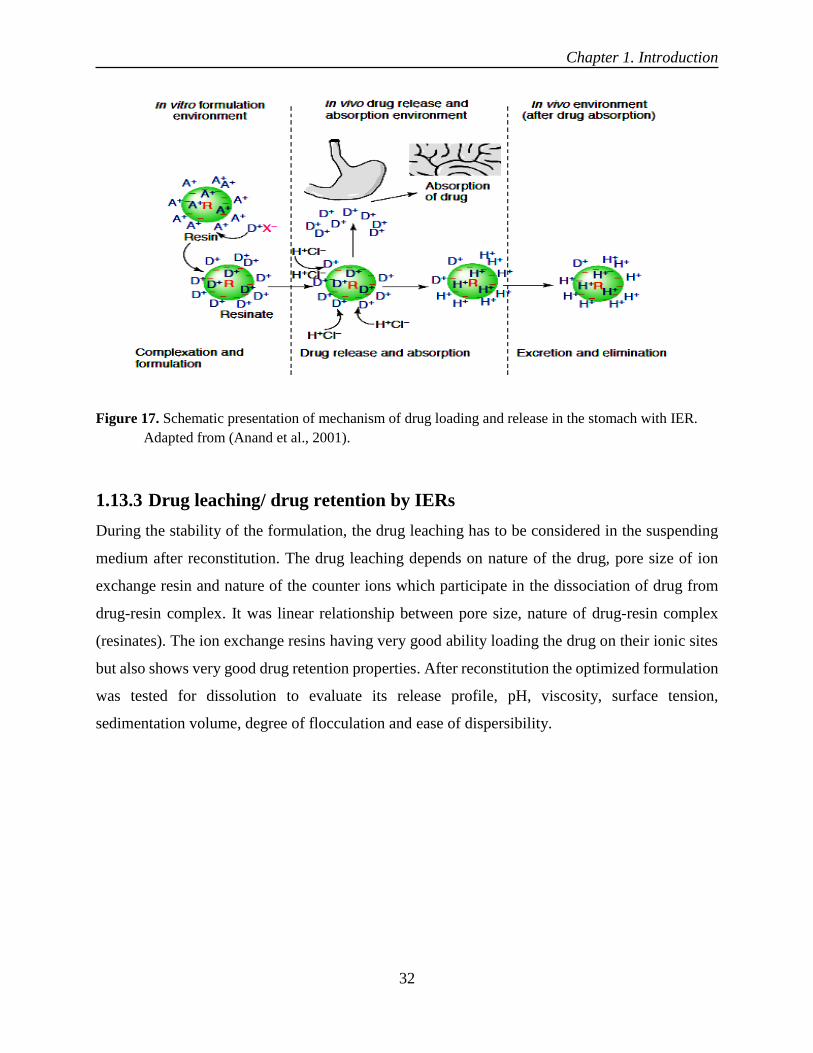
Figure 17. Schematic presentation of mechanism of drug loading and release in the stomach with IER.
Adapted from (Anand et al., 2001).
1.13.3 Drug leaching/ drug retention by IERs
During the stability of the formulation, the drug leaching has to be considered in the suspending
medium after reconstitution. The drug leaching depends on nature of the drug, pore size of ion
exchange resin and nature of the counter ions which participate in the dissociation of drug from
drug-resin complex. It was linear relationship between pore size, nature of drug-resin complex
(resinates). The ion exchange resins having very good ability loading the drug on their ionic sites
but also shows very good drug retention properties. After reconstitution the optimized formulation
was tested for dissolution to evaluate its release profile, pH, viscosity, surface tension,
sedimentation volume, degree of flocculation and ease of dispersibility.

Chapter 1. Introduction
33
1.14 Research Objectives
The present research work was focused on oral liquid formulations made of reconstituted powder
with the aim of controlled drug release. The work was divided into the following four main studies:
i) Evaluation of cation exchange resin Purolite® C100MRNS for controlled release
formulations by investigating formulation and process parameters and stability of
formulations before and after reconstitution.
ii) Evaluation of Amberlite® IR69F for enteric drug delivery system by investigating the
formulation and process parameters.
iii) Evaluation of Purolite® C100CaMRNS for drug combination delivery system with an
aim of complete drug release for both drugs and investigating the formulation and
process parameters.
iv) To enhance the robustness of reservoir system (pellets) by preventing diffusion of drug
into coated polymer by layering of drug-resin complex on NP core and to evaluate
drug-resin complex to enhance solubility of Ibuprofen.

Chapter 2. Materials and Methods
34
2 Chapter 2. Materials and Methods

Chapter 2. Materials and Methods
35
2 Materials and Methods
2.1 Materials
Model Drugs
Propranolol HCl (K-W Pfannenschmidt GmbH, Hamburg, Germany), diltiazem HCl (PCAS
Division Seloc France, Limay, France), ibuprofen (BASF SE, Ludwigshafen, Germany), tramadol
HCl (Heumann pharma GmbH, Germany).
Ion Exchange Resins
Sodium polystyrene sulfonate (AmberliteTM IRP 69 Rohm and Haas France S.A.S),
cholestyramine (DuoliteTM AP143/1093 Rohm and Haas France S.A.S), AmberliteTM IR69F
(Rohm and Haas France S.A.S), sodium polystyrene sulfonate (Purolite® C100MRNS Purolite
Ltd, Wales, UK), Purolite® C100CaMRNS (Purolite Ltd, Wales, UK), cholestyramine (Purolite®
A430MR, Purolite Ltd, Wales, UK), Purolite® WCA100 (Purolite Ltd, Wales, UK), Purolite®
C108DR (Purolite Ltd, Wales, UK).
Polymers
Hydroxypropyl methylcellulose (MethocelTM E5, Methocel® K15M Premium CR, Methocel®
K4M Premium CR and Methocel® K100LV Premium CR, Colorcon, Orpington, UK),
polyvinylpyrrolidone (PVP) (Kollidon® 30, BASF SE, Ludwigshafen, Germany), ethyl cellulose
(EC, Ethocel TM Standard 45 c.P premium, Ethocel TM Standard 100 c.P premium, (Colorcon,
Dartford Kent, UK), ethyl acrylate and methyl methacrylate copolymer aqueous dispersion
(Eudragit® NE 30 D, Evonik Industries AG, Darmstadt, Germany), polyvinyl acetate aqueous
dispersion (Kollicoat® SR 30 D, BASF SE, Ludwigshafen, Germany), methacrylic acid and ethyl
acrylate copolymer, powder and an aqueous dispersion (Eudragit® L 100-55, Eudragit® L 30 D-
55, Evonik Industries AG, Darmstadt, Germany), ethylcellulose aqueous dispersion (Aquacoat®
ECD, FMC BioPolymers, Cork, Ireland), ethyl acrylate and methyl methacrylate copolymer with
a low content of a methacrylic acid ester with quaternary ammonium groups, aqueous dispersion
(Eudragit® RS 100, Eudragit® RS 30 D, Evonik Industries AG, Darmstadt, Germany).

Chapter 2. Materials and Methods
36
Other excipients
Triethyl citrate (TEC) (Citroflex® 2; Morflex, Greensboro, NC, USA), tributyl citrate (BASF SE,
Ludwigshafen, Germany), Polyoxyethylene sorbitan monooleate (Tween® 80, Sigma-Aldrich
Chemie GmbH, Steinheim, Germany), sodium lauryl sulfate (Carl Roth GmbH + Co.KG,
Karlsruhe, Germany), Isopropanol, ethanol 96%, water. Inert sugar spheres, sieved size 710-850
μm (Suglets®, NP Pharm S.A.S., Bazainville, France), magnesium stearate (Herwe Chemisch-
technische rezeugnisse, Sinsheim Dühren, Germany), fumed silica (Aerosil® 200, Evonik
Industries AG, Darmstadt, Germany), talc (Luzenac pharma, Europe, Toulouse, France),
magnesium stearate (Caelo, Caeasar & Loretz GmbH, Hilden, Germany), sodium chloride (NaCl)
(Roth GmbH & Co. KG., Karlsruhe, Germany).
2.2 Methods
2.2.1 Purification of ion exchange resins
10 g of ion exchange resin was stirred with 200 ml of ethanol and washed with 200 ml of milli-Q®
water, then stirred with 200 ml of methanol and again washed with enough milli-Q® water. Finally,
the pre-washed resins were treated with 200 ml of NaOH to re-activate the Na+ ions on its ionic
sites. The resin was washed three times with milli-Q® water until the pH of filtrate turned neutral.
2.2.2 Moisture content of ion exchange resins
Moisture content was determined by loss on drying (LOD) by heating the sample at 103ºC for 3
to 5 minutes (HB43-S Halogen Mettler Toledo Germany). The 1.0 g of powder sample was placed
on the aluminium pan and the lid was closed.
2.2.3 Swelling of ion exchange resins
The swelling of resins was measured using a 10 ml graduated measuring cylinder. A specified
amount of resins was taken into the cylinder and volume was noted as (volume dry). The water
was added into the cylinder and wet volume was noted after a vigorous shaking and allowed it to
settle for half an hour. The % swelling of ion exchange resins was calculated by following formula.
% 𝑆𝑤𝑒𝑙𝑙𝑖𝑛𝑔 =(𝑉𝑜𝑙𝑢𝑚𝑒 𝑤𝑒𝑡 − 𝑉𝑜𝑙𝑢𝑚𝑒 𝑑𝑟𝑦)
𝑉𝑜𝑙𝑢𝑚𝑒 𝑑𝑟𝑦 × 100

Chapter 2. Materials and Methods
37
2.2.4 Particle size measurement
2.2.4.1 Powder laser diffraction
Particle size distribution of resins and resinates was measured by powder laser diffraction
(Sympatec particle size analyzer Helos BF Germany). The 2.0 g of powder sample was added into
the Powder Laser Diffraction at trigger condition, (STD trigger) with disperser RODOS STD
disperser with particle size range 0.5/4.5-175 μm and 0.5/4.5-875 μm. The particle size distribution
was obtained in X10, X50 and X90.
2.2.4.2 Liquid laser diffraction
Particle size distribution of formulation after reconstitution was determined by laser diffractometry
(LD) (Mastersizer 2000, Malvern Instruments, UK). The index of refraction for both, Purolite®
C100MRNS and Purolite® A430MR used was 1.59, and the absorption index was 0.001. The
sample was added to the dispersion unit containing water with obscuration range 4-6%, and then
the LD measurement was performed immediately at 1750 rpm stirring speed. The LD data obtained
by average of 3 measurements was evaluated using the volume distribution diameters (D50%,
D90% and D99%) which shows the percentage of particles having a diameter equal or lower than
the said value.
2.2.4.3 Light Microscopy
Light microscopy was performed using Leica Wild M3Z (Leica Microsystems (Schweiz) AG,
Heerbrugg, Switzerland). The used magnification was 20x10 fold. Each sample was observed 3
times. Drug-resin complexes were suspended in liquid paraffin and the crystallinity of the drug
and drug-resin complex was examined under polarized light.
2.2.5 Reduction of particle size of resinates
Particle size of resinates was reduced either in dry state by mortar and pestle or in wet state by wet
milling with HPMC E5 as binder in the formulation by the dyno mill (Retsch MM 2000 small ball
mill, Retsch GmbH, Haan, Germany) using glass beads. The sample to glass bead ratio was 3:1
(v/v), the total sample volume 600 ml. Each run of the machine took 5-8 minutes to complete the
cycle. The particle size of the sample was reduced by increasing the number of cycles or changing
Chapter 2. Materials and Methods
38
the beads for operation. After milling, the ion exchange resins were dried in an oven at 60°C and
finally sieved in a vibratory sieve shaker (Analysette 3 PRO, Fritsch GmbH, Idar-Oberstein,
Germany) using sieves of 45, 75, 106, 160 μm pore size at an amplitude of 0.8 mm for 2 minutes.
The size fractions <45, 75-106, 106-160 μm were used for the further drug loading studies. The
particle size of powders and microsuspensions was analysed according to section 2.2.4.
2.2.6 Drug loading on resin/preparation of drug-resin complex (DRC)
2.2.6.1 Batch method
The batch process was a quick, simple to perform drug loading method on ion exchange resins.
Furthermore, it was the only process suitable for very fine size of ion exchange resin particles.
This method was performed with Amberlite® IR 69F (Rohm and Hass France), Amberlite® IRP
69 (Rohm and Hass), Purolite® C100MRNS (Purolite Ltd, Wales, UK) and the model drugs
propranolol HCl, tramadol HCl and diltiazem HCl (Sigma Aldrich, USA). All the flasks containing
the drug solution were stirred with resin suspension for 24 hours at 300 RPM at 23ºC as mentioned
in Table 6. Finally, the samples were diluted and filtered using a syringe filter with 0.2 μm pore
size (Acrodisc® SOTAX syringe filters with GHP membrane, Pall Corporation, USA). The drug
association efficiency was determined indirectly through the drug contents in the supernatants
using a UV spectrophotometer (HP 8483, Agilent Technologies at 289 nm). To determine the drug
loading from drug-resin complex (w/w), a direct method was used. The drug-resin complex was
dissociated with 2N NaCl/KCl solution and the drug content was measured with a UV-
spectrophotometer (HP 8483, Agilent Technologies) at 289 nm after filtration through 0.2 μm
syringe filters (Acrodisc® SOTAX syringe filters with GHP membrane, Pall Corporation, USA).
The drug association efficiency and drug loading were calculated according to the following
equations.
% 𝐷𝑟𝑢𝑔 𝑎𝑠𝑠𝑜𝑐𝑖𝑎𝑡𝑖𝑜𝑛 𝑒𝑓𝑓𝑖𝑐𝑖𝑒𝑛𝑐𝑦 𝑤/𝑤 =𝑎𝑚𝑜𝑢𝑛𝑡 𝑜𝑓 𝑑𝑟𝑢𝑔 𝑎𝑠𝑠𝑜𝑐𝑖𝑎𝑡𝑒𝑑 (𝑚𝑔)
𝑎𝑚𝑜𝑢𝑛𝑡 𝑜𝑓 𝑑𝑟𝑢𝑔 𝑢𝑠𝑒𝑑 (𝑚𝑔) × 100
% 𝐷𝑟𝑢𝑔 𝑙𝑜𝑎𝑑𝑖𝑛𝑔 𝑤/𝑤 =𝑎𝑚𝑜𝑢𝑛𝑡 𝑜𝑓 𝑑𝑟𝑢𝑔 𝑒𝑙𝑢𝑡𝑒𝑑 (𝑚𝑔)
𝑎𝑚𝑜𝑢𝑛𝑡 𝑜𝑓 𝑑𝑟𝑢𝑔 𝑟𝑒𝑠𝑖𝑛 𝑐𝑜𝑚𝑝𝑙𝑒𝑥(𝑚𝑔) × 100

Chapter 2. Materials and Methods
39
Table 7. Summary of the formulations prepared with different drug and resin ratios w/w at 300 RPM for
different range of temperature
2.2.6.2 Column method
The column process was a feasible loading method for resins with a particle size of more than 500
µm. It involved passing a drug solution through a column packed with the IER until the effluent
concentration was the same as the eluent concentration.
2.2.7 Effect of temperature, pH and particle size on drug loading
For the evaluation of the effect of temperature on the drug loading process, 3 flasks containing
suspensions according to Table 6 with a 1:2 resin to drug ratio (w/w) were stirred for 24 hours in
a hot water bath set at 40 ± 5 ºC, 50 ± 5 ºC and 60 ± 5 ºC using a heating/stirring plate. (Variomag
Monotherm, H+P Labortechnik AG, Germany). The temperature was checked using an electronic
Heat-Probe Thermometer (Wahl Thermistor 700M, Palmerwahl, USA). In the end, all samples
were diluted and filtered through a 0.2 μm syringe filter (Acrodisc® SOTAX syringe filters with
GHP membrane, Pall Corporation, USA).
Ratio
Drug:Resin
Resin
(mg)
Propranolol
HCl (mg)
Milli-Q®
water (ml)
Temperature
(ºC)
Particle size
(µm)
pH of
loading
solution
1:1 100 100 25 23 106-150 2.0
2:1 100 200 - 23 106-150 5.9-6.5
2:1 100 200 - 40 106-150 2.0
2:1 100 200 - 50 45-75 2.0
2:1 100 200 - 60 45-75 4.0
3:1 100 300 - 23 45-75 4.0
4:1 100 400 - 23 75-106 4.0
1:2 200 100 - 23 75-106 6.0
1:3 300 100 - 23 75-106 6.0

Chapter 2. Materials and Methods
40
The influence of pH (2-8) and different particle size of resin (45-75µm, 75-106 µm, 106-160 µm)
on the drug loading was investigated. The different flasks were prepared containing 25 ml of Milli-
Q® water and different ratios of drug to resin (Table 6). All flasks were stirred for 24 hours, at 300
RPM and 23 ºC. Finally, samples were diluted, filtered using a 0.2 μm syringe filter (Acrodisc®
SOTAX syringe filters with GHP membrane, Pall Corporation, USA) and the drug content was
measured UV-spectrophotometrically.
2.2.8 Conductivity studies and change in pH
The change in conductivity and pH of the loading medium before and after the batch process of
drug loading was measured by a pH-meter (Metrohm 827 pH Lab, Metrohm AG, Germany) and
conductivity meter (inoLab wtw Metrohm, Germany).
2.2.9 Vacuum filtration and drying of resinates
Vacuum filtration was performed after completion of the batch process. The resinates were dried
overnight in the oven at 60 ºC and stored in the desiccator for further processing of granulation,
coating and particle size distribution.
2.2.10 Characterization of drug-resin complex (resinates)
2.2.10.1 Powder x-ray diffraction (PXRD)
X-ray diffractometry for resin, drug, drug resin physical mixture and drug-resin complex was
performed using a powder x-ray diffractometer (PW2273/20 copper anode, Analytical B.V.,
Almelo, The Netherlands; PW 1830 x-ray generator, PW 1710 diffraction control unit and PW
1820 vertical goniometer, Philips Industrial and Electroacoustic Systems Division, Almelo, The
Netherlands). The data collection and machine control was done externally (ADM version 3.5, A.
Wassermann Röntgenanalytik-Meßsysteme-Software, Kempten, Germany). The collected raw
data was taken into CSV files and evaluated using Microsoft Excel 2013. Samples were put on a
sample holder and mounted into the diffractometer. Spectra were collected at angles from 4° to
40° using a step size of 0.02° and a collection time of one second per data point.

Chapter 2. Materials and Methods
41
2.2.10.2 Fourier transforms infra-red spectroscopy (FTIR)
FTIR spectroscopy measurement was performed with an Excalibur 3100 FTIR spectrophotometer
(Varian Inc., Palo Alto, USA). The spectra from drug, drug resin physical mixture and drug- resin
complexes and resin alone were obtained in the scan range of 600 to 4,000 cm−1 at a resolution of
4 cm−1 and average of 32 scans, using a horizontal ATR accessory with a single reflection diamond
crystal (Pike Miracle, Pike Technologies, Madison, USA) and Varian software (Resolution Pro
4.0). Scans were taken every 2 minutes over 30 minutes using the Resolutions Pro Software
(Version 4.0, Varian Inc., Palo Alto, CA, USA). Data were exported to Microsoft Excel for further
evaluation.
2.2.11 Wet granulation of resinates
To improve the flowability of resinates during coating process, the resinates were wet granulated
with different binders at different ratios (Table. 8). After granulation, they were sieved and the
particle size fraction between 160-250µm was selected for the further coating process.
2.2.12 Coating of drug-resin complex (resinates)
The coating of resinates was performed in a fluidized bed coater (Mini-Glatt, Wurster insert, Glatt
GmbH, Binzen, Germany). For each batch process, 50-55 g of granulated resinates were used. The
coating processability was evaluated based on spray rate, product temperature, nozzle blockage,
the flow of resinates particles, agglomeration tendency and yield. Additionally, the coated resinates
were cured at 40 °C and 60 °C and 40°C/75% RH for 12 hours in aluminium pans in an oven.
After curing, the powder was equilibrated in a desiccator for 24 hours before further use.

Chapter 2. Materials and Methods
42
Table 8. Summary of concentration of different binders and granulation fluid during wet granulation of
resinates
Formulation Binders Granulation Fluid
Formulation
code
Resinates
% w/w
HPMC
E5
% w/w
Kollidon®
30
% w/w
Ethocel TM
% w/w
Eudragit®
NE 40D
% w/w
Ethanol:
H2O
IPA: H2O
w/w
F1 85 15 -- -- -- 88:12 --
F2 85 -- 15 -- -- 88:12 --
F3 85 -- -- 15 -- 88:12 --
F4 70 15 -- 15 -- 88:12 --
F5 70 15 -- 15 -- -- 88:12
F6 85 -- -- -- 15 -- 88:12
F7 70 -- -- 15 15 -- 88:12
2.2.12.1 Eudragit® NE 30D
The aqueous dispersion of Eudragit® NE 30D, was plasticized with TEC 10% w/w (based on dry
polymer mass) and the anti-tacking agent talc (35%-50% w/w based on dry polymer mass) was
dispersed in HPMC E5 5% w/w and 5% Tween® 80 based on dry polymer mass). The HPMC E5
was heated up to 55°C in 1/3 of the water, the rest 2/3 water was added to form the clear solution
of HPMC E5. The talc was added into clear solution of HPMC E5, with the dropwise addition of
Tween® 80. The final solid contents were adjusted to 15% w/w with milli-Q® water. The coating
was performed in a fluidized bed coater (Glatt GPCG-1, Glatt GmbH, Binzen, Germany) to obtain
coating level of 20-25% w/w (based on dry polymer mass and total weight gain). The coating
parameters were as follows: batch size = 50 g, inlet temperature = 28-30 °C, product temperature
= 18-20 °C ±2 °C, air flow = 72 m3/h, nozzle diameter = 0.5 mm, spray pressure = 0.9 bar, spray
rate = 0.8-1 g/min, final drying at 20-25 °C for 10 min. After coating, 0.5% (w/w) of colloidal
silica (Aerosil® 200) was mixed with coated resinates to avoid sticking during storage.
Chapter 2. Materials and Methods
43
2.2.12.2 Aquacoat® 30D and Surelease®
Aquacoat® 30D was plasticised with TEC 25% w/w (based on dry polymer mass) and talc 35%-
50% w/w (based on dry polymer mass) was added as anti-tacking agents to the polymer dispersion.
Surelease® was used without plasticizer but with talc 35% w/w (based on dry polymer mass) which
was added as anti-tacking and the final coating formulation was adjusted to 15% w/w solid
contents with milli-Q® water. The coating was performed in a fluidized bed coater (Mini-Glatt 4,
Glatt GmbH, Binzen, Germany) to achieve a coating level of 15-20% w/w (based on total weight
gain). The coating parameters were as follows: batch size = 45 g, inlet temperature = 60-62 °C,
product temperature = 35±2 °C, air flow = 0.2 m3/h, nozzle diameter = 0.5 mm, spray pressure =
0.9 bar, spray rate = 1-2 g/min, final drying at 30 °C for 10 min. After coating, 0.5% (w/w) of
colloidal silica (Aerosil® 200) was mixed with resinates to avoid sticking during storage of the
formulation.
2.2.12.3 Kollicoat® SR 30D
The aqueous dispersions of Kollicoat® RS 30D, was plasticized with TEC, 5% and talc 35%-50%
w/w (based on dry polymer mass) was added as anti-tacking agents into polymer dispersion and
the final solid contents were adjusted to 15% w/w with Milli-Q® water. The coating was performed
in a fluidized bed coater (Mini-Glatt 4, Glatt GmbH, Binzen, Germany) to achieve coating level
of 15-20% w/w (based on dry polymer mass or total weight gain). The coating parameters were as
follow: batch size = 45 g, inlet temperature = 28 °C, product temperature = 18-20±2 °C, air flow
= 0.2 m3/h, nozzle diameter = 0.5 mm, spray pressure = 0.9 bar, spray rate = 0.8-1g/min, final
drying at 20 °C for 10 min. After coating, 0.5% (w/w) of colloidal silica (Aerosil® 200) was mixed
with resinates to avoid sticking during storage of the formulation.
2.2.12.4 Eudragit® RS 30D and Eudragit® RL 30D
The aqueous dispersions of Eudragit® RS 30D and Eudragit® RL 30D, were plasticized with TEC
20% w/w (based on dry polymer mass) and talc 30% w/w (based on dry polymer mass) was added
as anti-tacking agent to avoid sticking/tackiness of resinates during the coating process. The final
formulation with solid contents were adjusted to 15% w/w by adding deionized water. The coating
was performed in a fluidized bed coater (Glatt GPCG-1, Glatt GmbH, Binzen, Germany) to obtain

Chapter 2. Materials and Methods
44
coating level of 20% w/w (based on dry polymer mass and total weight gain). The coating
parameters were as follow: batch size = 50 g, inlet temperature = 55-60 °C, product temperature =
30 ±2 °C, air flow = 72 m3/h, nozzle diameter = 0.5 mm, spray pressure = 0.9 bar, spray rate =
0.8-1 g/min, final drying at 30-35 °C for 10 min. After coating, 0.5% (w/w) of colloidal silica
(Aerosil® 200) was mixed with coated resinates to avoid sticking during storage.
2.2.12.5 Eudragit® L 30D-55
The aqueous dispersion of Eudragit® L30D-55, was plasticized with TEC 10% w/w (based on dry
polymer mass) and talc 35%-50% w/w (based on dry polymer mass) was added as anti-tacking
agent to avoid sticking of resinates during the coating process. The final solid contents were
adjusted to 15% w/w by adding deionized water. The coating was performed in a fluidized bed
coater (based on dry polymer mass and total weight gain). The coating parameters were as follows:
batch size = 55 g, inlet temperature = 45-50 °C, product temperature = 25-30 °C, air flow = 0.2
m3/h, nozzle diameter = 0.5 mm, spray pressure = 0.9 bar, spray rate = 0.8-1 g/min, final drying at
30-35 °C for 10 min. After coating, 0.5% (w/w) of colloidal silica (Aerosil® 200) was mixed with
coated resinates to avoid sticking during storage.
2.2.13 Diluent granules for reconstitution
The diluent granules for reconstitution were prepared with HPMC E5, HPMC E 50 LV and HPMC
K15M with help of water and dried at room temperature for 24 hours. The desired size of granules
500-800 µm were collected by sieving process. The % quantity of polymer granules were added
to coated resinates. It was selected on the basis of sufficient viscosity achieved upon reconstitution.
Table 9. Summary of concentration of granules used for reconstitution of formulation
Name of
stabilizer
Reconstituted
granules (g)
Coated resinates
(g)
Ratio used Water used
(ml)
HPMC E5 3 7 1:2.3 50
HPMC E50 LV 2 8 1:4 50
HPMC K15 M 1 9 1:9 50

Chapter 2. Materials and Methods
45
2.2.14 Stability studies
The long-term storage stability of coated powder of both extended release and enteric release were
evaluated. For this purpose, coated powder was put in petri dishes and stored in an incubation
chamber at isothermal stress conditions of 40°C and 75 % relative humidity (RH) and 25 ºC and
60% relative humidity (RH) for 12 weeks (3 months). Samples were taken after 1, 4, 8, 12 weeks
(n=3) according to the ICH guideline Q1A (R2) “Stability Testing of New Drug Substances and
Products” three batches for each formulation: (i) formulation with reconstitutable diluent granules
and (ii) formulation without reconstitutable diluent granules were selected.
.
2.2.15 Leaching of drug in suspending vehicle
The leaching of drug was evaluated at room temperature with three different types of vehicles to
form final suspending media. Milli-Q® water, 5mM and 10mM NaCl with conductivity (658 ±
10.65 µS/cm) and 5mM and 10mM KCl with conductivity (585.85 ± 10.45 µS/cm).
2.2.16 Medium uptake and weight loss
400 mg accurately weighed coated resinate powder (n=3) was submerged in 900 ml 6.8 pH sodium
phosphate buffer at 37 °C in a USP II paddle apparatus (Vankel VK 300, Vankel Industries,
Edison, NJ, USA). It was set at 100 RPM, at predetermined time points, the coated particles were
filtered and weighed (wet weight). The wet particles were then dried in an oven at 60 °C for 12
hours and additionally in a desiccator for 48 hours prior weighing (dry weight). The water uptake
and weight loss were calculated as follows.
% 𝑊𝑎𝑡𝑒𝑟 𝑢𝑝𝑡𝑎𝑘𝑒 =(𝑤𝑒𝑡 𝑤𝑒𝑖𝑔ℎ𝑡 − 𝑑𝑟𝑦 𝑤𝑒𝑖𝑔ℎ𝑡)
𝑑𝑟𝑦 𝑤𝑒𝑖𝑔ℎ𝑡 × 100
% 𝑊𝑎𝑡𝑒𝑟 𝑙𝑜𝑠𝑠 =(𝑖𝑛𝑖𝑡𝑖𝑎𝑙 𝑤𝑒𝑖𝑔ℎ𝑡 − 𝑑𝑟𝑦 𝑤𝑒𝑖𝑔ℎ𝑡)
𝑖𝑛𝑖𝑡𝑖𝑎𝑙 𝑤𝑒𝑖𝑔ℎ𝑡 × 100

Chapter 2. Materials and Methods
46
2.2.17 Determination of pore size inside ion exchange resin particles
The pore size, pore volume and surface area of resin before and after drug loading was measured
using the Quantachrome e4000 (NOVA 4000e surface area and pore size analyser GmbH &Co
Germany). The resin amount, 2.0 g samples were filled into calibrated sample cells and degassed
overnight. After degassing, samples were again taken into sample cells for analysis.
.
2.2.18 Preparation of pellets from drug-resin complex
2.2.18.1 Layering of DRC (drug-resin complex) on NP core
The drug-resin complex of ibuprofen and purolite® A430MR was layered onto non-pareil (NP)
cores (710-850 µm). The resinate layering suspension (20% w/w solid contents) was prepared in
an isopropanol: water (88:12) mixture using HPMC E5 (20% w/w based on drug-resin complex
mass) as binder. The layering of both drug and drug-resin complex was performed in a fluidized
bed coater (Aeromatic Strea-I, Binzen, Germany) to a weight gain of 10% and 20% (based on
initial weight of cores). The process parameters were as follows: batch size = 60 g, inlet
temperature = 50 °C, product temperature = 30 ± 2 °C air flow = 0.2 m3/h, nozzle diameter = 0.5
mm, spray pressure = 0.9 bar, spray rate = 2-3 g/min, final drying at 40 °C for 15 minutes.
2.2.19 Dissolution studies
The drug release from uncoated and coated resinates was investigated in an USP II paddle
apparatus (VK 7000, Vankel Industries, Edison, NJ, USA) at 50-150 rpm using 900 ml of 0.1 N
HCl or sodium phosphate buffer pH 6.8 at 37°C with an optional addition of 0.25% w/v SLS, 5,
10, 100, 300 mM of NaCl and KCl were used to adjust the ionic strength and the osmolality of the
release medium up to 600 mosmol/kg. At predetermined time points, samples were withdrawn,
and the drug amount was quantified UV-spectrophotometrically (HP 8453, Agilent Technologies
Deutschland GmbH, Waldbronn, Germany) at the following wavelengths: propranolol HCl: 289
nm, diltiazem HCl: 235 nm, tramadol HCl: 271, ibuprofen: 224 nm, diclofenac sodium: 276 nm,
naproxen sodium: 262 nm, ranitidine HCl: 313 nm.

Chapter 2. Materials and Methods
47
2.2.20 Video monitoring during drug release
The video monitoring of pellets during and after drug release was performed using a light
macroscope (Inteq® informationtechnik, GmbH, Berlin, Germany) supplied with an image
analysing software (IQ Easy measure®, Inteq® informationtechnik, GmbH, Berlin, Germany). It
was to observe the morphology and possible rupturing/cracking and swelling of the coating layer
during and after drug release.

Chapter 3. Results and Discussion
48
3 Chapter 3. Results and Discussion

Chapter 3. Results and Discussion
49
3.1 Development of controlled release drug delivery system using Purolite®
C100MRNS (cation exchange resin)
Ion exchange resins are high molecular weight polymers, which are insoluble in water and organic
solvents. They have cationic or anionic ionisable functional groups in their structures to carry basic
or acidic drugs in form of complexes called resinates. When the dose of the drug is high, tablets
are difficult to be swallowed in patients having some disease such as dysphagia or esophagitis.
Hence development of liquid controlled release dosage forms with flexible dosing is
recommended. So, loading the drug on an ion exchange resin is an option however the drug release
from uncoated resinates was rapid and incomplete. To control their rapid release, drugs were
coated with water insoluble, swellable polymer such as Eudragit® NE 30D. Drug leaching in water
was also considered because water is used to reconstitute powder for liquid dosage forms.
3.1.1 Drug loading
Drug loading was almost 55% w/w and it was remained unchanged for different particle size
ranges (71-106 µm, 45-71 µm and 45-150 µm). When both drug and resin were stirred together in
the batch process, maximum 38% drug was loaded in the first hour. It was increased up to 55% till
24 hours (Figure 18). Ionic sites of the ion exchange resin (Purolite® C100MRNS) became exposed
and got readily available for drug association or complexation (Borodkin, 1993).
Figure 18. Effect of Purolite® C100MRNS particle size on propranolol HCl loading (n = 3).

Chapter 3. Results and Discussion
50
During single batch process, for maximum drug loading of 53.62 ± 2.30 %, total used ion exchange
capacity of Purolite® C100MRNS was 2.5 meq/g, when drug and resin were taken in the ratio 2:1
w/w. The total 1% drug was adsorbed/attached hydrophobically on the surface of the resin. For
this range of drug loading, association efficiency was 57.99 ± 5.2%, which was further decreased
up to 36.89 ± 2.93 % for 3:1 w/w drug and resin ratio. This sharp decrease in association efficiency
was probably due to the total increased drug concentration in the system (Guo et al., 2009). The
maximum used ion exchange capacity of Purolite® C100MRNS was 2.9 meq/g, when drug loading
was 80.03 ± 0.66 % in fourth batch process (quadruple). On the other hand, 1.5 meq/g ion exchange
capacity of the resin was utilized when drug and resin were used in a ratio of 2:3 w/w. The drug
loading was reduced to 39.76 ± 0.05 %. It was might be due to the increased number of ionic sites
of resin in the system (Pongjanyakul et al., 2005) (Table 10).
Table 10. Effect of process time and drug: resin (Purolite® C100MRNS) ratio on the association efficiency
and the drug loading
* Superscript number means continued the respective batch
No
.
Batch
process
Drug
(mg)
Resin
(mg)
Drug:
resin
Association
efficiency,
%
Drug loading Attached
hydroph-
obically %
% w/w (±SD) meq/g
1
Single
100 100 1:1 97.01±1.75 49.23±0.45 1.6 0
2 200 100 2:1 57.99±5.2 53.62±2.30 2.5 1
3 300 100 3:1 36.89±2.93 52.48±2.00 2.5 1
4 400 100 4:1 24.67±2.04 49.61±2.06 2.5 1
5 200 200 1:1 98.26±1.95 49.56±0.10 1.6 0.5
6 200 300 2:3 99.03±2.13 39.76±0.05 1.5 1
7 100 200 1:2 99.05±0.41 33.12±0.09 1.3 1
8 100 300 1:3 99.35±0.03 24.87±0.07 0.9 1
9 Double *379² 100 53.99±5.21 68.94±1.62 2.6 0
10 Triple *4829 100 46.03±1.58 75.85±0.97 2.9 1
11 Quadru-
ple
*53010 100
43.31±0.45 80.03±0.66 2.9 1

Chapter 3. Results and Discussion
51
The drug loading remained constant both for pH and stirring speed of loading medium. When
different pH (2-8) and stirring speed (150-700 rpm) of loading medium was investigated. Both
drug and resin remained ionized during this pH and rpm range and drug is complexed with ionic
sites in the resin. It was due to the strong nature of the functional group R-SO-3Na+ of resin and
drug Prop- NH+ Cl- (Motycka, et al., 1979) (Figure 19A, B).
Figure 19. Effect of A) pH at 23 ºC and 300 rpm, B) stirring speed at pH 6 and 23 ºC and C) temperature
at pH 6 and 300 rpm and D) sequential batch process on drug loading at pH 6, 23 ºC and 300 rpm
(n = 3).

Chapter 3. Results and Discussion
52
In (Figure 19C) there was no significant increase in drug loading with temperature for an
investigation range of 23-60 ºC. It could be due to the ionization of ionic sites of both Purolite®
C100MRNS and propranolol HCl. Whereas, drug loading was increased up to 80.03 ± 0.66 % w/w
by sequential batch process. It could be due to removal of already exchanged ions in the single
batch process. If these ions remained in the system, they might compete again for the ionic sites in
the resin and reduced the drug loading. Therefore, during sequential batch process ions got
separated by filtration process. This additional step of filtration led to an increase of almost 22%
w/w extra drug loading on Purolite® C100MRNS by fourth batch process (Figure 19 D).
Figure 20. A) Effect of A) protonated and deprotonated forms of Purolite®C100MRNS on the drug loading
and B) drug loading on the pH of the loading medium (n = 3).
There was total 3% decrease in drug loading when Purolite® C100MRNS had H+ ion as counter
ion and in protonated state than deprotonated state. It was due to the higher charge density of H+
ions than Na+ ions. In the deprotonated state, pH was increased from 5.7 before drug loading to
6.8 after drug loading. This was probably due to the formation of NaCl in loading media during
exchange of drug with Na+ ions of Purolite® C100MRNS (Helfferich, 1962). Whereas, in case of
H+ as counter ion or protonated state, pH decreased from 4.4 to 2.3. The increased acidity might

Chapter 3. Results and Discussion
53
be due to the formation of acid HCl in the loading media when H+ ions were exchanged with
propranolol ions during drug loading process (Figure 20A,B).
3.1.2 Characterization of resinates
The shifting of stretching frequency of SO3-1 group of resin from 1176.57 cm-1, 1041.55 cm-1,
1010.35 cm-1 to the 1170.78 cm-1, 1033.84 cm-1, 1004.94 cm-1 in drug-resin complex respectively
and broadening of peaks shows the formation of drug-resin complex between –Prop-N+H Cl- and
R- SO3-1
group of resin (Borges et al., 2005; Li et al., 2007). Drug spectrum showed prominent
absorption bands at 1500.03 and 1000.69 cm–1, corresponding to the NH+ stretching vibration in
the amine group of the drug, disappeared (or shifted) in the resinate. In addition, new absorption
bands emerged around 750.89 cm–1 and 900 cm–1 in the resinate spectra. In comparison with
certain absorption bands around this wavelength position, the distinct increase in intensity of the
new absorption bands in the resinate spectra indicated, they were not usual absorption bands
associated with either resin or pure drug. The formation of new ionic bonds between propranolol
HCl and Purolite® C100MRNS would be expected, in emergence of additional absorption bands
or alterations in wavenumber position in resinates. The ionic association was referred to as a salt
bridge, like hydrogen-bonding effect, the degree of the ionic interaction, closely related to the
donating and accepting ability of an ion pair. It ultimately affected the NH+ stretching. As the ionic
association increases, the NH+ stretching of propranolol decreased and subsequently shifted to a
lower frequency and vice versa.
With 75% w/w drug loading the shifting and broadening of peaks was bit conspicuous as compared
to drug-resin complex made by single batch process, where drug loading was 17% low. In triple
batch process maximum ionic sites of resin were almost fully occupied with propranolol than
single batch process, where some ionic sites remained un-occupied due to low drug loading.
(Figure 21).

Chapter 3. Results and Discussion
54
Figure 21. FTIR Spectrum of resin, drug, drug: resin physical mixture and drug-resin complex with 75%
w/w drug loading.
The mean particle size of resinates increased to 17.39 ± 2.5 µm after drug loading. It might be due
to the expansion of resin matrix after drug loading, as resin expands gradually from 20% to 75%
drug loading and this expansion was irreversible. As even after the drug release, resin remained
expanded (Boyd et al., 1947). The meaning thereby, mean particle size of resin remains constant
69.53 µm as compared to actual size 52.14 µm. The hydrated drug molecules form shells inside
the structure of resin matrix with ionic sulphonic groups and led to the expansion of resin structure
(Figure 22).
% T
ran
smit
tan
ce

Chapter 3. Results and Discussion
55
Figure 22. Cumulative volume distribution of resinates before and after drug loading with respect to mean
particle size of unloaded and loaded resinates before coating (n = 3).
Effect of drug loading on the swelling, loss of moisture and appearance was investigated. The drug
loading was also related to the swelling of the resin during batch process. Purolite® C100MRNS
swelled by 22% and 30% in unloaded and loaded form respectively. The total moisture contents
were removed in total 4 minutes. The initial quick loss of moisture was happened in the first
minute. Then gradual increase of moisture loss was taken place and it was 10% for the unloaded
and 6% for the loaded ion exchange resin. The lower moisture of the loaded resin was probably
due to the occupancy of ionic sites with drug molecules and decreased the number of free ionic
sites of drug loaded ion exchange resin (Figure 23A, B). The ion exchange resin appeared to be
hollow from inside and with well-defined boundary in their swelled form under microscopic
examination (figure 23 C, D). The hollow structure of ion exchange resins from inside was might
be due to the increase of pore size upon expansion of resin matrix during swelling.

Chapter 3. Results and Discussion
56
Figure 23. Effect of drug loading on A) swelling of the resin B) Kinetics of moisture loss and C)
microscopic image of the resin before swelling and D) after swelling.
3.1.3 Drug release from uncoated and coated resinates
The drug release from uncoated resinates was performed in 0.1N HCl and sodium phosphate buffer
pH 6.8. The drug release was incomplete and only 56% of the drug was released in 0.1N HCl and
65% in phosphate buffer pH 6.8 (Figure 24). The incomplete release of drug from uncoated
resinates was probably due to the ionic equilibrium between drug and exchanging ions in the
release medium. Moreover, less release in 0.1 N HCl was probably due to the utilization of HCl
on two ways, it formed the propranolol HCl on one side and get dissociated the drug from resin
ionic sites on other side by providing H+ ions. Similar findings were observed by (Vuorio et al.,

Chapter 3. Results and Discussion
57
2003). The incomplete release of drug from the drug-resin complex from uncoated resinates was
probably due to the strong nature of the complex or non-availability of desired concentration of
ions in the release medium to break the complex during drug release.
Approximately, 95% drug release was achieved, when 22 g NaCl was added into the 0.1N HCl
medium (Figure 25A). It supported the second possibility of non-availability of desired
concentration of ions in the release medium to break the complex. Nevertheless, 5% less release
depicted the strong nature of complex or other hydrophobic interactions of drug inside or outside
surface of ion exchange resin.
Figure 24. Drug release from uncoated resinates in 0.1 N HCl and 6.8 pH sodium phosphate buffer.
Irrespective to the release medium, with increasing NaCl concentration of the medium, drug
release was increased. This could be due to the increased concentration of ionic strength of
medium, to dissociate the drug from the ionic sites of the resin.
On the other hand, there was gradual increase of drug release till maximum 92% in pH 6.8 when
simulated with 5.8g of NaCl to 17g NaCl. The total 372 mM of different ions (Na+, OH-, Cl-, H+,
PO4-3) in 6.8 sodium phosphate medium released the same amount of drug which was released by
500 mM of HCl simulated with NaCl (H+, Na+, Cl-). It could be due to the nature of higher affinity

Chapter 3. Results and Discussion
58
of anionic ions (OH-, PO4-3, Cl- ) for the propranolol to get dissociate it from the ionic sites of resin
(Figure 25B).
Figure 25. Effect of NaCl on A) the drug release from uncoated resinates in 0.1 N HCl and B) in pH 6.8
phosphate buffer.
In order to control the rapid drug release, resinates were coated with water insoluble, swellable
polymer Eudragit® NE 30D. During coating process, there was an increase in particle size, which
was due to the liquid bridges and nucleation of micro particle size of resinates 94.48 µm. It
increased the size of coated resinates and led to the granulation process (size enlargement). During
coating process both coating and granulation took place simultaneously. For every 10% coating
level, there was an average increase of 69.64 ± 6.85 µm particle size. This result was similar with
the findings by (Torres et al., 1998). Wherein, particle size of terbutaline–resin complex
microcapsules increased with increasing concentration of the coating polymer. For total 15%
coating level (% average weight gain) mean particle size increased from 94.48 ± 1.5476 µm to
213.22 ± 1.1548 µm which further grew up with 25% to 35% coating level. For maximum 35%
coating level, the average particle size grew up to 357.51 ± 2.5789 µm which exceeded the USP
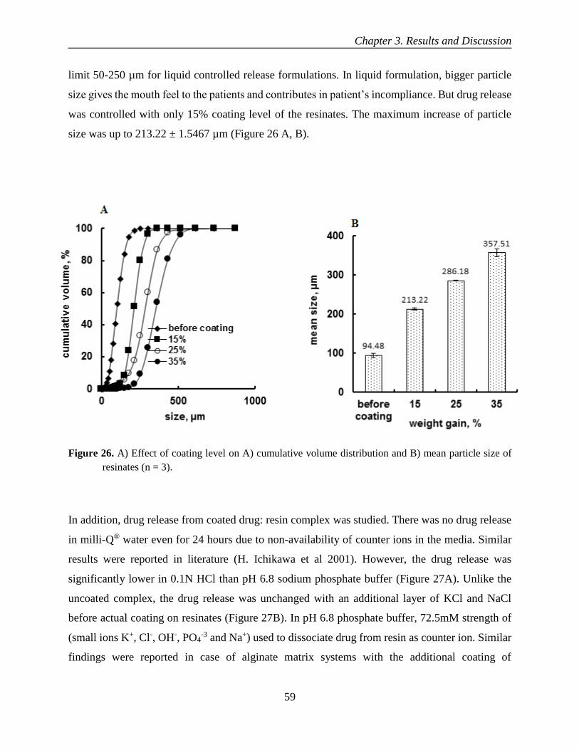
Chapter 3. Results and Discussion
59
limit 50-250 µm for liquid controlled release formulations. In liquid formulation, bigger particle
size gives the mouth feel to the patients and contributes in patient’s incompliance. But drug release
was controlled with only 15% coating level of the resinates. The maximum increase of particle
size was up to 213.22 ± 1.5467 µm (Figure 26 A, B).
Figure 26. A) Effect of coating level on A) cumulative volume distribution and B) mean particle size of
resinates (n = 3).
In addition, drug release from coated drug: resin complex was studied. There was no drug release
in milli-Q® water even for 24 hours due to non-availability of counter ions in the media. Similar
results were reported in literature (H. Ichikawa et al 2001). However, the drug release was
significantly lower in 0.1N HCl than pH 6.8 sodium phosphate buffer (Figure 27A). Unlike the
uncoated complex, the drug release was unchanged with an additional layer of KCl and NaCl
before actual coating on resinates (Figure 27B). In pH 6.8 phosphate buffer, 72.5mM strength of
(small ions K+, Cl-, OH-, PO4-3 and Na+) used to dissociate drug from resin as counter ion. Similar
findings were reported in case of alginate matrix systems with the additional coating of

Chapter 3. Results and Discussion
60
polycationic polymers (Xu et al., 2003; Zarate et al., 2011). However, drug release was
significantly higher in pH 4.5 when 200 mM NaCl was added than pH 6.5 added with 100 mM
NaCl and pH 1.6 added with 30 mM NaCl. The faster drug release in pH 4.5 (sodium acetate +
200 mM NaCl) might be due to higher concentration of ionic strength of Na+ in the buffer or
release medium (Figure 28A). On the other hand, the release of highly soluble drug, tramadol HCl
and diltiazem HCl, was faster and complete with same coating level than the soluble propranolol
HCl (Fig. 28B).
Figure 27. The effect of A) pH of the medium and Milli-Q® water B) an additional layer of salt and no
ionic layer coated with Eudragit® NE 30D 15% c.l cured at 60°C for 12 h.

Chapter 3. Results and Discussion
61
Figure 28. The effect of A) pH and NaCl and B) drug solubility on the release from coated resinates with
Eudragit® NE 30D 15% c.l cured at 60°C for 12 h.
The drug release was significantly higher in 200 mM and 300 mM NaCl than 100 mM NaCl. It
might be due to the increased ionic strength of release medium than 100 mM NaCl (Figure 29 A).
The drug release was almost similar for investigated range of particle sizes 160-250 µm, 250-
315µm but slower and incomplete for the particle size 315-425 µm. It could be due to the increased
diffusion path length for the drug after being dissociated from the ionic sites of the Purolite®
C100MRNS (Figure 29 B). However, drug release was slower with increased drug loading on the
resin for investigated range of 20% w/w to 75% w/w drug loading. The drug release from ion
exchange resin was always equilibrium dependent. There was always an equilibrium between
counter ion and associated drug in drug-resin complex. So, higher drug loading might shift the
equilibrium towards un-dissociated form, which ultimately made the drug release slow and
incomplete (Figure 29 C).

Chapter 3. Results and Discussion
62
Figure 29. Effect of A) the ionic strength (160-250 µm and 20% w/w drug loading), B) particle sizes (20%
w/w drug loading) and C) drug loadings (160-250 µm) on the release from complex coated with
Eudragit® NE 30D 15% c.l in phosphate buffer pH 6.8.
Different conditions of curing, presence of talc and coating levels were evaluated and investigated.
There was a fast release of drug from coated resinates in un-cured form. It might be due to
incomplete film formation. But drug release was decreased with cured resinates and slowest

Chapter 3. Results and Discussion
63
release was observed when curing was done at 40 ºC and 75% RH and 80 ºC. With increased
humidity, there could be an extra plasticization effect, which decreased the drug release
(Osterwald, 1985; Sun et al., 1999). With addition of 30% talc in the coating solution, there was a
fast drug release it could be due to reduced amount of polymer with an addition of talc in the
coating solution. During drug release talc particles leached out quickly and making the polymer
film more permeable and led to faster release as compared to the system where no talc was
involved (Li et al., 1990; Erdmann et al., 2000) (Figure 30 A, B). There was a fast and complete
release when resinates were coated only with 5% coating level (% weight gain). The drug release
was controlled with increasing coating level in the formulation. With 15% to 20% coating level, it
was presumed that all sides of resinates were completely covered by coated polymer and
underwent uniform swelling during drug release. With increase of the coating thickness, drug
release was slow even incomplete with higher coating level. It was probably due to the increased
diffusion path length of drug after being dissociated from ionic sites in the ion exchange resin.
(Figure 31).
Figure 30. Effect of A) curing conditions and B) anti-tackiness on propranolol HCl release form resinate
coated with Eudragit® NE 30D 15% c.l.

Chapter 3. Results and Discussion
64
Figure 31. Effect of different coating levels on propranolol HCl release in pH 6.8 sodium phosphate buffer.
The resinates were wet granulated with ethyl cellulose (ethocel c.P 100) and HPMC E5. There was
no effect on drug release when same concentration of both binders was used during granulation
process. As release profile was similar for formulations prepared with 15% w/w of each HPMC
E5 and ethyl cellulose (ethocel c.P 100) during granulation with both binders and in the
formulation without granulation. The only difference observed was the hardness of granules when
HPMC E5 and ethocel 100 c.P were used in higher concentration than 20% w/w. The granulation
only made the flow of particles better in the mini-glatt during coating process. The ethocel 100 c.P
was taken as binder and release retardant when used with combination of HPMC E5. But it did not
retard the release significantly (Figure 32A). Furthermore, drug release was similar with 5%, 10%
use of Tween® 80 in the coating formulation as compared to the formulation without Tween® 80.
It might be due to its talc dissolving property in the coating formulation without being had any
influence on the release of the drug from the system where drug was released with help of counter
ions in the system of ion exchange resins (Amighi and Moes, 1997, Bajdik et al., 2003) (Figure
32B).

Chapter 3. Results and Discussion
65
Figure 32. Effect of A) binders for granulation and B) Tween® 80 in the coating solution on propranolol
HCl release from coated resinate with Eudragit® NE 30D 15% c.l. in phosphate buffer pH 6.8.
It had been observed that the drug release from drug–resin complex and its microcapsules was
controlled by three possible mechanisms (Gyselinck et al., 1982). These were (1) the exchange
reaction between drug and the counter-ion (present in the release medium) and drug molecules in
the resinates (drug-resin complex). (2) The release of drug through the pores within the resin
structure with divinyl benzene (DVB) crosslinking (particle diffusion control). (3) The release of
drug across the thin layer of coated polymer around the resinates (membrane diffusion control).
However, particle diffusion played a pivotal role in the release of drug from drug–resin complex
(Irwin, 1987). The time for total 20%, 50% and 80% release were significantly lower for ion
exchange resin, having less cross- linking i.e. 4% than those having double (8%) DVB cross
linking (Table 11). The time for 80% release of Dowex® 50W was only 5 hours and it was lower
than the time for Purolite® C100MRNS which was almost 18 hours. When resinates of both ion
exchange resins were coated with 15% coating level of Eudragit® NE 30D cured at 60 ºC for 12
hours for same drug loading of almost 20% w/w for both resins. Similar findings were reported in
literature by Atyabi and his co-workers (Atyabi et al.,1995).

Chapter 3. Results and Discussion
66
Table 11. Effect of divinyl benzene cross linking (4% and 8%) on drug release from coated resinates
Release time
(h) Dowex® 50W Purolite® C100MRNS
4% DVB 8% DVB
t20 1 ± 0.12 4 ± 0.13
t50 3 ± 0.25 9 ± 0.35
t80 5 ± 0.34 18 ± 0.12
Contrary to the fast drug release with HPMC E5 when mixed in the coating formulation to disperse
talc homogenously in the coating solution. There was similar drug release with 0%, 5% and 10%
HPMC E5 (Figure 33A). It might be due to the two reasons. (1) 10% HPMC E5 was not sufficient
enough to create pores in the coating layer of flexible polymer Eudragit® NE 30D when used in
the coating formulation with said concentrations. (2) The non-dependent nature of drug release on
pores in coating layer. As drug release was only affect by ionic strength of release medium and
divinyl benzene cross (DVB) linking inside resin. On the other hand, no significant effect of
granulation fluid was observed as the release profile for all three formulations was similar when
different ratios of alcohol and water were used as granulation fluid (Figure 33B).
Figure 33. Effect of A) HPMC E5 in coating solution and B) granulation fluid (alcohol: water) on
propranolol HCl release from coated resinate coated with Eudragit® NE 30D 15% c.l.

Chapter 3. Results and Discussion
67
3.1.4 Stability Studies
The formulation was stable over one month when tested for its release profile at accelerated
conditions 40ºC/75% RH and optimum conditions 25ºC/60% RH defined by the ICH guidelines.
The drug release was slight faster on first day, but similar for one week to one month. When
formulation was tested at 40ºC with enhanced humidity 75% RH and 25ºC with relative humidity
60% RH (Figure 34). It could be due to the additive curing effect of the coated polymer with
increased humidity. The similar findings were reported by (Adeyeye et al. 2005), where release of
diclofenac–resin microcapsules in methylcellulose suspensions decreased with increased
temperature due to polymer relaxation, which sealed the drug within the matrix.
Therefore, the release profiles of coated resinates of propranolol at different storage temperatures,
humidity and times were equivalent to that of coated resinates tested at 2-8 °C after reconstitution,
leading to the conclusion that there was no effect of the storage time, temperature and humidity
over the release profile of the propranolol–resin reconstituted powder over one month.
Figure 34. Effect of storage conditions A) 40 ºC/75% RH and B) 25ºC/60% RH on propranolol HCl release
from (160-250 µm, 20% w/w drug loading) resinate coated with Eudragit® NE 30D 15% c.l.

Chapter 3. Results and Discussion
68
X-ray diffraction studies were undertaken to investigate the crystalline nature of the drug. The
drug showed characteristic crystalline peaks at 2θ = 7.5, 10.2, 13.5, 20, 22.5 and 25° and in
physical mixture with resin. While drug-resin complex, exhibited amorphous characteristic with
absence of crystalline peaks. The disappearance of the crystalline peaks of drug shows that upon
complexation with resin, drug became part of resin. It was molecularly attached to the binding
sites of the resin within the resin matrix. Similar findings were also observed by Vuorio and his
co-workers (Vuorio et al., 2004). Moreover, the drug-resin complex was stable over one day at
room temperature and one week when heated up to 60ºC (Figure 35).
Figure 35. Powder X- ray diffractogram of propranolol HCl, Purolite® C100MRNS, physical mixture,
drug-resin complex (DRC) at different time interval and temperature.
As drug always forms a complex with resin, to establish the interaction of quaternary ammonium
group of polymers with resin ionic sites of the resin, the complex was coated with Eudragit® RS
30D and Eudragit® RL 30D blend with the ratio of 7:3 w:w. The drug release from the drug-resin
complex coated with the blend was similar to that coated with Eudragit® RS 30D alone (Figure
36). Which meant, there was no additional effect of 5% extra quaternary ammonium group of
Eudragit® RL on drug release from resinates. It was probably due to the flexibility of Eudragit®
RS 30D which masks the net effect of Eudragit® RL 30D. Most recently, a similar results were
reported in the terbutaline-loaded resinates coated with Eudragit RS: RL polymers by (Cun˜a et

Chapter 3. Results and Discussion
69
al., 2000). As established, that drug was released by dissociation from ionic sites inside the resin
matrix. But drug release from Eudragit® RS 30D, Eudragit® RL 30D and Kollicoat® SR 30D used
to be sigmoidal from the matrix and reservoir systems (Ichikawa., et al, 2001).
The chloride counter ions (Cl-) of the quaternary ammonium groups were exchanged with the
anionic buffer species (PO4-3, OH-) in the release medium. The water uptake of the coated beads
correlated well with the drug release (Sun et al., 2001; Lehmann, 1986; Watts et al., 1991;
Bodmeier et al., 1996). No lag phase was observed, when drug-resin complex was coated with
Kollicoat® SR 30D even during one-month stability studies. The drug release was also stable over
one month at 40ºC/75% RH both from Eudragit® RS 30D and Eudragit® RL 30D blend (ratio 7:3
w/w) (Figure 37 A, B). The similar findings were also observed by (Ichikawa., et al, 2001).
Wherein, drug-loaded ion exchange resin with a Eudragit® RS 30D coating, the coating material
necessary for prolonged release was only 3% w/w based on the weight of the drug-loaded ion
exchange resin. Consequently, it was made stable for one week with high drug content of 54%
w/w in the resin. The drug release from coated resinates with Eudragit RS 30D 15% c.l. cured at
60°C for 12 h was slightly increased under accelerated storage conditions (Figure 38).
Figure 36. Effect of different polymers on propranolol HCl release from (160-250 µm, 20% w/w drug
loading) resinate coated with Eudragit® NE 30D 15% c.l.
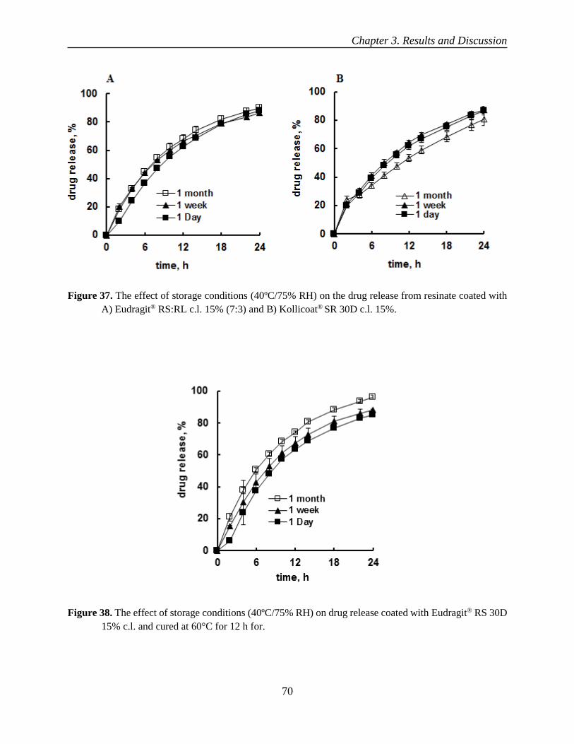
Chapter 3. Results and Discussion
70
Figure 37. The effect of storage conditions (40ºC/75% RH) on the drug release from resinate coated with
A) Eudragit® RS:RL c.l. 15% (7:3) and B) Kollicoat® SR 30D c.l. 15%.
Figure 38. The effect of storage conditions (40ºC/75% RH) on drug release coated with Eudragit® RS 30D
15% c.l. and cured at 60°C for 12 h for.

Chapter 3. Results and Discussion
71
3.1.5 Drug leaching and stability studies over one week after reconstitution
The formulation was mixed with extra soluble diluent granules of HPMC E5 of size 800 µm to aid
reconstitution. The soluble granules of HPMC E5 were quite separately visible in macroscopic
image of the formulation. The extra soluble granules upon contact with water immediately turned
into suspending medium for reconstitution. The nature and concentration of soluble granules
determined the final redispersibility, sedimentation and viscosity of the reconstitutable suspension
(Pongjanyakul et al., 2005). The formulation after reconstitution clearly showed suspended resin
particles without any agglomeration, which ultimately ensures the uniform dose after
reconstitution (Figure 39 A, B, C, D).
The reconstitutable granules were prepared with different grades of HPMC. The particle size
before reconstitution of the formulation was 212 µm. After reconstitution particle size was
increased to 218 µm. The 2% increase in particle size is due to the swelling behavior of either
coated Eudragit® NE 30D or the resin matrix itself. The coated resinates maintained their
independent shape and geometry without being agglomerated with each other which confirms the
stable nature of the formulation. Moreover, pH of the formulation was varied from 7.3 to 7.4. This
pH showed the almost neutral nature of the coated particles suspended in the HPMC medium.
Final dose of the formulation was 120 mg/ml (Table 12). Moreover, release profile of the
formulation after reconstitution when stored at (2 – 8 °C) was stable over one week (Figure 40).
In a nut shell, the medium taken for reconstitution does not affect the release profile of the
formulation. It was similar with the findings of (Obitte et al., 2009) regarding suspensions of
amoxicillin/clavulanic acid stored at (2 - 8°C) after reconstitution over one month.

Chapter 3. Results and Discussion
72
Figure 39. A) Formulation before reconstitution B) macroscopic image of formulation before reconstitution
C) formulation after reconstitution D) macroscopic image of formulation after reconstitution.
Table 12 Summary of the formulation before and after reconstitution
Coated resin pH
(0 day → 1 week)
Particle size
(μm)
Dose
(mg/ml)
1 week
before reconstitution --- 212.11 ± 3.98 ----
after reconstitution 7.3 ± 0.1 → 7.4 ± 0.1 218.15 ± 1.25 120 ± 8.75

Chapter 3. Results and Discussion
73
Figure 40. Effect of storage conditions at 2-8 °C after reconstitution on the drug release in pH 6.8 sodium
phosphate buffer.
As reconstitutable powder is always taken with water, drug leaching in water had to be undertaken
for one week. Less than 1% drug was leached in milli-Q® water. The 1% drug leaching was
observed in 5 mM to 10 mM NaCl and KCl over one week. When leaching studies of the
formulation were investigated till one week. However, drug leaching was slightly higher but not
significant (p ≥ 0.05) on 3rd day in 10 mM of NaCl and KCl as compared to 5 mM NaCl and KCl
(Figure 41). It was probably due to attainment of equilibrium between un-dissociated and
dissociated drug from the resin. It indirectly established the good drug retaining tendency of
Purolite® C100MRNS. As drug retention in the resin after ionically bind with its ionic sites based
on the pore size of the resin structure 12.54 Aº (Bilandi et al., 2014).
The pH of the leaching media varied in the range 6.8 ± 0.2. The conductivity of 5 mM KCl and
NaCl was 667.5 µS/cm and 659.33 µS/cm respectively. On the other hand, conductivity of leaching
medium of 10 mM KCl and NaCl was 1513.9 µS/cm and 1229.67µS/cm respectively. Both drug
and resin remained ionized in the said pH range but exhibited drug leaching was less than 1% for
both 5 and 10 mM KCl and NaCl over one week after reconstitution.

Chapter 3. Results and Discussion
74
Figure 41. A) Propranolol HCl leaching over one week in A) 5 mM and B) 10 mM of KCl, and NaCl and
in Milli-Q® water (n = 3).
3.1.6 Conclusion
A controlled release liquid formulation (reconstitutable powder) of propranolol HCl with high dose
was achieved with Purolite®C100MRNS. The release kinetics from coated drug-resin complex
was found to be particle diffusion controlled. Moreover, release had linear relationship with degree
of divinyl benzene (DVB) cross-linking of the resin. The time for 80% drug release of Dowex®
50W (having 4% DVB cross-linking) was significantly less than Purolite®C100MRNS (8% DVB
cross-linking). The drug release was also significantly influenced by thickness of the coating and
ionic strength of the release medium. The coated resinates with Eudragit® NE 30D were stable
over one month’s storage at 40°C/75% RH and 25°C/60% RH. Finally, the sustained release
reconstituted powder of propranolol showed chemical stability before and after reconstitution. As
release profiles remained unchanged over one week, when stored at 2-8°C after reconstitution. The
pH varied over one week was 7.3 ± 0.2. The ion exchange resin Purolite®C100MRNS exhibited
significantly higher drug retention as only 1% drug was leached from drug-resin complex in Milli-
Q® water. The drug leaching was less than 1% when formulation was treated with 5 mM to 10 mM
KCl and NaCl over one week after reconstitution. In summary, Purolite®C100MRNS could be used
as a carrier for preparation of a controlled-release liquid formulation, reconstitutable powder dosage
form.

Chapter 3. Results and Discussion
75
3.2 Development of enteric drug delivery system using Amberlite® IR69F
(cation exchange resin)
Conventionally, drugs are released from dosage forms with an immediate or extended fashion.
However, in present times, pulsatile drug release systems are gaining importance. A pulsatile drug
delivery system, able to release the drug after a well-defined lag-time (Bussemer et al., 2001;
Ritschel et al., 1994). The delivery of therapeutic agent at site specific to the intestinal region can
be accomplished by providing an enteric coating on the solid or liquid dosage forms
(reconstitutable powder). The enteric coated products are designed and formulated to release the
drug in the upper intestine. These coated products are formulated to remain intact in the stomach.
The rationale behind enteric coated formulations is well documented (Wilding, 2000).
The polymers used for enteric formulations are anionic polymethacrylates (copolymerisate of
methacrylic acid and either methylmethacrylate or ethyl acrylate (Eudragit®, cellulose based
polymers, e.g. cellulose acetate phthalate, polyvinyl derivatives, e.g. polyvinyl acetate phthalate).
In these polymers, there is a functional group which is insoluble in acidic pH and soluble at neutral
pH due to ionization and changing to soluble salt form in presence of sodium ions of the release
medium of intestine (Dangel et al., 2000).
The liquid-controlled release enteric coated formulations have been a challenge to the formulation
scientists. It was due to their stability concerns. To cop out, such challenges, drugs are first
ionically complexed with ion exchange resins and then coated with appropriate pH dependent
enteric polymer. The USP limit regarding release for such formulations, there is less than 10%
drug release as long as the formulation stays in the stomach. The liquid formulations in form of
reconstitutable powders has stability issues with respect to pH of the formulation and drug leaching
after reconstitution in the carrier vehicle. As per USP limit, drug leaching in the suspending
medium should not be more than 5%.
3.2.1 Drug loading
A drug loading of almost 7% w/w was achieved in 24 hours, when particle size of Amberlite® IR
69F was 300-1100 µm. However, drug loading was significantly increased up to 55% when
particle size was reduced to 71-160 µm. It was probably due to increased surface area of the resin,
which decreased the diffusion path length for the drug to reach ionic sites inside the resin.

Chapter 3. Results and Discussion
76
Although, 30% w/w drug loading was achieved quickly in the first hour when particle size of resin
was 71-160 µm. Moreover, for bigger particle size of 315-630 µm, loading was 28% after two
hours which, gradually increased up to 40% in 24 hours of batch process. There was absolutely no
drug loading on Amberlite® IR 69F till 2 hours when resin was taken in particle size 300-1100 µm.
It might be due to the decreased surface area and increased diffusion path length into the resin for
its ionic sites (Figure 42). Similar findings were also observed by Sriwongjanya and his coworkers
(Sriwongjanya et al.,1995). The un-milled and milled form of Amberlite® IR 69F were shown in
(Figure 43A, B).
Figure 42. Effect of Amberlite® IR69F particle size on propranolol HCl loading in batch process (n = 3).

Chapter 3. Results and Discussion
77
Figure 43. A) Image of Amberlite® IR69F (particle size 300-1100 µm) before milling B) after milling
(particle size (45-160 µm).
During single batch process, for maximum drug loading of 52.41 ± 1.22 % w/w, total used ion
exchange capacity of Amberlite® IR69F was 2.1 meq/g, when drug and resin were taken in ratio
2:1 w/w. The total 1% drug was adsorbed/attached hydrophobically on the surface of the resin.
For this range of drug loading, association efficiency was 55.25 ± 2.64 %, which was further
decreased up to 30.46 ± 8.96 % for 3:1 w/w drug and resin ratio. The sharp decrease in association
efficiency was probably due to the total increased drug concentration in the system (Guo et al.,
2009). The max used ion exchange capacity of Amberlite® IR69F was 2.9 meq/g, when drug
loading was 74.93 ± 0.14 % in triple batch process. On the other hand, 1.1 meq/g ion exchange
capacity of the resin was utilized when drug and resin were in a ratio of 1:2 w/w. The drug loading
was reduced to 32.80 ± 0.07 % (Pongjanyakul et al., 2005). It was might be due to the increased
number of ionic sites of resin in the system (Table 13).
The drug loading was independent of stirring speed (150-600) rpm (Figure 44). But it was
significantly increased when already competing ions were taken out from the system by filtration.
A total 24 % increase in drug loading was observed, from 52% to 76 % in triple batch process.
Moreover, drug loading was also found to be independent of pH (2–8). It was probably due to the
ionized state of both propranolol and resin at the experimental pH (2–8) during batch process
(Figure 45).

Chapter 3. Results and Discussion
78
Table 13. Summary of drug loading and drug association efficiency for Amberlite® IR69F
*Superscript number means continued the respective batch
Figure 44. Effect of stirring speed on drug loading on Amberlite® IR69F in the loading medium of pH 6
and at temp 23 ºC (n = 3).
No. Batch
process
Drug
(mg)
Resin
(mg)
Drug
:resin
ratio
Association
efficiency,
% + (±SD)
Drug loading
Attached
hydrophobically
%
% w/w (±SD) meq/g
1
Single
100 100 1:1 94.53±2.41 48.59±0.63 1.8 1
2 200 100 2:1 55.25±2.64 52.41±1.22 2.1 1
3 300 100 3:1 30.46±8.96 46.46±7.68 2.1 1
4 400 100 4:1 29.20±0.16 52.82±1.90 1.5 1
5 100 200 1:2 97.61±0.34 32.80±0.07 1.1 1
6 100 300 1:3 97.76±0.12 24.54±0.03 0.9 1
7 100 400 1:4 95.62±0.98 19.20±0.16 0.8 0.5
8 Double *345² 100 49.67±0.95 67.47±0.41 2.6 0
9 Triple *3787 100 45.70±2.52 74.93±0.14 2.9 1

Chapter 3. Results and Discussion
79
Figure 45. Effect of A) sequential batch process and B) pH of the loading medium on drug loading for
Amberlite® IR69F (n = 3).
3.2.2 Characterization of resinates
The drug crystals were visible with polarized light microscopy for drug alone and drug resin
physical mixture, but drug crystals were not visible in drug-resin complex. It confirmed the
amorphous nature of drug-resin complex. On the other hand, same was confirmed by powder X-
ray diffraction. It was undertaken to investigate the crystalline nature of the drug, drug showed
characteristic crystalline peaks at 2θ = 7.5, 12.5, 17.78, 18.76, 20 and 25° and in physical mixture
with resin. While drug-resin complex, exhibited amorphous characteristic with absence of
crystalline peaks (Figure 46). The disappearance of the crystalline peaks of drug showed the
complexation of drug with ion exchange resin. Similar findings were also observed by Vuorio and
his co-workers (Vuorio et al., 2004).

Chapter 3. Results and Discussion
80
Figure 46. Polarized light microscopic image A) of drug, B) physical mixture and C) drug-resin complex
(magnification 20x) and D) powder X-ray diffractogram of propranolol HCl, physical mixture, and
drug-resin complex.
As drug loading had linear relationship with swelling of ion exchange resin during batch process.
Amberlite® IR69F swelled 13 % in unloaded form and 28% in drug loaded form. Resins having
hydrophilic ionic sites in their structures which attract moisture on their exposure to environment.
The moisture contents of unloaded and drug loaded resins only 10% and 5% respectively. The
moisture was removed in total 9 minutes for unloaded and 4 minutes for drug-loaded resin. The
initial quick loss was in first minute. Then gradual increase up to 12% for unloaded and 5% for
loaded Amberlite® IR69F respectively. The 5% lower moisture for drug loaded resin was probably

Chapter 3. Results and Discussion
81
due to the occupancy of ionic sites with drug molecules. It ultimately reduced the free ionic sites
for moisture. The ion exchange resin appeared to be hollow from inside with well-defined
boundary in its swelled form under microscopic examination (Figure 47).
The overlay of FTIR spectra indicated the interactions between R-SO3-Na+ and Prop-NH2
+Cl – of
drug and ion exchange resin. The shifting of stretching frequencies of transmittance bands from
Figure 47. A) Swelling of Amberlite® IR69F in unloaded and drug loaded form. B) Kinetics of total loss on
drying of Amberlite® IR69F at 103°C. C) Light microscopic image before swelling D) after swelling.

Chapter 3. Results and Discussion
82
1180.43 cm-1,1039.62 cm-1 and 833.24 cm-1 to 1172.65 cm-1,1031.91 cm-1,792.74 cm-1
respectively and broadening of peaks confirms the complex between R-SO3-Na+ and Prop-NH2
+Cl-
(Figure 48). The above shifts in the wavenumbers and broadening of the peaks, suggested the ionic
interactions and other interactions like hydrogen bonding between ion exchange resin and
propranolol, resulted in the formation of drug-resin complex (Borges et al., 2005; Li et al., 2007;
Ribeiro et al., 2005; Sarmento et al., 2006; Shang et al., 2007).
When drug was loaded on the ion exchange resin. The particle size of the resin increased. The
increase in 12.15 ± 2.67 µm mean particle size of resinates was due to the expansion of resin matrix
on drug loading (Figure 49 A, B).
Figure 48. FTIR spectrum of Amberlite® IR69F, propranolol HCl physical mixture of drug and resin and
drug-resin complex.

Chapter 3. Results and Discussion
83
Figure 49. Effect of drug loading on A) cumulative volume distribution of resinates and B) mean particle
size of resinates (n = 3).
The mean particle size of the powder (resinates) before coating was 119 µm and it was increased
with increasing coating level (Figure 50). At 25% coating level, the size reached 414 µm which
exceeded the USP limits of particle size (50-250 µm) for liquid controlled release reconstitutable
formulations. Therefore, in order to avoid this problem of increasing particle size, resinates were
wet granulated in an open tray/petri dish and dried at 60°C in an oven for overnight. Finally, dried
granules were shifted to mini-Glatt for coating process. The mean particle size before coating was
191 µm which was negligibly increased up to 225 µm at 25% coating level (Figure 51). After
granulation, individual particles/resinates retained/maintained their geometry and size, which
further underwent negligibly increased in size on coating, instead of liquid bridging between non-
granulated resinates and agglomerated them into bigger particles (Torres et al., 1998).

Chapter 3. Results and Discussion
84
Figure 50. Effect of coating level on A) volume distribution of resinates and D) mean particle size of
resinates (n = 3).
Figure 51. Effect of coating level after granulation on A) cumulative volume distribution of resinates and
B) mean particle size before and after different coating levels (n = 3).

Chapter 3. Results and Discussion
85
3.2.3 Drug release and influence of ionic strength
There was less than 10% drug was released in 0.1 N HCl during two hours and more than 75%
release after first hour in pH 6.8 sodium phosphate buffer. It confirmed the enteric drug delivery
system with cation exchange resin (Amberlite® IR 69F) when coated with 25% coating level of
Eudragit® L 30D-55. The significance of ion exchange resin in the formulation was established,
as without making complex with resins, there was a higher coating level (% weight gain) up to
45% was required to make the formulation enteric release. Whereas, coating level was reduced to
only 25% when drug was complexed with resin. Moreover, almost 30% drug was released during
first two hours in 0.1N HCl, when propranolol granules alone (formed with 15% w/w of HPMC
E5) were coated with same thickness (25% c.l.) of Eudragit® L 30D-55 for the same size of 160 -
250 µm granules. Moreover, it was pertinent to described that, when granules were coated with
45% c.l. the final size of granules was increased and bigger than 250 µm (Figure 52A).
No change in release was observed when 200 mM and 400 mM NaCl was added to the release
medium of pH 6.8 (sodium phosphate buffer). The total drug release achievable in the release
medium pH 6.8 was up to almost 80% (Figure 52B). It was probably due to the strong nature of
ionic complex between drug and resin. It could also be due to the enhanced consumption of Na+
ions for the formation of salt of carboxylic group of Eudragit® L30D-55 and dissociation of
propranolol from ionic sites in the resin. This salt formation of carboxylic group of coated polymer
and dissociation of propranolol from the ionic sites of the resin, reduced the overall, concentration
of counter ions specially (Na+) in the release medium (Raju et al., 2011).
Furthermore, the drug release was remained unchanged after three months storage of the coated
resinates at 40 ºC/75% RH and 25 ºC/60% RH (Figure 53A). In addition to above, drug-resin
complex was stable at 40ºC and 60ºC over the period of one week, as there were no drug peaks
were visible in graph obtained by powder X-ray diffraction.
Moreover, clear and conspicuous peaks were exhibited by drug alone and its physical mixture at
at 2θ = 7.5, 12.5, 17.78, 18.76, 20 and 25°. Which confirmed the amorphous nature of the resinates
(Figure 53B).

Chapter 3. Results and Discussion
86
Figure 52. Effect of A) complexation and coating level B) ionic strength of the release medium in pH 6.8
Figure 53. Effect of A) storage condition for three months on propranolol HCl release in pH 1 for 2 h then
pH 6.8, and B) powder X-ray diffractogram.

Chapter 3. Results and Discussion
87
3.2.4 Drug leaching and stability over one week after reconstitution
As enteric coated reconstitutable powders were to be mixed with water or any other liquid as a
carrier (vehicle), prior to their administration. Therefore, there was need to establish leaching of
drug in the carrier. There was less than 1% drug leaching in milli-Q® water over one week.
Moreover, it was pertinent to mention that almost 1% drug was leached in 5mM and 10 mM
NaCl/KCl over one-week storage. It might be due to the attainment of equilibrium between
undissociated drug form and dissociated drug. Moreover, when ionic strength of the leaching
medium was increased up to double the strength of ion exchange capacity of the resin, drug
leaching was still less than 1% in both media NaCl and KCl. It indirectly, depicted the excellent
drug retaining tendency of Amberlite® IR 69F on its ionic sites (Figure 54 A, B).
The pH of the suspending medium during drug leaching was varied in range. 6.3 ± 0.2. Moreover,
no agglomeration of coated resinates was seen after reconstitution with suspending medium
(formed by HPMC E5 granules) during one-week storage (Figure 55).
Figure 54. Propranolol HCl leaching with A) 5 mM KCl and NaCl and milli-Q® water and B) 10 mM of
KCl and NaCl and milli-Q® water.

Chapter 3. Results and Discussion
88
Figure 55. Macroscopic image of coated drug-resin complex (resinates) after reconstitution at temperature
of 2-8°C.
3.2.5 Conclusion
A stable sustained release enteric formulation (reconstitutable powder) of propranolol HCl was
achieved with cation exchange resin Amberlite® IR69F. The choice of the polymer and the
thickness of the coated layer were critical to control the release of drug in pH 1 and pH 6.8 (sodium
phosphate buffer). The formulation was also stable over one week after reconstitution at
temperature of 2-8°C. The pH of the formulation during reconstitution was varied in range 6.3 ±
0.2. The ion exchange resin Amberlite® IR69F exhibited significantly higher drug retention as only
1% drug was leached from drug-resin complex in Milli-Q® water. The drug leaching was less than
1% when formulation was treated with 5 mM to 10 mM KCl and NaCl over one week after
reconstitution. In summary, Amberlite® IR69F could be used as a carrier for preparation of an
enteric liquid formulation of reconstitutable powder.

Chapter 3. Results and Discussion
89
3.3 Evaluation of Purolite® C100CaMRNS (cation exchange resin) as a drug
combination carrier
Oral combination drug delivery systems have been proven to be highly effective and beneficial in
the treatment of several diseases such as cancer, acquired immune deficiency syndrome (AIDS),
tuberculosis, diabetes (Type 2), heart diseases, central nervous system (CNS) disorders, and for
treating several other microbial infections (Mayer and Janoff, 2007). Combination therapy may be
achieved by prescribing/administering separate drugs, dosage forms that contain more than one
active ingredient. Combination drugs most commonly refers to a fixed-dose combination (FDCs),
which is a formulation including two or more active pharmaceutical ingredients (APIs) combined
in a single dosage form, which is manufactured and distributed in certain respective fixed doses.
The fix dose combinations are often claimed to make medicine-taking more convenient for patients
taking multiple medication (Taupitz et al., 2013).
Fix dose combinations are mostly tablets and capsules. There is a need to develop fix dose
combination for geriatric and pediatric patients in form of a liquid formulations in reconstitutable
powder form. Liquid dosage forms from ion exchange resins have certain key challenges with
respect to stability of the formulation. Ion exchange resins are polyelectrolytes, which are insoluble
both in water and organic solvents. Two drugs might react each other if they are not complexed
with ion exchange resins especially, it happened when free acid and free basic drugs are formulated
together. It is not confined to the two acidic/basic drugs. Two basic/acidic drugs might also
undergo incompatibility when taken into same formulation together, so chemical and physical
stability of such drugs in a formulation could be enhanced by complexing with ion exchange resins
(Jenquin et al.,1990).
3.3.1 Drug loading
Both drugs diltiazem HCl and tramadol HCl were loaded on the Purolite® C100CaMRNS by a
batch process at room temperature. The drug loading was significantly higher when both drugs
were individually/separately loaded on the Purolite® C100CaMRNS. The drug loading of tramadol
and diltiazem was 50% w/w and 42% w/w respectively. On the other hand, tramadol HCl loading
was reduced by 18% and diltiazem HCl was 17% when both drugs were loaded simultaneously
(Figure 56A). In the solution, during drug loading process, both drugs compete for the ionic sites
in the resin. Almost 18% less drug loading for diltiazem HCl might be due to its higher molecular

Chapter 3. Results and Discussion
90
weight (407 g/mol) when compared to tramadol HCl (260 g/mol) (Zhang et al., 2000). During
the first hour of drug loading, 20% of both drugs were loaded on the resin. Subsequently, tramadol
HCl and diltiazem HCl loadings were total 40% and 22% w/w respectively till 24 hours of batch
process (Figure 56B).
Figure 56. A) Tramadol HCl and diltiazem HCl loading on Purolite® C100CaMRNS individually and
together when conditions were pH 6, temp 23ºC and rpm 300 B) Steps of both drugs loading
for 24 hours (n = 3).
3.3.2 Characterization of resinates with X-ray powder diffraction and FTIR
The peaks of tramadol HCl and diltiazem HCl were clearly visible in powder X-ray diffractograms
in their pure form and physical mixture with Purolite® C100CaMRNS. It was undertaken to
investigate the crystalline nature of the drugs, both drugs (tramadol HCl and diltiazem HCl)
showed characteristic crystalline peaks at 2θ = 10, 10.5, 15, 17.9, 20, 22.3, and 25° and 8, 10, 15,
18.9, 20, 22.45, 25, 27° respectively. In physical mixture with resin crystalline peaks were at 2θ =
10, 15, 18, 20, 25, 30°. Both drugs peaks were disappeared in the drug-resin complex, which
ultimately confirmed the amorphous nature of the drug- resin complex (Vuorio et al., 2004).
On the other hand, shifting of frequency of transmittance bands of SO3-1 group of Purolite®
C100CaMRNS from 1039.62 cm-1 to 1035.91 cm-1 and 1030.62 cm-1 in drug-resin complex

Chapter 3. Results and Discussion
91
confirms the formation of ionic complex between -HN group of both drugs and -SO3-1 group of
Purolite® C100CaMRNS (Figure 57 A, B).
Figure 57. A) Powder X-ray diffractogram of tramadol HCl, diltiazem HCl, Purolite® C100CaMRNS, both
drugs and resin, physical mixture, drug-resin complex B) FTIR spectrum of purolite®
C100CaMRNS tramadol HCl, diltiazem HCl, physical mixture and drug-resin complex.
The drug loading depends on the swelling of resin during batch process. Purolite® C100CaMRNS
swelled up to 38% and the moisture was removed in total 9 minutes for unloaded form and took 6
minutes for drug loaded resinates. The initial quick loss of moisture was in first minute, then
gradual increase up to maximum 12% for the unloaded resin and 8% for the drug loaded resin. The
lower moisture in drug loaded resins compared to unloaded resins was probably due to the
occupancy of ionic sites with drug molecules (Figure 58 A, B). The Purolite® C100CaMRNS was
in powder form and viewed as irregular shape and it became quite conspicuous after swelling under
light microscope (Figure 59 A, B).

Chapter 3. Results and Discussion
92
Figure 58. Effect of drug loading on A) swelling of resin and B) kinetics of loss on drying for resin before
and after drug loading (n = 3).
Figure 59. Light microscopic image of Purolite® C100CaMRNS A) before swelling B) after swelling
(magnification 20x).
During batch process of drug loading, average conductivity of resin alone was 6.33 ± 0.67 µS/cm,
whereas, conductivity of diltiazem HCl and tramadol HCl were 670 ± 19.41 µS/cm and 1024 ±
3.57 µS/cm respectively. When temperature of the loading medium was 17.7 ± 1.23 °C during
conductivity measurement. The 52.83% lower conductivity of diltiazem HCl as compared to

Chapter 3. Results and Discussion
93
tramadol HCl could be due to its higher molecular weight (450 g/mol) than tramadol HCl (260
g/mol) (Table 14).
At the ending of the batch process, the conductivity increased up to 78.90%. The increase in total
conductivity of the loading medium after batch process might be due to formation of additional
ions of (Ca+2 and Cl-) CaCl2 during exchange of Cl- ions of both drugs and Ca+2 ions of Purolite®
C100CaMRNS (Burton et al., 1995). On the other hand, there was not any significant change in
pH of the loading medium, before and after drug loading of batch process (Figure 60).
Table 14. Conductivity studies during batch process
Sample Resin
4mg/ml
(µS/cm)
Diltiazem
4mg/ml
(µS/cm)
Tramadol
4mg/ml
(µS/cm)
after batch
process
(µS/cm)
Temp
ºC
A 7.10 695 1024 1832 18
B 5.62 659 1028 1810 17.5
C 6.32 656 1020 1815 17.2
Avg 6.33 670 1024 1819 17.7
S.D 0.67 19.41 3.57 11.53 1.23

Chapter 3. Results and Discussion
94
Figure 60. Effect of diltiazem HCl and tramadol HCl loading on pH resin during batch process (n =3).
Mean particle size of resin increased up to 21.39 ± 2.5 µm after drug loading. It might be due to
the expansion of resin matrix during drug loading. Ion exchange resin expands gradually during
single batch process to double batch process of drug loading. Moreover, this expansion of ion
exchange resin is irreversible. As ion exchange resin remained expanded even after the drug
release. The increased mean particle size of resin after drug release was 69.09 ± 2.54 µm as
compared to original size 47.14 ± 2.4587 µm (Figure 61 A, B). It could be due to the reason that
resin uncoiled from inside and drug molecules make a space in the resin pores. This whole process
ultimately, led to a steric hindrance (repulsion) between two adjacent drug molecules and caused
the resin to expand till its maximum strength (Torres et al., 1998).
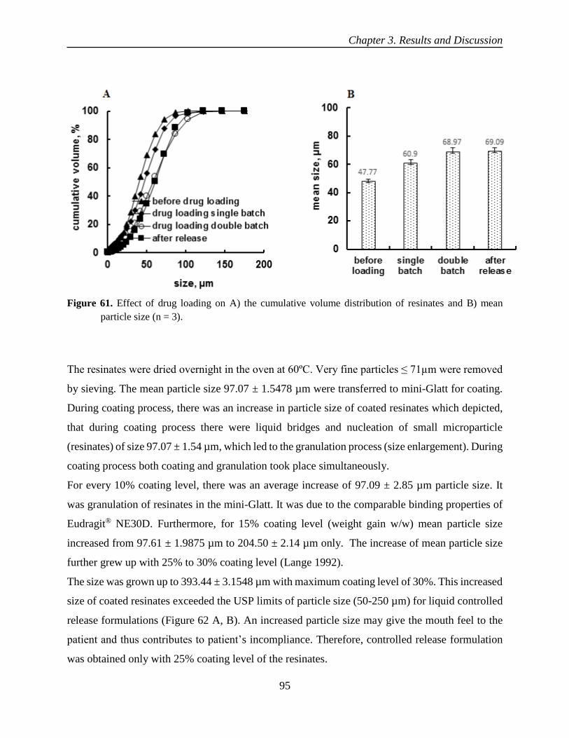
Chapter 3. Results and Discussion
95
Figure 61. Effect of drug loading on A) the cumulative volume distribution of resinates and B) mean
particle size (n = 3).
The resinates were dried overnight in the oven at 60ºC. Very fine particles ≤ 71µm were removed
by sieving. The mean particle size 97.07 ± 1.5478 µm were transferred to mini-Glatt for coating.
During coating process, there was an increase in particle size of coated resinates which depicted,
that during coating process there were liquid bridges and nucleation of small microparticle
(resinates) of size 97.07 ± 1.54 µm, which led to the granulation process (size enlargement). During
coating process both coating and granulation took place simultaneously.
For every 10% coating level, there was an average increase of 97.09 ± 2.85 µm particle size. It
was granulation of resinates in the mini-Glatt. It was due to the comparable binding properties of
Eudragit® NE30D. Furthermore, for 15% coating level (weight gain w/w) mean particle size
increased from 97.61 ± 1.9875 µm to 204.50 ± 2.14 µm only. The increase of mean particle size
further grew up with 25% to 30% coating level (Lange 1992).
The size was grown up to 393.44 ± 3.1548 µm with maximum coating level of 30%. This increased
size of coated resinates exceeded the USP limits of particle size (50-250 µm) for liquid controlled
release formulations (Figure 62 A, B). An increased particle size may give the mouth feel to the
patient and thus contributes to patient’s incompliance. Therefore, controlled release formulation
was obtained only with 25% coating level of the resinates.

Chapter 3. Results and Discussion
96
Figure 62. Effect of coating level on A) the cumulative volume distribution and B) mean particle size of
coated resinates (n = 3).
3.3.3 Drug release from uncoated and coated resinates
The release of both tramadol HCl and diltiazem HCl from uncoated resinates was burst and
incomplete both in 0.1 N HCl and pH 6.8 sodium phosphate buffer. This incomplete release in 0.1
N HCl could be due to the non-availability of desired strength of (H+, Cl-) ions in the release
medium or might be due to the strong nature of ionic complex of both drugs with resin (Xu et al.,
2003; Zarate et al., 2011).
However, drug release in release medium of pH 6.8 (sodium phosphate buffer) was almost 10%
more than 0.1N HCl. It was probably due to (Na+, Cl-, OH- and PO4-3) ions to dissociate the drug
from ionic sites of ion exchange resin (Figure 63).
On the other hand, there was complete release of drugs in both media when media was mixed with
300 mM of KCl (Figure 64). Drug release even from uncoated resinates was dependent on ionic
strength/concentration of the release medium (Zhang et al., 2000).

Chapter 3. Results and Discussion
97
Figure 63. Tramadol HCl and diltiazem HCl release from uncoated resinates in A) 0.1 N HCl and B) pH
6.8 phosphate buffer.

Chapter 3. Results and Discussion
98
Figure 64. Tramadol HCl and diltiazem HCl release A) 0.1 N HCl mixed with 300 mM of KCl. B) pH
6.8 (sodium phosphate buffer) mixed with 300 mM of KCl.
The tramadol HCl release was bit faster than diltiazem HCl, when their resinates were separately
coated. The bit faster release rate of tramadol HCl as compared to diltiazem HCl could be due to
its low molecular weight (263 g/mol) than diltiazem HCl (450 g/mol).
Whereas, both tramadol HCl and diltiazem HCl were released with the same rate, when they were
both complexed with the same resin and coated together with Eudragit® NE 30D. It was probably
due to their highly soluble nature of both drugs (Figure 65).

Chapter 3. Results and Discussion
99
Figure 65. Drug release after coating with Eudragit® NE 30D, 25% coating level cured at 60 ºC for 12
hours.
Finally, reconstitutable powder was to be administered with water so it was necessary to establish
drug leaching of the formulation in the water/carrier. There was less than 1% of both drugs
(tramadol HCl and diltiazem HCl) leached in 5mM of KCl solution, when its conductivity was in
range of 667.24 ± 8.256 µS/cm.
On the other hand, when strength of the leaching solution increased and made double (10 mM
KCl) than ion exchange capacity of Purolite® C100CaMRNS. The leaching of both drugs was still
less than 1%. when its conductivity was kept in the range 1513.9 ± 5.45 µS/cm. This negligible
amount of drug leaching after reconstitution of coated resinates for one week confirmed the stable
nature of the formulation (Figure 66).
Moreover, there was no leaching or negligible leaching of both drugs in milli-Q® water alone. It
was probably due to the non-availability of ions in the milli-Q® water. This meant that both drugs
ionically complexed with ionic sites of the resin and remained intact with resin even in the milli-
Q® water having no ions (Ichikawa et al 2001).

Chapter 3. Results and Discussion
100
Figure 66. Drug leaching from coated particles in milli-Q® water with A) 5 mM KCl and B) 10 mM KCl.
3.3.4 Conclusion
Both highly soluble drugs, tramadol HCl and diltiazem HCl were successfully complexed/loaded
with cation exchange resin (Purolite® C100CaMRNS). The nature of drug-resin complex for both
drugs was amorphous, characterized by powder X-ray diffraction. The ion exchange resin
Purolite® C100CaMRNS exhibited significantly higher drug retention as only 1% drug was
leached from drug-resin complex of both drugs in Milli-Q® water. The drug leaching was less than
1% when formulation was treated with 5 mM to 10 mM KCl over one week after reconstitution.
In summary, Purolite®C100CaMRNS could be used as a carrier for preparation of a controlled-
release liquid formulation of two highly soluble drugs (tramadol HCl and diltiazem HCl) in form
of reconstitutable powder.

Chapter 3. Results and Discussion
101
3.4 To enhance the robustness of reservoir system (pellets) by preventing
diffusion of drug into coated polymer by layering of drug-resin complex
on NP core
In reservoir multiparticulate systems, a lipophilic drug, such as ibuprofen which has a low melting
point, diffused into the coated polymer. When NP layered ibuprofen formulations were coated
with aqueous polymer dispersion Aquacoat® ECD. The drug diffusion was happened during
stability studies at accelerated conditions (Bodmeier et al., 1994). The same problem was
addressed with Kollicoat® SR 30D by Dashevsky and his co-workers (Dashevsky et al., 2005). To
prevent the drug diffusion into the coated polymer, an intermediate sealcoating was applied to the
formulation. This sealcoating should be of a hydrophilic polymer, for instance, hydroxypropyl
methylcellulose (HPMC) or polyethylene glycol (PEG). The aim of this study was to prevent the
diffusion of the layered lipophilic drug into the coated polymer with the help of a drug-resin
complex.
3.4.1 Drug loading on anion exchange resins
The ion exchange resins Purolite® A430MR and DuoliteTM AP 143/1093 swelled up to 58% and
78% respectively. Moisture content for DuoliteTM AP 143/1093 and Purolite® A430MR was 14%
and 6% respectively. For DuoliteTM AP 143/1093, the initial moisture loss occurred in the first
minute and then gradually increased up to 14%. For Purolite® A430MR, the 6% moisture was
removed/evaporated in the three minutes (Figure 67A, B). The adsorbed moisture may be due to
the free ionic sites of the unloaded resins. The ionic sites which were associated with the drug,
were not available for free moisture. Furthermore, during swelling studies water molecules went
into the resin matrix and form solvation shells inside the resin matrix and started to uncoil and
expand. This swelling of resin was inversely related to the percentage of divinyl benzene cross
linking in the resin. Once Purolite® A430MR and DuoliteTM AP 143/1093 swelled, they did not
come back to their original state even after heating, which meant that both ion exchange resins
were not flexible rather rigid and brittle. For both Purolite® A430MR and DuoliteTM AP 143/1093
their swelled state exhibited a definite boundary under light microscope (Figure 67C, D, E, F). The
moisture content for Purolite® A430MR were almost similar for both drug loaded and unloaded

Chapter 3. Results and Discussion
102
forms. Whereas, it was 5 percent less than for drug loaded form of DuoliteTM AP 143/1093 than
unloaded form (Figure 68).
Figure 67. A) Swelling of Purolite® A430MR and DuoliteTM AP 143/1093. B) kinetics of moisture content
of Purolite® A430MR and DuoliteTM AP 143/1093, and C) Light microscopic image of Purolite®
A430MR before swelling, and D) after swelling, and E) DuoliteTM AP 143/1093 before swelling,
F) after swelling.

Chapter 3. Results and Discussion
103
Figure 68. Moisture content of Purolite® A430MR and DuoliteTM AP 143/1093 before and after drug
loading.
The maximum drug loading for ibuprofen was 28% w/w. There was 58% drug loading for
diclofenac sodium with drug:resin ratio 3:1 w/w. The 30% higher drug loading of diclofenac
sodium was probably due to its higher solubility as compared to ibuprofen. The drug loading
remains constant even with three-fold increase of the drug in the system, meaning that no further
drug was loaded when the ionic sites of resin became saturated. On the other hand, the drug
association efficiency was almost 100% for diclofenac sodium (Table 15 and 16). The conductivity
studies of both diclofenac sodium and ibuprofen before and after drug loading were done in the
batch process. The conductivity of the DuoliteTM AP 143/1093 alone was 14 µS/cm and
conductivity of diclofenac alone was 1394 µS/cm before batch process. After batch process the
conductivity of the media was changed to 2045 µS/cm. 80% increase in conductivity after drug
loading was probably due to the exchange of ions and release of NaCl. The Na+ from drug and Cl-
from resin (Ashwini et al., 2012). On the other hand, ibuprofen alone had a conductivity of 7790
µS/cm. The 458% higher conductivity of ibuprofen than diclofenac sodium could be due to its
lower molecular weight. The conductivity after batch process was increased to 8250 µS/cm. This
30% increase in conductivity after batch process could be due to the formation of HCl, H+ from
ionization of ibuprofen and Cl- from the resin (Table 17).

Chapter 3. Results and Discussion
104
Table 15. Drug loading w/w and drug association efficiency for diclofenac sodium with DuoliteTM AP
143/1093 with batch process at 300 rpm at room temperature
Resin
(mg)
Drug
(mg)
Volume
(ml)
Drug : resin
ratio
(w/w) loading of
diclofenac (%) + (±SD)
Association efficiency of
diclofenac (%) + (±SD)
100 100 25 1:1 49.63±0.02 98.53±0.101
100 200 25 2:1 51.90±3.79 54.37±8.11
100 300 25 3:1 58.24±1.93 67.24±6.06
Table 16. Drug loading w/w and drug association efficiency for ibuprofen with Purolite® A430MR
Resin
(mg)
Drug
(mg)
Volume
(ml)
Drug:resin
ratio
(w/w) loading of
ibuprofen (%) + (±SD)
Association efficiency of
ibuprofen (%) + (±SD)
150 100 25 1:1.5 28.93±1.01 61.11±3.01
200 100 25 1:2 26.47±.83 72.02±3.07
Table 17. Conductivity studies of ibuprofen and diclofenac sodium during batch process at 23 ºC
Moreover, drug loading on Duolite TM AP143/1093, when it was having OH- and Cl- was similar
around 58% w/w. Initially, it was a quick drug loading in the first hour then equilibrium was
attained due to the saturation of ionic sites in the resin. It showed that drug loading remained
constant when DuoliteTM AP143/1093 had either OH- or Cl- ion as exchangeable counter ion. The
sample resin
alone 100mg
(µS/cm)
diclofenac
alone 100mg
(µS/cm)
ibuprofen
alone 100mg
(µS/cm)
diclofenac after
batch process
(µS/cm)
ibuprofen after
batch process
(µS/cm)
A 14.50 1407 7800 2060 8100
B 13.70 1378 7800 2048 8200
C 14.90 1397 7720 2038 8250
Avg 14.36 1394 7790 2045 8250
S.D 0.54 13.97 23.57 11.53 22.25

Chapter 3. Results and Discussion
105
change of pH was from 9.48 to 8.78 for DuoliteTM AP143/1093 during batch process before and
after diclofenac sodium loading (Figure 69).
Figure 69. Kinetics of diclofenac sodium loading on Duolite TM AP143/1093 for both OH- and Cl-.
The ion exchange resin, Purolite® A430MR was treated with 1N NaOH for twenty-four hours by
stirring at 300 rpm. Finally, it was recovered by vacuum filtration and washed with milli-Q® water
to remove excess NaOH from its surface. The initial pH of the eluent was 12.26 which became
9.15 and reached equilibrium on the fifth wash with milli-Q® water. On the other hand, Duolite TM
AP 143/1093 initial pH was 13.5 which became neutral, pH 7.15 on the fifth wash with milli-Q®
water (Figure 70). There was not any significant change in pH was observed before and after drug
loading when DuoliteTM AP 143/1093 (OH- ions) and Purolite® A430MR (Cl- ions) were used in
the batch process (Figure 71).

Chapter 3. Results and Discussion
106
Figure 70. A) Treatment of Purolite® A430MR with 1N NaOH and washed with milli-Q® water B)
Treatment of DuoliteTM AP 143/1093 with 1N NaOH and washed with milli-Q® water (n = 3).
Figure 71. Effect of drug loading on change of pH during batch process for A) DuoliteTM AP 143/1093
with OH- ions and B) Purolite® A430MR with Cl - ions.

Chapter 3. Results and Discussion
107
3.4.2 Increase in solubility of ibuprofen from drug-resin complex
The saturated solubility of Ibuprofen was 0.02 ± 0.04 mg/ml in purified water. Moreover, saturated
solubility of ibuprofen was 0.43 and 0.42 mg/ml, in NaCl (100 meq) and (300 meq) respectively
by shaking flask method. On the other hand, solubility increased significantly from drug-resin
complex up to 0.56 and 0.56 mg/ml in milli-Q® water when mixed with 100 meq and 300 meq
NaCl respectively. Almost 3.5 folds increase in solubility of ibuprofen could be due to change in
the salt form of ibuprofen in the presence of NaCl salts in their different concentrations (Table 18).
Table 18. Solubility of Ibuprofen from drug-resin complex
The particle size of Purolite® A430MR and Ibuprofen (drug-resin complex) was reduced by Dyno
mill with 5 cycles of milling to 10.65 ± 0.55 µm from 75.68 ± 0.54 µm (Figure 72,73). Moreover,
particle size distribution was varied from 4.14 ± 0.14 to 23.92 ± 1.5 with mean particle size 10.65
± 0.55 µm after five successive cycles of milling (Table 19).
Solubility in
Purified water
(mg/ml) + (±SD)
Solubility
in NaCl (mg/ml) + (±SD)
Solubility from
Drug resin complex in
NaCl (mg/ml) + (±SD)
Increase in
solubility
(folds)
100 meq 300 meq 100 meq 300 meq NaCl
0.02 ± 0.04 0.43 ± 0.19 0.42 ± 0.03 0.56 ± 0.13 0.56 ± 0.01 3.5

Chapter 3. Results and Discussion
108
Figure 72. Light microscopic image of drug-resin complex before milling with mean particle size.
Figure 73. Light microscopic image of drug-resin complex after milling with mean particle size.
Table 19 Particle size distribution before and after milling with dyno-mill
Milling D (0.1) (µm) (±SD) D (0.5) (µm) (±SD) D (0.9) (µm) (±SD)
before milling 12.75 ± 0.28 75.68 ± 0.54 172.91 ± 2.5
after milling 4.14 ± 0.14 10.65 ± 0.55 23.92 ± 1.5
before milling
after milling

Chapter 3. Results and Discussion
109
3.4.3 Layering of drug-resin complex on NP core and drug release
There was significant difference in initial release during one hour of the first day formulation. And
when it was stored at 40ºC/75%RH and 25°C/60%RH for one month. It was probably due to the
leaching of layered ibuprofen into the coated polymer during storage which ultimately released the
drug in the burst form for first hour (Figure 74).
The macroscopic images of pellets showed that pellets made by drug-resin complex layering were
irregularly increased in size as compared to the pellets formed by simple layering of ibuprofen on
NP core, it might be due to the non-uniform flow of pellets in the fluidized bed coater during
processing (Figure 75).
Figure 74. Ibuprofen release in pH 6.8 coated with 20% coating level of Kollicoat® SR 30D after storage
at 40ºC/75%RH and 25ºC/60%RH.

Chapter 3. Results and Discussion
110
Figure 75. Macroscopic images of A) ibuprofen layered NP core pellets B) drug-resin complex layered
NP core pellets.
In order to prevent the leaching of layered ibuprofen into coated polymer during storage, milled
drug-resin complex was layered on NP core 710-850 µm. But drug release was incomplete from
drug-resin complex layering. In order to get complete release, there was an additional layer of 10%
KCl (weight gain) was applied before the final coating with Kollicoat® SR 30D. The drug release
was bit fast and total release was increased up to 80% with this additional KCl layer but not of
significant (f2 = 82.52) (Figure 76).
It could be due to the increase in ionic concentration/strength in NP core below polymer coating.
Without this additional layer of KCl, drug release was only 62%. It was due to insufficient strength
of (Cl-, OH-, Na+ and PO4-3) ions in the sodium phosphate release medium of pH 6.8. Whereas
with an additional KCl layer there was overall increase in the cations and anions in form of K+ and
Cl- below the coating layer in the system. These ions release the drug from the drug-resin complex
layered NP core coated with Kollicoat® SR 30D (Woodworth et al., 1992).
Moreover, on macroscopic examination of pellets during and after release studies, it was evident
that pellets made by drug-resin complex underwent uniform swelling and gave a smooth release
profile even in the presence of weak points in the coating. On the other hand, swelling was
localized in resin-free pellets, which caused rupturing of surface of pellets and made the release
non-uniform (Figure 77, 78).

Chapter 3. Results and Discussion
111
Figure 76. Ibuprofen release in pH 6.8 coated with 20% coating level of Kollicoat® SR 30D.
Figure 77. Macroscopic images of drug-resin complex layered pellets after drug release in A) pH 1 and
B) pH 6.8 phosphate buffer.

Chapter 3. Results and Discussion
112
Figure 78. Macroscopic images of pellets without drug-resin complex layering after drug release in pH 1
A) and B) pH 6.8 phosphate buffer.
Furthermore, the drug release was similar, when drug-resin complex of ibuprofen and Purolite®
A430MR was taken in the ratios 2:1 and 1:1. It could be stated that increased drug concentration
in the complex had not any significant effect on the release. As drug release was only depend on
the availability of counter ions in the dissolution medium instead of drug concentration in the
complex (Figure 79). In drug-resin complex of ratio 2:1, less free ionic sites were available. It
meant that when drug resin complex was in ratio 1:1, higher number of ionic sites remained free
in the complex. These free ionic sites had no impact on retarding the drug release once the drug
was dissociated from ionic sites of the resin (Chowhan et al., 1982).
Moreover, drug layering efficiency of DRC (drug-resin complex) on NP core always depends on
the particle size of milled drug-resin complex by Dyno mill. The amount of binder was required
more than 20% w/w with respect to DRC was necessary to be taken when particle size was bigger
than 50 µm. Whereas, binder contents were reduced to 10-15% w/w when particle size of drug-
resin complex was reduced to around 15 µm. (Siepmann et al., 2008).
For different percentage/strengths of layering thickness of drug-resin complex on NP core, release
profile was similar, but coating level was reduced from 20% (where no resin in the formulation)
to 15% (drug-resin complex layering on NP core) (Figure 80).
The layering of drug resin complex was varied from 2.5% to 10% w/w for a constant amount of
coating level. The release profile was closer to zero order instead of first order. The reason of
reduction of coating level from 20% to 15% was due to the reason that already complex state of

Chapter 3. Results and Discussion
113
drug with resin retarded and controlled the release besides coating level. Moreover, drug release
remained unchanged of the formulation after one-month storage at 40ºC/75% RH and 25ºC/60%
RH (Figure 81).
Figure 79. Release of ibuprofen in pH 6.8 for different ratios of resin and drug in drug-resin complex.

Chapter 3. Results and Discussion
114
Figure 80. Release of ibuprofen from different layers of drug resin complex layering on NP core.
Figure 81. Effect of storage condition A) 25 ºC/60% RH and B) 40 ºC/75% RH on ibuprofen release in pH
6.8 phosphate buffer

Chapter 3. Results and Discussion
115
3.4.4 Conclusion
There was no burst effect and increase of drug release observed in first two hours. When NP core
(710-850 µm) was layered with drug-resin complex and stored for one-month storage at 40 ºC/75%
RH and 25 ºC/60%RH. The robustness of reservoir system (pellets) was successfully enhanced by
preventing of lipophilic drug (ibuprofen) into coated polymer Kollicoat® SR 30D by layering of
drug-resin complex on NP core. The coating level was reduced from 20% to 15% when drug-resin
complex layering was varied from 2.5% to 10% w/w on NP core. The saturated solubility of
ibuprofen was increased up to 3.5 folds from Purolite® A430MR and DuoliteTM AP143/1093 drug-
resin complex. In summary, anion exchange resin (Purolite® A430MR) could be used as carrier to
prevent the leaching of lipophilic drug into coated polymer.

Chapter 4. Summary
116
4 Chapter 4. Summary

Chapter 4. Summary
117
Summary
A drug loading up to 55% w/w was achieved on Purolite® C100MRNS, which was independent
of pH, temperature and stirring speed of the loading medium. The drug-resin complex was
characterized by FTIR for functional group interactions of both resin and drug. The amorphous
nature of drug-resin complex was characterized by powder laser diffraction. There was burst and
incomplete release from uncoated resinates and it was probably due to an equilibrium between
counter ions of release medium (H+, Na+) and exchanged drug molecules. An increase of 300 mM
(Na+ and Cl-) ions of release medium had released the 95% of drug in 0.1N HCl. On the contrary,
only 272.5 mM of (K+, Cl-, OH-, Na+ and PO4-3) ions of sodium phosphate buffer pH 6.8 were
sufficient to release almost 90% drug. The complete drug release was not achieved. This was
probably due to the strong nature of ionic complex between drug and resin or additional
hydrophobic interactions of drug due to its amphiphilic nature. There was a significantly faster
drug release with 4% DVB (divinyl benzene cross linking) than with 8%. As DVB cross linking
influenced the interparticle diffusion, when drug was dissociated from the ionic sites of the resin.
The release from Purolite® C100MRNS was depended on the pH and ionic strength of the media.
The higher drug loading on ion exchange resin contributed to a decreased release rate. There was
17.46 ± 2.53 µm increase in mean particle size of Purolite® C100MRNS due to expansion of resin
matrix on drug loading. Approximately 69.60 ± 6.95 µm increase of mean particle size of coated
resinates for every 10% increase of coating level. The drug-resin complex was stable at 40ºC and
60ºC for one week. Moreover, drug release was also un-changed up to three months of storage at
40ºC/ 75% RH and 20ºC/ 60% RH. The drug release from resinates coated with Eudragit® RS 30D
was increased after one month of storage at 40ºC/ 75% RH. This was probably due to an exchange
of Cl- with OH- which increased the permeability of the coating. A formulation with a higher dose
of 120.0 ± 9.13 mg/ml was successfully developed. The drug release remained unchanged over
one week after reconstitution with a pH of 7.4. The formulation does not agglomerate when
reconstituted with extra soluble granules of HPMC E5 of size 500-800 µm.
Another challenge with liquid controlled release formulations was the enteric coating due to the
stability concerns and the drug release in the stomach. To address these challenges, drugs were
first ionically complexed with Amberlite® IR 69F, then coated with the appropriate pH-dependent
polymer (Eudragit® L30D-55). The USP criteria for such formulations, is less than 10% drug
released in 0.1N HCl over two hours. The Amberlite® IR 69F swelled up to 45%. The drug loading

Chapter 4. Summary
118
was only 7% w/w for 72 hours, when resin was regular shape (spherical particles) with a size of
300 to1100 µm. The drug loading was significantly increased up to 55% w/w when resin was
milled by ball milling and particle size was reduced to 45-150 µm. The drug loading for a particle
size of 45-150 µm was fast within the first hour and remained constant till 24 hours. It was probably
due to saturation of all ionic sites in the resin. The shifting of transmittance peaks of -SO3-1 group
of resin from 1180.43 cm-1, 1039.62 cm-1 and 833.24 cm-1 to 1170.15 cm-1 1030.62 cm-1 and 794.67
cm-1 respectively, confirmed the formation of a complex between -N+HCl- group of propranolol
HCl and -SO3-group of Amberlite® IR69F. The absence of drug peaks in P-XRD and crystalline
nature of drug under polarized light microscope confirmed the amorphous state of the resinates.
There was increase of particle size 12 ± 3 µm during drug loading. It was due to the expansion of
resin matrix upon during loading process. There was increase in particle size of resinates up to
70.64 ± 5.85 µm for every 10% to 25% coating level (% weight gain). The drug release was less
than 10% in 250 ml of 0.1 N HCl within two hours and more than 70% release during first hour in
pH 6.8 sodium phosphate, confirmed the enteric drug delivery system. The drug-resin complex
was stable at 40ºC and 60ºC for a period of one week. Furthermore, the drug release remained un-
changed over 3 months storage at 40°C/75% RH and 25ºC/60% RH. Furthermore, there was no
agglomeration of coated particles during one-week storage at temperature of 2-8°C after its
reconstitution.
The model drugs (diltiazem HCl and tramadol HCl) were loaded on Purolite®C100CaMRNS
(cation exchange resin) by a batch process. The drug-resin complexes were characterized for
functional group interactions with FTIR and crystalline state of drug with P-XRD. The absence of
drug peaks in P-XRD confirmed the amorphous nature of the resinates. The particle size
distribution of powders was measured with powder laser diffraction. Uncoated resinates showed a
burst and incomplete release in both 0.1N HCl and pH 6.8 sodium phosphate buffer. The release
was successfully controlled by the Eudragit® NE30D, 25 % coating level (% weight gain) with a
curing procedure at 60°C for 12 hours. The complete drug release for both drugs was achieved
when the media was mixed with 300 mM KCl. The shifting of stretching vibrations of -SO3- group
of resin from 1039.62 cm-1 to 1035.91 cm-1 and 1030.62 cm-1 in drug- resin complex confirmed
the formation of a complex between -NH group of both drugs and -SO3-group of resin. The
leaching of drug was less than 1% when the formulation was treated with 5 mM KCl/NaCl over
one week with pH 6.08 ± 0.02.

Chapter 4. Summary
119
In reservoir multiparticulate systems, lipophilic drug such as ibuprofen, is leached out in the
coating polymer during the storage of the formulation at 40 ºC/75% RH. The leaching was
happened when the formulations were coated with an aqueous polymer dispersion like Aquacoat®
ECD and Kollicoat® SR 30D. This ultimately affects the robustness of the reservoir systems. In
order to prevent this drug leaching into the coated polymer, an intermediate seal coating of
(HPMC) or (PEG) was applied. At the same time, the additional seal coating increases the
production costs of the delivery system. So therefore, there was a need to address the leaching of
the lipophilic drug into the coated polymer with help of drug-resin complex. The drug Ibuprofen
was successfully loaded/complexed with anion exchange resin Purolite® A430MR and Duolite TM
AP143/1093. The particle size of drug-resin complex was reduced to 10.65 ± 0.55 µm using Dyno
mill. The drug-resin complex was layered on NP core 710-850 µm in a fluidized bed coater. The
layering formulation was prepared with 20% HPMC E5 as a binder w/w of drug-resin complex.
There was no increased drug release observed during first hour of the release, when formulation
was stored for one-month at 40 ºC/75% RH and 25 ºC/60% RH. This confirms the prevention of
leaching of lipophilic drug into the Kollicoat® SR 30D coating during storage with help of drug-
resin complex layering.
Chapter 5. Zusammenfassung
120
5 Chapter 5. Zusammenfassung

Chapter 5. Zusammenfassung
121
Eine Arzneistoffbeladung bis zu 55% (m/m) wurde mit Purolite® C100MRNS erreicht, welche
unabhängig von pH, Temperatur und Rührgeschwindigkeit des Beladungsmediums war. Der
Arzneistoff-Harz Komplex wurde durch FTIR auf Wechselwirkungen funktioneller Gruppen von
sowohl Harz als auch Arzneistoff hin charakterisiert. Die amorphe Natur des Arzneistoff-Harz
Komplexes wurde durch Powder Laser Diffraktometrie charakterisiert. Es gab spontane und
unvollständige Freisetzung von nicht-überzogenen Harzen und dies lag wahrscheinlich an einem
Gleichgewicht zwischen Gegenionen des Freisetzungsmedium (H+, Na+) und ausgetauschten
Arzneistoffmolekülen. Eine Zunahme von 300mM (Na+ and Cl-) Ionen des Freisetzungsmedium
setzte 95% des Arzneistoffs in 0,1 N HCl frei. Im Gegensatz dazu waren nur 272,5 mM von (K+,
Cl-, OH-, Na+ and PO4-3) Ionen des Natrium Phosphat Puffers pH 6,8 ausreichend um fast 90% des
Arzneistoffes freizusetzen. Die komplette Freisetzung des Arzneistoffes wurde nicht erreicht. Dies
liegt wahrscheinlich an einer starken Bindung des ionischen Komplexes zwischen Arneistoff und
Harz oder zusätzlichen hydrophoben Interaktionen des Arzneistoffes aufgrund seiner amphiphilen
Natur. Es gab signifikant schnellere Arzneistofffreisetzung mit 4% DVB (Divinyl Benzen
quervernetzt) als mit 8%. DVB quervernetzt beeinflusst die interpartikuläre Diffusion, wenn der
Arzneistoff von der ionischen Seite des Harzes dissoziert. Die Freisetzung aus Purolite®
C100MRNS war abhängig vom pH und der Ionenstärke des Mediums. Die höhere
Arzneistoffbeladung auf dem Ionenaustauscher Harz trug zu einer reduzierten Freisetzungsrate
bei. Es gab eine Steigerung von 17,45 ± 2,53 µm in der mittleren Partikelgröße von Purolite®
C100MRNS aufgrund der Ausdehnung der Harzmatrix nach Arzneistoffbeladung. Eine Zunahme
von ungefähr 69,60 ± 6,95 µm der mittleren Partikelgröße von überzogenen Harzen für jede
10%ige Überzugsstufe. Der Arzneistoff-Harz Komplex war stabil bei 40°C und 60°C für eine
Woche. Desweiteren war auch die Arzneistofffreisetzung unverändert nach drei monatiger
Lagerung bei 40°C/75% LF (relative Luftfeuchte) und 20°C/ 60% LF. Die Arzneistofffreisetzung
aus mit Eudragit® RS 30D überzogenen Harzen war erhöht nach einmonatiger Lagerung bei
40°C/75% LF. Dies lag wahrscheinlich an einem Austausch von Cl- gegen OH-, welcher die
Durchlässigkeit des Überzuges erhöhte. Eine Formulierung mit einer höheren Dosis von 120,0 ±
9,13 mg/ml wurde erfolgreich entwickelt. Die Arzneistofffreisetzung blieb unverändert für eine
Woche nach rekonstitutierung bei einem pH von 7,4. Die Formulierung agglomerierte nicht wenn
sie mit besonders löslichen Granulaten von HPMC E5 der Größe 500-800 µm rekonsitutiert wurde.
Eine weitere Herausforderung mit flüssigen kontrolliert freisetzenden Formulierungen war der

Chapter 5. Zusammenfassung
122
enterische Überzug aufgrund der Stabilitätsbedenken und der Arzneistofffreisetzung im Magen.
Um diese Herausforderung zu adressieren wurden Arzneistoffe zuerst ionisch komplexiert mit
Amberlite® IR 69F, dann überzogen mit dem angemessenen pH-abhängigen Polymer (Eudragit®
L30D-55). Die USP-Kriterien für solche Formulierungen ist, dass weniger als 10% Arzneistoff
freigesetzt wird in 0,1 N HCl über zwei Stunden. Das Amberlite® IR 69F quoll um bis zu 45%.
Die Arzneistoffbeladung war nur 7% (m/m) für 72 Stunden, wenn sie als reguläre Form
(sphärische Partikel) mit einer Größe von 300 bis 1100 µm genommen wurden. Die
Arzneistoffbeladung wurde signifikant auf bis zu 55% (m/m) erhöht, wenn das Harz gemahlen
wurde durch Kugelmahlung und die Partikelgröße auf 45 – 150 µm reduziert wurde. Die
Arzneistoffbeladung für Partikel der Größe 45 – 150 µm war schnell während der ersten Stunden
und blieb konstant für 24 Stunde. Dies lag wahrscheinlich an der Sättigung aller ionischen Stellen
im Harz. Die Verschiebung des Transmissionspeaks der SO3-1 Gruppen des Harzes von 1180.43
cm-1, 1039.62 cm-1 und 833.24 cm-1 zu 1170.15 cm-1 1030.62 cm-1 zu 794.67 cm-1 entsprechend,
bestätigte die Formulierung eines Komplexes zwischen -N+HCl- Gruppen des Propranolol-HCl
und den -SO3-Gruppen des Amberlite® IR69F. Die Abwesenheit von Arzneistoffpeaks in P-XRD
und die kristalline Natur des Arzneistoffs unter polarisiertem-Licht Mikroscope bestätigte die
amorphe Gestalt des Harzes. Es gab einen Zuwachs der Partikelgröße um 12 ± 3 µm während der
Arzneistoffbeladung. Dies lag an der Ausdehnung der Harzmatrix während des
Beladungsprozesses. Es gab eine Zunahme der Partikelgröße des Harzes um 70.64 ± 5.85 µm pro
10% bis 25% Überzugsmenge (% Gewichtszunahme). Die Arzneistofffreisetzung war geringer als
20% in 250 ml von 0,1 N HCl innerhalb von zwei Stunden. Es war mehr als 70% während der
ersten Stunde in pH 6,8 Sodium-phosphat Puffer. Dies bestätigte die enterische Arzneiform. Der
Arzneistoff-Harz Komplex war stabil bei 40°C und 60°C für die Dauer einer Woche. Desweiteren
blieb die Arzneistofffreisetzung unverändert über 3-monatige Lagerung bei 40°C/75% LF und
25ºC/60% LF. Desweiteren gab es keine agglomeration von überzogenen Partikeln während
einwöchiger Lagerung bei 2-8°C nach der Rekonstituierung.
Beide Modell-Arzneistoffe (Diltiazem HCl und Tramadol HCl) wurden auf
Purolite®C100CaMRNS (Kationen Austauscher Harz) in einem Chargenprozess geladen. Der
Arzneistoff-Harz Komplexe wurden in Bezug auf Interaktionen ihrer funktionellen Gruppen mit
FTIR und in Bezug auf die Kristallstruktur des Arzneistoffes mit P-XRD hin untersucht. Die
Abwesenheit von Arzneistoffpeaks in P-XRD bestätigte die amorphe Struktur der Harze. Die

Chapter 5. Zusammenfassung
123
Partikelgrößenverteilung der Pulver wurde mit Pulver Laser Diffraktomerie gemessen.
Unbeschichtete Harze zeigten eine plötzliche und unvollständige Freisetzung sowohl in 0,1 N HCl
und pH 6,8 Sodium Phosphat Puffer. Die Freisetzung wurde erfolgreich kontrolliert durch
Eudragit® NE30D, 25% Überzugsstufe (% Gewichtszunahme) mit einer
Nachbehandlungsprozedur bei 60°C für 12 Stunden. Die komplette Arzneistofffreisetzung für
beide Arzneistoffe wurde erreicht when das Medium mit 300 mM KCl gemischt wurde. Die
Verschiebung der Streckungsvibrationen der -SO3- Gruppe des Harzes von 1039,62 cm-1 zu
1035,91 cm-1 und 1030,62 cm-1 im Arzneistoff-Harz Komplex bestätigte die Bildung eines
Komplexes zwischen –NH Gruppen beider Arzneistoffe und der -SO3- Gruppe des Harze. Der
Austritt von Arzneistoffen war weniger als 1%, wenn die Formulierung mit 5 mM KCl/NaCL über
eine Woche mit pH 6,08 ± 0,02 behandelt wurde.
In Reservoir multipartikulären System, treten lipophile Arzneistoffe wie Ibuprofen, aus in das
Überzugspolymer während der Lagerung der Formulierung bei 40°C/75% LF. Dies passierte als
die Formulierung mit einer wässrigen Polymerdispersion überzogen wurden wie Aquacoat® ECD
und Kollicoat® SR 30D. Dies betrifft letztlich die Robustheit des Reservoirsystems. Um dieses
Austreten des Arzneistoffes in das Überzugspolymer zu verhindern, wurde ein intermediärer
Versiegelungsüberzug von (HPMC) oder (PEG) appliziert. Gleichzeitig erhöht der zusätzliche
Versiegelungsüberzug die Herstellungskosten der Formulierung. Daher gab es eine Notwendigkeit
das Austreten lipophiler Arzneistoffe in das Überzugspolymer zu adressieren mithilfe des
Arzneistoff-Harz Komplexes. Der Arzneistoff Ibuprofen wurde erfolgreichen geladen/komplexiert
mit dem Anionen-austauscher Harz Purolite® A430MR und Duolite TM AP143/1093. Die
Partikelgröße des Arzneistoff-Harz Komplexes wurde auf 10,65 ± 0,55 µm reduziert mithilfe der
Dynomill. Der Arzneistoff-Harz Komplex wurde aufgeschichtet auf NP-Kernen 710-850 µm in
einem fluidized bed coater. Die geschichtete Formulierung wurde hergestellt mit 20% HPMC E5
(m/m) als Bindemittel des Arzneistoff-Harz Komplexes. Es war keine erhöhte
Arzneistofffreisetzung sichtbar während der ersten Stunde der Freisetzung, wenn die
Formulierung einen Monat lang bei 40°C/75% LF und bei 25°C/60% LF gelagert wurde. Dies
bestätigt die Prävention des Austretens des lipophilen Arzneistoffes in den Kollicoat® SR 30D
Überzug während der Lagerung mithilfe der Schichtung des Arzneistoff-Harz Komplexes

Chapter 6. References
124
6 Chapter 6. References

Chapter 6. References
125
References
Achanta, A.S., Adusumilli, P.S., James, K.W., Rhodes, C.T.,. 1997. "Development of hot melt
coating methods." Drug Dev. Ind. Pharm 23: 441-449.
Adeyeye, M.C., Mwangi, E., Katpally, S., Fujioka, K., Ichikawa, H., Fukumori, Y. 2005.
"Suspensions of prolonged-release diclofenac–Eudragit® and ion-exchange resin
microcapsules. Improved dissolution stability." J. Microencap 22, 353–362.
Ahmed, A.R., Irfan, M., Dashevsky, A., Kolter, K., Bodmeier, R.,.2011. "Elimination of curing
effect of coatings with the aqueous dispersion of Kollicoat® SR 30D." AAPS. Atlanta,
GA, USA.
Akerman, S., Svarfvar, B., Kontturi, K.,. 1999. "Influence of ionic strength on drug adsorption
onto and release from a poly(acrylic acid) grafted poly(vinylidene fluoride) membrane."
Int J Pharm 178, 67-75.
Akerman, S., Viinikka, P., Svarfvar, B. 1998. "Transport of drugs across porous ion exchange
membranes." J Controlled Release 50: 153-66.
Amighi, K., Moes, A.J.,. 1997. "Influence of curing conditions on the drug release rate from
Eudragit NE 30D film coated sustained-release theophylline pellets." STP Pharm. Sci. 7:
141-147.
Anand, V., Kandarapu, R., Garg, S. 2001. "Ion-exchange resins: carrying drug delivery forward."
Drug Discovery Today 6, 905-14.
Ashwini R. M., Mangesh R. B., Rahul R. P., Nilkanth S. P., Devaki C. U. 2012. "Formulation and
Optimization of Drug-Resin Complex Loaded Mucoadhesive Chitosan Beads of
Repaglinide Using Factorial Design. 2(4),." (American Journal of Medicine and Medical
Sciences) 2(4): 62-70.
Atyabi, F. 1995. Preparation and potential applications of floating formulations based on ion
exchange resins. Ph.D. Thesis,University of Manchester.

Chapter 6. References
126
Atyabi, F., Sharma, H.L., Mohammad, H.A.H., Fell, J.T.,. 1996. "Controlled drug release from
coated floating ion exchange resin beads." Journal of Controlled Release 42: 25-28.
Bajdik, J., Pintye-Hódi, K., Regdon, G., Fazekas, P., Szabó-Révész, P., ErÞs, I.,. 2003. "The effect
of storage on the behaviour of Eudragit® NE 30 D free film." J. Therm. Anal. Calorim 73:
607-613.
Bando, H., McGinity, J. W.,. 2006. "Relationship between drug dissolution and leaching of
plasticizer for pellets coated with an aqueous Eudragit® S100:L100 dispersion." Int. J.
Pharm 323: 11-17.
Becker, B.A., Swift, J.G.,. 1959. Tox. Appl. Pharmacol. Vol. 1.
Bilandi, A., Mishra, A.K.,. 2014. "Pharmaceutical ion exchange resins." (A review International
Journal of Advanced Pharmaceutics.) 4: 134-145.
Bindschaedler, C., Gurny, R., Doelker, E.,. 1983. Labo. Pharma. Probl. Tech. Vol. 31, 389-394.
Bodmeier, R., Guo, X., Sarabia, R.E., Skultety, P.F. 1996. "The influence of buffer species and
strength on diltiazem HCl release from beads coated with the aqueous cationic polymer
dispersions. Eudragit® RS, RL 30 D." Pharm. Res 13: 52–56.
Bodmeier, R., Paeratakul, O. 1990. "Theophylline tablets coated with aqueous latexes containing
dispersed pore-formers." J. Pharm. Sci 79: 925-928.
Bodmeier, R., Paeratakul, O.,. 1994b. "The effect of curing on drug release and morphological
properties of ethyl cellulose pseudolatex-coated beads." Drug Dev. Ind. Pharm. 20: 1517-
1533.
Bodmeier, R., Pareatakul, O.,. 1997. "Plasticizer uptake by aqueous colloidal polymer dispersions
used for coating of dosage forms." Int. J. Pharm 152: 17-26.
Borges, O., Borchard, G., Verhoef, J.C., de Sousa, A., Junginger, H.E.,. 2005. "Preparation of
coated nanoparticles for a new mucosal vaccine delivery system." International Journal of
Pharmaceutics 299, 155-166.

Chapter 6. References
127
Borodkin, S., Sundberg, DP. 1971. "Polycarboxylic acid ion-exchange resin: adsorbates for taste
coverage in chewable tablets." J Pharm Sci 60: 1523–7.
Boyd, G.E., Adamson A.W., and Meyers L.S. Jr. 1947. "The exchange adsorption of ions from
aqueous solution by organic zeolites II. Kinetics." (J. Am. Chem. Soc) 69: 2836-2848.
Burton, S., Washington,N., Steele, R.J.C., Musson, R., Feely,L. 1995. "Intragastric distribution of
Ion exchange resin: a drug delivery system for the topical treatment of gastric mucosa,." J.
Pharm. Pharmacol 47: 901-906.
Chaudhry, NC., Saunders, L.,. 1956. "Sustained release of drugs from ion exchange resins." J
Pharm Pharmacol 8: 975–986.
Chen, L., Yang,G., Zhang, J. 1965. "A study of the exchange kinetics of ion-exchange fiber,."
React. Funct. Polym 29: 139–144.
Chowhan, Z.T., Amaro, A.A., Chi, L-H.,. 1982. "Comparative evaluations of aqueous film coated
tablet formulations by high humidity aging." Drug Dev. Ind. Pharm 8,: 713-737.
Christensen, F.N., Bertelsen, P. 1997. "Qualitative description of the Wurster-based fluid-bed
coating process." Drug Dev. Ind. Pharm 23: 451-463.
Conaghey, O.M., Corish, J., Corrgan, O.I.,. 1998a. "The release of nicotine from a hydrogel
containing ion exchange resins." Int. J. Pharm 170: 215–224.
Cun˜a, M., Vila Jato, J.L., Torres, D.,. 2000. "Controlled-release liquid suspensions based on ion-
exchange particles entrapped within acrylic microcapsules." Int. J. Pharm 199, 151–158.
Dangel, C., Schepky, G., Reich, H.B.,. 2000. "Comparative studies with Kollicoat® MAE 30D
and Kollicoat® MAE 30DP in aqueous spray dispersions and enteric coatings on highly
swellable caffeine cores." Drug Dev. Ind. Pharm. 26: 415-421.
Dardel., Francois de. 2015. " ion exchange resin
Strucure,“http://dardel.info/IX/resin_structure.html.”."
Dashvesky, A., Wagner, K., Krause, A., Kolter, K., Bodmeier, R. 1999. " Coating of pellets with
a new aqueous polymer dispersion, Kollicoat SR 30D." Pharm. Sci. Suppl. pp 5311.

Chapter 6. References
128
Deasy, P.B.,. 1991. "Microencapsulation of drugs by pan and air suspension techniques." Crit.
Rev. Ther. Drug 8: 39-89.
Eckersley, S.T., Rudin, A.,. 1990. "Mechansim of film formation from polymer latexes." J. Coat.
Technol 62: 89-100.
Ensslin, S., Moll, K.P., Mäder, K.,. 2008. "New insight into modified release pellets–Internal
structure and drug release mechanism." J. Control. Release 128: 149–156.
Erdmann, H., Scheiffele, S., Schepky, G., Kolter, K.,. 2000. "Coatings of different drugs with
optimized Kollicoat EMM 30D coatings." Proc. 3rd Worlds Meeting APV/APGI 3: 131-
132.
F. Atyabi, H.L. Sharma, H.A.H., Mohammad, J.T., Fell,. 1994. "Anovel floating system using ion
exchange resins." Proc. Int.Symp. Control. Release Bioact. Mater. 21: 806-807.
Fazal, Ur. R., Khan, S.,. 2012. "Therapeutic Applications of Ion Exchange Resins. In: Inamuddin
D, Luqman M, editors. Ion Exchange Technology II:." Springer Netherlands. 149-68.
Flößer, A., Kolter, K., Reich, H.B., Schepky, G. 2000. "Variation of composition of an enteric
formulation based on Kollicoat MAE 30 D." Drug Dev. Ind. Pharm. 26: 177-187.
François, Dardel., Thomas, V. Arden.,. n.d. ""Ion Exchangers" in Ullmann's Encyclopedia of
Industrial Chemistry. Wiley-VCH, Weinheim. doi:." 10.1002/14356007.a14_393.pub2.
Fukumori, Y., Ichikawa, H., Jono, K., Fukuda, T.,. 1993. "Effect of additives on agglomeration."
Chem. Pharm. Bull. 41: 725-730.
Goldberg, EP., Iwata, H., Longo, W.,. 1984. "Hydrophilic albumin and dextran ion-exchange
microspheres for localized chemotherapy." In: Davis SS, Illum L, McVie JG, Tomolinson
E, editors. Microsphere and drug therapy: pharmaceutical, immunological and medical
aspects.New York: Elsevier Science; 309–25.
Goodhart, F.W., Harris, M.R., Murthy, K.S., Nesbitt, R.U.,. 1984. "An evaluation of aqueous film-
forming dispersions for controlled release." Pharm. Technol 8: 64-71.

Chapter 6. References
129
Greco, F., Vicent, M.J.,. 2009. "Combination therapy: Opportunities and challenges for polymer–
drug conjugates as anticancer nanomedicines." Adv. Drug Deliv. Rev. 61: 1203-1213.
Guo, J.H.,. 1994. "A theoretical and experimental study of additive effects of physical aging and
antiplasticization on the water permeability of polymer film coatings." J. Pharm. Sci. 83:
447-449.
Guo, J.H., Robertson, R.E., Amidon, G.L.,. 1992. "Thermodynamic aspects of the disappearance
of antiplasticization in slightly plasticized polymer films." J. Pharm. Sci. 82: 1229-1230.
Guo, X., Chang, R.K., Hussain, M.A. 2009. "Ion-exchange resins as drug delivery carriers."
Journal of Pharmaceutical Sciences. 98: 886-902.
Gyselinck, P., Sueyaert, H., van Severen, R., Brackman, P.,. 1982. "Drug–polymer combinations.
Part 2. Evaluation of some mathematics approach to drug release from resinate."
Pharmazie 37: 190–192.
Hamed, E., Sakr, A.,. 2003. "Effect of curing conditions and plasticizer level on the release of
higly lipophilic drug from coated multiparticulate drug delivery system." Pharm. Dev.
Tech. 8: 397-407.
Harris, M.R., Ghebre-Sellassie, I.,. 1986. "A water based coating process for sustained release."
Pharm. Technol. 10: 102-107.
Helfferich, F.,. 1962. Ion exchange (New York: McGraw-Hill Book Company Inc.pp. 72-94.
Hite, W.C., Batheja, M., Sadat,Rezaei, M.,. 2013. Stabilized nicotine chewing gum. Google
Patents.
Illum, L.,. 1999. "A pulse effect, followed by controlled release and absorption of nicotine to
provide a sustained high level of absorbed nicotine Google Patents. US 4996047A"
Irwin, W,J., Machale, R., Watts, P,J.,. 1990. "Drug-Delivery By Ion-Exchange. Part VII: Release
of Acidic Drugs from Anionic Exchange Resinate Complexes." Drug Development and
Industrial Pharmacy. 16: 883-98.

Chapter 6. References
130
Jenke, D.R.,. 1989. "Drug Delivery via Ion exchange across a fiber membrane." Pharm Res. 6: 96-
99.
Jenquin, M.R., Liebowitz, S.M., Sarabia, RE., McGinity, J.W.,. 1990. "Physical and chemical
factors infuencing the release of drugs from acrylic resin." "lms. J Pharm Sci. 79: 81-86.
Jenquin, M.R., McGinity, J.W.,. 1994. "Characterization of acrylic resin matrix "lms and
mechanisms of drug-polymer interactions." Int J Pharm 101: 23-34.
Jeong, S.H., Park, K.,. 2008. "Drug loading and release properties of ion-exchange resin complexes
as a drug delivery matrix." International journal of pharmaceutics. 361: 26-32.
Jones, C., Burton, M.A., Gray, B.N., Hodgkin, J.,. 1989. "In vitro release of cytotoxic agents from
ion exchange resins." Journal of Controlled Release. 8: 251-57.
Jones, D.,. 1994. "Air suspension coating for multiparticulates." Drug Dev. Ind. Pharm. 20: 3175-
3206.
Jozwiakowski, M.J., Franz, R.M., Jones, D.M.,. 1990. "Characterization of a hot- melt fluid bed
coating process for fine granules." Pharm. Res. 7: 1119-1126.
Kelleher, W.J., Carpanzano, A.E.,. 1991. "Sustained release drug-resin complexes. Google
Patents. US 4996047A"
Keshikawa, T., Nakagami, H.,. 1994. "Film formation with coating systems of aqueous
suspensions and latex dispersions of ethylcellulose." Chem. Pharm. Bull. 42: 656-662.
Kolter, K., Ruchatz, F.,. 1999. " Influence of plasticizers on the physic-chemical properties of
Kollicoat SR 30D-films." Pharm. Sci. I. Suppl. 4225.
Lange. 1992. "Preparation and use of ion exchange resins loaded with quinocarboxylic acid
derivatives,US patent, 3,152,986,."
Langer, M.A., Chng, H.S., and Robinson, J.R.,. 1985. "Bioadhesive polymers as platforms for oral
controlled drug delivery III.Oral delivery of chlorthiazide using a bioadhesive polymer."
J. Pharm. Sci. 74: (406-411).

Chapter 6. References
131
Lehmann, K. 1994. "Coating of multiparticulate using polymeric solutions. In: Ghebre-Sellassie,
I. (Ed.),." Multiparticulate Oral Drug Delivery. Marcel Dekker,New York, pp. 51-78.
Lehmann, K.,. 1989. "Polymers for coating pharmaceutical preparations. In: Lehmann, K.,
Practical course in lacquer coating. Röhm GmbH, Darmstadt, pp. 2-4."
Lehmann, K.O.R.,. 1997. " Chemistry and application properties of polymethacrylate coating
systems. In: McGinity, J.W. (Ed.), Aqueous Polymeric Coatings for Pharmaceutical
Dosage Forms. Marcel Dekker, Inc., New York, USA, pp. 101–176."
Leong, K.C., Lu, G.Q., Rudolph, V.A.,. 1999. "Comparative study of the fluidized bed coating of
cylindrical metal surfaces with various thermoplastic polymer powders." J. Mater. Proc.
Tech. 89: 354-360.
Li, S.P., Mehta, G.N., Buehler, J.D., Grrim, W.M., Harwood, R.J.,. 1990. "The effect of
filmcoating additives on the in vitro dissolution release rate of ethyl cellulose-caoted
Theophylline granules." Pharm. Technol. 23: 21-24.
Li, T., Shi, X.W., Du, Y.M., Tang, Y.F.,. 2007. "Quaternized chitosan/alginate nanoparticles for
protein delivery." Journal of Biomedical Materials Research Part A 83, 383-390.
Lippold, B.H., Sutter, B.K., Lippod, B.C.,. 1989. "Parameters controlling drug release from pellets
coated with aqueous ethylcellulose dispersions." Int. J. Pharm 54: 15-25.
Liu, Z., Ballinger, J., Rauth, AM., Bendayan, B., Wu, XY.,. 2003. "Delivery of an anticancer drug
and a chemosensitizer to murine breast sarcoma by intratumoral injection of sulfopropyl
dextran microspheres." J Pharm Pharmacol. 55: 1063–73.
Liu, Z., Cheung, R., Wu. XY., Ballinger, JR., Bendayan, R., Rauth, AM.,. 2001. "A study of
doxorubicin loading onto and release from sulfopropyl dextran ion-exchange
microspheres." J Control Release 77: 213–24.
Lu MY, Borodkin S, Woodward L, Li P, Diesner C, Hernandez L,. 1991. "A polymer carrier
system for taste masking of macrolide antibiotics." Pharm Res 8: 706-712.

Chapter 6. References
132
Maejima, T., McGinity, J.W.,. 2001. "Influence of film additives on stabilizing drug release from
pellets coated with acrylic polymers." Pharm. Dev. Technol 6: 211-221.
Maejima, T., McGinity, J.W.,. 2001. "Influence of film additives on stabilizing drug release from
pellets coated with acrylic polymers." Pharm. Dev. Technol 6: 211-221.
Martin, G. J.,. 1955. "Ion Exchange and Adsorption Agents in Medicine (Little Brown &
Co.,Boston, MA)."
Mayer, L.D., Janoff, A.S. 2007. "Optimizing combination chemotherapy by controlling drug
ratios." Mol. Interventions 7: 216-223.
Mehta, A.M., Valazza, M.J., Ebele, S.E.,. 1986. "Evaluation of fluid bed process for enteric coating
systems." Pharm. Technol. 10: 46-56.
Mohammad J., Abdekhodaiea., Xiao, Yu Wu.,. 2006. "Drug loading onto ion-exchange
microspheres: Modeling study and experimental verification." Biomaterials 27: 3652–
3662.
Moldenhauer, M.G., Nairn, J.G.,. 1990. "Formulation parameters affecting the preparation and
properties of microencapsulated ion exchange resins containing theophylline." J. Pharm.
Sci. 79: 659-666.
Motycka, S., C.J.L. Newth., Nairn, J.G.,. 1985. "Preparation and evaluation of microencapsulated
and coated ion exchange resin beads containing theophylline." J. Pharm. Sci 74: 643-646.
Motycka, S., Nairn, J.G.,. 1979. "Preparation and evaluation of microencapsulated ion-exchange
resin beads." J. Pharm. Sci. 68: 211–215.
Muhammad, N.A., Boisvert, W., Harris, M.R., Weiss, J.,. 1992. "Modifying the release properties
of Eudragit L 30 D." Drug Dev. Ind. Pharm. 18: 519-538.
Muschert, S., Siepmann, F., Cuppok, Y., Leclercq, B., Carlin, B., Siepmann, J.,. 2009. "Improved
long term stability of aqueous ethylcellulose film coatings: Importance of the type of drug
and starter core." Int. J. Pharm. 368: 138-145.

Chapter 6. References
133
Obara, S., Maruyama, N., Nishiyama, Y., Kokubo, H.,. 1999. "Dry coating: An innovative enteric
coating emthod using a cellulose derivative." Eur. J. Pharm. Biopharm 47: 51-59.
Obitte, N.C., Chukwu, A., Odimegwu, D.C., and Nwoke, V.C. 2009. "Survey of drug storage
practice in homes, hospitals and patent medicine stores in Nsukka Nigeria." Scientific
Research and Essay 4(11) 1354-1359.
Onion, A.,. 1986a. "Film from water-based colloidal dispersions." Manuf. Chem. 12: 55-59.
Osterwald, H.P.,. 1985. "Properties of film-formers and their use in aqueous systems." Pharm.
Res. 2: 14-18.
Ove F, Stefan L, Claes L. 1975. "Chewable smoking substitute composition.Google Patents."
Pfeffer, R., Dave, R.N., Wie, D., Ramlakhan, M.,. 2001. "Synthesis of engineered particulates with
tailored properties using dry particle coating." Powder technol. 117: 40-67.
Pongjanyakul, T., Prakongpan, S., Rungsardthong, U., Chancham, P., Priprem, A.,. 2005.
"Characteristics and in vitro release of dextromethorphan resinates." Powder Technol. 152:
100–106.
Qi, X., Jiang, Y., Zhang, H., Wu, Z,. 2014. "Tablets compressed with gastric floating pellets coated
with acrylic resin for gastro retention and sustained release of famotidine: in-vitro and in-
vivo study." The Journal of pharmacy and pharmacology 6: 78-87.
Raghunathan, Y.,. 1989. "Controlled release pharmaceutical preparations. US Patent No. 4 847
077."
Raju D, Padmavathy J, Saraswathi VS, Saravanan D, Lakshmi I. 2011. "A Formulation and
development of enteric coated tablets of prednisolone as a colon targeted drug delivery."
IJPSR 2(3): 685-690.
Reichenberg. D. 1953. "Properties of ion exchange resins in relation to their structure III. Kinetics
of exchange." J. Am. Chem. Soc. 75: 589-597.

Chapter 6. References
134
Rekhi, G.S., Porter, S.C., Jambhekar, S.S.,. 1995. "Factors affecting the release of propranolol
hydrochloride from beads coated with aqueous polymeric dispersions." Drug Dev. Ind.
Pharm. 21: 709-729.
Ribeiro, A.J., Silva, C., Ferreira, D., Veiga, F.,. 2005. "Chitosan-reinforced alginate microspheres
obtained through the emulsification/internal gelation technique ." European Journal of
Pharmaceutical Sciences 25, 31-40.
Rock, T.C., Grellmann, I., Schepky, G., Kolter, K.,. 2000. "A new in-vitro test model to predict
the suitability of films for colon targeting. Proc. 3rd World Meeting APV/APGI 3, 33–34."
Samani, M.S., Adrangui, M., Farid, D.J., Nokhodchi, A.,. 1999. "Effect of polysorbates on atenolol
release from film-coated tablets." Drug Dev. Ind. Pharm. 25: 513-516.
Saul, B., Paul, S.D.,. 1971. "Chewable tablets including coated particles of pseudoephedrine-weak
cation exchange resin. Google Patents; US 3594470A."
Sawaya, A., Benoit, J.P., Benita, S.,. 1987. "Binding mechanism of doxorubicin in lon-exchange
albumin microcapsules." Journal of Pharmaceutical Sciences 76: 475-80.
Shang, L., Wang, Y., Jiang, J., Dong, S. 2007. "pH-dependent protein conformational changes in
albumin:gold nanoparticle bioconjugates: a spectroscopic study ." Langmuir 23, 2714-
2721.
Shao, Z.J., Morales, I.L., Diaz, S., Muhammad, N.A.,. 2002. "Drug Release from Kollicoat SR
30D-Coated Nonpareil Beads: Evaluation of Coating Level, Plasticizer Type, and Curing
Condition. AAPS Pharm. Sci. Tech. Article 3."
Sharma, O.P., Shah, M.V., Parikh, D.C., Mehta, TA.,. 2014 . "Formulation optimization of
gastroretentive drug delivery system for allopurinol using experimental design." Expert
opinion on drug delivery. 5: 1-12.
Siepmann, F., Hoffmann, A., Leclercq, B., Carlin, B., Siepmann, J.,. 2007. "How to adjust desired
drug release patterns from ethylcellulose-coated dosage forms." J. Control. Release 119:
182–189.

Chapter 6. References
135
Siepmann, F., Hoffmann, A., Leclercq, B., Carlin, B., Siepmann, J.,. 2007. "How to adjust desired
drug release patterns from ethylcellulose-coated dosage forms." J. Control. Release 119:
182–189.
Siepmann, F., Muschert, S., Leclercq, B., Carlin, B., Siepmann, J.,. 2008. "How to improve the
storage stability of aqueous polymeric film coatings." J. Control. Release 126: 26-33.
Spitael, J., Kinget, R.,. 1977. "Preparation and evaluation of free-films: Influence of method of
preparation and solvent composition upon permeability." Pharm. Acta. Helv. 52: 47-50.
Srikanth, M. K., Rao, N.S., Uhumwango MU.,. 2010. "Ion-Exchange Resins as Controled Drug
Delivery Carriers." J. Sci. Reseach. 2: 597–611.
Sriwongjanya, M., Bodmeier, R.,. 1998. "Effect of ion exchange resin on the drug release from
matrix tablets,." Eur. J. Pharm. and Biopharm. 46: 321-327.
Steward, P.A., Hearn, J., Wilkinson, M.C.,. 2000. "An Overview of polymer latex film formation
and properties." Adv. Colloid Interfac. 86: 195-267.
Sun, Y.M., Huang, W.F., Chang, C.C.,. 1999. "Spray–coated and solution–cast ethylcellulose
pseudo latexes membranes." J. Membrane Sci. 157: 159-170.
Sun, Y-M., Hsu, S.C., Lai, J.Y.,. 2001. "Transport properties of ionic drugs in the ammonio
methacrylate copolymer membrane." Pharm. Res. 18: 304–310.
T. Bussemer, I. Otto, R. Bodmeier,. 2001. "Pulsatile drug-delivery systems." Crit. Rev. Ther. Drug
Carrier Syst 18 (5) 433–458.
Taupitz, T.T., Dressman, J.B., Klein, S.,. 2013 . "New formulation approaches to improve
solubility and drug release from fixed dose combinations: Case examples
pioglitazone/glimepiride and ezetimibe/simvastatin." Eur. J. Pharm. Biopharm 84, 208-
218.
Thairs, S., Ruck, S., Jackson, S.J., Steele, R.J.C., Feely, L.C., Washington, C.,. 1998. "Effect of
dose size, food and surface coating on the gastric residence and distribution of an ion
exchange resin." International journal of pharmaceutics. 176: 47-53.

Chapter 6. References
136
Torres, D., Baodo, L., Blanco, D.,Vila-Jato, J.,. 1998. "Comparison between aqueous and non-
aqueous solvent evaporation methods for microencapsulation of drug–resin complexes."
Int. J. Pharm 173, 171–182.
Umamaheshwari, R.B., Jain, S., Jain, NK.,. 2003. "A new approach in gastroretentive drug
delivery system using cholestyramine." Drug delivery. 10: 151-160.
Upadrashta, S.M., Katikaneni, P.R., Hileman,G.A., Neau,S.H., Rowlings, C.E. 1994.
"Compressibility and compactibility properties of ethylcellulose,." Int. J. Pharm. 112: 173-
179.
Vanderfoff, J.W.,. 1970. "Mechanism of film Formation of Latices." Br. Polym. J 2: 161-173.
Voyutskii, S.S.,. 1971. "Interfacial contact and bonding in autohesion." Com. J. Adh. 3: 69-74.
Vuorio, M., Manzanares, Ki.L., Murtoma, Ki. L., Hirvonenc, kia. J., Kankkunenc T.,. 2003. "Ion-
exchange fibers and drugs a transient study." Journal of Controlled Release 91: 439–448.
Vuorio, M., Murtoma ki.L., Hirvonen. Kia J.,Kontturi.K.,. 2004. "Ion-exchange fibers and drugs:
a novel device for the screening of iontophoretic systems." Journal of Controlled Release
97: 485– 492.
W.A. Ritschel, H. Forusz,. 1994. "Chronopharmacology: a review of drugs studies." Exp. Clin.
Pharmacol. 16 (1) 57–75.
Wan, L.S.C., Lai, W.F. 1993. "The influence of anti-tack additives on drug release from film
caoted granules." Int. J. Pharm. 94: 39-47.
Watts, P-J., Davies, M.C., Melis, C.D.,. 1991. "Encapsulation of 5- aminosalicylic acid into
Eudragit® RS microspheres and modulation of their release characteristics by use of
surfactants." J. Control. Rel. 19: 311–318.
Wesseling, M., Bodmeier, R.,. 2001. "Influence of plasticization time, curing conditions, storage
time, and core properties on the drug release from aquacoat-coated pellets." Pharm. Dev.
Tech 6: 325-331.

Chapter 6. References
137
Wesseling, M., Kuppler, F., Bodmeier, R.,. 1999. "Tackiness of acrylic and cellulosic polymer
films used in the coating of solid dosage forms." Eur. J. Pharm. Biopharm. 47: 73–78.
Wesseling, M., Kuppler, F., Bodmeier, R.,. 1999. "Tackiness of acrylic and cellulosic polymer
films used in the coating of solid dosage forms." Eur. J. Pharm. Biopharm. 47: 73–78.
Whitehead, L., Fell, J.T., Collett, J.H., Sharma, H.L., Smith, A.,. 1998. "Floating dosage forms: an
in vivo study demonstrating prolonged gastric retention. Journal of controlled release."
official journal of the Controlled Release Society 55: 3-12.
Wilding, I.R., Newman, S.P.,. 1998. "Saving time in the drug development process using gamma
scintigraphy." Pharm. Tech. Eur. Feb, 10: 26–31.
Woodworth, J.R., Ludden, T.M., Ludden, L.K., Shepherd, A.M.,. 1992. "Rotenberg KS.
Comparative bioavailability of a sustained release ion-exchanged hydralizine product with
a potassium (cation) challenge." J Pharm Sci; 81: 541–2.
Wulf, M., Uhlmann, P., Michel, S., Grundke, K.,. 2000. "Surface tension studies of levelling."
Prog. Org. Coat. 38: 59-66.
Yoshida, T., Lai, T.C., Kwon, G.S., Sako, K.,. 2013. "pH- and ion-sensitive polymers for drug
delivery." Expert opinion on drug delivery. 10: 497-513.
Yuan J, Liu T, Li H, Shi T, Xu J, Liu H. 2014. "Oral sustained-release suspension based on a novel
taste-masked and mucoadhesive carrier-ion-exchange fiber." International journal of
pharmaceutics. 472: 74-81.
Yuasa, H., Nakano, T., Kanaya, Y.,. 1997. "Suppression of agglomeration in a fluidized bed
coating I. Suppression of agglomeration by adding NaCl." Int. J. Pharm. 158: 195-201.
Yuen, K.H., Deshmukh, A.A., Newton, J.M.,. 1993. "Development and in-vitro evaluation of a
multiparticulate sustained release theophylline formulation." Drug Dev. Ind. Pharm. 19:
855- 874.
Zhang, Zhi.Yan., Qi-Neng Ping, Bin Xiao.,. 2000. "Microencapsulation and characterization of
tramadol–resin complexes." Journal of Controlled Release 66: 107–113.

Chapter 6. References
138
Zhou, L., Cheng, R., Tao, H., Ma, S., Guo, W., Meng, F.,. 2011. "Endosomal pH-activatable
poly(ethylene oxide)-graft-doxorubicin prodrugs: synthesis, drug release, and
biodistribution in tumor-bearing mice." Biomacromolecules. 12: 1460-7.

Chapter 7. Publications
139
7 Chapter 7. Publications

Chapter 7. Publications
140
Publications
Khan N.I., Dashevskiy A., Irfan M., Bodmeier. R., An insight into the oral liquid-controlled
release formulations with ion exchange resins. (under preparation)
Khan N.I., Dashevskiy A., Irfan M., Bodmeier. R., Evaluation of Purolite® C100CaMRNS (cation
exchange resin) as drug combination delivery carrier. (under preparation)
Poster Presentations
N.I. Khan, A. Dashevskiy, R. Bodmeier “Evaluation of Purolite® C100MRNS Ion Exchange
Resin as Drug Delivery Carrier” Presented at American Association of Pharmaceutical Scientists
(AAPS) 2015 conference held in Orlando, Florida, USA.
N.I. Khan, M. Irfan, R. Bodmeier “Evaluation of Purolite® C100CaMRNS (cation exchange
resin) as drug combination delivery carrier” Presented at “Tag der Pharmazie” held on 1st July
2016 Freie Universität Berlin Deutsche Pharmazeutische Gesellschaft (DPhG).
N.I. Khan, M. Irfan, R. Bodmeier “Evaluation of Amberlite® IR69F (cation exchange resin) as
an enteric drug delivery carrier” Presented at American Association of Pharmaceutical Scientists
(AAPS) 2016 conference held in Denver, Colorado, USA).
L.Toufik, K. Mäder, R. Ali, N. I. Khan, A. Dashevskiy, R. Bodmeier “Cellulose acetate butyrate
as an ethanol-resistant polymer for preparing coated multiparticulate” Presented at “Tag der
Pharmazie” held on 1st July 2016 Freie Universität Berlin Deutsche Pharmazeutische Gesellschaft
(DPhG).
R. Ali, N.I. Khan, R. Bodmeier “Improving compactibility of high pellet-loaded tablets using
Eudragit®RL as top-coating polymer of pellets” Presented at American Association of
Pharmaceutical Scientists (AAPS) 2016 conference held in Denver, Colorado, USA).

Chapter 8. Curriculum Vitae
141
8 Chapter 8. Curriculum Vitae

Chapter 8. Curriculum Vitae
142
For reasons of data protection, the curriculum vitae is not included in the online
version