oral mucosa and skin reactions related to amalgam t · oral mucosa and skin reactions related to...
TRANSCRIPT

ORAL MUCOSA AND SKIN REACTIONSRELATED TO AMALGAM
P. HOLMSTRUPDepartment of PeriodontologyRoyal Dental CollegeNorre Alle 20DK-2000 Copenhagen N, Denmark
Adv Dent Res 6:120-124, September, 1992
Abstract—Documented cases of oral mucosa and skinaffections related to amalgam restorations are rare, althoughthe exact incidence is unknown. Lesions of the oral mucosamay be due to specific immunologic or non-specific toxicreactions toward products generated from restorations. Theimmunologic reaction most probably involved in mucosalaffections related to amalgam is the delayed or cell-mediated(type IV) reaction. Such reactions are seen in contact allergy,and the term "contact lesions of the oral mucosa" has beenused. There is a much lower tendency of sensitization throughmucous membranes than through skin, and it is questionablewhether mercury released from amalgam restorations is able tosensitize a patient.
A chronic toxic reaction may be established due to repeatedor constant influence to toxic agents in low concentrations overlong periods. Such reactions are most frequently localized tothe contact zone with the toxic agent. Chronic toxic reactionsmay possibly be seen in areas of the oral mucosa in directcontact with amalgam fillings. Since the clinical features ofthese lesions do not differ from those of lesions due to contacthypersensitivity, the diagnosis is obtained by exclusion basedon a negative patch test.
This manuscript is published as part of the proceedings of theNIH Technology Assessment Conference on Effects and Side-effects of Dental Restorative Materials, August 26-28,1991,National Institutes of Health, Bethesda, Maryland, and did notundergo the customary journal peer-review process.
T he purpose of this presentation is to review availabledocumentation on affections of oral mucosa and skinrelated to amalgam restorations. However, it is importantto keep in mind that the reactions described are not
restricted to amalgam, but may occur adjacent to virtually anydental restorative material.
Lesions related to amalgam fillings which have recentlybeen reviewed (Holmstrup, 1991) may be leukoplakia- orlichen planus-like whitish or reddish, sometimes ulcerative,but usually without symptoms (Bolewska et ai, 1990a). Thelesions have previously been denoted as electrogalvanic orgalvanic lesions or as lesions related to dental restorations(Banoczy et aL, 1979; Axell et aL, 1984), but since neitherclinical nor experimental studies have supported the hypothesisof a relationship between electrogalvanic currents and oralmucosal affections, the terms "electrogalvanic" or "galvanic"lesions are confusing and should be avoided (Knychalska-Karwan, 1966; Phillips etal., 1968).
Lesions of the oral mucosa caused by dental restorations,which is the main topic of the present review, are due to specificimmunologic or non-specific toxic reactions to productsgenerated from restorations.
HYPERSENSITIVITY REACTIONS OF THE ORAL MUCOSAThe immunologic basis for lesions caused by products fromrestorations is contact allergy or Type IV hypersensitivity,which is a manifestation of an excessive immune response toan antigen, leading to tissue damage, but whereas contactallergic reactions of the skin are common, such reactions in theoral mucosa are rare (Merritt, 1986).
There seems, however, to be a great discrepancy in thereported incidence of hypersensitivity reactions inherent withthe use of amalgam restorations. Djerassi and Berova (1969)reported that about 16% exhibited a positive reaction to theepicutaneous test performed with amalgam and its compounds.
A questionnaire study pertinent to allergy possibly causedby dental materials indicated that nearly half of the dentists hadseen or diagnosed an allergy associated with dental treatment.About 10% of the lesions could be ascribed to reactionsassociated with amalgams (Franz, 1982). A study ofhypersensitivity reactions to dental materials in a referredgroup of patients showed that 12 (7.9%) of 152 patientssubjected to epicutaneous tests had positive skin reactions tomercury salts; 5 of the 12 had oral mucosal changes (Stenmanand Bergman, 1989). This corresponds to 3.3% of the referredpatients. The epicutaneous test was carried out by use of astandard test series of dental materials applied on the skin asdescribed by Fregert (1981). Kallus and Mjor (1991), in arecently published study of the incidence of adverse effects ofdental materials, found that lichenoid reactions in the oralmucosa adjacent to amalgam restorations occurred more oftenthan other long-term side-effects of dental materials. A review
120
VOL. 6 ORAL MUCOSA AND AMALGAM 121
of reports on mercurial hypersensitivity from mercury exposurein dentistry has been given by Bauer and First (1982), but notall case presentations were supported by relevant tests.
Two of the essential problems in the diagnosis ofhypersensitivity reactions are to prove that the reaction isindeed due to a hypersensitivity reaction and to identify theantigen causing that reaction. Hypersensitivity reactions dueto corrosion products of amalgam restorations seldom occur.They seem to be related to mercury in almost all cases.
It is not fully understood why some patients react and othersdo not, and why patients react in some instances and not inothers. It is well-recognized, although the reasons for it are notunderstood, that there is a lower tendency for sensitizationthrough mucous membranes than through skin. Furthermore,it is uncertain whether mercury released from amalgamrestorations is able to sensitize a patient. Most probably, othersources of mercury contact are more common causes ofsensitization, examples being mercury-containing wounddisinfectants and mercury preservatives in vaccines.
In order for a contact allergic reaction to be established,metal ions which are leached from amalgam have to penetratethe epithelial lining, bind with host proteins, and stimulate theimmune system by interaction with macrophages andlymphocytes.
So far, information pertinent to mucosal diffusion ofcorrosion products of dental alloys seems to be scarce (Brune,1986). An increased mercury content has been observed ingingival biopsies from areas in close contact with amalgam(Freden et al., 191 A). Moreover, accumulations of mercuryhave been found in lysosomes of macrophages and fibroblastsof submucous connective tissue of contact lesions. However,mercury has also been identified in normal mucosa and in orallichen planus lesions with or without any relationship toamalgam (Bolewska et al., 1990b). Therefore, it appears thatmercury is taken up by damaged oral mucosa, but undercertain, at present unknown, conditions, mercury may also betaken up by the intact oral mucosa without causing clinical orhistopathologic changes.
A recent study demonstrated a different response of lichenoidmucosal lesions dependent on the extent of the lesions. Thoselesions, which were confined to the area of contact withamalgam, denoted contact lesions, showed total or almost totaldisappearance without recurrence after replacement of therestorations; whereas lesions exceeding the contact zone showedonly minor changes (Bolewska et al., 1990a). Patients withcontact lesions showed a positive skin test to 0.05% mercurychloride in 52% of cases, whereas the corresponding figureamong patients with lesions exceeding the contact area was5%. These findings indicate a different etiology for the twotypes of lesions, and it was proposed that lesions exceeding thecontact zone are lichen planus lesions; whereas the others maybe due to Type IV hypersensitivity or, in some instances, totoxic reactions to mercury.
Patients with signs of hypersensitivity reactions should beevaluated by a specialist in allergology or dermatology. Thesespecialists may perform an epicutaneous test, which is a usefultechnique for documentation of Type IV hypersensitivity(Fregert, 1981). A positive skin reaction is frequently taken as
proof that the patient is allergic to those antigens causing thepositive reactions. However, a positive patch test is not proofthat the patient's current lesion is caused by the antigen. Thenext step is usually to avoid the allergen. If the lesion subsides,it is taken as suggestive evidence that the antigen was the cause.The final step may be to re-introduce the allergen and observeif the lesion recurs. However, it is rarely possible for such a fulltest regime to be completed.
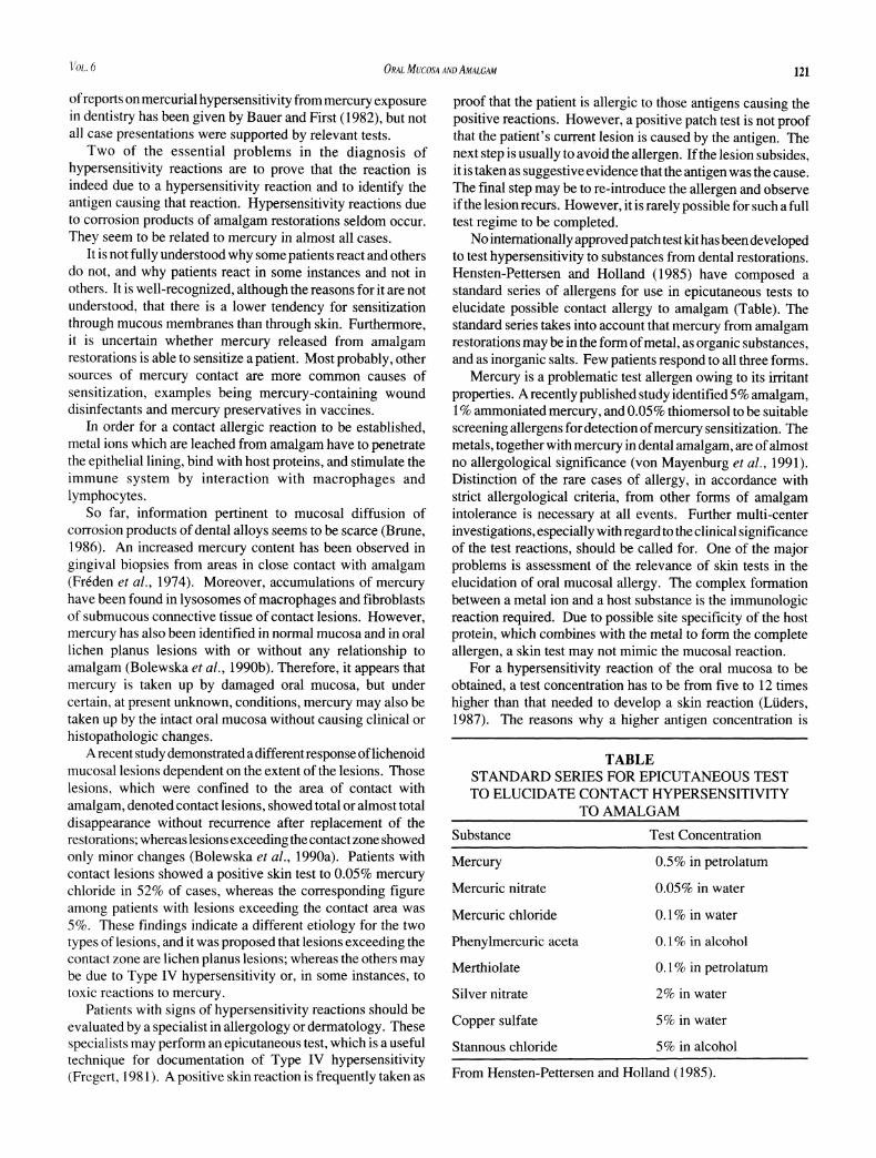
No internationally approved patch test kit has been developedto test hypersensitivity to substances from dental restorations.Hensten-Pettersen and Holland (1985) have composed astandard series of allergens for use in epicutaneous tests toelucidate possible contact allergy to amalgam (Table). Thestandard series takes into account that mercury from amalgamrestorations may be in the form of metal, as organic substances,and as inorganic salts. Few patients respond to all three forms.
Mercury is a problematic test allergen owing to its irritantproperties. A recently published study identified 5% amalgam,1 % ammoniated mercury, and 0.05% thiomersol to be suitablescreening allergens for detection of mercury sensitization. Themetals, together with mercury in dental amalgam, are of almostno allergological significance (von Mayenburg et al., 1991).Distinction of the rare cases of allergy, in accordance withstrict allergological criteria, from other forms of amalgamintolerance is necessary at all events. Further multi-centerinvestigations, especially with regard to the clinical significanceof the test reactions, should be called for. One of the majorproblems is assessment of the relevance of skin tests in theelucidation of oral mucosal allergy. The complex formationbetween a metal ion and a host substance is the immunologicreaction required. Due to possible site specificity of the hostprotein, which combines with the metal to form the completeallergen, a skin test may not mimic the mucosal reaction.
For a hypersensitivity reaction of the oral mucosa to beobtained, a test concentration has to be from five to 12 timeshigher than that needed to develop a skin reaction (Liiders,1987). The reasons why a higher antigen concentration is
TABLESTANDARD SERIES FOR EPICUTANEOUS TESTTO ELUCIDATE CONTACT HYPERSENSITIVITY
TO AMALGAM
Substance Test Concentration
Mercury
Mercuric nitrate
Mercuric chloride
Phenylmercuric aceta
Merthiolate
Silver nitrate
Copper sulf ate
Stannous chloride
0.5% in petrolatum
0.05% in water
0.1% in water
0.1% in alcohol
0.1% in petrolatum
2% in water
5% in water
5% in alcohol
From Hensten-Pettersen and Holland (1985).

122 HOLMSTRUP ADV DENT RES SEPTEMBER 1992
needed are unknown, but one modifying consequence of thehigh concentration needed may be false-positive registrationdue to a toxic reaction.
Hensten-Pettersen and Holland (1985) list the basis forrequiring allergologic examination of dental patients:
- the presence of oral mucosal lesions as lichen planus andstomatitis resistant to treatment; and
- clear, anatomic relationship between an oral mucosal lesionand the suspected dental restorative material.
The authors emphasize that such uncharacteristic oralphenomena as smarting pain, burning sensations, diffuse pain,battery sensation, and metallic or other taste sensations are notindicative of the need for allergologic examination. Thisattitude is supported by studies of patients with orofacialcomplaints in which no difference in patch test reactions wasfound between patients and controls (Axell et al, 1983;Yontchev etal., 1986). Clinically relevant contact allergies arealso rare in patients with burning mouth syndrome (Lindmaierand Lindemayr, 1989), and patch testing is not indicated(BaiirleandSchonberger, 1986). Likewise, distant symptoms,such as headache or paresthesia, are not indications for patchtesting (Hensten-Pettersen and Holland, 1985).
Lichen planus is one of the most common oral mucosaldisorders (Axell, 1976). It has an unknown cause and showsvarious clinical manifestations, including papules, reticularpatterns, erythema, plaque-like changes, and bullae (Thorn etal, 1988).
It has been proposed that hypersensitivity to mercury fromcorroding amalgam fillings plays an important part in theetiology of oral lichen planus (Hensten-Pettersen and Holland,1985). Such a hypothesis has been supported by studiesdemonstrating hypersensitivity to mercury among 16-62% ofpatients with oral lichen planus (Finne et al, 1982; Eversoleand Ringer, 1984; Lundstrom, 1984; Mobacken et al., 1984;James et al., 1987), whereas mercury hypersensitivity in thegeneral population has been found in 1-4% (Magnusson et al,1968).
One explanation may be that the clinical and histopathologicchanges of lichen planus lesions resemble those of Type IVhypersensitivity reactions, and, consequently, it may be difficult,or even impossible, to distinguish between a contact lesion dueto Type IV mercury hypersensitivity and one due to oral lichenplanus. Furthermore, the presence of lichen planus affectionsof the oral mucosa may well render the host more susceptibleto mercury hypersensitivity, due to increased penetration of theaffected oral mucosa by allergens. The latter possibility wasnot supported by our findings in a group of patients with lichenplanus exceeding the contact area with amalgam. Only 5% hada positive skin reaction (Bolewska et al., 1990a).
Another cause of lesions related to dental restorations maybe immunologic or toxic reactions to plaque accumulations onthe surfaces of the restorations. Although this phenomenonhas received little attention, lesions due to plaque accumulationon restorations are important in differential diagnosis andshould always be kept in mind. Such lesions may disappear
after improved oral hygiene—supported, for instance, bychlorhexidine mouthrinses. A change in oral lichen planuslesions as the result of amalgam fillings being replaced withresin composite, porcelain-fused-to-metal, or other materialshas been observed in some cases by the author and has alsobeen reported (Hensten-Pettersen, 1984; Lind et al, 1986).However, it is important to exclude plaque accumulations onrestorations as an etiologic factor before replacing fillings withalternative materials. Plaque reduction may also have surprisingeffects on mucosal lesions of lichen planus (Holmstrup et al,1990). The reason why some oral lichen planus affectionschange their clinical appearance after replacement of fillings isprobably due to changes of the surface microbiology of therestorations.
GENERALIZED HYPERSENSITIVITY REACTIONSGeneralized symptoms caused by Type IV mercuryhypersensitivity reactions are extremely rare (White and Smith,1984). They include eczema and urticaria occurring on theface and limbs, predominantly on the flexural aspects (Juhlinand Oman, 1968; Thomson and Russell, 1970). These canoccur concurrently with symptoms manifested in the mouth(Fernstrom et al, 1962). In some cases, the patient presents apersonal or family history of past allergies to various sources,including mercury (Bauer and First, 1982).
White and Smith (1984) were able to find only 28 casereports in the dental and dermatological literature of individualswith rashes attributed to dental restorations. More than half ofthese had been sensitized previously by a local application tothe skin and not by the restorations. It is likely, therefore, thatthe incidence of sensitization by mercury in amalgam is low.This observation was further substantiated in a study whichwas unable to demonstrate any significant difference in patchtest reactions to mercury in individuals with and withoutamalgam fillings (Sprengler, 1958; GotzandFortmann, 1959).
Mercury vapor liberated during insertion of new amalgamfillings and during removal of old ones may give rise to thedevelopment of mercury exanthema in highly sensitizedindividuals. A series of 15 patients has been reported withgeneralized rash, mostly appearing one or two days after thebreaking of a clinical thermometer or during dental treatment(Nakayama et al, 1983). The patients had similar skinmanifestations, i.e., diffuse symmetrical erythema,predominantly on major flexural areas. Most of the patientshad a previous history of contact dermatitis to mercurochrome.They were found, by being subjected to patch testing, to havecontact allergy to several mercurials. The clinical findingsindicated that the patients had developed systemic Type IVhypersensitivity due to inhalation of mercury vapor, but theprimary induction was to compounds as mercurochrome.
From the cases reported as mercurial hypersensitivity, thelesions are usually self-limiting and subside after two to threeweeks, even without the removal of the allergen. Most likely,this is because of a decrease in mercury release to a level notable to maintain the hypersensitivity reaction (Frykholm,1957). Some authors suggest antihistamine therapy duringtreatment for relief of symptoms and, in sensitized individuals,as a prophylactic pre-medicament to reduce the patients' post-

VOL. 6 ORAL MUCOSA AND AMALGAM 123
operative discomfort (Wright, 1971; White and Brandt, 1976).In situations in which symptoms do not resolve, removal ofoffending fillings and restorations with non-mercurial dentalmaterials may still be indicated. The use of rubber dam, waterspray, and high-speed evacuation can minimize tissue contactwith the sensitizer. This procedure is especially important,because the time of exposure is more closely related to insertionor removal of amalgam restorations than to existing fillings inthe mouth (Bauer and First, 1982).
Occupational contact with amalgam compounds appears toplay a much more significant role in mercury sensitization thando amalgam restorations. White and Brandt (1976) showedthat positive patch tests to mercuric chloride increased indental students from 2% among freshmen to 10.8% amongseniors. None of the students suffered from symptoms due tomercury sensitivity.
It has been found that patients with epidermalhypersensitivity to mercury, producing clinical symptoms ofdermatitis, seldom present lesions of the oral mucosa related todental amalgam (Gaul, 1966). This observation was consideredto be due to the epidermal specificity of the antigen formed bymercury and protein, i.e., the specific protein being presentonly in the epidermis. Another possible explanation is thehigher concentration needed for a reaction to be obtained inoral mucosa than in skin.
TOXIC REACTIONSA chronic toxic reaction may be established due to repeated orconstant influence of toxic agents in low concentrations overlong periods. Such reactions are most frequently localized tothe contact zone with the toxic agent (Hensten-Pettersen andHolland, 1985). Chronic toxic reactions may be seen in areasof the oral mucosa in direct contact with amalgam fillings.Since the clinical features of these lesions do not differ fromthose of lesions due to contact hypersensitivity, the diagnosisis obtained by exclusion based on a negative patch test.Differential diagnosis is difficult, because a negative skin testmay reflect a possible site-specific mucosal contact allergy.
Little is known about toxic reactions of the oral mucosa todental restorative materials, including amalgam. A pronouncedcytotoxic effect of dental amalgam on monolayer cultures ofhuman epithelial cells has been reported (Leirskar, 1974).However, the amounts of silver, mercury, copper, and tin inmedia from the cultures were below those found to be toxic tothe cells, and the author suggests that the release of zinc ionsmight be of major importance for the cytotoxicity of silveramalgam. This has been confirmed in another in vitro studydemonstrating the higher cytotoxicity of zinc-containingamalgam than of other amalgams (Kaga et aL, 1988). Thecytotoxicity of amalgam decreased with aging time, possiblydue to the combined effects of surface oxidation and furtheramalgamation.
CONCLUSIONS
Lesions of the oral mucosa caused by amalgam restorations arerare. They may be due to Type IV contact hypersensitivity orto toxic reactions. The lesions—which are reddish, whitish, orsometimes ulcerated—are characterized by a clear anatomic
relationship to the fillings. It is important that improved oralhygiene exclude plaque accumulations on the surfaces of thefillings as a possible etiologic factor. Further, differentialdiagnostic considerations include lichen planus and leukoplakia.
Patients with signs of hypersensitivity reactions should beevaluated by a specialist in allergology or dermatology. Thesespecialists may perform an epicutaneous test to elucidatepossible contact allergy to amalgam. Hypersensitivity reactionsto amalgam seem to be related to mercury in almost all cases.
The basis for requiring allergologic examination of patientssuspected of contact hypersensitivity to amalgam is the presenceof whitish or reddish, sometimes ulcerative, oral mucosallesions with a clear anatomic relation to amalgam fillings.Replacement of such restorations with resin composite,porcelain-fused-to-metal, or other materials, in most casesresults in total, or almost total, disappearance of the mucosallesions. Generalized symptoms caused by Type IV mercuryhypersensitivity are extremely rare, and the incidence ofsensitization by mercury in amalgam is low.
The clinical features of lesions due to toxic reactions fromamalgam restorations do not differ from those of lesions due tocontact hypersensitivity, and the diagnosis is obtained byexclusion based on a negative patch test.
Further research is needed with respect to (a) relevant testallergens and methods to elucidate mucosal contact allergy; (b)penetration of the oral mucosa by products from restorativematerials; and (c) toxic reactions of the oral mucosa fromrestorative materials.
REFERENCESAxell T (1976). A prevalence study of oral mucosal lesions in
an adult Swedish population (thesis). Odontol Revy 27 (36Suppl): 1-103.
Axell T, Holmstrup P, Kramer IRH, Pindborg JJ, Shear M(1984). International seminar on oral leukoplakia andassociated lesions related to tobacco habits. CommunityDent Oral Epidemiol 12:145-154.
Axell T, Nilner K, Nilsson B (1983). Clinical evaluation ofpatients referred with symptoms related to oral galvanism.Swed Dent J 1:169-11%.
Banoczy J, Roed-Petersen B, Pindborg JJ, Inovay J (1979).Clinical and histologic studies on electrogalvanicallyinduced oral white lesions. Oral Surg Oral Med OralPathol 48:319-323.
Bauer JG, First HO (1982). The toxicity of mercury in dentalamalgam. CA Dent Assoc J 10:47-61.
Baurle G, Schonberger A (1986). Glossodynie—indikationzur epikutantestung? Z Hautkr 61:1175-1184.
Bolewska J, Hansen HJ, Holmstrup P, Piindborg JJ, StangerupM (1990a). Oral mucosal lesions related to silver amalgamrestorations. Oral Surg Oral Med Oral Pathol 70:55-58.
Bolewska J, Holmstrup P, M0ller-Madsen B, Kenrad B,Danscher G (1990b). Amalgam associated mercuryaccumulations in normal oral mucosa, oral mucosal lesionof lichen planus and contact lesions associated with amalgam./ Oral Pathol Med 19:39-42.
Brune D (1986). Metal release from dental biomaterials.Biomaterials 7:163-175.

124 HOLMSTRUP ADV DENT RES SEPTEMBER 1992
Djerassi E, Berova N (1969). The possibilities of allergicreactions from silver amalgam restorations. Int Dent J19:481-488.
Eversole LR, Ringer M (1984). The role of dental restorativemetals in the pathogenesis of oral lichen planus. Oral SurgOral Med Oral Pathol 57:383-387.
Fernstrom AlB, Frykholm KO, Huldt S (1962). Mercuryallergy with eczematous dermatitis due to silver-amalgamfillings. Br Dent J 113:204-206.
Finne K, Goransson K, Winckler L (1982). Oral lichen planusand contact allergy to mercury. Int J Oral Surg 11:236-239.
Franz G (1982). The frequency of allergy to dental materials.JDent Assoc S Afr 37:805-810.
Freden H, Hellden L, Milleding P (1974). Mercury content ingingival tissues adjacent to amalgam fillings. OdontolRevy25:207-210.
Fregert S (1981). Manual of contact dermatitis. 2nd ed.Copenhagen (Denmark): Munksgaard, 71-83.
Frykholm KO (1957). Allergy to mercury from amalgamrestorations. Acta Odontol Scand (22 Suppl) 15:61-70.
Gaul LE (1966). Immunity of the oral mucosa in epidermalsensitization to mercury. Arch Dermatol 93:45-46.
Gotz H, Fortmann I (1959). Bewirken amalgamfullungen derzahne eine quecksilbersensibilisierung der haut? ZHautkr26:34-36.
Hensten-Pettersen A (1984). Allergiskereaksjonerp&dentalematerialer. Nor Tandlaegeforen Tid 94:573-578.
Hensten-Pettersen A, Holland RI (1985). Bivirkninger avdentala material. Tandldkartidningen 77:1103-1125.
Holmstrup P (1991). Oral mucosa reactions related to silveramalgam. In: Horsted-Bindslev P, Magos L, Holmstrup P,Arenholdt-Bindslev D, editors. Dental amalgam—a healthhazard? Copenhagen (Denmark): Munksgaard, 62-82.
Holmstrup P, Schi0tz AW, Westergaard J (1990). Effect ofdental plaque control on gingival lichen planus. Oral SurgOral Med Oral Pathol 69:585-590.
James J, Ferguson MM, Forsyth A, Tulloch N, Lamey P-J(1987). Oral lichenoid reactions related to mercurysensitivity. Br J Oral Maxillofac Surg 25:474-480.
Juhlin L, Ohman S (1968). Allergic reaction to mercury in redtattoos and mucosa adjacent to amalgam fillings. ActaDerm Venereol 48:103-105.
Kaga M, Seale NS, Hanawa T, Ferracane JL, Okabe T (1988).Cytotoxicity of amalgams. J Dent Res 67:1221-1224.
Kallus T, Mjor IA (1991). Incidence of adverse effects ofdental materials. Scand J Dent Res 99:236-240.
Knychalska-Karwan Z (1966). Przyczynek do roli pradowelektrogalwanicznych w leukoplaky jamy ustnej.Czasopismo Stomatol 19:10-15.
Leirskar J (1974). On the mechanism of cytotoxicity of silverand copper amalgams in a cell culture system. Scand J DentRes 82:74-81.
LindPO, HurlenB, LybergT, Aas E (1986). Amalgam-relatedoral lichenoid reaction. Scand J Dent Res 94:448-451.
Lindmaier A, Lindemayr H (1989). Probleme mitzahnprothesen und zahnfullungsmaterialien: epicutantester-gebnisse, konsequenzen und nachbeobachtung. Z Hautkr64:24-30.
Lundstrom JM (1984). Allergy and corrosion of dental materialsin patients with oral lichen planus. Int J Oral Surg 13:16-24.
Liiders G (1987). Exogen induzierte erkrankungen dermundschleimhaut. Z Hautkr 62:603-612.
Magnusson B, Blohm S-G, Fregert S, Hjort N, H0vding G,PirilaV, SkogE(1968). Routine patch testing (IV). ActaDerm Venereol 48:110-114.
Merritt K (1986). Biochemistry. Hypersensitivity. Clinicalreaction. In: International workshop. Biocompatibility,toxicity and hypersensitivity to alloy systems used indentistry. Ann Arbor (MI): The University of MichiganSchool of Dentistry.
Mobacken H, Hersle K, Sloberg K, Thilander H (1984). Orallichen planus: hypersensitivity to dental restoration material.Contact Dermatitis 10:10-15.
Nakayama H, Niki F, Shono M, Hada S (1983). Mercuryexanthem. Contact Dermatitis 9:411-417.
Phillips RW, Schnell RJ, Shafer WG (1968). Failure ofgalvanic current to produce leukoplakia in rats. J Dent Res47:666.
Sprengler H (1958). Krankheiten der mundschleimhaut undder lippen. Stuttgart (Germany): Thieme Verlag.
Stenman E, Bergman M (1989). Hypersensitivity to dentalmaterials in a referred group of patients. Scand J Dent Res97:76-83.
Thomson J, Russell JA (1970). Dermatitis due to mercuryfollowing amalgam dental restorations. Br J Dermatol82:292-297.
Thorn JJ, Holmstrup P, Rindum J, Pindborg JJ (1988). Thecourse of various clinical forms of oral lichen planus. Aprospective follow-up study of 611 patients. J Oral Pathol17:213-218.
von Mayenburg J, Rakoski J, Szliska C (1991). Patch testingwith amalgam at various concentrations. Contact Dermatitis24:266-269.
White IR, Smith BGN (1984). Dental amalgam dermatitis. BrDent J 156:259-270.
White RR, Brandt RL (1976). Development of mercuryhypersensitivity among dental students. J Am Dent Assoc92:1204-1207.
Wright F (1971). Allergic reaction to mercury after dentaltreatment. NZ Dent J 67:251-252.
Yontchev E, Meding B, HedegSrd B (1986). Contact allergyto dental materials in patients with orofacial complaints. /OralRehabil 13:183-190.